miles et al 1983 host si.pdf
TRANSCRIPT

Am. J. Trop. Med. Hyg., 32(6), 1983, pp. 1251-1259Copyright @ 1983 by The American Society of Tropical Medicine and Hygiene
VERTEBRATE HOSTS AND VECTORS OF TRYPANOSOMARANGELI IN THE AMAZON BASIN OF BRAZIL
M. A. MILES,*]. R. ARlAS,t S. A. S. VALENTE,:!: R. D. NAIFF,tA. A. de SOUZA,:!:M. M. POVOA,:!:]. A. N. LIMA,:!:AND R. A. CEDILLOS§
*WeUcome Parasitology Unit, Section 01 Parasitology, Instituto Evandro Chagas da FundaçãoSESP, Caixa Postal 3, 66.000 Belém, Pará, Brazil, tDivision 01 Medical Sciences, Instituto
Nacional de Pesquisas de Amazônia, 69.000 Manaus, Amazonas, Brazil, :tSeção de Parasitologia,Instituto Evandro Chagas da Fundação SESP, 66.000 Belém, Pará, Brazil, and §Pan American
Health Organization, Washington, D.C. 20037
Abstract. A total of 46 Trypanosoma rangeli stocks were isolated from naturally infectedmammals and triatomine vectors. Twenty-two stocks were from the common opossum (Di-delphis marsupialis), one from the brown "4-eyed" opossum (Metachirus nudicaudatus), onefrom the anteater (Tamandua tetradactyla), one from the coati (Nasua nasua), seven fromRhodnius pictipes and 14 from Rhodnius robustus. Two stocks were also isolated fromrecently fed sandflies (Lutzomyia sp., Shannoni group). The stocks were identified as T.rangeli on the basis of natural or experimental salivary gland infections in Rhodnius, inoc-ulative (anterior station) transmission to mice, morphological parameters in parasitemic miceand com pari sons of isozyme profiles with a known stock of T. range li isolated from manoThree other trypanosome stocks from D. marsupialis, T. tetradactyla and the three-toed sloth(Bradypus tridactylus) were morphologically similar to T. range li in culture but had quitedifferent isozyme profiles and were not identified. It is concluded that T. rangeli is widelydistributéd in Amazonas, Pará and Rondonia States of Brazil, and probably extends into otherregions where R. pictipes and R. robustus are known to occur. R. pictipes is light-attractedinto houses and occasionally transmits Chagas' disease to mano It is likely that T. range li isalso occasionally transmitted to man in the Amazon basin.
Trypamosoma rangeli was first described in1920, by Tejera, on the basis of flagellatesmorphologically distinct from those of Trypano-soma cruzi that were seen in the teces of the tri-atomine bug Rhodnius prolixus, captured in housesin Venezuela. In 1936, De Leon found the organ-ism in man in Guatemala, although it may havebeen seen much earlier in Peru under the nameT. escomeli.l T. range li infection is pathogenic toboth naturally and experimentally infected vec-tors, yet human infections are apparently quiteharmless. 2,3 Transmission is by inoculation of in-
fected saliva during bug-feeding; nevertheless, theorganism (subgenus Herpetosoma) is consideredas belonging to the section Stercoraria in whichtransmission is usually contaminative.4 T. range liis widely distributed in Central and northern SouthAmerica and is often sympatric with T. cruzi, andserological cross-reactions between sera from hu-man T. cruzi and T. range li infections are said toconfuse epidemiological studies. The interesting
.Accepted 26 May 1983.
l' ,
history and biology of T. rangeli have been re-viewed in detail by both Hoare4 and D' Alessan-dro.1
J)' Alessandro accepts records of T. rangeli fromtriatomine bugs and mammals in the Americas asbeing biologlca1ly proven only when substantiatedby salivary gland infections or by transmission ex-periments.1 Many T. rangeli-like organisms whichmay undergo some development in triatomine bugshave been reported, and several are na:rned as dis-tinct species, although their relationship to T. ran-geli is unclear .1,4 By D' Alessandro's criteria p~pv-en natural vectors of T. range li are R. prolixus,R. paUescens, R. ecuadoriensis, R. pictipes, R.robustus, Triatoma dimidiata capitata, and pos-sibly R. brethesi. 1. 5 Proven naturally infected
mammal hosts are Didelphis marsupialis (com-mon opossum), Philander opossum (Grey, "4-eyed"opossum), Tamandua tetradactyla (anteater), CaviaproceUus (guinea pig), Oryzomys concolor (rice rat),Eira barbara (tayra), Procyon lotor (raccoon), andthe primares, Cebus spp. (capuchin), Saimiri spp.(squirrel monkey), and Saguinus (=Leontocebus)geoffroyi (tamarin).l The known distribution of:1
1251
1252 MILES ET AL
F-;j..
"\(
" ."
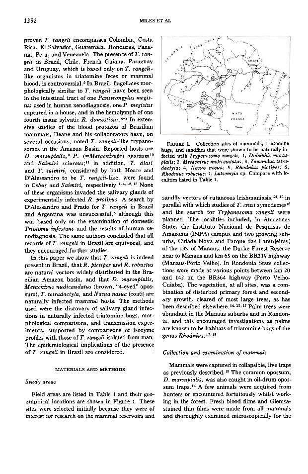
FIGURE 1. Collection sites oí mammals, triatominebugs, and sandflies that were shown to be naturally in-íected with Trypanosoma rangeli, 1, Didelphis marsu-pialisj 2, Metachirus nudicaudatusj 3, Tamandua tetra-dactylaj 4, Nasua nasuaj 5, Rhodnius pictipesj 6,Rhodnius robustusj 7, Lutzomyia sp. Compare with 10-calities listed in Table 1.
sandfly vectors of cutaneous leishmaniasis, 14,15 inparallel with which studies of T. cruz i zymodemes16and the search for Trypanosoma rangeli wereplanned. The localities included, in AmazonasState, the Instituto Nacional de Pesquisas deAmazonia (INPA) campus and two growing sub-urbs, Cidade Nova and Parque das Laranjeiras,of the city of Manaus, the Ducke Forest Reservenear to Manaus and km 65 on the BR319 highway(Manaus-Porto Velho). In Rondonia State collec-tions were made at various points between km 20and 142 on the BR364 highway (Porto Velho-Cuiaba). The vegetation, at alI sites, was a com-bination of disturbed primary forest and second-~ry growth, cleared of most large trees, as hasbeen described elsewhere.14, 15, 17 Palm trees were
abundant in the Manaus suburbs and in R(>ndon-ia, and this encouraged investigations as palmsare known to be habitats of triatomine bugs of thegenus Rhodnius.17, 18
proven T. range li encompasses Colombia, CostaRica, EI Salvador, Guatemala, Honduras, Pana-ma, Peru, and Venezuela. The presence of T. ran-geli in Brazil, Chile, French Guiana, Paraguayand Uruguay, which is based only on T. rangeli-like organisms in triatomine feces or mammalblood, is controversial.5 In Brazil, flagellates mor-phologically similar to T. range li have been seenin the intestinal tract of one Panstrongylus megis-tus used in human xenodiagnosis, one P. megistuscaptured in a house, and in the hemolymph of onefourth instar sylvatic R. domesticus.6-8 In exten-sive studies of the blood protozoa of Brazilianmammals, Deane and his collaborators have, onseveral occasions, noted T. rangeli-like trypano-somes in the Amazon Basin. Reported hosts areD. marsuPialis,9 P. (=Metachirops) opossuml0and Saimiri sciureus;ll in addition, T. diasiand T. saimiri, considered by both Hoare andD'Alessandro to be T. rangeli-like, were foundin Cebus and Saimiri, respectively.1.4. 12, 13 None
of these organisms invaded the salivary glands ofexperimentally infected R. prolixus. A se~rch byD'Alessandro and Prado for T. range li in Braziland Argentina was unsuccessful,5 although thiswas based only on the examination of domesticTriatoma infestans and the results of human xe-nodiagnosis. The same authors concluded that alIrecords of T. rangeli in Brazil are equivocal, andthey encouraged further studies.
In this paper we show that T. rangeli is indeedpresent in Brazil, that R. pictipes and R. robustusare natural vectors widely distributed in the Bra-zilian Amazon basin, and that D. marsupialis,Metachirus nudicaudatus (brown, "4-eyed" opos-sum), T. tetradactyla, and Nasua nasua (coati) arenaturally infected mammal hosts. The methodsused were the discovery of salivary gland infec-tions in naturally infected triatomine bugs, mor-phological comparisons, and transmission exper-iments, supported by comparisons of isozymeprofiles with those of T. range li isolated from manoThe epidemiological implications of the presenceof T. rangeli in Brazil are considered. Collection and examination 0/ mammals
Mammals were captured in collapsible, Iive trapsas previously described. 19 The common opossum,
D. marsupialis, was also caught in oil-drum opos-sum traps.14 A few animaIs were acquired fromhunters or encountered fortuitously whilst work-ing in the forest. Fresh bIood fiIms and Giemsa-stained thin fiIms were made from alI mammalsand thoroughly examined microscopically for the
MATERIALS AND METHODS
Studyareas
Field areas are listed in Table 1 and their geo-graphicallocations are shown in Figure 1. Thesegires were selected initially because they were ofinterest for research on the mammal reservoirs and
1253HOSTS AND VECTORS OF TRYPANOSOMA RANGEU IN BRAZIL
TABLE 1I solation histories of 48* Trypanosoma rangeli stocks examined
Host speciesDidelphis
marsuPialis I
(common opossum)AMAZONAS
Manaus Cidade Nova IM449(A)Manaus Parque das Laranjeiras (*)IM273(A); IM3l0(A); (*)IM5l0(A)Ducke Forest Reserve AMOlO km 26 IM68(A)BR3l9 km 65 (*)IM568(B)
RONDONIA
BR364 km 20-65 IM88(A); IM92(A); (*)IMlOl(A);(*)IM122(A); (*)IM123(A); IMl44(B);IMl46(B); (*)IMl58(A); (*)IMl70(A);(*)IM1284(B,C); IM1296(B);(*)IM1298(B); IMl3l6(B)
(*)IMl389(B)(*)IMlO6(A)
(*)IMl3l4(B)
IM1269(B)
1M 1249(A)
M etachirus nudicaudatus(brown, "4-eyed" opossum)
Tamandua tetradactyla(anteater)
Nasua nasua(coati)
Rhodnius pictipes
km 111Estrada Belnfunte km 11
RONDONIA
BR364 km 54
RONDONIA
BR364 km 92
RONDONIA
BR364 km 92
AMAZONAS
Manaus Parque das Laranjeiras BUG 1333/2 INT(B)j (*)BUG 1333/9INT(B); BUG 1333/9 GL(B); BUG1814 INT(B); BUG 1821 INT(B)jBUG 1823 INT(B)
(*)BUG 861 INT(B)
Rhodnius
robustus
RONDONIA
BR364 km '23
AMAZONASManaus Parque das Laranjeiras *)BUG 1752 INT(B)j BUG 1752
GL(B)j BUG 1791 INT(B); BUG1798 INT(B)j BUG 1798 GL(B)jBUG 1801 GL(B); BUG 1946INT(B); BUG 1946 GL(B)j BUG1949 GL(B); BUG 1958 INT(B)jBUG 1958 GL(B)j BUG 1959GL(B)j CX 503(C); CX 507(C)
RONDONIA
BR364 km 20 IMI086 INT(B); IMI087 INT(B),..
Lutzomyia sp.(Shannoni group)
* Stocks derived by different routes from lhe salDe bost are considered separately"t Soe Figure 1.* (*) indicates stocks of T rangeli tbat were mixed with T. c",zi. ,.§ (A), stocks isolated by culture of cardia.: blood from lhe natural vertebrate host on biphasic blood-agar medium;2' (B), stocks obtained by culture of
naturally infected triatomine bug fetos, salivary glands, or fetos of xenodiagnosis bugs; in addition, two stocks isolated by culture of sandfly intestinaltra.:tz. INT = culture of intestinal tra.:t or fetos; GL ~ culture of salivary glands; (C), stocks obtained by transmission to mito during feeding of naturallyinfected bugs or by intraperitoneal inoculation of infected xenodiagnosis bug fetos, and subsequently cultured from infected mouse cardia.: blood.
" Prevalence of T. rangeli infection in D. marsupialis was 10-30% in localities listed.
presence of trypanosomes. Trypanosomes wereisolated and grown from mammal hosts using thethree p~ocedures of blood culture, xenodiagnosis,and mouse inoculation. Fifth instar R. prolixusor, initially, Dipetalogaster maximus were em-ployed for xenodiagnoses. Although D. maximusreadily acquired intestinal T. rangeli infections its
use was restricted as susceptibility to salivary glandinfection was uncertain.20 The methods used forindividual T. rangeli stocks are indicated in Table1 by A, B, and C, A refers to stocks isolated bythe direct culture of cardiac blood from the nat-ural vertebrate host on biphasic, blood-agar me-dium;21 B refers to stocks isolated by xenodiag-
Localityt

1251
MILES ET AL.
nosis and grown in vitro by culture ofxenodiagnosis bug teces or salivary glands usinggentamycin and 5'-fluorocytosine to prevent con-tamination;22 C refers to stocks isolated by xeno-diagnosis, passaged into mice by intraperitonealinoculation of xenodiagnosis bug teces, and grownin vitro by the culture of mouse cardiac blood.
acterization of T. cruz i stocks,21 and included theroutine preparation and exarnination of Giemsa-stained films of each population of organisms thatwas harvested. The 11 enzymes used in this studywere: malate dehydrogenase (E.C. 1.1.1.37,MDH); malate dehydrogenase (oxaloacetate de-carboxylating) (NADP+) (E.C. 1.1.1.40, ME); iso-citrate dehydrogenase (NADP+) (E.C. 1.1.1.42,ICD); phosphogluconate dehydrogenase (decar-boxylating) (E.C. 1.1.1.44, 6PGDH); glucose-6-phosphate dehydrogenase (E.C. 1.1.1.49, G6PD);aspartate aminotransferase (E.C. 2.6.1.1, ASAT);alanine arninotransferase (E.C. 2.6.1.2, ALAT);phosphoglucomutase (E.C. 2.7.5.1, PGM); ami-nopeptidase (cytosol) (E.C. 3.4.11.1; PEP); acon-itate hydratase (E.C. 4.2.1.3, ACON), and glu-cosephosphate isomerase (E.C. 5.3.1.9, GPI). Theisozyme profiles of T. range li stocks were com-pared visually with those of T. rangeli stock R1625isolated from a patient in EI Salvador by cultureof venous blood on Seneckjie's medium, and kind-ly provided by Dr M. Sauerbrey (CARS/CDC). T.cruzi stocks representing T. cruzi zymodemes werealgo included in electrophoresis to detect mixed T.rangeli/T. cruzi infections.
Collection and examination of triatomine bugs
The majority of triatomine bugs were collectedby the systematic dissection of palm trees18-the"inajá" (Maximiliana regia) palm in the vicinityof Manaus and the "babaçu" palm (Orbignya spe-ciosa) in Rondonia.17 A smalI number of adultR.pictipes were captured in light-traps like those de-scribed by Whitlaw and Chaniotis.23 The intesti-nal tracts and salivary glands of samples of R.pictiPes and R. roliustus from each field colIectionwere dissected and examined by phase microscopyfor T. range li and/or T. cruzi. Hemolymph wasnot routinely examined. Almost alI isolares of T.rangeli from naturalIy infected R. pictipes and R.robustus were made by the direct culture of in-fected bug feces or salivary glands (B in Table 1).Occasionally, T. range li stocks were obtained byfeeding bugs on mice or by intraperitoneal inoc-ulation of infected feces into mice and subsequentculture of mouse cardiac blood (C in Table 1).Two T. range li stocks were also isolated by directculture of the intestinal tracts of recently fed Lut-zomyia sp. (Shannoni group) sandflies.
Experimental transmission
Trypanosoma range li stocks were passaged ex-perimentally, through the entire life cycle, bymembrane feeding laboratory-reared Rhodniusspp. on cultures and subsequently refeeding onuninfected, juvenile mice. These studies will bedescribed in detail in a later reportoM ouse infections and morphological comparisons
Mouse infections were established either by al-lowing naturally infected triatomines to feed onmice, by intraperitoneal inoculation of organismsfrom cultures past peak growth into mice, or byintraperitoneal inoculation of naturally infected orxenodiagnosis bug teces. Laboratory-bred 4- to 5-week-old mice were used throughout. Fresh bloodfilms and Giemsa-stained thin films were exam-ined periodically from days 4-30. Trypanosomeswere photographed, measured and comparativeíndices were calculated according to Hoare.4
RESULTS
Enzyme electrophoresis
A total of 46 trypanosome stocks identified asT. range li were isolated from naturally rnfectedmammals or triatomine bugs (Table 1). Twenty-two stocks were from D. marsupialis, one fromM. nudicaudatus, one from T. tetradactyla, onefrom N. nasua, seven from R. pictipes and 14from R. robustus. In addition, two stocks wereisolated from sandflies, which contained bloodmeals of an unknown mammal host. The stocksfrom triatomine bugs included 10 isolated directlyor indirectly from salivary glands (Table 1).
Trypanosomes were very rarely seen in freshfilms of naturally infected mammals and Dever insufficient numbers to allow satisfactory morpho-logical studies in Giemsa-stained thin films of par-asitemic blood. AlI stocks grew rapidly in the bi-
Bulk growth of T. rangeli stocks was in largetubes of blood-agar medium.21 Harvesting of or-ganisms, preparation of enzyme extracts and en-zyme electrophoresis were as used for the char-

1255HOSTS AND VECTORS OF TRYPANOSOMA RANGEU IN BRAZn.
TABLE 2
Morphological comparisons between Brazilian T. rangeli stocks, T. rangeli isolatedfrom man, and published records
ParameterstNo.mea- .sured* L KIPK KN F NIStock code
28.3-36.7 2.7-4.3 7.0-11.7 7.0-12.0 7.0-11.0 1.0-1.8 1.3-1.5
(32.7) (3.4) (9.4) (10.4) (9.6) (1.3) (1.4)
27.7-35.3 2.7-4.3 8.0-10.7 6.7-12.3 7.3-13.0 1.0-2.0 1.3-1.5
(31.7) (3.5) (9.6) (9.2) (9.3) (1.5) (1.4)
27.0-37.0 1.7-5.7 7.0-12.7 6.6-11.7 5.0-10.3 0.9-2.2 1.2-1.5
(29.1) (3.3) (9.2) (9.1) (7.5) (1.4) (1.4)
25.7-33.3 2.7-4.0 7.0-12.0 5.0-10.6 7.3-12.7 1.à-2.7 1.2-1.5
(30.8) (3.2) (9.9) (7.9) (9.7) (1.8) (1.3)
28.0-36.0 2.7-4.0 9.3-12.7 6.3-11.7 7.0-10.7 1.1-2.2 1.2-1.4
(32.1) (3.3) (10.7) (8.8) (9.4) (1.6) (1.3)
25.0-37.0 1.8-7.0 8.2-10.0 (27.0-32.2) ---(7.9-9.5) (1.2-1.7) (1.6-2.0)
25.0-37.0 1.8-7.0 8.2-10.0 5.0-12.0 5.0-11.0 -1.1-2.8
(26.4-33.8) (3.4-4.4) (9.5-9.7) (6.9-8.9) (8.1-9.5) -(1.6-2.0)
.Trypomastigotes in Giemsa-stained tmn tilms of parasitemic mouse blood.t Mler Hoare.' L, totallength; PK, distance from posterior to kinetoplast; KN, distance from kinetoplast to nucleus; NA, distance from nucleus to
anterior; F, length of free flaKOllum; NI, nuclear index (PN/NA); KI, kinetoplast index (PN/KN). Ranges given with means in parentheses.* But 1768 GL, isolated from R. robustus salivary glands (Manaus, Parque das Laranjeiras) is not listed in Table 1 as it was not characterized
biochemically.§ From Hoare.'II From O' Alessandro. '
IM144 (D. marsupialis) 20
BUG 1768GL:I: (R. robustus) 27
BUG 1798 GL (R. Tobustus) 25
BUG 1801 GL (R. robustus) 10
R1625 (man) 23
Various§
Variousll
phasic blood agar culture medium and theirmorphology, as assessed by phase microscopy andexamination of Giemsa-stained thin films, waseitherthat of T. rangeli or T. rangeli/T. cruzi mix-tures. Similar morphological forms were seen innaturally infected triatomine bugs or xenodiag-nosis bugs. A total of 16 stocks were shown to beT. rangeli/T. cruzi mixtures on the basis of mor-pholQgy and/or the presence of superimposed iso-zyme profiles (Table 1). Infections of 14 stockswere established in mice by inoculation of culturespast peak growth; they were: three from D. mar-supialis, two from R. pictipes, eight from R. ro-bustus and the T. range li standard stock R1625.The morphology of trypomastigotes in infectedmice was characteristic of T. rangelij occasionallyT. cruzi was algO noted in mice inoculated withmixed T. rangeli/T. cruzi stocks. Trypomastigotesof four stocks were measured in Giemsa-stainedthin films of parasitemic mouse blood and theirdimensions were compared with those of the stan-dard R1625 as shown in Table 2.
The isozyme profiles of the newly isolated stockscorresponded with that of the T. rangeli standardR1625 (Fig. 2) except for slight variation in theposition of the principal, fast, ALA T band or mi-nor mobility differences in the main PEP com-
ponents, similar to those reported for T. cruzi.21Mixed T. rangelilT. cruz i infections were easilyrecognized by the presence of two superimposedcharacteristic patterns within a single enzyme ex-tract. The intensity of either the T. rangeli or T.cruzi pattern in these mixed stocks was frequentlyweak, and some mixtures in which one populationovêrgrew the other may have been missed.
Several of the Brazilian T. range li stocks weretransmitted experimentally through the entire lifecycle using laboratory-bred triatomine bugs andmice. In alI aspects behavior and morphology wereconsistent with those of T. rangeli. These resultswill be described in detail elsewhere. '
The geographical distribution of the originalhosts of the T. rangeli isolares is shown in Figúre1. D. marsuPialis was commonly infected in thevicinity of Manaus, and both R. pictipes and R.robustus were vectors. In Rondonia State D. mar-supialis, M. nudicaudatus, T. tetradactyla andN.nasua were infected, as was R. pictipes. One of38 R. pictipes and 12 of 61 R. robustus examinedfrom the Parque das Laranjeiras had natural sal-ivary gland infections. Morphological forms in theglands were as described for T. rangelij1.4 someinfections were massive, with glands packed withmetatrypanosomes. No salivary gland infections

1256 MILES ET AL.
FIGURE 2. Examples of isozyrne profiles. The enzyrnes shown are: (a) ME; (b) ASATj (c) PGM; .and (d) GPI.In each case the trypanosome stocks are (from left to right): I, R 1625-Trypanosoma rangeli from man, EI Salvador;2, IM92-T. range li from Didelphis marsupialis, Rondonia State, Brazilj 3, Bug 333/9 GL-T. rangeli from Rhod-nius PictiPes, Manaus, Amazonas State, Brazil; 4, Bug 1752 GL-T. rangeli from Rhodnius robustus, Manaus,Amazonas State, Brazil; 5, IMI087-T. rangeli from Lutzomyia sp. (Shannoni group), Rondonia State, Brazilj 6,IM418-unidentified, from Bradypus tridactylus, Manaus, Amazonas State, Brazilj 7, 1M 1269-T. rangeli fromTamandua tetradactyla, Rondonia State, Brazil. Scale: the application threads at the origin measure approximately1 cm.
DISCUSSION
The presence of T. rangeli in Brazil has longbeen suspected from records of morphologicallysimilar trypanosomes in the intestinal tract oftriatomine bugs and in the blood of Didelphismarsupialis and the grey "4-eyed" opossum (Phi-lander opossum).1.4 Conclusive evidence has beenlacking because salivary gland infections have notbeen seen in naturally or experimentally infectedbugs, I. 4 Dor have intrinsic characters, such as iso-
zyme profiles, been used to compare Brazilian iso-lates with biologically-proven T. range li fromelsewhere. We have found natural salivary glandinfections in both R. pictipes and R. robustus,and demonstrated inoculative (anterior station)transmission to mice. The morphological param-eters of trypanosomes from parasitemic mice werevery similar to those reported for T. range li (Table2). The isozyme profiles of stocks isolated fromthe mammal and vector species listed in Table 1were, with the exception ofminor ASAT and PEPdifferences, indistinguishable from the profile of astandard T. range li stock isolated from man in ElSalvador. We consider that these results, whichfulfill the cri teria of D' Alessandro, are irrefutableevidence that T. range li is indeed present in Bra-zil.
were seen in bugs from Rondonia State but glandsof only 18 R. robustus have been dissected.*
In addition to the T. rangeU stocks given inTable 1, three other stocks, morphologically sim-ilar to T. rangeli in culture, were isolated from D.marsupialis (IM1013, t BR364, km 49), T. tetra-dactyla (1M311, BR364, km 92) and Bradypus tri-dactylus (IM41.8, 3-toed sloth; Manaus, Parquedas Laranjeiras). The isozyme profiles of thesestocks were quite different from those of the 48T. range li stocks listed in Table 1 (Fig. 2): theywere separated from the T. range li stocks by themobilities of alI but two (MDH, ME) of the en-zymes examined. We have yet to perform mor-phological studies in mice and transmission ex-periments to determine whether this organismconforms to the criteria adopted for identifying T.rangeli or, as we suspect, represents another try-panosome species.
* Massive salivary gland infections were algO seen inone of 14 R. pictiPes from Mosqueiro Island, Pará State,and one R. PictiPes collected from the city of Belém..Pará State (the latter infection found fortuitously duringelectron rnicroscopy24), but neither of these stocks wasisolated for morphological or biochemical characteriza-tion.
t INPA Manaus stock code numbers.

HOSTS AND VECTORS OF TRYPANOSOMA RANGELI IN BRAZIL
Trypanosoma rangeli infections of R. pictipesand R. robustus were recently discovered in Ven-ezuela.19 Although this was not the impetus forthe present study, in retrospect these observationsin Venezuelasuggest that T. range li was likely tobe found in the same triatomine species in theBrazilian Amazon. The known geographical dis-tribution of R. pictipes and R. robustus is, for R.pictipes: Bolivia, Brazil (Amazonas, Goiás, MatOGrosso, Pará and Rondonia States) , Colombia,Ecuador, French Guiana, Guyana, Peru, Suri-nam, Trinidad and Venezuela, and for R. robus-tus: Bolivia, Brazil (Amazonas, Pará and Ron-donia States), Colombia, Ecuador, French Guiana,Peru and Venezuela.17, 26, 27 It seems predictable,
therefore, that T. range li will be found in otherarfas such as Goiás State, Brazil, and Trinidad,in association with the same vector species, es-pecially as the most common mammal host of T.rangeli in our localities (Table 1), the opossum D.marsupialis, is ubiquitous throughout the rangesof R. pictipes and R. robustus. Both R. pictipesand R. robustus are associated with palm treehabitats.17 Collection of bugs from palms, and ex-amination by either the cultivation of teces fol-lowed by isozyme characterization of trypanosomestocks, or by the simple technique described byAnez,28 would rapidly enhance understanding ofthe geographical distribution of T. rangeli.
We have found slight isozyme heterogeneityamongst the T. range li stocks examined. Otherauthors have reported heterogeneity on the basisof enzyme electrophoresis29 or the variable sus-ceptibility of triatomine bug species to T. range liisolated in different regions.1 A complex T. range liepidemiology is implied from the number of T.rangeli-like species, with controversial taxonomicstatus, that have been described.L 4 The ecotopes
of R. pictipes and R. robustus are not fuIly under-stood, 17 and there may be differences between their
mammal hosts and roles as vectors of T. range li.As far as we are aware, little work has been doneto assess the capacity of other Amazonian triato-mine species,18 including species of the genusRhodnius such as R. paraensis, to transmit T.rangeli. The role of primate species as potentialhosts of T. rangeli in the Amazon Basin clearlymerits further investigation. T. rangeli-like organ-isms have been seen in S. sciureus; arboreal tria-tomine bugs such as Panstrongylus lignarius andR. pictipes may live in close association with pri-mates and even form part of their diet.11
The value of isozyme profiles in epidemiological
studies of this kind is emphasized by the cleardistinction (Fig. 2) of three unidentified"stocks fromsingle specimens of D. marsupialis, T. tetradac-tyla and B. tridactylus, from the T. range li stockslisted in Table 1. The importance of biochemicalcharacterization in elucidating complex epide-miologies has been clearly demonstrated previ-ously with Leishmania and T. cruzi. 30 Monoclonal
antibodies31 and genetic probes32 provide alter-native techniques directly applicable to such epi-demiological investigations.
Trypanosoma rangeli and T. cruzi are sympatricin many regions; in some localities T. range li in-fections in man may be much more common thanT. cruz i infections. I. 33 In natu,rally infected pri-
mate species T. range li has been found to be moreprevalent than T. cruzi. 34. 35 This difference prob-
ably reflects the ease of transmission by the in-oculative rather than the contaminative route. Al-though T. range li is not pathogenic to man,serological cross reaction may hinder epidemio-logical surveys and individual diagnosis of T. cru-zi infection.1 A differential diagnostic test is notavailable but has become more feasible with theadvent of species-specific monoclonal antibod-ies.36 Chagas' disease is rare in the Amazon basinand only sporadic autochthonous cases have beenreported.37 R. pictiPes, light-attracted into housesfrom infested palm trees, has been implicated asa source of such infections.17 The fact that R. pic-tipes and R. robustus are vectors of T. range li inthe Brazilian Amazon basin implies, especially inview of the inoculative transmission route, thatoccasional human T. rangeli infections in the re-gion are to be expected.
ACKNOWLEDGMENTS
We thank F. S. Gomes, R. M. Almeida, R. N.L. Santos, J. Vidal and R. A. Freitas for assis-tance in the field; C. R. Sena and D. S. Lima ofthe Instituto Nacional de Pesquisas da Amazpnia(CNPq) for technial assistance, and Colo JorgeTeixeira, Govemor of the State of Rondonia, foraccess to study areas and logistical support. MAMis a Wellcome Trust Senior Lecturer and we aregrateful to the following for financial support:the Wellcome Trust (London); the UNDP/WorldBank/WHO Special programme for research andtraining in tropical diseases; CNPq Grandes En-demias Grant No. 222.8.087/80 (Brazil)j Funda-ção SESP (Brazil); Instituto Nacional de Pesquisasda Amazônia CNPq (Brazil); and the FederalGovemment of the State of Rondonia (Brazil).
1257

1258 MILES ET AL.
REFERENCES
1. D'Alessandro, A., 1976. Biology of Trypanosoma(Herpetosoma) rangeli Tejera, 1920. Pages328-403 in W. H. R. Lumsden and D. A. Evans,eds., Biology 01 the Kinetoplastida, Volume 1.Academic Press, London.
2. Marinkelle, C. J., 1968. Pathogenicity of Trypa-nosoma rangeli for Rhodnius prolixus StaI in na-ture. J. Med. Entomol., 5: 497-499.
3. D'Alessandro, A., and Mandei, S., 1969. Naturalinfections and behavior of Trypanosoma rangeliand Trypanosoma cruzi in the vector Rhodniusprolixus in Colombia. J. Parasitol., 55: 846-852.
4. Hoare, C. A., 1972. The Trypanosomes 01 Mam-mais. Blackwell Scientific Publications, Oxford.
5. D'Alessandro, A., and Prado, C. E. dei, 1977.Search for Trypanosoma rangeli in endemic areasof Trypanosoma cruzi in Argentina and Brazil.Am. J. Trop. Med. Hyg., 26: 623-627.
6. Lucena, D. T. de, and Marques, R. J., 1954. Pri-meiro caso de infecção humana por Trypanosomarangeli Tejera 1920 no Brasil. Rev.Brasil. Med.,lI: 535-540.
7. Lucena, D. T. de, and Vergetti, J. G., 1973. In-fecção natural de Panstrongylus megistus (Bur-meister, 1835) por Trypanosoma rangeli (Tejera,1920), no interior do estado de Alagoas. Rev.Inst. Med. Trop. S. Paulo, 15: 171-178.
8. Barrett, T. V., and Oliveira, T. S. de, 1977. Atrypanosome, indistinguishable from Trypanoso-ma rangeli, in the haemolymph of Rhodnius do-mesticus from Brazil. Trans. R. SOCo Trop. Med.Hyg., 71: 445-446.
9. Deane, L. M., 1958. Encontro de tripanossomo dotipo rangeli em gambás da espécie Didelphis mar-suPialis marsupialis, no estado do Para. Rev.Brasil. Malariol. Doenças Trop., 10: 451-458.
10. Deane, L. M., 1958. Novo hospedeiro de tripa-nossomos dos tipos cruzi e rangeli encontrado noestado do Pará: o marsupial M etachirops opossumopossum. Rev. Brasil. Malariol. Doenças Trop.,10: 531-541.
11. Deane, L. M., Almeida, F. B., Ferreira, Neto, J.A., and Silva, J. E. de, 1972. "Trypanosoma cru-zi" e outros tripanosomas em primatas brasileiros.Rev. SOCo Brasil. Med. Trop., 4: 361.
12. Deane, L. M., and Martins, R., 1952. Sobre umtripanosoma encontrado em macaco da Amazôniae qui evolui em triatomíneos. Rev. Brasil. Ma-lariol. Doenças. Trop., 4: 47-61.
13. Deane, L. M., and Damasceno, R. G., 1961. Tri-panosomideos de mamíferos da região Amazôni-ca. 11. Tripanosomas de macacos da Zona do Sal-gado, Estado do Pará. Rev. Inst. Med. Trop. S.Paulo, 3: 61-70,
14. Arias, J. R., and Naiff, R. D., 1981. The principalreservoir host of cutaneous leishmaniasis in theurban areas of Manaus, central Amazon of Bra-zil. Mem. Inst. Osw. Cruz., 76: 279-286.15.
Biancardi, C. B., 1981. Aspectos da epidemiologiada leishmaniase cutanea na Rodovia BR-364. MScThesis presented to the Instituto Nacional de Pes-quisas de Amazonia, Manaus, Amazonas, Brazil.
16. Povoa, M. M., Naiíí, R. D., Miles, M. A., Arias,J. R., Souza, A. A. de, Naiíí, M. C., and Bian-cardi, C. B., 1983. Chagas' disease in the Am-azon Basin: IV. Host records oí Trypanosoma cruzizymodemes in the States oí Amazonas and Ron-donia, Brazil. Ann. Trop. Med., Parasitol. (Inpress.)
17. Miles, M. A., Arias, J. R., and Souza, A. A. de,1983. Chagas' disease in the Amazon Basin: V.Periurban palms as habitats oí Rhodnius robus-tus and Rhodnius pictiPes-triatomine vectors oíChagas' disease. Mem. Inst. Osw. Cruz. (Inpress.)
18. Miles, M. A., Souza, A. A. de, andPovoa, M., 1981.Chagas' disease in the Amazon Basin, li: Eco-topes oí ten triatomine bug species (Hemiptera:Reduviidae) írom the vicinity oí Belem, Para State,Brazil. J. Med. Entomol., 18: 266-278.
19. Miles, M. A., Souza, A. A. de, and Povoa, M. M.,1981. Mammal tracking and nest location inBrazilian íorest with an improved spool-and-linedevice. J. Zool., Lond., 195: 331-347.
20. Marsden, P. D., Cuba, C. C., Alvarenga, N. J.,and Barreto, A. C., 1979. Report on a field col-lection oí DiPetalogaster maximus (Hemiptera,Triatominae) (Uhler, 1894). Rev. Inst. Med. Trop.S. Paulo, 21: 202-206.
21. Miles, M. A., Lanham, 5. M., Souza, A. A. de,and Povoa, M., 1980. Further enzymic charac-ters oí Trypanosoma cruzi and their evaluationfor strain identification. Trans. R. Soco Trop.Med. Hyg., 74: 221-237.
22. Miles, M. A., 1982. Leishmania-culture and bio-chemical comparisons-some diíficulties. Pages123-137 in M. L. Chance and B. C. Walton,eds., Biochemical Characterization of Leishman-ia. UNDP!World Bank/WHO, Geneva.
23. Whitlaw, J. T., Jr., and Chaniotis, B. N., 1978.Palm trees and Chagas' disease in Panama. Am.J. Trop. Med. Hyg., 27: 873-881.
24. Ellis, D. S., Evans, D. A., and Stamíord, 5., 1982.Studies by electron microscopy oí the giant íormsoí some Aírican and South American trypano-somes íound other than within their mammalianhost. Folia Parasitol, (Praha), 29: 5-11.
25. Carcavallo, R. U., Martinez Silva, R., Otero, M.A., and Tonn, R. J., 1975. Iníeccion natural deRhodnius robustus Larrouse y Rhodnius plctipesStal por T. cruzi y T. rangeli en Venezuela. Bol.Dir. Malariol. Sano Amb., 15: 117-119.
26. Miles, M. A., 1919. Transmission cycles and theheterogeneity oí Trypanosoma cruzi. Pages 117-196 in W. H. R. Lumsden and D. A. Evans, eds.,Biology of the Kinetoplastida, Volume 2. Aca-demic Press, London.
27. Lent, H., and Wygodzinsky, P., 1979. Revision oíthe Triatominae (Hemiptera, Reduviidae) , andtheir significance as vectors oí Chagas' disease.BuU. Am. Mus. Nat. Hist., 163: 520 pp.
28. Anez, N., 1980. Detection oí Trypanosoma range liby salivation oí iníected Rhodnius prolixus onglass slides. Ann. Trop. Med. Parasitol., 74: 561-562.
29. Kreutzer, R. D., and Souza, D. E., 1981. Bio-

HOSTS AND VECTORS OF TRYPANOSOMA RANGELI IN BRAZIL
crofllariae in Panamanian monkeys. Am. J. Trop.Med. Hyg., 23: 862-868.35.
Souza, O. E., and Dawson, G. A., 1976. Try-panosome infections in the marmoset (Saguinusgeoffroyi) from the Panama Canal Zone. Am. J.Trop. Med. Hyg., 25: 407-409.36.
Anthony, R. L., Cody, T. S., and Constantine, N.T., 1981. Antigenic differentiation of Trypano-soma cruzi and Trypanosoma rangeli by means ofmonoclonal-hybridoma antibodies. Am. J. Trop.Med. Hyg., 30: 1192-1197.37.
Lainson, R., Shaw, J. J., Fraiha, H., Miles, M. A.,andDraper, C. C., 1979. Chagas'sdiseaseintheAmazon Basin, I: Trypanosoma cruzi infectionsin silvatic mammals, triatomine bugs and manin the State ofPará, north Brazil. Trans. R. SocoTrop. Med. Hyg., 73: 193-204.38.
World Health Organization, 1978. Proposals forthe nomenclature of salivarian trypanosomes andfor the maintenance of referente collections. Bull.W.H.O., 56: 467-480.
chemical characterization oí Trypanosoma spp. byisozyme electrophoresis. Am. J. Trop. Med. Hyg.,
30: 308-317.30. Miles, M. A., 1983. Trypanosoma and Leishmania:
The contribution oí enzyme studies to epidemiol-ogy and taxonomy, In G. S. Oxford and D. Rol-linson, eds., Protein Polymorphism: Adaptive andTaxonomic Significance. Academic Press, Lon-dono31.
Pratt, D. M., and David, J. R., 1981. Monoclonalantibodies that distinguish New World species oíLeishmania. Nature, 291: 581-583.32.
Wirth, D. F., and Pratt, D. M., 1982. Rapid iden-tification oí Leishmania species by specific hy-bridization oí kinetoplast DNA in cutaneous le-sions. Proc. Natl. Acad. Sci., 79: 6999-7003.33.
Souza, D. E., and Johnson, C. M., 1971. Fre-quency and distribution oí Trypanosoma cnlzi andTrypanosoma rangeli in the Republic oí Panama.Am. J. Trop. Med. Hyg., 20: 405-410.34.
Souza, O. E., Rossan, R. N., and Baerg, D. C.,1974. The prevalence oí trypanosomes and mi-
1259