migrating focal seizures and myoclonic status in arv1
TRANSCRIPT

ARTICLE OPEN ACCESS
Migrating Focal Seizures and Myoclonic Statusin ARV1-Related EncephalopathyFrancesca Darra FnMD Tommaso Lo Barco MD Roberta Opri PhD Elena Parrini PhD
Claudia Bianchini PhD Elena Fiorini MD Alessandro Simonati MD Bernardo Dalla Bernardina MD
Gaetano Cantalupo MD and Renzo Guerrini MD FRCP
Neurol Genet 20217e593 doi101212NXG0000000000000593
Correspondence
Dr Darra
francescadarraunivrit
AbstractObjectiveTo report longitudinal clinical EEG and MRI findings in 2 sisters carrying compound het-erozygous ARV1 mutations and exhibiting a peculiar form of developmental and epilepticencephalopathy (DEE) Neuropathologic features are also described in one of the sisters
MethodsClinical course description video-EEG polygraphic recordings brain MRI skin and musclebiopsies whole-exome sequencing (WES) and brain neuropathology
ResultsSince their first months of life both girls exhibited severe axial hypotonia visual inattentiondyskinetic movements severe developmental delay and slow background EEG activity In-tractable nonmotor seizures started in both at the eighth month of life exhibiting the elec-troclinical characteristics of epilepsy of infancy with migrating focal seizures (EIMFS) In thesecond year of life continuous epileptiform EEG activity of extremely high amplitude appearedin association with myoclonic status leading to severely impaired alertness and responsivenessRepeated brain MRI revealed progressive atrophic changes and severe hypomyelination WESidentified a compound heterozygous in the ARV1 gene [(pSer122Glnfs7) and (pTrp163)]in one patient and was subsequently confirmed in the other Both sisters died prematurelyduring respiratory infections Postmortem neuropathologic examination of the brain per-formed in one revealed atrophic brain changes mainly involving the cerebellum
ConclusionsThis report confirms that biallelic ARV1 mutations cause a severe form of DEE and addsepilepsy with migrating focal seizures and myoclonic status to the spectrum of epilepsy phe-notypes Considering the potential role of human ARV1 in glycosylphosphatidylinositol (GPI)anchor biosynthesis this severe syndrome can be assigned to the group of inherited GPIdeficiency disorders with which it shares remarkably similar clinical and neuroimaging featuresARV1 should be considered in the genetic screening of individuals with EIMFS
From the Child Neuropsychiatry Unit(FD) Department of Surgical Sciences Dentistry Gynecology and Pediatrics University of Verona Child Neuropsychiatry Unit(TLB) De-partment of Surgical Sciences Dentistry Gynecology and Pediatrics University of Verona PhD Program in Clinical and Experimental Medicine (TLB) University of Modena andReggio Emilia Pediatric Unit (RO) Department of Surgical Sciences Dentistry Gynecology and Pediatrics University of Verona Pediatric Neurology (EP) Neurogenetics andNeurobiology Unit and Laboratories Meyer Childrenrsquos Hospital University of Florence Pediatric Neurology (CB) Neurogenetics and Neurobiology Unit and Laboratories MeyerChildrenrsquos Hospital University of Florence Child Neuropsychiatry Unit (EF) Azienda Ospedaliera Universitaria Integrata di Verona Neurology (Child Neurology and Neuropathology)(AS) Department of Neuroscience Biomedicine and Movement University of Verona CREP (Research Center for Pediatric Epilepsies) (BDB) Azienda Ospedaliera UniversitariaIntegrata di Verona and Pediatric Neurology (RG) Neurogenetics and Neurobiology Unit and Laboratories Meyer Childrenrsquos Hospital University of Florence Italy
Go to NeurologyorgNG for full disclosures Funding information is provided at the end of the article
The Article Processing Charge was funded by the authors
This is an open access article distributed under the terms of the Creative Commons Attribution-NonCommercial-NoDerivatives License 40 (CC BY-NC-ND) which permits downloadingand sharing the work provided it is properly cited The work cannot be changed in any way or used commercially without permission from the journal
MORE ONLINE
Video
Copyright copy 2021 The Author(s) Published by Wolters Kluwer Health Inc on behalf of the American Academy of Neurology 1
The ARV1 gene encodes for Acyl-CoA cholesterol acyltransferasendashrelated enzyme 2 required for viability 1(ARV1) which is a highly conserved protein located in theendoplasmic reticulum membrane Although extensivefunctional studies in yeasts and mice have explored theconsequences of ARV1 mutations1-4 the association ofbiallelic mutations of this gene with human disease is lim-ited to a few individuals exhibiting early infantile epilepticencephalopathy with intractable seizures and severe de-velopmental delay5-8
In 1 patient the epilepsy phenotype featured drug-resistantmultifocal clustered seizures disorganized background EEGactivity and modified hypsarrhythmia5 in the remainingchildren details on the epileptic encephalopathy were notprovided
We describe 2 sisters carrying compound heterozygous ARV1mutations and exhibiting an early-onset epileptic encepha-lopathy with peculiar electroclinical features
Case DescriptionPatient 1Patient 1 is a first-born girl with nonconsanguineous parentswho was delivered by elective C-section at 36 + 5 gestationalweeks Her birth weight was 2650 g (50th percentile) length46 cm (25th percentile) head circumference 327 cm (40thpercentile) and Apgar Index 89
At 4 months hypotonia was noticed with dyskinetic move-ments opisthotonus posturing and absence of eye tracking
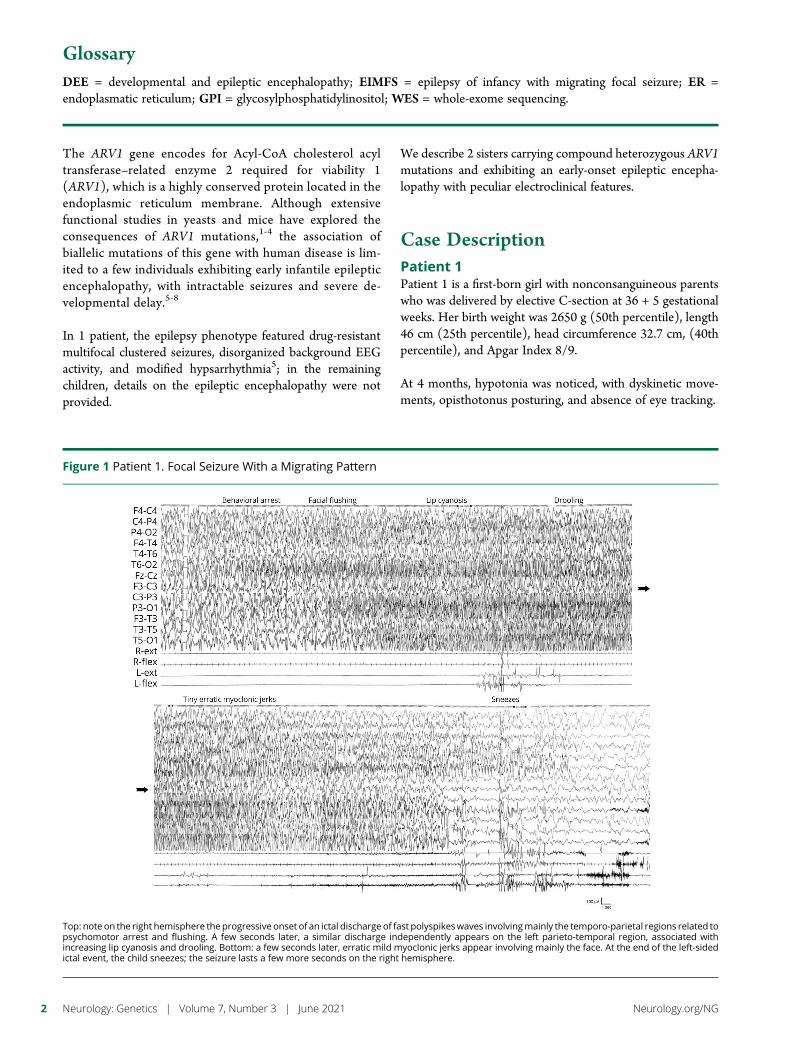
Figure 1 Patient 1 Focal Seizure With a Migrating Pattern
Top note on the right hemisphere the progressive onset of an ictal discharge of fast polyspikeswaves involvingmainly the temporo-parietal regions related topsychomotor arrest and flushing A few seconds later a similar discharge independently appears on the left parieto-temporal region associated withincreasing lip cyanosis and drooling Bottom a few seconds later erratic mild myoclonic jerks appear involving mainly the face At the end of the left-sidedictal event the child sneezes the seizure lasts a few more seconds on the right hemisphere
GlossaryDEE = developmental and epileptic encephalopathy EIMFS = epilepsy of infancy with migrating focal seizure ER =endoplasmatic reticulum GPI = glycosylphosphatidylinositol WES = whole-exome sequencing
2 Neurology Genetics | Volume 7 Number 3 | June 2021 NeurologyorgNG
At 6 months severe axial hypotonia abnormal movementsand absent eye fixation persisted EEG showed slow high-amplitude background activity (gt400 μV) replaced 1 monthlater by subcontinuous high-amplitude multifocal spikes andwaves on both hemispheres
From age 8 months multiple per-day focal nonmotor seizuresstarted with unresponsiveness chewing automatisms and per-ioral cyanosis lasting less than a minute Despite treatment withvalproate at age 12 months seizures increased in frequencyappearing at times in clusters showing multifocal origin andinvolving either hemisphere with a migrating pattern (figure 1)prompting repeated intensive care unit admissions Transitionfrom focal seizures to hemiclonic status was often seen especiallyduring fever (figure 2) Polygraphic EEG recordings revealed anunusual pattern of myoclonic status featuring subcontinuousrhythmic or arrhythmic spikes and slow waves of very highamplitude associated with reduced responsiveness and erraticmyoclonus predominating in the arms and face During sleepmyoclonus disappeared but the abnormal EEG patternremained subcontinuous (figure 3A)
Myoclonic status and clustered multifocal seizures persisteddespite several pharmacologic trials (including phenobarbital
phenytoin ethosuximide levetiracetam carbamazepine andhydrocortisone) Only the ketogenic diet reduced seizurefrequency for 10 consecutive months from several per-day tomonthly clusters triggered by fever remission of myoclonicstatus episodes and improved awareness
By age 3 years head growth dropped to below the thirdpercentile and the overall clinical picture worsened because ofalmost continuous focal seizures and myoclonic status (figure3B) with recurrent respiratory infections leading to demise at4 years 2 months due to bronchopneumonia during statusepilepticus No clinical or electrocardiographic signs of car-diomyopathy were noticed
Extensive biological and metabolic investigations on urineblood and CSF karyotype Comparative Genomic Hybridiza-tion array (CGH-array) histologic and ultrastructural exami-nation of skin and muscle biopsy and study of mitochondrialrespiratory chain enzymes were all unrevealing Eye fundussensitive evoked potential (SEP) and ERG persisted in normallimits whereas visual evoked potentials (VEP) progressivelyextinguished BrainMRI performed at 4 months showed a thincorpus callosum (figure 4A) A second MRI at 24 monthsshowed mild brain atrophy mainly involving the right temporal
Figure 2 Patient 1 Hemiclonic Seizure
On the top during an ictal discharge of polyspikes and waves with intermixed fast activity involving the right centro-parietal region associated with rhythmicjerks of the left arm a similar ictal discharge independently involves the left temporo-occipital region and is associated with eye deviation to the right lipcyanosis and intense tachycardia On the bottom cyanosis and tachycardia resolve only at the end of the left temporal discharge which lasts longer than thatinvolving the right hemisphere
NeurologyorgNG Neurology Genetics | Volume 7 Number 3 | June 2021 3
lobe hypomyelination and cerebellar atrophy more severelyaffecting the vermis (figure 4BndashD)
Postmortem examination identified the cause of death withacute respiratory distress syndrome and a hypertrophic car-diomyopathy with subvalvular stenosis Brain neuropathologydocumented cerebral atrophy more severe in the right tem-poral lobe with concomitant Ammon horn sclerosis and ab-normal morphology of the parahippocampal gyrus The corpuscallosum was thin and the cerebellum atrophic with reducedcortical thickness especially in the vermis The cytoarchitec-tonic organization of the cerebral cortex was normal whereas
severe and diffuse loss of Purkinje cells was present in thecerebellar cortex with dystrophic dendrites in the molecularlayer cell rarefaction in the inner granular layer and Bergmanngliosis There was in addition an abnormal organization of thedentate gyri bilaterally with mild gliosis Subcortical gray nu-clei brainstem and the spinal cord were normal
Patient 2Patientrsquos one younger sister was born at 37 gestational weeksfollowing an uneventful pregnancy at birth her weight was3210 g (75deg percentile) and head circumference was 347 cm(75deg percentile)
Figure 3 Patient 1 Myoclonic Status
(A) Note on the EEG during wakefulness subcontinuous diffuse high-amplitude spike waves intermingled with polyspikes synchronous and asynchronouson the hemispheres associated with subcontinuous erratic myoclonic jerks partially masked by dyskinetic movements During sleep continuous EEGdischarges persist in the absence of recognizable physiologic elements whereas motor manifestations disappear (B) Note on the EEG the continuous SW ofvery high amplitude involving both hemispheres accompanied on EMG channels by rhythmic but bilaterally asynchronous myoclonic activity
4 Neurology Genetics | Volume 7 Number 3 | June 2021 NeurologyorgNG
Severe axial hypotonia with opisthotonos posturing and jitteri-ness resulting in a hypotonic-dyskinetic quadriparesis was alreadyapparent at 4months (figure 5A) Visual pursuit was poor ocularfundus examination VEPs and SEPs were normal Head growthslowed down to around the 3rd percentile before age 2 years
From age 8 months the girl exhibited multiple seizures perday Video-EEG recordings captured focal ictal activity arisingindependently from the centro-temporal-parietal regions ofeither hemisphere (video 1) causing psychomotor arrestapnea cyanosis deviated gaze and lasting 30ndash60 secondsProlonged focal motor seizures triggered by fever at timesevolved as hemiclonic status epilepticus Interictal EEG ac-tivity was of high amplitude (gt400 μV) intermingled withmultifocal discharges Phenobarbital was ineffective the ke-togenic diet started at 11 months determined a temporaryreduction in seizure frequency Seizures remained un-controlled and at 18 months polygraphic EEG recordingsdocumented the same peculiar myoclonic status epilepticusobserved in this patientrsquos sister (figure 5B) Dilated cardio-myopathy with partial hemodynamic decompensation wasdiagnosed ECG documented I- and II-degree atrioventricularblock and physical examination showed hepatosplenomegaly
During follow-up head growth slowed down to below the 3rdpercentile The frequency of focal seizures decreased to onecluster per month mostly during fever myoclonic statusremained unchanged (figures 5C5D) until demise at age 9years during pneumonia
Brain MRI performed at 7 months showed a thin corpuscallosum A new MRI at age 3 years showed a mildlyatrophic cerebellar vermis a small right hippocampus andhypomyelination (figure 4EndashG) At age 8 years atrophic changeswere more obvious mainly involving the cerebellum(figure 4H)
Whole-exome sequencing performed in patient 2 at age 8years identified compound heterozygous mutations in theARV1 gene (NM_0227863) c363_364del(pSer122Glnfs7) inherited from the mother and c489 G gtA (pTrp163) inherited from the father Sanger sequencingperformed in a DNA sample from formalin-fixed paraffin-embedded brain tissue of patient 1 confirmed both muta-tions The pSer122Glnfs7 mutation is present in the gno-mAD control database (gnomadbroadinstituteorg) with afrequency of 4250498 alleles and is not reported in theHuman Gene Mutation Database (HGMD) (portalbiobase-internationalcomhgmdprostartphp) The pTrp163mutation is not present in the gnomAD control database(gnomadbroadinstituteorg) and in the HGMD mutationdatabase (portalbiobase-internationalcomhgmdprostartphp) Both mutations introduce a premature stop codon inthe mRNA and are predicted to elicit a rapid degradation ofthe ARV1 mRNA through nonsense-mediated mRNA decayThe MutationTaster (mutationtasterorg) tool predicts bothmutations to be disease causing We classified both mutationsas pathogenic according to the international guidelines of theACMG Laboratory Practice Committee Working Group9
Figure 4 MRI Findings of Two Patients
Patient 1 (A) Sagittal T1W section showing a thin corpus callosum (B) Coronal T1W section showing signs of mild atrophymainly involving the right temporallobe which shows gyral simplification and a dilated temporal horn (C) T2W axial section showing high signal intensity of the white matter consistent withhypomyelination (D) Sagittal T1W section showing atrophy of the cerebellar vermis Patient 2 (E) Sagittal T1W section showing a thin corpus callosum and amildly atrophic cerebellar vermis (F) Coronal T1W image showing signs of mild atrophymainly involving the right temporal lobe which shows enlarged sulciand a dilated temporal horn (G) T2Waxial section showing high signal intensity of thewhitematter consistent with hypomyelination (H) Sagittal T1W sectionshowing more severe atrophy of the cerebellar vermis
NeurologyorgNG Neurology Genetics | Volume 7 Number 3 | June 2021 5
Data AvailabilityThe data that support the findings of this study are availableon request from the corresponding author The data are notpublicly available because of privacy or ethical restrictions
DiscussionThese 2 sisters were compound heterozygous for thepSer122Glnfs7 and the pTrp163 truncating mutations andexhibited severe clinical features that overlap with those de-scribed in patients with ARV1-related encephalopathy allharboring homozygous variants Indeed both truncating
ARV1 mutations found in our patients are predicted to resultin loss of function the molecular mechanism known to beassociated with ARV1 deficiency both in humans and mice58
ARV1 mapping to 1q422 encodes for a transmembraneprotein of endoplasmatic reticulum (ER) implied in sterolhomeostasis in eukaryotic cells with a role in sterol transportbetween the ER and plasmatic membrane3 As already stated5
because of its implication in glycosylphosphatidylinositol(GPI)-anchor synthesis1011 ER stress induced by accumu-lation of immature GPI-anchored proteins or aberrant lipidmetabolism may be responsible for the diseases associatedwith ARV1 deficiency11
Figure 5 Patient 2 Abnormal Background Activity and Myoclonic Status
(A) Note on the EEG the unusually slow and high-amplitude poorly modulated theta-delta activity on the parieto-occipital regions and on the EMG theabnormal dyskinetic movements (B) Note on the EEG continuous diffuse high-amplitude (about 400 μV) epileptiform activity associating polyspike-wavesand a diffuse very fast activity related to continuous rhythmic jerks This electroclinical picture remains unchanged throughout the evolution (C) up to 9 years(D)
6 Neurology Genetics | Volume 7 Number 3 | June 2021 NeurologyorgNG
Evidence of a genetic syndrome resulting from deficiency ofARV1 in humans is currently based on the description of 14patients from 5 families all harboring homozygous variantsThe overall common reported clinical features include severedevelopmental delay and intractable seizures with onset in thefirst year of life in all and seizure onset within the 6th monthsin 50 Additional clinical features included poor head con-trol (1014) hypotonia (814) visual impairment (914)and dystonia (614) Seven patients died prematurely becauseof respiratory infections or aspiration pneumonia mostlyduring status epilepticus (1ndash5 years) Six remaining patientswere still alive at last follow-up between age 2 years 5 monthsand 18 years5-8 Progressive deceleration of head growth isreported in 5 patients and mild-moderate dilated cardiomy-opathy in 27
Reported MRI findings include cerebellar atrophy in 3 in-dividuals delayed myelination in 2 a thin corpus callosumin 1 and a hyperintense signal on T2-weighted with re-stricted diffusion in the cerebral central tegmental tract inone578 No MRI abnormalities were observed in 2 indi-viduals aged 7 months and 3 years respectively78 Overallreported clinical features are similar to those observed inour patients who harbored compound heterozygousvariants
In the 2 sisters we describe severe axial hypotoniadystonicdyskinetic movements poor visual contact andhead growth deceleration were already manifested a fewweeks after birth and from the third to the fourth monthof life subcontinuous myoclonic jerks intermingled todyskinetic movements appeared The associated electro-clinical picture was quite distinctive consisting of in-tractable migrating focal motor and nonmotor seizures1213
and a peculiar myoclonic status mainly involving the faceand upper limbs resembling the electroclinical syndromeknown as myoclonic status of nonprogressive epilepticencephalopathies1415 The overall neurologic conditionprogressively worsened leaving both girls severely hypo-tonic with almost continuous abnormal movements pooreye contact absent speech profound intellectual disabilityand recurrent respiratory infections The older girl was af-fected by dilated myocardiopathy and her sister exhibitedleft ventricular hypertrophy which was documented atautopsy not to be associated with abnormalities of themyocardial tissue
Brain MRI findings in both patients documented malforma-tive characteristics in association with progressive cerebellaratrophy The association of these findings favors the hy-pothesis of a GPI-anchorndashrelated disorder which plays a keyrole in embryogenesis neurogenesis and synaptic forma-tion16 To additionally support this hypothesis the coresymptoms of patients with ARV1 deficiency reported pre-viously5 and in our study are also seen in inherited GPIdeficiency conditions17-20 Of note migrating focal seizures
dilated cardiomyopathy and fever sensitivity can also befound in both of these conditions2122
This report confirms the evidence for an autosomal recessiveearly-onset developmental and epileptic encephalopathy(DEE) caused by biallelic ARV1 mutations in humans anddescribes the first family with compound heterozygous mu-tations It also suggests that ARV1 mutations should be con-sidered a possible cause of epilepsy of infancy with migratingfocal seizures
Considering the potential role of human ARV1 in GPI-anchorbiosynthesis this genetic syndrome could be considered inthe group of inherited GPI deficiency disorders19202223
This syndrome epitomizes difficulties in sharply partingthe category of the DEEs from that of the progressiveneurologic disorders at the early stages of clinical di-agnosis Such dichotomous classification is obviously un-satisfactory when applied to peculiar clinical features suchas those reported in this study in which the combination ofsevere neurologic impairment and almost continuous ep-ileptic activity makes it difficult to identify the main de-terminants of clinical presentation The DEE category inthis context is more a tentative broad category awaitingdiagnostic finalization
Study FundingNo targeted funding reported
DisclosureThe authors report no disclosures Go to NeurologyorgNGfor full disclosures
Publication HistoryReceived byNeurology Genetics August 16 2020 Accepted in final formMarch 23 2021
Appendix Authors
Name Location Contribution
FrancescaDarra FnMD
University ofVerona Italy
Major role in the acquisition of datadesigned and conceptualized the studyanalyzed the data and drafted themanuscript for intellectual content
Tommaso LoBarco MD
University ofVerona Italy
Acquisition of data analyzed the datadrafted the manuscript for intellectualcontent and prepared figures
Roberta OpriMD
University ofVerona Italy
Interpreted the data and revised themanuscript for intellectual content
Elena ParriniPhD
AOU MeyerFirenze Italy
Performed genetic test interpretedgenetic data and revised themanuscript for intellectual content
ClaudiaBianchini PhD
AOU MeyerFirenze Italy
Performed genetic test interpretedgenetic data and revised themanuscript
Continued
NeurologyorgNG Neurology Genetics | Volume 7 Number 3 | June 2021 7
References1 Tinkelenberg AH Liu Y Alcantara F et al Mutations in yeast ARV1 alter intracellular
sterol distribution and are complemented by human ARV1 J Biol Chem 2000275(52)40667-40670
2 Shechtman CF Henneberry AL Seimon TA et al Loss of subcellular lipid transportdue to ARV1 deficiency disrupts organelle homeostasis and activates the unfoldedprotein response J Biol Chem 2011286(14)11951-11959
3 Georgiev AG Johansen J Ramanathan VD Sere YY Beh CT Menon AK Arv1regulates PM and ER membrane structure and homeostasis but is dispensable forintracellular sterol transport Traffic 201314(8)912-921
4 Lagor WR Tong F Jarrett KE et al Deletion of murine Arv1 results in a leanphenotype with increased energy expenditure Nutr Diabetes 20155(10)e181
5 Palmer EE Jarrett KE Sachdev RK et al Neuronal deficiency of ARV1 causes an auto-somal recessive epileptic encephalopathy Hum Mol Genet 201625(14)3042-3054
6 Alazami AM Patel N Shamseldin HE et al Accelerating novel candidate gene dis-covery in neurogenetic disorders via whole-exome sequencing of prescreened mul-tiplex consanguineous families Cell Rep 201510(2)148-161
7 Segel R Aran A Gulsuner S et al A defect in GPI synthesis as a suggested mechanism forthe role of ARV1 in intellectual disability and seizures Neurogenetics 202021(4)259-267
8 Davids M Menezes M Guo Y et al Homozygous splice-variants in human ARV1cause GPI-anchor synthesis deficiency Mol Genet Metab 2020130(1)49-57
9 Richards S Aziz N Bale S et al Standards and guidelines for the interpretation ofsequence variants a joint consensus recommendation of the American College ofMedical genetics and genomics and the association for molecular pathology GenetMed 201517(15)405-424
10 Kajiwara K Watanabe R Pichler H et al Yeast ARV1 is required for efficient delivery of anearly GPI intermediate to the first mannosyltransferase during GPI assembly and controlslipid flow from the endoplasmic reticulumMol Biol Cell 200819(5)2069-2082
11 Ikeda A Kajiwara K Iwamoto K et al Complementation analysis reveals a potentialrole of human ARV1 in GPI anchor biosynthesis Yeast 201633(2)37-42
12 Coppola G Plouin P Chiron C Robain O Dulac OMigrating partial seizures in infancy aMalignant disorder with developmental arrest Epilepsia 199536(10)1017-1024
13 Caraballo RH Fontana E Darra F et al Migrating focal seizures in infancy analysis ofthe electroclinical patterns in 17 patients J Child Neurol 200823(5)497-506
14 Dalla Bernardina B Fontana E Darra F Myoclonic status in nonprogressive en-cephalopathies Adv Neurol 200595(50 suppl 5)59-70
15 Caraballo RH Cersosimo RO Espeche A Arroyo HA Fejerman N Myoclonicstatus in nonprogressive encephalopathies study of 29 cases Epilepsia 200748(1)107-113
16 Um JW Ko J Neural glycosylphosphatidylinositol-anchored proteins in synapticspecification Trends Cell Biol 201727(12)931-945
17 Nguyen TTM Murakami Y Sheridan E et al Mutations in GPAA1 encoding a GPItransamidase Complex protein cause developmental delay epilepsy cerebellar at-rophy and osteopenia Am J Hum Genet 2017101(5)856-865
18 Knaus A Kortum F Kleefstra T et al Mutations in PIGU impair the function of theGPI transamidase Complex causing severe intellectual disability epilepsy and brainanomalies Am J Hum Genet 2019105(2)395-402
19 Tarailo-Graovac M Sinclair G Stockler-Ipsiroglu S et al The genotypic and phe-notypic spectrum of PIGA deficiency Orphanet J Rare Dis 201527(10)23
20 Kato M Saitsu H Murakami Y et al PIGA mutations cause early-onset epilepticencephalopathies and distinctive features Neurology 201482(18)1587-1596
21 Burgess R Wang S McTague A et al The genetic landscape of epilepsy of infancywith migrating focal seizures Ann Neurol 201986(6)821-831
22 Bayat A Knaus A Pendziwiat M et al Lessons learned from 40 novel PIGA patientsand a review of the literature Epilepsia 202061(6)1142-1155
23 Vetro A Pisano T Chiaro S et al Early infantile epileptic-dyskinetic encephalopathydue to biallelic PIGP mutations Neurol Genet 20206(1)e387
Appendix (continued)
Name Location Contribution
Elena Fiorini MD AOUI VeronaItaly
Acquisition of data and revised themanuscript
AlessandroSimonati MD
University ofVerona Italy
Acquisition of data and performedneuropathologic examination
Bernardo DallaBernardina MD
University ofVerona Italy
Major role in the acquisition of datacontributed to conceptualize the studyanalyzed the data and revised themanuscript
GaetanoCantalupo MD
University ofVerona Italy
Acquisition of data analyzed the datacontributed to prepare figures andrevised the manuscript
Renzo GuerriniMD FRCP
AOU MeyerFirenze Italy
Performed genetic test interpretedgenetic data analyzed the datacontributed to draft the manuscript forintellectual content and revised themanuscript for intellectual content
8 Neurology Genetics | Volume 7 Number 3 | June 2021 NeurologyorgNG
DOI 101212NXG000000000000059320217 Neurol Genet
Francesca Darra Tommaso Lo Barco Roberta Opri et al Related EncephalopathyARV1-Migrating Focal Seizures and Myoclonic Status in
This information is current as of May 18 2021
reserved Online ISSN 2376-7839Published by Wolters Kluwer Health Inc on behalf of the American Academy of Neurology All rightsan open-access online-only continuous publication journal Copyright Copyright copy 2021 The Author(s)
is an official journal of the American Academy of Neurology Published since April 2015 it isNeurol Genet
ServicesUpdated Information amp
httpngneurologyorgcontent73e593fullhtmlincluding high resolution figures can be found at
References httpngneurologyorgcontent73e593fullhtmlref-list-1
This article cites 23 articles 4 of which you can access for free at
Subspecialty Collections
httpngneurologyorgcgicollectionstatus_epilepticusStatus epilepticus
rders-myoclonushttpngneurologyorgcgicollectionmyoclonus_see_movement_disoMyoclonus see Movement Disordersmyoclonus
httpngneurologyorgcgicollectionmetabolic_disease_inheritedMetabolic disease (inherited)
httpngneurologyorgcgicollectioneeg_see_epilepsy-seizuresEEG see EpilepsySeizures
httpngneurologyorgcgicollectionall_geneticsAll Geneticsfollowing collection(s) This article along with others on similar topics appears in the
Permissions amp Licensing
httpngneurologyorgmiscaboutxhtmlpermissionsits entirety can be found online atInformation about reproducing this article in parts (figurestables) or in
Reprints
httpngneurologyorgmiscaddirxhtmlreprintsusInformation about ordering reprints can be found online
reserved Online ISSN 2376-7839Published by Wolters Kluwer Health Inc on behalf of the American Academy of Neurology All rightsan open-access online-only continuous publication journal Copyright Copyright copy 2021 The Author(s)
is an official journal of the American Academy of Neurology Published since April 2015 it isNeurol Genet
The ARV1 gene encodes for Acyl-CoA cholesterol acyltransferasendashrelated enzyme 2 required for viability 1(ARV1) which is a highly conserved protein located in theendoplasmic reticulum membrane Although extensivefunctional studies in yeasts and mice have explored theconsequences of ARV1 mutations1-4 the association ofbiallelic mutations of this gene with human disease is lim-ited to a few individuals exhibiting early infantile epilepticencephalopathy with intractable seizures and severe de-velopmental delay5-8
In 1 patient the epilepsy phenotype featured drug-resistantmultifocal clustered seizures disorganized background EEGactivity and modified hypsarrhythmia5 in the remainingchildren details on the epileptic encephalopathy were notprovided
We describe 2 sisters carrying compound heterozygous ARV1mutations and exhibiting an early-onset epileptic encepha-lopathy with peculiar electroclinical features
Case DescriptionPatient 1Patient 1 is a first-born girl with nonconsanguineous parentswho was delivered by elective C-section at 36 + 5 gestationalweeks Her birth weight was 2650 g (50th percentile) length46 cm (25th percentile) head circumference 327 cm (40thpercentile) and Apgar Index 89
At 4 months hypotonia was noticed with dyskinetic move-ments opisthotonus posturing and absence of eye tracking
Figure 1 Patient 1 Focal Seizure With a Migrating Pattern
Top note on the right hemisphere the progressive onset of an ictal discharge of fast polyspikeswaves involvingmainly the temporo-parietal regions related topsychomotor arrest and flushing A few seconds later a similar discharge independently appears on the left parieto-temporal region associated withincreasing lip cyanosis and drooling Bottom a few seconds later erratic mild myoclonic jerks appear involving mainly the face At the end of the left-sidedictal event the child sneezes the seizure lasts a few more seconds on the right hemisphere
GlossaryDEE = developmental and epileptic encephalopathy EIMFS = epilepsy of infancy with migrating focal seizure ER =endoplasmatic reticulum GPI = glycosylphosphatidylinositol WES = whole-exome sequencing
2 Neurology Genetics | Volume 7 Number 3 | June 2021 NeurologyorgNG
At 6 months severe axial hypotonia abnormal movementsand absent eye fixation persisted EEG showed slow high-amplitude background activity (gt400 μV) replaced 1 monthlater by subcontinuous high-amplitude multifocal spikes andwaves on both hemispheres
From age 8 months multiple per-day focal nonmotor seizuresstarted with unresponsiveness chewing automatisms and per-ioral cyanosis lasting less than a minute Despite treatment withvalproate at age 12 months seizures increased in frequencyappearing at times in clusters showing multifocal origin andinvolving either hemisphere with a migrating pattern (figure 1)prompting repeated intensive care unit admissions Transitionfrom focal seizures to hemiclonic status was often seen especiallyduring fever (figure 2) Polygraphic EEG recordings revealed anunusual pattern of myoclonic status featuring subcontinuousrhythmic or arrhythmic spikes and slow waves of very highamplitude associated with reduced responsiveness and erraticmyoclonus predominating in the arms and face During sleepmyoclonus disappeared but the abnormal EEG patternremained subcontinuous (figure 3A)
Myoclonic status and clustered multifocal seizures persisteddespite several pharmacologic trials (including phenobarbital
phenytoin ethosuximide levetiracetam carbamazepine andhydrocortisone) Only the ketogenic diet reduced seizurefrequency for 10 consecutive months from several per-day tomonthly clusters triggered by fever remission of myoclonicstatus episodes and improved awareness
By age 3 years head growth dropped to below the thirdpercentile and the overall clinical picture worsened because ofalmost continuous focal seizures and myoclonic status (figure3B) with recurrent respiratory infections leading to demise at4 years 2 months due to bronchopneumonia during statusepilepticus No clinical or electrocardiographic signs of car-diomyopathy were noticed
Extensive biological and metabolic investigations on urineblood and CSF karyotype Comparative Genomic Hybridiza-tion array (CGH-array) histologic and ultrastructural exami-nation of skin and muscle biopsy and study of mitochondrialrespiratory chain enzymes were all unrevealing Eye fundussensitive evoked potential (SEP) and ERG persisted in normallimits whereas visual evoked potentials (VEP) progressivelyextinguished BrainMRI performed at 4 months showed a thincorpus callosum (figure 4A) A second MRI at 24 monthsshowed mild brain atrophy mainly involving the right temporal
Figure 2 Patient 1 Hemiclonic Seizure
On the top during an ictal discharge of polyspikes and waves with intermixed fast activity involving the right centro-parietal region associated with rhythmicjerks of the left arm a similar ictal discharge independently involves the left temporo-occipital region and is associated with eye deviation to the right lipcyanosis and intense tachycardia On the bottom cyanosis and tachycardia resolve only at the end of the left temporal discharge which lasts longer than thatinvolving the right hemisphere
NeurologyorgNG Neurology Genetics | Volume 7 Number 3 | June 2021 3
lobe hypomyelination and cerebellar atrophy more severelyaffecting the vermis (figure 4BndashD)
Postmortem examination identified the cause of death withacute respiratory distress syndrome and a hypertrophic car-diomyopathy with subvalvular stenosis Brain neuropathologydocumented cerebral atrophy more severe in the right tem-poral lobe with concomitant Ammon horn sclerosis and ab-normal morphology of the parahippocampal gyrus The corpuscallosum was thin and the cerebellum atrophic with reducedcortical thickness especially in the vermis The cytoarchitec-tonic organization of the cerebral cortex was normal whereas
severe and diffuse loss of Purkinje cells was present in thecerebellar cortex with dystrophic dendrites in the molecularlayer cell rarefaction in the inner granular layer and Bergmanngliosis There was in addition an abnormal organization of thedentate gyri bilaterally with mild gliosis Subcortical gray nu-clei brainstem and the spinal cord were normal
Patient 2Patientrsquos one younger sister was born at 37 gestational weeksfollowing an uneventful pregnancy at birth her weight was3210 g (75deg percentile) and head circumference was 347 cm(75deg percentile)
Figure 3 Patient 1 Myoclonic Status
(A) Note on the EEG during wakefulness subcontinuous diffuse high-amplitude spike waves intermingled with polyspikes synchronous and asynchronouson the hemispheres associated with subcontinuous erratic myoclonic jerks partially masked by dyskinetic movements During sleep continuous EEGdischarges persist in the absence of recognizable physiologic elements whereas motor manifestations disappear (B) Note on the EEG the continuous SW ofvery high amplitude involving both hemispheres accompanied on EMG channels by rhythmic but bilaterally asynchronous myoclonic activity
4 Neurology Genetics | Volume 7 Number 3 | June 2021 NeurologyorgNG
Severe axial hypotonia with opisthotonos posturing and jitteri-ness resulting in a hypotonic-dyskinetic quadriparesis was alreadyapparent at 4months (figure 5A) Visual pursuit was poor ocularfundus examination VEPs and SEPs were normal Head growthslowed down to around the 3rd percentile before age 2 years
From age 8 months the girl exhibited multiple seizures perday Video-EEG recordings captured focal ictal activity arisingindependently from the centro-temporal-parietal regions ofeither hemisphere (video 1) causing psychomotor arrestapnea cyanosis deviated gaze and lasting 30ndash60 secondsProlonged focal motor seizures triggered by fever at timesevolved as hemiclonic status epilepticus Interictal EEG ac-tivity was of high amplitude (gt400 μV) intermingled withmultifocal discharges Phenobarbital was ineffective the ke-togenic diet started at 11 months determined a temporaryreduction in seizure frequency Seizures remained un-controlled and at 18 months polygraphic EEG recordingsdocumented the same peculiar myoclonic status epilepticusobserved in this patientrsquos sister (figure 5B) Dilated cardio-myopathy with partial hemodynamic decompensation wasdiagnosed ECG documented I- and II-degree atrioventricularblock and physical examination showed hepatosplenomegaly
During follow-up head growth slowed down to below the 3rdpercentile The frequency of focal seizures decreased to onecluster per month mostly during fever myoclonic statusremained unchanged (figures 5C5D) until demise at age 9years during pneumonia
Brain MRI performed at 7 months showed a thin corpuscallosum A new MRI at age 3 years showed a mildlyatrophic cerebellar vermis a small right hippocampus andhypomyelination (figure 4EndashG) At age 8 years atrophic changeswere more obvious mainly involving the cerebellum(figure 4H)
Whole-exome sequencing performed in patient 2 at age 8years identified compound heterozygous mutations in theARV1 gene (NM_0227863) c363_364del(pSer122Glnfs7) inherited from the mother and c489 G gtA (pTrp163) inherited from the father Sanger sequencingperformed in a DNA sample from formalin-fixed paraffin-embedded brain tissue of patient 1 confirmed both muta-tions The pSer122Glnfs7 mutation is present in the gno-mAD control database (gnomadbroadinstituteorg) with afrequency of 4250498 alleles and is not reported in theHuman Gene Mutation Database (HGMD) (portalbiobase-internationalcomhgmdprostartphp) The pTrp163mutation is not present in the gnomAD control database(gnomadbroadinstituteorg) and in the HGMD mutationdatabase (portalbiobase-internationalcomhgmdprostartphp) Both mutations introduce a premature stop codon inthe mRNA and are predicted to elicit a rapid degradation ofthe ARV1 mRNA through nonsense-mediated mRNA decayThe MutationTaster (mutationtasterorg) tool predicts bothmutations to be disease causing We classified both mutationsas pathogenic according to the international guidelines of theACMG Laboratory Practice Committee Working Group9
Figure 4 MRI Findings of Two Patients
Patient 1 (A) Sagittal T1W section showing a thin corpus callosum (B) Coronal T1W section showing signs of mild atrophymainly involving the right temporallobe which shows gyral simplification and a dilated temporal horn (C) T2W axial section showing high signal intensity of the white matter consistent withhypomyelination (D) Sagittal T1W section showing atrophy of the cerebellar vermis Patient 2 (E) Sagittal T1W section showing a thin corpus callosum and amildly atrophic cerebellar vermis (F) Coronal T1W image showing signs of mild atrophymainly involving the right temporal lobe which shows enlarged sulciand a dilated temporal horn (G) T2Waxial section showing high signal intensity of thewhitematter consistent with hypomyelination (H) Sagittal T1W sectionshowing more severe atrophy of the cerebellar vermis
NeurologyorgNG Neurology Genetics | Volume 7 Number 3 | June 2021 5
Data AvailabilityThe data that support the findings of this study are availableon request from the corresponding author The data are notpublicly available because of privacy or ethical restrictions
DiscussionThese 2 sisters were compound heterozygous for thepSer122Glnfs7 and the pTrp163 truncating mutations andexhibited severe clinical features that overlap with those de-scribed in patients with ARV1-related encephalopathy allharboring homozygous variants Indeed both truncating
ARV1 mutations found in our patients are predicted to resultin loss of function the molecular mechanism known to beassociated with ARV1 deficiency both in humans and mice58
ARV1 mapping to 1q422 encodes for a transmembraneprotein of endoplasmatic reticulum (ER) implied in sterolhomeostasis in eukaryotic cells with a role in sterol transportbetween the ER and plasmatic membrane3 As already stated5
because of its implication in glycosylphosphatidylinositol(GPI)-anchor synthesis1011 ER stress induced by accumu-lation of immature GPI-anchored proteins or aberrant lipidmetabolism may be responsible for the diseases associatedwith ARV1 deficiency11
Figure 5 Patient 2 Abnormal Background Activity and Myoclonic Status
(A) Note on the EEG the unusually slow and high-amplitude poorly modulated theta-delta activity on the parieto-occipital regions and on the EMG theabnormal dyskinetic movements (B) Note on the EEG continuous diffuse high-amplitude (about 400 μV) epileptiform activity associating polyspike-wavesand a diffuse very fast activity related to continuous rhythmic jerks This electroclinical picture remains unchanged throughout the evolution (C) up to 9 years(D)
6 Neurology Genetics | Volume 7 Number 3 | June 2021 NeurologyorgNG
Evidence of a genetic syndrome resulting from deficiency ofARV1 in humans is currently based on the description of 14patients from 5 families all harboring homozygous variantsThe overall common reported clinical features include severedevelopmental delay and intractable seizures with onset in thefirst year of life in all and seizure onset within the 6th monthsin 50 Additional clinical features included poor head con-trol (1014) hypotonia (814) visual impairment (914)and dystonia (614) Seven patients died prematurely becauseof respiratory infections or aspiration pneumonia mostlyduring status epilepticus (1ndash5 years) Six remaining patientswere still alive at last follow-up between age 2 years 5 monthsand 18 years5-8 Progressive deceleration of head growth isreported in 5 patients and mild-moderate dilated cardiomy-opathy in 27
Reported MRI findings include cerebellar atrophy in 3 in-dividuals delayed myelination in 2 a thin corpus callosumin 1 and a hyperintense signal on T2-weighted with re-stricted diffusion in the cerebral central tegmental tract inone578 No MRI abnormalities were observed in 2 indi-viduals aged 7 months and 3 years respectively78 Overallreported clinical features are similar to those observed inour patients who harbored compound heterozygousvariants
In the 2 sisters we describe severe axial hypotoniadystonicdyskinetic movements poor visual contact andhead growth deceleration were already manifested a fewweeks after birth and from the third to the fourth monthof life subcontinuous myoclonic jerks intermingled todyskinetic movements appeared The associated electro-clinical picture was quite distinctive consisting of in-tractable migrating focal motor and nonmotor seizures1213
and a peculiar myoclonic status mainly involving the faceand upper limbs resembling the electroclinical syndromeknown as myoclonic status of nonprogressive epilepticencephalopathies1415 The overall neurologic conditionprogressively worsened leaving both girls severely hypo-tonic with almost continuous abnormal movements pooreye contact absent speech profound intellectual disabilityand recurrent respiratory infections The older girl was af-fected by dilated myocardiopathy and her sister exhibitedleft ventricular hypertrophy which was documented atautopsy not to be associated with abnormalities of themyocardial tissue
Brain MRI findings in both patients documented malforma-tive characteristics in association with progressive cerebellaratrophy The association of these findings favors the hy-pothesis of a GPI-anchorndashrelated disorder which plays a keyrole in embryogenesis neurogenesis and synaptic forma-tion16 To additionally support this hypothesis the coresymptoms of patients with ARV1 deficiency reported pre-viously5 and in our study are also seen in inherited GPIdeficiency conditions17-20 Of note migrating focal seizures
dilated cardiomyopathy and fever sensitivity can also befound in both of these conditions2122
This report confirms the evidence for an autosomal recessiveearly-onset developmental and epileptic encephalopathy(DEE) caused by biallelic ARV1 mutations in humans anddescribes the first family with compound heterozygous mu-tations It also suggests that ARV1 mutations should be con-sidered a possible cause of epilepsy of infancy with migratingfocal seizures
Considering the potential role of human ARV1 in GPI-anchorbiosynthesis this genetic syndrome could be considered inthe group of inherited GPI deficiency disorders19202223
This syndrome epitomizes difficulties in sharply partingthe category of the DEEs from that of the progressiveneurologic disorders at the early stages of clinical di-agnosis Such dichotomous classification is obviously un-satisfactory when applied to peculiar clinical features suchas those reported in this study in which the combination ofsevere neurologic impairment and almost continuous ep-ileptic activity makes it difficult to identify the main de-terminants of clinical presentation The DEE category inthis context is more a tentative broad category awaitingdiagnostic finalization
Study FundingNo targeted funding reported
DisclosureThe authors report no disclosures Go to NeurologyorgNGfor full disclosures
Publication HistoryReceived byNeurology Genetics August 16 2020 Accepted in final formMarch 23 2021
Appendix Authors
Name Location Contribution
FrancescaDarra FnMD
University ofVerona Italy
Major role in the acquisition of datadesigned and conceptualized the studyanalyzed the data and drafted themanuscript for intellectual content
Tommaso LoBarco MD
University ofVerona Italy
Acquisition of data analyzed the datadrafted the manuscript for intellectualcontent and prepared figures
Roberta OpriMD
University ofVerona Italy
Interpreted the data and revised themanuscript for intellectual content
Elena ParriniPhD
AOU MeyerFirenze Italy
Performed genetic test interpretedgenetic data and revised themanuscript for intellectual content
ClaudiaBianchini PhD
AOU MeyerFirenze Italy
Performed genetic test interpretedgenetic data and revised themanuscript
Continued
NeurologyorgNG Neurology Genetics | Volume 7 Number 3 | June 2021 7
References1 Tinkelenberg AH Liu Y Alcantara F et al Mutations in yeast ARV1 alter intracellular
sterol distribution and are complemented by human ARV1 J Biol Chem 2000275(52)40667-40670
2 Shechtman CF Henneberry AL Seimon TA et al Loss of subcellular lipid transportdue to ARV1 deficiency disrupts organelle homeostasis and activates the unfoldedprotein response J Biol Chem 2011286(14)11951-11959
3 Georgiev AG Johansen J Ramanathan VD Sere YY Beh CT Menon AK Arv1regulates PM and ER membrane structure and homeostasis but is dispensable forintracellular sterol transport Traffic 201314(8)912-921
4 Lagor WR Tong F Jarrett KE et al Deletion of murine Arv1 results in a leanphenotype with increased energy expenditure Nutr Diabetes 20155(10)e181
5 Palmer EE Jarrett KE Sachdev RK et al Neuronal deficiency of ARV1 causes an auto-somal recessive epileptic encephalopathy Hum Mol Genet 201625(14)3042-3054
6 Alazami AM Patel N Shamseldin HE et al Accelerating novel candidate gene dis-covery in neurogenetic disorders via whole-exome sequencing of prescreened mul-tiplex consanguineous families Cell Rep 201510(2)148-161
7 Segel R Aran A Gulsuner S et al A defect in GPI synthesis as a suggested mechanism forthe role of ARV1 in intellectual disability and seizures Neurogenetics 202021(4)259-267
8 Davids M Menezes M Guo Y et al Homozygous splice-variants in human ARV1cause GPI-anchor synthesis deficiency Mol Genet Metab 2020130(1)49-57
9 Richards S Aziz N Bale S et al Standards and guidelines for the interpretation ofsequence variants a joint consensus recommendation of the American College ofMedical genetics and genomics and the association for molecular pathology GenetMed 201517(15)405-424
10 Kajiwara K Watanabe R Pichler H et al Yeast ARV1 is required for efficient delivery of anearly GPI intermediate to the first mannosyltransferase during GPI assembly and controlslipid flow from the endoplasmic reticulumMol Biol Cell 200819(5)2069-2082
11 Ikeda A Kajiwara K Iwamoto K et al Complementation analysis reveals a potentialrole of human ARV1 in GPI anchor biosynthesis Yeast 201633(2)37-42
12 Coppola G Plouin P Chiron C Robain O Dulac OMigrating partial seizures in infancy aMalignant disorder with developmental arrest Epilepsia 199536(10)1017-1024
13 Caraballo RH Fontana E Darra F et al Migrating focal seizures in infancy analysis ofthe electroclinical patterns in 17 patients J Child Neurol 200823(5)497-506
14 Dalla Bernardina B Fontana E Darra F Myoclonic status in nonprogressive en-cephalopathies Adv Neurol 200595(50 suppl 5)59-70
15 Caraballo RH Cersosimo RO Espeche A Arroyo HA Fejerman N Myoclonicstatus in nonprogressive encephalopathies study of 29 cases Epilepsia 200748(1)107-113
16 Um JW Ko J Neural glycosylphosphatidylinositol-anchored proteins in synapticspecification Trends Cell Biol 201727(12)931-945
17 Nguyen TTM Murakami Y Sheridan E et al Mutations in GPAA1 encoding a GPItransamidase Complex protein cause developmental delay epilepsy cerebellar at-rophy and osteopenia Am J Hum Genet 2017101(5)856-865
18 Knaus A Kortum F Kleefstra T et al Mutations in PIGU impair the function of theGPI transamidase Complex causing severe intellectual disability epilepsy and brainanomalies Am J Hum Genet 2019105(2)395-402
19 Tarailo-Graovac M Sinclair G Stockler-Ipsiroglu S et al The genotypic and phe-notypic spectrum of PIGA deficiency Orphanet J Rare Dis 201527(10)23
20 Kato M Saitsu H Murakami Y et al PIGA mutations cause early-onset epilepticencephalopathies and distinctive features Neurology 201482(18)1587-1596
21 Burgess R Wang S McTague A et al The genetic landscape of epilepsy of infancywith migrating focal seizures Ann Neurol 201986(6)821-831
22 Bayat A Knaus A Pendziwiat M et al Lessons learned from 40 novel PIGA patientsand a review of the literature Epilepsia 202061(6)1142-1155
23 Vetro A Pisano T Chiaro S et al Early infantile epileptic-dyskinetic encephalopathydue to biallelic PIGP mutations Neurol Genet 20206(1)e387
Appendix (continued)
Name Location Contribution
Elena Fiorini MD AOUI VeronaItaly
Acquisition of data and revised themanuscript
AlessandroSimonati MD
University ofVerona Italy
Acquisition of data and performedneuropathologic examination
Bernardo DallaBernardina MD
University ofVerona Italy
Major role in the acquisition of datacontributed to conceptualize the studyanalyzed the data and revised themanuscript
GaetanoCantalupo MD
University ofVerona Italy
Acquisition of data analyzed the datacontributed to prepare figures andrevised the manuscript
Renzo GuerriniMD FRCP
AOU MeyerFirenze Italy
Performed genetic test interpretedgenetic data analyzed the datacontributed to draft the manuscript forintellectual content and revised themanuscript for intellectual content
8 Neurology Genetics | Volume 7 Number 3 | June 2021 NeurologyorgNG
DOI 101212NXG000000000000059320217 Neurol Genet
Francesca Darra Tommaso Lo Barco Roberta Opri et al Related EncephalopathyARV1-Migrating Focal Seizures and Myoclonic Status in
This information is current as of May 18 2021
reserved Online ISSN 2376-7839Published by Wolters Kluwer Health Inc on behalf of the American Academy of Neurology All rightsan open-access online-only continuous publication journal Copyright Copyright copy 2021 The Author(s)
is an official journal of the American Academy of Neurology Published since April 2015 it isNeurol Genet
ServicesUpdated Information amp
httpngneurologyorgcontent73e593fullhtmlincluding high resolution figures can be found at
References httpngneurologyorgcontent73e593fullhtmlref-list-1
This article cites 23 articles 4 of which you can access for free at
Subspecialty Collections
httpngneurologyorgcgicollectionstatus_epilepticusStatus epilepticus
rders-myoclonushttpngneurologyorgcgicollectionmyoclonus_see_movement_disoMyoclonus see Movement Disordersmyoclonus
httpngneurologyorgcgicollectionmetabolic_disease_inheritedMetabolic disease (inherited)
httpngneurologyorgcgicollectioneeg_see_epilepsy-seizuresEEG see EpilepsySeizures
httpngneurologyorgcgicollectionall_geneticsAll Geneticsfollowing collection(s) This article along with others on similar topics appears in the
Permissions amp Licensing
httpngneurologyorgmiscaboutxhtmlpermissionsits entirety can be found online atInformation about reproducing this article in parts (figurestables) or in
Reprints
httpngneurologyorgmiscaddirxhtmlreprintsusInformation about ordering reprints can be found online
reserved Online ISSN 2376-7839Published by Wolters Kluwer Health Inc on behalf of the American Academy of Neurology All rightsan open-access online-only continuous publication journal Copyright Copyright copy 2021 The Author(s)
is an official journal of the American Academy of Neurology Published since April 2015 it isNeurol Genet