migraine.tens.2015 article 543
DESCRIPTION
study about Migraine and microcurrentsTRANSCRIPT
Straube et al. The Journal of Headache and Pain (2015) 16:63 DOI 10.1186/s10194-015-0543-3
RESEARCH ARTICLE Open Access
Treatment of chronic migraine withtranscutaneous stimulation of the auricularbranch of the vagal nerve (auricular t-VNS): arandomized, monocentric clinical trial
Andreas Straube1*, J. Ellrich2,3, O. Eren1, B. Blum1 and R. Ruscheweyh1Abstract
Background: Aim of the study was assessment of efficacy and safety of transcutaneous stimulation of the auricularbranch of the vagal nerve (t-VNS) in the treatment of chronic migraine.
Methods: A monocentric, randomized, controlled, double-blind study was conducted. After one month of baseline,chronic migraine patients were randomized to receive 25 Hz or 1 Hz stimulation of the sensory vagal area at the leftear by a handhold battery driven stimulator for 4 h/day during 3 months. Headache days per 28 days were comparedbetween baseline and the last month of treatment and the number of days with acute medication was recorded TheHeadache Impact Test (HIT-6) and the Migraine Disability Assessment (MIDAS) questionnaires were used to assessheadache-related disability.
Results: Of 46 randomized patients, 40 finished the study (per protocol). In the per protocol analysis, patients in the1 Hz group had a significantly larger reduction in headache days per 28 days than patients in the 25 Hz group(−7.0 ± 4.6 vs. −3.3 ± 5.4 days, p = 0.035). 29.4 % of the patients in the 1 Hz group had a ≥50 % reduction in headachedays vs. 13.3 % in the 25 Hz group. HIT-6 and MIDAS scores were significantly improved in both groups, without groupdifferences. There were no serious treatment-related adverse events.
Conclusion: Treatment of chronic migraine by t-VNS at 1 Hz was safe and effective. The mean reduction of headachedays after 12 weeks of treatment exceeded that reported for other nerve stimulating procedures.
Keywords: Sensory nerve; Neuromodulation; Clinical study; Chronic headache; Electrical pulses
BackgroundMigraine is a frequent neurological disorder. In somepatients, episodic migraine (with < 15 headache days permonth) evolves towards chronic migraine, which is char-acterized by ≥15 headache days per month of which ≥ 8have migraine-like features [1], see also: http://ihs-classi-fication.org/de/0_downloads/. Chronic migraine affectsapproximately 1.3 to 2.4 % of the general population [2].It is associated with significant disability and reducedhealth-related quality of life and often complicated by
* Correspondence: [email protected] und Poliklinik für Neurologie, Oberbayerisches Kopfschmerzzentrum,Klinikum Großhadern, Ludwig-Maximilians-Universität München, Marchioninistr.15, 81377 Munich, GermanyFull list of author information is available at the end of the article
© 2015 Straube et al. This is an Open Access a(http://creativecommons.org/licenses/by/4.0), wprovided the original work is properly credited
overuse of acute pain medications [3, 4]. Up to now, ran-domized controlled trials showing a significant effect inthe treatment specifically of chronic migraine have beenpublished only for topiramate and onabotulinumtoxin A[5, 6]. Treatment of chronic migraine is often difficult,with significant numbers of patients not responding topharmacological management.In recent years, neuromodulation was introduced in the
treatment of headache [7]. Invasive occipital nerve stimu-lation (ONS) has been investigated for the treatment ofchronic migraine, with inconsistent results [8–10]. Signifi-cant reduction in headache days was demonstrated in onlyone of the three studies, which however did not meet itsprimary endpoint (a 50 % reduction of mean daily painratings) [10]. A major disadvantage of ONS is the safety
rticle distributed under the terms of the Creative Commons Attribution Licensehich permits unrestricted use, distribution, and reproduction in any medium,.
Straube et al. The Journal of Headache and Pain (2015) 16:63 Page 2 of 9
profile with frequent adverse events such as infections, leadmigration or lead disconnection [8, 10]. This is also the rea-son why in some health markets the reimbursement of ONSwas stopped by the regulatory administration. Thus, lessinvasive forms of neuromodulation such as transcutaneouselectrical nerve stimulation are under investigation. For ex-ample, supraorbital transcutaneous stimulation for 3 monthshas been shown to be effective for the preventive treatmentof episodic migraine (active treatment: 38 % responders,sham: 12 % responders, p < 0.05) [11].Vagal nerve stimulation using implanted electrodes is
used as a treatment option in otherwise therapy-refractory epilepsy and depression [12]. Case reports andsmall series of patients who received an implanted vagalnerve stimulator for treatment of epilepsy and had comor-bid migraine suggest that VNS may have a preventive ef-fect in migraine [13–16]. A recently developed medicaldevice (NEMOS®, cerbomed, Erlangen, Germany) allowsfor non-invasive, transcutaneous stimulation of the auricu-lar branch of the vagus nerve (auricular t-VNS) using aspecial ear electrode. Auricular t-VNS excites thick mye-linated sensory Aβ-fiber afferents in the vagal nerve, acti-vating the nucleus of the solitary tract [17, 18]. Effects onautonomous activity have been demonstrated in healthysubjects where auricular t-VNS increases heart rate vari-ability [19]. Anticonvulsive effects in rodents are similar tothose achieved with invasive VNS [18]. Functional imagingduring auricular t-VNS has shown a pattern consistentwith afferent vagal stimulation [20, 21]. Both invasive VNSand auricular t-VNS reduce pinprick and pressure pain inhumans [22, 23]. In addition, a recent observational studyhas suggested that t-VNS to the right cervical branch ofthe vagus nerve (cervical t-VNS) may be effective for acutemigraine treatment [24]. In the present study, we investi-gated the effect of auricular t-VNS on chronic migraine.
MethodsThis was a monocentric, prospective, double-blind, ran-domized, parallel-group, controlled trial analyzed both
Fig. 1 Study design
on intention-to-treat basis (ITT), and on per protocolbasis (PP). The trial was conducted in a German tertiaryheadache outpatient clinic (Department of Neurology,University of Munich). The study was approved by theethics committee of the medical faculty of the University ofMunich and written informed consent was obtained fromall participants. The study is registered in the GermanClinical Trials Register (DRKS00003681).
Study participantsMen or women between 18 and 70 years with a diagnosisof chronic migraine according to the ICHD-IIR (codeA1.5.1.) (http://ihs-classification.org/de/0_downloads/), dur-ation of ≥ 6 months, no migraine-prophylactic medicationor stable migraine-prophylactic medication for ≥1 month,and stable acute medication were eligible, medication over-use was not an exclusion criterion.Patients were excluded if they suffered from other primary
or secondary headaches, severe neurologic or psychiatricdisorders including opioid- or tranquilizer-dependency,cranio-mandibulary dysfunction, fibromyalgia, had a Beck’sDepression Inventory (BDI [25]) score >25 at the screeningvisit, anatomic or pathologic changes at the left outer ear,currently participated in another clinical trial, or were unableto keep a headache diary. Pregnant or breast-feeding womenwere also excluded. A pregnancy test was performed at thescreening visit in women of childbearing potential and theywere required to use a reliable means of contraception. Inaddition, patients who had less than 15 headache days per28 days during the 4-week baseline period were excluded.
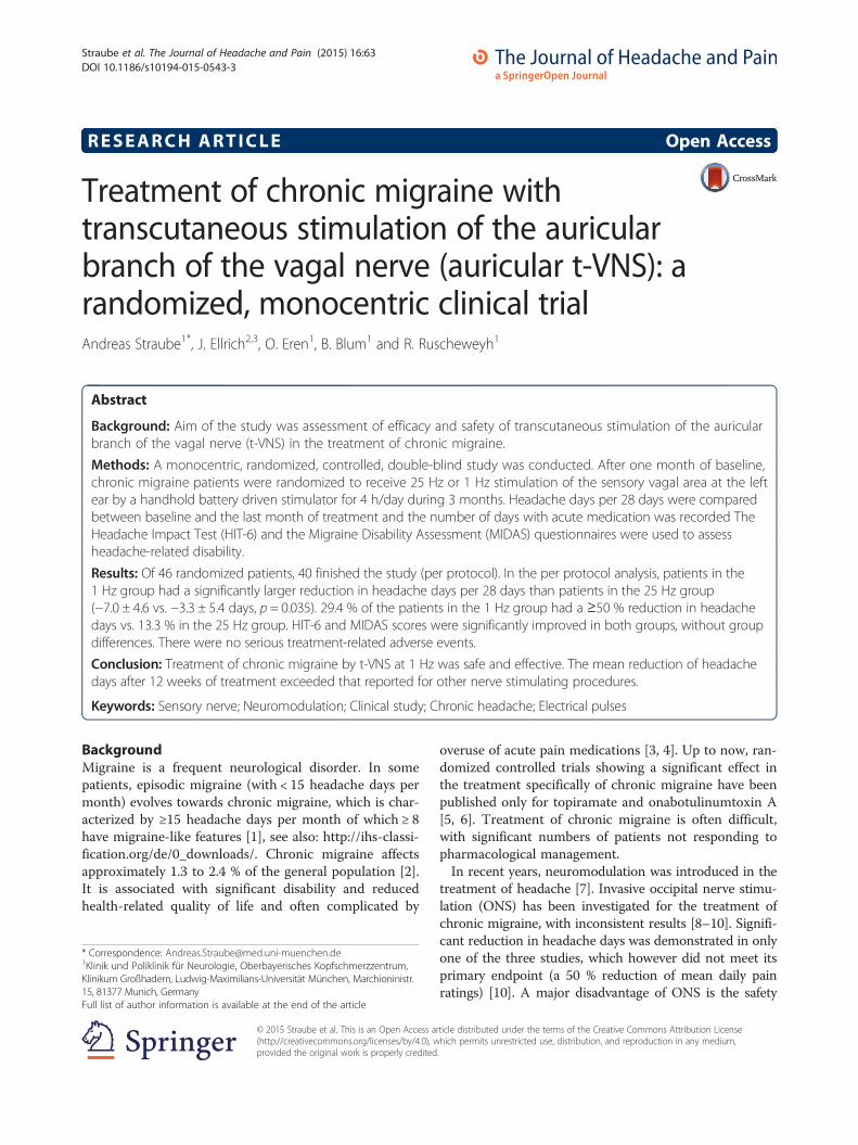
Study design (Fig. 1)The study consisted of a 4-week screening period
(“baseline”) followed by a 12-week randomized, double-blind, parallel-group treatment period with either 1 Hzor 25 Hz tVNS with the NEMOS® device (Fig. 2). Ad-verse events were recorded at visits 2 to 6. Compliancewith stimulation was checked at visits 3 to 6 by readingout the NEMOS® device and quantified in percent of the
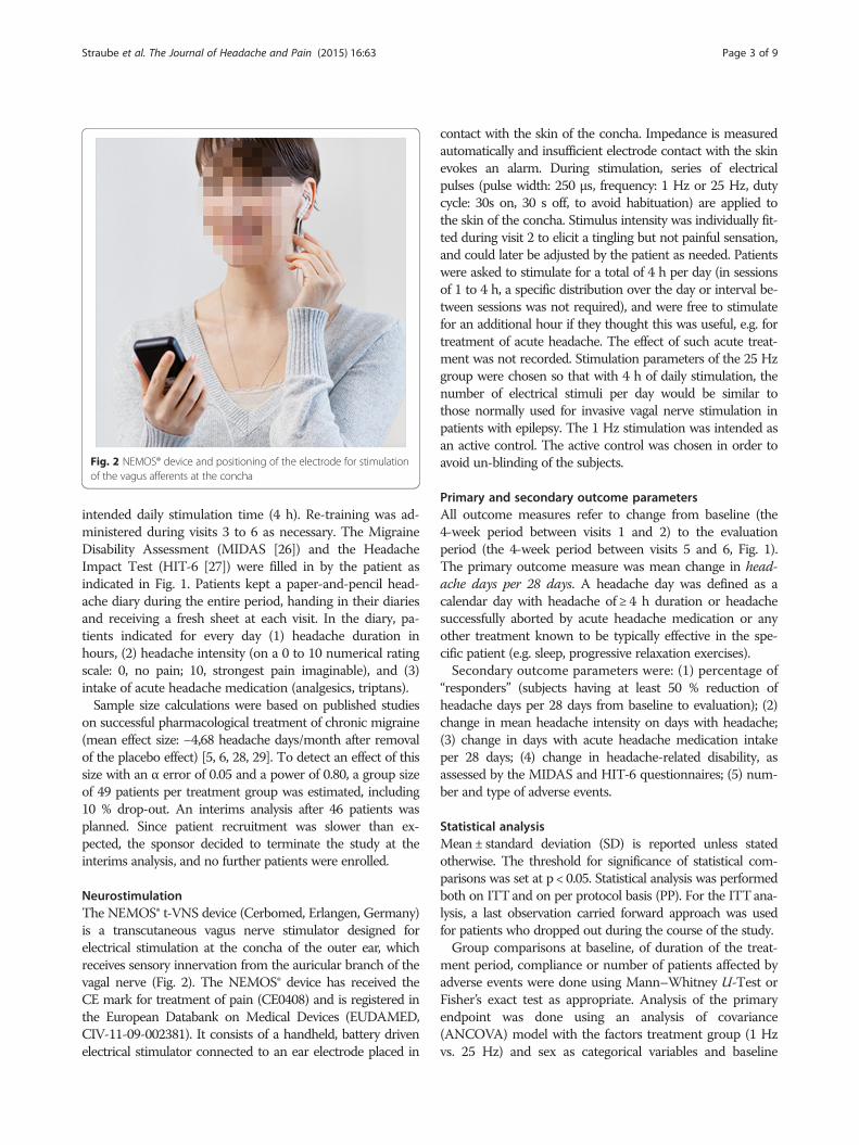
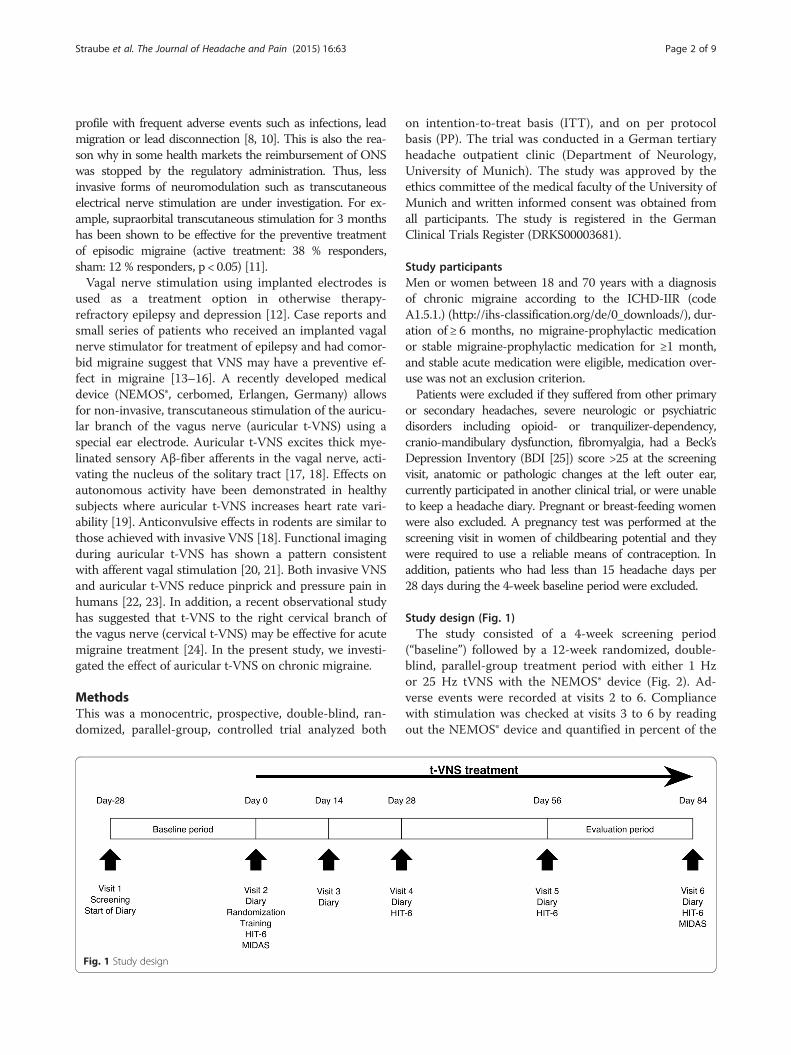
Fig. 2 NEMOS® device and positioning of the electrode for stimulationof the vagus afferents at the concha
Straube et al. The Journal of Headache and Pain (2015) 16:63 Page 3 of 9
intended daily stimulation time (4 h). Re-training was ad-ministered during visits 3 to 6 as necessary. The MigraineDisability Assessment (MIDAS [26]) and the HeadacheImpact Test (HIT-6 [27]) were filled in by the patient asindicated in Fig. 1. Patients kept a paper-and-pencil head-ache diary during the entire period, handing in their diariesand receiving a fresh sheet at each visit. In the diary, pa-tients indicated for every day (1) headache duration inhours, (2) headache intensity (on a 0 to 10 numerical ratingscale: 0, no pain; 10, strongest pain imaginable), and (3)intake of acute headache medication (analgesics, triptans).Sample size calculations were based on published studies
on successful pharmacological treatment of chronic migraine(mean effect size: −4,68 headache days/month after removalof the placebo effect) [5, 6, 28, 29]. To detect an effect of thissize with an α error of 0.05 and a power of 0.80, a group sizeof 49 patients per treatment group was estimated, including10 % drop-out. An interims analysis after 46 patients wasplanned. Since patient recruitment was slower than ex-pected, the sponsor decided to terminate the study at theinterims analysis, and no further patients were enrolled.
NeurostimulationThe NEMOS® t-VNS device (Cerbomed, Erlangen, Germany)is a transcutaneous vagus nerve stimulator designed forelectrical stimulation at the concha of the outer ear, whichreceives sensory innervation from the auricular branch of thevagal nerve (Fig. 2). The NEMOS® device has received theCE mark for treatment of pain (CE0408) and is registered inthe European Databank on Medical Devices (EUDAMED,CIV-11-09-002381). It consists of a handheld, battery drivenelectrical stimulator connected to an ear electrode placed in
contact with the skin of the concha. Impedance is measuredautomatically and insufficient electrode contact with the skinevokes an alarm. During stimulation, series of electricalpulses (pulse width: 250 μs, frequency: 1 Hz or 25 Hz, dutycycle: 30s on, 30 s off, to avoid habituation) are applied tothe skin of the concha. Stimulus intensity was individually fit-ted during visit 2 to elicit a tingling but not painful sensation,and could later be adjusted by the patient as needed. Patientswere asked to stimulate for a total of 4 h per day (in sessionsof 1 to 4 h, a specific distribution over the day or interval be-tween sessions was not required), and were free to stimulatefor an additional hour if they thought this was useful, e.g. fortreatment of acute headache. The effect of such acute treat-ment was not recorded. Stimulation parameters of the 25 Hzgroup were chosen so that with 4 h of daily stimulation, thenumber of electrical stimuli per day would be similar tothose normally used for invasive vagal nerve stimulation inpatients with epilepsy. The 1 Hz stimulation was intended asan active control. The active control was chosen in order toavoid un-blinding of the subjects.
Primary and secondary outcome parametersAll outcome measures refer to change from baseline (the4-week period between visits 1 and 2) to the evaluationperiod (the 4-week period between visits 5 and 6, Fig. 1).The primary outcome measure was mean change in head-ache days per 28 days. A headache day was defined as acalendar day with headache of ≥ 4 h duration or headachesuccessfully aborted by acute headache medication or anyother treatment known to be typically effective in the spe-cific patient (e.g. sleep, progressive relaxation exercises).Secondary outcome parameters were: (1) percentage of
“responders” (subjects having at least 50 % reduction ofheadache days per 28 days from baseline to evaluation); (2)change in mean headache intensity on days with headache;(3) change in days with acute headache medication intakeper 28 days; (4) change in headache-related disability, asassessed by the MIDAS and HIT-6 questionnaires; (5) num-ber and type of adverse events.
Statistical analysisMean± standard deviation (SD) is reported unless statedotherwise. The threshold for significance of statistical com-parisons was set at p < 0.05. Statistical analysis was performedboth on ITTand on per protocol basis (PP). For the ITTana-lysis, a last observation carried forward approach was usedfor patients who dropped out during the course of the study.Group comparisons at baseline, of duration of the treat-
ment period, compliance or number of patients affected byadverse events were done using Mann–Whitney U-Test orFisher’s exact test as appropriate. Analysis of the primaryendpoint was done using an analysis of covariance(ANCOVA) model with the factors treatment group (1 Hzvs. 25 Hz) and sex as categorical variables and baseline
Straube et al. The Journal of Headache and Pain (2015) 16:63 Page 4 of 9
values as covariate. The same type of ANCOVA was usedfor the analysis of the following secondary outcome param-eters: change in mean headache intensity, change in dayswith acute headache medication intake per 28 days andchange in MIDAS and HIT-6 scores. The number ofresponders was compared between groups using a logisticregression model that included treatment group and sex asfactor and the number of headache days per 28 days atbaseline as covariate. An estimate of the treatment oddsratio (Wald method) was derived from this model.
ResultsThe study was conducted between March 2012 and July2014. A total of 46 patients were randomized to the 1 Hzgroup (n = 22) or the 25 Hz group (n = 24, ITT). 6 patientsdropped out during the study. Reasons for dropouts were:adverse events in 4 patients (treatment-related stimulationsite ulcer in 3 patients, gastrectomy not related to treatmentin 1 patient), insufficient compliance in 1 patient, patient’srequest in 1 patient. One additional patient was excludedfrom the per protocol (PP) analysis after the end of thestudy because of violation of inclusion criteria (<15 head-ache days per 28 days in the screening period). This left 17patients in the 1 Hz group and 22 patients in the 25 Hzgroup for the PP analysis (Fig. 3) Demographic and head-ache characteristics of the population are shown in Table 1.There were no significant differences between both groups.
Primary outcome measurePP-analysis indicated a significant decrease in headachedays per 28 days from baseline to evaluation, which was
Fig. 3 Patient disposition
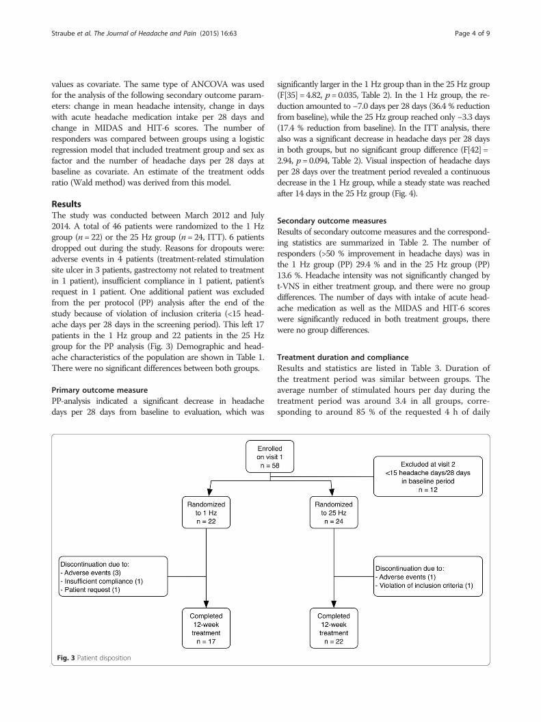
significantly larger in the 1 Hz group than in the 25 Hz group(F[35] = 4.82, p= 0.035, Table 2). In the 1 Hz group, the re-duction amounted to −7.0 days per 28 days (36.4 % reductionfrom baseline), while the 25 Hz group reached only −3.3 days(17.4 % reduction from baseline). In the ITT analysis, therealso was a significant decrease in headache days per 28 daysin both groups, but no significant group difference (F[42] =2.94, p= 0.094, Table 2). Visual inspection of headache daysper 28 days over the treatment period revealed a continuousdecrease in the 1 Hz group, while a steady state was reachedafter 14 days in the 25 Hz group (Fig. 4).
Secondary outcome measuresResults of secondary outcome measures and the correspond-ing statistics are summarized in Table 2. The number ofresponders (>50 % improvement in headache days) was inthe 1 Hz group (PP) 29.4 % and in the 25 Hz group (PP)13.6 %. Headache intensity was not significantly changed byt-VNS in either treatment group, and there were no groupdifferences. The number of days with intake of acute head-ache medication as well as the MIDAS and HIT-6 scoreswere significantly reduced in both treatment groups, therewere no group differences.
Treatment duration and complianceResults and statistics are listed in Table 3. Duration ofthe treatment period was similar between groups. Theaverage number of stimulated hours per day during thetreatment period was around 3.4 in all groups, corre-sponding to around 85 % of the requested 4 h of daily
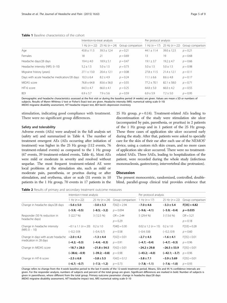
Table 1 Baseline characteristics of the cohort
Intention-to-treat analysis Per protocol analysis
1 Hz (n = 22) 25 Hz (n = 24) Group comparison 1 Hz (n = 17) 25 Hz (n = 22) Group comparison
Age 43.8 ± 11.5 39.3 ± 12.4 p = 0.21 44.1 ± 11.4 39.0 ± 12.5 p = 0.21
Females 18 21 p = 0.69 13 19 p = 0.68
Headache days/28 days 19.4 ± 4.0 18.9 ± 5.1 p = 0.47 19.1 ± 3.7 19.2 ± 4.7 p = 0.66
Headache intensity (NRS: 0–10) 5.2 ± 1.5 5.0 ± 1.5 p = 0.73 5.0 ± 1.5 5.0 ± 1.5 p = 0.98
Migraine history (years) 27.1 ± 13.0 20.4 ± 12.1 p = 0.08 27.8 ± 11.5 21.4 ± 12.1 p = 0.11
Days with acute headache medication/28 days 10.3 ± 6.4 8.2 ± 4.9 p = 0.24 11.1 ± 6.6 8.6 ± 4.8 p = 0.17
MIDAS score 76.8 ± 64.8 83.6 ± 56.0 p = 0.55 77.2 ± 70.1 82.1 ± 58.0 p = 0.71
HIT-6 score 64.3 ± 4.7 66.0 ± 4.1 p = 0.25 64.8 ± 5.0 66.0 ± 4.2 p = 0.55
BDI 6.9 ± 5.7 7.9 ± 5.6 p = 0.59 6.9 ± 5.9 7.2 ± 5.0 p = 0.95
Demographic and headache characteristics assessed at the first visit or during the baseline period (4 weeks) are given. Values are mean ± SD or numbers ofsubjects. Results of Mann–Whitney U test or Fisher’s Exact test are given. Headache intensity (NRS: numerical rating scale 0–10)MIDAS migraine disability assessment, HIT headache impact test, BDI beck’s depression inventory
Straube et al. The Journal of Headache and Pain (2015) 16:63 Page 5 of 9
stimulation, indicating good compliance with treatment.There were no significant group differences.
Safety and tolerabilityAdverse events (AEs) were analysed in the full analysis set(safety set) and summarized in Table 4. The number oftreatment emergent AEs (AEs occurring after initiation oftreatment) was higher in the 25 Hz group (112 events, 76treatment-related events) as compared to the 1 Hz group(67 events, 39 treatment-related events, Table 4),. Most AEswere mild or moderate in severity and resolved withoutsequelae. The most frequent treatment-related AE werelocal problems at the stimulation site, such as mild ormoderate pain, paresthesia, or pruritus during or afterstimulation, and erythema, ulcer or scab (31 events in 10patients in the 1 Hz group, 70 events in 17 patients in the
Table 2 Results of primary and secondary treatment outcome meas
Intention-t-treat analysis
1 Hz (n = 22) 25 Hz (n = 24) Gr
Change in headache days/28 days −5.6 ± 5.0 −3.0 ± 5.3 F[4
(−5.9; −0.5) (−8.5; −3.2) p =
Responder (50 % reduction inheadache days)
5 (22.7 %) 3 (12.5 %) OR
p =
Change in headache intensity(NRS 0 – 10)
−0.1 ± 1.1 (n = 20) 0.2 ± 1.0 F[4
(−0.2; 0.9) (−0.4; 0.7) p =
Change in days with acute headachemedication in 28 days
−2.0 ± 4.2 −1.3 ± 4.4 F[4
(−4.2; −0.3) (−4.4; −0.3) p =
Change in MIDAS score −18.7 ± 28.0 −21.8 ± 54.5 F[4
(−38.6; −0.9) (−39.2; −0.8) p =
Change in HIT-6 score −2.5 ± 6.8 −3.8 ± 5.5 F[4
(−6.7; −0.7) (−7.3; −1.2) p =
Change refers to change from the 4-week baseline period to the last 4 weeks of thgiven. For the responder analysis, numbers of subjects and percent of the total grogiven in parentheses, where different from the total group. Primary outcome paramMIDAS migraine disability assessment, HIT headache impact test, NRS numerical rati
25 Hz group, p= 0.14). Treatment-related AEs leading todiscontinuation of the study were stimulation site ulcer(accompanied by pain, paresthesia, or pruritus) in 2 patientsof the 1 Hz group and in 1 patient of the 25 Hz group.These three cases of application site ulcer occurred earlyduring the study. After that, patients were asked to speciallycare for the skin of their ear after each use of the NEMOS®device, using a custom rich skin cream, and no more casesof application site ulcer occurred. There were no treatment-related SAEs. Three SAEs, leading to hospitalization of thepatient, were recorded during the whole study (infectiousmononucleosis, gastrectomy, intervertebral disc protrusion).
DiscussionThe present monocentric, randomized, controlled, double-blind, parallel-group clinical trial provides evidence that
ures
Per protocol analysis
oup comparison 1 Hz (n = 17) 25 Hz (n = 22) Group comparison
2] = 2.94 −7.0 ± 4.6 −3.3 ± 5.4 F[35] = 4.82
0.094 (−9.6; −4.1) (−5.9; −0.4) p = 0.035
= 2.44 5 (29.4 %) 3 (13.6 %) OR = 3.21
0.29 p = 0.18
0] = 0.30 0.02 ± 1.2 (n = 15) 0.2 ± 1.0 F[33] = 0.28
0.58 (−0.4; 0.8) (−0.2; 0.9) p = 0.60
2] = 0.01 −2.7 ± 4.5 −1.6 ± 4.1 F[35] < 0.01
0.91 (−4.7; −0.4) (−4.7; −0.3) p = 0.96
2] < 0.01 −24.2 ± 29.8 −26.5 ± 53.9 F[35] < 0.01
0.98 (−43.2; −4.0) (−42.1; −3.7) p = 0.96
2] = 0.12 −3.8 ± 7.1 −3.9 ± 5.69 F[35] = 0.01
0.73 (−7.8; −1.1) (−7.6; −1.0) p = 0.93
e 12-week treatment period. Means, SDs and 95 % confidence intervals areup are given. Significant differences are marked in bold. Number of subjects iseter: change in headache days/28 daysng scale 0–10
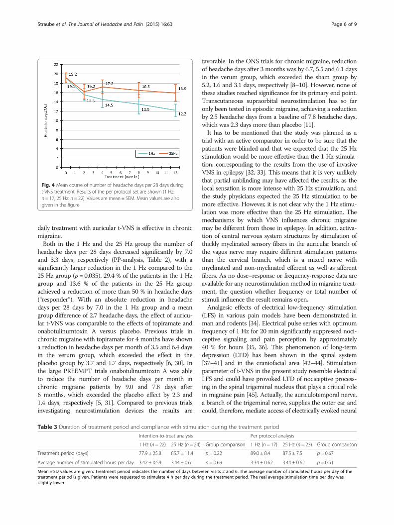
Fig. 4 Mean course of number of headache days per 28 days duringt-VNS treatment. Results of the per protocol set are shown (1 Hz:n = 17, 25 Hz: n= 22). Values are mean ± SEM. Mean values are alsogiven in the figure
Straube et al. The Journal of Headache and Pain (2015) 16:63 Page 6 of 9
daily treatment with auricular t-VNS is effective in chronicmigraine.Both in the 1 Hz and the 25 Hz group the number of
headache days per 28 days decreased significantly by 7.0and 3.3 days, respectively (PP-analysis, Table 2), with asignificantly larger reduction in the 1 Hz compared to the25 Hz group (p = 0.035). 29.4 % of the patients in the 1 Hzgroup and 13.6 % of the patients in the 25 Hz groupachieved a reduction of more than 50 % in headache days(“responder”). With an absolute reduction in headachedays per 28 days by 7.0 in the 1 Hz group and a meangroup difference of 2.7 headache days, the effect of auricu-lar t-VNS was comparable to the effects of topiramate andonabotulinumtoxin A versus placebo. Previous trials inchronic migraine with topiramate for 4 months have showna reduction in headache days per month of 3.5 and 6.4 daysin the verum group, which exceeded the effect in theplacebo group by 3.7 and 1.7 days, respectively [6, 30]. Inthe large PREEMPT trials onabotulinumtoxin A was ableto reduce the number of headache days per month inchronic migraine patients by 9.0 and 7.8 days after6 months, which exceeded the placebo effect by 2.3 and1.4 days, respectively [5, 31]. Compared to previous trialsinvestigating neurostimulation devices the results are
Table 3 Duration of treatment period and compliance with stimula
Intention-to-treat analysis
1 Hz (n = 22) 25 Hz (n = 24)
Treatment period (days) 77.9 ± 25.8 85.7 ± 11.4
Average number of stimulated hours per day 3.42 ± 0.59 3.44 ± 0.61
Mean ± SD values are given. Treatment period indicates the number of days betwtreatment period is given. Patients were requested to stimulate 4 h per day durinslightly lower
favorable. In the ONS trials for chronic migraine, reductionof headache days after 3 months was by 6.7, 5.5 and 6.1 daysin the verum group, which exceeded the sham group by5.2, 1.6 and 3.1 days, respectively [8–10]. However, none ofthese studies reached significance for its primary end point.Transcutaneous supraorbital neurostimulation has so faronly been tested in episodic migraine, achieving a reductionby 2.5 headache days from a baseline of 7.8 headache days,which was 2.3 days more than placebo [11].It has to be mentioned that the study was planned as a
trial with an active comparator in order to be sure that thepatients were blinded and that we expected that the 25 Hzstimulation would be more effective than the 1 Hz stimula-tion, corresponding to the results from the use of invasiveVNS in epilepsy [32, 33]. This means that it is very unlikelythat partial unblinding may have affected the results, as thelocal sensation is more intense with 25 Hz stimulation, andthe study physicians expected the 25 Hz stimulation to bemore effective. However, it is not clear why the 1 Hz stimu-lation was more effective than the 25 Hz stimulation. Themechanisms by which VNS influences chronic migrainemay be different from those in epilepsy. In addition, activa-tion of central nervous system structures by stimulation ofthickly myelinated sensory fibers in the auricular branch ofthe vagus nerve may require different stimulation patternsthan the cervical branch, which is a mixed nerve withmyelinated and non-myelinated efferent as well as afferentfibers. As no dose–response or frequency-response data areavailable for any neurostimulation method in migraine treat-ment, the question whether frequency or total number ofstimuli influence the result remains open.Analgesic effects of electrical low-frequency stimulation
(LFS) in various pain models have been demonstrated inman and rodents [34]. Electrical pulse series with optimumfrequency of 1 Hz for 20 min significantly suppressed noci-ceptive signaling and pain perception by approximately40 % for hours [35, 36]. This phenomenon of long-termdepression (LTD) has been shown in the spinal system[37–41] and in the craniofacial area [42–44]. Stimulationparameter of t-VNS in the present study resemble electricalLFS and could have provoked LTD of nociceptive process-ing in the spinal trigeminal nucleus that plays a critical rolein migraine pain [45]. Actually, the auriculotemporal nerve,a branch of the trigeminal nerve, supplies the outer ear andcould, therefore, mediate access of electrically evoked neural
tion during the treatment period
Per protocol analysis
Group comparison 1 Hz (n = 17) 25 Hz (n = 23) Group comparison
p = 0.22 89.0 ± 8.4 87.5 ± 7.5 p = 0.67
p = 0.69 3.34 ± 0.62 3.44 ± 0.62 p = 0.51
een visits 2 and 6. The average number of stimulated hours per day of theg the treatment period. The real average stimulation time per day was
Table 4 Overview of adverse events (safety set)
1 Hz (n = 22) 25 Hz (n = 24)
Number of events Number of patients (%) Number of events Number of patients (%)
Treatment emergent AEs 67 17 (77.3 %) 112 19 (79.2 %)
Treatment-related AEs 39 11 (50.0 %) 76 17 (70.8 %)
Stimulation site treatment-related TEAEs 31 10 (45.5 %) 70 17 (70.8 %)
All serious AEs (including pre-treatment SAEs) 2 2 (9.1 %) 0 0
Serious treatment emergent AEs 2 2 (9.1 %) 0 0
Serious treatment-related AEs 0 0 0 0
Treatment-related AEs leading to discontinuation of study 8 4 (18.2 %) 4 1 (4.2 %)
Death 0 0 0 0
Straube et al. The Journal of Headache and Pain (2015) 16:63 Page 7 of 9
signals to brainstem nuclei of the trigeminal nerve [46]. Thus,LTD could be a mechanism that might, at least, contribute tothe analgesic effect of t-VNS in the present study.In fact, other stimulation parameters might be even
more effective than the 1 Hz stimulation, and the 25 Hzstimulation might have been partially active in the presentstudy, possibly reducing the effect in the group compari-son. Indeed, 25–30 Hz stimulation has been shown to sig-nificantly reduce experimental pain in humans [23] andseizures in rodents [18]. In addition, in the present studyboth groups significantly improved in headache-relateddisability measures (MIDAS and HIT-6), and reducedtheir intake of acute headache medication, although it can-not be determined if this is due to the placebo effect ordue to stimulation effects in both groups. The missing sig-nificant difference in the reduction of the MIDAS andHIT6 between the 1Hz and the 25Hz group is probablydue to the too small sensitivity of these tests in detectingdifferences in quality of life. Furthermore, it is unclear if25 Hz stimulation also have a mood stabilizing effectwhich influences the ratings in the used tests.Furthermore, it is still not clear how vagus nerve
stimulation interferes with migraine generation. Onepossibility is a direct or indirect inhibition of nociceptivetrigeminal neurons by vagal activation. Indeed, animaldata show that afferent vagal stimulation can reduce theactivation of nociceptive neurons in the caudal trigemi-nal nucleus in response to noxious stimulation of theface or dura [47–49]. This might be due to the existenceof dense reciprocal connections between the spinal tri-geminal nucleus and the nucleus tractus solitarii (NTS)which is the major target of vagal afferents [50]. Re-sponses of spinal trigeminal neurons might also be re-duced by activation of the descending pain inhibitorysystems. Although this has not been shown directly forthe trigeminal area, animal studies showed that vagalnerve stimulation can activate descending pain inhibi-tory systems, probably involving projections from theNTS to the nucleus raphe magnus and the locus coeruleus,which are at the origin of serotonergic and noradrenergic
descending pain inhibitory pathways [51]. Alternatively,VNS might exert migraine prophylactic actions by modify-ing cortical excitability. Altered cortical excitability inchronic migraine has been demonstrated in various elec-trophysiological measurements is thought to contribute toits pathogenesis [52]. Several lines of evidence indicate thatthe cortical excitability is increased in chronic migraine pa-tients: 1) There is a reduced habituation of the blink reflexinterictally [53]. 2) The magnetic suppression of perceptualaccuracy was decreased in patients with chronic migrainecompared to episodic migraine and controls which mayindicate also a higher cortical excitability [54]. 3) Analysisof the high frequency somatosensory evoked potentialsshowed early response sensitization and late habituation,most probably due an increased coupling between thal-amus and cortex in chronic migraine [55]. Afferent vagalinformation is relayed via the NTS and the parabrachialnucleus to several subcortical and cortical regions, includingthalamus, insula and lateral prefrontal cortex. In addition,the NTS has strong projections to the locus coeruleus andthe nucleus raphe magnus which provide widespread norad-renergic and serotonergic innervation of the cortex [56].Modulation of cortical excitability via these pathways isthought to be important for the anticonvulsant effects ofVNS [33]. Increased GABA levels have been found in thecerebrospinal fluid of epilepsy patients treated with VNS,suggesting an increase in inhibitory neurotransmission [57].Auricular t-VNS increases parasympathetic activity and/orreduces sympathetic activity [19], which might also affectcortical excitability, maybe by mechanisms similar to thoseassumed for the migraine preventive effects of beta-blockingagents [58]. In summary, VNS is well positioned to alter cor-tical excitability, especially to reduce cortical hyperexcitabil-ity. Direct evidence that this interferes with pain processingor migraine generation is currently lacking. It would be inter-esting to repeat the above described experiments whichshowed an increased cortical excitability in chronic migraineunder t-VNS stimulation. A third possibility is that the anti-migraine action of VNS relies on modification of transmitterrelease from efferent parasympathetic fibers innervating
Straube et al. The Journal of Headache and Pain (2015) 16:63 Page 8 of 9
dural vessels, e.g. fibers stemming from the spheno-palatineganglion. The release of neurotransmitters, especially calci-tonin-gene related peptide (CGRP), at dural vessels withsubsequent neurogenic inflammation and sensitization ofprimary afferents is thought to play an important role inmigraine pathophysiology [59]. Parasympathetic fibres in-nervating the dura mater release vasoactive intestinal poly-peptide (VIP) and pituitary adenylate cyclase-activatingpolypeptide (PACAP), which are potent vasodilatators andthought to contribute to sensitization of nociceptive trigemi-nal primary afferents. Increased peripheral blood VIP levelshave been detected in chronic migraine [60], and intraven-ous administration of PACAP has been shown to inducemigrainous headache in migraine patients [61], suggestingthat both transmitters are related to migraine patho-physiology. Although auricular t-VNS stimulates only vagalafferents, there are close connections between afferent andefferent parasympathetic brainstem centers, making an in-fluence of VNS on dural efferents likely.A major practical advantage of auricular t-VNS is good
tolerability and safety. For comparison, in the pooled topir-amate trial analysis, 1 out of 4 patients (25 %) dropped outbecause of intolerable adverse effects [62]. In our studyonly 3 of 46 patients (7 %) dropped out due to side effectsof t-VNS. All three cases occurred early in the study andwere due to stimulation site ulcer which later in the studycould be prevented by appropriate skin care. Another ad-vantage of t-VNS therapy is that it can be combined withany other drug treatment without risking cumulative ad-verse effects or pharmacodynamic interactions. In addition,auricular t-VNS allows patients to continue routine activ-ities, leading to a high compliance with stimulation times(around 85 % on average). However, long-term effects andsustainability of efficacy of t-VNS are still unknown andneed to be demonstrated in appropriate open-label trials.
ConclusionsIn conclusion, the present parallel-group randomized con-trolled trial, provides evidence that auricular t-VNS at 1 Hzfor 4 h daily is effective for chronic migraine preventionover 3 months. The absolute reduction in headache days(7.0) and the difference between groups (2.7 headache days)is comparable to the effects of topiramate and onabotuli-num toxin A in chronic migraine prevention. The t-VNStreatment also results in a meaningful improvement in thequality of life as assessed by MIDAS and HIT 6. The safetyprofile was favourable and compliance with daily stimula-tion was high.
Competing interestsCerbomed funded the study.A. Straube has received honoraries by Pharm Allergan, Boehringer Ingelheim,Hormosan, electroCore, CerboMed. Grants from the German Sciencefoundation, German Minister of Research and Education and the Kröner-Freseniusfoundation.
J. Ellrich was employed as Chief Medical Officer by the company cerbomedGmbH.O. Eren has nothing to discloseB. Blum has nothing to discloseR. Ruscheweyh has received honaries by Pharm Allergan, MSD,Mundipharma, Pfizer and grants from the Else Kröner Fresenius Stiftung.
Authors’ contributionsThe study was planned by JE and AS. AS, RR, OE, BB recruited the patientsand collected the data. Statistical analysis was performed by MetronomiaClinical Research GmbH (Munich, Germany). Cerbomed supported thepreparation of the figures and the layout. The paper was written by theauthors and all authors participated in the decision to publish the paper andhad full access to all study data. All authors read and approved the finalmanuscript.
AcknowledgementThe authors thank Nadine Wolf, PhD for her contribution to the clinicalinvestigation plan and A. Hartlep, PhD and V. Koepke for their help in thepreparation of the manuscript.
Author details1Klinik und Poliklinik für Neurologie, Oberbayerisches Kopfschmerzzentrum,Klinikum Großhadern, Ludwig-Maximilians-Universität München, Marchioninistr.15, 81377 Munich, Germany. 2Department of Health Science and Technology,Professor Dr. med. Jens Ellrich, Aalborg University, Fredrik Bajers Vej 7D2,DK-9220 Aalborg, Denmark. 3Cerbomed GmbH, Medical Valley Center, Henkestr.91, 91052 Erlangen, Germany.
Received: 5 May 2015 Accepted: 16 June 2015
References1. Headache Classification Subcommittee of the International Headache
Society (2013) The International Classification of Headache Disorders, 3rdedition (beta version). Cephalalgia 33:629–808
2. Bigal ME, Serrano D, Reed M, Lipton RB (2008) Chronic migraine in thepopulation: burden, diagnosis, and satisfaction with treatment. Neurology71:559–566
3. Dodick DW (2006) Clinical practice. Chronic daily headache. N Engl J Med354:158–165
4. Lipton RB, Bigal ME (2003) Chronic daily headache: is analgesic overuse acause or a consequence? Neurology 61:154–155
5. Diener HC, Dodick DW, Aurora SK, Turkel CC, DeGryse RE, Lipton RB,Silberstein SD, Brin MF (2010) OnabotulinumtoxinA for treatment of chronicmigraine: results from the double-blind, randomized, placebo-controlledphase of the PREEMPT 2 trial. Cephalalgia 30:804–814
6. Silberstein SD, Lipton RB, Dodick DW, Freitag FG, Ramadan N, Mathew N,Brandes JL, Bigal M, Saper J, Ascher S, Jordan DM, Greenberg SJ, Hulihan J(2007) Efficacy and safety of topiramate for the treatment of chronicmigraine: a randomized, double-blind, placebo-controlled trial. Headache47:170–180
7. Martelletti P, Jensen RH, Antal A, Arcioni R, Brighina F, de Tommaso M,Franzini A, Fontaine D, Heiland M, Jurgens TP, Leone M, Magis D,Paemeleire K, Palmisani S, Paulus W, May A (2013) Neuromodulation ofchronic headaches: position statement from the European HeadacheFederation. J Headache Pain 14:86
8. Saper JR, Dodick DW, Silberstein SD, McCarville S, Sun M, Goadsby PJ (2011)Occipital nerve stimulation for the treatment of intractable chronic migraineheadache: ONSTIM feasibility study. Cephalalgia 31:271–285
9. Lipton RB, Goadsby PJ, Cady RK, Aurora SK, Grosberg BM, Freitag FG,Silberstein SD, Whiten DM, Jaax KN (2009) PRISM study: occipital nervestimulation for treatment-refractory migraine. Cephalalgia 29:30
10. Silberstein SD, Dodick DW, Saper J, Huh B, Slavin KV, Sharan A, Reed K,Narouze S, Mogilner A, Goldstein J, Trentman T, Vaisman J, Ordia J, Weber P,Deer T, Levy R, Diaz RL, Washburn SN, Mekhail N (2012) Safety and efficacyof peripheral nerve stimulation of the occipital nerves for the managementof chronic migraine: results from a randomized, multicenter, double-blinded,controlled study. Cephalalgia 32:1165–1179
11. Schoenen J, Vandersmissen B, Jeangette S, Herroelen L, Vandenheede M,Gerard P, Magis D (2013) Migraine prevention with a supraorbital
Straube et al. The Journal of Headache and Pain (2015) 16:63 Page 9 of 9
transcutaneous stimulator: a randomized controlled trial. Neurology80:697–704
12. Beekwilder JP, Beems T (2010) Overview of the clinical applications of vagusnerve stimulation. J Clin Neurophysiol 27:130–138
13. Hord ED, Evans MS, Mueed S, Adamolekun B, Naritoku DK (2003) The effectof vagus nerve stimulation on migraines. J Pain 4:530–534
14. Sadler RM, Purdy RA, Rahey S (2002) Vagal nerve stimulation abortsmigraine in patient with intractable epilepsy. Cephalalgia 22:482–484
15. Mauskop A (2005) Vagus nerve stimulation relieves chronic refractorymigraine and cluster headaches. Cephalalgia 25:82–86
16. Lenaerts ME, Oommen KJ, Couch JR, Skaggs V (2008) Can vagus nervestimulation help migraine? Cephalalgia 28:392–395
17. Ellrich J (2011) Transcutaneous vagus nerve stimulation. Eur Neurol Rev6:254–256
18. He W, Jing XH, Zhu B, Zhu XL, Li L, Bai WZ, Ben H (2013) The auriculo-vagalafferent pathway and its role in seizure suppression in rats. BMC Neurosci14:85
19. Clancy JA, Mary DA, Witte KK, Greenwood JP, Deuchars SA, Deuchars J(2014) Non-invasive vagus nerve stimulation in healthy humans reducessympathetic nerve activity. Brain Stimul 7:871–877
20. Kraus T, Kiess O, Hosl K, Terekhin P, Kornhuber J, Forster C (2013) CNS BOLDfMRI effects of sham-controlled transcutaneous electrical nerve stimulationin the left outer auditory canal - a pilot study. Brain Stimul 6:798–804
21. Frangos E, Ellrich J, Komisaruk BR (2015) Non-invasive access to the vagusnerve central projections via electrical stimulation of the external Ear: fMRIevidence in humans. Brain Stimul 8:624–636
22. Kirchner A, Birklein F, Stefan H, Handwerker HO (2000) Left vagus nervestimulation suppresses experimentally induced pain. Neurology 55:1167–1171
23. Busch V, Zeman F, Heckel A, Menne F, Ellrich J, Eichhammer P (2013) Theeffect of transcutaneous vagus nerve stimulation on pain perception–anexperimental study. Brain Stimul 6:202–209
24. Goadsby PJ, Grosberg BM, Mauskop A, Cady R, Simmons KA (2014) Effect ofnoninvasive vagus nerve stimulation on acute migraine: an open-label pilotstudy. Cephalalgia 34:986–993
25. Beck AT, Erbaugh J, Ward CH, Mock J, Mendelsohn M (1961) An inventoryfor measuring depression. Arch Gen Psychiatry 4:561–571
26. Stewart WF, Lipton RB, Dowson AJ, Sawyer J (2001) Development andtesting of the Migraine Disability Assessment (MIDAS) questionnaire toassess headache-related disability. Neurology 56:S20–S28
27. Kosinski M, Bayliss MS, Bjorner JB, Ware JE Jr, Garber WH, Batenhorst A, CadyR, Dahlof CG, Dowson A, Tepper S (2003) A six-item short-form survey formeasuring headache impact: the HIT-6. Qual Life Res 12:963–974
28. Freitag FG, Diamond S, Diamond M, Urban G (2008) Botulinum toxin type Ain the treatment of chronic migraine without medication overuse.Headache 48:201–209
29. Silvestrini M, Bartolini M, Coccia M, Baruffaldi R, Taffi R, Provinciali L (2003)Topiramate in the treatment of chronic migraine. Cephalalgia 23:820–824
30. Diener HC, Bussone G, Van Oene JC, Lahaye M, Schwalen S, Goadsby PJ(2007) Topiramate reduces headache days in chronic migraine: a randomized,double-blind, placebo-controlled study. Cephalalgia 27:814–823
31. Aurora SK, Dodick DW, Turkel CC, DeGryse RE, Silberstein SD, Lipton RB,Diener HC, Brin MF (2010) OnabotulinumtoxinA for treatment of chronicmigraine: results from the double-blind, randomized, placebo-controlledphase of the PREEMPT 1 trial. Cephalalgia 30:793–803
32. The Vagus Nerve Stimulation Study Group (1995) A randomized controlledtrial of chronic vagus nerve stimulation for treatment of medicallyintractable seizures. Neurology 45:224–230
33. Milby AH, Halpern CH, Baltuch GH (2008) Vagus nerve stimulation forepilepsy and depression. Neurotherapeutics 5:75–85
34. Ellrich J (2006) Long-term depression of orofacial somatosensory processing.Suppl Clin Neurophysiol 58:195–208
35. Ellrich J (2004) Electric low-frequency stimulation of the tongue induceslong-term depression of the jaw-opening reflex in anesthetized mice.J Neurophysiol 92:3332–3337
36. Jung K, Rottmann S, Ellrich J (2009) Long-term depression of spinal nociception andpain in man: influence of varying stimulation parameters. Eur J Pain 13:161–170
37. Rottmann S, Jung K, Vohn R, Ellrich J (2010) Long-term depression of pain-relatedcerebral activation in healthy man: an fMRI study. Eur J Pain 14:615–624
38. Rottmann S, Jung K, Ellrich J (2010) Electrical low-frequency stimulationinduces long-term depression of sensory and affective components of painin healthy man. Eur J Pain 14:359–365
39. Jung K, Larsen LE, Rottmann S, Ellrich J (2011) Heterotopic low-frequencystimulation induces nociceptive LTD within the same central receptive fieldin man. Exp Brain Res 212:189–198
40. Jung K, Lelic D, Rottmann S, Drewes AM, Petrini L, Ellrich J (2012) Electricallow-frequency stimulation induces central neuroplastic changes of painprocessing in man. Eur J Pain 16:509–521
41. Rottmann S, Jung K, Ellrich J (2008) Electrical low-frequency stimulationinduces homotopic long-term depression of nociception and pain fromhand in man. Clin Neurophysiol 119:1895–1904
42. Aymanns M, Yekta SS, Ellrich J (2009) Homotopic long-term depression oftrigeminal pain and blink reflex within one side of the human face. ClinNeurophysiol 120:2093–2099
43. Yekta SS, Lamp S, Ellrich J (2006) Heterosynaptic long-term depression ofcraniofacial nociception: divergent effects on pain perception and blinkreflex in man. Exp Brain Res 170:414–422
44. Ellrich J, Schorr A (2004) Low-frequency stimulation of trigeminal afferentsinduces long-term depression of human sensory processing. Brain Res996:255–258
45. Noseda R, Burstein R (2013) Migraine pathophysiology: anatomy of thetrigeminovascular pathway and associated neurological symptoms, CSD,sensitization and modulation of pain. Pain. 154 Suppl 1.
46. Peuker ET, Filler TJ (2002) The nerve supply of the human auricle. Clin Anat15:35–37
47. Lyubashina OA, Sokolov AY, Panteleev SS (2012) Vagal afferent modulationof spinal trigeminal neuronal responses to dural electrical stimulation in rats.Neuroscience 222:29–37
48. Multon S, Schoenen J (2005) Pain control by vagus nerve stimulation: fromanimal to man…and back. Acta Neurol Belg 105:62–67
49. Bossut DF, Maixner W (1996) Effects of cardiac vagal afferentelectrostimulation on the responses of trigeminal and trigeminothalamicneurons to noxious orofacial stimulation. Pain 65:101–109
50. Zerari-Mailly F, Buisseret P, Buisseret-Delmas C, Nosjean A (2005) Trigemino-solitarii-facial pathway in rats. J Comp Neurol 487:176–189
51. Randich A, Gebhart GF (1992) Vagal afferent modulation of nociception.Brain Res Brain Res Rev 17:77–99
52. Coppola G, Pierelli F, Schoenen J (2009) Habituation and migraine.Neurobiol Learn Mem 92:249–259
53. De Marinis M, Pujia A, Colaizzo E, Accornero N (2007) The blink reflex in“chronic migraine”. Clin Neurophysiol 118:457–463
54. Aurora SK, Barrodale PM, Tipton RL, Khodavirdi A (2007) Brainstemdysfunction in chronic migraine as evidenced by neurophysiological andpositron emission tomography studies. Headache 47:996–1003
55. Coppola G, Iacovelli E, Bracaglia M, Serrao M, Di LC, Pierelli F (2013)Electrophysiological correlates of episodic migraine chronification: evidencefor thalamic involvement. J Headache Pain 14:76
56. Henry TR (2002) Therapeutic mechanisms of vagus nerve stimulation.Neurology 59:S3–S14
57. Ben-Menachem E, Hamberger A, Hedner T, Hammond EJ, Uthman BM,Slater J, Treig T, Stefan H, Ramsay RE, Wernicke JF (1995) Effects of vagusnerve stimulation on amino acids and other metabolites in the CSF ofpatients with partial seizures. Epilepsy Res 20:221–227
58. Richter F, Mikulik O, Ebersberger A, Schaible HG (2005) Noradrenergicagonists and antagonists influence migration of cortical spreadingdepression in rat-a possible mechanism of migraine prophylaxis andprevention of postischemic neuronal damage. J Cereb Blood Flow Metab25:1225–1235
59. Pietrobon D, Striessnig J (2003) Neurobiology of migraine. Nat Rev Neurosci4:386–398
60. Cernuda-Morollon E, Martinez-Camblor P, Alvarez R, Larrosa D, Ramon C,Pascual J (2015) Increased VIP levels in peripheral blood outside migraineattacks as a potential biomarker of cranial parasympathetic activation inchronic migraine. Cephalalgia 35:310–316
61. Schytz HW, Birk S, Wienecke T, Kruuse C, Olesen J, Ashina M (2009)PACAP38 induces migraine-like attacks in patients with migraine withoutaura. Brain 132:16–25
62. Bussone G, Diener HC, Pfeil J, Schwalen S (2005) Topiramate 100 mg/day inmigraine prevention: a pooled analysis of double-blind randomisedcontrolled trials. Int J Clin Pract 59:961–968