microspheres as a high capacity anode for lithium ion ... · hierarchically porous and nitrogen,...
TRANSCRIPT
1
Hierarchically porous and nitrogen, sulfur-codoped graphene-like
microspheres as a high capacity anode for lithium ion batteries
Dongfei Sun,ab Juan Yanga and Xingbin Yan*ac
a Laboratory of Clean Energy Chemistry and Materials, Lanzhou Institute of Chemical
Physics, Chinese Academy of Science, Lanzhou 730000, P. R. China
b Graduate University of Chinese Academy of Sciences, Beijing 100080, P. R. China
c State Key Laboratory of Solid Lubrication, Lanzhou Institute of Chemical Physics, Chinese
Academy of Sciences, Lanzhou 730000, P. R. China
Experimental section
Preparation of Ni template
Powdery Ni microspheres were prepared by reducing the commercial nickel hydroxide
microspheres which can be obtained from everywhere (Henan Kelong New Energy Co., Ltd.).
Typically, Ni microspheres were obtained by calcining the powdery nickel hydroxide
microspheres at 500 °C for 2 h in air, followed by reducing at 600 °C for 2 h in a hydrogen
atmosphere.
Preparation of 3D NS-GSs
3D NS-GSs were prepared by a precursor-assisted chemical vapor deposition method.
Typically, Ni microspheres (6 g) and poly(vinylpyrrolidone) (PVP)/(NH4)2S2O8 aqueous
solution (5ml, the concentrations of PVP and (NH4)2S2O8 are 0.2 g/ml and 0.5 g/ml,
respectively) were mixed uniformly. After been dried, the mixture was placed into a quartz
tube and heated to 400 °C under Ar (100 sccm)/H2 (100 sccm) atmosphere for 1 h, and
subsequently the temperature was increased to 800 °C for 1 h under the same conditions.
Then, the sample was cooled rapidly to room temperature. Finally, the as-obtained
Electronic Supplementary Material (ESI) for ChemComm.This journal is © The Royal Society of Chemistry 2014
2
graphene/Ni product was etched in HCl solution (1 mol L-1) at 80 °C for 48 h to remove Ni,
and followed by drying at 60 °C for 48 h to obtain 3D NS-GSs. For preparing 3D GSs, the
same procedures were carried out just without adding (NH4)2S2O8. Furthermore, in order to
investigate the influence of oxygen-containing group on the structure and performance of 3D
GSs, another carbon source (polyethylene glycol, PEG) with high oxygen content was also
used.
Structural characterizations
The morphology and microstructure of the as-prepared samples was investigated using a
field-emission scanning electron microscope (FE-SEM, JSM-6701F), and a transmission
electron microscope (TEM, Tecnai G20). The structure and composition of the samples was
recorded using a micro-Raman spectroscopy (JY-HR800, the excitation wavelength of 532nm)
and an X-ray diffraction (XRD, Philips X’ Pert Pro). The surface chemical species of the as-
prepared 3D NS-GSs sample were examined on a Perkin-Elmer PHI-5702 multifunctional X-
ray photoelectron spectroscope (XPS, Physical Electronics, USA). Nitrogen adsorption-
desorption isotherm measurements were performed on a Micrometitics ASAP 2020
volumetric adsorption analyzer at 77 K.
Electrochemical measurements
The working electrodes and lithium-ion half-cells were prepared as follows. Typically, a
mixture of 85 wt% active material (3D GSs or 3D NS-GSs) and 15 wt% polyvinylidene
difluoride (PVDF) was milled with N-methyl pyrrolidone (NMP) to form a homogeneous
slurry, and the mixture was coated onto a copper foil. As-prepared working electrodes were
dried under vacuum at 110 °C for 10 h. After being pressed, the electrodes were assembled
into coin cells (CR2032) in an argon-filled glove box by using 1 mol L-1 LiPF6 in dimethyl
carbonate (DMC) and ethylene carbonate (EC) (1:1, v/v) as the electrolyte and pure lithium
foil as the counter electrode. The assembled coin cells were tested in the voltage range of
3
0.01~3.0 V on a CT2001A cell test instrument (LAND Electronic Co.). All the
electrochemical measurements were carried out at 25 °C in a digital biochemical incubator
and the specific capacity was calculated based on the weight of each active material. Cyclic
voltammetry (CV) (0.01~3 V, 0.01 mV s-1) and electrochemical impedance spectroscopy (EIS)
measurements were carried out on a CHI 660D electrochemical workstation.
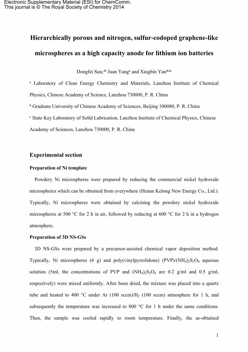
Fig. S1 (a-c) SEM images of nickel hydroxide microspheres and (d-f) nickel microspheres.
The Ni microspheres maintain the spherical structure, and each Ni microsphere consists of
Ni nanoparticles with many pores, which forms a continuous and irregular porous structure in
the micropheres. The porous structure can ensuing that liquid precursor are easily filled in the
interstices of the Ni microspheres, meanwhile, the liquid precursor can prevent the
agglomeration or deformation of the porous Ni microspheres at the CVD process.
4
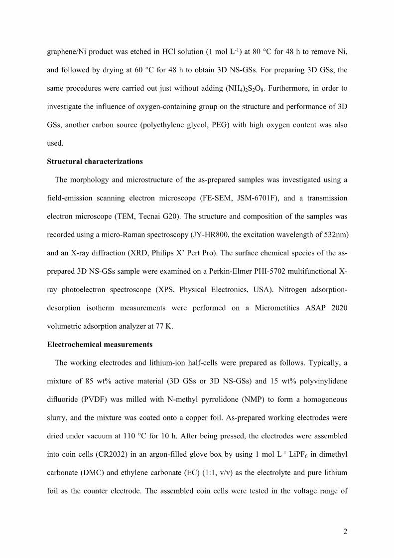
Fig. S2 (a) and (b) SEM images of 3D GSs.
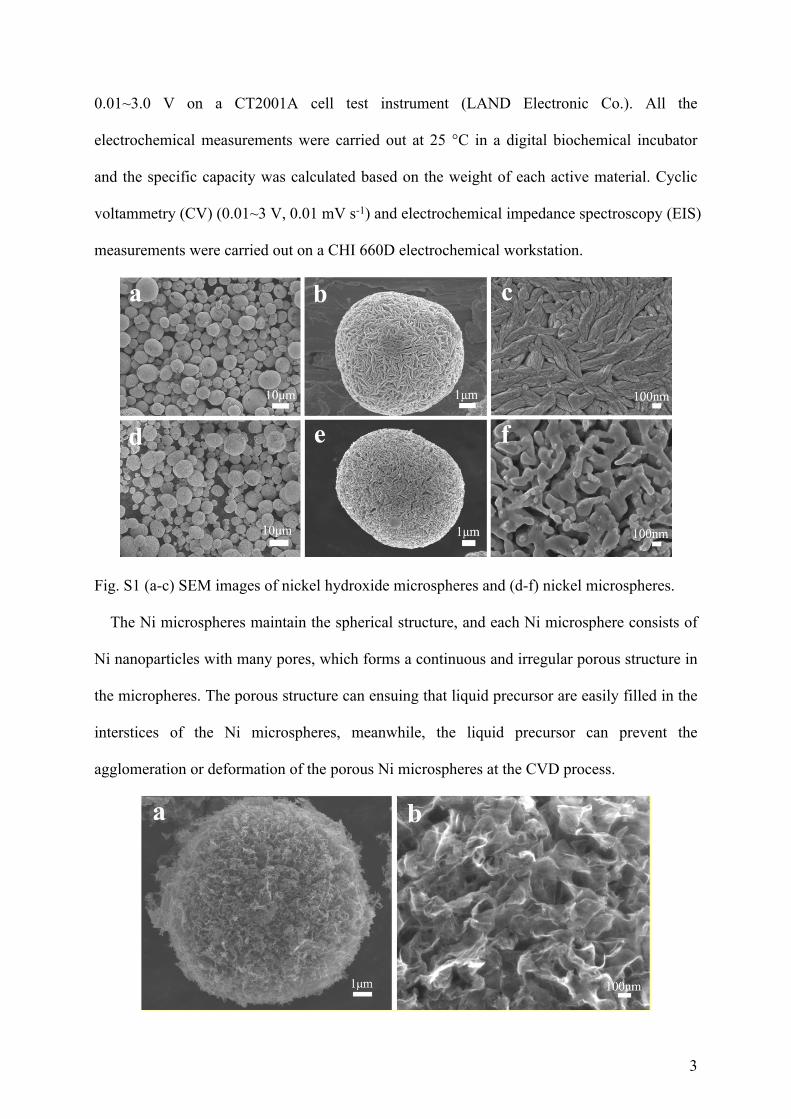
Fig. S3 (a) The dark-TEM image of a NS-GS, (b) SAED pattern of 3D NS-GSs, (c) and (d)
higher magnification TEM images of 3D NS-GSs, and (e) the line profiles (extracted from (d))
of the d-spacing of graphene sheets.
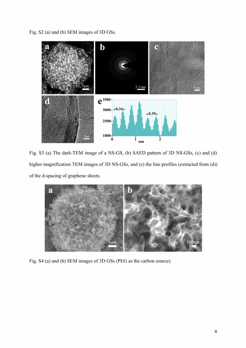
Fig. S4 (a) and (b) SEM images of 3D GSs (PEG as the carbon source).
5
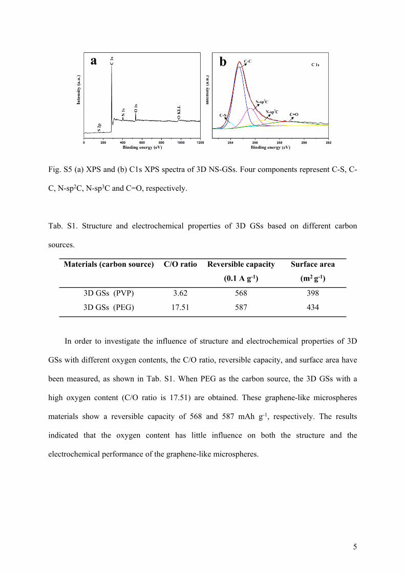
Fig. S5 (a) XPS and (b) C1s XPS spectra of 3D NS-GSs. Four components represent C-S, C-
C, N-sp2C, N-sp3C and C=O, respectively.
Tab. S1. Structure and electrochemical properties of 3D GSs based on different carbon
sources.
Materials (carbon source) C/O ratio Reversible capacity
(0.1 A g-1)
Surface area
(m2 g-1)
3D GSs (PVP) 3.62 568 398
3D GSs (PEG) 17.51 587 434
In order to investigate the influence of structure and electrochemical properties of 3D
GSs with different oxygen contents, the C/O ratio, reversible capacity, and surface area have
been measured, as shown in Tab. S1. When PEG as the carbon source, the 3D GSs with a
high oxygen content (C/O ratio is 17.51) are obtained. These graphene-like microspheres
materials show a reversible capacity of 568 and 587 mAh g-1, respectively. The results
indicated that the oxygen content has little influence on both the structure and the
electrochemical performance of the graphene-like microspheres.
6
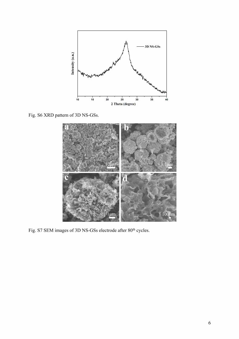
Fig. S6 XRD pattern of 3D NS-GSs.
Fig. S7 SEM images of 3D NS-GSs electrode after 80th cycles.

7
Fig. S8 Nyquist plots of the 3D NS-GSs electrode after 5th and 80th cycles at the current
density of 0.1 A g-1.
Fig. S9 (a) Galvanostatic charge-discharge voltage versus capacity profiles of the 3D GSs
electrode at a current density of 0.1 A g-1, and (b) cycling performance and Columbic
efficiency of the 3D GSs electrode at 0.1 A g-1.
8
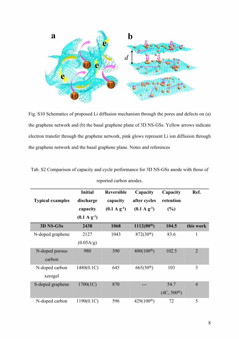
Fig. S10 Schematics of proposed Li diffusion mechanism through the pores and defects on (a)
the graphene network and (b) the basal graphene plane of 3D NS-GSs. Yellow arrows indicate
electron transfer through the graphene network, pink glows represent Li ion diffusion through
the graphene network and the basal graphene plane. Notes and references
Tab. S2 Comparison of capacity and cycle performance for 3D NS-GSs anode with those of
reported carbon anodes.
Typical examples
Initial
discharge
capacity
(0.1 A g-1)
Reversible
capacity
(0.1 A g-1)
Capacity
after cycles
(0.1 A g-1)
Capacity
retention
(%)
Ref.
3D NS-GSs 2438 1068 1112(80th) 104.5 this work
N-doped graphene 2127
(0.05A/g)
1043 872(30th) 83.6 1
N-doped porous
carbon
980 390 400(100th) 102.5 2
N-doped carbon
xerogel
1480(0.1C) 645 665(50th) 103 3
S-doped graphene 1700(1C) 870 --- 54.7
(4C, 500th)
4
N-doped carbon 1190(0.1C) 596 429(100th) 72 5

9
nanoparticles
nitrogen-doped
carbon spheres
1700
(0.05A/g)
816 660(50th) 80.8 6
mesoporous carbon 2385 1011 889(100th) 87.9 7
Notes and references
1 Z. S. Wu, W. C. Ren, L. Xu, F. Li and H. M. Cheng, ACS Nano, 2011, 5, 5463-5471.
2 L. Wang, Y. L. Zheng, X. H. Wang, S. H. Chen, F. G. Xu, L. Zuo, J. F. Wu, L. L. Sun, Z.
Li, H. Q. Hou and Y. H. Song, ACS Appl. Mater. Interfaces, 2014, 6, 71170-7125.
3 X. C. Liu, S. M. Li, J. Mei, W. M. Lau, R. Mi, Y. C. Li, H. Liu and L. M. Liu, J. Mater.
Chem. A, 2014, 2, 14429-14438.
4 Y. S. Yun, V. D. Le, H. Kim, S. J. Chang, S. J. Baek, S. Park, B. H. Kim, Y. H. Kim, K.
Kang and H. J. Jin, J. Power Sources, 2014, 262, 79-85.
5 D. Bhattacharjya, H. Y. Park, M. S. Kim, H. S. Choi, S.. N. Inamdar and J. S. Yu,
Langmuir, 2014, 30, 318-324.
6 T. Q. Chen, L. K. Pan, T. A. J. Loh, D. H. C. Chua, Y. F. Yao, Q. Chen, D. S. Li, W. Qin
and Z. Sun, Dalton Trans., 2014, 43, 14931-14935.
7 M. S. Kim, D. Bhattacharjya, B. Z. Fang, D. S. Yang, T. S. Bae and J. S. Yu, Langmuir,
2013, 29, 6754-6761.