microscopic identification of wood … identification of wood species an important step in furniture...
TRANSCRIPT
European Journal of Science and Theology, August 2013, Vol.9, No.4, 243-252
_______________________________________________________________________
MICROSCOPIC IDENTIFICATION OF WOOD
SPECIES
AN IMPORTANT STEP IN FURNITURE
CONSERVATION
Maria-Cristina Timar, Lidia Gurau
*, Mihaela Porojan and
Emanuela Beldean
Transilvania University of Brasov, Faculty of Wood Engineering, 29 Eroilor Street, 500036,
Brasov, Romania
(Received 30 April 2012, revised 20 March 2013)
Abstract
Wooden religious artefacts such as iconostasis, polychromic wood statues and church
furniture represent an important part of the cultural heritage, often needing careful
conservation and restoration. Any restoration work of such an object that involves
completion of missing elements or a replacement of severely damaged parts should be
very well documented and in accordance with the original material. Identification of
wood species becomes, therefore, a compulsory step prior to any intervention. A direct
visual evaluation of species on an investigated object, based on the characteristic
macroscopic features, is most often not possible or is not conclusive either because of
the natural ageing of wood, possible biological degradation or due to an opaque finishing
or painting. Therefore, a microscopic approach is more reliable.
This paper presents a case study of wood species identification for a bishop throne, dated
1838, from the Berislăvesti hermitage in Vâlcea County. For species identification, three
small wooden samples, coded J1, J2, J3, were collected and prepared as slides for
investigation by transmitted light microscopy. For objective measurements, a specialised
image analysis software, offering a quantitative method to separate, measure and
statistical data process for some anatomical features of interest, was employed. Two
samples (J1, J3) were identified as walnut (Juglans regia) and one (J2) as lime (Tilia
cordata).
Keywords: wood species identification, microscopy, imageJ analysis, furniture
1. Introduction
Since ancient times, wood has been widely used by mankind for different
uses, so that almost any important step of the culture and civilisation evolution,
as well as the spiritual values or the technical achievements have a wooden
* E-mail: [email protected]
Timar et al/European Journal of Science and Theology 9 (2013), 4, 243-252
244
materialised proof as an artistic and/or functional object [1, 2]. Wooden objects
represent, therefore, an important part of the cultural heritage. Artefacts such as
iconostasis, polychromic wood statues and church furniture are representative
elements for both religious art and wooden cultural heritage.
Old furniture items cumulate the flavour of old-times and patina with an
important historical, aesthetical and technical value resulting from the ingenuity
of structures, elegance of shapes, skilfulness of decoration and diversity of
finishing techniques employing natural materials [3, 4]. A very special
symbolistic value and particular structural characteristics related to a special
functionality are often extra definitory elements for church furniture.
Due to the susceptibility of wood to degradation and deterioration caused
by diverse biotic and non-biotic factors, it is often the case that these objects
need careful conservation and restoration. Scientific investigation is well
acknowledged as a component of conservation and in this respect the
identification of wooden species is an important step in furniture/wooden objects
conservation [5, 6], as any restoration intervention that involves wood
completion or a replacement should be very well documented and in accordance
with the original material.
Identification of wooden species is often a challenging task due to both
the diversity of wooden species used throughout history and the ageing and
degradation phenomena affecting the wooden material appearance and
sometimes even its structural integrity. Only seldom, the identification of
common wooden species can be made by a macroscopic investigation of the
characteristic macroscopic anatomical features, on a cleaned not degraded area,
but most often such an attempt is not conclusive. Consequently, relevant samples
have to be extracted and adequately prepared for a microscopic investigation to
observe the characteristic microscopic anatomical features. Microscopic
identification keys and reference samples are then employed in order to conclude
on the wood species involved [5, 6]. Moreover, microscopic measurements of
different anatomical elements offer the possibility to compare these data with
relevant literature information [7] for a more reliable identification. ImageJ
[http://en.wikipedia.org/wiki/ImageJ] is a specialised software for microscopic
data processing, successfully used for such purposes [8, 9].
It has to be mentioned that non-destructive advanced techniques, such as
X-ray phase contrast micro-tomography [10] enable the 3D-analysis throughout
the volume of the wood without physical sectioning. However, as this technique
and other tomographic methods are not readily available, the most used remain
the typical wood anatomy imaging techniques that include classical transmission
light microscopy through thin microtome sections.
This paper presents a case study of microscopic species identification for
three small samples extracted from a Bishop’s throne, dated 1838, from the
Berislăvesti hermitage in Vâlcea County.
Microscopic identification of wood species
245
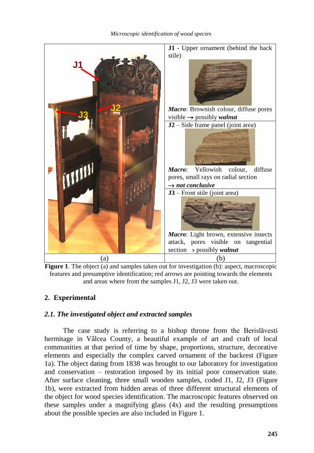
J1 - Upper ornament (behind the back
stile)
Macro: Brownish colour, diffuse pores
visible possibly walnut
J2 – Side frame panel (joint area)
Macro: Yellowish colour, diffuse
pores, small rays on radial section
not conclusive
J3 – Front stile (joint area)
Macro: Light brown, extensive insects
attack, pores visible on tangential
section possibly walnut
(a) (b) Figure 1. The object (a) and samples taken out for investigation (b): aspect, macroscopic
features and presumptive identification; red arrows are pointing towards the elements
and areas where from the samples J1, J2, J3 were taken out.
2. Experimental
2.1. The investigated object and extracted samples
The case study is referring to a bishop throne from the Berislăvesti
hermitage in Vâlcea County, a beautiful example of art and craft of local
communities at that period of time by shape, proportions, structure, decorative
elements and especially the complex carved ornament of the backrest (Figure
1a). The object dating from 1838 was brought to our laboratory for investigation
and conservation – restoration imposed by its initial poor conservation state.
After surface cleaning, three small wooden samples, coded J1, J2, J3 (Figure
1b), were extracted from hidden areas of three different structural elements of
the object for wood species identification. The macroscopic features observed on
these samples under a magnifying glass (4x) and the resulting presumptions
about the possible species are also included in Figure 1.
J1
J3 J2
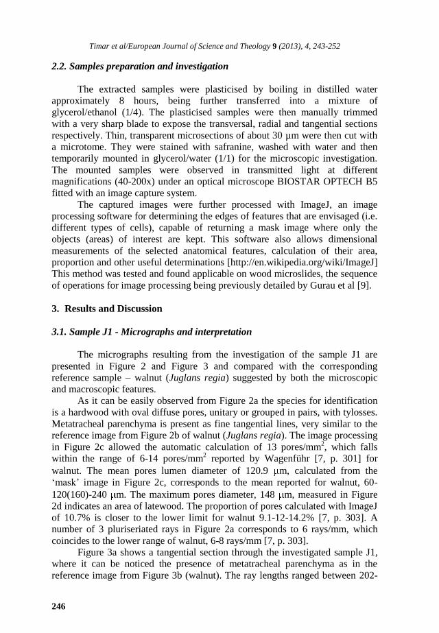
Timar et al/European Journal of Science and Theology 9 (2013), 4, 243-252
246
2.2. Samples preparation and investigation
The extracted samples were plasticised by boiling in distilled water
approximately 8 hours, being further transferred into a mixture of
glycerol/ethanol (1/4). The plasticised samples were then manually trimmed
with a very sharp blade to expose the transversal, radial and tangential sections
respectively. Thin, transparent microsections of about 30 µm were then cut with
a microtome. They were stained with safranine, washed with water and then
temporarily mounted in glycerol/water (1/1) for the microscopic investigation.
The mounted samples were observed in transmitted light at different
magnifications (40-200x) under an optical microscope BIOSTAR OPTECH B5
fitted with an image capture system.
The captured images were further processed with ImageJ, an image
processing software for determining the edges of features that are envisaged (i.e.
different types of cells), capable of returning a mask image where only the
objects (areas) of interest are kept. This software also allows dimensional
measurements of the selected anatomical features, calculation of their area,
proportion and other useful determinations [http://en.wikipedia.org/wiki/ImageJ]
This method was tested and found applicable on wood microslides, the sequence
of operations for image processing being previously detailed by Gurau et al [9].
3. Results and Discussion
3.1. Sample J1 - Micrographs and interpretation
The micrographs resulting from the investigation of the sample J1 are
presented in Figure 2 and Figure 3 and compared with the corresponding
reference sample – walnut (Juglans regia) suggested by both the microscopic
and macroscopic features.
As it can be easily observed from Figure 2a the species for identification
is a hardwood with oval diffuse pores, unitary or grouped in pairs, with tylosses.
Metatracheal parenchyma is present as fine tangential lines, very similar to the
reference image from Figure 2b of walnut (Juglans regia). The image processing
in Figure 2c allowed the automatic calculation of 13 pores/mm2, which falls
within the range of 6-14 pores/mm2 reported by Wagenführ [7, p. 301] for
walnut. The mean pores lumen diameter of 120.9 m, calculated from the
‘mask’ image in Figure 2c, corresponds to the mean reported for walnut, 60-
120(160)-240 m. The maximum pores diameter, 148 m, measured in Figure
2d indicates an area of latewood. The proportion of pores calculated with ImageJ
of 10.7% is closer to the lower limit for walnut 9.1-12-14.2% [7, p. 303]. A
number of 3 pluriseriated rays in Figure 2a corresponds to 6 rays/mm, which
coincides to the lower range of walnut, 6-8 rays/mm [7, p. 303].
Figure 3a shows a tangential section through the investigated sample J1,
where it can be noticed the presence of metatracheal parenchyma as in the
reference image from Figure 3b (walnut). The ray lengths ranged between 202-
Microscopic identification of wood species
247
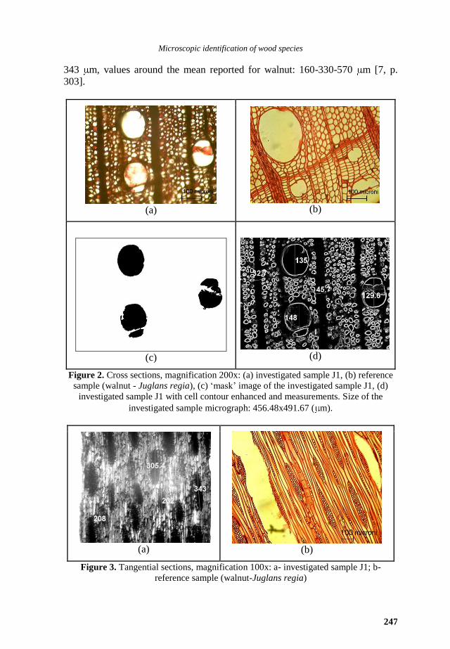
343 m, values around the mean reported for walnut: 160-330-570 m [7, p.
303].
(a)
(b)
(c)
(d)
Figure 2. Cross sections, magnification 200x: (a) investigated sample J1, (b) reference
sample (walnut - Juglans regia), (c) ‘mask’ image of the investigated sample J1, (d)
investigated sample J1 with cell contour enhanced and measurements. Size of the
investigated sample micrograph: 456.48x491.67 ( m).
(a)
(b)
Figure 3. Tangential sections, magnification 100x: a- investigated sample J1; b-
reference sample (walnut-Juglans regia)
Timar et al/European Journal of Science and Theology 9 (2013), 4, 243-252
248
(a)
(b)
(c)
(d)
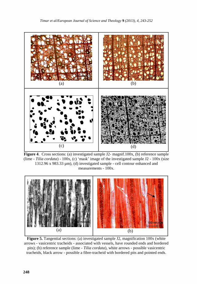
Figure 4. Cross sections: (a) investigated sample J2- magnif.100x, (b) reference sample
(lime - Tilia cordata) - 100x, (c) ‘mask’ image of the investigated sample J2 - 100x (size
1312.96 x 983.33 µm), (d) investigated sample - cell contour enhanced and
measurements - 100x.
(a)
(b)
Figure 5. Tangential sections: (a) investigated sample J2, magnification 100x (white
arrows - vasicentric tracheids - associated with vessels, have rounded ends and bordered
pits); (b) reference sample (lime - Tilia cordata), white arrows - possible vasicentric
tracheids, black arrow - possible a fibre-tracheid with bordered pits and pointed ends.
Microscopic identification of wood species
249
Based on the above observation and analysis, the investigated sample is
most probably walnut – Juglans regia. This result confirms the presumptions
based on the macroscopic features.
3.2. Sample J2 - Micrographs and interpretation
The micrographs resulting from the investigation of the sample J2 are
presented in Figures 4 and 5 and are compared with the corresponding reference
sample – lime (Tilia cordata) suggested by the microscopic features.
The species for identification from Figure 4a is a hardwood, with diffuse
pores dispersion with no pronounced difference between pores size from
earlywood compared to latewood. Pores are numerous, have no tiles and appear
unitary or grouped 2-3 in radial rows (Figure 4a). Similar pores distribution and
size is visible in Figure 4b which describe the reference species, lime (Tilia
cordata).
The crosssection in Figure 4a shows the presence of quite a large number
of vessels with vasicentric tracheids, with a darker appearance. The vasicentric
tracheids are visible also in Figure 5a of the investigated sample, as cells
associated with vessels, having rounded ends, thin walls and bordered pits, but
also in the reference sample in Figure 5d. However, the frequent occurrence of
the vasicentric tracheids is more common for juvenile wood rather than mature
wood of lime [11].
By processing with ImageJ the image of the investigated species from
Figure 4a, a number of 83 pores/mm2
were identified, which falls within the
range of 70-130 pores/mm2 reported by Wagenführ [7, p. 243] for lime. It has to
be mentioned, that vessels with vasicentric tracheids were not identified,
respectively counted by the program and if they were, the pores number would
have increased to 100/mm2. Also, it was difficult to distinguish the small pores
from the fibro-tracheids, some of the latter being kept in the evaluation.
The mean pores lumen diameter of 55.2 m, calculated from the ‘mask’
image in Figure 4c is close to the mean reported for lime, 20-60-90 m. The
maximum pores diameters 87-88.8-98.5 m, measured in Figure 4d are close to
the upper size limit reported for lime 90 µm [7, p. 243], while the smallest pores
in Figure 4d, had values of 29.8-31-33 m, towards the lower limit for lime, 20
µm. The vessels with vasicentric tracheids were smaller with measured
diameters of 24-28 µm (Figure 4d). The proportion of pores calculated with
ImageJ for the species for identification was 19.8%. For lime, Wagenführ [7, p.
243] appreciates pores proportion in the mature wood around 17%. The vessels
contain distinct dense thickenings visible on tangential sections for both, the
investigated species in Figure 5a and in lime, Figure 5b.
Apotracheal and metatracheal parenchyma are present as fine tangential
lines in Figure 4a similar to the reference image from Figure 4b of lime (Tilia
cordata).
The rays of the investigated species appear distanced with several pores
diameters. From Figure 4a, a number of 4-5 rays/mm were calculated, which
Timar et al/European Journal of Science and Theology 9 (2013), 4, 243-252
250
falls in the frequency reported for lime, 2-3-9 rays/mm [7, p. 243]. Their widths
and heights were measured in Figure 5a. For lime, ray heights vary in the range
180-240-1250 m and their widths vary from 10 to 30 m Wagenführ [7, p.
243]. In Figure 5a, one complete pluriseriate ray was measured, 678 m, and
one uniseriate, 228 m, values which fall in the interval for lime. The rays
widths, were of 18-33-46 m, near the values for lime.
Based on the above observation and analysis, the investigated sample is
most probably wood from lime – Tilia cordata, perhaps cut from a region near
the pith. For this sample the macroscopic examination was not conclusive,
pointing only towards a hardwood diffuse porous species. This case is a proof
for the utility of microscopic method of wood species identification based on
identification keys, reference sample images and image data processing, as
presented and used in this paper.
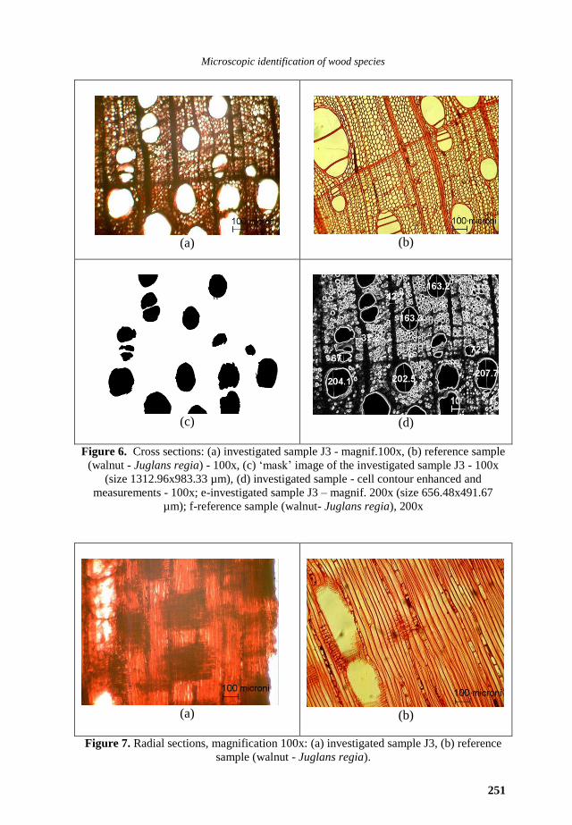
3.3. Sample J3 – Micrographs and interpretation
The micrographs resulting from the investigation of the sample J3 are
presented in Figure 6 and Figure 7 and compared with the corresponding
reference sample – walnut (Juglans regia) suggested by the microscopic
features.
The species for identification from Figure 6a is a hardwood having diffuse
pores dispersion. The pores appear oval in shape, unitary or in groups of 2-3,
with tylosses in the latewood clearly visible in Figure 6a and Figure 7a. The
pores distribution and size is comparable with images of walnut (Juglans regia)
in Figure 6 b.
By processing with ImageJ the image of the investigated species from
Figure 6a, a number of 13 pores/mm2
were identified, which falls within the
range of 8-20 pores/mm2 reported by Wagenführ [7, p. 301] for walnut. The
mean pores lumen diameter of 135.5 m, calculated from the ‘mask’ image in
Figure 6c is around the mean reported for walnut, 60-120(160)-240 m. The
maximum pores diameters in earlywood, 202.5-207.7 m, measured in Figure
6d are close to the upper size limit of walnut earlywood pores, 160-240 µm. The
pores from the latewood next to the annual ring limit had values of 67-72.4 µm
(Figure 6d) close to the lower limit for walnut pore size range in the latewood,
60-120 µm. The proportion of pores calculated with ImageJ of 19% is higher
than the upper value reported for walnut 9.1-12-14.2% [7, p. 303], but probably
the sample comes from a young walnut tree or from a region closer to the pith.
Metatracheal parenchyma is present as fine tangential lines in Figure 6a
and longitudinal lines in Figure 7a, very similar to the reference images from
Figure 6b and Figure 7b of walnut (Juglans regia).
Based on the above observation and analysis, the investigated sample is
most probably walnut – Juglans regia. This confirms initial presumptions.
Microscopic identification of wood species
251
(a)
(b)
(c)
(d)
Figure 6. Cross sections: (a) investigated sample J3 - magnif.100x, (b) reference sample
(walnut - Juglans regia) - 100x, (c) ‘mask’ image of the investigated sample J3 - 100x
(size 1312.96x983.33 µm), (d) investigated sample - cell contour enhanced and
measurements - 100x; e-investigated sample J3 – magnif. 200x (size 656.48x491.67
µm); f-reference sample (walnut- Juglans regia), 200x
(a)
(b)
Figure 7. Radial sections, magnification 100x: (a) investigated sample J3, (b) reference
sample (walnut - Juglans regia).
Timar et al/European Journal of Science and Theology 9 (2013), 4, 243-252
252
4. Conclusions
Restoration work on wooden religious objects, as components of the
cultural heritage, requires that species of origin are identified and their degree of
deterioration is analysed. A case study of a bishop throne from 1838 looked at
the species identification of three furniture parts, whose visual evaluation was
not conclusive. As this could be the case often occurring in practice, a
microscopic approach was proposed and used in this paper. Examination and
feature evaluation of microscopic microslides with ImageJ and comparison with
reference data from literature has identified walnut and lime as species of origin
for the furniture object under restoration.
Acknowledgement
The authors are grateful to the Berislavesti Hermitage for offering the
opportunity to study and investigate for conservation-restoration purposes such a
beautiful and valuable object.
References
[1] D. Lica, Civilizatia lemnului (Wood civilisation), University Transilvania, Brasov,
2006, 85.
[2] D. Lica and C. Cosereanu, Civilizatia lemnului la români (Wood civilisation at
Romanians), University Transilvania, Brasov, 2010, 103.
[3] M. Cionca, Stiluri si ornamente la mobilier – Renasterea italiană (Styles and
ornaments for furniture – Italian Renaissance), University Transilvania, Brasov,
2004, 120.
[4] M. Cionca, Proiectarea mobilei stil – Cassoni din Renasterea florentină (Design of
art furniture – Cassoni from Florence Renaissance), University Transilvania,
Brasov, 2004, 205.
[5] M. Romagnoli, M. Sarlatto, F. Terranova, E. Bizzarri and S. Cesetti, International
Association of Wood Anatomists Journal, 28(2) (2007) 109-123.
[6] N. Macchioni, Scientific examination for the investigation of paintings. A
Handbook of conservator – restores, D. Pinna, M. Galeotti & R. Mazzeo (eds.)
Centro Di Firenze, Firenze, 2009, 21-22.
[7] R. Wagenfuhr, Holzatlas, 5 Auflage, Hanser Fachbuchverlag, Leipzig, 2000, 707.
[8] G.C. Carrasco, Microscopy and Computerized Image Analysis of Wood Pulp Fibres
Multi-Scale Structures, in Microscopy: Science, Technology, Applications and
Education, A. Mèndez-Vilas and J. Diaz (eds.), Formatex Research Center,
Bendajoz 2010, 2182-2189.
[9] L. Gurău, M.C. Timar, M. Cionca, A. Olărescu and R. Dumitrascu, Pro Ligno, 6(1)
(2010) 35-45.
[10] S. Mayo, R. Evans, F. Chen and R. Lagerstrom, Journal of Physics: Conference
Series, 186 (2009) 012105, 1-3.
[11] K. Fukazawa and J. Ohtani, International Association of Wood Anatomists
Bulletin, 3(3-4) (1982) 201.