microrna mir-466 inhibits lymphangiogenesis by targeting
TRANSCRIPT

Seo et al. Journal of Biomedical Science (2015) 22:3 DOI 10.1186/s12929-014-0104-0
RESEARCH Open Access
MicroRNA miR-466 inhibits Lymphangiogenesisby targeting prospero-related homeobox 1 in thealkali burn corneal injury modelMinkoo Seo1, Jun-Sub Choi2, Chang Rae Rho2,3, Choun-Ki Joo2,4 and Suk Kyeong Lee1*
Abstract
Background: Lymphangiogenesis is one of the major causes of corneal graft rejection. Among thelymphangiogenic factors, vascular endothelial growth factor (VEGF)-C and -D are considered to be the most potent.Both bind to VEGF receptor 3 (VEGFR3) to activate Prospero homeobox 1 (Prox1), a transcription factor essential forthe development and maintenance of lymphatic vasculature. MicroRNAs (miRNAs) bind to the 3' untranslatedregions (3' UTRs) of target genes in a sequence-specific manner and suppress gene expression. In the current study,we searched for miRNAs that target the pro-lymphangiogenic factor Prox1.
Results: Among the miRNAs predicted by the bioinformatic analysis to seed match with the 3' UTR of Prox-1, wechose 3 (miR-466, miR-4305, and miR-4795-5p) for further investigation. Both the miR-466 and miR-4305 mimics, butnot the miR-4795-5p mimic, significantly reduced the luciferase activity of the Prox-1 3' UTR reporter vector. Inprimary lymphatic endothelial cells (HDLEC), miR-466 mimic transfection suppressed Prox1 mRNA and proteinexpression, while miR-4305 mimic transfection did not. Experiments using mutated reporter constructs of the twopossible seed match sites on the 3' UTR of Prox1 suggested that the target site 2 directly bound miR-466. HDLECtransfected with the miR-466 mimic suppressed tube formation as compared to the scrambled control. Furthermore,HDLEC transfected with a miR-466 inhibitor showed enhanced tube formation as compared to control inhibitortransfected cells, and this inhibitory effect was counteracted by Prox1 siRNA. The miR-466 mimic reduced angiogenesisand lymphangiogenesis resulting in clearer corneas in an cornea injury rat model compared to the scrambled control.
Conclusions: Our data suggest that miR-446 may have a protective effect on transplanted corneas by suppressingProx1 expression at the post-transcriptional level. The results of the current study may provide insights into themechanisms of lymphangiogenesis resulting from corneal graft rejection and alkali-burn injuries, as well as into thedevelopment of new treatments for lymphangiogenic eye diseases.
Keywords: MicroRNA, Prox1, miR-466, miR-181, Tube Formation, Lymphangiogenesis, Cornea transplantation, Alkaliburn
BackgroundApproximately 10%–50% of cornea transplantation recipi-ents experience graft rejection within one year [1]. Cor-neal graft rejection takes place when the immune cells ofthe host recognize the donor tissues as antigens and attackthem. The normal cornea maintains avascularity by balan-cing positive and negative angiogenesis-regulating mole-cules such as vascular endothelial growth factor (VEGF)
* Correspondence: [email protected] of Medical Lifescience, College of Medicine, The CatholicUniversity of Korea, Seoul, KoreaFull list of author information is available at the end of the article
© 2015 Seo et al.; licensee BioMed Central. ThCommons Attribution License (http://creativecreproduction in any medium, provided the orDedication waiver (http://creativecommons.orunless otherwise stated.
and angiostatin, respectively. Stimulation of cornea causedby corneal transplantation promotes the production ofVEGF, disrupts the balance, and results in capillary endo-thelial cell proliferation and neovascularization [2,3]. Cor-neal lymphangiogenesis is also induced after cornealtransplantation [4]. While the blood vessels provide aroute of entry for CD4+ alloreactive T lymphocytes andmemory T lymphocytes [5], newly formed corneal lymph-atic vessels enable effective access of antigen presentingcells and antigenic materials to lymph nodes where accel-erated sensitization to graft antigens occurs [6].
is is an Open Access article distributed under the terms of the Creativeommons.org/licenses/by/4.0), which permits unrestricted use, distribution, andiginal work is properly credited. The Creative Commons Public Domaing/publicdomain/zero/1.0/) applies to the data made available in this article,
Seo et al. Journal of Biomedical Science (2015) 22:3 Page 2 of 12
Angiogenesis, defined as the sprouting of new bloodvessels from existing blood vessels, is promoted by pro-angiogenic factors, such as VEGF, angiopoietins, andintegrins [7]. The potent angiogenic inducer VEGF-A canbind to both VEGF receptor 1 (VEGFR1) and VEGFR2.However, the signals responsible for inducing prolifera-tion and migration of vascular endothelial cells aremainly transduced via VEGFR2 [8].Lymphangiogenesis is known to be closely associated
with inflammation, wound healing, corneal graft rejection,and tumors. The cell survival, proliferation, and migrationof epithelial cells are important process in lymphangiogen-esis, which depends on VEGF-C and -D signalling path-ways through VEGFR-2 and VEGFR-3 [9,10]. Especially,VEGF-C and –D bind with VEGFR-3 and activate Pros-pero homeobox 1 (Prox1) [11]. Prox1 is homolog of thedrosophila homeobox protein prospero [12]. Prox1 is atranscription factor essential for the embryonic develop-ment of vertebrates, and the development and mainten-ance of lymphatic vasculature in adulthood [13-15].Following transplantation of corneas from C57BL/6
mice into BALB/C mice, the graft survival rates werecompared between two experimental groups [16]. In onegroup, VEGF-TrapR1R2 was used to inhibit both lym-phangiogenesis and angiogenesis, and in the other,VEGFR3 Ab mF4-31C was used to inhibit lymphangio-genesis only. Results showed that the survival rates of thecorneal grafts were comparable in the two groups, indi-cating that lymphangiogenesis but not angiogenesis wasan important determinant for graft survival rates.Inflammation-induced lymphangiogenesis has been re-
ported to be attributable to the increased expression ofProx1 stimulated by inflammatory responses [17]. In par-ticular, Prox1 promotes the expression of the VEGF-Creceptor, VEGFR3 [18]. In Prox1+/− mice, milky chyleleaked from the mesenteric lymphatic vessels, andabnormal lymphatic ducts were formed [19]. Further-more, embryos of Prox1-knockout mice showed a loss oflymphangiogenesis without disrupted hemangiogenesisfrom the cardinal vein [13]. Therefore, inhibiting Prox1function or reducing Prox1 expression may be effectivestrategies for inhibiting corneal lymphangiogenesis.MicroRNAs (miRNAs) are highly conserved small non-
coding RNAs (19–25 nucleotides) that can modulategene expression. Primary miRNA transcripts are proc-essed consecutively to produce mature miRNAs by thetwo RNase III endonucleases, Drosha and Dicer. MaturemiRNAs function as negative gene regulators throughcomplementary sequence pairing with the 3' untranslatedregions (3' UTRs) of target genes [20].Kazenwadel et al. [21] reported that the over-expression
of miR-181a in mouse lymphatic endothelial cells directlytargeted the 3' UTR of Prox1, and the expression of miR-181a was lower in vascular endothelial cells than in
lymphatic endothelial cells. Furthermore, the expressionof miR-181a was inversely related to the expression ofProx1 [21]. Other investigators found that miR-31 targetsthe 3' UTR of Prox1 to suppress its expression in humanlymphatic endothelial cells, and that over-expressed miR-31 led to defective lymphangiogenesis in Xenopus andzebrafish embryos [22]. However, there may be other un-known miRNAs capable of down-regulating Prox1 ex-pression as well.In the current study, miR-466, miR-4305, and miR-
4795-5p were chosen as new miRNA candidates thatcould target the 3' UTR of Prox1 based on the results ofa bioinformatics analysis. The ability of these miRNAs tosuppress the expression of Prox1 in vitro was then inves-tigated. The in vivo inhibitory effects of these miRNAson lymphangiogenesis were also assessed using an ex-perimental alkali corneal burn animal model.
MethodsCellsHuman dermal lymphatic endothelial cells (HDLEC)were purchased from PromoCell (Heidelberg, Germany)and cultured in MV2 media (PromoCell). HEK293T werecultured in DMEM (Gibco BRL, Grand Island, NY, USA)supplemented with 10% fetal bovine serum and antibi-otics (100 U/mL penicillin and 100 μg/mL streptomycin;Gibco BRL). Both cells were incubated at 37°C and sup-plemented with 5% CO2.
miRNA mimics, siRNA, and miRNA inhibitorThe miRNA mimics, siRNA, and scrambled miRNA usedas a negative control were purchased from GenolutionPharmaceuticals (Seoul, South Korea). The sequences areas follows: scrambled control sense, 5′-UUUUAACUCAGUAUUUUUA-3′ and antisense, 5′-UAAAAAUACUGAGUUAAAA-3′; Prox1 siRNA sense, 5′- GAGUUGACAUUGGAGUGAA-3′ and antisense, 5′- UUCACUCC AAUGUCAACUC-3′. The LNA™ microRNA Power In-hibitor for hsa-miR-466 and the negative control inhibitor(NC inhibitor) were purchased from Exiqon (Vedbaek,Denmark). The sequences are as follows: inhibitor forhsa-miR-466, 5′-GTGTTGCGTGTATGTGTA-3′; NCinhibitor, 5′-GTGTAACACGTCTATACGCCCA-3′.
Plasmid construction and site-directed mutagenesisThe full length 3' UTR of Prox1 was amplified from thegenomic DNA of HEK293T cells and cloned between theRenilla luciferase coding sequence and the poly(A) site ofthe psiCHECK-2 plasmid (Promega, Madison, WI, USA)using XhoI/NotI sites to produce psiC-Prox1. The primersused for the amplification of 3′ UTR of Prox1 were as fol-lows: 5′-TCGACTCGAGTGCCTACAAGAGCTGCTTCA-3′ and 5′-GGCCGCGGCCGCATTTGGCCTTTTGGGGTACT-3′. Mutations were introduced into the putative

Seo et al. Journal of Biomedical Science (2015) 22:3 Page 3 of 12
seed match sequences of psiC-Prox1 using an EZchangesite-directed mutagenesis kit (Enzynomics, Daejeon,South Korea). The sequences are as follows: psiC-Prox1-m1, 5′-AATGACTTATATATGAAATCAAAATCTAGACACAT-3′ and 5′-GGGAGGCATGGATATGTTATG-3′;psiC-Prox1-m2, 5′-TTATGACTCGCCAACATTCTTTTTC-3′ and 5′-GTCTCTATTAGCAATGAAGGGAATTTGT-3′.
Luciferase reporter assayTo test whether the miRNAs directly target the 3' UTRof Prox1, luciferase reporter assay was carried out. Forthis, HEK293T cells were seeded in a 96-well plate (5 ×10³ cells/well). After 24 h, cells were co-transfected with20 ng psiC-Prox1 or its mutants (psiC-Prox1-m1, psiC-Prox1-m2, and psiC-Prox1-m1m2), and 10 nM each ofthe miRNA mimics. Luciferase activities were measured48 h post-transfection using the Dual-Glo™ luciferase re-porter assay system (Promega). Renilla luciferase activitywas normalized using firefly luciferase activity for eachsample.
Transfection of HDLECCells were seeded 24 h prior to transfection in 60- or100-mm-diameter dishes containing 10 mL culturemedium. Transfection was performed with 20 nM eachof miRNA mimic, siRNA, and/or miRNA inhibitor usingLipofectamine™ 2000 (Invitrogen, Carlsbad, CA, USA) ac-cording to the manufacturer's protocol. Cells were har-vested for RNA and protein extraction 48 h aftertransfection.
Quantitative reverse transcription-polymerase chain reac-tion (qRT-PCR)HDLEC were harvested and total RNA was extractedusing the RNAzol™ B reagent (Tel-Test, Friendswood,TX, USA) according to the manufacturer's instruction.cDNA was synthesized using 1 μg total RNA, oligo(dT)(Macrogen, Seoul, South Korea), and M-MLV reversetranscriptase (Invitrogen). Real-time PCR for Prox1 wascarried out using a SYBR green qPCR kit (Takara, Tokyo,Japan) with an Mx3000P™ Real-Time PCR System(Stratagene, La Jolla, CA, USA). The sequences of theprimers were as follows: Prox1; 5′-ATCCCAGCTCCAA-TATGCTG-3′ and 5′-GTACTGGTGACCCCATCGTT-3′, glyceraldehyde phosphate dehydrogenase (GAPDH);5′- ATGGGGAAGGTGAAGGTCG-3′ and 5′- GGGGTCATTGATGGCAACAATA-3′. The PCR conditionswere 95°C for 10 min, followed by 40 cycles at 95°C for20 s, 60°C for 30 s, and 72°C for 30 s. To confirm specificamplification of the PCR product, dissociation curveswere checked routinely. For this, the PCR products wereincubated at 95°C for 10 s and ramped up from 55°C to95°C with a heating rate of 0.1°C/s, and fluorescence wasmeasured continuously. Relative gene expression was
calculated according to the comparative Ct method usingGAPDH as an internal standard.
Western blot analysisTo detect the Prox1 protein, cell lysate in RIPA buffer(50 μg) was mixed with NuPAGE LDS sample buffer(4×) and heated at 70°C for 10 min. The samples wereelectrophoretically separated on 8% SDS-PAGE gel, andthen transferred to a nitrocellulose membrane (Invitro-gen). The membrane was incubated overnight at 4°Cwith mouse monoclonal antibody against Prox1 (1:500,Abnova, Taipei City, Taiwan). After washing, the blotswere incubated for 2 h at room temperature with horse-radish peroxidase-conjugated anti-mouse secondaryantibody (1:5000, Santa Cruz Biotechnology, Dallas, TX,USA). Protein bands were visualized using an enhancedchemiluminescence detection system (Amersham Bio-sciences). β-Actin antibody (Cell Signaling Technology,Danvers, MA, USA) was used to confirm comparableloading. The density of each protein band was read andquantified using Fujifilm Multi Gauge software (version3.0).
Tube formation assayEndothelial cells plated on a reconstituted basementmembrane matrix have been known to rapidly attach,align, and form capillary-like tubules [23]. As this endo-thelial cell specific process is rapid and quantifiable, tubeformation assay has been used to study angiogenic andanti-angiogenic factors, to investigate mechanisms ofangiogenesis, and to define endothelial cell populations[23]. To assess the effect of miRNAs on lymphangiogen-esis of HDLEC, tube formation experiments were per-formed using MILLIPORE® In Vitro Angiogenesis AssayKit (MILLIPORE, Billerica, MA, USA) according to themanufacturer's protocol. Ninety six-well plates were coatedwith cold liquid ECMatrix (70 μl/well) and incubated at37°C in a humidified 5% CO2 incubator for 1 h to promotesolidification. miRNA-transfected cells (7 × 10³cells/well)were seeded into 96-well plates pre-coated with polymer-ized ECMatrix and incubated with conditioned media at37°C for 4–6 h. Formation of tube-like structures was ob-served under a phase-contrast microscope and quantifiedby counting the number of tubes formed in 3 randomlychosen fields using ImageJ software.
Experimental corneal alkali burn animal modelMale Sprague–Dawley rats (body weight, approximately250–300 g) were used in this study. All of the animalswere treated in accordance with the guidelines of the As-sociation for Research in Vision and Ophthalmology(ARVO) Statement for the Use of Animals in Ophthalmicand Vision Research, and the study protocol was ap-proved by the Committee for Animal Research, Catholic

Seo et al. Journal of Biomedical Science (2015) 22:3 Page 4 of 12
University of Medicine. The rats were deeply anesthetizedvia intraperitoneal injection of 50 mg/kg tiletamine pluszolazepam (Zoletil; Virbac, Carros, France) and 15 mg/kgxylazine hydrochloride (Rompun; Bayer, Leuverkeusen,Germany). Alkali injuries to the eyes were induced via 10 sexposure of the central cornea to a 4-mm-diameter disk offilter paper soaked in 1 N NaOH, followed by rinsing withsterile saline (10 mL). To avoid any corneal infection, onedrop of antibiotic (0.5% levofloxacin; Cravit; Santen,Osaka, Japan) was instilled onto the ocular surface imme-diately after the alkali burn injury. The animals were thenrandomly allocated to three treatment groups: scrambledcontrol, miR-181a, and miR-466. Each group (n = 10) wastreated with a single subconjunctival injection with 20 μlof 20 nM miRNA mimic immediately after the alkali burninjury.
ImmunostainingFormalin-fixed corneas from each group of animal wereembedded in paraffin and 4 μm sections were preparedfor examination. To access lymphangiogenesis, cornealsections were stained with and anti-mouse lymphaticvessel endothelial hyaluronan receptor (LYVE)-1 anti-body (1:500; Abcam, Eugene, OR, USA) for 16 h at 4°C.After three washes with PBS for 15 min, the sectionswere then stained with a Texas Red-conjugated second-ary antibody (Abcam). To detect F-actin, corneal sectionswere incubated with rhodamine-conjugated phalloidin(dilution 1:500, Abcam) for 1 h and washed three timeswith PBS. The stained sections were incubated withhoechst solution to stain nucleus before examined byfluorescence microscopy at 100× magnification.
ResultsScreening of miRNAs that can target Prox1In order to screen miRNAs which may target Prox1, weused publicly available TargetScan program (http://www.targetscan.org) and found 17 human microRNAs thatshowed a good seed match with the 3' UTR of humanProx1 mRNA (Additional file 1: Table S1). Among the17 miRNAs, we selected miR-4305 and miR-4795-5p forfurther study, as they both showed 8mer seed matcheswith the 3' UTR of human Prox1(Figure 1A). This wasconsidered to be important due to similarity with miR-181a, which was previously shown to target Prox1. Add-itionally, miR-466 was also selected, as the 3' UTR ofProx1 contained two putative binding sites for thismiRNA (7mer-m8 and 7mer-1A sites), unlike other miR-NAs. Although miR-4262 showed an 8mer seed matchwith the 3' UTR of Prox1, it was excluded from furtherstudy because the 8mer seed sequence of miR-4262 wasidentical to that of miR-181a. As the target sequences ofmiRNAs are frequently conserved in many species, weanalyzed conservation of the seed match sequences in
the 3' UTR of Prox1. The 7mer-1A site complementaryto the target site 2 of miR-466 and the 8mer site com-plementary to the seed region of miR-4305 were wellconserved among species (Figure 1B). However, the7mer-m8 site complementary to the target site 1 of miR-466 and the 8mer site complementary to the seed regionof miR-4795-5p were not well conserved. Subsequently,a luciferase reporter assay was conducted to assesswhether miR-466, miR-4305, and miR-4795-5p directlytargeted the 3' UTR of Prox1. First, HEK293T cells wereco-transfected with psiC-Prox1 and each miRNA mimic.miR-181a, which is known to target Prox1, was includedas a positive control, while a scrambled miRNA wasused as a negative control. As expected, miR-181a sig-nificantly reduced the luciferase activity of psiC-Prox1 ascompared to the scrambled control. The miR-466 andmiR-4305 mimics, but not the miR-4795-5p mimic, sig-nificantly reduced the luciferase activity of the reportervector as compared to that of the scrambled control(Figure 1C).
Effect of miRNA mimics on the expression of Prox1 mRNAand proteinTo test whether miR-466 and miR-4305 have the abilityto modulate Prox1 expression, HDLEC were harvested48 h after miRNA mimic transfection. qRT-PCR revealedthat the Prox1 mRNA level was reduced by approxi-mately 50% following transfection with the miR-181amimic. Similarly, the Prox1 mRNA level was decreasedby about 50% following transfection with the miR-466mimic as compared with the scrambled control (Figure 2A).miR-4305 transfection did not affect Prox1 mRNA levelsignificantly. Western blot analysis also showed that thelevel of Prox1 protein was reduced by transfection with themiR-181a and miR-466 mimics as compared to levels ob-served following transfection with the scrambled control(Figure 2B). However, the level of Prox1 protein was notsignificantly affected by miR-4305.
Dose-dependent effect of the miR-466 mimicAs non-specific effects can obscure the results ofmiRNA mimic transfection experiments, we carried outa luciferase assay using increasing doses of the miR-466mimic. To accomplish this, HEK293T cells were co-transfected with increasing concentrations of the miR-466mimic and psiC-Prox1 reporter plasmid. Transfectionwith 5 nM miR-466 slightly reduced the luciferaseactivity, however the reduction was not statistically sig-nificant as compared to the scrambled control transfec-tion (Figure 3). Transfecting the cells with 10 nM orhigher concentrations of the miR-466 mimic caused adose-dependent reduction in the luciferase activity ofpsiC-Prox1 (Figure 3). To minimize any possible non-

Figure 1 Search for miRNAs targeting Prox1. (A) Seed matches between the 3' UTR of Prox1 and miR-466, miR-4305, and miR-4795-5p. Thecoordinates for miR-466 target sites 1 and 2, as well as the target site for miR-4305 and miR-4795-5p, are shown in parentheses (GenBankaccession number NM_002763). (B) Cross-species sequence alignment of the 3' UTR of Prox1. The shaded boxes are putative miR-466, miR-4305,and miR-4795-5p target sites. Con indicates conserved residues. Alignment of the Prox1 3' UTR shows the sequence conservation among thehuman (has), chimpanzee (ptr), mouse (mmu), rat (rno), rabbit (ocu), and dog (cfa). Numbers indicate the nucleotide position after the stop codonof Prox1. (C) Direct targeting of the Prox1 3' UTR by miR-466, miR-4305, and miR-4795-5p. Luciferase activity was measured in HEK293T cellsco-transfected with psiC-Prox1 and the miR-466, miR-4305, or miR-4795-5p mimics. miR-181a, which was reported to target Prox1, was used as apositive control. Luciferase activity was normalized using internal firefly luciferase activity, and expressed as a ratio to the luciferase activityobtained from the scrambled control-transfected cells. Error bars indicate SDs (n = 3 per experiment). *P < 0.05. †P < 0.01.
Seo et al. Journal of Biomedical Science (2015) 22:3 Page 5 of 12
specific effects, we used 10–20 nM miRNA mimicsthroughout the experiments.
Confirming target sites for miR-466 in the Prox1 3' UTRsThe 3' UTR of Prox1 contains two putative binding sitesfor miR-466 (Figure 1A). To test whether both weredirectly targeted by miR-466, point mutations were intro-duced to psiC-Prox1 to produce psiC-Prox1-m1, psiC-Prox1-m2, and psiC-Prox1-m1m2 (Figure 4A and B).Each of these vectors was co-transfected with the miR-466 mimic into HEK293T cells, and the luciferase assay
was conducted. Luciferase activity was partially reduced inthe cells transfected with the miR-466 mimic togetherwith either psiC-Prox1 or psiC-Prox1-m1 (Figure 4C).However, luciferase activity was unaffected in the cellstransfected with the miR-466 mimic together with psiC-Prox1-m2 (Figure 4C). As expected from the fact thatboth of the putative seed match sites were eliminated,luciferase activity was not affected when psiC-Prox1-m1m2 was co-transfected with miR-466 (Figure 4C).Luciferase activity was not affected in the cells co-transfected with wild-type or mutant Prox1 3' UTR

Seo et al. Journal of Biomedical Science (2015) 22:3 Page 6 of 12
reporter vectors and miR-466 m (mutant form of miR-466) or the scrambled control (Figure 4C). These re-sults showed that target site 2, but not the target site 1,on the 3' UTR of the Prox1 was targeted by miR-466.
Effect of miRNAs on tube formation in HDLECTo test whether miR-466 has an anti-lymphangiogenesiseffect, an in vitro tube formation assay was conducted.Forty eight hours after transfection with miR-466 mimic,HDLEC were cultured on a Matrigel-coated 96 well platefor 4–6 h and the extent of tube formation was assessed.Formation of a rich network of tubular structures was ob-served in HDLEC transfected with the scrambled miRNA.miR-466 mimic significantly impaired the tube-forming ac-tivity of HDLEC as compared to the scrambled control(Figure 5A). Tube formation was then quantified by meas-uring the number of tubes formed using image manipula-tion software (ImageJ). The miR-181a mimic exerted thehighest level of inhibitory effects on tube formation (ap-proximately 84%) followed by the miR-466 mimic (ap-proximately 57%; Figure 5B) when compared to thescrambled control. We also tested whether the inhibitionof miR-466 enhanced tube formation in HDLEC andwhether Prox1 siRNA counteracted the effect of a miR-466inhibitor. As expected, Prox1 siRNA transfection signifi-cantly decreased Prox1 expression in HDLEC as comparedto transfection with the scrambled control (Figure 5C).HDLEC were then transfected with a miR-466 inhibitoralone or together with Prox1 siRNA. HDLEC transfectedwith the miR-466 inhibitor alone showed significantly in-creased tube formation (Figure 5D and E). However, co-transfected Prox1 siRNA counteracted the effect of themiR-466 inhibitor (Figure 5D and E).
Effect of miRNAs on angiogenesis and lymphangiogenesisin the corneal alkali burn animal modelThe effects of miRNAs on corneal opacity, angiogenesis,and lymphangiogenesis were examined in an experimentalanimal model. Immediately after inducing an alkali burn,each miRNA mimic was subconjunctivally injected once,and the effect of each miRNA was evaluated two weekslater. miR-181a was used as a positive control, while thescrambled miRNA was used as a negative control. In thescrambled control-injected animals, the injured centralcorneal stroma appeared opaque with a distinct edematousmargin (Figure 6A). In contrast, miR-466- or miR-181a-injected animals showed reduced opacity (Figure 6B and6C). Under direct microscopic observation, blood vascularinfiltration was about 46% of the scrambled control grouplevel in the miR-181a-treated group. When the animalswere injected with miR-466, blood vascular infiltration was51% of the scrambled control group level (Figure 6J). Infil-tration of blood vessel into the cornea was examined bystaining with phalloidin to detect F-actin (Figure 6D-F),
and positive cells were quantified using the ImageJ soft-ware. The cornea section from the scrambled miRNA-injected eyes showed a thickened cornea and strong scat-tered F-actin staining. In the miR-181a-treated group, thecornea showed almost half the thickness and F-actin posi-tive staining was about 38% compared to the scrambledcontrol group (Figure 6K). When the animals were injectedwith miR-466, corneal thickness was between those ob-served for the scrambled control and the miR-181a-injected animals, while F-actin positive staining was 56% ofthe scrambled control group level (Figure 6K). To analyzethe effects of miRNAs on lymphangiogenesis of the cornea,corneal sections from each animal group were analyzed byimmunohistofluorescence staining with anti-LYVE-1 anti-bodies (Figure 6G-I). miR-466- and miR-181a-injected cor-neas showed significantly reduced levels of LYVE-1staining (~33% and ~30%, respectively) compared to thescrambled control-treated corneas (Figure 6L).
DiscussionThe results of the current study showed that the expres-sion of Prox1 was inhibited by miR-466 at both themRNA and protein levels. The luciferase assay showedthat miR-466 directly targeted the well conserved 7mer-1A site in the 3' UTR of Prox1 (target site 2), as thesuppressive effect of the miR-466 mimic on luciferase ac-tivity was almost abolished when this site was mutated.The target site 2, unlike the target site 1, was well con-served among species. Previous reports showed that add-itional Watson-Crick pairing to four or five sequentialnucleotides at nucleotides 12–17 enhanced miRNA tar-geting [24]. The target site 2 contains five contiguous se-quences that can be used for an effective 3' pairing withthe nucleotides 14 ~ 18 of miR-466, while the target site1 does not. Thus, the target site 2 seems to have betterchance to be targeted with miR-466 than the target site 1.In the alkali burn corneal injury model, miR-466 de-
creased corneal opacity and inhibited both lymphangiogen-esis and angiogenesis, possibly attributable to decreasedProx1 expression. According to the results of a recentstudy [25], growth and migration of vascular endothelialcells were decreased by the addition of culture supernatantderived from Prox1 siRNA-treated oral squamous cell car-cinoma cell cultures. In that experiment, down-regulationof VEGF-C was observed following silencing of the Prox1gene [25]. These results suggested that Prox1 acted as aregulator of angiogenesis and lymphangiogenesis in oralsquamous cell carcinoma [25]. Prox1 is known to controlthe expression of angiopoietin-2, which promotes angio-genesis in vascular endothelial cells [26]. VEGF-C andangiopoietin-2 are also important modulators of angiogen-esis [27,28]. Therefore, suppressed Prox1 expression bymiR-466 may have inhibited the expression of angiogenicmodulators such as VEGF-C and angiopoietin-2, resulting

Figure 2 Effects of miR-466 and miR-4305 on Prox1 expression. (A) Reduction in Prox1 mRNA levels by miR-466. HDLEC were transfectedwith the miR-181a, miR-466, miR-4305 mimics, or the scrambled control. The cells were then cultured for 48 h and harvested for use in real-timeqRT-PCR analyses (n = 3). Error bars indicate SDs. †P < 0.01. (B) Effects of miR-466 and miR-4305 on the expression of Prox1 protein. HDLEC weretransfected with miR-181a, miR-466, miR-4305 mimics, or the scrambled control. The cells were cultured for 48 h and then harvested for analysis.The band densities obtained using an anti-Prox1 antibody were divided by those obtained using an anti–beta-actin antibody to normalize proteinloading. Western blotting was conducted using three sets of independently transfected HDLEC. Bands were quantified using Fujifilm Multi Gaugesoftware (version 3.0). Means were calculated using data from all three independent experiments and are expressed as ratios to the valueobtained from the scrambled control-transfected HDLEC. Error bars indicate SDs. *P < 0.05. †P < 0.01.
Seo et al. Journal of Biomedical Science (2015) 22:3 Page 7 of 12
in reduced angiogenesis in the alkali burn corneal injuryanimal model. However, as miRNAs usually have multipletargets, our observations may be attributable to angiogenicmodulators other than Prox1 targeted by miR-466. Thisnotion is supported by previous findings demonstratingthat miR-466 induced apoptosis by targeting a few anti-
Figure 3 Dose-dependent effect of the miR-466 mimic.Luciferase activity was measured in HEK293T cells co-transfectedwith increasing concentrations of the miR-466 mimic and psiC-Prox1. Error bars indicate SDs (n = 3 per experiment). *P < 0.05.
apoptotic genes [29], and that this induced apoptosis re-sulted in the inhibition of angiogenesis [30,31].mmu-miR-466, which can be induced by apoptosis [29]
and metabolic oxidative stress [32], was shown to en-hance viral replication by inhibition of INF-α [33]. Inaddition, over-expression of mmu-miR-466 inhibitedNfat5 expression and was associated with renal dysfunc-tion [34]. However, the functions of hsa-miR-466 in hu-man cells have yet to be fully elucidated. The results ofthe current study demonstrated that hsa-miR-466 inhib-ited Prox1 expression and suppressed tube formation inhuman primary lymphatic endothelial cells. Furthermore,miR-466 reduced angiogenesis and lymphangiogenesis,resulting in clearer corneas than those observed in thescrambled control-treated mice in an animal cornea in-jury model.The inhibitory effects of miR-466 on Prox1 expression,
tube formation, and lymphatic vessel formation werecomparable to those of miR-181. However, the inhibitoryeffect of miR-466 on blood vessel formation in thein vivo corneal injury model was slightly weaker than thatof miR-181a. Findings of discrepant efficiencies attrib-uted to miRNAs measured using different methods arenot rare [35,36] and may be due to different experimentalsettings, or may reflect experimental errors. Another

Figure 4 Target site search for miR-466 in the Prox1 3' UTR. (A) Illustration showing (i) the location of possible seed match sites betweenmiR-466 and the 3' UTR regions and (ii) the sites altered to produce mutant forms of psiC-Prox1. Site-directed mutagenesis was performed toproduce mutant versions of the 3' UTR of Prox1 seed match sequence (psiC-Prox1-m1, psiC-Prox1-m2, and psiC-Prox1-m1m2). (B) Seed matchesbetween miR-466 and the mutated 3' UTRs of Prox1. wt, wild type. (C) Luciferase activity was measured in HEK293T cells co-transfected with themiR-466 mimic and a luciferase reporter vector containing the wild-type or mutated 3' UTRs of Prox1. The scrambled control and miR-466 m(mutant form) were used to confirm sequence-specific binding between miR-466 and the 3' UTRs. Luciferase activity was normalized using fireflyluciferase activity and expressed as a ratio of the luciferase activity to the activity obtained from the scrambled control-transfected cells. Error barsindicate SDs (n = 3 per experiment). *P < 0.05. †P < 0.01.
Seo et al. Journal of Biomedical Science (2015) 22:3 Page 8 of 12
possibility is that miR-181a may also target angiogenesis-related genes other than Prox1 more effectively thanmiR-466.miR-4305 also significantly reduced the luciferase activ-
ity of the Prox1 3' UTR reporter vector as compared tothe scrambled control in this study. However, Prox1 ex-pression was not changed following miR-4305 mimictransfection of HDLEC compared to the scrambled con-trol transfection. It is not clear why the miR-4305 mimicshowed discrepant results in the luciferase assay, and inqRT-PCR and western blot experiments. These discrep-ant findings may be due to the use of only the 3' UTR of
Prox1, as opposed to the whole Prox1 mRNA, in the lu-ciferase assay. The seed match sequence on the 3' UTRof Prox1 in the luciferase reporter construct may havebeen available for miR-4305 binding, while the confirm-ation of that sequence in the Prox1 mRNA may have notallowed for miR-4305 binding. In addition, HEK293Tcells were used for the luciferase assay, while HDLECwere used for other experiments.Bevacizumab is a recombinant humanized immuno-
globulin G1 monoclonal antibody against all isoforms ofVEGF-A [37]. Bevacizumab binds VEGF-A and preventsthe interaction of VEGF-A to its receptor on the surface
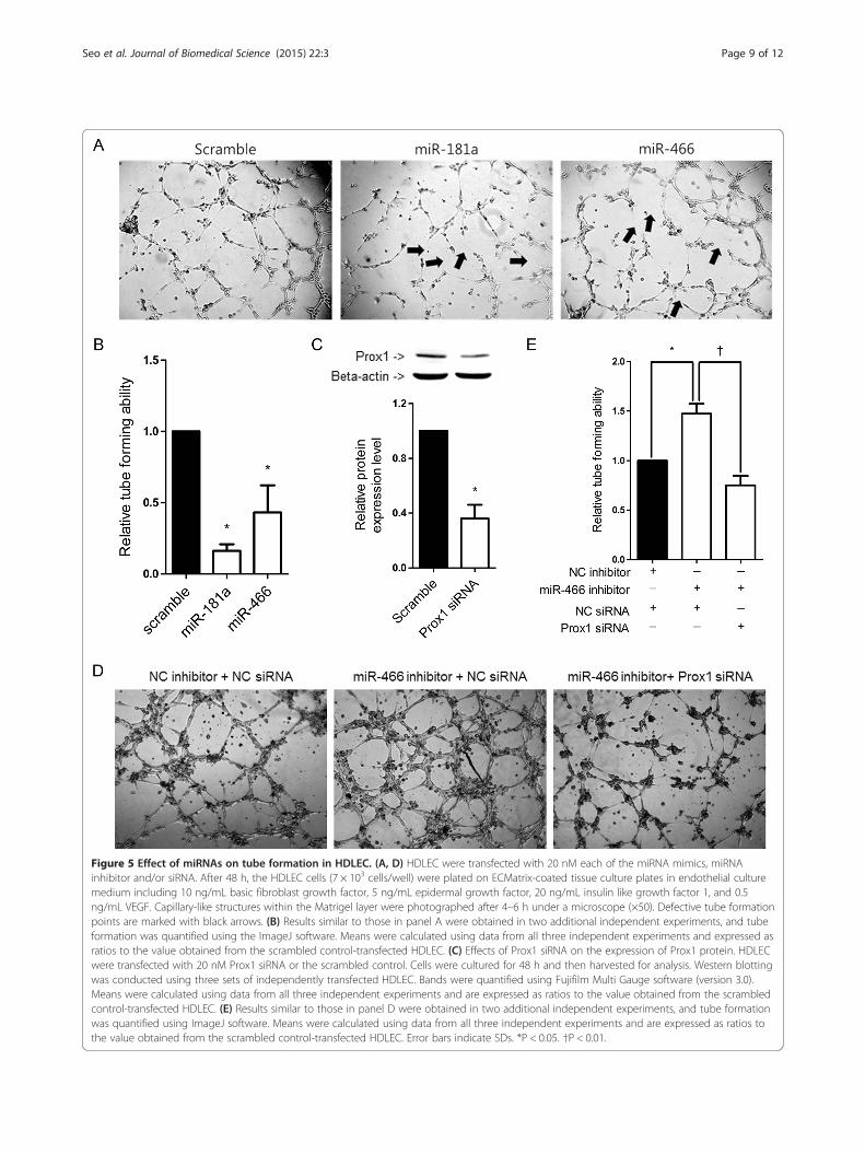
Figure 5 Effect of miRNAs on tube formation in HDLEC. (A, D) HDLEC were transfected with 20 nM each of the miRNA mimics, miRNAinhibitor and/or siRNA. After 48 h, the HDLEC cells (7 × 10³ cells/well) were plated on ECMatrix-coated tissue culture plates in endothelial culturemedium including 10 ng/mL basic fibroblast growth factor, 5 ng/mL epidermal growth factor, 20 ng/mL insulin like growth factor 1, and 0.5ng/mL VEGF. Capillary-like structures within the Matrigel layer were photographed after 4–6 h under a microscope (×50). Defective tube formationpoints are marked with black arrows. (B) Results similar to those in panel A were obtained in two additional independent experiments, and tubeformation was quantified using the ImageJ software. Means were calculated using data from all three independent experiments and expressed asratios to the value obtained from the scrambled control-transfected HDLEC. (C) Effects of Prox1 siRNA on the expression of Prox1 protein. HDLECwere transfected with 20 nM Prox1 siRNA or the scrambled control. Cells were cultured for 48 h and then harvested for analysis. Western blottingwas conducted using three sets of independently transfected HDLEC. Bands were quantified using Fujifilm Multi Gauge software (version 3.0).Means were calculated using data from all three independent experiments and are expressed as ratios to the value obtained from the scrambledcontrol-transfected HDLEC. (E) Results similar to those in panel D were obtained in two additional independent experiments, and tube formationwas quantified using ImageJ software. Means were calculated using data from all three independent experiments and are expressed as ratios tothe value obtained from the scrambled control-transfected HDLEC. Error bars indicate SDs. *P < 0.05. †P < 0.01.
Seo et al. Journal of Biomedical Science (2015) 22:3 Page 9 of 12

Figure 6 Inhibition of corneal neovascularization by miRNAs in a corneal injury animal model. Alkali burn-induced corneas of Sprague–Dawley rats were treated once with each miRNA mimic by subconjunctival injection on the day of injury. (A-C) Photographic images of corneastreated with each miRNA mimic. Immunofluorescent stain of corneal sections to detect vessel formation (D-F) or lymphangiogenesis (G-I). Redstains F-actin (D-F) or LYVE-1 (G-I), which is a marker of vessels and lymphatic vessels, and blue shows hoechst staining (D-I). Photographs ofcorneas were taken two weeks after the treatment. (J) Quantification of new blood vessel formation, F-actin-stained area (K), and LYVE-1-stainedarea (L). Relative rate was quantified using ImageJ software and normalized to the value obtained from the scrambled control-injected rats. Errorbars indicate SDs. †P < 0.01.
Seo et al. Journal of Biomedical Science (2015) 22:3 Page 10 of 12
of endothelial cells, leading to inhibition of endothelialcell proliferation and new blood vessel formation [38].Since the FDA approved bevacizumab for the treatmentof metastatic colorectal cancer in combination with5-fluorouracil, bevacizumab has been used for var-ious malignancies [38,39]. Although bevacizumab-basedtreatment on eye diseases has not been approved by theFDA, intravitreal bevacizumab injection led to improve-ment of visual acuity and regression of retinal neovascu-larization in proliferative diabetic retinopathy patients[40]. Likewise, intraocular pressure and angiogenesis
decreased following the administration of bevacizumabeye drops in neovascular glaucoma patients [41]. Fur-thermore, subconjunctival administration of bevacizu-mab after corneal transplantation decreased the numberand caliber of vessels, however the effects were transi-ent [42]. Bevacizumab inhibits only VEGF-A-inducedlymphangiogenesis and has no effects on the lymphan-giogenesis caused by VEGF-C or VEGF-D [43]. To treatlymphangiogenesis-related disease more effectively, it isnecessary to develop drugs that directly target theprocess of lymphangiogenesis.

Seo et al. Journal of Biomedical Science (2015) 22:3 Page 11 of 12
The results of the current study demonstrated thatmiR-466 inhibited Prox1, which is known to be activatedby various growth factors including VEGF-A, −C, and -D[11,44]. Thus, miR-466 is expected to have broader andgreater inhibitory effects on lymphangiogenesis-relateddiseases than bevacizumab.
ConclusionsOur results on human lymphatic endothelial cell and al-kali burn corneal injury model demonstrated that miR-466 directly targeted the 3' UTR of Prox1 and suppressedthe expression of Prox1, resulting in inhibition of lym-phangiogenesis. Therefore, miR-466 may be useful for in-vestigating the mechanisms of lymphangiogenesis and fordeveloping treatments for lymphangiogenic eye diseases.
Additional file
Additional file 1: TableS1. Predicted miRNAs on target Prox1 gene(TargetScan 6.2, http://www.targetscan.org/).
AbbreviationsVEGF: Vascular endothelial growth factor; VEGFR: VEGF receptor;Prox1: Prospero homeobox 1; miRNA: MicroRNA; 3' UTR: 3' untranslatedregions; HDLEC: Human primary lymphatic endothelial cell;GAPDH: Glyceraldehyde phosphate dehydrogenase; LYVE: Lymphatic vesselendothelial hyaluronan receptor; qRT-PCR: Quantitative reverse transcription-polymerase chain reaction.
Competing interestsThe authors declare that they have no competing interests.
Authors’ contributionsMS and SKL designed overall experiments and wrote the manuscript. MScarried out the in vitro experiments. JC, CRR and CJ discussed theexperimental design and participated in manuscript writing. JC, CRR carriedout the in vivo experiments. All authors read and approved the finalmanuscript.
AcknowledgementsThis study was supported by a grant of the Korean Health Technology R&DProject, Ministry for Health & Welfare, Republic of Korea. (HI09C1555), and bythe GRRC program of Gyeonggi province [2014-B05, Development ofbio-medical lead compounds using RNAi].
Author details1Department of Medical Lifescience, College of Medicine, The CatholicUniversity of Korea, Seoul, Korea. 2Catholic Institute for Visual Science,College of Medicine, The Catholic University of Korea, Seoul, Korea.3Department of Ophthalmology and Visual Science, Daejeon St. Mary’sHospital, Daejeon, Korea. 4Department of Ophthalmology and Visual Science,Seoul St. Mary’s Hospital, Seoul, Korea.
Received: 20 August 2014 Accepted: 3 December 2014
References1. Panda A, Vanathi M, Kumar A, Dash Y, Priya S: Corneal graft rejection. Surv
Ophthalmol 2007, 52:375–396.2. Dawson DW, Volpert OV, Gillis P, Crawford SE, Xu H, Benedict W, Bouck NP:
Pigment epithelium-derived factor: a potent inhibitor of angiogenesis.Science 1999, 285:245–248.
3. Zhang P, Wu D, Ge J, Zhu Z, Feng G, Yue T, Lin J, Zheng H: Experimentalinhibition of corneal neovascularization by endostatin gene transfectionin vivo. Chin Med J (Engl) 2003, 116:1869–1874.
4. Ling S, Lin H, Xiang D, Feng G, Zhang X: Clinical and experimental researchof corneal lymphangiogenesis after keratoplasty. Ophthalmologica 2008,222:308–316.
5. Cursiefen C, Chen L, Dana MR, Streilein JW: Corneal lymphangiogenesis:evidence, mechanisms, and implications for corneal transplantimmunology. Cornea 2003, 22:273–281.
6. Liu Y, Hamrah P, Zhang Q, Taylor AW, Dana MR: Draining lymph nodes ofcorneal transplant hosts exhibit evidence for donor majorhistocompatibility complex (MHC) class II-positive dendritic cells derivedfrom MHC class II-negative grafts. J Exp Med 2002, 195:259–268.
7. Otrock ZK, Mahfouz RA, Makarem JA, Shamseddine AI: Understanding thebiology of angiogenesis: review of the most important molecularmechanisms. Blood Cells Mol Dis 2007, 39:212–220.
8. Matsumoto T, Claesson-Welsh L: VEGF receptor signal transduction. SciSTKE 2001, 2001:re21.
9. Joukov V, Pajusola K, Kaipainen A, Chilov D, Lahtinen I, Kukk E, Saksela O,Kalkkinen N, Alitalo K: A novel vascular endothelial growth factor, VEGF-C,is a ligand for the Flt4 (VEGFR-3) and KDR (VEGFR-2) receptor tyrosinekinases. Embo j 1996, 15:1751.
10. Achen MG, Jeltsch M, Kukk E, Makinen T, Vitali A, Wilks AF, Alitalo K, StackerSA: Vascular endothelial growth factor D (VEGF-D) is a ligand for thetyrosine kinases VEGF receptor 2 (Flk1) and VEGF receptor 3 (Flt4). ProcNatl Acad Sci U S A 1998, 95:548–553.
11. Tammela T, Alitalo K: Lymphangiogenesis: Molecular mechanisms andfuture promise. Cell 2010, 140:460–476.
12. Oliver G, Sosa-Pineda B, Geisendorf S, Spana EP, Doe CQ, Gruss P: Prox 1, aprospero-related homeobox gene expressed during mousedevelopment. Mech Dev 1993, 44:3–16.
13. Wigle JT, Harvey N, Detmar M, Lagutina I, Grosveld G, Gunn MD, JacksonDG, Oliver G: An essential role for Prox1 in the induction of thelymphatic endothelial cell phenotype. Embo j 2002, 21:1505–1513.
14. Hong YK, Harvey N, Noh YH, Schacht V, Hirakawa S, Detmar M, Oliver G:Prox1 is a master control gene in the program specifying lymphaticendothelial cell fate. Dev Dyn 2002, 225:351–357.
15. Hong YK, Detmar M: Prox1, master regulator of the lymphatic vasculaturephenotype. Cell Tissue Res 2003, 314:85–92.
16. Dietrich T, Bock F, Yuen D, Hos D, Bachmann BO, Zahn G, Wiegand S, ChenL, Cursiefen C: Cutting edge: lymphatic vessels, not blood vessels,primarily mediate immune rejections after transplantation. J Immunol2010, 184:535–539.
17. Flister MJ, Wilber A, Hall KL, Iwata C, Miyazono K, Nisato RE, Pepper MS,Zawieja DC, Ran S: Inflammation induces lymphangiogenesis throughup-regulation of VEGFR-3 mediated by NF-kappaB and Prox1. Blood 2010,115:418–429.
18. Petrova TV, Makinen T, Makela TP, Saarela J, Virtanen I, Ferrell RE, FinegoldDN, Kerjaschki D, Yla-Herttuala S, Alitalo K: Lymphatic endothelialreprogramming of vascular endothelial cells by the Prox-1 homeoboxtranscription factor. Embo j 2002, 21:4593–4599.
19. Harvey NL, Srinivasan RS, Dillard ME, Johnson NC, Witte MH, Boyd K,Sleeman MW, Oliver G: Lymphatic vascular defects promoted by Prox1haploinsufficiency cause adult-onset obesity. Nat Genet 2005,37:1072–1081.
20. Perron MP, Provost P: Protein interactions and complexes in humanmicroRNA biogenesis and function. Front Biosci 2008, 13:2537–2547.
21. Kazenwadel J, Michael MZ, Harvey NL: Prox1 expression is negativelyregulated by miR-181 in endothelial cells. Blood 2010, 116:2395–2401.
22. Pedrioli DM, Karpanen T, Dabouras V, Jurisic G, van de Hoek G, Shin JW,Marino D, Kalin RE, Leidel S, Cinelli P, Schulte-Merker S, Brandli AW, DetmarM: miR-31 functions as a negative regulator of lymphatic vascularlineage-specific differentiation in vitro and vascular development in vivo.Mol Cell Biol 2010, 30:3620–3634.
23. Arnaoutova I, George J, Kleinman HK, Benton G: The endothelial cell tubeformation assay on basement membrane turns 20: state of the scienceand the art. Angiogenesis 2009, 12:267–274.
24. Grimson A, Farh KK, Johnston WK, Garrett-Engele P, Lim LP, Bartel DP:MicroRNA targeting specificity in mammals: determinants beyond seedpairing. Mol Cell 2007, 27:91–105.
25. Sasahira T, Ueda N, Yamamoto K, Kurihara M, Matsushima S, Bhawal UK,Kirita T, Kuniyasu H: Prox1 and FOXC2 act as regulators oflymphangiogenesis and angiogenesis in oral squamous cell carcinoma.PLoS One 2014, 9:e92534.

Seo et al. Journal of Biomedical Science (2015) 22:3 Page 12 of 12
26. Harada K, Yamazaki T, Iwata C, Yoshimatsu Y, Sase H, Mishima K, Morishita Y,Hirashima M, Oike Y, Suda T, Miura N, Watabe T, Miyazono K: Identificationof targets of Prox1 during in vitro vascular differentiation fromembryonic stem cells: functional roles of HoxD8 in lymphangiogenesis.J Cell Sci 2009, 122:3923–3930.
27. Augustin HG, Koh GY, Thurston G, Alitalo K: Control of vascularmorphogenesis and homeostasis through the angiopoietin-Tie system.Nat Rev Mol Cell Biol 2009, 10:165–177.
28. Cao Y, Linden P, Farnebo J, Cao R, Eriksson A, Kumar V, Qi JH, Claesson-Welsh L, Alitalo K: Vascular endothelial growth factor C inducesangiogenesis in vivo. Proc Natl Acad Sci U S A 1998, 95:14389–14394.
29. Druz A, Chu C, Majors B, Santuary R, Betenbaugh M, Shiloach J: A novelmicroRNA mmu-miR-466 h affects apoptosis regulation in mammaliancells. Biotechnol Bioeng 2011, 108:1651–1661.
30. Dimmeler S, Zeiher AM: Endothelial cell apoptosis in angiogenesis andvessel regression. Circ Res 2000, 87:434–439.
31. Folkman J: Angiogenesis and apoptosis. Semin Cancer Biol 2003,13:159–167.
32. Druz A, Betenbaugh M, Shiloach J: Glucose depletion activatesmmu-miR-466 h-5p expression through oxidative stress and inhibition ofhistone deacetylation. Nucleic Acids Res 2012, 40:7291–7302.
33. Li Y, Fan X, He X, Sun H, Zou Z, Yuan H, Xu H, Wang C, Shi X: MicroRNA-466 l inhibits antiviral innate immune response by targetinginterferon-alpha. Cell Mol Immunol 2012, 9:497–502.
34. Luo Y, Liu Y, Liu M, Wei J, Zhang Y, Hou J, Huang W, Wang T, Li X, He Y,Ding F, Yuan L, Cai J, Zheng F, Yang JY: Sfmbt2 10th intron-hostedmiR-466(a/e)-3p are important epigenetic regulators of Nfat5 signaling,osmoregulation and urine concentration in mice. Biochim Biophys Acta1839, 2014:97–106.
35. Yan X, Chen X, Liang H, Deng T, Chen W, Zhang S, Liu M, Gao X, Liu Y,Zhao C, Wang X, Wang N, Li J, Liu R, Zen K, Zhang CY, Liu B, Ba Y: miR-143and miR-145 synergistically regulate ERBB3 to suppress cell proliferationand invasion in breast cancer. Mol Cancer 2014, 13:220.
36. Renjie W, Haiqian L: MiR-132, miR-15a and miR-16 synergistically inhibitpituitary tumor cell proliferation, invasion and migration by targetingSox5. Cancer Lett 2015, 356:568–578.
37. Ferrara N, Hillan KJ, Gerber HP, Novotny W: Discovery and development ofbevacizumab, an anti-VEGF antibody for treating cancer. Nat Rev DrugDiscov 2004, 3:391–400.
38. Syrigos KN, Karapanagiotou E, Boura P, Manegold C, Harrington K:Bevacizumab-induced hypertension: pathogenesis and management.BioDrugs 2011, 25:159–169.
39. Giantonio BJ, Catalano PJ, Meropol NJ, O'Dwyer PJ, Mitchell EP, Alberts SR,Schwartz MA, Benson AB 3rd: Bevacizumab in combination withoxaliplatin, fluorouracil, and leucovorin (FOLFOX4) for previously treatedmetastatic colorectal cancer: results from the Eastern CooperativeOncology Group Study E3200. J Clin Oncol 2007, 25:1539–1544.
40. Spaide RF, Fisher YL: Intravitreal bevacizumab (Avastin) treatment ofproliferative diabetic retinopathy complicated by vitreous hemorrhage.Retina 2006, 26:275–278.
41. Waisbourd M, Shemesh G, Kurtz S, Rachmiel R, Moisseiev E, Zayit-Soudri S,Loewenstein A, Barequet I: Topical bevacizumab for neovascularglaucoma: a pilot study. Pharmacology 2014, 93:108–112.
42. Awadein A: Subconjunctival bevacizumab for vascularized rejectedcorneal grafts. J Cataract Refract Surg 2007, 33:1991–1993.
43. Bock F, Onderka J, Dietrich T, Bachmann B, Kruse FE, Paschke M, Zahn G,Cursiefen C: Bevacizumab as a potent inhibitor of inflammatory cornealangiogenesis and lymphangiogenesis. Invest Ophthalmol Vis Sci 2007,48:2545–2552.
44. Hirakawa S, Kodama S, Kunstfeld R, Kajiya K, Brown LF, Detmar M: VEGF-Ainduces tumor and sentinel lymph node lymphangiogenesis andpromotes lymphatic metastasis. J Exp Med 2005, 201:1089–1099.
Submit your next manuscript to BioMed Centraland take full advantage of:
• Convenient online submission
• Thorough peer review
• No space constraints or color figure charges
• Immediate publication on acceptance
• Inclusion in PubMed, CAS, Scopus and Google Scholar
• Research which is freely available for redistribution
Submit your manuscript at www.biomedcentral.com/submit