micropropagation of an antidiabetic medicinal plant...
TRANSCRIPT

491
http://journals.tubitak.gov.tr/botany/
Turkish Journal of Botany Turk J Bot(2014) 38: 491-498© TÜBİTAKdoi:10.3906/bot-1204-27
Micropropagation of an antidiabetic medicinal plant, Artemisia pallens
Varsha Nitin NATHAR1, Ghulam Mohiuddin YATOO2,*1Department of Botany, Sant Gadge Baba Amravati University, Amravati, Maharashtra, India
2Plant Tissue Culture Laboratory, Department of Botany, Sant Gadge Baba Amravati University, Amravati, Maharashtra, India
* Correspondence: [email protected]
1. IntroductionArtemisia pallens Wall. is a small aromatic herbaceous plant about 60 cm in height belonging to the family Asteraceae, native to the southern part of India, especially to the states of Karnataka, Tamil Nadu, Andhra Pradesh, and Maharashtra. It is commonly known as “davana”. An ethnomedical search conducted by Tropical Botanic Garden and Research Institute, Palode, Trivandrum (India), revealed that this plant is used in southern India as a folk remedy for the treatment of diabetes mellitus. Reports showed that oral administration of the methanol extract of the aerial parts of Artemisia pallens lowers the level of blood glucose in normal and diabetic rats (Subramoniam et al., 1996). Davanone, davan ether, davana furan, and linalool are the major constituents of davana oil. Methyl cinnamate, ethyl cinnamate, bicyclogermacrene, 2-hydroxyisodavanone, farnesol, geranyl acetate, sesquiterpene lactones, and germacranolides are also found (Pujar et al., 2000). Leaves and flowers are highly valued in the making of floral decorations and oils. Leaves are very small, bluish green with yellow flowers, and inconspicuous. It is utilized in traditional Ayurvedic medicinal formulations. Essential oil of Artemisia pallens is used as an antiseptic and disinfectant and is commercially important due to its fragrance (Misra et al., 1991). Artemisia pallens also
possesses antibacterial activity (Ruikar et al., 2009). Santonin, a sesquiterpene lactone, is widely distributed in the genus Artemisia within the family Asteraceae. It possesses antiinflammatory, antipyretic, and analgesic properties (Al-Harbi et al., 2000). The plant has also been screened for anthelmintic activity (Akhtar et al., 2000). Ruthless exploitation has resulted in severe reduction of this natural antidiabetic herb. Hence, it became extremely important to develop an in vitro protocol for rapid regeneration of Artemisia pallens. The aim of the present investigation was to establish an easy in vitro micropropagation protocol for A. pallens that would be an important parameter for the genetic transformation of Artemisia pallens to enhance the production of secondary metabolites. Plant tissue culture provides an efficient system for transgenic production (Hansen and Wright, 1999). We have reported an easy in vitro system to establish and maintain regeneration in Artemisia pallens. The present study reports the range of callus formation using different plant growth regulators for the first time.
2. Materials and methodsExperimental studies were conducted on Artemisia pallens Wall. in the Plant Tissue Culture Laboratory of the Department of Botany, Sant Gadge Baba Amravati University, Amravati, India.
Abstract: Artemisia pallens Wall. is an important aromatic medicinal plant used as a folk remedy for the treatment of diabetes mellitus. The present study was initiated to explore in vitro propagation of Artemisia pallens using different explants on Murashige and Skoog (MS) media supplemented with varying concentrations and combinations of growth regulators. Highest callogenic response (100%) was shown by shoot tip explants with 2 mg/L 2,4-dichlorophenoxyacetic acid. The highest number of shoots (14.25 ± 1.65) and highest shoot length (4.25 ± 0.47 cm) were observed on MS medium with 3 mg/L kinetin. After 40 days, shoots grown in vitro were transferred to rooting media. The highest number of roots (12 ± 0.08) and root length (8.15 ± 1.13 cm) were recorded on MS medium with 3 mg/L indole-3-butyric acid. The regenerated plantlets after 30 days were hardened in plastic cups containing sterile garden soil, farmyard soil, and sand (2:1:1) and were transferred to a greenhouse.
Key words: Acclimatization, Artemisia, callogenesis, MS medium, regeneration
Received: 22.04.2012 Accepted: 13.12.2013 Published Online: 31.03.2014 Printed: 30.04.2014
Research Article

NATHAR and YATOO / Turk J Bot
492
2.1. Plant materialSeeds of Artemisia pallens were collected from Chandur Bazar, Amravati (Maharashtra, India), in December 2010. The seeds were surface disinfected with 0.1% (W/V) aqueous mercuric chloride for 4 min, followed by rinsing 3 to 5 times in sterile distilled water. The seeds were then surface sterilized with 40% alcohol for 3 min and finally washed with autoclaved distilled water for 3–5 min to remove traces of surface sterilants. Surface disinfected seeds were inoculated into sterile test tubes containing 20 mL of plain Murashige and Skoog (MS) medium. Shoot tip (with 1 to 2 leaf primordia), leaf, and petiole explants excised from 40-day-old seedlings grown in vitro were used as the explant source for the present study. Artemisia pallens was identified by the Botanical Survey of India (BSI), Western Regional Centre, Koregaon Road Pune, India. A voucher specimen, as voucher specimen number GMYAP2, was deposited at the BSI. The plant specimens were also deposited in the Departmental Museum of the Department of Botany, Sant Gadge Baba Amravati University, Amravti, as voucher specimen number 0252.2.2. Media preparation and explant inoculationThe basal medium used during the entire study was MS salt solution (Murashige and Skoog, 1962). Growth regulators used throughout the study were 6-benzylaminopurine (BAP), indole-3-acetic acid (IAA), indole-3-butyric acid (IBA), 1-naphthalene acetic acid (NAA), 2,4-dichlorophenoxyacetic acid (2,4-D), and kinetin. All culture media were fortified with 3% sucrose solidified with 0.8% agar. In all experiments, the chemicals used were of analytical grade (HiMedia, India; Sigma-Aldrich, USA; and E. Merck, Germany). All inoculations were carried out under aseptic conditions in a laminar air flow cabinet. Shoot tip, leaf, and petiole explants were inoculated by inserting their cut ends in MS medium supplemented with various concentrations and combinations of growth regulators. The cultures were maintained in a culture room incubated with a 16-h light cycle in every 24 h. The temperature was regulated at 20 ± 2 °C.2.3. Callus inductionShoot tip, leaf, and petiole explants excised from 40-day-old seedlings grown in vitro were inoculated on MS media supplemented with different concentrations of BAP, 2,4-D, IAA, kinetin, and NAA singly or in combination with each other (Table 1) for callus induction.2.4. Multiple shoot inductionCalli formed after 40 days of culture were subcultured on MS media containing different concentrations/combinations of BAP, kinetin, NAA, and IAA (Table 2) to induce multiple shoots.
2.5. Multiple root inductionMultiple shoots formed were transferred to MS media with different concentrations/combinations of auxins (Table 3) to induce multiple rooting in them.2.6. Acclimatization and transfer of plantlets to soilRegenerated plantlets were isolated from the culture media and washed with sterile double distilled water to remove adhering medium. They were transferred to plastic cups containing sterile garden soil, farmyard soil, and sand (2:1:1). Each potted plantlet was irrigated initially with distilled water every 3 days for 3 weeks. The potted plantlets were initially maintained in a programmable environmental chamber for 5 weeks. The temperature and humidity were adjusted to 20 ± 2 °C and 50%, respectively. The temperature of the programmable environmental chamber was raised by 1 °C every 5 days. The plantlets were then transferred to normal laboratory conditions for 2 weeks. Finally, after the 57th day, the plantlets were transplanted to the Departmental Botanical Garden and placed under shade for further growth and development. The morphological characteristics, growth characteristics, and survival efficiency were observed.
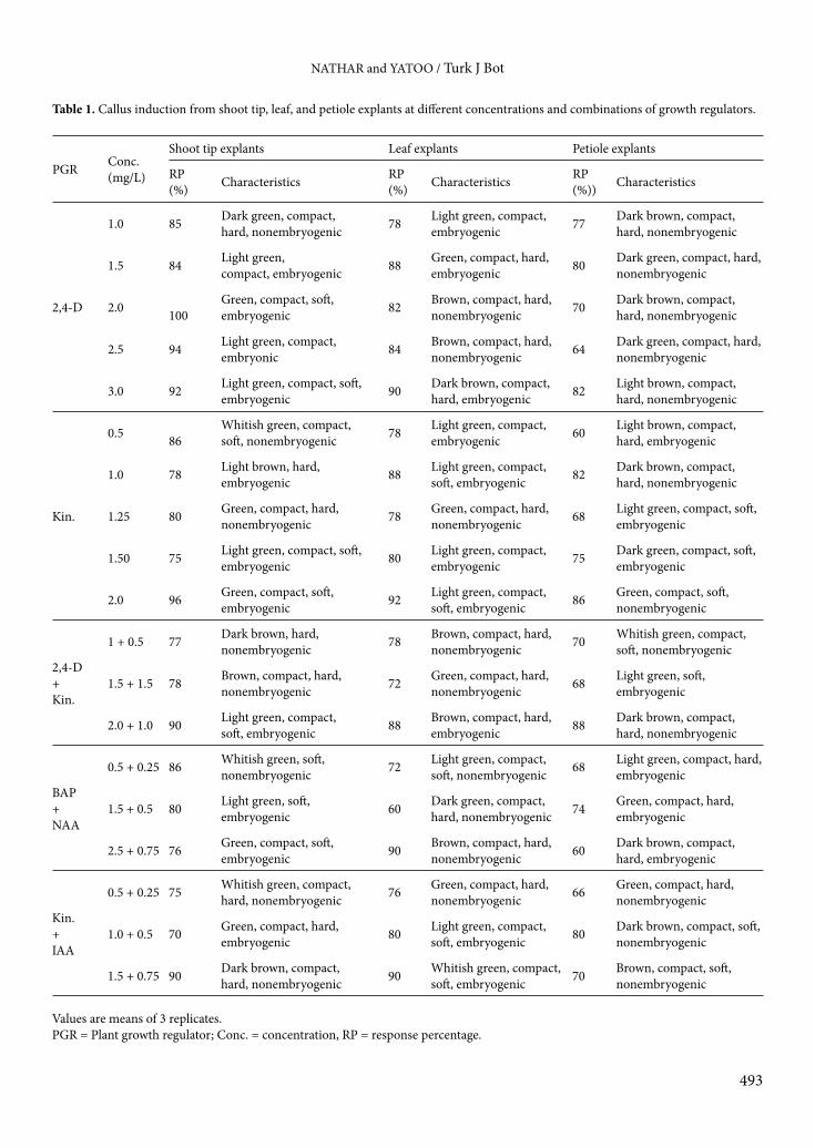
3. Results and discussion3.1. Seed germinationSurface sterilized seeds were germinated on MS media (Figure 1); 95% of the seeds had germinated after 5–7 days of dark exposure at 23 °C. Fully grown seedlings were observed within 40 days after transfer to photoperiodic conditions for 16/8 h of light/dark at 20 ± 1 °C. Similar results were reported in A. vulgaris, except that 10% filtered sterilized coconut water was added to media (Sujatha and Kumari, 2007). Shoot tip, leaf, and petiole segments of A. pallens were cultured on MS media supplemented with 2,4-D, kinetin, 2,4-D+kinetin, BAP+NAA, and kinetin+IAA in different concentrations to test the efficiency of sprouting of various explants. It was observed that after 9–12 days of inoculation, the explants showed callus initiation in all the concentrations.3.2. Callus inductionThe callogenic response showed variations among the hormonal combinations/concentrations and types of explants used. The callus color ranged from white to dark green, while texture ranged from soft to compact-hard and from embryogenic to nonembryogenic. The callus response varied among 70%–100%, 72%–90%, and 60%–80% in shoot tip, leaf, and petiole explants, respectively. Callus initiation was observed after the ninth day of inoculation. Among different types of explants used, shoot tips proved to be best for callus induction. Highest callogenic response, i.e. 100%, was noticed with shoot tip explants at 2 mg/L 2,4-D (Figure 1). The callus obtained
NATHAR and YATOO / Turk J Bot
493
Table 1. Callus induction from shoot tip, leaf, and petiole explants at different concentrations and combinations of growth regulators.
PGR Conc.(mg/L)
Shoot tip explants Leaf explants Petiole explants
RP(%) Characteristics RP
(%) Characteristics RP(%)) Characteristics
2,4-D
1.0 85 Dark green, compact, hard, nonembryogenic 78 Light green, compact,
embryogenic 77 Dark brown, compact, hard, nonembryogenic
1.5 84 Light green,compact, embryogenic 88 Green, compact, hard,
embryogenic 80 Dark green, compact, hard, nonembryogenic
2.0 100Green, compact, soft, embryogenic 82 Brown, compact, hard,
nonembryogenic 70 Dark brown, compact, hard, nonembryogenic
2.5 94 Light green, compact, embryonic 84 Brown, compact, hard,
nonembryogenic 64 Dark green, compact, hard, nonembryogenic
3.0 92 Light green, compact, soft, embryogenic 90 Dark brown, compact,
hard, embryogenic 82 Light brown, compact, hard, nonembryogenic
Kin.
0.5 86Whitish green, compact, soft, nonembryogenic 78 Light green, compact,
embryogenic 60 Light brown, compact, hard, embryogenic
1.0 78 Light brown, hard, embryogenic 88 Light green, compact,
soft, embryogenic 82 Dark brown, compact, hard, nonembryogenic
1.25 80 Green, compact, hard, nonembryogenic 78 Green, compact, hard,
nonembryogenic 68 Light green, compact, soft, embryogenic
1.50 75 Light green, compact, soft, embryogenic 80 Light green, compact,
embryogenic 75 Dark green, compact, soft, embryogenic
2.0 96 Green, compact, soft, embryogenic 92 Light green, compact,
soft, embryogenic 86 Green, compact, soft, nonembryogenic
2,4-D+Kin.
1 + 0.5 77 Dark brown, hard, nonembryogenic 78 Brown, compact, hard,
nonembryogenic 70 Whitish green, compact, soft, nonembryogenic
1.5 + 1.5 78 Brown, compact, hard, nonembryogenic 72 Green, compact, hard,
nonembryogenic 68 Light green, soft, embryogenic
2.0 + 1.0 90 Light green, compact, soft, embryogenic 88 Brown, compact, hard,
embryogenic 88 Dark brown, compact, hard, nonembryogenic
BAP+NAA
0.5 + 0.25 86 Whitish green, soft, nonembryogenic 72 Light green, compact,
soft, nonembryogenic 68 Light green, compact, hard, embryogenic
1.5 + 0.5 80 Light green, soft, embryogenic 60 Dark green, compact,
hard, nonembryogenic 74 Green, compact, hard, embryogenic
2.5 + 0.75 76 Green, compact, soft, embryogenic 90 Brown, compact, hard,
nonembryogenic 60 Dark brown, compact, hard, embryogenic
Kin.+IAA
0.5 + 0.25 75 Whitish green, compact, hard, nonembryogenic 76 Green, compact, hard,
nonembryogenic 66 Green, compact, hard, nonembryogenic
1.0 + 0.5 70 Green, compact, hard, embryogenic 80 Light green, compact,
soft, embryogenic 80 Dark brown, compact, soft, nonembryogenic
1.5 + 0.75 90 Dark brown, compact, hard, nonembryogenic 90 Whitish green, compact,
soft, embryogenic 70 Brown, compact, soft, nonembryogenic
Values are means of 3 replicates.PGR = Plant growth regulator; Conc. = concentration, RP = response percentage.

NATHAR and YATOO / Turk J Bot
494
Table 2. Multiple shoot induction from callus at different concentrations/combinations of growth regulators.
Plant growthregulator
Concentration(mg/L)
Response(%)
Average no.of shoots*
Average shoot length*(cm)
BAP
0.25 80 9 ± 1.29 2.75 ± 0.48
0.5 78 5 ± 0.29 1.97 ± 0.36
1.0 64 10.7 ± 1.10 2.02 ± 0.34
1.5 78 7 ± 1.29 1.87 ± 0.42
2.0 94 10 ± 1.82 2.32 ± 0.57
3.0 80 9 ± 1.29 3.4 ± 0.40
Kin.
1.0 66 5.75 ± 0.85 2.42 ± 0.54
1.5 75 6 ± 0.91 2.22 ± 0.66
2.0 64 9 ± 1.29 2.3 ± 0.43
3.0 92 14.25 ± 1.65 4.25 ± 0.47
Kin.+NAA
1.0 + 0.25 75 9.25 ± 1.49 1.87 ± 0.13
1.5 + 0.50 88 6.5 ± 1.5 1.87 ± 0.21
2.0 + 1.0 92 7 ± 1.29 2.05 ± 0.21
BAP+IAA
1.0 + 0.5 78 5.75 ± 0.85 1.37 ± 0.24
2.0 + 0.75 64 11.2 ± 0.75 1.77 ± 0.33
3.0 + 1.0 88 5.25 ± 0.94 2.37 ± 0.25
*Mean ± standard error of 3 replicates.
Table 3. Multiple root induction from shoots grown in vitro with different concentrations/combinations of growth regulators.
Plant growthregulator
Concentration(mg/L)
Response(%)
Average no.of roots*
Average root length*(cm)
IAA
1 80 2.5 ± 0.64 4.1 ± 0.98
2 90 5.75 ± 0.85 3.52 ± 0.63
3 88 6.75 ± 1.10 3.22 ± 1.09
IBA
1 75 4 ± 0.91 2.25 ± 0.33
2 68 8 ± 0.70 3.62 ± 0.53
3 94 12 ± 0.08 8.15 ± 1.13
NAA
0.5 72 4 ± 0.91 4.17 ± 1.08
1.5 60 7 ± 1.29 2.87 ± 0.42
2.0 78 8.25 ± 0.85 4.3 ± 0.50
3.0 90 4 ± 1.08 3.85 ± 0.46
IAA+BAP
1.0 + 0.5 84 4.75 ± 0.75 3.95 ± 0.92
1.0 + 0.75 74 3.75 ± 1.54 3.95 ± 0.73
1.0 + 1.0 80 3 ± 1.08 5.1 ± 1.26
*Mean ± standard error of 3 replicates.

NATHAR and YATOO / Turk J Bot
495
at this concentration was green, compact, soft, and embryogenic. Cytokinin (BAP) alone at low concentrations did not induce dedifferentiation, but at higher doses it was able to induce calli (Nin et al., 1996). Callus formation was also reported in Artemisia scopario with 2,4-D alone and in combination with kinetin (Aslam et al., 2006). Benjamin et al. (1990) reported 3 types of cultures in A. pallens on MS media supplemented with different concentrations/combinations of hormones. Unorganized calli were obtained on MS medium supplemented with BAP+2,4-D and semiorganized calli on BAP+IAA from shoot buds. The effect of plant growth regulators on callus formation showed that, in Rosa gallica L., 2 mg/L of 2,4-D and 1 mg/L of BAP were the optimum concentrations, increasing callogenesis more than 90% in vegetative (leaf, stem, and petiole) and petal explants and more than 80%
in flower explants (pistil and anther). Callus induction in Artemisia absinthium from leaf explant on MS medium supplemented with 2,4-D+NAA (1.0 + 0.5 mg/L) was also reported (Yatoo, 2010). The callus obtained after 40 days was subcultured on shooting media.3.3. Multiple shootingThe calli that formed from shoot tip explants were used for multiple shoot induction. Multiple shoot initiation started from the callus after 10 days when subcultured onto fresh MS medium containing different cytokinins like BAP and kinetin and auxins like NAA and IAA. Kinetin among the various cytokinins proved best for multiple shoot induction. The highest number of shoots, i.e. 14.25 ± 1.65, and highest shoot length, i.e. 4.25 ± 0.47 cm, were observed at 3 mg/L kinetin (Figure 1). The cytokinin BAP has been commonly used for the induction of organogenesis in
Figure 1. In vitro studies on Artemisia pallens. a- Seed germination on plain MS medium; b- callus formation from shoot tip explants with 2 mg/L 2,4-D; c and d- multiple shoot formation with 3 mg/L kinetin; e- multiple root formation with 3 mg/L IBA; f- acclimatized plant.

NATHAR and YATOO / Turk J Bot
496
many plants (Zilis et al., 1979; Parrot et al., 1992; Mneny and Mantell, 2002). Effectiveness of different cytokinins to induce multiple shoot formation was revealed in the order of BAP > kinetin > zeatin > adenine (Prakash et al., 1994). BAP was essential for shoot production in both cultivars since no shoot development occurred on media without BAP (Gürel and Gülşen, 1998). Chalageri and Babu in 2012 showed that among different concentrations of growth regulators used in Viola patrinii, NAA (2.68 µM) with kinetin (23.25 µM) showed 88% shoot regeneration, which was achieved in 4 weeks. Multiple shooting was obtained on MS media with BAP+IAA+NAA (Benjamin et al., 1990). Several studies have demonstrated that an excess of cytokinins can promote the regeneration of shoots in tissue cultured plants (Kevers et al., 1984). BAP has been used in preference to other cytokinins to induce multiple shoots in A. pallens. Encapsulated shoot buds grown in vitro on MS media supplemented with NAA (5.3 μm) + BA (1.33 μm) + biotin (1 mg/L) + casein hydrolysate (3 mg/L) showed controlled multiplication (Sharief et al., 1997). Nair et al. (1979) stated that BAP is most effective for meristem, shoot tip, and bud culture. When BAP+kinetin in combination was tested, the frequency of response was still much better (Sujatha and Kumari, 2007). In Macrotyloma uniflorum, a large number of shoots developed from shoot tip explants on MS media supplemented with BAP alone or BAP+kinetin (Shamsudeen et al., 1999). Similar combinations produced greater numbers of shoots in Arachis hypogaea, Carthamus tinctorius, and Eclipta alba (Venkatachalam and Jayabalan, 1997; Baskaran and Jayabalan, 2005; Kumar and Kumari, 2005). In the present investigation, higher concentrations of BAP reduced shoot number and length, which is in agreement with the findings of Hu and Wang (1983) and Indhra and Dhar (2000).3.4. Multiple rootingTo induce rooting, individual elongated shoots after 40 days were cultured on MS media augmented with different auxin concentrations. For root induction MS medium was augmented with different hormones like IAA (1–3 mg/L), IBA (1–3 mg/L), NAA (0.5–3 mg/L), and IAA+BAP (1 + 0.5–1.0 mg/L). Roots were visible within 8–12 days following the transfer of elongated shoots to the rooting media. The highest number of roots (12 ± 0.08) and highest root length (8.15 ± 1.13 cm) were encountered with 3 mg/L IBA (Figure 1). Gürel et al. in 2001 reported that over 90% of the regenerated shoots of Beta vulgaris L. could be readily rooted when cultured on medium containing 3.0 mg/L IBA.
IAA was reported as a potential auxin for rooting of Arachis stenosperma and Arachis villosa (Vijayalakshmi and Giri, 2003). Similar results were obtained in Sesbania drummondii (Cheepala et al., 2004). However, in the present
study, IBA was found to be most beneficial, followed by NAA. The present finding confirms the previous work on Artemisia judaica with IBA (Liu et al., 2003) and on Morus alba with NAA (Anuradha and Pullaiah, 1992).3.5. AcclimatizationThe hardened plantlets were initially maintained in a programmable environmental chamber for 5 weeks. Regenerated plantlets were isolated from the culture media and washed with sterile double distilled water to remove adhering medium. These plantlets were transferred to plastic cups that contained sterile garden soil, farmyard soil, and sand (2:1:1). The survival rate after hardening for the first 5 weeks was 100%. It decreased to 80% after 10 weeks of acclimatization. There was no detectable variation among the acclimatized plants with respect to morphological and growth characteristics. All of the micropropagated plants were free from external defects (Figure 1) and are maintained in the departmental garden.
4. ConclusionsThe main objective of this study was to develop a system for mass propagation and aseptic growth of Artemisia pallens. The plant is used for different medicinal purposes. The literature reveals different regeneration systems for mass or in vitro propagation of many Artemisia species. Shoot tip explants supplemented with 2,4-D had the best callogenic response (Figure 2). Kinetin showed maximum shooting response both in terms of number of shoots and shoot length from subcultured calli (Figure 3). Maximum number of roots and maximum root length from shoots grown in vitro were achieved with NAA and IBA, respectively (Figure 4). In the present work, surprisingly, all the explants produced calli; this is the first report on in vitro callogenesis in Artemisia pallens. A wide range of
22
2+10.5+0.25
1.5+0.753 2 2+1 2.5+0.5 1.5+0.75
32 2+1
1.5+0.51+0.5
0
20
40
60
80
100
120
2,4- D Kin. 2,4-D + Kin. BAP + NAA Kin. + IAA
Resp
onse
per
cent
age (
%)
Shoot tip explant Leaf explant Petiole explant
Figure 2. Maximum callogenic response from shoot tip, leaf, and petiole explants. The concentration of growth regulator is in mg/L.

NATHAR and YATOO / Turk J Bot
497
calli were obtained, which make extraction of biologically actively compounds easier. This will help in its conservation as well as in establishing a genetic transformation system.
The plants produced from excised tissues will be helpful for genetic engineering and cell culture techniques in the future.
1
3
1+0.25
2+0.75
33
2+1 3+1
0
2
4
6
8
10
12
14
16
BAP Kin. Kin. + NAA BAP + IAA
Number of shootsShoot length (cm) 3
2 2
1+0.51
3
21+1
0
1
2
3
4
5
6
7
8
9
IAA IBA NAA IAA+BAP
Number of rootsRoot length (cm)
Figure 3. Multiple shoot induction from calli. The concentration of growth regulator is in mg/L.
Figure 4. Multiple root induction from shoots grown in vitro. The concentration of growth regulators is in mg/L. Values are means ± standard errors of 3 replicates.
References
Akhtar MS, Iqbal Z, Khan MN, Lateef M (2000). Anthelmintic activity of medicinal plants with particular reference to their use in animals in the Indo-Pakistan subcontinent. Small Ruminant Res 38: 99–107.
Al-Harbi M, Qureshi S, Ahmed M, Raza M, Miana G, Shah A (2000). Studies on the anti-inflammatory antipyretic and analgesic activity of medicinal plants with particular reference to their use in animals in the Indo-Pakistan subcontinent. Small Ruminant Res 38: 99–107.
Anuradha M, Pullaiah T (1992). Micropropagation of mulberry (Morus alba L.). Annali Di Botanica 15: 35–41.
Aslam N, Zia M, Chaudhary F (2006). Callogenesis and direct organogenesis of Artemisia scorpario. Pak J Biol Sci 9: 1783–1786.
Baskaran P, Jayabalan N (2005). An efficient micropropagation system for Eclipta alba—a valuable medicinal herb. In Vitro Cell Dev Bio Pl 41: 532–539.
Benjamin BD, Sipahimalani AT, Heble MR (1990). Tissue cultures of Artemisia pallens: organogenesis, terpenoid production. Plant Cell Tiss Organ Cult 21: 159–164.
Chalageri G, Babu UD (2012). In vitro plant regeneration via petiole callus of Viola patrinii and genetic fidelity assessment using RAPD markers. Turk J Bot 36: 358–368.
Cheepala SB, Sharma NC, Sahi SV (2004). Rapid in vitro regeneration of Sesbania drummondii. Plant Biol 48: 13–18.
Gürel S, Gülşen Y (1998). The effects of IBA and BAP on in vitro shoot production of almond (Amygdalus communis L.). Turk J Bot 22: 375–379.
Gürel S, Gürel E, Kaya Z (2001). Callus development and indirect shoot regeneration from seedling explants of sugar beet (Beta vulgaris L.) cultured in vitro. Turk J Bot 25: 25–33.
Hansen G, Wright MS (1999). Recent advances in the transformation of plants. Trends Plant Sci 4: 226–231.
Hu CY, Wang PJ (1983). Meristem shoot tip and bud culture. In: Evans DA, Sharp WR, Ammirato PV, Yamada Y, editors. Handbook of Plant Cell Culture. New York, NY, USA: Macmillan, pp. 177–227.
Indhra DB, Dhar U (2000). Micropropagation of Indian wild strawberry. Plant Cell Tiss Organ Cult 60: 83–88.
Kevers C, Coumans M, Coumans G, Caspar T (1984). Physiological, biochemicals events leading to vitrification of plants cultured in vitro. Plant Physiol 61: 69–74.
Kumar VJ, Kumari BDR (2005). Effect of phytohormones on multiple shoot bud induction in cv. NARI-6 of safflower (Carthamus tinctorius L.). J Plant Biotech 7: 149–153.
Liu CZ, Murch SJ, El-Demerdash M, Saxena PK (2003). Regeneration of the Egyptian medicinal plant Artemisia judaica L. Plant Cell Rep 21: 525–530.
Misra L, Chandra A, Thakur R (1991). Fragrant components of oil from Artemisia pallens. Phytochem 30: 549–552.
Mneny EE, Mantell SH (2002). Clonal propagation of cashew (Anacardium occidentale L.) by tissue culture. J Hortic Sci 77: 649–657.
Murashige T, Skoog F (1962). A revised medium for rapid growth and bio assays with tobacco tissue cultures. Physiol Plantarum 15: 473–497.

NATHAR and YATOO / Turk J Bot
498
Nair NG, Kartha KK, Gamborg OL (1979). Effect of growth regulators on plant regeneration from shoot tip meristems of cassava (Manihot esculenta Crantz) and the culture of internodes in vitro. Z Pflanzen Physiol 95: 51–56.
Nin S, Morosi E, Schiffs S, Bennici A (1996). Callus cultures of Artemisia absinthium L.: initiation, growth, optimization and organogenesis. Plant Cell Tiss Organ Cult 45: 67–72.
Parrot WA, Bailey MA, Durham RE, Mathews HV (1992). Tissue cultures and regeneration in legumes. In: Moss JP, editor. Biotechnology and Crop Improvement in Asia. Patancheru, India: ICRISAT, pp. 115–148.
Prakash SN, Deepak P, Neera BS (1994). Regeneration of pigeon pea (Cajanus cajan) from cotyledonary node via multiple shoot formation. Plant Cell Rep 13: 623–627.
Pujar PP, Sawaikar DD, Rojatkar SR, Nagasampagi BA (2000). A new germacranolide from Artemisia pallens. Fitoterapia 71: 590–592.
Ruikar AD, Kamble GS, Puranik VG, Deshpande NR (2009). Antimicrobial screening of medicinal plant–Artemisia pallens. Int J Pharmtech Res 4: 1164–1166.
Shamsudeen VM, Jawahar M, Thiruvengadam M, Jeyakumar M, Jayabalan N (1999). Effect of cytokinins on the proliferation of multiple shoots in horse gram (Macrotyloma uniflorum (Lam.) Verdc.). J Plant Biotech 1: 79–83.
Sharief U, Jagadishchandra KS, Johnson TS, Ravishankar GA (1997). Propagation of Artemisia pallens by encapsulated in vitro grown shoot buds. J Med Arom Plant Sci (India) 19: 712–717.
Subramoniam A, Pushpangadan P, Rajasekharan S, Evans D, Latha P, Valsaraj R (1996). Effect of Artemisia pallens Wall. on blood glucose levels in normal and alloxan-induced diabetic rats. J Ethnopharmacol 50: 13–17.
Sujatha G, Kumari BDR (2007). Effect of phytohormones on micropropagation of Artemisia vulgaris L. Acta Physiol Plant 29: 189–195.
Venkatachalam P, Jayabalan N (1997). Effect of auxins and cytokinins on efficient regeneration and multiple shoot formation from cotyledons and cotyledonary node explants of groundnut (Arachis hypogaea L.) by in vitro culture technology. Appl Biochem Biotechnol 67: 237–247.
Vijayalakshmi G, Giri C (2003). Plant regeneration via organogenesis from shoot base derived callus of Arachis stenosperma, Arachis villosa. Curr Sci 85: 1624–1629.
Yatoo GM (2010). Callogenesis and direct organogenesis from aseptic plant of Artemisia absinthium L. growing wild in Kashmir. J Ind Bot Soc 89: 248–252.
Zilis M, Zwagerman D, Lamberts D, Kurtz L (1979). Commercial propagation of herbaceous perennials by tissue culture. Proceedings International Plant Propagator’s Society 9: 404–414.