microfluidic chemotaxis platform for differentiating the ...... · microfluidic chemotaxis platform...
TRANSCRIPT
Microfluidic Chemotaxis Platform forDifferentiating the Roles of Soluble andBound Amyloid-b on MicroglialAccumulationHansang Cho1, Tadafumi Hashimoto2, Elisabeth Wong1, Yukiko Hori2, Levi B. Wood3, Lingzhi Zhao2,4,Kevin M. Haigis3, Bradley T. Hyman2 & Daniel Irimia1
1BioMEMS Resource Center, Massachusetts General Hospital, Harvard Medical School, 2MassGeneral Institute forNeurodegenerative Disease, Massachusetts General Hospital, Harvard Medical School, 3Molecular Pathology Unit, MassachusettsGeneral Hospital, Harvard Medical School, 4Appel Alzheimer’s Disease Research Institute, Department of Neurology andNeuroscience, Weill Cornell Medical College.
Progressive microglial accumulation at amyloid-b (Ab) plaques is a well-established signature of thepathology of Alzheimer’s disease, but how and why microglia accumulate in the vicinity of Ab plaques isunknown. To understand the distinct roles of Ab on microglial accumulation, we quantified microglialresponses to week-long lasting gradients of soluble Ab and patterns of surface-bound Ab in microfluidicchemotaxis platforms. We found that human microglia chemotaxis in gradients of soluble Ab42 was mosteffective at two distinct concentrations of 23 pg.mL21 and 23 ng.mL21 Ab42 in monomers and oligomers.We uncovered that while the chemotaxis at higher Ab concentrations was exclusively due to Ab gradients,chemotaxis at lower concentrations was enhanced by Ab-induced microglial production of MCP-1.Microglial migration was inhibited by surface-bound Ab42 in oligomers and fibrils above 45 pg.mm22.Better understanding of microglial migration can provide insights into the pathophysiology of senileplaques in AD.
Microglial cells are resident macrophages in the central neural system (CNS) and have multiple functions innormal and pathological brains1–6. Ramified microglia are believed to constantly scan the brain and clearthe CNS of toxic agents and debris7,8. In the context of Alzheimer’s disease (AD), diminished clearance of
Ab due to impaired microglia is postulated to be one of the mechanisms of Ab plaque formation9. Once Ab formsplaques, microglia become hyper-activated10, accumulate around the plaques, and consequently secrete neurotoxicagents including neuroinflammatory mediators, reactive oxygen species, and free radicals11–14. Understanding themechanisms that lead to microglial accumulation and inflammatory responses may provide useful insights intodeveloping strategies for AD treatment. However, several studies report different interactions between microgliaand other factors15–17. Previous in vivo studies of microglia migration were hampered by both the inability toobserve long-term effects of Ab on microglia18 and the complexity of senile plaques, which are deposits ofoligomeric and fibrillar Ab, surrounded by a ‘‘halo’’ of soluble oligomeric Ab19, activated glia, and dystrophicneurites12. Activated microglial cells take on various morphologies: rounded, ramified shapes, rods to amoeboids,which complicate the visual tracking of these cells individually. Furthermore, previous in vitro attempts to studyrat microglia migration in the presence of short lived, damaged axons20, could not establish long-lasting gradientsand were not conclusive for differentiating slow accumulation of microglia from random navigation21,22.
Here, we have developed a novel microfluidic chemotaxis platform to study the motility of microglia respond-ing to various types of Ab in a regulated manner. The new platform models soluble and bound Ab environmentsin AD brains and enables us to measure the distinct effects of both gradients of soluble Ab, lasting longer than aweek and surface-bound patterns of Ab on the migration of microglia.
ResultsA microfluidic platform provides a gradient of soluble Ab and allows monitoring of microglial motility.Based on in vivo observation of microglial accumulation on Ab plaques, we hypothesize that soluble Ab is an
SUBJECT AREAS:ASSAY SYSTEMS
ALZHEIMER’S DISEASE
BIOMEDICAL ENGINEERING
NEUROIMMUNOLOGY
Received18 January 2013
Accepted23 April 2013
Published10 May 2013
Correspondence andrequests for materials
should be addressed toD.I. (dirimia@hms.
harvard.edu)
SCIENTIFIC REPORTS | 3 : 1823 | DOI: 10.1038/srep01823 1
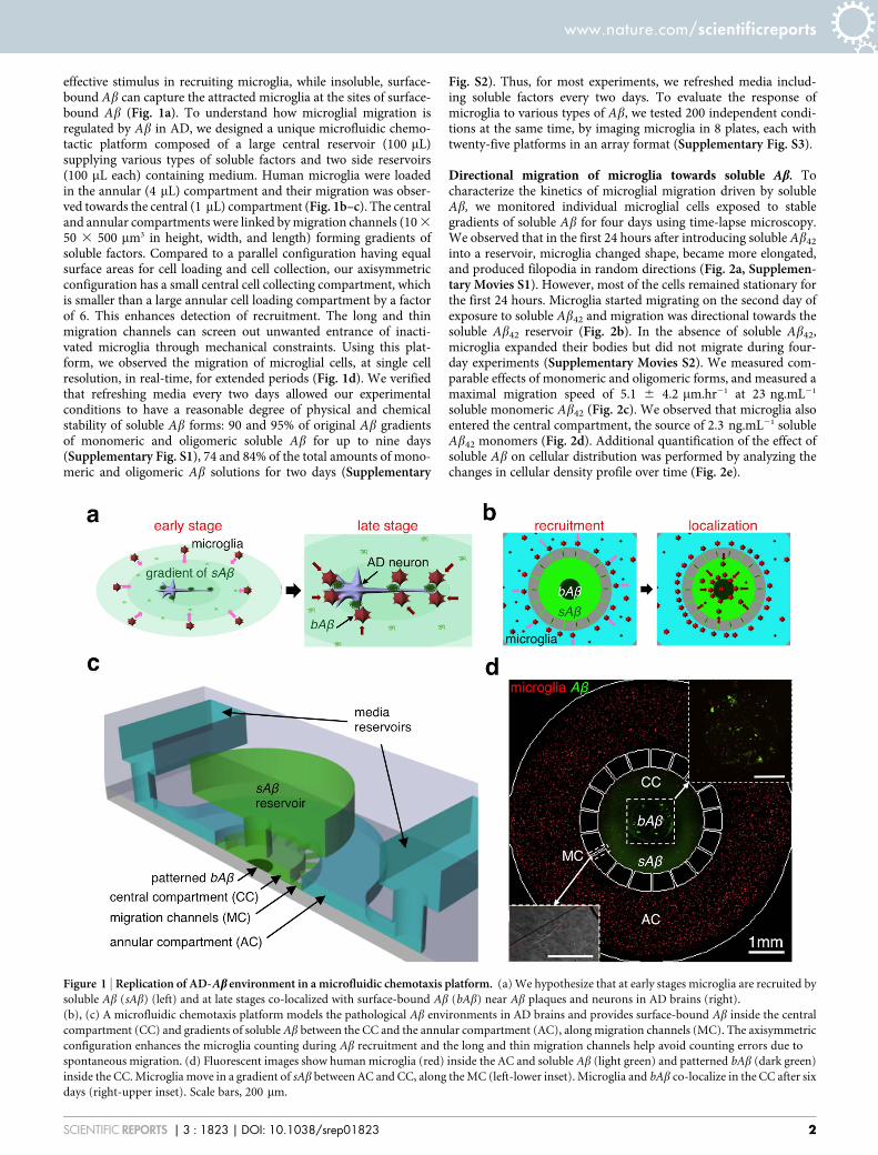
effective stimulus in recruiting microglia, while insoluble, surface-bound Ab can capture the attracted microglia at the sites of surface-bound Ab (Fig. 1a). To understand how microglial migration isregulated by Ab in AD, we designed a unique microfluidic chemo-tactic platform composed of a large central reservoir (100 mL)supplying various types of soluble factors and two side reservoirs(100 mL each) containing medium. Human microglia were loadedin the annular (4 mL) compartment and their migration was obser-ved towards the central (1 mL) compartment (Fig. 1b–c). The centraland annular compartments were linked by migration channels (10 3
50 3 500 mm3 in height, width, and length) forming gradients ofsoluble factors. Compared to a parallel configuration having equalsurface areas for cell loading and cell collection, our axisymmetricconfiguration has a small central cell collecting compartment, whichis smaller than a large annular cell loading compartment by a factorof 6. This enhances detection of recruitment. The long and thinmigration channels can screen out unwanted entrance of inacti-vated microglia through mechanical constraints. Using this plat-form, we observed the migration of microglial cells, at single cellresolution, in real-time, for extended periods (Fig. 1d). We verifiedthat refreshing media every two days allowed our experimentalconditions to have a reasonable degree of physical and chemicalstability of soluble Ab forms: 90 and 95% of original Ab gradientsof monomeric and oligomeric soluble Ab for up to nine days(Supplementary Fig. S1), 74 and 84% of the total amounts of mono-meric and oligomeric Ab solutions for two days (Supplementary
Fig. S2). Thus, for most experiments, we refreshed media includ-ing soluble factors every two days. To evaluate the response ofmicroglia to various types of Ab, we tested 200 independent condi-tions at the same time, by imaging microglia in 8 plates, each withtwenty-five platforms in an array format (Supplementary Fig. S3).
Directional migration of microglia towards soluble Ab. Tocharacterize the kinetics of microglial migration driven by solubleAb, we monitored individual microglial cells exposed to stablegradients of soluble Ab for four days using time-lapse microscopy.We observed that in the first 24 hours after introducing soluble Ab42
into a reservoir, microglia changed shape, became more elongated,and produced filopodia in random directions (Fig. 2a, Supplemen-tary Movies S1). However, most of the cells remained stationary forthe first 24 hours. Microglia started migrating on the second day ofexposure to soluble Ab42 and migration was directional towards thesoluble Ab42 reservoir (Fig. 2b). In the absence of soluble Ab42,microglia expanded their bodies but did not migrate during four-day experiments (Supplementary Movies S2). We measured com-parable effects of monomeric and oligomeric forms, and measured amaximal migration speed of 5.1 6 4.2 mm.hr21 at 23 ng.mL21
soluble monomeric Ab42 (Fig. 2c). We observed that microglia alsoentered the central compartment, the source of 2.3 ng.mL21 solubleAb42 monomers (Fig. 2d). Additional quantification of the effect ofsoluble Ab on cellular distribution was performed by analyzing thechanges in cellular density profile over time (Fig. 2e).
Figure 1 | Replication of AD-Ab environment in a microfluidic chemotaxis platform. (a) We hypothesize that at early stages microglia are recruited by
soluble Ab (sAb) (left) and at late stages co-localized with surface-bound Ab (bAb) near Ab plaques and neurons in AD brains (right).
(b), (c) A microfluidic chemotaxis platform models the pathological Ab environments in AD brains and provides surface-bound Ab inside the central
compartment (CC) and gradients of soluble Ab between the CC and the annular compartment (AC), along migration channels (MC). The axisymmetric
configuration enhances the microglia counting during Ab recruitment and the long and thin migration channels help avoid counting errors due to
spontaneous migration. (d) Fluorescent images show human microglia (red) inside the AC and soluble Ab (light green) and patterned bAb (dark green)
inside the CC. Microglia move in a gradient of sAb between AC and CC, along the MC (left-lower inset). Microglia and bAb co-localize in the CC after six
days (right-upper inset). Scale bars, 200 mm.
www.nature.com/scientificreports
SCIENTIFIC REPORTS | 3 : 1823 | DOI: 10.1038/srep01823 2

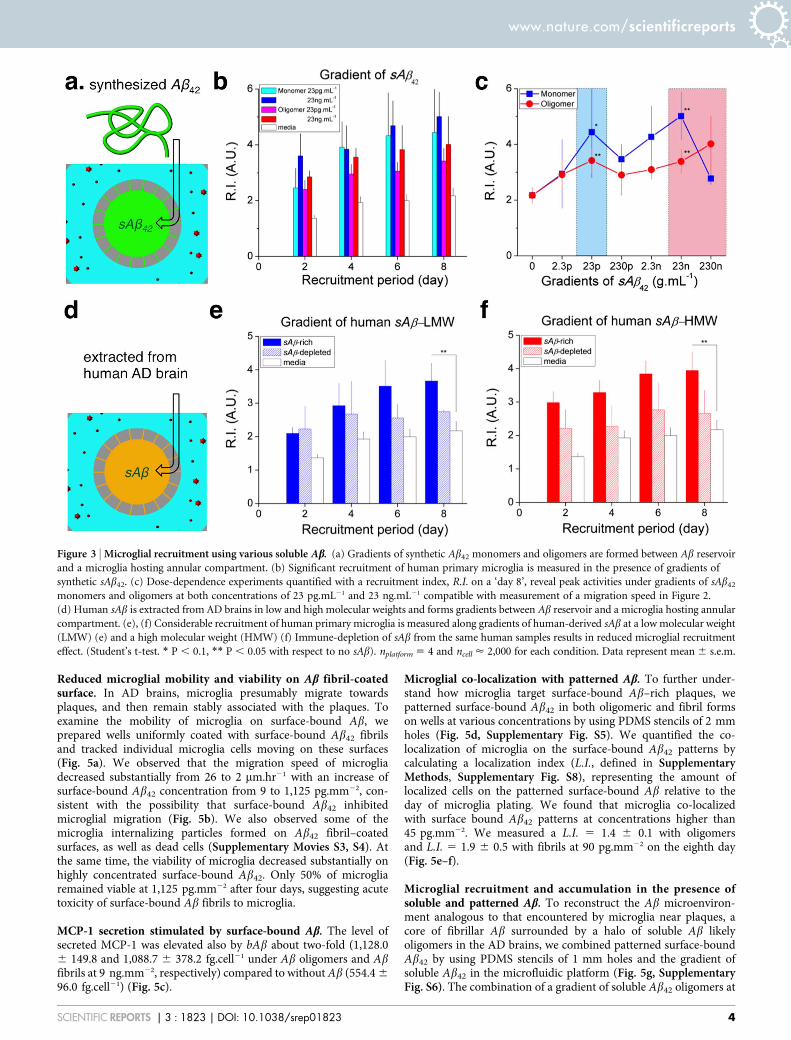
To compare the effect of soluble Ab in various forms and concen-trations on microglial recruitment, we defined a recruitment index,R.I. (as defined in Supplementary Methods, Supplementary Fig.S7), representing the fraction of microglia cells recruited to the cent-ral compartment after loading soluble Ab in the central compart-ment. We compared the R.I. values under various types of solubleAb42 for eight days (Fig. 3a, Supplementary Fig. S4) and found thatthe R.I. value reached 5.0 6 0.9 after exposure to monomeric syn-thetic Ab42 at 23 ng.mL21 and 4.0 6 1.0 after exposure to oligomericforms at 230 ng.mL21, on ‘day 8’ (Fig. 3b). Measuring the concen-tration dependence of directional migration speed and R.I. revealedtwo peaks of activity at two concentrations of soluble Ab42 that werethree orders of magnitude apart (23 pg.mL21 and 23 ng.mL21)(Fig. 2c and Fig. 3c). Interestingly, the low peak concentrationscorresponds to the levels in normal and early AD brains, while thehigh peak to late AD brains23.
Microglial recruitment by human-derived soluble Ab. To validatethe biological relevance of our platform, we measured the microglialresponse to gradients of soluble Ab enriched or depleted samplesderived from tris-buffered saline (TBS) extracts of human ADbrains as previously described24. For this purpose, low (8 to20 kDa) and high molecular weight fractions (more than 100 kDa)were separated by size exclusion chromatography (Fig. 3d). Bothlow-molecular-weight soluble Ab (LMW) and high-molecular-weight soluble Ab (HMW) fractions were similar in recruitingmicroglia towards the central compartment on the eighth day (R.I.5 3.7 6 0.5 for LMW and R.I. 5 3.9 6 0.6 for HMW) (Fig. 3e–f).Removing the soluble Ab by immune-precipitation considerablyreduced the microglial accumulation (R.I 5 2.7 6 0.1 for LMWand 2.7 6 0.7 for HMW), suggesting that a major factor of theTBS extract inducing microglial migration was Ab.
Self-promoting microglial recruitment by Ab-induced MCP-1secretion. Because typical chemotactic responses of various cells
display only one activity peak, we investigated further themechanisms behind the double peak chemotactic activities ofmicroglia in response to Ab. To probe if a second chemoattractantmay explain the unusual double chemotactic peak, we utilized amultiplexed cytokine assay to examine 27 cytokines expressed intothe media collected from microglia cultured in the presence ofsoluble Ab42 at 2.3 pg.mL21 for two days. We identified fivecytokines at detectable levels: IL-1ra, IL-6, IL-8, MCP-1, and MIP-1b while the remaining cytokines were either below, or just atthe threshold of detectability (Fig. 4a). Arrows indicate saturatedmeasurement of cytokines relative to the standard curve. BecauseMCP-1, MIP-1a, and MIP-1b are known chemoattractant mole-cules for macrophages3,7,25, we measured microglial recruitmentunder gradients of soluble Ab42 in addition to a mixture ofneutralizing antibodies against MCP-1, MIB-1a, and MIB-1b andobserved reduced activity at 23 pg.mL21 but not at 23 ng.mL21 ofsoluble Ab42 (Fig. 4c). In control experiments, we validated thatMCP-1 alone can promote microglial recruitment and verified thatneutralizing antibodies can reduce microglial recruitment towardsthe single cytokine (Fig. 4d). We further measured the MCP-1concentration in media from microglia stimulated with variousconcentrations of both monomeric and oligomeric soluble Ab42.We estimated the maximum secretion of MCP-1 at 1,433.9 6
816.0 and 1,457.3 6 109.4 fg.cell21 in the presence of 2.3 pg.mL21
of soluble monomeric and oligomeric Ab42, respectively and 554.4 6
224.3 fg.cell21 in the absence of soluble Ab42 (Fig. 4b). Interestingly,the MCP-1 secretion in the presence of 2.3 pg.mL21 Ab42 wassignificantly higher than at other concentrations of Ab42 oligo-mers. While the migration of microglia occurs at higher levels ofthe gradient (23 pg.mL21) than uniform Ab42 (2.3 pg.mL21), thedifference could be explained by the lower effective concentrationat the initial region of a gradient. It is possible that MCP-1 plays a rolein the peak microglia recruitment activity at a gradient of 23 pg.mL21
soluble Ab42, in addition to other reported roles in microglia-mediated neurodegeneration26.
Figure 2 | Inducement of microglial directional migration by gradients of soluble Ab. (a) Individual microglia migrate directionally along the gradient
of sAb monomers formed in migration channels. (b) Activation of directional motility can be discerned after 24 hours-exposure to gradients of sAb42 in
monomers at 23 pg.mL21 and 23 ng.mL21. (c) Dose-dependence of microglia migration speed during 4 days observations reveal two peak activities under
gradients of soluble Ab42 monomers and oligomers at concentrations of 23 pg.mL21 and 23 ng.mL21 (ranges highlighted in cyan and pink colors,
respectively). (d) Fluorescence images present detectable microglia accumulation toward the source of soluble Ab42 at day 6 compared to day 0.
(e) Microglial density profiles at different days quantify the migration of microglia populations toward the source of soluble Ab42 in monomers at
2.3 ng.mL21. See the details on the stability and the preparation of soluble monomeric and oligomeric Ab in Figs. S1, S2, and Supplementary methods.
(Student’s t-test. * P , 0.01 with respect to no sAb42). ncell 5 18 for each condition. Data represent mean 6 s.e.m.
www.nature.com/scientificreports
SCIENTIFIC REPORTS | 3 : 1823 | DOI: 10.1038/srep01823 3
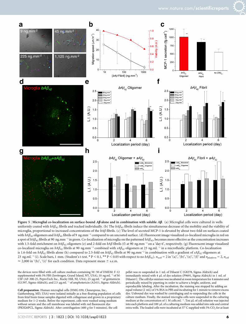
Reduced microglial mobility and viability on Ab fibril-coatedsurface. In AD brains, microglia presumably migrate towardsplaques, and then remain stably associated with the plaques. Toexamine the mobility of microglia on surface-bound Ab, weprepared wells uniformly coated with surface-bound Ab42 fibrilsand tracked individual microglia cells moving on these surfaces(Fig. 5a). We observed that the migration speed of microgliadecreased substantially from 26 to 2 mm.hr21 with an increase ofsurface-bound Ab42 concentration from 9 to 1,125 pg.mm22, con-sistent with the possibility that surface-bound Ab42 inhibitedmicroglial migration (Fig. 5b). We also observed some of themicroglia internalizing particles formed on Ab42 fibril–coatedsurfaces, as well as dead cells (Supplementary Movies S3, S4). Atthe same time, the viability of microglia decreased substantially onhighly concentrated surface-bound Ab42. Only 50% of microgliaremained viable at 1,125 pg.mm22 after four days, suggesting acutetoxicity of surface-bound Ab fibrils to microglia.
MCP-1 secretion stimulated by surface-bound Ab. The level ofsecreted MCP-1 was elevated also by bAb about two-fold (1,128.06 149.8 and 1,088.7 6 378.2 fg.cell21 under Ab oligomers and Abfibrils at 9 ng.mm22, respectively) compared to without Ab (554.4 6
96.0 fg.cell21) (Fig. 5c).
Microglial co-localization with patterned Ab. To further under-stand how microglia target surface-bound Ab–rich plaques, wepatterned surface-bound Ab42 in both oligomeric and fibril formson wells at various concentrations by using PDMS stencils of 2 mmholes (Fig. 5d, Supplementary Fig. S5). We quantified the co-localization of microglia on the surface-bound Ab42 patterns bycalculating a localization index (L.I., defined in SupplementaryMethods, Supplementary Fig. S8), representing the amount oflocalized cells on the patterned surface-bound Ab relative to theday of microglia plating. We found that microglia co-localizedwith surface bound Ab42 patterns at concentrations higher than45 pg.mm22. We measured a L.I. 5 1.4 6 0.1 with oligomersand L.I. 5 1.9 6 0.5 with fibrils at 90 pg.mm22 on the eighth day(Fig. 5e–f).
Microglial recruitment and accumulation in the presence ofsoluble and patterned Ab. To reconstruct the Ab microenviron-ment analogous to that encountered by microglia near plaques, acore of fibrillar Ab surrounded by a halo of soluble Ab likelyoligomers in the AD brains, we combined patterned surface-boundAb42 by using PDMS stencils of 1 mm holes and the gradient ofsoluble Ab42 in the microfluidic platform (Fig. 5g, SupplementaryFig. S6). The combination of a gradient of soluble Ab42 oligomers at
Figure 3 | Microglial recruitment using various soluble Ab. (a) Gradients of synthetic Ab42 monomers and oligomers are formed between Ab reservoir
and a microglia hosting annular compartment. (b) Significant recruitment of human primary microglia is measured in the presence of gradients of
synthetic sAb42. (c) Dose-dependence experiments quantified with a recruitment index, R.I. on a ‘day 8’, reveal peak activities under gradients of sAb42
monomers and oligomers at both concentrations of 23 pg.mL21 and 23 ng.mL21 compatible with measurement of a migration speed in Figure 2.
(d) Human sAb is extracted from AD brains in low and high molecular weights and forms gradients between Ab reservoir and a microglia hosting annular
compartment. (e), (f) Considerable recruitment of human primary microglia is measured along gradients of human-derived sAb at a low molecular weight
(LMW) (e) and a high molecular weight (HMW) (f) Immune-depletion of sAb from the same human samples results in reduced microglial recruitment
effect. (Student’s t-test. * P , 0.1, ** P , 0.05 with respect to no sAb). nplatform 5 4 and ncell < 2,000 for each condition. Data represent mean 6 s.e.m.
www.nature.com/scientificreports
SCIENTIFIC REPORTS | 3 : 1823 | DOI: 10.1038/srep01823 4

5 nM and surface-bound Ab42 fibrils at 90 pg.mm22 achieved themost effective localization by 2.5 times compared to 1.6 6 0.2 timeswith surface-bound Ab42 fibrils at 90 pg.mm22 only (Fig. 5h–i).
DiscussionOur study demonstrates that soluble monomeric and oligomeric Abserve as a ‘‘recruiting signal’’ and bound fibrillar and oligomericsurface-bound Ab acts as a ‘‘targeting signal’’ during microgliarecruitment and localization. Together, soluble and insoluble Abhave synergistic effects on microglial accumulation to sites of Abdeposits, and could explain microglial accumulation in the vicinityof Ab plaques in the AD cortex.
Moreover, the MCP-1 dependent mechanism of microglia recruit-ment in response to lower doses of Ab may be important duringphysiological neuroinflammation and could be relevant to the earlystages of microglial activation in AD. The microfluidic platform canbe extended to the study of migration of other cells relevant to theprogression of neurodegenerative diseases27,28, and could help quant-ify the potency of various cytokines and chemokines, which havebeen detected in the brain alone and in combinations11, in modulat-ing microglia recruitment and accumulation. With high-throughputcapabilities and regulated microenvironments, our platforms can
facilitate systematic monitoring of microglial migration and itsmodulation by compounds for the treatment of neuroinflammationin various disease conditions, including Alzheimer’s disease.
MethodsMicrofluidic platform fabrication. Negative photoresists, SU-8 50 and SU-8 100(MicroChem, Newton, MA, USA), were sequentially patterned using standardlithography on a 4’’ silicon wafer to create a mold for cell migration channels of 50 mmin height and chemokine compartments of 100 mm in height. A mixture of a base anda curing agent with a 1051 weight ratio (SYLGARD 184 A/B, Dowcorning, Midland,MI, USA) was poured onto the SU-8 mold and cured for one hour at roomtemperature under vacuum and, subsequently, cured for more than 3 hours in anoven at 80uC. The cured polydimethyl-siloxane (PDMS) replica was peeled off fromthe mold and holes were punched for fluid reservoirs. Arrayed holes were alsolaser-cut (Zing 24, Epilog Laser, Golden, CO, USA) into a thin PDMS membrane of250 mm in thickness (HT 6240, Bisco Silicones, Elk Grove, IL, USA) and an acrylicplate of 6 mm in thickness. The machined membrane and the plate were gluedtogether using uncured PDMS and incubated at 80uC overnight. This assembly wasirreversibly bonded first to the PDMS replica using oxygen plasma at 50 mW, 5 ccm,for 30 seconds (PX-250, March Plasma Systems, Petersburg, FL, USA), and later to aglass-bottomed UniWell plate (MGB001-1-2-LG, Matrical Bioscience, Spokane, WA,USA). Immediately after the bonding, 10 mL of poly (l-lysine) solution (PLL, M.W.70,000–150,000, 1.0 mg.mL21, Sigma-Aldrich Co. LLC, St. Louis, MO, USA) wasinjected into the each platform and incubated for 2 hours at a room temperature topromote cellular adhesion. PLL-treated surface was rinsed with autoclaved and0.2 mm filtered water (AM9920, Life Technologies, Grand Island, NY, USA) and then
Figure 4 | MCP-1 secretion from microglia under stimulation of soluble Ab and self-promoted microglial recruitment. (a) Microglial cells are cultured
in wells containing sAb42 and discernibly expressed five cytokines (MCP-1, MIP-1b, IL-ra1, IL-6, and IL-8) are measured among tested twenty-seven
human cytokines from extracted solutions. (b) Highest levels of a cytokine, MCP-1 are measured when microglia are cultured under sAb42 in monomers
and oligomers at 2.3 pg.mL21. (c) Microglial cells are cultured in the presence of gradients of cytokine-neutralizing antibody (Ab) combined with sAb42 or
cytokines and reduction of recruitment index is measured in the presence of neutralizing antibody against MCP-1 and sAb42 at 23 pg.mL21 but not
23 ng.mL21. (d) MCP-1 is validated to be a potent chemoattractant for microglia and the recruitment is inhibited by immune-neutralization of MCP-1.
However, MIP-1a and MIP-1b have no microglial chemoattractant activity in this assay. (Student’s t-test. *P , 0.01 for oligomers with respect to no
sAb42). nwell 5 3, ncell < 2,000 in ‘(a)’, ‘(b)’ and nplatform 5 4, ncell < 2,500 in ‘(c)’, ‘(d)’ for each condition. Data represent mean 6 s.e.m.
www.nature.com/scientificreports
SCIENTIFIC REPORTS | 3 : 1823 | DOI: 10.1038/srep01823 5
the devices were filled with cell culture medium containing 50550 of DMEM: F-12supplemented with 5% FBS (Invitrogen, Grand Island, NY, USA), 10 ng.mL21 of M-CSF (AF-300-25, PeproTech Inc., Rocky Hill, NJ, USA), 25 mg.mL21 of gentamicin(G1397, Sigma-Aldrich), and 2.5 mg.mL21 of amphotericin (A2411, Sigma-Aldrich).
Cell preparation. Human microglial cells (HMG 030, Clonexpress, Inc.,Gaithersburg, MD, USA) were isolated initially as a free-floating population of cellsfrom fetal brain tissue samples digested with collagenase and grown in a proprietarymedium for 1–2 weeks. Before the experiment, cells were washed using mediumwithout serum and the cell membrane was labeled with red fluorescent dye(PKH26PCL, Sigma-Aldrich). After centrifugation (400 g for 5 minutes), the cell
pellet was re-suspended in 1 mL of Diluent C (G8278, Sigma-Aldrich) andimmediately mixed with 4 mL of dye solution (P9691, Sigma-Aldrich) in 1 mL ofDiluent C. The cell/dye mixture was incubated at room temperature for 4 minutes andperiodically mixed by pipetting in order to achieve a bright, uniform, andreproducible labeling. After the incubation, the staining was stopped by adding anequal volume (2 mL) of 1% BSA in PBS and incubating for 1 minute to remove excessdye. Unbound dye was washed by centrifuging and re-suspending the cells in theculture medium. Finally, the stained microglia cells were suspended in the culturingmedium at the concentration of 1 M cells.mL21. Ten mL of cell solution was injectedinto each platform and 100 mL of a culturing medium was added into side and centralextra wells. The loaded cells were incubated at 37uC supplied with 5% CO2 for at least
Figure 5 | Microglial co-localization on surface-bound Ab alone and in combination with soluble Ab. (a) Microglial cells were cultured in wells
uniformly coated with bAb42 fibrils and tracked individually. (b) The bAb42 fibrils induce the simultaneous decrease of the mobility and the viability of
microglia, proportional to increased concentrations of the bAb fibrils. (c) The level of secreted MCP-1 is elevated by about two-fold on surfaces coated
with bAb42 oligomers and bAb42 fibrils of 9 ng.mm22 compared to an uncoated surface. (d) Fluorescent image visualized co-localized microglia in red on
a spot of bAb42 fibrils at 90 ng.mm22 in green. Co-localization of microglia on the patterned bAb42, becomes more effective as the concentration increases
with 1.5-fold enrichment on bAb42 oligomers (e) and 2-fold on bAb fibrils (f) at 90 ng.mm22 on a ‘day 6’, respectively. (g) Fluorescent image visualized
co-localized microglia on bAb42 fibrils at 90 ng.mm22 combined with sAb42 oligomers at 23 ng.mL21 in a microfluidic platform. Co-localization
is 1.6-fold on bAb42 fibrils alone (h) compared to 2.5-fold on bAb42 fibrils at 90 ng.mm22 in combination with a gradient of sAb42 oligomers at
23 ng.mL21 (i). Scale bars, 1 mm. (Student’s t-test. * P , 0.1, ** P , 0.05 with respect to no bAb42). nwell 5 2 in ‘(a)’, ‘(b)’, ‘(e)’, ‘(f)’ and nplatform 5 2, ncell
< 2,000 in ‘(h)’, ‘(i)’ for each condition. Data represent mean 6 s.e.m.
www.nature.com/scientificreports
SCIENTIFIC REPORTS | 3 : 1823 | DOI: 10.1038/srep01823 6

two days before adding the various Ab solutions. In the experiments, the cell culturemedia and Ab solutions were replaced every two days while collecting the usedsolutions for Ab analysis.
Sourcing of human brain tissue. Brains from human subjects with a diagnosis ofAlzheimer’s disease were obtained through the Massachusetts Alzheimer’s DiseaseResearch Center. The Massachusetts Alzheimer’s Disease Research Center (ADRC)serves, among other clinical and research activities, as a repository of samples frompatients with Alzheimer’s disease. ADRC clinical and research activities are reviewedamong others by the Institutional Review Board at the Massachusetts GeneralHospital. In addition, the ADRC has a Certificate of Confidentiality issued by the USDepartment of Health & Human Services/National Institutes of Health to protect theprivacy of individuals who are enrolled in this research against forced disclosure inany civil, criminal, administrative, legislative or other proceeding, either at the federal,state of local level. The human samples used in this study were anonymised. TheInstitutional Review Board at the Massachusetts General Hospital viewed the use ofanonymized autopsy material for biochemistry assays as exempt from review.
Ab extraction from human brain tissues. Cortical gray matter from frontal lobe ofAD patient brains was homogenized in 5 volumes of TBSI (Tris-buffered saline withprotease inhibitor cocktail (Roche)) with 25 strokes on a mechanical Douncehomogenizer and centrifuged at 260,000 3 g for 30 min at 4uC. The supernatant wasused as a TBS-soluble fraction28,29. 750 mL of TBS-soluble fraction of human brainswas separated by a size exclusion chromatography on double superdex 75 columns(GE Healthcare Life Sciences, Piscataway, NJ, USA) in 50 mM ammonium acetatepH 8.5 with an AKTA purifier 103,30. The individual fractions separated by SEC wereanalyzed by immunoblotting and Ab specific sandwich ELISA and fraction 6 to 9(.100 kDa) was used as a HMW Ab and fraction 30 to 33 (20 , 8 kDa) was used as aLMW Ab30. Immediately after the size-exclusion chromatography, we removedsoluble Ab including soluble alpha Ab precursor protein (APP) from the fractions byimmune-depleting with 6E10 antibody (Signet, Dedham, MA, USA).
Time-lapse imaging. For continuous time-lapse imaging to track individual cells, wekept the UniWell plate on a fully automated microscope (Eclipse Ti, Nikon Inc.,Melville, NY, USA) integrated with a heated incubating stage (LiveCell 05-11-0032Rev B, Pathology Devices Inc., Westminster, MD, USA), which was set at 37.7uC, 5%CO2, and 85% humidity. We imaged cells at every 1-hour (NIS Elements, Nikon Inc.)using a bright field and every 6-hour using a TRITC fluorescence microscope for 4days with a 103 objective lens and a perfect focusing system in a phase contrast mode.For continual time-lapse imaging to count cells, we kept the plate in an incubationchamber set at 37.7uC and 5% CO2 and then imaged it using 43 objective lens in alarge-area mode of 8 3 8 mm2 with a 15% stitching, a phase contrast mode, and aTRITC fluorescent microscope every two–three days for seven days. For continualtime-lapse imaging to monitor gradient stability, we kept the plate at roomtemperature in a dark room, and imaged using 43 objective lens in a large-area modeof 12 3 10 mm2 without stitching, a phase contrast mode, and a TRITC fluorescentmicroscope every two–three days for nine days.
1. Nakajima, K. & Kohsaka, S. Microglia: activation and their significance in thecentral nervous system. J. Biochem. 130, 169–175 (2001).
2. Muzio, L., Martino, G. & Furlan, R. Multifaceted aspects of inflammation inmultiple sclerosis: the role of microglia. J. Neuroimmunol. 191, 39–44 (2007).
3. Khoury El, J. et al. Ccr2 deficiency impairs microglial accumulation andaccelerates progression of Alzheimer-like disease. Nat Med 13, 432–438 (2007).
4. Ransohoff, R. M. & Perry, V. H. Microglial physiology: unique stimuli, specializedresponses. Annu. Rev. Immunol. 27, 119–145 (2009).
5. Milligan, E. D. & Watkins, L. R. Pathological and protective roles of glia in chronicpain. Nat. Rev. Neurosci. 10, 23–36 (2009).
6. Aguzzi, A., Barres, B. A. & Bennett, M. L. Microglia: scapegoat, saboteur, orsomething else? Science 339, 156–161 (2013).
7. Nimmerjahn, A., Kirchhoff, F. & Helmchen, F. Resting microglial cells are highlydynamic surveillants of brain parenchyma in vivo. Science 308, 1314–1318 (2005).
8. Hanisch, U.-K. & Kettenmann, H. Microglia: active sensor and versatile effectorcells in the normal and pathologic brain. Nat. Neurosci. 10, 1387–1394 (2007).
9. Mawuenyega, K. G. et al. Decreased clearance of CNS b-amyloid in Alzheimer’sdisease. Science 330, 1774 (2010).
10. Combs, C. K., Karlo, J. C., Kao, S.-C. & Landreth, G. E. b-amyloid stimulation ofmicroglia and monocytes results in TNF a-dependent expression of induciblenitric oxide synthase and neuronal apoptosis. J. Neurosci. 21, 1179–1188 (2001).
11. Xia, M. Q. & Hyman, B. T. Chemokines/chemokine receptors in the centralnervous system and Alzheimer’s disease. J. Neurovirol. 5, 32–41 (1999).
12. Serrano-Pozo, A. et al. Stable size distribution of amyloid plaques over the courseof Alzheimer disease. J. Neuropathol. Exp. Neurol. 71, 694–701 (2012).
13. Bolmont, T. et al. Dynamics of the microglial/amyloid interaction indicate a rolein plaque maintenance. J. Neurosci. 28, 4283–4292 (2008).
14. Grathwohl, S. A. et al. Formation and maintenance of Alzheimer’s disease beta-amyloid plaques in the absence of microglia. Nature Neuroscience 12, 1361–1363(2009).
15. Meyer-Luehmann, M. et al. Rapid appearance and local toxicity of amyloid-bplaques in a mouse model of Alzheimer’s disease. Nature 451, 720–724 (2008).
16. Takata, K. et al. Microglial transplantation increases amyloid-b clearance inAlzheimer model rats. FEBS Lett. 581, 475–478 (2007).
17. Burguillos, M. A. et al. Caspase signalling controls microglia activation andneurotoxicity. Nature 472, 319–324 (2011).
18. Grienberger, C. et al. Staged decline of neuronal function in vivo in an animalmodel of Alzheimer’s disease. Nat. Comm. 3, 774 (2012).
19. Koffie, R. M. et al. Apolipoprotein E4 effects in Alzheimer’s disease are mediatedby synaptotoxic oligomeric amyloid-b. Brain 135, 2155–2168 (2012).
20. Hosmane, S., Yang, I. H., Ruffin, A., Thakor, N. & Venkatesan, A. Circularcompartmentalized microfluidic platform: study of axon–glia interactions. Lab ona Chip 10, 741–747 (2010).
21. Taylor, A. M. et al. A Microfluidic culture platform for CNS axonal injury,regeneration and transport. Nat. Methods 2, 599–605 (2005).
22. Kim, S., Kim, H. J. & Jeon, N. L. Biological applications of microfluidic gradientdevices. Integr Biol (Camb) 2, 584–603 (2010).
23. Fagan, A. M. et al. Decreased cerebrospinal fluid Ab42 correlates with brainatrophy in cognitively normal elderly. Annals of Neurology 65, 176–183 (2009).
24. Wu, H.-Y. et al. Amyloid beta induces the morphological neurodegenerative triadof spine loss, dendritic simplification, and neuritic dystrophies throughcalcineurin activation. J. Neurosci. 30, 2636–2649 (2010).
25. Ishizuka, K. et al. Identification of monocyte chemoattractant protein-1 in senileplaques and reactive microglia of Alzheimer’s disease. Psychiatry Clin Neurosci 51,135–138 (1997).
26. Yang, G. et al. Neuronal MCP-1 mediates microglia recruitment andneurodegeneration induced by the mild impairment of oxidative metabolism.Brain Pathol. 21, 279–297 (2011).
27. Franciosi, S., Choi, H. B., Kim, S. U. & McLarnon, J. G. IL-8 enhancement ofamyloid-beta (Ab 1-42)-induced expression and production of pro-inflammatorycytokines and COX-2 in cultured human microglia. J. Neuroimmunol. 159, 66–74(2005).
28. Fuhrmann, M. et al. Microglial Cx3cr1 knockout prevents neuron loss in a mousemodel of Alzheimer’s disease. Nat. Neurosci. 13, 411–413 (2010).
29. Hashimoto, T. et al. CLAC: a novel Alzheimer amyloid plaque component derivedfrom a transmembrane precursor, CLAC-P/collagen type XXV. EMBO J. 21,1524–1534 (2002).
30. Townsend, M., Shankar, G. M., Mehta, T., Walsh, D. M. & Selkoe, D. J. Effects ofsecreted oligomers of amyloid b-protein on hippocampal synaptic plasticity: apotent role for trimers. J. Physiol. 572, 477–492 (2006).
AcknowledgmentsThis work was supported in part by funding from the National Institutes of Health, grantsAG005134 – pilot project 27.3, GM092804 and EB002503. We thank the BioMEMS RC forthe use of fabrication facilities, Dr. Salil Desai for advise on microscale patterningtechniques, and Dr. Ken Arai for discussion of Ab stability.
Author contributionsH.C., L.Z., B.T.H. and D.I. conceived the microfluidic platforms. H.C. and T.H. performedexperiments and analysed data. T.H. prepared and analyzed human-derived soluble Ab.H.C. and E.W. developed the algorithms for analyzing microglial mobility. Y.H. executedELISA, western blot assays, EM imaging, and analyzed data. L.B.W. and K.M.H. executed amultiple cytokine assays and analyzed data. H.C., L.B.W., B.T.H. and D.I. wrote themanuscript.
Additional informationSupplementary information accompanies this paper at http://www.nature.com/scientificreports
Competing financial interests: The authors declare no competing financial interests.
License: This work is licensed under a Creative CommonsAttribution-NonCommercial-ShareAlike 3.0 Unported License. To view a copy of thislicense, visit http://creativecommons.org/licenses/by-nc-sa/3.0/
How to cite this article: Cho, H. et al. Microfluidic Chemotaxis Platform for Differentiatingthe Roles of Soluble and Bound Amyloid-b on Microglial Accumulation. Sci. Rep. 3, 1823;DOI:10.1038/srep01823 (2013).
www.nature.com/scientificreports
SCIENTIFIC REPORTS | 3 : 1823 | DOI: 10.1038/srep01823 7