microdeletion at chromosome 4q21 defines a new emerging
TRANSCRIPT

Microdeletion at chromosome 4q21 defines a newemerging syndrome with marked growth restriction,mental retardation and absent or severelydelayed speech
C Bonnet,1 J Andrieux,2 M Beri-Dexheimer,1 B Leheup B,1,3 O Boute,4 S Manouvrier,4
B Delobel,5 H Copin,6 A Receveur,6 M Mathieu,7 G Thiriez,8 C Le Caignec,9 A David,9
MC de Blois,10 V Malan,10,11 A Philippe,11,12 V Cormier-Daire,11,12 L Colleaux,12
E Flori,13 H Dollfus,14 V Pelletier,14 C Thauvin-Robinet,15 A Masurel-Paulet,15
L Faivre,15 M Tardieu,16 N Bahi-Buisson,17 P Callier,18 F Mugneret,18 P Edery,19
P Jonveaux,1 D Sanlaville19
ABSTRACTBackground Genome-wide screening of large patientcohorts with mental retardation using microarray-basedcomparative genomic hybridisation (array-CGH) hasrecently led to identification several novel microdeletionand microduplication syndromes.Methods Owing to the national array-CGH networkfunded by the French Ministry of Health, sharedinformation about patients with rare disease helped todefine critical intervals and evaluate their gene content,and finally determine the phenotypic consequences ofgenomic array findings.Results In this study, nine unrelated patients withoverlapping de novo interstitial microdeletions involving4q21 are reported. Several major features are commonto all patients, including neonatal muscular hypotonia,severe psychomotor retardation, marked progressivegrowth restriction, distinctive facial features and absentor severely delayed speech. The boundaries and the sizesof the nine deletions are different, but an overlappingregion of 1.37 Mb is defined; this region contains fiveRefSeq genes: PRKG2, RASGEF1B, HNRNPD, HNRPDLand ENOPH1.Discussion Adding new individuals with similar clinicalfeatures and 4q21 deletion allowed us to reduce thecritical genomic region encompassing two genes, PRKG2and RASGEF1B. PRKG2 encodes cGMP-dependentprotein kinase type II, which is expressed in brain and incartilage. Information from genetically modified animalmodels is pertinent to the clinical phenotype. RASGEF1Bis a guanine nucleotide exchange factor for Ras familyproteins, and several members have been reported askey regulators of actin and microtubule dynamics duringboth dendrite and spine structural plasticity.Conclusion Clinical and molecular delineation of 4q21deletion supports a novel microdeletion syndrome andsuggests a major contribution of PRKG2 and RASGEF1Bhaploinsufficiency to the core phenotype.
The recent use of microarray-based comparativegenomic hybridisation (array-CGH) has acceleratedthe identification of novel cytogenetic abnormali-ties. In contrast to the earlier “phenotype-first”approach, array-CGH allows a “reverse dysmor-phology”. Combining array-CGH results from
a large cohort of patients with mental retardationand dysmorphic features can help define a criticalchromosome region that is unbalanced in severalpatients. The clinical descriptions of these patientscan secondarily be compared to determine commonphenotypic features. Some recurrent rearrange-ments were recently described: 17q21.31 deletionand duplication syndromes, 15q13.3 deletionsyndrome, 15q24 deletion syndrome, 1q41q42deletion syndrome, 2p15p16.1 deletion syndromeand 9q22.3 deletion syndrome.1
We report here the clinical and molecularcharacterisation of a novel 4q21 microdeletionsyndrome in nine unrelated individuals witha distinctive phenotype including severe mentalretardation, absent or severely delayed speech,facial dysmorphism and marked progressive growthdelay. We defined a 1.37 Mb critical regioncontaining five RefSeq genes and pinpointed toPRKG2 and RASGEF1B as the most promisingcandidate genes involved in the matched pheno-typic features.
CLINICAL REPORTSThe clinical characteristics of nine unrelated indi-viduals with a de novo microdeletion involving4q21 are summarised in table 1 and were ordered byage at diagnosis ranging from 9 months in case 1 to23 years in case 9.
Patient 1This girl was born at term with normal neonatalmeasurements: birth weight was 3180 g(25the50th centile), length was 49 cm (25the50thcentile) and head circumference was 35.5 cm(75the90th centile). In the neonatal period,a severe hypotonia was noted. At the age of8 months, her height was 66 cm (�1 SD), weightwas 8200 g (median) and head circumference was45 cm (1 SD); at the age of 14 months, herheight was 70.5 cm (�1.5 SD), weight was 9750 g(median) and head circumference was 48 cm(2 SD). She had a large anterior fontanel, broadforehead, frontal bossing and hypertelorism. Shehad a flat nasal bridge, bulbous nasal tip, full
For numbered affiliations seeend of article.
Correspondence toPhilippe Jonveaux, Laboratoirede Genetique, EA 4368-IFR111,Nancy Universite, CentreHospitalier et Universitaire(CHU) de Nancy, rue duMorvan, 54511 Vandoeuvre lesNancy, France; [email protected]
Received 31 July 2009Revised 26 October 2009Accepted 9 November 2009
J Med Genet 2010;47:377e384. doi:10.1136/jmg.2009.071902 377
Original article
on Novem
ber 30, 2021 by guest. Protected by copyright.
http://jmg.bm
j.com/
J Med G
enet: first published as 10.1136/jmg.2009.071902 on 3 June 2010. D
ownloaded from

cheeks, long philtrum, thin lips and downturned corners of themouth. She had a short neck, short limbs, small hands and feet,and brachydactyly. She showed a profound congenital deafnesswith small ear canal. Psychomotor development was delayed: atthe age of 9 months, she was severely hypotonic and was unableto support her head or to sit unaided. Cerebral MRI showed largeventricles, hypoplasia of corpus callosum, hypoplasia of cere-bellar vermis and frontal cerebral hypoplasia.
Patient 2This 15-month-old boy was born at 40 weeks of gestation withnormal neonatal measurements: birth weight was 3310 g(50the75th centile), length was 51 cm (75the90th centile) andhead circumference was 37 cm (>90th centile). At the age of1 year, his height was 72 cm (�1 SD), weight was 8900 g(�1 SD) and head circumference was 51 cm (>3 SD). In additionto macrocephaly, a large anterior fontanel, high forehead,hypertelorism and epicanthus were noted. There were a widenasal bridge, low-set ears, small mouth with downturnedcorners, small hands and feet and brachymetacarpy. He haddorsal kyphosis with hypoplastic dorsal vertebrae. He presentedwith global developmental delay: he was hypotonic, sat inde-pendently at the age of 15 months and had no speech. CerebralMRI showed frontotemporal hypoplasia. Skeletal radiographiesshowed skeletal age delay, large anterior fontanel, abnormalmetaphyses and epiphyses, and short long bones.
Patient 3This boy was born after a pregnancy that was complicated byintrauterine growth retardation (birth weight was <10thcentile). He showed neonatal hypotonia, feeding problems ininfants and seizures. Delayed epiphyseal ossification and delayedcarpal ossification were noted in the neonatal period. At the ageof 2 years, his height was 68 cm (�5 SD) and his head circum-ference was 48 cm (�0.75 SD). On clinical examination, hepresented with a broad forehead, frontal bossing, hypertelorism,anteverted nostrils, full cheeks, short philtrum, downturnedcorners of the mouth, short neck, short limbs, small hands andfeet, and brachydactyly. His psychomotor development wasdelayed: at the age of 24 months, he could only sit indepen-dently and was unable to speak. Cerebral MRI was normal.Skeletal radiographies showed abnormal modelling of distalphalanges (hands and feet), coxa valga with enlargement of theproximal femoral metaphyses, small epiphyses and short longbones.
Patient 4This 8-year-old girl with mental retardation was the third childof healthy, non-consanguineous parents. The familial historywas unremarkable. The pregnancy was complicated byhydramnios and fetal pleural effusion. Birth occurred bycaesarean at 34 weeks of gestation. Birth weight was 1800 g(25th centile), length was 42 cm (10the25th centile) and headcircumference was 32.5 cm (75th centile). Apgar scores were 5, 8and 8 at 1, 5 and 10 min, respectively. The neonatal period wasmarked by hypotonia and respiratory distress. She underwentsurgery for emphysema of inferior lobe of the left lung. Shepresented with severe scoliosis. Psychomotor development wasseverely delayed. At the age of 8 years, she was unable to walkand to speak. Cerebral MRI showed ventricular dilatation anddiscrete cerebral hypoplasia. At the age of 8 years, her height was100 cm (�5 SD), weight was 16 400 kg (�3 SD) and headcircumference was 51 cm (�0.5 SD). On clinical examination,there were frontal bossing, bilateral ptosis, left microcoria,Ta
ble1
Clinicalfeatures
ofpatientswith
microdeletionencompassingthe4q21
region
Patient
1Patient
2Patient
3Patient
4Patient
5Patient
6Patient
7Patient
8Patient
9
Current
age(years)
9/12
13/12
28
1213
1323
23
Deletion
4q21.1q21.22
4q21.1q21.23
4q13.3q21.21
4q21.21q22.3
4q21.21q21.22
4q13.3q21.23
4q21.21q21.23
4q21.21q21.23
4q21.21
Size(M
b)6.6
9.6
13.5
15.1
3.2
9.7
6.3
4.5
5.5
Distinctivefacialfeatures
++
++
++
++
+
(a)Frontalbossing,
broadforehead
++
++
�+
++
�(b)Hypertelorism
++
+�
+�
++
�(c)Shortphiltrum
��
++
++
�+
+
Developmentaldelay/severe
mentalretardation
++
++
++
++
+
(a)Severelydelayedspeech
Na
Na
++
++
++
+
(b)Neonatalhypotonia
++
++
++
++
+
Measurementabnorm
alities
++
++
++
++
+
(a)IUGRbirthweight
�25thto
50th
centile
�50thto
75th
centile
+<10th
centile
�25thcentile
+<10th
centile
�25thcentile
�50thto
75th
centile
+<10th
centile
�25thto
50th
centile
(b)Postnatalgrow
thdelay
�1.5SD
e1SD
+�5
SD
+�5
SD
+�3
.5SD
+�4
SD
+�5
.9SD
+<�4
SD
+<�4
SD
(c)Conserved
head
circum
ference
++2SD
+>+3SD
+�0
.75SD
+�0
.5SD
+�1
.5SD
+Median
+Median
+Median
+Median
(d)Smallhandsandsm
allfeet
++
+�
++
�+
�(e)Brachydactyly
+�
+�
++
��
�Cerebralimagery
Ventricular
dilatation,
corpus
callosum
hypoplasia,cerebellar
verm
ishypoplasia
andfrontalcerebral
hypoplasia
Frontotemporal
hypoplasia
Normal
Cerebral
hypoplasia,
ventricular
dilatation
Hypoplastic
cerebellar
verm
is
Frontalcerebral
hypoplasia,large
ventricles
Discretecerebral
hypoplasia,
periventricular
gliosis
Cerebral
hypoplasia,
ventricular
dilatation
Globalcerebellar
hypoplasia
+,Featurepresent;�,
featureabsent;Na,
notassessable.
378 J Med Genet 2010;47:377e384. doi:10.1136/jmg.2009.071902
Original article
on Novem
ber 30, 2021 by guest. Protected by copyright.
http://jmg.bm
j.com/
J Med G
enet: first published as 10.1136/jmg.2009.071902 on 3 June 2010. D
ownloaded from

anteverted nostrils, short philtrum, low-set ears without lobule,short neck, and bilateral syndactyly of second and third toes andbilateral brachymesophalangy of fifth fingers.
Patient 5This 12-year-old girl with mental retardation was born aftera pregnancy complicated by intrauterine growth retardation(birth weight was <10th centile) due to maternal hypertension.At birth, neonatal hypotonia, hypertrichosis and short limbswere noted. Psychomotor development was severely delayed:she walked at the age of 27 months and was unable to speak atthe age of 12 years. Endocochlear sensorial deafness was noted.Neurological examination showed resting and intention tremors,hyperactive reflexes and wide-based, unstable gait. Brain MRIwas normal at the age of 4 years; at the age of 10 years, brainMRI showed cerebellar vermis hypoplasia. At the age of12 years, her height was 127 cm (�3.5 SD), weight was 37 kg(median) and head circumference was 51 cm (�1.5 SD); herhands and feet were small with syndactyly, brachydactyly andfetal pads. Facially, she had some distinctive features: hyper-telorism, synophrys, very long eyelashes, long palpebral fissures,flat nasal bridge, anteverted nostrils, short philtrum, rolled upupper lip and short neck. Skeletal radiographies showedabnormal modelling of distal phalanges (hands and feet), coxavalga with enlargement of the proximal femoral metaphyses,small epiphyses and short femoral neck.
Patient 6The boy was born at 39 weeks after an uneventful pregnancy.Birth weight was 2750 g (25th centile), length was 47 cm (25thcentile) and head circumference was 35 cm (75the90th centile).The neonatal period was marked by respiratory distress at day 2with stridor due to laryngeal diplegia and neonatal hypotonia.There was severe psychomotor retardation: he walked at the ageof 4 years and he remained unable to speak. Brain MRI at the ageof 2 years showed frontal cerebral hypoplasia, ventricular dila-tation and primary empty sella syndrome. At the age of 6 years,his height was 96.5 cm (<�4 SD) and his weight was 15 kg(�2 SD), with a conserved head circumference of 52 cm(median). His hands and feet were small (<3rd percentile and<9th percentile) with brachydactyly. On physical examinationat the age of 13 years, his height was 125 cm (�4 SD), weightwas 34.2 kg (�1 SD) and head circumference was 54 cm(median). His face showed a broad and high forehead, frontalbossing, sparse and broad eyebrows, synophrys, horizontalpalpebral fissures, wide nasal bridge, full cheeks, short philtrum,thin upper lip, small overlapping teeth and micrognathism.Mental retardation was considered as severe.
Patient 7She was born at 38.5 weeks of gestation by caesarean: birthweight was 3130 g (50the75th centile), length was 46 cm (10the25th centile) and head circumference was 34 cm (50the75thcentile). Neonatal period was marked by hypotonia, feedingproblems and neonatal seizures. Psychomotor development wasseverely delayed: she could not walk and speak. Mental retar-dation was severe with behavioural troubles including self-aggressiveness and hetero-aggressiveness, repetitive stereotypedhand movements (hands clapping) and sleeping troubles. Cere-bral MRI at the age of 5 years showed periventricular gliosis anddiscrete cerebral hypoplasia. At the age of 11 years, her heightwas �5.9 SD. Facial features included a triangular face, broadforehead, hypertelorism, long slant down palpebral fissures, low-set posteriorly rotated ears and small mouth with downturned
corners. Neurological examination showed hyperactive reflexesand upper limb flexor spasms. Skeletal radiographies showedabnormal modelling of distal phalanges (hands and feet), coxavalga with enlargement of the proximal femoral metaphyses,small epiphyses and scoliosis.
Patient 8She was born with intrauterine growth retardation (birthweight was <10th centile). Psychomotor development wasseverely delayed: she walked at the age of 5½ years and hadsevere language impairment. She used only a few words. CT ofthe brain showed ventricular dilatation and discrete cerebralhypoplasia. At the age of 19 years, her height was 137 cm (below�4 SD) and her head circumference was 55 cm (median). Onclinical examination, she presented with a broad and highforehead, frontal bossing, hypertelorism, hypoplastic alae nasi,short philtrum, and small hands and feet anomalies includingbrachymetatarsy.
Patient 9This patient has been reported previously.2 This 23-year-old manwas born at 39 weeks of gestation. His birth weight was 3020 g(25the50th centile), length was 49 cm (50th centile) and headcircumference was 34 cm (50the75th centile). Pregnancy wasmarked by a toxoplasmic seroconversion between months 5 and8 of gestation. At the age of 1 year, he developed postnatal shortstature; at the age of 20 years, his height was 146 cm (<�4 SD),with normal bone age and normal head circumference. Facialfeatures included convergent strabismus, short philtrum andretrognatism. He had Meckel diverticulum, ectopic testes,hypospadias, lumbar scoliosis and abnormal tooth position.Psychomotor development was severely delayed: he walked atthe age of 4 years and spoke with only few words. Mentalretardation is severe with behavioural problems including self-aggressiveness. He fulfilled the DSM IV criteria for autism. Twoepileptic seizure episodes were noted at the age of 14 years.Neurological examination showed hyperactive reflexes anddyspraxia. Cerebral CT showed periventricular calcifications. Atage 14 years, brain MRI showed global cerebellar hypoplasia.
MATERIALS AND METHODSAscertainment of patientsAll patients were referred for array-CGH analysis by clinicalgeneticists. The patients took part in a clinical diagnostic testingfor genomic imbalance using array-CGH following initial testingfor karyotype (results normal). A collaborative study was set up,thanks to the national array-CGH network funded by theFrench Ministry of Health. Informed consents were available forall tested patients.
Cytogenetic investigationsChromosome analysis was performed on peripheral bloodlymphocytes by means of GTG banding. Fluorescence in situhybridisation (FISH) experiments were conducted with bacterialartificial chromosomes (BAC) clones containing chromosome-4-specific sequences (RP11-646K24 and RP11-450H10 for patient2; RP11-57B24 for patient 5 and 6; RP11-449B1 for patients 3,4 and 9; RP11-263F19 for patient 1 and 7; RP11-29L8 and RP11-147K21 for patient 8), in accordance with publicly availablegenome resources (National Center for Biotechnology Informa-tion (NCBI) Map Viewer: http://www.ncbi.nlm.nih.gov; SantaCruz Human Genome Browser: http://www.genome.ucsc.edu).BACs were obtained from the RPCI-11 library (BACPAC
J Med Genet 2010;47:377e384. doi:10.1136/jmg.2009.071902 379
Original article
on Novem
ber 30, 2021 by guest. Protected by copyright.
http://jmg.bm
j.com/
J Med G
enet: first published as 10.1136/jmg.2009.071902 on 3 June 2010. D
ownloaded from

Resources Center, CHORI, Oakland, California, USA) andselected according to their positions on chromosomes 4. BACDNAs were labelled by nick translation.
Array-CGH analysisArray-CGH analysis was performed with the Agilent kit 44A forpatients 5, 6 and 8 or 105A for patients 1, 2, 3, 4, 7 and 9 (AgilentTechnologies, Santa Clara, California, USA). Genomic DNAwasextracted from leucocytes of EDTA-treated peripheral bloodusing a manual procedure (QIAamp DNA midi kit, Qiagen,Hilden, Germany). The concentration and purity of the prepa-ration were determined by measuring UVabsorbance at 260 and280 nm. Hybridisation strategy used was hybridisation witha reference DNA for patients 1, 2, 5, 6 and 8; patient trios forpatients 3, 4 and 9 (three patients are hybridised on threedifferent arrays: patient 1 vs patient 2 on array 1, patient 3 vspatient 2 on array 2, and patient 1 vs patient 3 on array 3); andtrios with parents from patient 7 (the patient is hybridised withthe father on the first array and with the mother on the secondarray). DNAs (1 mg) were double digested with RsaI and AluI(Promega, Madison, Wisconsin, USA) for 2 h at 378C. Afterinactivation of the enzymes at 658C, digested DNAs werelabelled by random priming (Agilent Technologies) for 2 h at378C using Cy5-dUTP or Cy3-dUTP. Labelled products werecolumn purified (Microcon YM30, Millipore, Bedford, MA).After probe denaturation and pre-annealing with 25 mg of Cot1DNA (Invitrogen, Canada), hybridisation was performed at 658Cwith rotation for 24 h (44A) or 40 h (105A). After two washingsteps, the array was analysed with the Agilent scanner and theFeature Extraction software (v9.5.3.1). A graphical overview wasobtained using the CGH analytics software (v3.5.14).
Molecular investigations: real-time quantitative PCRReal-time quantitative PCR (qPCR) was performed on an ABIPRISM 7500 Sequence Detection System (Applied Biosystems,Foster City, California, USA). We designed three primer setswithin the deleted region (table 2). qPCR was carried out ina total volume of 20 mL containing 10 mL of Fast SYBR GreenMaster Mix (Applied Biosystems), 0.4 mM of each primer and10 ng of genomic DNA. Thermal cycling conditions were 958Cfor 20 s, followed by 40 cycles with 958C for 3 s and 608C for30 s. The RPPH1 gene was selected as the control amplicon.
Validation experiments demonstrated that amplification effi-ciency of the control and all target amplicons were approxi-mately equal. All samples were run in triplicate. The dosage ofeach amplicon relative to RPPH1 and normalised to controlDNA was determined using the 2eDDCt method.
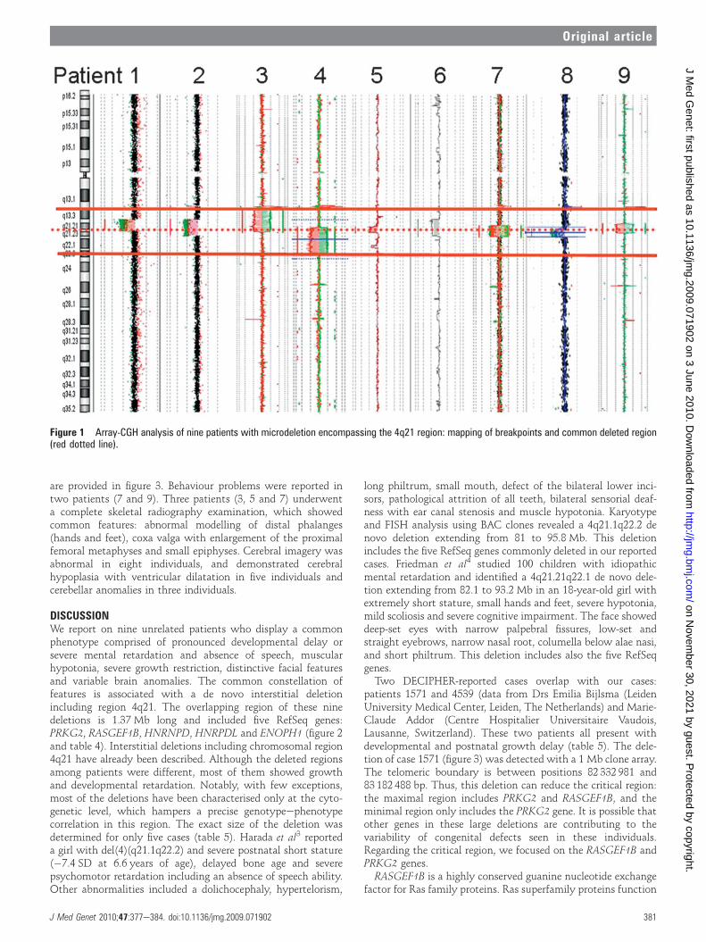
RESULTSArray-CGH analysisArray-CGH results (figure 1) are summed up in table 3 (NCBIMarch 2006 genome build). The shortest deletion is 3.2 Mb insize (patient 5) and the longest is 15.1 Mb in size (patient 4).The minimal critical region extends from 82 228 875 to83601 083 bp and includes five RefSeq genes: PRKG2, RASGEF1B,HNRNPD, HNRPDL and ENOPH1 (figure 2). For validationexperiments, the nine deletions were confirmed by FISH and/orqPCR in patients, while parents showed a normal result, provingthe de novo occurrence of the nine deletions. The deletedgenomic regions of the nine patients have never been describedas copy number polymorphism in the database of genomicvariants (http://projects.tcag.ca/variation/?source¼hg18). Exceptfor polymorphic regions, no copy number alterations were ob-served in other chromosomes (data not shown). None of thepatients shared common breakpoints. In addition, the absence ofsegmental duplications at the proximal and distal breakpointintervals suggested that these deletions were not generated bynon-allelic homologous recombination.
Clinical details of the studied subjectsIn three cases (patients 3, 5 and 8), the pregnancy was compli-cated by intrauterine growth retardation. The youngest patients(patients 1 and 2) still have measurements within the normalrange. Strikingly, a growth restriction combining postnatal shortstature with conserved head circumference was noted in theseven other patients. Small hands and small feet were reportedin the majority of cases (78%), while brachydactyly wasreported in four patients. In all patients, global psychomotordevelopmental delay was noted from an early age with a markedhypotonia. The level of developmental delay was estimated assevere. Three patients (3, 4 and 7) did not walk, and walkingability started after the age of 4 years in three other patients(6, 8 and 9). The speech and language development wereparticularly affected in all patients, with a profound speechdeficit in five patients (3, 4, 5, 6 and 7). Congenital sensorialdeafness was reported only in two individuals (1 and 5); thiscould be due to incomplete penetrance or to a second event onthe other allele. Distinctive facial features present in themajority of the individuals included a broad forehead, frontalbossing, hypertelorism, a short philtrum and downturnedcorners of the mouth. Facial photographs of seven individuals
Table 2 Primer pairs used for quantitative real-time PCR
Gene Forward primer (59/39) Reverse primer (59/39)
GK2 TTCTCAGGATGGAACGATTTGA TGTGGCATAACGAATTTCACTTTC
HNRPD GGTGAAGTTGTAGACTGCACTCTGA ACAAAGCCAAAACCCCTTGA
WDFY3 GGCTGCTCGGTGAGGTTTT GCTGACCACAGTTCCTGCAA
Table 3 Molecular features of patients with microdeletion encompassing the 4q21 region
Deletion Size (Mb) Start End Nomenclature
Patient 1 Agilent 105 K 4q21.1q21.22 6.6 76949115 83601083 arr 4q21.1q21.22(76949115�83601083)3 1
Patient 2 Agilent 105 K 4q21.1q21.23 9.6 77174381 86773353 arr 4q21.1q21.23(77174381�86773353)31
Patient 3 Agilent 105 K 4q13.3q21.22 13.5 70547247 84066553 arr 4q13.3q21.22 (70547247�84066553)31
Patient 4 Agilent 105 K 4q21.21q22.3 15.1 81706999 96867539 arr 4q21.21q22.3 (81706999�96867539)31
Patient 5 Agilent 44 K 4q21.21q21.22 3.2 80926692 84215893 arr 4q21.21q21.22 (80926692�84215893)31
Patient 6 Agilent 44 K 4q13.3q21.23 9.7 76077557 85788996 arr 4q13.3q21.23 (80293554�86615654)31
Patient 7 Agilent 105 K 4q21.21q21.23 6.3 80293554 86615654 arr 4q21.21q21.23 (80293554�86615654)31
Patient 8 Agilent 44 K 4q21.21q21.23 4.5 82228875 86711060 arr 4q21.21q21.23 (82228875�86711060)31
Patient 9 Agilent 105 K 4q21.1q21.23 5.5 78995025 84459549 arr 4q21.1q21.23 (78995025�84459549)31
Data are from the National Center for Biotechnology Information March 2006 genome build.
380 J Med Genet 2010;47:377e384. doi:10.1136/jmg.2009.071902
Original article
on Novem
ber 30, 2021 by guest. Protected by copyright.
http://jmg.bm
j.com/
J Med G
enet: first published as 10.1136/jmg.2009.071902 on 3 June 2010. D
ownloaded from
are provided in figure 3. Behaviour problems were reported intwo patients (7 and 9). Three patients (3, 5 and 7) underwenta complete skeletal radiography examination, which showedcommon features: abnormal modelling of distal phalanges(hands and feet), coxa valga with enlargement of the proximalfemoral metaphyses and small epiphyses. Cerebral imagery wasabnormal in eight individuals, and demonstrated cerebralhypoplasia with ventricular dilatation in five individuals andcerebellar anomalies in three individuals.
DISCUSSIONWe report on nine unrelated patients who display a commonphenotype comprised of pronounced developmental delay orsevere mental retardation and absence of speech, muscularhypotonia, severe growth restriction, distinctive facial featuresand variable brain anomalies. The common constellation offeatures is associated with a de novo interstitial deletionincluding region 4q21. The overlapping region of these ninedeletions is 1.37 Mb long and included five RefSeq genes:PRKG2, RASGEF1B, HNRNPD, HNRPDL and ENOPH1 (figure 2and table 4). Interstitial deletions including chromosomal region4q21 have already been described. Although the deleted regionsamong patients were different, most of them showed growthand developmental retardation. Notably, with few exceptions,most of the deletions have been characterised only at the cyto-genetic level, which hampers a precise genotypeephenotypecorrelation in this region. The exact size of the deletion wasdetermined for only five cases (table 5). Harada et al3 reporteda girl with del(4)(q21.1q22.2) and severe postnatal short stature(�7.4 SD at 6.6 years of age), delayed bone age and severepsychomotor retardation including an absence of speech ability.Other abnormalities included a dolichocephaly, hypertelorism,
long philtrum, small mouth, defect of the bilateral lower inci-sors, pathological attrition of all teeth, bilateral sensorial deaf-ness with ear canal stenosis and muscle hypotonia. Karyotypeand FISH analysis using BAC clones revealed a 4q21.1q22.2 denovo deletion extending from 81 to 95.8 Mb. This deletionincludes the five RefSeq genes commonly deleted in our reportedcases. Friedman et al4 studied 100 children with idiopathicmental retardation and identified a 4q21.21q22.1 de novo dele-tion extending from 82.1 to 93.2 Mb in an 18-year-old girl withextremely short stature, small hands and feet, severe hypotonia,mild scoliosis and severe cognitive impairment. The face showeddeep-set eyes with narrow palpebral fissures, low-set andstraight eyebrows, narrow nasal root, columella below alae nasi,and short philtrum. This deletion includes also the five RefSeqgenes.Two DECIPHER-reported cases overlap with our cases:
patients 1571 and 4539 (data from Drs Emilia Bijlsma (LeidenUniversity Medical Center, Leiden, The Netherlands) and Marie-Claude Addor (Centre Hospitalier Universitaire Vaudois,Lausanne, Switzerland). These two patients all present withdevelopmental and postnatal growth delay (table 5). The dele-tion of case 1571 (figure 3) was detected with a 1 Mb clone array.The telomeric boundary is between positions 82 332 981 and83 182 488 bp. Thus, this deletion can reduce the critical region:the maximal region includes PRKG2 and RASGEF1B, and theminimal region only includes the PRKG2 gene. It is possible thatother genes in these large deletions are contributing to thevariability of congenital defects seen in these individuals.Regarding the critical region, we focused on the RASGEF1B andPRKG2 genes.RASGEF1B is a highly conserved guanine nucleotide exchange
factor for Ras family proteins. Ras superfamily proteins function
Figure 1 Array-CGH analysis of nine patients with microdeletion encompassing the 4q21 region: mapping of breakpoints and common deleted region(red dotted line).
J Med Genet 2010;47:377e384. doi:10.1136/jmg.2009.071902 381
Original article
on Novem
ber 30, 2021 by guest. Protected by copyright.
http://jmg.bm
j.com/
J Med G
enet: first published as 10.1136/jmg.2009.071902 on 3 June 2010. D
ownloaded from

Figure 2 Map of the deleted region (National Center for Biotechnology Information March 2006 genome build). (A) Region 4q13.2q22.3. (B) Zoomedview showing the 4q21.21q21.22 region.
382 J Med Genet 2010;47:377e384. doi:10.1136/jmg.2009.071902
Original article
on Novem
ber 30, 2021 by guest. Protected by copyright.
http://jmg.bm
j.com/
J Med G
enet: first published as 10.1136/jmg.2009.071902 on 3 June 2010. D
ownloaded from

as molecular switches in fundamental events such as signaltransduction, cytoskeleton dynamics and intracellular traf-ficking. RASGEF1B shows a high level of central nervous systemexpression (Genecards) and is, therefore, a good candidate for thecentral nervous system phenotype. A growing number of genesthat are related to mental retardation have been involved inRho-GTPase signalling pathways.5 Among them, ARHGEF6 andFGD1 encode guanine nucleotide exchange factors and regulateRac and Cdc42, two members of the Rho family of smallGTPases, which are considered as key regulators of actin andmicrotubule dynamics during both dendrite and spine structuralplasticity.6
PRKG2 encodes cGMP-dependent protein kinase type II(cGKII). cGKII is a membrane-bound kinase, which is activatedby intracellular cGMP, and is known to be expressed abundantlyin the intestinal mucosa, kidney, lung, brain and cartilage. Pfeiferet al7 generated mice carrying a null mutation of the cGKII gene(cGKII�/� mice); these mice developed postnatal dwarfismthat was caused by a severe defect in endochondral ossificationat the growth plates and impaired chondrocyte hypertrophy.Chikuda et al8 described the Komeda miniature rat Ishikawa.This rat is a naturally occurring mutant caused by an autosomalrecessive mutation mri. They identified the mri mutation asa deletion in cGKII. The Komeda miniature rat Ishikawaexhibited longitudinal growth retardation and showed expandedgrowth plates.
Kugimiya et al9 and then Kawasaki et al10 investigated themechanism of cGKII-mediated chondrocyte hypertrophy. cGKIIphosphorylates glycogen synthase kinase-3b (GSK-3b). Phos-phorylation inactivates GSK-3b. GSK-3b is a negative regulatorof b-catenin through its phosphorylation and degradation.When GSK-3b is phosphorylated and inactivated, b-catenin level
increases and b-catenin can go into the nucleus to enhancechondrocyte hypertrophy. cGKII also phosphorylates Sox9, aninhibitor of chondrocyte hypertrophy, and suppresses its nuclearentry, leading to enhancement of chondrocyte hypertrophy.Chondrocyte hypertrophy in the growth plates is a rate-limitingstep for longitudinal skeletal growth. In cGKII�/� mice, chon-drocyte hypertrophy and elongation of growth plates areimpaired, resulting in postnatal dwarfism. These experimentaldata provide support to the hypothesis that haploinsufficiencyof PRKG2 could explain severe growth delay observed in thereported patients. Additionally, Ulher11 showed that cGKIItranscript is abundant in brain. Serulle et al12 13 also showed thatcGKII and GluR1 form a complex in brain; more specifically inthis complex, cGKII can phosphorylate GluR1, leading toincreased GluR1 surface levels and to the surface expression ofAMPA receptors at extrasynaptic sites. This mechanism plays animportant role in long-term potentiation in the hippocampalneurons, a critical step involved in learning and memory. GluR1accumulation in the plasma membrane also plays a pivotal rolein synaptic plasticity. Therefore, PRKG2 haploinsufficiencycould also participate to the severe cognitive developmentaldelay reported in patients with 4q21 microdeletion.In summary, we have characterised a novel microdeletion
syndrome at chromosome 4q21 with a recognisable clinicalphenotype including severe mental retardation, absent speech,distinctive facial features and severe growth delay. In theminimal critical region, we identified PRKG2 and RASGEF1B asmajor determinants of the 4q21 deletion phenotype. Furtherdetailed clinical examination of additional patients witha similar microdeletion is needed to get more insight into thephenotype and to establish guidelines for anticipatory health-care management across the lifespan.
Figure 3 Front and lateral views of seven patients with microdeletion encompassing the 4q21 region. (A and B) Patient 1; (C and D) patient 2; (E andF) patient 3; (G and H) patient 4; (I and J) patient 5; (K and L) patient 7; (M) patient 9. Parental informed consent was obtained for the publication ofthis figure.
Table 4 List of genes within the common deleted region
Start Stop Symbol Model evidence Cyto Description
82228861 82345239 PRKG2 Best RefSeq 4q13.1q21.1 Protein kinase, cGMP-dependent, type II
82567243 82612085 RASGEF1B Best RefSeq 4q21.21q21.22 RasGEF domain family, member 1B
83493491 83514173 HNRPD Best RefSeq 4q21.1q21.2 Heterogeneous nuclear ribonucleoprotein D (AU-richelement RNA binding protein 1.37 kDa)
83563371 83570402 HNRPDL Best RefSeq 4q13q21 Heterogeneous nuclear ribonucleoprotein D-like
83570787 83601264 ENOPH1 Best RefSeq 4q21.22 Enolaseephosphatase 1
Data are from http://www.ncbi.nlm.nih.gov/mapview/maps.cgi?ORG¼hum, build 36.3.
J Med Genet 2010;47:377e384. doi:10.1136/jmg.2009.071902 383
Original article
on Novem
ber 30, 2021 by guest. Protected by copyright.
http://jmg.bm
j.com/
J Med G
enet: first published as 10.1136/jmg.2009.071902 on 3 June 2010. D
ownloaded from

Author affiliations:1Laboratoire de Genetique, EA 4368-IFR111, Nancy Universite, Centre Hospitalier etUniversitaire (CHU) de Nancy, Nancy, France2Laboratoire de Genetique Medicale, Hopital Jeanne de Flandre, CHU de Lille, Lille,France3Service de Medecine Infantile III et Genetique Clinique, CHU de Nancy, Nancy, France4Service de Genetique Clinique, Hopital Jeanne de Flandre, CHU de Lille, Lille, France5Centre de Genetique Chromosomique, Hopital St Vincent de Paul, Lille, France6UF de Cytogenetique, Centre de Gynecologie-Obstetrique, Hopital Nord, Amiens,France7Service de Pediatrie et Genetique, Hopital Nord, Amiens, France8Service de Pediatrie, CHU de Besancon, Besancon, France9CHU de Nantes, Service de Genetique Medicale, Nantes, France, et INSERM UMR915, Institut du Thorax, Nantes, France10Service de Cytogenetique, Hopital Necker Enfants Malades, Paris, France
11Departement de Genetique, Hopital Necker Enfants Malades, Paris, France12INSERM U781, Hopital Necker Enfants Malades, Paris, France13Service de Cytogenetique, Hopitaux Universitaires de Strasbourg, Strasbourg, France14Departement de Genetique, Hopitaux Universitaires de Strasbourg, Strasbourg,France15Centre de Genetique et Centre de Reference Maladies Rares Anomalies duDeveloppement et Syndromes Malformatifs, Hopital d’Enfants, Dijon, France16Service de Neurologie pediatrique, Hopital Bicetre, Paris, France17Service de Neuropediatrie, Hopital Necker-Enfants Malades, Paris, France18Service de Cytogenetique, CHU de Dijon, Dijon, France19Service de Cytogenetique Constitutionnelle, CBPE, Hospices Civils de Lyon, Bron,France
Acknowledgements We wish to express our sincere gratitude to all the patientsand their family who have participated in this study. We also thank the cytogeneticsand molecular genetics staff from the different medical genetics departments. Thisstudy was supported by grants from the French Ministry of Health(DHOS-2007e2008).
Funding Other funders: French Ministry of Health (DHOS).
Competing interests None.
Patient consent Obtained.
Ethics approval This study was conducted with the approval of the universityhospitals.
Provenance and peer review Not commissioned; externally peer reviewed.
REFERENCES1. Slavotinek AM. Novel microdeletion syndromes detected by chromosome
microarrays. Hum Genet 2008;124:1e17.2. Jacquemont ML, Sanlaville D, Redon R, Raoul O, Cormier-Daire V, Lyonnet S, Amiel
J, Le Merrer M, Heron D, de Blois MC, Prieur M, Vekemans M, Carter NP, MunnichA, Colleaux L, Philippe A. Array-based comparative genomic hybridisation identifieshigh frequency of cryptic chromosomal rearrangements in patients with syndromicautism spectrum disorders. J Med Genet 2006;43:843e9.
3. Harada N, Nagai T, Shimokawa O, Niikawa N, Matsumoto N. A 4q21-q22 deletionin a girl with severe growth retardation. Clin Genet 2002;61:226e8.
4. Friedman JM, Baross A, Delaney AD, Ally A, Arbour L, Armstrong L, Asano J, BaileyDK, Barber S, Birch P, Brown-John M, Cao M, Chan S, Charest DL, Farnoud N,Fernandes N, Flibotte S, Go A, Gibson WT, Holt RA, Jones SJ, Kennedy GC,Krzywinski M, Langlois S, Li HI, McGillivray BC, Nayar T, Pugh TJ, Rajcan-Separovic E,Schein JE, Schnerch A, Siddiqui A, Van Allen MI, Wilson G, Yong SL, Zahir F, EydouxP, Marra MA. Oligonucleotide microarray analysis of genomic imbalance in childrenwith mental retardation. Am J Hum Genet 2006;79:500e13.
5. Chelly J, Khelfaoui M, Francis F, Cherif B, Bienvenu T. Genetics andpathophysiopathology of mental retardation. Eur J Hum Genet 2006;14:701e13.
6. Newey SE, Velamoor V, Govek EE, Van Aelst L. RhoGTPases, dendritic structure,and mental retardation. J Neurobiol 2005;64:58e74.
7. Pfeifer A, Aszodi A, Seidler U, Ruth P, Hofmann F, Fassler R. Intestinal secretorydefects and dwarfism in mice lacking cGMP-dependent protein kinase II. Science1996;274:2082e6.
8. Chikuda H, Kugimiya F, Hoshi K, Ikeda T, Ogasawara T, Shimoaka T, Kawano H,Kamekura S, Tsuchida A, Yokoi N, Nakamura K, Komeda K, Chung UI, Kawaguchi H.Cyclic GMP-dependent protein kinase II is a molecular switch from proliferation tohypertrophic differentiation of chondrocytes. Genes Dev 2004;18:2418e29.
9. Kugimiya F, Chikuda H, Kamekura S, Ikeda T, Hoshi K, Ogasawara T, Nakamura K,Chung UI, Kawaguchi H. Involvement of cyclic guanosine monophosphate-dependentprotein kinase II in chondrocyte hypertrophy during endochondral ossification. ModRheumatol 2005;15:391e6.
10. Kawasaki Y, Kugimiya F, Chikuda H, Kamekura S, Ikeda T, Kawamura N, Saito T,Shinoda Y, Higashikawa A, Yano F, Ogasawara T, Ogata N, Hoshi K, Hofmann F,Woodgett JR, Nakamura K, Chung UI, Kawaguchi H. Phosphorylation of GSK-3betaby cGMP-dependent protein kinase II promotes hypertrophic differentiation of murinechondrocytes. J Clin Invest 2008;118:2506e15.
11. Uhler MD. Cloning and expression of a novel cyclic GMP-dependent protein kinasefrom mouse brain. J Biol Chem 1993;268:13586e91.
12. Serulle Y, Zhang S, Ninan I, Puzzo D, McCarthy M, Khatri L, Arancio O, Ziff EB.A GluR1-cGKII interaction regulates AMPA receptor trafficking. Neuron2007;56:670e88.
13. Serulle Y, Arancio O, Ziff EB. A role for cGMP-dependent protein kinase II in AMPAreceptor trafficking and synaptic plasticity. Channels (Austin) 2008;2:230e2.
Table 5 Clinical features of individuals with overlapping microdeletionpreviously reported in the literature
Patient 10DECIPHER1571
Patient 11DECIPHER4539
Patient 12Friedmanet al4
Patient 13Haradaet al3
Deletion 4q13.2q21.21 4q21.21q21.23 4q21.21q22.1 4q21.1q22.2
Size (Mb) 15 3.2 11 14.8
Distinctive facialdysmorphism
ND + ND +
(a) Frontal bossing,broad forehead
�
(b) Hypertelorism +
(c) Short philtrum + �Developmentaldelay/severe mentalretardation
ND + + +
(a) Severely delayedspeech
+ + ND +
(b) Neonatalhypotonia
ND + + +
Measurementabnormalities
+ + + +
(a) IUGR ND � ND �(b) Postnatal growthdelay
+ + + +�7.4 SD
(c) Conserved headcircumference
ND + ND ND
(d) Small hands andsmall feet
+ +
(e) Brachydactyly
Cerebral imagery ND ND ND ND
+, Feature present; �, feature absent; ND, no data available.IUGR (intrauterine growth retardation).
Web resources
< NCBI Map Viewer: http://www.ncbi.nlm.nih.gov/mapview/maps.cgi?ORG¼hum
< UCSC: Santa Cruz Human Genome Browser: http://www.genome.ucsc.edu
< DGV: Database of Genomic Variants: http://projects.tcag.ca/variation/
< DECIPHER: DatabasE of Chromosomal Imbalance and Pheno-type in Humans using Ensembl Resources: https://decipher.sanger.ac.uk/
< GENECARDS: http://www.genecards.org/
384 J Med Genet 2010;47:377e384. doi:10.1136/jmg.2009.071902
Original article
on Novem
ber 30, 2021 by guest. Protected by copyright.
http://jmg.bm
j.com/
J Med G
enet: first published as 10.1136/jmg.2009.071902 on 3 June 2010. D
ownloaded from