microct image based simulation to design heating … filemicroct image based simulation to design...
TRANSCRIPT
Journal of Thermal Biology 62 (2016) 129–137
Contents lists available at ScienceDirect
Journal of Thermal Biology
http://d0306-45
n CorrE-m
journal homepage: www.elsevier.com/locate/jtherbio
MicroCT image based simulation to design heating protocols inmagnetic nanoparticle hyperthermia for cancer treatment
Alexander LeBrun, Ronghui Ma, Liang Zhu n
Department of Mechanical Engineering, University of Maryland Baltimore County, Baltimore, MD 21250, United States
a r t i c l e i n f o
Article history:Received 2 February 2016Accepted 29 June 2016Available online 7 July 2016
Keywords:Magnetic nanoparticle hyperthermiaInjection strategyImage-based simulationHeating protocol designBioheat transfer
x.doi.org/10.1016/j.jtherbio.2016.06.02565/& 2016 Elsevier Ltd. All rights reserved.
esponding author.ail address: [email protected] (L. Zhu).
a b s t r a c t
Objectives: The objective is to design heating protocols to completely damage PC3 tumors after a singlemagnetic nanoparticle hyperthermia session with minimal collateral thermal damage, based on microCTimage generated tumor and mouse models.Methods: Tumor geometries and volumetric heat generation rate distributions that are generated frommicroCT scans in our previous study are imported into COMSOL 4.3s multiphysics for heat transfersimulations and heating protocol design using the Arrhenius damage model. Then, parametric studies areperformed to evaluate how significantly the infusion rate affects the protocol design and its resultedcollateral thermal damage.Results: The simulated temperature field in the generated tumor geometry and volumetric heat gen-eration rate distribution are reasonable and correlates well with the amount of the total thermal energydeposited into the tumors. The time needed for complete thermal damage is determined to be ap-proximately 12 min or 25 min if one uses the Arrhenius integral Ω equal to 1 or 4 as the damagethreshold, when the infusion rate is 3 μL/min. The heating time increases 26% or 91% in the higherinfusion rate groups of 4 or 5 μL/min, respectively. Collateral thermal damage to the surrounding tissue isalso assessed. Although the two larger infusion rate groups can still cause thermal damage to the entiretumor, the collateral thermal damage would have exceeded the design criterion of 5%, while the as-sessment criterion is acceptable only in the infusion rate group of 3 μL/min. Based on the results of thisstudy, we identify an injection strategy and heating protocols to be implemented in future animal ex-periments to evaluate treatment efficacy for model validation.
& 2016 Elsevier Ltd. All rights reserved.
1. Introduction
Currently, the main treatment options for cancer are surgery,radiation, and chemotherapy. Although those traditional inter-ventions have increased the five-year survival rate in cancer pa-tients, none of them have been able to consistently kill or removeall cancerous tissues at the original site. The remaining tumor cellsmay contribute to tumor recurrence and/or cancer metastasis. Thissuggests a need to develop an effective treatment strategy toeliminate all cancerous tissues while ensuring safety of the sur-rounding healthy tissue.
In the past decade, it has been demonstrated that magneticnanoparticles can deliver confined thermal energy to tumorswhen subject to an alternating magnetic field, therefore holding ahigh cell-killing potential while minimizing collateral thermaldamage to the surrounding tissue in cancer treatment (Ito et al.,
2003; Johannsen et al., 2005, 2007; Jordan et al., 2006; Wust et al.,2006; Maier-Hauff et al., 2007; Zhao et al., 2012). Existing studiesalso suggest that nanoparticle distribution in tumors is a majorfactor in determining treatment efficacy (Wust et al., 2006; Jo-hannsen et al., 2007; Attaluri et al., 2011). Recent experimentalstudies using microCT and other imaging techniques have pro-vided strong evidence that nanoparticle distributions in tumorsare often unpredictable (Kalambur et al., 2005; Johannsen et al.,2007; Attaluri et al., 2011; LeBrun et al., 2013; Wabler et al., 2014).In systemic delivery of nanoparticles to tumor sites, vascular per-meability, local blood perfusion distribution, and particle pene-tration in the tumor region are often difficult to quantify (Kongand Dewhirst, 1999; Urono et al., 1999; McGuire and Yuan, 2001;Liu et al., 2005; Dreher et al., 2006). In intratumoral injections totumors, nanoparticle deposition in tumors is mainly determinedby diffusion and advection of the carrier solution in heterogeneousmicrostructure of tumors. Unfortunately, particle distribution intumors is difficult to predict due to the heterogeneous tumorporosity, micro-crack formation during injection, and the random

A. LeBrun et al. / Journal of Thermal Biology 62 (2016) 129–137130
nature of particle interactions with cells in the extracellular matrixsuch as particle deposition on the cell surface, intercellular uptake,and particle aggregation. In addition, tumors in clinical settingsmay have various sizes and shapes, which result in different na-noparticle distribution from one tumor to another. It is therefore,important to design individualized heating protocols based onimage-generated tumor geometry and nanoparticle distribution sothat the spatial tumor temperature elevations can be preciselycontrolled in hyperthermia treatment. The image-based designapproaches need to be verified via carefully designed in vivo ani-mal studies before applied to clinical settings.
We have shown in a previous study that microCT imagingtechnology can be utilized to determine nanoparticle distributionvolume in opaque PC3 tumors after injecting 0.1 cc commerciallyavailable ferrofluid at the tumor center (LeBrun et al., 2016). In thatstudy, the nanoparticle distribution volume is used to evaluatehow far the nanoparticles spread by average from the injection siteof the tumor. It has been shown that a low intratumoral injectionrate minimizes variation of the nanoparticle distribution volume.Therefore, an injection strategy has been identified to result inrelatively repeatable and controllable nanoparticle distributionpatterns in PC3 tumors. It is expected that one will obtain similarnanoparticle distribution volume in the same type of tumors, ifone implements the same injection strategy. In addition, the mi-croCT imaging system has been utilized to generate tumor geo-metries and volumetric heat generation rate distribution, settingthe foundation for designing individualized treatment protocols.
Accuracy of simulated temperature distributions in tumors in-duced by magnetic nanoparticles relies on precise description oftumor geometry and its surroundings, tissue thermal and phy-siological properties, and local concentration distribution of na-noparticles. Therefore, realistic and accurate models of the tumorgeometry and nanoparticle distribution are imperative to guideclinicians to achieve effective and safe treatment through devel-opment of individualized heating protocols. In this study, tumorgeometries and volumetric heat generation rate distributionsgenerated in our previous study (LeBrun et al., 2016) are importedinto COMSOL 4.3s multiphysics (Stockholm, Sweden) for heattransfer simulations and heating protocol design. The goal of thisstudy is to estimate the time it takes to completely damage thetumor after a single heating session and to evaluate the extent ofcollateral thermal damage. Then, parametric studies are per-formed to evaluate how significant the infusion rate affects theprotocol design and resultant collateral thermal damage.
2. Methods and materials
2.1. Importing tumor geometry, mouse model, and heat generationrate distribution
The tumor geometry generated in our previous study (LeBrunet al., In press) was imported into COMSOL 4.3s multiphysics viathe Pro/Engineer LiveLink™ interface. Using the built-in tools ofCOMSOL, the tumor was scaled to the appropriate size and prop-erly oriented to coincide with the imported files. Then a mousemodel generated in Pro/Engineer based on microCT scans of amouse body was used as a mounting surface for the tumor(Manuchehrabadi et al., 2013). The mouse model was smoothedand processed using the same tools as that to generate the tumorgeometry, allowing for meshing while maintaining the originalvolume and shape. The hind and forepaws, and the tail weresimplified in the model to avoid any singularities during themeshing process. The tumor was mounted in a way so that the flatarea of the tumor created during resection was the contact surfacewith the mouse body. Fig. 1 illustrates views of a tumor implanted
on a mouse body imported to COMSOL. A meshed mouse modelwith the embedded tumor for heat transfer simulation is shown inFig. 2.
As described in our previous study, the ferrofluid infusion ratestrongly influences the resulted nanoparticle distribution in thePC3 tumors. In this study, three volumetric heat generation ratedistributions representing possible nanoparticle distribution in thethree infusion rate groups were evaluated and tested. For eachinfusion rate, its volumetric heat generation rate distribution wasimported into COMSOL as a source file. Note that the center of thecoordinate system was adjusted so that it was consistent to that inthe COMSOL geometry of the mouse model. Linear interpolationand constant extrapolation were used between elements toachieve a smooth volumetric heat generation rate distribution asclose to the original volumetric heat generation distribution aspossible without incurring singularities.
2.2. Heat transfer modeling in COMSOL
The Pennes bioheat equation (Pennes, 1948) was employed inthe modeling of the transient temperature elevations in tissue.This is a continuum model that has been used extensively inmodeling thermal effect of local blood perfusion in biological tis-sue (Zhu, 2010). The Pennes bioheat equation was applied to boththe mouse body and tumor, shown as:
( )ρ ω ρ∂∂
= ∇ + − + ( )cTt
k T c T T Q 1t tt
t t t b b b t met t2
,
( )ρ ω ρ∂∂
= ∇ + − + + ( )cTt
k T c T T Q Q 2c cc
c c c b b b c met c MNH2
,
where k is thermal conductivity (W/m K), ρ is density (kg/m3), c isspecific heat (J/kg K), Tb is the prescribed arterial blood tempera-ture equal to 37 °C, ω is the local blood perfusion rate (1/s), andQmet is the local metabolic heat generation rate (W/m3). The sub-scripts t, b, and c denote tissue, blood, and tumor tissue, respec-tively. The nanoparticle induced volumetric heat generation rate(W/m3), or QMNH, was only applied to the tumor region since it isassumed that the nanoparticles are confined into the tumor re-gion. Thermal and physiological properties used in the model arepresented in Table 1. It was assumed that the thermal and phy-siological properties remained constant and isotropic within eachdomain to simplify the model (Rylander et al., 2006; Trakic et al.,2006). The temperature dependency of the tumor blood perfusionrate was considered in this study (Lang et al., 1999), and shown as:
⎧
⎨⎪⎪
⎩⎪⎪
⎫
⎬⎪⎪
⎭⎪⎪
⎛⎝⎜
⎞⎠⎟
( )ω =
× < °
× −−
×° ≤ ≤ °
× > ° ( )
−
−
−
T
TT
T
s
0. 833 10 37 C
0. 833 1037
5. 438 1037 C 42 C
0. 416 10 42 C
1
3
c
3
34.8
3
3
As the temperature increases, the tumor blood perfusion ratedecreases. The temperature dependency of the metabolic heatgeneration rate was not considered since the effect has not beenwell documented for the temperature range in this study.
2.3. Boundary conditions
The mouse body was considered to be exposed to a naturalconvection condition where the ambient air temperature wasprescribed as 25 °C and the heat transfer coefficient was 10 W/m2 K (Manuchehrabadi et al., 2013). The bottom of the mousemodel was prescribed as a uniform temperature of 37 °C to si-mulate the effect of the heating pad. A convective boundary

3.5 cmFig. 1. (a) A simplified mouse model with an implanted tumor and (b) the head-on view of the model.
h = 10 W/m2·KTair = 25°C
h = 3.7 – 4.2 W/m2·KTair = 25°C
T = 37°C
Fig. 2. The mouse model with the embedded tumor after meshing and the pre-scribed boundary conditions.
Table 1Material, thermal, and physiological properties of PC3 tumors and tissue (Trakicet al., 2006).
Parameter Symbol Value
Density ρt, ρc, ρb 1000 (kg/m3)Specific heat ct, cc, cb 3500 (J/kg K)Thermal conductivity kt, kc 0.642 (W/m K)Metabolic heat generation of tissue Qmet,t 25,829 (W/m3)Metabolic heat generation of tumor Qmet,c 2708 (W/m3)Blood perfusion rate of tissue ωt 0.003 (1/s)
Table 2Arrhenius parameters for PC3 tumors (Rylander et al., 2006).
Parameter Symbol Temperature (°C) PC3 Cells Tissue
Frequency factor A Tr54 1.8�1036 6.36�1019
(1/s) T454 7�1017 –
Activation energy Ea Tr54 2.38�105 1.38�105
(J/mol) T454 1.24�105 –
A. LeBrun et al. / Journal of Thermal Biology 62 (2016) 129–137 131
condition was also applied to the tumor using a correlation for asphere:
= ( )hNu k
D 4aD av air,
⎧⎨⎪⎪
⎩⎪⎪ ⎡
⎣⎢⎤⎦⎥
⎫⎬⎪⎪
⎭⎪⎪( )
= ++
( )
NuRa
Pr0. 60
0. 387
1 0. 59/4b
D avD
,
1/6
9/16 8/27
2
where NuD, av is the Nusselt number with respect to the averagediameter of the tumor, h is the heat transfer coefficient, D is theaverage diameter of the tumor, kair is the thermal conductivity of
air at 25 °C (0.028 W/m K), RaD is the Rayleigh number, and Pr isthe Prandtl number with a value of 0.707. This approximation wasused since the heat transfer coefficient for the exact tumor geo-metry would be difficult to determine. PC3 tumors tend to favormore hemispherical growth (Rylander et al., 2006). Typical valuesof the heat transfer coefficient h calculated from this study rangedfrom 3.7 to 4.2 W/m2 K, depending on the size of the tumor.
2.4. Numerical simulation parameters
The mesh was generated by COMSOL 4.3 and it was finer insidethe tumor with a growth factor of 1.4 starting from the tumorboundary towards the normal tissue. The maximum element sizewas 0.008 m while the minimum element size was 0.0005 m. Thetotal number of the tetrahedral elements using the fine meshsetting was 146,062. The mesh sensitivity was checked via in-creasing the mesh setting to extremely fine meshing with an in-crease in the total number of elements to 644,035. The four-foldincrease in the mesh elements resulted in a difference of less than0.1 °C in the average temperature in the tumor. The temporal re-solution was selected as 0.01 s. Lowering the temporal resolutionby half resulted in a less than 0.1% change in the average tumortemperature.
2.5. Thermal damage assessment
Thermal damage in tissue was assessed by a first-order ther-mal-chemical rate equation coupled with the Pennes bioheatequation simulating the temperature field. Although it is widelyrecognized that tissue injury is the result of complicated reactionmechanisms, progression of thermal injury can be considered as auni-molecular process where native molecules are transformedinto a coagulated/denatured state leading to cell death (Marqaet al., 2011; Dewhirst et al., 2015). The temperature-time historydetermines the extent of thermal damage at a specific location (x,y, z). It is quantified using a parameter, Ω, calculated from theArrhenius equation:
A. LeBrun et al. / Journal of Thermal Biology 62 (2016) 129–137132
⎡⎣⎢⎢
⎤⎦⎥⎥ ∫( ) ( )τ
ςς τ
Ω( ) =( )
=( )
τ −x y z ln A e dt, , ,
0
5
ER T x y z t
0, , ,a
u t
where ς(0) is the initial concentration of healthy cells, ς(τ) is theconcentration of healthy cells remaining after heating of a dura-tion τ (seconds), A is the frequency factor (1/s), Ea is the activationenergy (J/mol), Ru is the universal gas constant (8.23 J/mol K), andTt(x,y,z,t) is the absolute tissue temperature at a given location. Thevalues for the frequency factor and activation energy are cell lineand tissue dependent. In this study, the temperature dependencyof the frequency factor and activation energy for PC3 cells wasfrom a previous study and listed in Table 2. Before treatment, Ω iszero, it then increases with heating. Based on Eq. (5), 63% and 99%of the cells are damaged (denaturation of proteins occurs), whenΩ is 1 and 4, respectively.
2.6. Criteria for designing treatment protocols
One of the features of COMSOL software is its multiphysicsplatform. In this study, calculation of the thermal damage para-meter,Ω, is implemented in the COMSOL software to solve for thediscretized form of Eq. (5), and this equation can be solved si-multaneously with the temperature field in the tumor and itssurrounding tissue to determine the progression of the thermaldamage distribution with time.
The goal for designing treatment protocols of hyperthermiatherapy is to predict the extent of heating-induced cell death. Anideal thermal therapy for cancer treatment is to induce irreversibledamage to the cancerous cells to prevent tumor recurrence ormetastasis, and to preserve the surrounding healthy tissue. In or-der to identify a treatment protocol, two criteria are proposed tobe satisfied in this study: 100% damage to the tumor cells and aratio of the damaged normal tissue to the tumor volume less than5%. Previous studies usually used a percentage of damage in thenormal tissue to a defined normal tissue volume (Manuchehrabadiand Zhu, 2014). It seems that the definition of normal tissue vo-lume is very arbitrary and subjective in previous studies. Sincethere is no established standard for an acceptable amount of col-lateral thermal damage, our damage criterion is defined as theratio of the damaged normal tissue to the known tumor volume.The percentage will be calculated by the following equation:
⎛⎝⎜
⎞⎠⎟= ×
( )V
V
V% 100%
6damage
damagednormaltissue
tumor
where Vdamaged normal tissue is the volume of thermally damaged
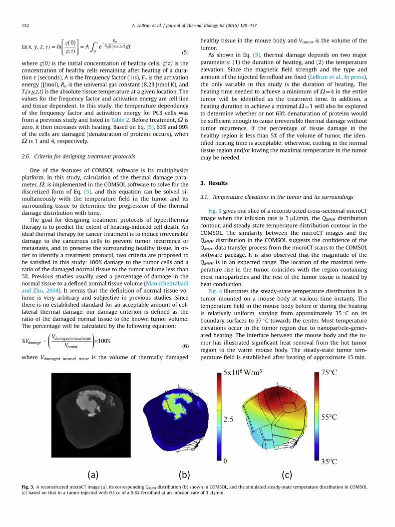
Fig. 3. A reconstructed microCT image (a), its corresponding QMNH distribution (b) show(c) based on that in a tumor injected with 0.1 cc of a 5.8% ferrofluid at an infusion rate
healthy tissue in the mouse body and Vtumor is the volume of thetumor.
As shown in Eq. (5), thermal damage depends on two majorparameters: (1) the duration of heating, and (2) the temperatureelevation. Since the magnetic field strength and the type andamount of the injected ferrofluid are fixed (LeBrun et al., In press),the only variable in this study is the duration of heating. Theheating time needed to achieve a minimum of Ω¼4 in the entiretumor will be identified as the treatment time. In addition, aheating duration to achieve a minimal Ω¼1 will also be exploredto determine whether or not 63% denaturation of proteins wouldbe sufficient enough to cause irreversible thermal damage withouttumor recurrence. If the percentage of tissue damage in thehealthy region is less than 5% of the volume of tumor, the iden-tified heating time is acceptable; otherwise, cooling in the normaltissue region and/or lowing the maximal temperature in the tumormay be needed.
3. Results
3.1. Temperature elevations in the tumor and its surroundings
Fig. 3 gives one slice of a reconstructed cross-sectional microCTimage when the infusion rate is 3 μL/min, the QMNH distributioncontour, and steady-state temperature distribution contour in theCOMSOL. The similarity between the microCT images and theQMNH distribution in the COMSOL suggests the confidence of theQMNH data transfer process from the microCT scans to the COMSOLsoftware package. It is also observed that the magnitude of theQMNH is in an expected range. The location of the maximal tem-perature rise in the tumor coincides with the region containingmost nanoparticles and the rest of the tumor tissue is heated byheat conduction.
Fig. 4 illustrates the steady-state temperature distribution in atumor mounted on a mouse body at various time instants. Thetemperature field in the mouse body before or during the heatingis relatively uniform, varying from approximately 35 °C on itsboundary surfaces to 37 °C towards the center. Most temperatureelevations occur in the tumor region due to nanoparticle-gener-ated heating. The interface between the mouse body and the tu-mor has illustrated significant heat removal from the hot tumorregion to the warm mouse body. The steady-state tumor tem-perature field is established after heating of approximate 15 min.
n in COMSOL, and the simulated steady-state temperature distribution in COMSOLof 3 μL/min.

75°C
35°C
55°C
Fig. 4. Slices of the temperature fields in the tumor and mouse body: (a) before heating, (b) 3 min, (c) 6 min, (d) 9 min, (e) 12 min, and (f) 15 min after the heating starts.Note that the steady-state temperature field is established after 15 min.
A. LeBrun et al. / Journal of Thermal Biology 62 (2016) 129–137 133
3.2. Designing heating protocols for individual tumors
Once the transient temperature distribution is determined, it isthen used in the Arrhenius integral to determine the distributionof the damage parameter, Ω. Fig. 5 shows the damages on onecross-sectional plane of a tumor before treatment and after 5, 12,15, 20, and 25 min of heating, respectively. The void region re-presents the damaged tumor region when ΩZ4. It can be clearlyseen that before the steady-state temperature field is established,not much damage occurs in the tumor. As the heating time in-creases and heat conducts throughout the tissue, more and moredamage in the tumor is observed. Our results have shown that ittakes more than 25 min to completely damage the entire tumor(Fig. 5f). Fig. 5b and c are also used to estimate the heating time ifΩZ1 is used to define thermal damage. The tumor region is se-parated by a heavy white line representing the tumor locationswith Ω¼1. One notices that the heavy white line is located at thebottom boundary of the tumor when the heating time is ap-proximately 12 min (Fig. 5c), implying a heating duration of12 min is sufficient to induce ΩZ1 in the entire tumor.
To verify whether the prescribed heating duration for inducingcomplete thermal damage to the tumor also satisfies the imposed2nd criteria, the volume of tissue in the mouse body with a valuegreater thatΩ¼4 is determined. Based on the damage contours inFig. 6, it is clear that collateral thermal damage to the surroundinghealthy tissue is noticeable at the end of the heating duration of25 min.
Simulations are performed for one tumor in each infusion rateto evaluate how the infusion rate affects the heating protocol de-sign. Fig. 7 compares the heating time when using either ΩZ1 orΩZ4 as a definition for irreversible thermal damage in tumors in
the three infusion groups. Heating time required to achieve ΩZ1is approximately half of that when the definition of damage isΩZ4. When ΩZ4 is used as the definition of damage, theheating duration is about 1500 s (25 min) if the infusion rate is3 μL/min. As shown in Fig. 7, the heating time increases to 1900 swhen the infusion rate is 4 μL/min, further to 2900 s when theinfusion is 5 μL/min.
3.3. Evaluation of thermal damage in healthy tissue
Collateral thermal damage in the healthy tissue is inevitable. Ithas been shown that a healthy tissue volume of 55 mm3 is da-maged in the infusion group of 3 μL/min, the collaterally damagedhealthy tissue volumes are much bigger in the larger infusion rategroups of 4 μL/min (78 mm3) and 5 μL/min (141 mm3). Since thevolume of all the tumors used in the study varies(10137114 mm3), it is important to evaluate the percentage of thevolume of collateral thermal damage to the healthy tissue to thetumor volume. Showing in Fig. 8, the percentage is 4.8%, 8.6% and14.1% for the infusion rate of 3 μL/min, 4 μL/min, and 5 μL/min,respectively. The percentage in the infusion group of 3 μL/min isbelow our imposed criterion of 5%, while the collateral damagepercentages in the other two infusion groups are above the im-posed criterion. Therefore, only the results obtained for the infu-sion group of 3 μL/min satisfy our two design criteria.
4. Discussion
Our study illustrates that a heat transfer model based on mi-croCT image-generated tumor geometry and volumetric heat

4
0
2
Fig. 5. Evolution of damage contours (a) before heating, (b) 5 min, (c) 12 min, (d) 15 min, (e) 20 min, and (f) 25 min after heating. The dark red line represents the Ω¼4contour. The heavy white lines in (b) and (c) represent the Ω¼1 contour.
Ω = 4 contour
Tumor and body interface
Collateral thermal damage region
Fig. 6. Damage contours in a single slice after the heating session of 25 min. The region of the collateral thermal damage to the healthy mouse tissue is shown in theshadowed pink region. (For interpretation of the references to color in this figure legend, the reader is referred to the web version of this article.)
A. LeBrun et al. / Journal of Thermal Biology 62 (2016) 129–137134

0
500
1000
1500
2000
2500
3000
3500
3 4 5
Tim
e (s
)
Infusion rate (μL/min)
Omega = 1Omega = 4
Fig. 7. Effects of the infusion rate on the designed heating time, when either Ω¼1or Ω¼4 is selected as the damage threshold.
0
2
4
6
8
10
12
14
16
3 4 5
Perc
ent V
olum
e C
olla
tera
l Th
erm
al D
amag
e (%
)
Infusion Rate (μL/min)
Fig. 8. Percent volume of collateral thermal damage to the surrounding healthytissue after complete thermal treatment (ΩZ4).
A. LeBrun et al. / Journal of Thermal Biology 62 (2016) 129–137 135
generation rate distribution should greatly improve model pre-dicting power, and the approach has the potential of developingindividualized treatment designs for various tumor sizes and typesin the future. The theoretical simulation results suggest that thenanoparticle deposition distribution in the infusion rate group of3 μL/min not only results in the shortest heating time to causeirreversible thermal damage to tumors, but also leads to thesmallest amount of collateral damage to the surrounding healthytissue. The predicted transient temperature fields and thermaldamage assessments imply that an injection strategy of using the3 μL/min satisfies the two designing criteria. On the other hand,although the other two larger infusion rate groups can still causethermal damage to the entire tumor, the heating time is at least26% longer to achieve the goal and the collateral thermal damagewould have exceeded the designed criterion of 5%.
Thermal dosage needed in a tumor depends on the size andshape of the tumor, boundary conditions, the extent of the averagespreading of nanoparticles from the injection site, the shape of thenanoparticle distribution region, and the total heat generation ratein the tumor. The infusion rate is an important factor affecting theheating time, since it plays a crucial role in determining theaverage spreading of the nanoparticles from the injection site.From the results obtained in the theoretical simulations, it can beseen that the average heating time for the tumors decreases from2900 s in the group of 5 μL/min to 1500 s in the group of 3 μL/min,resulting in a drop of 49% in the heating time. A higher infusionrate results in a larger nanoparticle distribution volume and/ormore nanoparticle accumulation at tumor periphery (Flessneret al., 2005; Attaluri et al., 2011; LeBrun et al., In press), therefore,heating is more spread in the higher infusion rate groups. Thisleads to higher temperatures at the tumor surface, possibly re-sulting in more convection heat loss to the cold ambient
environment. It is believed that more heat loss to the air en-vironment in the higher infusion rate groups also results in lowertemperatures at the tumor-mouse body interface. Examining the3-D temperature contours in the tumor and its surrounding mousebody, one notices that the predicted minimal tumor temperatureat the tumor-mouse body interface is lower in the higher infusionrate groups, therefore, requiring a longer heating time to causeirreversible thermal damage at the interface. If the objective of adesigned heating protocol is to achieve 100% thermal damage totumors, the tumor location where the minimal temperature occursis of special importance in assessment of tumor damage. Theore-tical simulation can be used in the future to guide clinicians onwhere to place a temperature sensor during heating, therefore,providing real-time monitoring of thermal dosage.
One result obtained in this study is in the collateral thermaldamage assessment in surrounding healthy tissue. It is found thatlarger volumes of the damaged normal tissue are associated withhigher infusion rates. One reason may be due to the resultedlonger heating time required to cause irreversible thermal damageto the entire tumor. A longer heating duration results in more heatspreading from the hot tumor to the warm mouse body, therefore,leading to more collateral damage. Unlike drug delivery to tumorswhere a more uniform distribution of drug concentration in tu-mors is more desirable, we believe that in hyperthermia treat-ment, a more spreading nanoparticle distribution to tumor per-iphery may induce more collateral thermal damage to surroundinghealthy tissue.
Previous investigators have used either Ω¼1 or Ω¼4 as theminimal threshold for irreversible thermal damage. Our theore-tical simulation predicts the heating time for both thresholds. It isexpected that usingΩ¼4 would lead to more than double heatingtime when comparing to that usingΩ¼1. WhenΩ¼1 is used as athreshold, there would have been a significant portion of the tu-mor region that may not be completely damaged. This may laterlead to tumor recurrence and/or metastasis. However, future ani-mal/clinical studies are need to evaluate whether Ω¼1 as theminimal threshold is still sufficient to damage the entire tumorand to maintain the shrinkage of the tumor for a long period oftime. The predicted temperature elevations in the tumors agreewith experimental measurements in previous studies. We mea-sured the temperature elevations inside PC3 tumors using a si-milar magnetic field strength and the same ferrofluid, except thatthe infusion rate is much larger in our previous study (5, 10, and20 μL/min) (Attaluri et al., 2011). The measured temperature ele-vations along a central path of tumors varied greatly from 8 °C to35 °C above the baseline temperature of 37 °C. In this study oursimulation suggests that tumor temperature may be elevated 8–41 °C above its baseline values. The predicted range of temperatureelevations falls in the range of the experimental results. Themaximum temperature observed in our study also agrees wellwith those found in other clinical and experimental studies (Jo-hannsen et al., 2005; 2007; Maier-Hauff et al., 2011). Future ex-perimental studies will be performed to provide a more directcomparison to measured temperature elevation in PC3 tumors,following the designed heating protocol.
There are several limitations with the model used in this study.First, the location of the tumor on each mouse was difficult toprecisely quantify, although PC3 tumor cells were injected at thesame flank location. For simplicity, we only constructed a singlemouse model and attach all tumors to it. Although major tem-perature elevations occur in the tumor region and the temperaturedistribution in the mouse body is relatively uniform, the variationon the size and shape of the mouse could potentially affect thetransient temperature distribution and the total heating time forcomplete thermal damage.
The temperature dependencies on all thermal and

A. LeBrun et al. / Journal of Thermal Biology 62 (2016) 129–137136
physiological properties were not considered in this study, basedon previous studies that properties such as density and heat ca-pacity do not vary much in the temperature range experienced inthis study (Rylander et al., 2006). Another assumption used in thisstudy is that the tissue properties are homogenous. Imagingtechnology could potentially be used to determine the spatialheterogeneous properties of tissue based on the variations inradiodensity. For this study, the blood perfusion rate inside thetumors was not measured directly during the experiments and anassumed relationship between blood perfusion rates and localtemperatures is used. In an ideal situation, there should have beensimultaneous measurements of the local blood perfusion rate andtemperatures. Not being able to measure them simultaneously isstill a limitation of any theoretical simulation of temperature fieldsin tissue. Finally, although the Arrhenius model has been used inmore than half a century, recent investigations have suggestedthat it overestimates cell death fraction (Pearce, 2015). A recentpaper (Pearce, 2015) has proposed a modified Arrhenius modeladding a “shoulder” region before the typical linear curve. It hasbeen shown that agreement between experimental measurementsof cell death and prediction by the Arrhenius model is improvedsignificantly once a temperature-dependent time delay (“should-er”) is implemented. Future experimental studies should be de-signed to examine its effect on the thermal dosage and treatmentefficacy.
The QMNH file implemented in COMSOL software package isbased on quantification of heat generation in dilute ferrofluid,without taking into consideration of inter-particle interactions. Itis not clear how the local temperature elevation may contribute toenhanced nanoparticle interactions. When particles aggregate,their heat generation mechanisms (i.e. relaxation, hysteresis, andeddy-currents) may change to affect the overall amount of heatgeneration rate (Etheridge et al., 2014). The calibration methodsused to determine the correlation between the nanoparticle con-centration and QMNH were assumed to hold in vivo. Another factorthat was not considered in our model is possible changes in theporous structure of the tumors during heating, since the move-ment of nanoparticles due to increased pore volume duringheating may occur. In fact, it has been shown that the nanoparticledistribution volume after heating is bigger than that withoutheating, suggesting a re-distribution of nanoparticles during theheating (Attaluri et al., 2011). It has been well documented thathyperthermia can enhance vascular permeability in tumors to fa-cilitate extravasation of drugs to tumor interstitial and furtherspreading to tumor periphery (Ceelen et al., 2000; Ponce et al.,2006; González-Moreno et al., 2010). The migration of nano-particles in a porous medium is possible due to diffusion or fluc-tuation of interstitial pressure. Future experimental studies arewarranted to continue to explore this issue.
In summary, we have designed heating protocols that utilize avolumetric heat generation rate distribution and tumor geometrygenerated from microCT scans. The simulated temperature field inthe generated tumor geometry and simplified volumetric heatgeneration rate distribution are reasonable and correlate well withthe amount of the total thermal energy deposited into the tumorsand with results found in previous animal and clinical studies. Thetime needed for complete thermal damage is determined to beapproximately 25 min when the infusion rate is 3 μL/min, whilethe heating time is much longer in the higher infusion rate groups.Collateral thermal damage to the surrounding tissue is also as-sessed and the assessment outcome is only acceptable in the in-fusion rate group of 3 μL/min. Based on the results of this study,we identify an injection strategy and heating protocols to be im-plemented in future animal experiments to evaluate treatmentefficacy for model validation.
Acknowledgment
This study is support in part by an NSF research grant CBET-1335958 and the GAANN Scholarship Program at UMBC. The re-search is performed in partial fulfillment of the requirements forthe Ph.D. degree from UMBC by Alexander LeBrun.
References
Attaluri, A., Ma, R., Qiu, Y., Li, W., Zhu, L., 2011. Nanoparticle distribution andtemperature elevations in prostatic tumors in mice during magnetic nano-particle hyperthermia. Int. J. Hyperth. 27 (5), 491–502.
Ceelen, W.P., Hesse, U., de Hemptinne, B., Pattyn, P., 2000. Hyperthermic in-traperitoneal chemoperfusion in the treatment of locally advanced intra-ab-dominal cancer. Br. J. Surg. 87 (8), 1006–1015.
Dewhirst, M.W., Abraham, J.P., Viglianti, B.L., 2015. Evolution of thermal dosimetryfor application of hyperthermia treatment to cancer. Adv. Heat Transf. 47,397–421.
Dreher, M.R., Liu, W., Michelich, C.R., Dewhirst, M.W., Yuan, F., Chilkoti, A., 2006.Tumor vascular permeability, accumulation, and penetration of macro-molecular drug carriers. J. Natl. Cancer Inst. 98 (5), 335–344.
Etheridge, M.L., Hurley, K.R., Zhang, J., Jeon, S., Ring, H.L., Hogan, C., et al., 2014.Accounting for biological aggregation in heating and imaging of magnetic na-noparticles. Technology 2 (3), 214–228.
Flessner, M.F., Choi, J., Credit, K., Deverkadra, R., Henderson, K., 2005. Resistance oftumor interstitial pressure to the penetration of intraperitoneally deliveredantibodies into metastatic ovarian tumors. Clin. Cancer Res. 11 (8), 3117–3125.
González-Moreno, S., González-Bayón, L.A., Ortega-Pérez, G., 2010. Hyperthermicintraperitoneal chemotherapy: rationale and technique. World J. Gastrointest.Oncol. 2 (2), 68–75.
Ito, A., Tanaka, K., Honda, H., Abe, S., Yamaguchi, H., Kobayashi, T., 2003. Completeregression of mouse mammary carcinoma with a size greater than 15 mm byfrequent repeated hyperthermia using magnetite nanoparticles. J. Biosci.Bioeng. 96 (4), 364–369.
Johannsen, M., Gneveckow, U., Eckelt, L., Feussner, A., Waldöfner, N., Scholz, R.,et al., 2005. Clinical hyperthermia of prostate cancer using magnetic nano-particles: presentation of a new interstitial technique. Int. J. Hyperth. 21 (7),637–647.
Johannsen, M., Gneveckow, U., Thiesen, B., Taymoorian, K., Cho, C.H., Waldöfner, N.,et al., 2007. Thermotherapy of prostate cancer using magnetic nanoparticles:feasibility, imaging, and three-dimensional temperature distribution. Eur. Urol.52, 1653–1662.
Jordan, A., Scholz, R., Maier-Hauff, K., van Landeghen, F.K.H., Waldöfner, N., Teich-graeber, U., et al., 2006. The effect of thermotherapy using magnetic nano-particles on rat malignant glioma. J. Neuro-Oncol. 78, 7–14.
Kalambur, V.S., Han, B., Hammer, B.E., Shield, T.W., Bischof, J.C., 2005. In vitrocharacterization of movement, heating and visualization of magnetic nano-particles for biomedical applications. Nanotechnology 16, 1221.
Kong, G., Dewhirst, M.W., 1999. Hyperthermia and liposomes. Int. J. Hyperth. 15 (5),345–370.
Lang, J., Erdmann, B., Seebass, M., 1999. Impact of nonlinear heat transfer ontemperature control in regional hyperthermia. IEEE Trans. Biomed. Eng. 46 (9),1129–1138.
LeBrun, A., Joglekar, T., Bieberich, C., Ma, R., Zhu, L., 2016. Identification of infusionstrategy for achieving repeatable nanoparticle distribution and quantifiablethermal dosage in magnetic nanoparticle hyperthermia. Int. J. Hyperth. 32 (2),132–143.
LeBrun, A., Manuchehrabadi, N., Attaluri, A., Wang, F., Ma, R., Zhu, L., 2013. MicroCTimage-generated tumour geometry and SAR distribution for tumour tempera-ture elevation simulations in magnetic nanoparticle hyperthermia. Int. J. Hy-perth. 29 (8), 730–738.
Liu, P., Zhang, A., Xu, Y., Xu, L.X., 2005. Study of non-uniform nanoparticle liposomeextravasation in tumor. Int. J. Hyperth. 21 (12), 259–270.
Maier-Hauff, K., Rothe, R., Scholz, R., Gneveckow, U., Wust, P., Thiesen, B., et al.,2007. Intracranial thermotherapy using magnetic nanoparticles combined withexternal beam radiotherapy: results of feasibility study on patients with glio-blastoma multiforme. J. Neuro-Oncol. 81 (1), 53–60.
Maier-Hauff, K., Ulrich, F., Nestler, D., Niehoff, H., Wust, P., Thiesen, B., et al., 2011.Efficacy and safety of intratumoral thermotherapy using magnetic iron-oxidenanoparticles combined with external beam radiotherapy on patients withrecurrent glioblastoma multiforme. J. Neuro-Oncol. 103 (2), 317–324.
Manuchehrabadi, N., Zhu, L., 2014. Development of a computational simulation toolto design a protocol for treating prostate tumours using transurethral laserphotothermal therapy. Int. J. Hyperth. 30 (6), 349–361.
Manuchehrabadi, N., Chen, Y., LeBrun, A., Ma, R., Zhu, L., 2013. Computational si-mulation of temperature elevation in tumors using Monte Carlo method andcomparison to experimental measurements in laser photothermal therapy.ASME J. Biomech. Eng. 135; p. 121007.
Marqa, M.F., Colin, P., Nevoux, P., Mordon, S.R., Betrouni, N., 2011. Focal laser ab-lation of prostate cancer: numerical simulation of temperature and damagedistribution. Biomed. Eng. Online 10, 45.

A. LeBrun et al. / Journal of Thermal Biology 62 (2016) 129–137 137
McGuire, S., Yuan, F., 2001. Quantitative analysis of intratumoral infusion of colormolecules. Am. J. Physiol. 281 (2), H715–H721.
Pearce, J.A., 2015. Improving accuracy in Arrhenius models to cell death: adding atemperature-dependent time delay. J. Biomech. Eng. 137 (12), 121006.
Pennes, H.H., 1948. Analysis of tissue and arterial blood temperatures in the restingforearm. J. Appl. Physiol. 1, 93–122.
Ponce, A.M., Vujaskovic, Z., Yuan, F., Needham, D., Dewhirst, M.W., 2006. Hy-perthermia mediated liposomal drug delivery. Int. J. Hyperth. 22 (3), 205–213.
Rylander, M.N., Feng, Y., Zhang, Y., Bass, J., Stafford, R.J., Volgin, A., et al., 2006.Optimizing heat shock protein expression induced by prostate cancer lasertherapy through predictive computational models. J. Biomed. Opt. 11 (4),041113.
Trakic, A., Lui, F., Crozier, S., 2006. Transient temperature rise in a mouse due tolow-frequency regional hyperthermia. Phys. Med. Biol. 51, 1673–1691.
Urono, M., Kuroda, M., Nishimura, Y., 1999. For the clinical application of thermo-chemotherapy given at mild temperatures. Int. J. Hyperth. 15 (2), 79–107.
Wabler, M., Zhu, W., Hedayati, M., Attaluri, A., Zhou, H., Mihalic, J., 2014. Magneticresonance imaging contrast of iron oxide nanoparticles developed for hy-perthermia is dominated by iron content. Int. J. Hyperthermia 30, 192–200.
Wust, P., Gneveckow, U., Johannsen, M., Böhmer, D., Henkel, T., Kahmann, F., et al.,2006. Magnetic nanoparticles for interstitial thermotherapy-feasibility, toler-ance and achieved temperatures. Int. J. Hyperth. 22 (8), 673–685.
Zhao, Q., Wang, L., Cheng, R., Mao, L., Arnold, R., Howerth, E.W., et al., 2012. Mag-netic nanoparticle-based hyperthermia for head & neck cancer in mousemodels. Theranostics 2 (1), 113–121.
Zhu, L., 2010. Recent developments in biotransport. ASME J. Thermodyn. Sci. Eng.Appl. 2 (4), 040801.