microbial pathogens trigger host dna double-strand … song j, bent af ... dna damage in response to...
TRANSCRIPT

Microbial Pathogens Trigger Host DNA Double-StrandBreaks Whose Abundance Is Reduced by Plant DefenseResponsesJunqi Song, Andrew F. Bent*
Department of Plant Pathology, University of Wisconsin - Madison, Madison, Wisconsin, United States of America
Abstract
Immune responses and DNA damage repair are two fundamental processes that have been characterized extensively, butthe links between them remain largely unknown. We report that multiple bacterial, fungal and oomycete plant pathogenspecies induce double-strand breaks (DSBs) in host plant DNA. DNA damage detected by histone c-H2AX abundance orDNA comet assays arose hours before the disease-associated necrosis caused by virulent Pseudomonas syringae pv. tomato.Necrosis-inducing paraquat did not cause detectable DSBs at similar stages after application. Non-pathogenic E. coli andPseudomonas fluorescens bacteria also did not induce DSBs. Elevation of reactive oxygen species (ROS) is common duringplant immune responses, ROS are known DNA damaging agents, and the infection-induced host ROS burst has beenimplicated as a cause of host DNA damage in animal studies. However, we found that DSB formation in Arabidopsis inresponse to P. syringae infection still occurs in the absence of the infection-associated oxidative burst mediated by AtrbohDand AtrbohF. Plant MAMP receptor stimulation or application of defense-activating salicylic acid or jasmonic acid failed toinduce a detectable level of DSBs in the absence of introduced pathogens, further suggesting that pathogen activitiesbeyond host defense activation cause infection-induced DNA damage. The abundance of infection-induced DSBs wasreduced by salicylic acid and NPR1-mediated defenses, and by certain R gene-mediated defenses. Infection-inducedformation of c-H2AX still occurred in Arabidopsis atr/atm double mutants, suggesting the presence of an alternativemediator of pathogen-induced H2AX phosphorylation. In summary, pathogenic microorganisms can induce plant DNAdamage. Plant defense mechanisms help to suppress rather than promote this damage, thereby contributing to themaintenance of genome integrity in somatic tissues.
Citation: Song J, Bent AF (2014) Microbial Pathogens Trigger Host DNA Double-Strand Breaks Whose Abundance Is Reduced by Plant Defense Responses. PLoSPathog 10(4): e1004030. doi:10.1371/journal.ppat.1004030
Editor: Shengyang He, Michigan State University, United States of America
Received July 11, 2013; Accepted February 12, 2014; Published April 3, 2014
Copyright: � 2014 Song, Bent. This is an open-access article distributed under the terms of the Creative Commons Attribution License, which permitsunrestricted use, distribution, and reproduction in any medium, provided the original author and source are credited.
Funding: This work was supported by a grant from the U.S. National Science Foundation (IOS1022397) to AFB. The funders had no role in study design, datacollection and analysis, decision to publish, or preparation of the manuscript.
Competing Interests: The authors have declared that no competing interests exist.
* E-mail: [email protected]
Introduction
Organisms continuously encounter many types of DNA damage
and have evolved elegant mechanisms to maintain their genomic
integrity [1,2]. DNA damage can be induced by a variety of
exogenous stresses such as ultraviolet light or genotoxic chemicals,
and by endogenous insults such as reactive oxygen species and
DNA replication errors [1–3]. DNA double-strand breaks (DSBs)
can trigger cell cycle arrest and programmed cell death, and are
among the most serious types of DNA damage. Surveillance for
DSBs and signaling in response to DSBs are therefore critical for
cells to orchestrate DNA repair pathways not only in the germ line
but also in somatic tissues, to sustain genome stability and survival
of the organism [1,2].
Pathogen management of their own (microbial) DNA integrity
has a long history of study [4], as does the study of interactions
between viruses and host DNA damage repair processes [5]. There
have been far fewer reports or studies of damage to host DNA
caused by microbial pathogens. However, it has recently been
established that microbial pathogens of animals can induce host
DNA damage [6–12].
Multicellular organisms are continuously exposed to microbes
and have developed effective immune systems to resist attacks by
pathogens [13,14]. Organisms are challenged to balance the
health-promoting impacts of antimicrobial responses and the
potential toxic effects on surrounding tissue caused by excessive or
chronic inflammation. In animal pathogenesis studies, carcino-
genic effects of innate immune responses mediated by Toll-like
receptors have been reported [15]. An oxidative burst is a
common element of plant and animal antimicrobial responses
[14,16,17], but reactive oxygen species (ROS) also have well-
known DNA damaging activities [3,18]. There is evidence that a
significant component of the host genotoxicity of certain microbial
infections in animals is attributable to host-generated ROS [8–11].
In plants, the relative contribution of the defense-associated ROS
burst to pathogen restriction as opposed to genotoxicity (DNA
damage) remains to be explored.
Plant DNA damage repair pathways have received extensive
study [19,20]. Although the tie-ins of plant DNA damage to other
aspects of organismal physiology are often plant-specific, many
elements of the plant DNA damage repair pathways resemble
those of animals due to conservation of core DNA damage repair
PLOS Pathogens | www.plospathogens.org 1 April 2014 | Volume 10 | Issue 4 | e1004030

mechanisms [19,20]. A hallmark DNA damage response con-
served across multicellular organisms is the rapid phosphorylation
of histone variant H2AX in the chromatin that flanks break sites,
forming c-H2AX [21,22]. The phosphatidylinositol 3-kinase-like
kinases ATM (ataxia telangiectasia mutated) and ATR (ataxia
telangiectasia mutated and Rad3-related) are central mediators of
these and other cellular responses to DSBs [23–25]. c-H2AX is
one of the most sensitive indicators of DNA DSBs [26].
Associations between DNA damage and plant immune
responses have been identified. Exposure of plants to the salicylic
acid analogs BTH or INA, or inoculation with the oomycete
pathogen H. arabidopsidis, was shown to increase the frequency of
somatic homologous recombination [27]. Infection of tobacco and
Arabidopsis leaves with either tobacco masaic virus or oilseed rape
mosaic virus (ORMV) results in both local and systemic increases
in homologous recombination frequency, and ORMV inocula-
tions elicited DNA damage [28,29]. DNA damaging agents induce
pathogenesis-related gene expression [30,31], and the DNA
damage repair proteins RAD51D, BRCA2 and SSN2 are now
known to be involved in regulation of gene expression during plant
immune responses [32–34]. Poly(ADP-ribosyl)ation, a process
frequently associated with DNA damage repair, has been shown to
impact plant responses to microbial pathogens [35,36]. Lastly,
during the programmed cell death of the hypersensitive response
to pathogens expressing effectors (Avr gene products) recognized by
a corresponding plant R gene product, increased signal in TUNEL
(terminal deoxynucleotidyl transferase-mediated dUTP nick end
labeling) assays can be observed, as is also common in animal cell
apoptosis [37,38]. In spite of the above, DNA damage during
plant interactions with virulent pathogens is largely undescribed,
and whether DNA damage arises during responses activated by
core plant defense mediators such as salicylic acid, jasmonic acid
or activated microbe-associated molecular pattern (MAMP)
receptors also is not known.
Here, we demonstrate that diverse microbial pathogens induce
DNA double-strand breaks in host plant genomes. Surprisingly (in
light of the mutagenic nature of ROS in many settings), infection-
associated AtrbohD- and AtrbohF-dependent ROS production is not
required for pathogen-induced elevation of c-H2AX. Instead, we
find that plant antimicrobial defense mechanisms contribute to
suppressed formation and/or rapid repair of c-H2AX-associated
lesions. DNA DSB damage is apparently a common aspect of
plant pathogenesis by virulent microbial pathogens, and protection
against DNA damage is an important feature of effective plant
disease resistance.
Results
Induction of DNA double-strand breaks by bacterialpathogens
To investigate interactions between pathogen infection and
genome stress, we used c-H2AX [21,26] to monitor the extent of
DNA damage in response to bacterial pathogens in Arabidopsis.
Wild-type Arabidopsis Col-0 plants were challenged with virulent
Pseudomonas syringae pv. tomato (Pst) strain DC3000 and levels of c-
H2AX at various time points after infection were determined using
an anti-c-H2AX antibody. Accumulation of c-H2AX was readily
detected as early as 2 h after infiltration, with a progressive
increase at the indicated time points after infiltration (Figures 1A
and 1B). No c-H2AX accumulation was observed after mock
treatment with 10 mM MgCl2, suggesting that the elevated levels
of DNA damage were triggered by the pathogen rather than by
any physical perturbations associated with plant inoculation.
We also measured the phosphorylation of H2AX in response to
Pst DC3000(avrRpt2), a strain that is isogenic with Pst DC3000
except for its expression of the effector AvrRpt2. AvrRpt2 induces
a strong host resistance response (an R gene-mediated incompat-
ible interaction) in plants that express the resistance gene RPS2
[39,40]. Across four independent experiments, the induction of
c-H2AX levels between 2 and 48 h after inoculation was relatively
similar between Pst DC3000(avrRpt2) and Pst DC3000. At the early
2, 4 and 8 h time points, minor differences between the two strains
in the c-H2AX levels induced at the same time after inoculation
were not reproducible across experiments. To determine whether
the high c-H2AX accumulation after infection is related to the
suite of virulence-promoting bacterial effectors delivered via type
III secretion, Arabidopsis Col-0 plants were infected with Pst
DC3000(DhrcC) that carries a deletion in the hrcC gene that
encodes a key component of the type III secretion system [41].
Decreased c-H2AX accumulation was observed with Pst
DC3000(DhrcC) 4 to 8 h after infiltration compared with virulent
Pst DC3000 treatment, and the difference became statistically
significant at 24 and 48 h after infiltration (Figures 1A, 1B and S1).
As points of reference, our group and other laboratories have
previously found that the onset of Pst DC3000-induced plant cell
death is not observed until ,16–24 h after infection, and Pst
DC3000(avrRpt2)-induced hypersensitive response cell death also
occurs relatively late, with an onset ,14–18 h after inoculation
[e.g., 42,43–45]. Electrolyte leakage (an early sign of the resistance
response) is first detected 5–6 h after inoculation of resistant
Arabidopsis with Pst DC3000(avrRpt2) [46], and with Pst DC3000,
cytosolic Ca2+ increases are not observed for at least 150 min after
inoculation [47], well after the 2 h time point at which c-H2AX
accumulation is first detected (Figure 1A).
Independent evidence that P. syringae pv. tomato infection
increases DSBs in Arabidopsis was obtained in comet assays
(Figure 1C and 1D). Comet assays measure DNA damage by
directly monitoring the increased capacity of fractured DNA to
electrophoretically migrate out of isolated nuclei [48,49]. The
roughly comparable levels of DNA damage caused by Pst
DC3000(avrRpt2) and Pst DC3000 may be a result of infiltration
directly into the leaf interior of the relatively high (16107 cfu/ml)
Author Summary
Multicellular organisms are continuously exposed tomicrobes and have developed sophisticated defensemechanisms to counter attack by microbial pathogens.Organisms also encounter many types of DNA damageand have evolved multiple mechanisms to maintain theirgenomic integrity. Even though these two fundamentalresponses have been characterized extensively, the rela-tionship between them remains largely unclear. Our studydemonstrates that microbial plant pathogens with diverselife styles, including bacteria, oomycete and fungalpathogens, induce double-strand breaks (DSBs) in thegenomes of infected host plant cells. DSB induction isapparently a common feature during plant-pathogeninteractions. DSBs are the most deleterious form of DNAdamage and can lead to chromosomal aberrations andgene mutations. In response to pathogen infection, plantimmune responses are activated and contribute tosuppressing pathogen-induced DSBs, thereby maintainingbetter genome integrity and stability. The findings identifyimportant ways that the plant immune and DNA damagerepair responses are interconnected. Awareness of theabove phenomena may foster future development ofdisease management approaches that improve cropproductivity under biotic stress.
Host DNA Damage by Plant Pathogens
PLOS Pathogens | www.plospathogens.org 2 April 2014 | Volume 10 | Issue 4 | e1004030

and equivalent populations of the two pathogen strains. However,
these initial results also suggested that stress may be imposed on
plant DNA by plant defense responses.
A number of non-pathogenic bacteria were then tested for their
ability to induce DNA DSBs in the host plant. When wild-type
Arabidopsis Col-0 plants were infiltrated with E. coli strain DH5aat a dose similar to the above Pst inoculations, no c-H2AX was
observed (Figure 2A). Three plant-associated bacterial strains that
are not pathogenic on Arabidopsis were also tested: P. syringae pv.
glycinea (Psg) is a soybean pathogen that multiplies and produces
few visible symptoms when introduced into Arabidopsis leaf
mesophyll [50,51], Psg (avrRpt2) expresses the effector AvrRpt2 and
induces R gene-mediated defenses in Arabidopsis Col-0 despite the
low virulence of the parent strain [44] and P. fluorescens WCS417r
is a biological control strain that has been widely used to trigger
induced systemic resistance (ISR) in plants [52,53]. Similar to E.
coli strain DH5a, P. fluorescens WCS417r did not induce detectable
levels of c-H2AX. However, Psg and Psg (avrRpt2) caused elevated
phosphorylation of H2AX (Figure 2B).
To compare the extent of Pst-induced DSB damage to other
known stress conditions, Col-0 plants were irradiated with gamma-
rays. Phosphorylation of H2AX was readily detected after
exposure to 100 Gy of ionizing gamma irradiation (Figure 2C).
Interestingly, the c-H2AX band induced by Pst migrated slightly
slower in SDS-PAGE than that induced by gamma-rays,
suggesting that additional sites may be phosphorylated upon
Figure 1. Host DNA damage by Pseudomonas syringae pv.tomato (Pst). (A–B) Accumulation of c-H2AX during infection. Wild-type Arabidopsis Col-0 plants were vacuum-inoculated with (left toright) 10 mM MgCl2, Pst DC3000, Pst DC3000(avrRpt2) or PstDC3000(DhrcC) at 16107 cfu/ml. The level of c-H2AX was monitoredat (A) 2, 4, 8 h, or (B) 12, 24, 48 h after inoculation, by immunoblot usinganti-c-H2AX antibody. Controls for equivalent loading included a non-specific band detected by the antibody (control) or Ponceau S stainingof the same blot. Similar results were obtained in at least three separateexperiments. (C) Representative Pst-induced DNA damage detected bycomet assay. Wild-type Col-0 plants were inoculated with 10 mMMgCl2, or with Pst DC3000 or Pst DC3000(avrRpt2) at 16107 cfu/ml.Tissues were collected 8 or 16 h after inoculation and nuclei weresubjected to comet assays. (D) Comet assay data presented as mean6 SE from at least 200 randomly selected nuclei for each treatment;data for 8 and 16 h are from separate experiments. **: significantlydifferent from MgCl2-treated control (ANOVA P,0.01).doi:10.1371/journal.ppat.1004030.g001
Figure 2. Accumulation of c-H2AX induced by non-pathogenicpathogens and abiotic stresses. (A) Arabidopsis Col plants werevacuum-infiltrated with an E. coli DH5a strain or Pst DC3000 at aconcentration of 16107 cfu/ml. The level of c-H2AX was assessed at 0, 4and 8 h postinoculation by immunoblot using anti-c-H2AX antibody.(B) Arabidopsis Col plants were vacuum-infiltrated with Psg, Psg(avrRpt2) or P. fluorescens WCS417r at a concentration of 16107 cfu/ml. The level of c-H2AX was assessed at 2, 4 and 8 h postinoculation byimmunoblot using anti-c-H2AX antibody. (C) Arabidopsis Col plantswere irradiated with 100 Gy of gamma-rays and harvested at 10, 20 and40 min, or vacuum-infiltrated with Pst DC3000 at a concentration of16107 cfu/ml and harvested at 2, 4 and 8 h postinoculation. The levelof c-H2AX was assessed by immunoblot using anti-c-H2AX antibody.(D) Arabidopsis Col plants were treated with 2.5 mg/ml of bleomycin orvacuum-infiltrated with Pst DC3000 at a concentration of 16107 cfu/ml.The level of c-H2AX was assessed at 0, 2, 4 and 8 h post treatment byimmunoblot using anti-c-H2AX antibody. Equivalent loading of laneswas verified using Ponceau S stain. Similar results were obtained inseparate replicate experiments.doi:10.1371/journal.ppat.1004030.g002
Host DNA Damage by Plant Pathogens
PLOS Pathogens | www.plospathogens.org 3 April 2014 | Volume 10 | Issue 4 | e1004030

pathogen infection other than the highly conserved serine at the
C-terminus of H2AX protein (Figure 2C). Similarly, when plants
were treated with bleomycin, a DNA damage agent that generates
DNA DSBs, accumulation of c-H2AX was induced and a small
size difference was observed when compared with the c-H2AX
triggered by Pst (Figure 2D).
Oomycete and fungal pathogens of multiple plantspecies induce DSBs
To investigate whether DSBs are induced by pathogens other
than bacteria, and in other plant species, we examined the level of
c-H2AX in potato and tomato in response to strains of the
oomycete pathogen Phytophthora infestans. Katahdin, a potato
variety susceptible to late blight disease, was challenged with a
US23 isolate of P. infestans. Figure 3 shows that accumulation of
c-H2AX was induced 3 days after inoculation and significantly
increased between 5–7 days after inoculation, a time coincident
with visible lesion formation. Extensive c-H2AX accumulation
was similarly detected in tomato variety Bonny Best after
inoculation with a US22 isolate of P. infestans that is virulent on
Bonny Best (Figure 3). At late time points after compatible
interactions of potato or tomato with P. infestans (i.e., 7 days post-
infection) an additional more slowly migrating band was also
detected with the anti-c-H2AX antibody (Figures 3A and 3B). We
speculate that H2AX may by that point carry other post-
translational modifications [54,55]. For example, ionizing radia-
tion can induce formation of a ubiquitinated H2AX that migrates
at a higher molecular weight and is detected using anti-c-H2AX
antibodies [56].
Incompatible interactions were also tested with these pathogens,
to determine if a net increase or decrease in DNA damage is
observed relative to compatible interactions in which R gene-
mediated defenses are not prominent. US23 isolates of P. infestans
are recognized by the product of the RB resistance gene, and RB
mediates a mild hypersensitive response in potato [57,58]. Much
less accumulation of c-H2AX was detected when the US23 isolate
infected the resistant Katahdin SP951 transgenic potato line
(Figure 3A), relative to Katahdin lines that lack the single
transgene copy of RB. In tomato as well (Figure 3B), significantly
less c-H2AX accumulation was observed when the above-noted
US22 P. infestans isolate was sprayed onto the tomato variety
Mountain Magic that carries the Ph-2 and Ph-3 loci that confer
resistance to P. infestans [59]. This is consistent with the finding that
TMV triggered systemic activation of homologous recombination
is blocked when resistance gene N is absent [28].
The inducibility of DSBs by microbial plant pathogens was
further examined in Arabidopsis infected by the necrotrophic
fungal pathogen Botrytis cinerea. c-H2AX was induced and its
presence sustained between 2 and 4 days after inoculation, and
then c-H2AX increased significantly on the 5th day after
inoculation (Figure 3C). Host cell death and leaf collapse also
became prevalent on the 5th day after inoculation.
Paraquat-associated plant cell death does not includestrong c-H2AX induction
Virulent P. syringae, P. infestans and B. cinerea all eventually cause
tissue necrosis and plant cell death. To investigate the hypothesis
that the elevation of DSBs in plants infected with these virulent
pathogens is an event common to any dying plant cells,
experiments were conducted with paraquat (methyl viologen).
Paraquat is an herbicide that blocks photosynthetic electron
transport and causes excess superoxide generation leading to plant
cell death [60], but we found no evidence of strong DSB induction
by paraquat. Paraquat was applied to Arabidopsis in the same
experiment described above in which Botrytis cinerea induced
c-H2AX accumulation prior to the appearance of necrotic lesions.
With application of 50 mM paraquat, leaves started to wilt 8 h
after spraying and developed extensive necrotic lesions by 24 h,
but only minimal increases in c-H2AX abundance were observed
(Figure 3C). When 5 mM paraquat was misted onto Arabidopsis
leaves in a separate experiment, multiple isolated necrotic lesions
formed over the next few days but no elevation of c-H2AX was
observed (in contrast to the Pst-inoculated positive control; Figure
S2).
Pathogen-triggered ROS are not a primary cause ofpathogen-induced DSBs
Elevated ROS are a primary feature of plant defense responses
and a possible source of the host DNA damage associated with
pathogen infections [3,17,18,61,62]. Virulent P. syringae elicit a
rapid but transient accumulation of ROS in plants over
approximately the first half hour after infection, while P. syringae
expressing a recognized avirulence gene induce the first wave as
well as a second wave of elevated ROS that is more massive and
prolonged [16]. However, ROS production induced by bacterial
and oomycete pathogens is nearly eliminated in Arabidopsis
Figure 3. Accumulation of c-H2AX induced by oomycete andfungal pathogens but not paraquat. The level of c-H2AX wasassessed at indicated times by immunoblot using anti-c-H2AX antibody.(A) Katahdin and transgenic Katahdin potato plants carrying a singlecopy of the RB gene were spray-inoculated with 56104 sporangia/ml ofa US23 isolate of P. infestans. dpi: days post-inoculation. (B) Two tomatovarieties, Bonny Best (susceptible) and Mountain Magic (resistant),were spray-inoculated with 56104 sporangia/ml of a US22 isolate ofP. infestans. (C) Wild-type Arabidopsis Col-0 plants were spray-treatedwith either a Botrytis cinerea spore suspension (16105 spores/ml) or50 mM paraquat, and leaf samples were removed for analysis at theindicated days (d) or hours (h) after treatment. Equivalent loading oflanes was verified using Ponceau S stain or a non-specific banddetected by the antibody (control).doi:10.1371/journal.ppat.1004030.g003
Host DNA Damage by Plant Pathogens
PLOS Pathogens | www.plospathogens.org 4 April 2014 | Volume 10 | Issue 4 | e1004030

atrbohD single and atrbohDF double mutants with disruptions in the
corresponding NADPH oxidase catalytic subunits [17,63]. We
examined the c-H2AX level in response to pathogen infections in
the atrbohD and atrbohDF mutant plants. There was no obvious
reduction of c-H2AX (Figure 4), in response to either virulent Pst
DC3000 or avirulent Pst DC3000(avrRpt2), indicating that
pathogen-triggered NADPH-derived ROS production is not the
primary cause or a required component of the formation of
pathogen-induced DSBs.
Salicylate-mediated defenses reduce pathogen-inducedDSBs
Salicylic acid (SA) is a key signaling molecule that activates
defense responses against pathogens in plants, including cellular
redox shifts and other physiological responses that could lead to
DNA damage [64–66]. We investigated if SA induces c-H2AX
accumulation. After wild-type Arabidopsis plants were sprayed
with 1 mM SA (a defense-inducing level [67]), no c-H2AX
accumulation was detected at time points up to 48 h after SA
treatment (Figure S3). To test if SA-mediated defenses reduce
pathogen-induced DNA damage, plants were treated with 1 mM
SA for 1 day to induce systemic acquired resistance (SAR), then
vacuum-inoculated with virulent Pst DC3000. Pretreatment with
SA strongly reduced the c-H2AX accumulation caused by Pst
DC3000, compared with H2O-pretreated controls (Figures 5A and
S4A). The finding that SAR reduces Pst-induced DNA damage
prompted us to investigate the accumulation of c-H2AX in the
SAR-deficient Arabidopsis mutant npr1. We detected increased
c-H2AX accumulation induced by Pst DC3000 in the npr1mutant
(Figures 5B and S4B). These data indicate that, rather than
causing greater DNA damage, SA-mediated signaling reduces Pst-
induced damage to host DNA.
Other defense mutants that exhibit altered SA perception and/
or signaling were also examined. Arabidopsis cpr5 plants exhibit
constitutive defense responses such as PR gene expression, and
constitutively elevated levels of SA [68,69]. A low but constitutive
presence of c-H2AX was detected in the cpr5 mutant prior to
pathogen infection (Figures 5C and S4C). In the Arabidopsis SA
signaling mutants eds1 and pad4, and in the SA synthesis mutant
sid2, no detectable changes of c-H2AX accumulation after Pst
infection were observed (Figures 5D and S4D).
We also tested the plant defense signaling molecule jasmonic
acid [66]. Similar to SA, jasmonic acid did not induce c-H2AX
accumulation (Figure S3).
MAMPs do not induce detectable DSBsWe then investigated whether pathogen-free activation of
MAMP-induced defense signaling induces c-H2AX accumulation.
The response was monitored from 15 min. to 2 h, which is beyond
the half-hour time period when the ROS burst, MAP kinase
activation, ethylene synthesis, changes in gene expression and the
other primary responses to MAMPs arise [70]. When wild-type
Arabidopsis Col-0 seedlings were exposed to 0.1 mM of the
bacterial EF-Tu epitope elf18 (a dose sufficient to saturate
induction of most elf18-induced plant defense responses [71]),
no c-H2AX accumulation was detected (Figure S5A). The ability
of MAMPs to induce c-H2AX accumulation was also investigated
using the flagellin epitope flg22. Again, no c-H2AX accumulation
was observed after a high-dose 1 mM flg22 treatment (Figure S5B),
suggesting that typical MAMP-induced plant defense responses do
not in general induce sufficient DNA DSBs to cause detectable
phosphorylation of H2AX.
Minimal impact of sni1, ssn2 and rad51D mutants on Pst-induced DNA damage
The DNA damage (DNA protection) proteins SNI1, SSN2, and
RAD51D have been shown to play roles in both homologous
recombination and defense gene transcription [32,34]. To test for
a possible contribution of these proteins to prevention or reduction
of Pst-induced DNA damage, the level of c-H2AX in response to
Pst was examined in the respective Arabidopsis Col-0 mutants.
SNI1 is a subunit of the Structural Maintenance of Chromosome
(SMC) 5/6 complex involved in DNA damage response [72] and
functions as a negative regulator of some plant defense responses
[32–34]. The sni1 single mutant was recently reported to exhibit a
constitutive DNA damage response [72]. Consistent with this, we
observed phosphorylation of H2AX in -sni1 plants in the absence
of pathogen infection (Figures 6 and S6A). However, no
reproducibly significant changes in the time course of Pst-induced
c-H2AX were observed. SSN2 is a SWIM-domain containing
protein that acts at early steps of homologous recombination, and
the ssn2 mutation partially suppresses sni1 [34,73]. Rad51D
complexes with SSN2 and SNI1 during homologous recombina-
tion and the rad51d mutation also suppresses sni1 [32]. With rad51d
and ssn2 single mutants, we found that the time course of Pst-
induced c-H2AX again was comparable to that in wild-type plants
(Figures 6 and S6A). When the rad51d mutant was grown under
short-day conditions, it displayed a spontaneous lesion phenotype
and exhibited elevated levels of c-H2AX without pathogen
infection (Figures 6 and S6B).
ATR and ATM contribute to restriction of virulent andavirulent Pst bacteria but are not required for pathogen-induced H2AX phosphorylation
The accumulation of c-H2AX after exposure to ionizing
radiation is largely dependent on ATM in Arabidopsis, although
ATR can contribute to formation of DSBs to a lesser extent [25].
To determine whether Arabidopsis ATR or ATM is required for
the phosphorylation of H2AX in response to pathogen infection,
Figure 4. Pst-induced c-H2AX accumulation is independent ofPst-triggered ROS production. Four-week old wild-type ArabidopsisCol, atrbohD and atrbohDF plants were vacuum-infiltrated with (A) PstDC3000 or (B) Pst DC3000(avrRpt2) at a concentration of 16107 cfu/ml.The level of c-H2AX was assessed at 2, 4 and 8 h postinoculation byimmunoblot using anti-c-H2AX antibody. Equivalent loading of laneswas verified using Ponceau S stain.doi:10.1371/journal.ppat.1004030.g004
Host DNA Damage by Plant Pathogens
PLOS Pathogens | www.plospathogens.org 5 April 2014 | Volume 10 | Issue 4 | e1004030

we examined c-H2AX levels in atr and atm single mutants and in
atr atm double mutant plants. We used SALK T-DNA insertion
lines atr-2 and atm-2 in the Col-0 background, that carry an
insertion in exon 10 of ATR and intron 64 of ATM respectively,
and have been characterized previously [74,75]. Similar levels of
c-H2AX were induced by virulent Pst DC3000 in the atr-2 and
atm-2 single mutants and the atr-2 atm-2 double mutants compared
with wild-type Col-0 (Figure 7A). To verify this result we also
tested atr-3 and atm-1, which carry in the Ws genetic background a
T-DNA insertion in the highly conserved C-terminal kinase
domain (atr-3) or in the 39 region of the gene (atm-1), and both
likely act as null alleles [74]. c-H2AX induction after Pst infection
was detected in atr-3 and atm-1 single mutants and in atr-3 atm-1
double mutants at comparable levels to wild-type (Figure S7).
Similar results were obtained in two independent experiments with
the Col atr and atm mutants, and in three independent experiments
with the Ws atr and atm mutants. These experiments are consistent
with the slightly larger c-H2AX band observed after Pst treatment
as opposed to gamma-rays or bleomycin (Figure 2), and suggest
that protein kinases other than ATR and ATM are engaged to
mediate pathogen-induced c-H2AX formation.
The response of atr-2 and atm-2 mutants to pathogen infection
was examined using Pst DC3000. The atr-2 atm-2 double mutants
were more susceptible to infection than wild type plants whereas
the atr-2 and atm-2 single mutants were similar to wild type
(Figure 7B), indicating that ATR and ATM play overlapping roles
in basal defense. To test whether ATR and ATM are required for
effector triggered immunity, atr-2 and atm-2 lines were inoculated
with the avirulent pathogen Pst DC3000(avrRpt2). As shown in
Figure 7B, the atr-2 atm-2 double mutants exhibited enhanced
susceptibility. Taken together with previously published findings
[74–78], these bacterial growth data provide another example that
Arabidopsis ATR and ATM can play broad roles in plant
development, DNA damage repair, and now, plant immunity.
Discussion
The present study discovered that host DNA damage is
induced, both in the model organism Arabidopsis thaliana and in
tomato and potato crop plants, in response to plant pathogens with
diverse life styles including a hemibiotrophic bacterial species, an
oomycete and a necrotrophic fungus. Similar or reduced levels of
DNA DSBs were induced during incompatible interactions when
compared with compatible interactions. Plant defense mediators
such as ROS, jasmonic acid and MAMP receptors did not on their
own increase DSBs, and SA-mediated defenses reduced rather
than elevated pathogen-induced DNA damage. These findings
provide a new type of evidence of links between the plant immune
and DNA damage responses. Prevention and repair of DNA
Figure 5. Pst-induced c-H2AX accumulation during salicylic acid signaling and perception. (A) Wild-type Arabidopsis Col-0 plants werepretreated with H2O (W) or 1 mM SA for 1 day and then vacuum-inoculated with Pst DC3000 at a concentration of 16106 cfu/ml. The level of c-H2AXwas assessed at 1 and 3 days postinoculation by immunoblot using anti-c-H2AX antibody. (B) Wild-type Col-0 and Col-0 npr1 mutant plants werespray-inoculated with Pst DC3000 at a concentration of 16107 cfu/ml. The level of c-H2AX was assessed at 2, 4 and 8 h postinoculation. (C)Arabidopsis Col-0 and Col-0 cpr5 mutant plants were vacuum-inoculated with Pst DC3000 at a concentration of 16107 cfu/ml. The level of c-H2AXwas assessed at 0, 4 and 8 h postinoculation by immunoblot using anti-c-H2AX antibody. Results with both shorter and longer exposure times wereshown. (D) Arabidopsis Col-0, eds1, pad4 and sid2 plants were vacuum-inoculated with Pst DC3000 at a concentration of 16107 cfu/ml. The level ofc-H2AX was assessed at 0, 4 and 8 h postinoculation by immunoblot using anti-c-H2AX antibody. Equivalent loading of lanes was verified usingPonceau S stain. Similar results were obtained in two additional experiments.doi:10.1371/journal.ppat.1004030.g005
Figure 6. Pst-induced c-H2AX accumulation in mutants in-volved in homologous-recombination pathway. Wide-type Ara-bidopsis Col, sni1, ssn2, rad51d plants grown under short-day conditions(A), or wild-type Arabidopsis Col and rad51d plants grown under long-day conditions (B), were vacuum-inoculated with Pst DC3000 at aconcentration of 16107 cfu/ml. The level of c-H2AX was assessed at 0, 4and 8 h after inoculation by immunoblot using anti-c-H2AX antibody.Equivalent loading of lanes was verified using Ponceau S stain. Similarresults were obtained in two additional experiments.doi:10.1371/journal.ppat.1004030.g006
Host DNA Damage by Plant Pathogens
PLOS Pathogens | www.plospathogens.org 6 April 2014 | Volume 10 | Issue 4 | e1004030

damage is needed, to a greater extent than was previously
understood, as an element of the plant defense response.
Previously discovered associations between DNA damage and
plant immune responses were noted in the introduction [27–38].
In addition, Yan et al. very recently reported that salicylic acid
activates DNA damage responses as part of the plant immune
response [72]. Similar associations have been established in animal
systems. For example, DNA damage can regulate human
inflammatory responses through activation of the tumor suppres-
sor p53 and elevated expression of Toll-like receptors [79], and the
DNA damage response induces expression of innate immune
system ligands of the NKG2D receptor [80]. The fact that DNA
damage induces immune responses suggests that multicellular
organisms associate DNA damage with, among other things,
microbial infections. The present work and [29] provide
experimental evidence for a key part of this arrangement, by
showing that diverse plant pathogens elicit plant DNA damage.
One point of note is the short time after Pst infection at which
c-H2AX becomes apparent. The flagellin or EF-Tu MAMPs flg22
or elf18 did not elicit detectable DNA DSBs, but c-H2AX was
reproducibly present within 2 h after Pst infection. The path-
breaking findings of [27,28] indicate that pathogens do not even
need to be physically present at a cell for that cell to experience
pathogenesis-associated genome stress. They measured homolo-
gous recombination rather than directly monitoring DNA
damage, but as one example, those researchers reported increased
homologous recombination in non-inoculated leaves as early as
8 h after inoculation of tobacco with Tobacco mosaic virus [28].
This is faster than the virus itself moves. The SA analogs BTH or
INA induced a 1.5 to 7 fold increase of homologous recombination
frequency 14 days after chemical treatment [28], while we did not
observe increases in DNA DSBs after treatment with SA or JA.
However, we examined the level of c-H2AX at much earlier times
points 12, 24 and 48 h after SA or JA application. Yan et al.
reported, from comet assays, that SA can induce DNA damage in
wild-type and npr1 mutant plants [72]. This discrepancy between
the two studies may have been caused by the use of different DNA
damage detection methods or by differences in plant growth and
treatment conditions. The main conclusion of Yan et al. [72], that
SA activates DNA damage responses to potentiate plant immu-
nity, is highly consistent with our main finding that pathogenic
microorganisms can induce plant DNA damage and that plant
defense mechanisms help to suppress rather than promote this
damage.
Production of ROS is one of the earliest cellular responses of
plants to pathogens and is also a common response to pathogens in
animals [14,16,17]. The genotoxicity of certain microbial infec-
tions in animals has been attributed to host-generated ROS [8–
11]. We found that Arabidopsis rbohD and rbohDF mutants that are
defective in pathogenesis-induced ROS burst still produced
extensive pathogenesis-induced DSBs. In addition, the strong
ROS inducer paraquat failed to induce extensive DSBs. Further-
more, despite the contrasting induction of a less strong early ROS
burst in response to virulent pathogens vs. the combined early
ROS burst and a stronger and more prolonged later ROS burst in
response to avirulent pathogens [16], both types of Pst DC3000
pathogens induced similar formation of DSBs. Recognition of
diverse MAMPs including both flg22 and elf18 also triggers an
oxidative burst [70] but failed to induce the generation of DSBs.
Collectively, these findings indicate that ROS are not key
mediators required for pathogen induction of DSBs in the plant
pathosystems that we analyzed. A recent paper has analogously
suggested that the host ROS triggered by H. pylori infection is not
required for DSB formation in animals [7].
Because in our studies the MAMPs flg22 and elf18, the signaling
molecules salicylic acid and jasmonic acid, and ROS-generating
Figure 7. Pst-induced c-H2AX accumulation is independent of ATR and ATM but atr atm double mutants are more susceptible togrowth of Pst bacteria. (A) Wild-type Arabidopsis Col, Col atr-2 or Col atm-2 single mutants, or Col atr-2 atm-2 double mutant plants were vacuum-inoculated with Pst DC3000 at a concentration of 16107 cfu/ml. The level of c-H2AX was assessed at 2, 4 and 8 h after inoculation by immunoblotusing anti-c-H2AX antibody. Equivalent loading of lanes was verified using Ponceau S stain. Similar results were obtained in three additionalexperiments. (B) Growth of Pst within leaves. Plants were infiltrated with Pst DC3000 or Pst DC3000(avrRpt2) at a concentration of 16105 cfu/ml. Errorbars are SEM for four replicates for each sample within the experiment. * indicates significant difference from Col-0 (ANOVA, Tukey pairwisecomparisons, P,0.05). Similar results were obtained in two additional experiments.doi:10.1371/journal.ppat.1004030.g007
Host DNA Damage by Plant Pathogens
PLOS Pathogens | www.plospathogens.org 7 April 2014 | Volume 10 | Issue 4 | e1004030

paraquat each failed to induce detectable level of DSBs, we
postulate that direct interaction with one or more pathogen-
derived effectors, toxins, or other molecules is required for
pathogen induction of DNA damage. Type-III secretion-defective
Pst DC3000 DhrcC induced fewer DSBs, suggesting a contribution
of one or more Type III-secreted effectors to bacteria-induced
plant DNA damage. However, that contribution may be direct, or
indirect through effector elicitation of specific host responses, or
indirect due to general enhancement of pathogen population sizes.
The more significant result of the DC3000 DhrcC experiments may
be that substantial DSB induction was evident even when the
Type-III secretion system was disabled.
In the present study, host DNA DSB induction was observed
following infection by plant pathogens but not after introduction of
non-pathogenic bacteria. Prior work provides some context for this
result. For example, the non-pathogenic bacteria E. coli DH5a and
P. fluorescens Pf101 elicit defense transcript accumulation and
phytoalexin biosynthesis in bean [81]. Inoculation of Arabidopsis
roots with P. fluorescens strain WCS417r (the strain used in this
study) activates induced systemic resistance, which is independent
of SA accumulation and pathogenesis-related (PR) gene activation
but primes plants to respond faster or stronger to pathogen attack
[82,83]. Induction of those responses, then, is not likely to elicit
DNA DSBs, although the failure of those non-pathogenic bacteria
to induce DNA DSBs may alternatively be attributable to the weak
defense response they trigger. In contrast to P. fluorescens
WCS417r, the soybean pathogen Psg was previously shown to
trigger a marked systemic increase of SA level and PR gene
expression in Arabidopsis, leading to elevated systemic resistance
to secondary infection, although to a lesser extent compared to
those induced by virulent Pst and avirulent Pst avrRpm1 [50]. So
although we do not yet know the mechanism of DNA DSB
induction by plant pathogens, and did not observed its induction
by ROS, SA, JA, or MAMPs, strength of host defense induction is
a feature that correlates with the DNA DSB-inducing behavior of
the strains we studied. Recent work with Psg on Arabidopsis
reminds us that molecular mechanisms of non-host resistance
against different plant pathogens can be distinct. The Arabidopsis
non-host resistance gene PSS1 confers a new form of non-host
resistance against both a hemibiotrophic oomycete pathogen,
P. sojae and a necrotrophic fungal pathogen, F. virguliforme, but not
the bacterial pathogen Psg [84].
ATM and ATR are the two primary known signal transducers
of DNA breakage, and they initiate a phosphorylation-mediated
signal transduction cascade that leads to cell-cycle arrest and
repair of DSBs [23–25]. In addition to ATM and ATR, a related
mammalian enzyme, the DNA-dependent protein kinase (DNA-
PK), is capable of phosphorylating H2AX in response to DSBs
[85]. The relative roles of ATM and ATR in DSB-dependent
c-H2AX induction have been debated, and apparently vary
depending on the biological context. For example, Kuhne et al.
provided evidence that ATM contributes to ionizing radiation-
induced c-H2AX formation in mouse fibroblasts [86], whereas a
separate report suggested that ATM was not required or played
minor role in ionizing radiation-dependent c-H2AX accumulation
[87]. In Arabidopsis the accumulation of c-H2AX in response to
ionizing radiation-induced DSBs is dependent on both ATM and
ATR, with a predominant role for ATM [25]. We found that
pathogen-induced c-H2AX accumulation was not reduced in two
different Arabidopsis atr atm double mutant lines. Our finding
unsettles the concept that only ATR and ATM carry out this
process in plants. Because no obvious homologs of DNA-PK are
present in nonvertebrates, our experiments suggest that plants
have one or more kinases other than ATM, ATR or DNA-PK that
can phosphorylate H2AX, and which do so in response to
pathogen infection. This finding of pathogen-induced c-H2AX
accumulation in Arabidopsis atr atm double mutants is supported
by a recent discovery made using primary cultures of human renal
proximal tubule epithelial cells, where knockdown of all three
major phosphatidylinositol 3-kinase-like kinases (ATM, ATR, and
DNA-PKcs) did not abolish the activation of c-H2AX during viral
infection by BKPyV [88]. Viral infection by BKPyV did cause
severe DNA damage in the absence of ATM or ATR [88], and as
previously noted, numerous animal and plant studies have shown
that ATM and ATR play central roles in DNA damage repair
[19,24,25]. Hence it is not overly surprising that Arabidopsis atr
atm double mutant plants exhibit heightened disease susceptibility,
even though ATM and ATR are not the sole means through
which plant H2AX can be phosphorylated after pathogen
infection.
The discovery of pathogen-induced plant DNA damage
[present study and 29] opens intriguing avenues for future study.
For example, research to pinpoint pathogenesis-induced DSB sites
may reveal if preferential sites exist. Investigation of the pathogen
factors or pathogen-induced plant factors that lead to infection-
associated DNA damage will be a priority, and this may lend
insight into disease management mechanisms that can protect the
genome from the damage induced by pathogens. The subject has
clear implications for improved crop productivity under conditions
of biotic stress [19,20].
Materials and Methods
Plant treatmentsArabidopsis plants were grown at 22uC under 9-h light/15-h
cycles in Fison’s Sunshine Mix #1. The rad51d mutant line was
also grown at 16-h light/8-h dark cycles. Five-week-old Arabi-
dopsis plants were typically used in various treatments unless
otherwise indicated. Pseudomonas syringae pv. tomato bacterial strains
used in this study were Pst DC3000, Pst DC3000(avrRpt2) [39] and
Pst DC3000(DhrcC) [41]. Nonpathogenic bacteria strains included
in this study were E. coli DH5a grown on LB plates, or P. syringae
pv. glycinea (Psg), Psg (avrRpt2) [44] or P. fluorescens WCS417r [52]
grown on NYGA plates. For DNA damage experiments, above-
soil portions of intact plants were briefly inverted into a bacterial
solution at 16107 cfu/ml in 10 mM MgCl2, bacteria were
introduced into leaf mesophyll by vacuum infiltration [89], plants
were returned to their normal growth environment, and samples
were collected at the specified time points. For SAR induction,
plants were pretreated with 10 mM MgCl2 or 1 mM SA for 1 day
followed by vacuum-infiltration with virulent Pst DC3000 at
16106 cfu/ml. For ionizing radiation, Arabidopsis Col-0 plants
were irradiated at 100 Gy with a 137Cs source and collected at
indicated times. For bleomycin treatment, Arabidopsis Col-0
plants were incubated with 2.5 mg/ml of bleomycin for indicated
times. For Botrytis cinerea infection [90], plants were sprayed with
16105 spores/ml and samples were collected 1 to 5 days post
inoculation. Inoculation of Phytophthora infestans was carried out by
spraying plants with sporangial suspensions according to previ-
ously described procedures [57]. Potato and tomato plants were
infected with US23 or US22 P. infestans isolates, respectively,
provided courtesy of Amilcar Sanchez-Perez and Dennis Halter-
man. Treatment with paraquat (methyl viologen; Sigma-Aldrich,
St. Louis, MO) was performed on Arabidopsis plants by spraying
an aqueous suspension to run-off at concentrations of 5 or 50 mM.
To obtain atr atm double mutants, progeny plants from self-
fertilized atr-3/atr-3,ATM/atm-1 or atr-2/atr-2,ATM/atm-2 lines
[74,75] were genotyped by PCR for presence of the relevant
Host DNA Damage by Plant Pathogens
PLOS Pathogens | www.plospathogens.org 8 April 2014 | Volume 10 | Issue 4 | e1004030
T-DNA insertion and separately for absence of the wild-type allele
using the methods of http://signal.salk.edu/tdnaprimers.2.html.
The resulting double mutant lines were observed to be sterile.
Bacterial growth assayFive-week-old Arabidopsis seedlings were inoculated with Pst
DC3000 or Pst DC3000(avrRpt2) at 16105 cfu/ml by infiltra-
tion of leaf mesophyll using a 1 cc plastic syringe with no
needle or vacuum infiltration of immersed rosette leaves. After
3 days, leaf discs were taken from eight inoculated fully
expanded rosette leaves and samples from two leaves were
combined to form a single replicate and macerated in 10 mM
MgCl2. The samples were then diluted serially, plated on
NYGA plates and colony counts were recorded two days after
incubation at 28uC.
Histone preparations and immunoblottingHistones were extracted from plant leaf tissue nuclear prepa-
rations as previously described [25,91]. Protein samples were
subjected to SDS-PAGE, blotted and immunodetected with rabbit
anti-human c-H2AX antibody at 1:5000 dilution (Sigma-Aldrich,
St. Louis, MO). Band intensity on immunoblots was quantified
using Image Studio Lite software version3.1 (LI-COR Biosciences,
Lincoln, Nebraska) and statistical tests of significance were
performed on the resulting data as described.
Comet assayComet assays [48,49] were performed using the CometAssay kit
from Trevigen (Gaithersburg, MD) with minor modifications. Leaf
tissues were cut into pieces with a razor blade in 500 ml 16 PBS
buffer supplemented with 20 mM EDTA on ice. Nuclei suspen-
sion was filtered into an Eppendorf tube through 50 mm nylon
mesh, combined with Comet low-melting-point agarose at a ratio
of 1: 10 and pipetted onto CometSlides. After incubation in lysis
solution for 1 h at 4uC, the slides were placed in 16Tris-Acetate
electrophoresis buffer for 30 min prior to electrophoresis in the
same buffer for 10 min at 4uC. Nuclei were stained with SYBR
green. Images were captured and quantified with CometScore
software (Tritek Co., Sumerduck, VA). At least 200 nuclei were
scored per slide.
Arabidopsis gene numbersH2AXa: AT1G08880; H2AXb: AT1G54690; NPR1: AT1G64280;
CPR5: AT5G64930; EDS1: AT3G48090; PAD4: AT3G52430; SID2:
AT1G74710; FLS2: AT5G46330; EFR: AT5G20480; SNI1:
AT4G18470; SSN2: AT4G33925; RAD51D: AT1G07745; ATR:
AT5G40820; ATM: AT3G48190.
Supporting Information
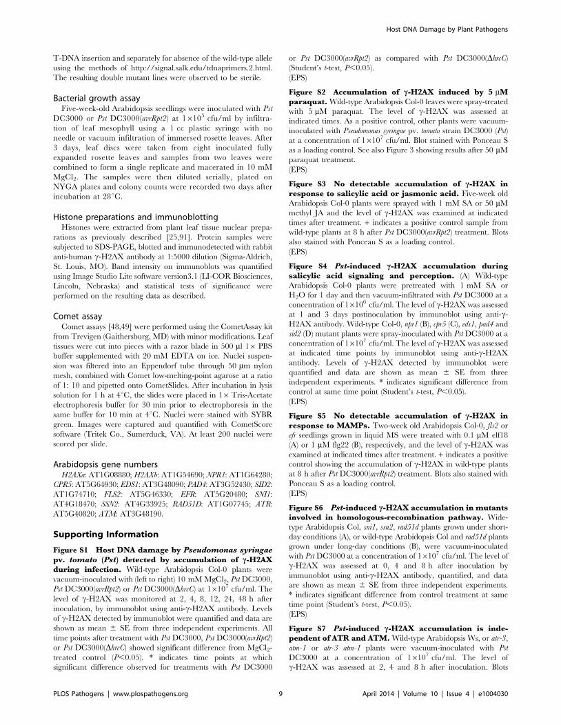
Figure S1 Host DNA damage by Pseudomonas syringaepv. tomato (Pst) detected by accumulation of c-H2AXduring infection. Wild-type Arabidopsis Col-0 plants were
vacuum-inoculated with (left to right) 10 mM MgCl2, Pst DC3000,
Pst DC3000(avrRpt2) or Pst DC3000(DhrcC) at 16107 cfu/ml. The
level of c-H2AX was monitored at 2, 4, 8, 12, 24, 48 h after
inoculation, by immunoblot using anti-c-H2AX antibody. Levels
of c-H2AX detected by immunoblot were quantified and data are
shown as mean 6 SE from three independent experiments. All
time points after treatment with Pst DC3000, Pst DC3000(avrRpt2)
or Pst DC3000(DhrcC) showed significant difference from MgCl2-
treated control (P,0.05). * indicates time points at which
significant difference observed for treatments with Pst DC3000
or Pst DC3000(avrRpt2) as compared with Pst DC3000(DhrcC)
(Student’s t-test, P,0.05).
(EPS)
Figure S2 Accumulation of c-H2AX induced by 5 mMparaquat. Wild-type Arabidopsis Col-0 leaves were spray-treated
with 5 mM paraquat. The level of c-H2AX was assessed at
indicated times. As a positive control, other plants were vacuum-
inoculated with Pseudomonas syringae pv. tomato strain DC3000 (Pst)
at a concentration of 16107 cfu/ml. Blot stained with Ponceau S
as a loading control. See also Figure 3 showing results after 50 mM
paraquat treatment.
(EPS)
Figure S3 No detectable accumulation of c-H2AX inresponse to salicylic acid or jasmonic acid. Five-week old
Arabidopsis Col-0 plants were sprayed with 1 mM SA or 50 mM
methyl JA and the level of c-H2AX was examined at indicated
times after treatment. + indicates a positive control sample from
wild-type plants at 8 h after Pst DC3000(avrRpt2) treatment. Blots
also stained with Ponceau S as a loading control.
(EPS)
Figure S4 Pst-induced c-H2AX accumulation duringsalicylic acid signaling and perception. (A) Wild-type
Arabidopsis Col-0 plants were pretreated with 1 mM SA or
H2O for 1 day and then vacuum-infiltrated with Pst DC3000 at a
concentration of 16106 cfu/ml. The level of c-H2AX was assessed
at 1 and 3 days postinoculation by immunoblot using anti-c-
H2AX antibody. Wild-type Col-0, npr1 (B), cpr5 (C), eds1, pad4 and
sid2 (D) mutant plants were spray-inoculated with Pst DC3000 at a
concentration of 16107 cfu/ml. The level of c-H2AX was assessed
at indicated time points by immunoblot using anti-c-H2AX
antibody. Levels of c-H2AX detected by immunoblot were
quantified and data are shown as mean 6 SE from three
independent experiments. * indicates significant difference from
control at same time point (Student’s t-test, P,0.05).
(EPS)
Figure S5 No detectable accumulation of c-H2AX inresponse to MAMPs. Two-week old Arabidopsis Col-0, fls2 or
efr seedlings grown in liquid MS were treated with 0.1 mM elf18
(A) or 1 mM flg22 (B), respectively, and the level of c-H2AX was
examined at indicated times after treatment. + indicates a positive
control showing the accumulation of c-H2AX in wild-type plants
at 8 h after Pst DC3000(avrRpt2) treatment. Blots also stained with
Ponceau S as a loading control.
(EPS)
Figure S6 Pst-induced c-H2AX accumulation in mutantsinvolved in homologous-recombination pathway. Wide-
type Arabidopsis Col, sni1, ssn2, rad51d plants grown under short-
day conditions (A), or wild-type Arabidopsis Col and rad51d plants
grown under long-day conditions (B), were vacuum-inoculated
with Pst DC3000 at a concentration of 16107 cfu/ml. The level of
c-H2AX was assessed at 0, 4 and 8 h after inoculation by
immunoblot using anti-c-H2AX antibody, quantified, and data
are shown as mean 6 SE from three independent experiments.
* indicates significant difference from control treatment at same
time point (Student’s t-test, P,0.05).
(EPS)
Figure S7 Pst-induced c-H2AX accumulation is inde-pendent of ATR and ATM. Wild-type Arabidopsis Ws, or atr-3,
atm-1 or atr-3 atm-1 plants were vacuum-inoculated with Pst
DC3000 at a concentration of 16107 cfu/ml. The level of
c-H2AX was assessed at 2, 4 and 8 h after inoculation. Blots
Host DNA Damage by Plant Pathogens
PLOS Pathogens | www.plospathogens.org 9 April 2014 | Volume 10 | Issue 4 | e1004030

also stained with Ponceau S as a loading control. Similar
experiments with Arabidopsis Col atr atm mutants are shown in
Figure 7.
(EPS)
Acknowledgments
We are grateful to Dr. Anne Britt for providing the atr-3, atm-1 and atr-3/
atr-3,ATM/atm-1 segregating lines and helpful advice, Dr. Masaaki Umeda
for providing the atr-2, atm-2 and atr-2/atr-2,ATM/atm-2 segregating
lines, Drs. Dennis Halterman and Jiming Jiang for providing the Katahdin
SP951 transgenic potato lines, Amilcar Sanchez-Perez for P. infestans
inoculations, and Brian Keppler for suggestions regarding the manuscript.
Author Contributions
Conceived and designed the experiments: JS AFB. Performed the
experiments: JS. Analyzed the data: JS AFB. Contributed reagents/
materials/analysis tools: JS AFB. Wrote the paper: JS AFB.
References
1. Jackson SP, Bartek J (2009) The DNA-damage response in human biology and
disease. Nature 461: 1071–1078.
2. Ciccia A, Elledge SJ (2010) The DNA damage response: making it safe to play
with knives. Mol Cell 40: 179–204.
3. Sedelnikova OA, Redon CE, Dickey JS, Nakamura AJ, Georgakilas AG, et al.
(2010) Role of oxidatively induced DNA lesions in human pathogenesis. Mutat
Res 704: 152–159.
4. Ambur OH, Davidsen T, Frye SA, Balasingham SV, Lagesen K, et al. (2009)
Genome dynamics in major bacterial pathogens. FEMS Microbiol Rev 33: 453–
470.
5. Weitzman MD, Lilley CE, Chaurushiya MS (2010) Genomes in conflict:
maintaining genome integrity during virus infection. Annu Rev Microbiol 64:
61–81.
6. Nougayrede JP, Homburg S, Taieb F, Boury M, Brzuszkiewicz E, et al. (2006)
Escherichia coli induces DNA double-strand breaks in eukaryotic cells. Science
313: 848–851.
7. Toller IM, Neelsen KJ, Steger M, Hartung ML, Hottiger MO, et al. (2011)
Carcinogenic bacterial pathogen Helicobacter pylori triggers DNA double-strand
breaks and a DNA damage response in its host cells. Proc Natl Acad Sci U S A
108: 14944–14949.
8. Chakraborty SP, Kar Mahapatra S, Sahu SK, Das S, Tripathy S, et al. (2011)
Internalization of Staphylococcus aureus in lymphocytes induces oxidative stress and
DNA fragmentation: possible ameliorative role of nanoconjugated vancomycin.
Oxid Med Cell Longev 2011: 942123.
9. Rakkestad KE, Skaar I, Ansteinsson VE, Solhaug A, Holme JA, et al. (2010)
DNA damage and DNA damage responses in THP-1 monocytes after exposure
to spores of either Stachybotrys chartarum or Aspergillus versicolor or to T-2 toxin.
Toxicol Sci 115: 140–155.
10. Touati E (2010) When bacteria become mutagenic and carcinogenic: lessons
from H. pylori. Mutat Res 703: 66–70.
11. Mangerich A, Knutson CG, Parry NM, Muthupalani S, Ye W, et al. (2012)
Infection-induced colitis in mice causes dynamic and tissue-specific changes in
stress response and DNA damage leading to colon cancer. Proc Natl Acad
Sci U S A 109: E1820–1829.
12. Arthur JC, Perez-Chanona E, Muhlbauer M, Tomkovich S, Uronis JM, et al.
(2012) Intestinal inflammation targets cancer-inducing activity of the microbiota.
Science 338: 120–123.
13. Dodds PN, Rathjen JP (2010) Plant immunity: towards an integrated view of
plant-pathogen interactions. Nat Rev Genet 11: 539–548.
14. Murphy K, Travers P, Walport M, Janeway C (2012) Janeway’s immunobiol-
ogy. New York: Garland Science. xix, 868 p. p.
15. Rakoff-Nahoum S, Medzhitov R (2009) Toll-like receptors and cancer. Nat Rev
Cancer 9: 57–63.
16. Lamb C, Dixon RA (1997) The Oxidative Burst in Plant Disease Resistance.
Annu Rev Plant Physiol Plant Mol Biol 48: 251–275.
17. O’Brien JA, Daudi A, Butt VS, Bolwell GP (2012) Reactive oxygen species and
their role in plant defence and cell wall metabolism. Planta 236: 765–779.
18. Roldan-Arjona T, Ariza RR (2009) Repair and tolerance of oxidative DNA
damage in plants. Mutat Res 681: 169–179.
19. Waterworth WM, Drury GE, Bray CM, West CE (2011) Repairing breaks in the
plant genome: the importance of keeping it together. New Phytol 192: 805–822.
20. Balestrazzi A, Confalonieri M, Macovei A, Dona M, Carbonera D (2011)
Genotoxic stress and DNA repair in plants: emerging functions and tools for
improving crop productivity. Plant Cell Rep 30: 287–295.
21. Rogakou EP, Pilch DR, Orr AH, Ivanova VS, Bonner WM (1998) DNA double-
stranded breaks induce histone H2AX phosphorylation on serine 139. J Biol
Chem 273: 5858–5868.
22. Iacovoni JS, Caron P, Lassadi I, Nicolas E, Massip L, et al. (2010) High-
resolution profiling of gammaH2AX around DNA double strand breaks in the
mammalian genome. EMBO J 29: 1446–1457.
23. Abraham RT (2001) Cell cycle checkpoint signaling through the ATM and ATR
kinases. Genes Dev 15: 2177–2196.
24. Sperka T, Wang J, Rudolph KL (2012) DNA damage checkpoints in stem cells,
ageing and cancer. Nat Rev Mol Cell Biol 13: 579–590.
25. Friesner JD, Liu B, Culligan K, Britt AB (2005) Ionizing radiation-dependent
gamma-H2AX focus formation requires ataxia telangiectasia mutated and ataxia
telangiectasia mutated and Rad3-related. Mol Biol Cell 16: 2566–2576.
26. Kinner A, Wu W, Staudt C, Iliakis G (2008) Gamma-H2AX in recognition and
signaling of DNA double-strand breaks in the context of chromatin. NucleicAcids Res 36: 5678–5694.
27. Lucht JM, Mauch-Mani B, Steiner HY, Metraux JP, Ryals J, et al. (2002)
Pathogen stress increases somatic recombination frequency in Arabidopsis. NatGenet 30: 311–314.
28. Kovalchuk I, Kovalchuk O, Kalck V, Boyko V, Filkowski J, et al. (2003)Pathogen-induced systemic plant signal triggers DNA rearrangements. Nature
423: 760–762.
29. Yao Y, Kathiria P, Kovalchuk I (2013) A systemic increase in the recombinationfrequency upon local infection of Arabidopsis thaliana plants with oilseed rape
mosaic virus depends on plant age, the initial inoculum concentration and thetime for virus replication. Front Plant Sci 4: 61.
30. Choi JJ, Klosterman SJ, Hadwiger LA (2001) A comparison of the effects of
DNA-damaging agents and biotic elicitors on the induction of plant defensegenes, nuclear distortion, and cell death. Plant Physiol 125: 752–762.
31. Kunz BA, Cahill DM, Mohr PG, Osmond MJ, Vonarx EJ (2006) Plant
responses to UV radiation and links to pathogen resistance. Int Rev Cytol 255:1–40.
32. Durrant WE, Wang S, Dong XN (2007) Arabidopsis SNI1 and RAD51Dregulate both gene transcription and DNA recombination during the defense
response. Proc Natl Acad Sci U S A 104: 4223–4227.
33. Wang S, Durrant WE, Song J, Spivey NW, Dong X (2010) Arabidopsis BRCA2and RAD51 proteins are specifically involved in defense gene transcription
during plant immune responses. Proc Natl Acad Sci U S A 107: 22716–22721.
34. Song J, Durrant WE, Wang S, Yan S, Tan EH, et al. (2011) DNA repairproteins are directly involved in regulation of gene expression during plant
immune response. Cell Host Microbe 9: 115–124.
35. Adams-Phillips L, Wan J, Tan X, Dunning FM, Meyers BC, et al. (2008)
Discovery of ADP-ribosylation and other plant defense pathway elements
through expression profiling of four different Arabidopsis-Pseudomonas R-avr
interactions. Mol Plant Microbe Interact 21: 646–657.
36. Adams-Phillips L, Briggs AG, Bent AF (2010) Disruption of poly(ADP-ribosyl)ation mechanisms alters responses of Arabidopsis to biotic stress. Plant
Physiol 152: 267–280.
37. Mur LA, Kenton P, Lloyd AJ, Ougham H, Prats E (2008) The hypersensitiveresponse; the centenary is upon us but how much do we know? J Exp Bot 59:
501–520.
38. Ryerson DE, Heath MC (1996) Cleavage of nuclear DNA into oligonucleosomalfragments during cell death induced by fungal infection or by abiotic treatments.
Plant Cell 8: 393–402.
39. Kunkel BN, Bent AF, Dahlbeck D, Innes RW, Staskawicz BJ (1993) RPS2, an
Arabidopsis disease resistance locus specifying recognition of Pseudomonas syringae
strains expressing the avirulence gene avrRpt2. Plant Cell 5: 865–875.
40. Yu GL, Katagiri F, Ausubel FM (1993) Arabidopsis mutations at the RPS2 locus
result in loss of resistance to Pseudomonas syringae strains expressing the avirulencegene avrRpt2. Mol Plant Microbe Interact 6: 434–443.
41. Boch J, Joardar V, Gao L, Robertson TL, Lim M, et al. (2002) Identification of
Pseudomonas syringae pv. tomato genes induced during infection of Arabidopsis
thaliana. Mol Microbiol 44: 73–88.
42. Whalen MC, Innes RW, Bent AF, Staskawicz BJ (1991) Identification of
Pseudomonas syringae pathogens of Arabidopsis and a bacterial locus determiningavirulence on both Arabidopsis and soybean. Plant Cell 3: 49–59.
43. Ritter C, Dangl JL (1996) Interference between two specific pathogenrecognition events mediated by distinct plant disease resistance genes. Plant
Cell 8: 251–257.
44. Yu I-C, Parker J, Bent AF (1998) Gene-for-gene disease resistance without thehypersensitive response in Arabidopsis dnd1 mutant. Proc Natl Acad Sci USA 95:
7819–7824.
45. Gao X, Chen X, Lin W, Chen S, Lu D, et al. (2013) Bifurcation of ArabidopsisNLR immune signaling via Ca(2)(+)-dependent protein kinases. PLoS Pathog 9:
e1003127.
46. Zhang C, Gutsche AT, Shapiro AD (2004) Feedback control of the Arabidopsis
hypersensitive response. Mol Plant Microbe Interact 17: 357–365.
47. Grant M, Brown I, Adams S, Knight M, Ainslie A, et al. (2000) The RPM1 plantdisease resistance gene facilitates a rapid and sustained increase in cytosolic
calcium that is necessary for the oxidative burst and hypersensitive cell death.Plant J 23: 441–450.
Host DNA Damage by Plant Pathogens
PLOS Pathogens | www.plospathogens.org 10 April 2014 | Volume 10 | Issue 4 | e1004030

48. Collins AR, Azqueta A (2012) DNA repair as a biomarker in human
biomonitoring studies; further applications of the comet assay. Mutat Res 736:
122–129.
49. Dhawan A, Bajpayee M, Parmar D (2009) Comet assay: a reliable tool for the
assessment of DNA damage in different models. Cell Biol Toxicol 25: 5–32.
50. Mishina TE, Zeier J (2007) Pathogen-associated molecular pattern recognition
rather than development of tissue necrosis contributes to bacterial induction of
systemic acquired resistance in Arabidopsis. Plant J 50: 500–513.
51. Huynh TV, Dahlbeck D, Staskawicz BJ (1989) Bacterial blight of soybean:
Regulation of a pathogen gene determining host cultivar specificity. Science 245:
1374–1377.
52. Pieterse CM, van Wees SC, Hoffland E, van Pelt JA, van Loon LC (1996)
Systemic resistance in Arabidopsis induced by biocontrol bacteria is independent
of salicylic acid accumulation and pathogenesis-related gene expression. Plant
Cell 8: 1225–1237.
53. Haas D, Defago G (2005) Biological control of soil-borne pathogens by
fluorescent pseudomonads. Nat Rev Microbiol 3: 307–319.
54. Lukas J, Lukas C, Bartek J (2011) More than just a focus: The chromatin
response to DNA damage and its role in genome integrity maintenance. Nat Cell
Biol 13: 1161–1169.
55. Srivastava N, Gochhait S, de Boer P, Bamezai RN (2009) Role of H2AX in
DNA damage response and human cancers. Mutat Res 681: 180–188.
56. Huen MS, Grant R, Manke I, Minn K, Yu X, et al. (2007) RNF8 transduces the
DNA-damage signal via histone ubiquitylation and checkpoint protein assembly.
Cell 131: 901–914.
57. Song J, Bradeen JM, Naess SK, Raasch JA, Wielgus SM, et al. (2003) Gene RB
cloned from Solanum bulbocastanum confers broad spectrum resistance to potato
late blight. Proc Natl Acad Sci U S A 100: 9128–9133.
58. Kramer LC, Choudoir MJ, Wielgus SM, Bhaskar PB, Jiang J (2009) Correlation
between transcript abundance of the RB gene and the level of the RB-mediated
late blight resistance in potato. Mol Plant Microbe Interact 22: 447–455.
59. Gardner RG, Panthee DR (2010) NC 1 CELBR and NC 2 CELBR: Early
Blight and Late Blight-resistant Fresh Market Tomato Breeding Lines.
Hortscience 45: 975–976.
60. Bowler C, van Montagu M, Inze D (1992) Superoxide dismutase and stress
tolerance. Ann Rev Plant Physiol Plant Mol Biol 43: 83–116.
61. Levine A, Tenhaken R, Dixon R, Lamb CJ (1994) H2O2 from the oxidative
burst orchestrates the plant hypersensitive disease resistance response. Cell 79:
583–593.
62. Suzuki N, Miller G, Morales J, Shulaev V, Torres MA, et al. (2011) Respiratory
burst oxidases: the engines of ROS signaling. Curr Opin Plant Biol 14: 691–699.
63. Torres MA, Dangl JL, Jones JD (2002) Arabidopsis gp91phox homologues
AtrbohD and AtrbohF are required for accumulation of reactive oxygen
intermediates in the plant defense response. Proc Natl Acad Sci U S A 99:
517–522.
64. Rao MV, Paliyath G, Ormrod DP, Murr DP, Watkins CB (1997) Influence of
salicylic acid on H2O2 production, oxidative stress, and H2O2-metabolizing
enzymes. Salicylic acid-mediated oxidative damage requires H2O2. Plant
Physiol 115: 137–149.
65. Durrant WE, Dong X (2004) Systemic acquired resistance. Annu Rev
Phytopathol 42: 185–209.
66. Pieterse CM, Van der Does D, Zamioudis C, Leon-Reyes A, Van Wees SC
(2012) Hormonal modulation of plant immunity. Annu Rev Cell Dev Biol 28:
489–521.
67. Cao H, Bowling SA, Gordon S, Dong X (1994) Characterization of an
Arabidopsis mutant that is nonresponsive to inducers of systemic acquired
resistance. Plant Cell 6: 1583–1592.
68. Kirik V, Bouyer D, Schobinger U, Bechtold N, Herzog M, et al. (2001) CPR5 is
involved in cell proliferation and cell death control and encodes a novel
transmembrane protein. Curr Biol 11: 1891–1895.
69. Bowling SA, Clarke JD, Liu Y, Klessig DF, Dong X (1997) The cpr5 mutant of
Arabidopsis expresses both NPR1-dependent and NPR1-independent resistance.
Plant Cell 9: 1573–1584.
70. Boller T, Felix G (2009) A renaissance of elicitors: perception of microbe-
associated molecular patterns and danger signals by pattern-recognitionreceptors. Annu Rev Plant Biol 60: 379–406.
71. Kunze G, Zipfel C, Robatzek S, Niehaus K, Boller T, et al. (2004) The N
terminus of bacterial elongation factor Tu elicits innate immunity in Arabidopsisplants. Plant Cell 16: 3496–3507.
72. Yan S, Wang W, Marques J, Mohan R, Saleh A, et al. (2013) Salicylic acidactivates DNA damage responses to potentiate plant immunity. Mol Cell 52:
602–610.
73. Martin V, Chahwan C, Gao H, Blais V, Wohlschlegel J, et al. (2006) Sws1 is aconserved regulator of homologous recombination in eukaryotic cells. Embo J
25: 2564–2574.74. Culligan K, Tissier A, Britt A (2004) ATR regulates a G2-phase cell-cycle
checkpoint in Arabidopsis thaliana. Plant Cell 16: 1091–1104.75. Garcia V, Bruchet H, Camescasse D, Granier F, Bouchez D, et al. (2003)
AtATM is essential for meiosis and the somatic response to DNA damage in
plants. Plant Cell 15: 119–132.76. Vespa L, Couvillion M, Spangler E, Shippen DE (2005) ATM and ATR make
distinct contributions to chromosome end protection and the maintenance oftelomeric DNA in Arabidopsis. Genes Dev 19: 2111–2115.
77. Amiard S, Depeiges A, Allain E, White CI, Gallego ME (2011) Arabidopsis
ATM and ATR kinases prevent propagation of genome damage caused bytelomere dysfunction. Plant Cell 23: 4254–4265.
78. Adachi S, Minamisawa K, Okushima Y, Inagaki S, Yoshiyama K, et al. (2011)Programmed induction of endoreduplication by DNA double-strand breaks in
Arabidopsis. Proc Natl Acad Sci U S A 108: 10004–10009.79. Menendez D, Shatz M, Azzam K, Garantziotis S, Fessler MB, et al. (2011) The
Toll-like receptor gene family is integrated into human DNA damage and p53
networks. PLoS Genet 7: e1001360.80. Gasser S, Orsulic S, Brown EJ, Raulet DH (2005) The DNA damage pathway
regulates innate immune system ligands of the NKG2D receptor. Nature 436:1186–1190.
81. Jakobek JL, Lindgren PB (1993) Generalized Induction of Defense Responses in
Bean Is Not Correlated with the Induction of the Hypersensitive Reaction. PlantCell 5: 49–56.
82. Pieterse CMJ, van Wees SCM, van Pelt JA, Knoester M, Laan R, et al. (1998) Anovel signaling pathway controlling induced systemic resistance in Arabidopsis.
Plant Cell 10: 1571–1580.83. Verhagen BW, Glazebrook J, Zhu T, Chang HS, van Loon LC, et al. (2004)
The transcriptome of rhizobacteria-induced systemic resistance in arabidopsis.
Mol Plant Microbe Interact 17: 895–908.84. Sumit R, Sahu BB, Xu M, Sandhu D, Bhattacharyya MK (2012) Arabidopsis
nonhost resistance gene PSS1 confers immunity against an oomycete and afungal pathogen but not a bacterial pathogen that cause diseases in soybean.
BMC Plant Biol 12: 87.
85. Park EJ, Chan DW, Park JH, Oettinger MA, Kwon J (2003) DNA-PK isactivated by nucleosomes and phosphorylates H2AX within the nucleosomes in
an acetylation-dependent manner. Nucleic Acids Res 31: 6819–6827.86. Kuhne M, Riballo E, Rief N, Rothkamm K, Jeggo PA, et al. (2004) A double-
strand break repair defect in ATM-deficient cells contributes to radiosensitivity.Cancer Res 64: 500–508.
87. Karlsson KH, Stenerlow B (2004) Focus formation of DNA repair proteins in
normal and repair-deficient cells irradiated with high-LET ions. Radiat Res 161:517–527.
88. Jiang M, Zhao L, Gamez M, Imperiale MJ (2012) Roles of ATM and ATR-mediated DNA damage responses during lytic BK polyomavirus infection. PLoS
Pathog 8: e1002898.
89. Katagiri F, Thilmony R, He SY (2002) The Arabidopsis thaliana-pseudomonas
syringae interaction. Arabidopsis Book 1: e0039.
90. Genger RK, Jurkowski GI, McDowell JM, Lu H, Jung HW, et al. (2008)Signaling pathways that regulate the enhanced disease resistance of Arabidopsis
‘‘defense, no death’’ mutants. Mol Plant Microbe Interact 21: 1285–1296.
91. Jackson JP, Johnson L, Jasencakova Z, Zhang X, PerezBurgos L, et al. (2004)Dimethylation of histone H3 lysine 9 is a critical mark for DNA methylation and
gene silencing in Arabidopsis thaliana. Chromosoma 112: 308–315.
Host DNA Damage by Plant Pathogens
PLOS Pathogens | www.plospathogens.org 11 April 2014 | Volume 10 | Issue 4 | e1004030

0
2
4
6
8
10
12
14
0 10 20 30 40 50 60
Indu
ctio
n of
γ-H
2AX
(log 2)
Time after Inoculation (hr)
Pst
Pst(avrRpt2)
Pst(hrcC)
MgCl2
****
Figure S1. Host DNA damage by Pseudomonas syringae pv. tomato (Pst) detected by accumulation of γ-H2AX during infection. Wild-type Arabidopsis Col-0 plants were vacuum-inoculated with (left to right) 10 mM MgCl2, Pst DC3000, Pst DC3000(avrRpt2) or Pst DC3000(ΔhrcC) at 1 × 10⁷ cfu/ml. The level of γH2AX was monitored at 2, 4, 8, 12, 24, 48 hr after inoculation, by immunoblot using anti-γ-H2AX antibody. Levels of γH2AX detected by immunoblot were quantified and data are shown as mean ± SE from three independent experiments. All time points after treatment with Pst DC3000, Pst DC3000(avrRpt2) or Pst DC3000(ΔhrcC) showed significant difference from MgCl2-treated control (P < 0.05). * indicates time points at which significant difference observed for treatments with Pst DC3000 or Pst DC3000(avrRpt2) as compared with Pst DC3000(ΔhrcC) (student’s t-test, P < 0.05).

anti-γH2AX0
Ponceau S
4h 8h 4h2h 8h1d 2d 3d 4d 5d 6d 7dparaquat Pst
Figure S2. Accumulation of γ-H2AX induced by 5 μM paraquat. Wild-type Arabidopsis Col-0 leaves were spray-treated with 5 μM paraquat. The level of γ-H2AX was assessed at indicated times. As a positive control, other plants were vacuum-inoculated with Pseudomonas syringae pv. tomato strain DC3000 (Pst) at a concentration of 1 × 10⁷ cfu/ml. Blot stained with Ponceau S as a loading control. See also Figure 3 showing results after 50 μM paraquat treatment.

0 12 24 48 12 24 48 +SA JA
hpi
Ponceau S
anti-γH2AX
Figure S3. No detectable accumulation of γ-H2AX in response to salicylic acid or jasmonic acid. Five-week old Arabidopsis Col-0 plants were sprayed with 1 mM SA or 50 μM methyl JA and the level of γ-H2AX was examined at indicated times after treatment. + indicates a positive control sample from wild-type plants at 8 hr after Pst DC3000(avrRpt2) treatment. Blots also stained with Ponceau S as a loading control.

0
1
2
3
4
5
6
7
8
0 2 4 6 8 10
Indu
ctio
n of
γ-H
2AX
(log 2)
WTnpr1
0
1
2
3
4
5
6
7
0 2 4 6 8 10
Ind
uct
ion
of
γ-H
2AX
(lo
g 2)
Time after Inoculation (hr)
WT
eds1
pad4
sid2
0
1
2
3
4
0 1 2 3 4
Ind
uct
ion
of
γ-H
2AX
(lo
g 2)
Time after Inoculation (day)
Water
SA
0
1
2
3
4
5
0 2 4 6 8 10Time after Inoculation (hr)
WT
cpr5
Ind
uct
ion
of
γ-H
2AX
(lo
g 2)
A B
C D
Time after Inoculation (day)
*
*
*
*
*
Figure S4. Pst-induced γ-H2AX accumulation during salicylic acid signaling and perception. (A) Wild-type Arabidopsis Col-0 plants were pretreated with 1 mM SA or H2O for 1 day and then vacuum-infiltrated with Pst DC3000 at a concentration of 1 × 10⁶ cfu/ml. The level of γ-H2AX was assessed at 1 and 3 days postinoculation by immunoblot using anti-γ-H2AX antibody. Wild-type Col-0, Col-0 npr1 (B), cpr5 (C), eds1, pad4 and sid2 (D) mutant plants were spray-inoculated with Pst DC3000 at a concentration of 1 × 10⁷ cfu/ml. The level of γ-H2AX was assessed at indicated time points by immunoblot using anti-γ-H2AX antibody. Levels of γH2AX detected by immunoblot were quantified and data are shown as mean ± SE from three independent experiments. * indicates significant difference from control at same time point (student’s t-test, P < 0.05).

Col0 15 60 120 0 15 60 120 + min
efr
Ponceau S
anti-γH2AX
Col0 15 60 120 0 15 60 120 + min
fls2
Ponceau S
anti-γH2AX
A
B
Figure S5. No detectable accumulation of γ-H2AX in response to MAMPs. Two-week old Arabidopsis Col-0, fls2 or efr seedlings grown in liquid MS were treated with 0.1 μM elf18 (A) or 1 μM flg22 (B), respectively, and the level of γ-H2AX was examined at indicated times after treatment. + indicates a positive control showing the accumulation of γ-H2AX in wild-type plants at 8 hr after Pst DC3000(avrRpt2) treatment. Blots also stained with Ponceau S as a loading control.

0
1
2
3
4
5
6
0 2 4 6 8 10
Indu
ctio
n of
γ-H
2AX
(log
2)
Time after Inoculation (hr)
WT
sni1
ssn2
rad51d
A
B
**
0
1
2
3
4
5
6
7
8
0 2 4 6 8 10
Indu
ctio
n of
γ-H
2AX
(log 2)
Time after Inoculation (hr)
WT
rad51d
Figure S6. Pst-induced γ-H2AX accumulation in mutants involved in homologous recombination pathway. Wild-type Arabidopsis Col, sni1, ssn2 and rad51d plants were vacuum-inoculated with Pst DC3000 at a concentration of 1 × 10⁷ cfu/ml. The level of γ-H2AX was assessed at 0, 4 and 8 hr after inoculation by immunoblot using anti-γ-H2AX antibody, quantified, and data are shown as mean ± SE from three independent experiments. * indicates significant difference from control treatment at same time point (student’s t-test, P < 0.05).

2 4 8 2 4 8 2 4 8 2 4 8WS atr atm atratm
Ponceau S
anti-γH2AXh
Figure S7. Pst-induced γ-H2AX accumulation is independent of ATR and ATM. Wild-type Arabidopsis Ws, or atr-3, atm-1 or atr-3 atm-1 plants were vacuum-inoculated with Pst DC3000 at a concentration of 1 × 10⁷ cfu/ml. The level of γ-H2AX was assessed at 2, 4 and 8 hr after inoculation. Blots also stained with Ponceau S as a loading control. Similar experiments with Arabidopsis Col atr atm mutants are shown in Figure 7.