microbial enhanced oil recovery ability of...
TRANSCRIPT

MICROBIAL ENHANCED OIL RECOVERY ABILITY OF THE BIOSURFACTANT PRODUCED FROM SERRATIA
MARCESCENS UEO15
ELEMBA, O. M1., IJAH, U. J. J2. and CHIBUNNA, M3.
1Department of Biology/Microbiology/Biotechnology, Federal University Ndufu Alike Ikwo, P.M.B. 1010 Abakaliki Nigeria.
2Department of Microbiology, Federal University of Technology, Minna.P.M.B. 65, Minna, Nigeria.
3Imo state university, Owerri, Nigeria

INTRODUCTION Problem
Biosurfactant surface active molecules produced by microorganisms
majorly bacteria, yeast and few fungi.
They have two active ends that are referred to be amphipathic:
Hydrophilic (water-loving) moiety comprising of carbohydrates, alcohol and their derivatives
Hydrophobic (water-hating) moiety is composed of fatty acids, ester and their derivatives.

Different microorganisms produced different biosurfactants which are influenced by the type of carbon substrates used
The environment in which organism was isolated
The nitrogen content of the medium to which the organism was cultured
Classification and characterization based mainly on their chemical structure and microbial origin

AIM
This study focussed on the use of biosurfactant which are biodegradable, biocompatible and non-toxic in the recovery of soil polluted with oil to test the efficacy of biosurfactant.

MATERIALS AND METHODS
Sample collection
Hydrocarbon contaminated soil was collected (using soil auger) at the depth of 10-15cm from NNPC depot in Chanchaga Local Government Area, Minna, Niger state, Nigeria
Isolation of bacteria

Biosurfactant assay
Haemolytic activity
Oil displacement method
Emulsification capacity

Characterization and identification of biosurfactant producers • Isolates were characterized based on Gram’s reaction,
and biochemical tests. The biochemical tests performed included reduction of nitrate, spore formation, utilization of citrate, production of indole, and methyl red-voges proskauer test (MR-VP).
• The ability of the isolates to utilize the following carbohydrates was tested: glucose, arabinose, inositol, xylose, fructose, mannitol and sucrose.
• The probable identities of the isolates were determined using the schemes of Krieg et al.2000 and Holt et al., 2004.

Production and Extraction of biosurfactants One hundred millilitres of mineral salt medium of
Jacobucci et al. (2001) was dispensed in conical flasks containing 1 ml of diesel was sterilized by autoclaving at 121°C for 15 minutes,
the medium was allowed to cool before being inoculated with 2mI of nutrient broth culture of the bacterial isolates. Two of the flasks were left uninoculated and served as a control.
The flasks were incubated shaking at 250rpm at room temperature (30oC) and pH 7.1. For the period of 8 days.
After which the cultures were centrifuged at 500rpm for 30minutes, then it was filtered using whatman No.1 filter paper.

The supernatants was collected and subjected to solvent extraction three times using Chloroform and Methanol in the ratio of 2:1.
Two phase separation occurred (the upper solvent and the whitish bottom phase which is the crude biosurfactant),
the bottom sediment was collected using separating funnel and washed with distilled water, after which the left over solvent was allowed to evaporate to dryness over a water bath at 45oC for 24hours.
Quantity of the dried biosurfactant was determined by measuring the dry weight using the formula:
Quantity of biosurfactant= Weight of the plate after drying -weight of the empty plate

Plate I: Extracted crude biosurfactant
Biosurfactant

Characterization of the biosurfactant produced The biosurfactants produced was purified and
characterized using TLC, FTIR and GCMS respectively.
Purification was done using thin layer chromatography:
Thin layer chromatography was carried out by spotting the crude biosurfactant on TLC Plates already pre-coated (G60, Merck, Darmstadt, Germany)
The plate which was commercially prepared was developed and activated by placing it in an hot air oven at a temperature of 100oC for 30minutes before
The surfactant were spotted on the plate using capillary tube.

Chloroform-methanol-acetic acid and water (85:10:5:1) was used to separate the spots.
The TLC plates were placed inside the tank containing the solvents the tank was cover with lid and was observed for movement.
After one hour the movement stopped and was assumed separation has ended.
The plates were removed from the tank allowed to air dried and then was viewed under a UV light to identify the separated fractions. After was the plates were sprayed with anthrone reagent and ninhydrin solution.
The spots were scraped from the plate and extracted with chloroform-methanol mixture.

The quantity produced after this was also weighed and measured.
The Rf value was determined by using the formula, distance travelled by the test sample divided by the distance travelled by the solvent
Rf = Distance travelled by the test sample Distance travelled by the solvent

Fourier transform Infrared Spectroscopy (FTIR) The Infrared (IR) spectroscopy of the biosurfactant
was carried out using 8400S Fourier transform infrared spectrophotometer by Shimadzu, Japan
The IR spectra were scanned between 500 and 4500cm-1 wave numbers (per cm) with a resolution of two measures per wave number
using potassium bromide as background reference.

Gas chromatography /mass spectroscopy (GC-MS)
The GC-MS analysis was carried out using GCMS QP2010 Plus Shimudza, Japan equipped with capillary column and selective detector (AOC-20i)
which was set to scan from m/z 20 to m/z 310 at a scan rate of 1.5 scans per seconds with an initial oven temperature of 80oC for three minutes
with a carrier gas (Helium)
at a flow rate of 1.58ml/min and a split ratio of 50:1.0

OIL RECOVERY ABILITY OF THE BIOSURFACTANT Oil recovery ability of the biosurfactant was carried out
using soil column study method by Pruthi and Cameotra (1997).
Two Glass columns were packed with 100g of sandy loam soil each and they were saturated with 20ml of crude oil and kerosene each this was allowed to stand for thirty days.
The efficiency of the biosurfactant solution in releasing the oil from the soil was tested by adding 100 ml aqueous solution of 1.0% of the biosurfactant solution to the column.
Distilled water only was used as control.

Statistical Analysis
Analysis of variance (ANOVA) was used in determining the significance of differences among the means.
All values are averages of three readings and have been shown as mean ± SD.

Results

Iso
late
co
de
Gra
m
rea
cti
on
Ca
tala
se
La
cta
se
Glu
co
se
Su
cro
se
Cit
rate
Mo
bil
ity
Ind
ole
Ure
ase
HS
Ga
s
Mr
Vp
Sp
ore
Ox
ida
se
Co
nfi
rme
d
iso
late
s
UEO1 + rod + – + + – + – + + – – + + – Bacillus licheniformis UEO1
UEO2 + rod + + + + – + – – – – + – + – Bacillus circulans UEO2
UEO3 + rod + + + + _ + – – – – + – + – Bacillus circulans UEO3
UEO4 + rod + – + - - + – + – – + – + – Bacillus lentus UEO4
UEO5 - rod + + + + + + – – – – – – – – Pseudomonas nautica UEO5
UEO6 +cocci + – + + – + – – + – – + – – Micrococcus sp UEO6
UEO7 +rod + – + + – + – _ _ _ + _ + – Bacillus Sp UEO7
UEO8 - rod + – + – – + – – + Pseudomonas sp UEO8
UEO9 +rod + – + + – + – – – – + – + – Bacillus firmus UEO9
UEO10 -rod + – + – + + – – – – – + – – Achromobacter oryzihabitan
UEO10
UEO11 -rod + – + + ND ND – – Proteus mirabilis UEO11
UEO12 -rod + – + + ND ND – – Proteus vulgaris UEO12
UEO13 +rod + – + – – + – + – – + – + – Bacillus lentus UEO13
UEO14 -rod + – + _ + + – – – – – + – – Serratia marcescens UEO14
UEO15 -rod + – + + + + – + – – + + – – Serratia marcescens UEO15
Table 1: Identification and characterization of the bacterial isolates
Key: + =positive; – =negative: HS=hydrogen sulphide; Mr= methyl red; Vp= voges proskauer; ND= not determined

S/N
o
Isolates Beta(β)-Haemolysis Alpha (α)-Haemolysis
1 Bacillus licheniformis UEO1 +
2 Bacillus circulans UEO2 +
3 Bacillus circulans UEO3 +
4 Bacillus lentus UEO4 +
5 Pseudomonas nautica UEO5
6 Micrococcus sp UEO6 +
7 Bacillus Sp UEO7 +
8 Pseudomonas sp UEO8 +
9 Bacillus firmus UEO9 +
10
Achromobacter oryzihabitan
UEO10
+
11 Proteus mirabilis UEO11 +
12 Proteus vulgaris UEO12 +
13 Bacillus lentus UEO13 +
14 Serratia marcescens UEO14 +
15 Serratia marcescens UEO15 +
Haemolytic activity of isolates On blood agar plate, three isolates UEO1, UEO9, UEO15 produced a transparent clear zone (β-haemolysis) around the colonies causing lysis of the blood (Table 2); this is a clear indication that those isolates are potent producers of biosurfactant
Table 2: Haemolytic ability of isolates
Key +: positive

Displacement, Emulsification capacity and Rf value of the isolates
The diameter and zone of displacement by these isolates (Bacillus licheniformis UEO1, Bacillus firmus UEO9 and Serratia marcescens UEO15
Table 3: Diameter of displacement and Emulsification capacity of the isolates
Isolates Diameter of
displacement(cm)
Emulsification
capacity (%E24)
(for diesel)
Emulsification
capacity (%E24)
for engine oil
Rf
Serratia marcescens UEO15 5.6±0.01ab 78.90 ± 3.0c 66.79±2.2c 0.75
Bacillus firmus UEO9 5.2±0.01a 56.63 ±1.39a 52.22±0.5a 0.46
Bacillus licheniformis UEO1 5.7±0.11ab 58.80 ±0.64ab 58.10±0.02b 0.53

0
2
4
6
8
10
12
14
S. marcescens UEO15 B. firmus UEO9 B. licheniformis UEO1Qu
an
tity
of
bio
surf
act
an
ts p
rod
uce
d (
g/l
)
Isolates
Quantity of biosurfactants crude
Quantity of biosurfactants purified
Quantity of biosurfactant produced The quantity/amount of crude and purified biosurfactant produced by these isolates differ from one another, with Serratia marcescens UEO15 having the largest amount (12.5 and 1.34g/l) of biosurfactant as compared to Bacillus licheniformis UEO1 and B. firmus UEO9 with 9.16; 0.27g/l and 5.78; 0.6g/l respectively (Figure 1)
Figure 1: quantities of biosurfactant produced

Characterization of biosurfactant from S.marcescens UEO15
S. marcescens UEO15 was further characterized using GCMS and FTIR this isolate
was chosen because of the reasonable amount of surfactant recovered after purification; so that it can effectively be subjected and used in further analysis.
The Infra-Red (FTIR) Analysis
Infrared analysis of the biosurfactants produced by Serratia marcescens is shown in Table 4 (Figure 2). Revealed strong band of CH2 stretching in the region with wave number 2908.65cm-1 which is typical of alkanes (C-H), weak bands that stretched at 2400.85 cm-1 confirms the presence of phosphines (P-H3). Conjugated weak bands stretching at 1662.69 cm-1 revealed characteristic for amines (N-H).
Carboxylic acids and their derivatives (C=O) had strong bands that stretched at 1105.25 cm-1. The alcohols and phenols (OH) were also proved from the broad bands stretching at 978.74 cm-1.
Weak bands that stretched at 500.34 cm-1 indicated disulphides. Which are characteristic component of Sewarratin (biosurfactant from S. marcescens)

Figure 2: FTIR Spectroscopy of S. marcescens UEO15

Wave No (cm-1) Intensity Identified functional Group
2908.65 75.251 C-H (s) stretch of Alkane
2400.85 36.110 Phosphine P-H3(w) stretch
1662.69 50.700 Amine (N-H)
1105.25 42.823 Carboxylic acids (C=O) and their
derivatives
978.74 30.442 Alcohols and phenols (OH)
500.34 15.531 Disulphide (w)
Table 4: Interpretation of FT-IR for S. marcescens UEO15
Key: s=strong; w=weak
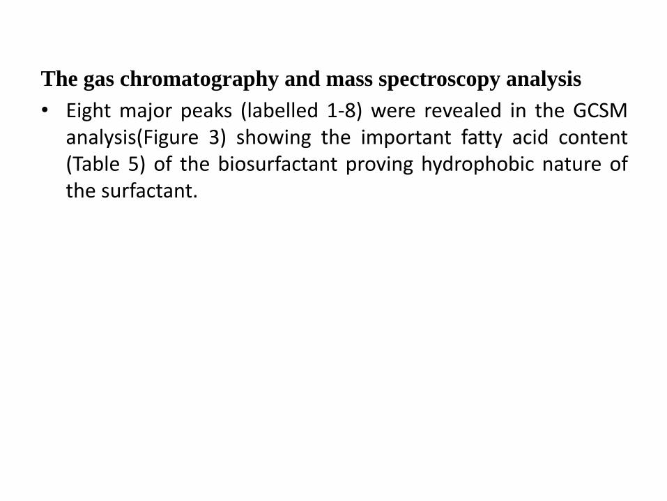
The gas chromatography and mass spectroscopy analysis
• Eight major peaks (labelled 1-8) were revealed in the GCSM analysis(Figure 3) showing the important fatty acid content (Table 5) of the biosurfactant proving hydrophobic nature of the surfactant.

Figure 3: GC-MS of S. marcescens UEO15, showing the scans (mz) and probably structure present in the biosurfactant

Table 5: The hydrophobic component of the biosurfactant (SMUEO15) from S. marcescens
Peak
Number
Compound name Molecular Formula
(mol. weight)
1 Palmitic acid, C17H34O2 (270)
2 Tetradecanoic acid C14H28O2 (228)
3 9-Octadecenoic acid, C19H36O2 (296)
4 Octadecanoic acid C19H38O2 (298)
5 Oleic Acid C18H34O2 (282)
6 Octadecanoic acid, 2-(2-hydroxyethoxy)ethyl ester C22H44O4 (372)
7 1-[[[(2-aminoethoxy)hydroxyphosphinyl]oxy]methyl]-1,2-
ethanediyl ester
C37H74NO8P (691)
8 Hexadecanoic acid, 2-hydroxy-1-(hydroxymethyl)ethyl ester C19H38O4 (330)
The presence of a Phosphine P-H3(w) stretch as seen in IR spectrum and
hydroxyphosphinyl group as revealed in peak 7 confirmed the biosurfactant to be a
phospholipid

Oil recovery ability of the biosurfactant Soil column study, which tested the effectiveness of the biosurfactant in possible microbial
enhanced oil recovery, revealed that the biosurfactant SMUEO15 recovered 78% and 58% of
crude oil and kerosene from soil as compared to that of distilled water (10% and 25%)
respectively (Figure 4).
0
10
20
30
40
50
60
70
80
90
crude oil kerosene
oil
rem
ova
l eff
ice
ncy
(%
)
Substrate
Oil removal efficacy (%) SMUEO15
Oil removal efficacy (%) Distlled water
Figure 4: Oil recovery efficacy of biosurfactant SMUEO15

DISCUSSION Serial dilution of soil sample and subsequent plating on nutrient agar
resulted in isolation of fifteen isolates. Strains were tested for haemolytic activity, which is regarded by some authors as indicative for biosurfactant production and used as a preliminary method for bacterial screening (Mulligan et al., 1974; Banat et al., 2000; Carrillo et al., 1996).
Haemolytic activity observed in all the fifteen isolated strains(Table 2); UEO1, 9 and 15 gave beta(β) (complete) haemolytic activity while the other strains had alpha (incomplete) haemolytic activity.
Based on this only the three isolates were selected and subjected to other analysis.
Three of the isolates successfully displace the oil hence confirmation to production of biosurfactant. Displacement of oil clearly is a sign of extracellular surfactants present in the supernatant of cultures (Carrillo et al., 1996).

The emulsification index (78.90%) of the S.marcescens UEO15 in this study compares favourably with that of Yu-Hong Wei et al. (2008) who had similar result (77%) in their studies with Serratia marcescens as good emulsifiers, and (79.92%) Serratia marcescens UCP 1549 by Alves et al. (2014).
The compounds present as revealed from the IR and GCMS confirm the presence of the bonds formed between carbon atoms and hydroxyl groups in the chemical structures of serrawettin as reported by Guo and Zang (2004).
Therefore the biosurfactant (SMUEO15) produced and used in this study is a Serrawettin with phosphine esterified to hydroxyl-amine group (Table 5) which confirms it to be a phospholipid in contrast to lipopeptides reported by Anyanwu, et al. (2011) and Alves, et al. (2014); both produced from Serratia marcescens NSK-1and Serratia marcescens UCP 1549 respectively

These differences may be attributed to the different sources from where
the bacteria were isolated, the strain used, the substrates used as carbon source during production and the environmental conditions of the different area were the research were carried out (Lang and Wanger (1987); Robert et al., 1989; Cameotra et al., 2010)
This corresponds with the statement that it is possible to obtain different types of biosurfactants from one species of microorganism (Rocha et al., 1992; Mukherjee et al., 2006; Kim et al., 2014).
Soil column study (Table 6) which tested the effectiveness of the biosurfactant in possible microbial enhanced oil recovery or remediation of contaminated soil revealed that 78% and 59% of crude oil and kerosene respectively were recovered by the addition of the biosurfactant solution while only 10% and 25% of crude oil and kerosene respectively were recovered with distilled water.

The removal efficacy obtained in this present study was higher than that
reported by Anyanwu et al. (2011) who recorded 60% and 51% removal of
engine oil and kerosene respectively and also that reported by
Scheibenbogen et al. (1994) which ranged between 23% and 59%.
However, Pruthi and Cameotra (1997) reported much more efficiency
recovery of 85% - 90% of the oil from a sand pack column using
biosurfactant solution from Arthrobacter protophormie.
These difference in efficacy maybe attributed to the different strains ability.
The results indicated that the biosurfactants were able to remove significant
amount of crude oil from the contaminated soil.
Therefore Serratia marcescens UEO15 biosurfactant, can be concluded to
have potential application in microbial enhanced oil recovery and
bioremediation of oil polluted environment.

CONCLUSION
The results obtained in this present study showed that the isolate S. marcescens UEO15 was an effective biosurfactant producer which produced a phospholipid serrewattin type of biosurfactant named biosurfactant SMUO15.
The study also led to the suggestion that the strain can be an effective agent for environmental clean-up in area of oil spill remediation and recovery.

Thanks
for your
attention