microaerophilic fe‐oxidizing micro‐organisms in …...doi: 10.1111/gbi.12376 original article...
TRANSCRIPT
Geobiology. 2019;00:1–28. wileyonlinelibrary.com/journal/gbi | 1© 2020 John Wiley & Sons Ltd
Received: 29 December 2018 | Revised: 10 September 2019 | Accepted: 30 November 2019
DOI: 10.1111/gbi.12376
O R I G I N A L A R T I C L E
Microaerophilic Fe-oxidizing micro-organisms in Middle Jurassic ferruginous stromatolites and the paleoenvironmental context of their formation (Southern Carpathians, Romania)
Mihaela Grădinaru1 | Iuliana Lazăr1 | Mihai N. Ducea1,2 | Lucian Petrescu3
1Department of Geology, Faculty of Geology and Geophysics, University of Bucharest, Bucharest, Romania2Department of Geosciences, University of Arizona, Tucson, AZ, USA3Department of Mineralogy, Faculty of Geology and Geophysics, University of Bucharest, Bucharest, Romania
CorrespondenceMihaela Grădinaru and Iuliana Lazăr, Department of Geology, Faculty of Geology and Geophysics, University of Bucharest, 1 N. Bălcescu Bd, 010041 Bucharest, Romania.Emails: [email protected] (M.G.); [email protected] (I.L.)
Funding informationResearch Institute of the University of Bucharest (ICUB), Grant/Award Number: 28544/2017; Romanian Executive Agency for Higher Education, Research, Development and Innovation Funding (UEFISCDI) project, Grant/Award Number: PN-III-P4-ID-PCCF-2016-0014
AbstractFerruginous stromatolites occur associated with Middle Jurassic condensed deposits in several Tethyan and peri-Tethyan areas. The studied ferruginous stromatolites oc-curring in the Middle Jurassic condensed deposits of Southern Carpathians (Romania) preserve morphological, geochemical, and mineralogical data that suggest microbial iron oxidation. Based on their macrofabrics and accretion patterns, we classified stro-matolites: (1) Ferruginous microstromatolites associated with hardground surfaces and forming the cortex of the macro-oncoids and (2) Domical ferruginous stroma-tolites developed within the Ammonitico Rosso-type succession disposed above the ferruginous microstromatolites (type 1). Petrographic and scanning electron micro-scope (SEM) examinations reveal that different types of filamentous micro-organisms were the significant framework builders of the ferruginous stromatolitic laminae. The studied stromatolites yield a large range of δ56Fe values, from −0.75‰ to +0.66‰ with predominantly positive values indicating the prevalence of partial ferrous iron oxidation. The lowest negative δ56Fe values (up to −0.75‰) are present only in domi-cal ferruginous stromatolites samples and point to initial iron mobilization where the Fe(II) was produced by dissimilatory Fe(III) reduction of ferric oxides by Fe(III)-reducing bacteria. Rare-earth elements and yttrium (REE + Y) are used to decipher the nature of the seawater during the formation of the ferruginous stromatolites. Cerium anomalies display moderate to small negative values for the ferruginous mi-crostromatolites, indicating weakly oxygenated conditions compatible with slowly reducing environments, in contrast to the domical ferruginous stromatolites that show moderate positive Ce anomalies suggesting that they formed in deeper, an-oxic–suboxic waters. The positive Eu anomalies from the studied samples suggest a diffuse hydrothermal input on the seawater during the Middle Jurassic on the sites of ferruginous stromatolite accretion. This study presents the first interpretation of REE + Y in the Middle Jurassic ferruginous stromatolites of Southern Carpathians, Romania.
K E Y W O R D S
ferruginous stromatolites, iron isotopes, Jurassic, microbial signatures, rare-earth elements, Romania

2 | GRĂDINARU et Al.
1 | INTRODUC TION
Iron-rich sedimentary formations are widespread during the Archaean and Proterozoic but are restricted temporally and spatially during the Phanerozoic. Some of these formations feature clear stro-matolitic textures. However, most of them do not show distinctive stromatolitic layering or microbialite textures. It has been suggested that the genesis of these deposits could be associated with global episodic geologic events, generated from interactions between geologic, geochemical and biological processes (Chan, Emerson, & Luther, 2016 and references therein). Here, we refer to Middle Jurassic ferruginous stromatolites, related to stratigraphic discon-tinuities and carbonate condensed successions in several Tethyan and peri-Tethyan areas. These ferruginous laminated textures usu-ally form crusts associated with complex hardground surfaces and/or form the laminated cortex of macro-oncoids, oncoids, and ooids. Considering their diverse morphology, texture, and geochemistry, iron mineralized laminated structures associated with Middle-Upper Jurassic condensed successions have been reported in the literature under different terms such us: ferruginous microbialites (Burkhalter, 1995; Reolid & Abad, 2018), ferruginous stromatolites, microstro-matolites (Gradziński, Tyszka, Uchman, & Jach, 2004; Jenkyns, 1971; Lazăr & Grădinaru, 2014; Lazăr, Grădinaru, & Petrescu, 2013; Mamet & Préat, 2003; Palmer & Wilson, 1990; Préat, Mamet, Ridder, Boulvain, & Gillan, 2000; Reolid & Nieto, 2010), and Fe-Mn den-drolitic structures (Böhm & Brachert, 1993; Kazmierczak & Kempe, 2006; Reolid & Molina, 2010).
The genesis of the iron-rich laminated deposits (e.g., banded iron formation, ferruginous stromatolites/microstromatolites) had long been a subject of many contradictory discussions and continues to be a topic of research interest. Numerous authors suggest that the genesis of the ferruginous stromatolites and banded iron formations could be related to the microbial activity that plays an important role in precipitation of iron compounds of these structures (e.g., Chan et al., 2016; Grădinaru, 2011; Gradziński et al., 2004; Konhauser et al., 2002; Krepski, Emerson, Hredzak-Showalter, Luther, & Chan, 2013; Lazăr & Grădinaru, 2014; Lazăr et al., 2013; Mamet & Préat, 2003; Palmer & Wilson, 1990; Planavsky et al., 2009; Préat, Jong, Mamet, & Mattielli, 2008; Reolid & Nieto, 2010; Salama, Aref, & Gaupp, 2013; Shapiro & Konhauser, 2015). Others suggested a diagenetic origin of these structures (e.g., Sandoval & Checa, 2002). However, the formation mechanisms of iron-rich stromatolitic deposits are still widely debated.
In the recent decades, redox-sensitive tracers such as iron iso-topes and rare-earth elements were used for the study of Proterozoic iron-rich stromatolites, suggesting that these ancient microbial benthic ecosystems were possibly dominated by microaerophilic
iron-oxidizing bacteria as primary producers (Lin, Tang, Shi, Zhou, & Huang, 2019; Planavsky et al., 2009, 2012). Regarding the Middle Jurassic ferruginous stromatolites, there are only few studies con-cerning the textural, compositional features, geochemistry, mineral-ogy, and the environmental context of their genesis. These studies suggest a possible microbial origin (e.g., Burkhalter, 1995; Grădinaru, 2011; Lazăr et al., 2013; Mamet & Préat, 2003; Préat et al., 2008; Préat et al., 2000; Préat, Mamet, Stefano, Martire, & Kolo, 2011; Reolid, Abad, & Martín-García, 2008; Reolid & Molina, 2010; Reolid & Nieto, 2010) and only a few of them (Abad & Reolid, 2012; Préat et al., 2008; Reolid et al., 2008; Reolid & Nieto, 2010) report data concerning iron isotopes and REE content of such stromatolites structures.
The aim of the present paper is to report new morphological, geochemical, and mineralogical data to decipher the paleoenviron-mental context and to link micro-organisms to iron oxidation in-volved in the genesis of Middle Jurassic ferruginous stromatolites.
2 | GEOLOGIC AL SET TING
The studied ferruginous stromatolites were recorded from the Middle Jurassic deposits of two major tectonic units of the Southern Carpathians (Figure 1a): a structurally higher, Getic Unit (or Median Dacides) and the lower, Danubian Unit (or Marginal Dacides) (Săndulescu, 1984). These represent a basement-domi-nated mid-Cretaceous fold and thrust belt resulting from the clo-sure of a segment of the Alpine Tethys (Săndulescu, 1984, 1994). The sedimentary cover in the eastern part of the Getic Unit is
Key points
• Morphological, geochemical, and mineralogical data from the ferruginous stromatolites suggest a microbial origin.
• Iron isotopes, rare-earth elements, and yttrium were used as palaeoenvironmental indicators.
• Eu anomalies indicate a diffuse hydrothermal input on the seawater during formation of Middle Jurassic fer-ruginous stromatolites.
• The lack of true negative Ce anomalies in the microfos-siliferous ferruginous stromatolites suggests low oxy-gen conditions at water–sediment interfaces providing appropriate settings for microaerophilic Fe-oxidizing micro-organisms.
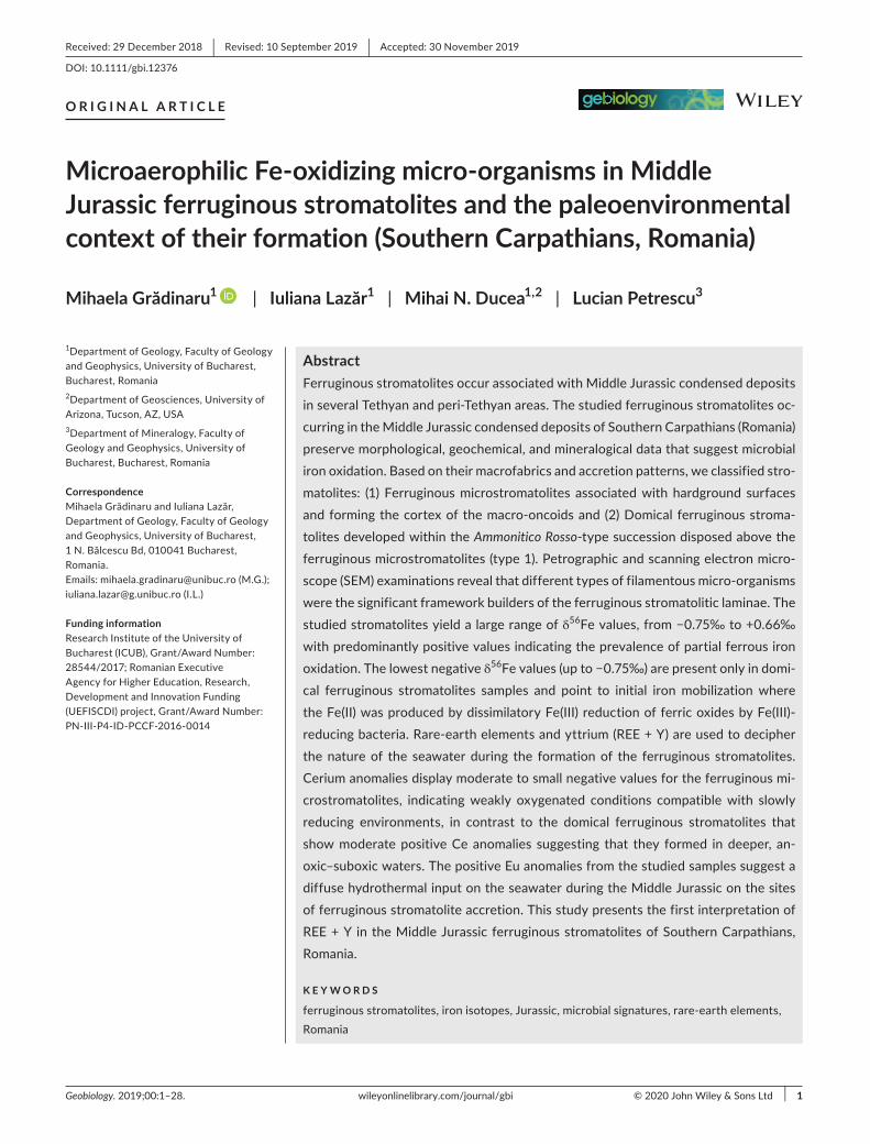
F I G U R E 1 Location of the studied sections: (a) general location of the studied zones within the Southern Carpathians (based on the geotectonic map of Romania, Săndulescu, 1984). (b) Location of the section Strunga Pass on the geological outline map of the Bucegi Mountains (based on Patrulius, 1969). (c) Location of the section Purcărete Valley on the geological outline map of the Rucăr zone (based on Patrulius, 1969). (d) Location of the section Saraorschi Valley on the geological outline map of the Almăjului Mountains (based on Codarcea et al., 1966)
| 3GRĂDINARU et Al.

4 | GRĂDINARU et Al.
represented by Triassic–Lower Cretaceous sediments. They are resting on a peri-Gondwanan terrane composed of Cambro-Silurian island arcs (Balintoni, Balica, Ducea, & Hann, 2014) meta-morphosed during two distinct events in the Paleozoic (Ducea et al., 2016). The Danubian Units consist of several variably metamor-phosed arc terranes of pan African affinity (Balintoni et al., 2014) and un-metamorphosed granitic rocks of earliest Permian age as well as Paleozoic cover rocks. Unlike the basement units of the Getic realm, there is no Variscan or younger metamorphism in the Danubian, in all making these basement terrains quite distinctive. Both represent continental margin rocks to the Jurassic Severin basin and both were covered by Upper Jurassic–Lower Cretaceous carbonate platforms prior to their juxtaposition during the mid-Cretaceous. The facies of the Jurassic to Lower Cretaceous sedi-mentary series reveal a complex paleogeography of the Danubian realm and were described by numerous authors starting with Răileanu (1953, 1960), Popa, Năstăseanu, and Antonescu (1977) Năstăseanu (1979) among others and a synthesis was presented by Grădinaru in Haas et al., 2011 and references therein).
During the Middle Jurassic, some areas of these units (e.g., Bucegi Mountains, Rucăr zone in the Getic unit) accumulated mixed carbonate–siliciclastic condensed deposits characterized by reduced stratigraphic thickness (less than one meter to few tens of meters) compared with the stratigraphic thickness of the con-temporaneous expanded successions (of hundreds of meters, e.g., Reșita-Moldova Nouă area in the western part of the Getic unit in the Southern Carpathians). These successions contain condensed beds associated with diachronous hardgrounds which reveal signs of reduced sedimentation rates, omission, erosion, in situ rework-ing, taphonomic condensation, and synsedimentary cementation (Lazăr et al., 2013).
Three Middle Jurassic sections from the Southern Carpathians have been studied: Strunga Pass (Bucegi Mountains, Figure 1b), Purcărete Valley (Rucăr zone, Figure 1c) from Getic Unit and Saraorschi Valley (Almăjului Mountains, Figure 1d) from Danubian Units.
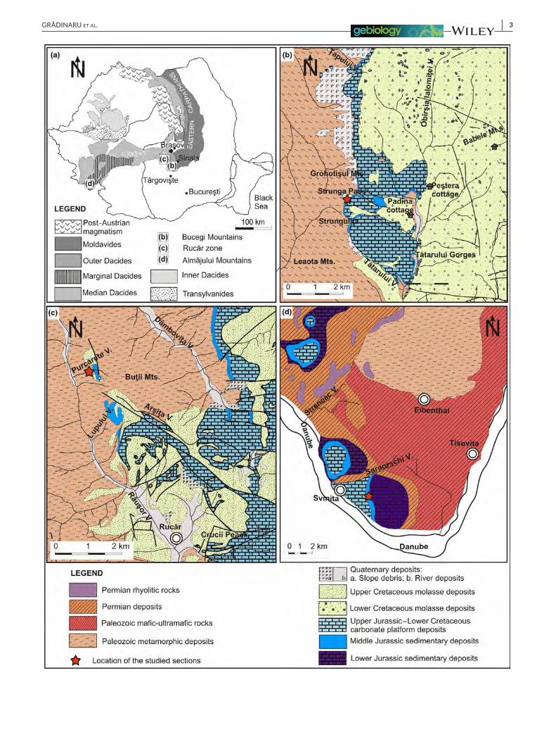
Strunga Pass is located on the western slope of the Bucegi Mountains. The biostratigraphy and lithostratigraphy of this sec-tion were presented in detail by Patrulius (1969) and Lazăr (2006). The lower part of the succession is represented by yellowish ooidal bioclastic grainstone and ooidal bioclastic grainstone–packstone (2.5–5.5 m thick) with thin, discontinuous, quartzitic micro-con-glomerate intercalations (Figures 2a and 3a,h). The top of these deposits is marked by a sharp hardground discontinuity (Figures 2a and 3a). The next bed (0.65–1 m thick) has a nodular aspect and consists of oncoidal floatstone, respectively, gray-green to reddish bioclastic ooidal packstone–grainstone (Figures 2a–d and 3e–g) with cavities and fractures lined by Fems (type 1) and filled with bioclastic wackestone–packstone (Figures 2a and 3b,d). This bed contains numerous ferruginous macro-oncoids (Figures 2b–e and 3e), ferruginous ooids and a fossil assemblage with ammonites, belemnites along with representatives of benthic faunas (rare sponges, bivalves, gastropods, brachiopods, echinoids, crinoids,
and extremely rare solitary corals; Lazăr et al., 2013). The top of this bed is represented by an uneven discontinuity hardground surface connected to cavities, fissures, and fractures developed within the underlying bed (Figure 2a,c,d). The hardground surface and the walls of the cavities and fissures are encrusted with 0.5- to 3-cm-thick ferruginous microstromatolites (Fems) (Figures 2b–e and 3d–g). This last unit represents a condensed bed correspond-ing to the Early Bathonian–Early Callovian time interval consid-ering the ammonite fauna accumulated in this bed (cf. Patrulius, 1969). The mixture of non-contemporaneous fossil specimens (belonging to several biozones) with different degrees of pres-ervation, as well as the occurrence of macro-oncoids that have nuclei represented by ammonite steinkerns with different preser-vation states, indicate faunal condensation (sensu Fürsich, 1978) and taphonomic condensation (sensu Gómez & Fernández-López, 1994). The overlying unit (1–2.5 m thick) is represented by red marly limestones with rhyncholites and foraminifera (Figure 3b) (Middle Callovian–Oxfordian, Neagu, Manea, & Gavrilescu, 1983), followed by 0.5- to 1-m-thick Middle Oxfordian jasper, radiolarites and 0.8-m-thick cherty limestones (Beccaro & Lazăr, 2007).
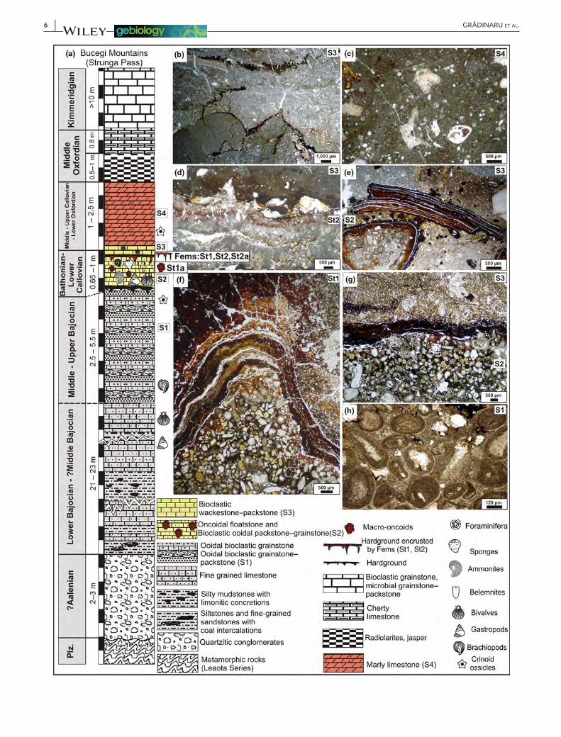
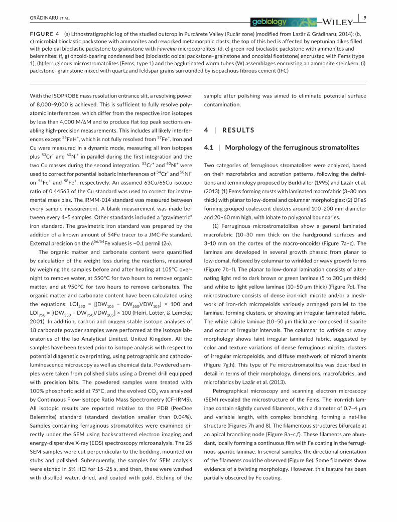
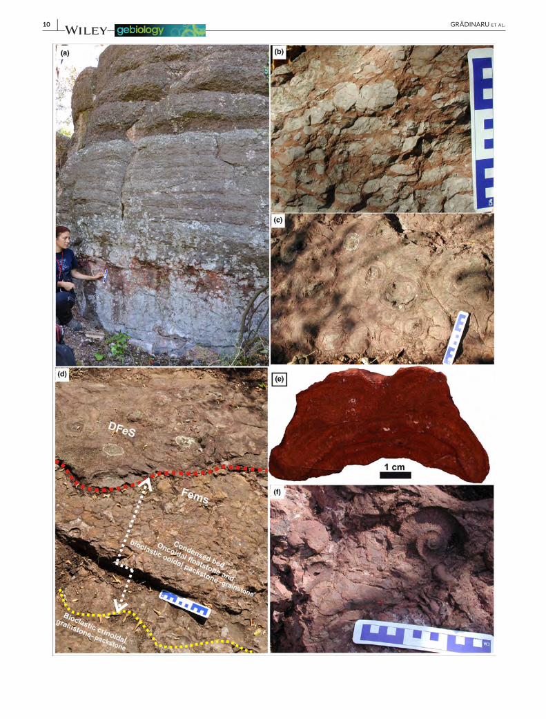
In the Rucăr zone, the Middle Jurassic succession from Purcărete Valley (Figure 4a) (a tributary of Râușor Valley) comprises of mixed siliciclastic–carbonate rocks (coarse sandstones and fine grained sandy limestones with discontinuous quartzite conglomerate inter-calations; 16–21 m thick), followed upwards by yellowish bioclastic ooidal limestones (packstone–grainstone mixed with quartz and feldspar grains, Figure 4i, 2.5–5.5 m thick). The top of this unit is marked by a sharp hardground discontinuity, encrusted, with 1- to 2-cm-thick ferruginous crusts (Figures 2f and 4h) yielding aggluti-nated polychaete worm tubes, rare bryozoan colonies, crinoid os-sicles, echinoid spines, small solitary corals, barnacles, fragmented belemnites, and reworked ammonites, along with ferruginous mac-ro-oncoids and abundant ferruginous ooids (Lazăr & Grădinaru, 2014). The overlying bed (0.7–0.8 m thick) consists of gray–green to yellowish bioclastic ooidal packstone–grainstone and oncoidal float-stone (Figure 4f,g). This bed reveals numerous cavities and fractures filled with bioclastic packstone (Figure 2f). The nodular aspect of this bed is generated by the numerous ferruginous macro-oncoids (ranging from 1 up to 7 cm in diameter); the bed contain also nu-merous ferruginous ooids, reworked ammonites, belemnites, cri-noids, oysters, along with rare gastropods, brachiopods, echinoids, sponges, and solitary corals (Figure 4f). This bed represents a con-densed unit corresponding to the (?)Bathonian–Callovian interval (Patrulius, 1969; Patrulius et al., 1980). The early lithification of the condensed unit is evidenced by the precipitation of several types of early cement (isopachous fibrous and radiaxial cements), which appear synchronously with the colonization of the substrate by the macro- and microfauna. The top of the condensed bed is marked by a second, uneven hardground discontinuity covered by 0.5- to 2-cm-thick Fems (Figure 4f). The next unit is represented by green-red bioclastic packstone with ammonites (1.6 m thick, Figure 4d,e), red to pink microbial bioclastic packstone (0.86 m thick, Figure 4b,c) containing coarse to medium sized, angular rock fragments derived

| 5GRĂDINARU et Al.
F I G U R E 2 (a–e) Bucegi Mountains: (a) outcrop view of the oncoid-bearing condensed bed bounded by two sharp discontinuities (dotted lines); red dotted line indicate the hardground surface encrusted by Fems; (b, c) surface view of the oncoid-bearing condensed bed encrusted with Fems (type 1); (c) fracture encrusted with Fems (yellow dotted line); dotted red line outlines a macro-oncoid; (d) uneven hardground surface encrusted with 0.5- to 3-cm-thick Fems crust (white arrows); (e) polished slab on the oncoid-bearing condensed bed showing cavities filled with Fems (type 1) (white arrows); (f) Rucăr zone (Purcărete Valley): surface view of the oncoid-bearing condensed bed: yellow dotted lines outline the irregular cavities filled with ferruginous microstromatolites (Fems, type 1) containing agglutinated worm tubes (yellow arrows); irregular cavity encrusted with Fems and filled with green bioclastic packstone (red dotted lines); black arrows indicate fragments of large bivalves shells encrusted by Fems
6 | GRĂDINARU et Al.

| 7GRĂDINARU et Al.
from the underlying lithostratigraphic units and a mixture of numer-ous non-contemporaneous fossil specimens of ammonites from the Middle to Late Callovian time interval (Grigore, Lazăr, & Gheucă, 2015); these are followed by 1.25-m-thin-bedded pink limestones (bioclastic packstone–wackestone) and by Oxfordian cherty lime-stones (3–4 m thick).
The studied succession from Saraorschi Valley (Figure 6a) (a left tributary of the Danube River) belongs to the Danubian Units, Sirinia zone. The Jurassic deposits in this zone are represented by:
• Lower Jurassic calcareous–siliciclastic succession containing rich bivalves, brachiopods, nautiloids, and ammonite faunas developed within Late Sinemurian–Toarcian interval (Callomon & Grădinaru, 2005; Popa et al., 1977; Popa & Patrulius, 1996; Răileanu, 1953);
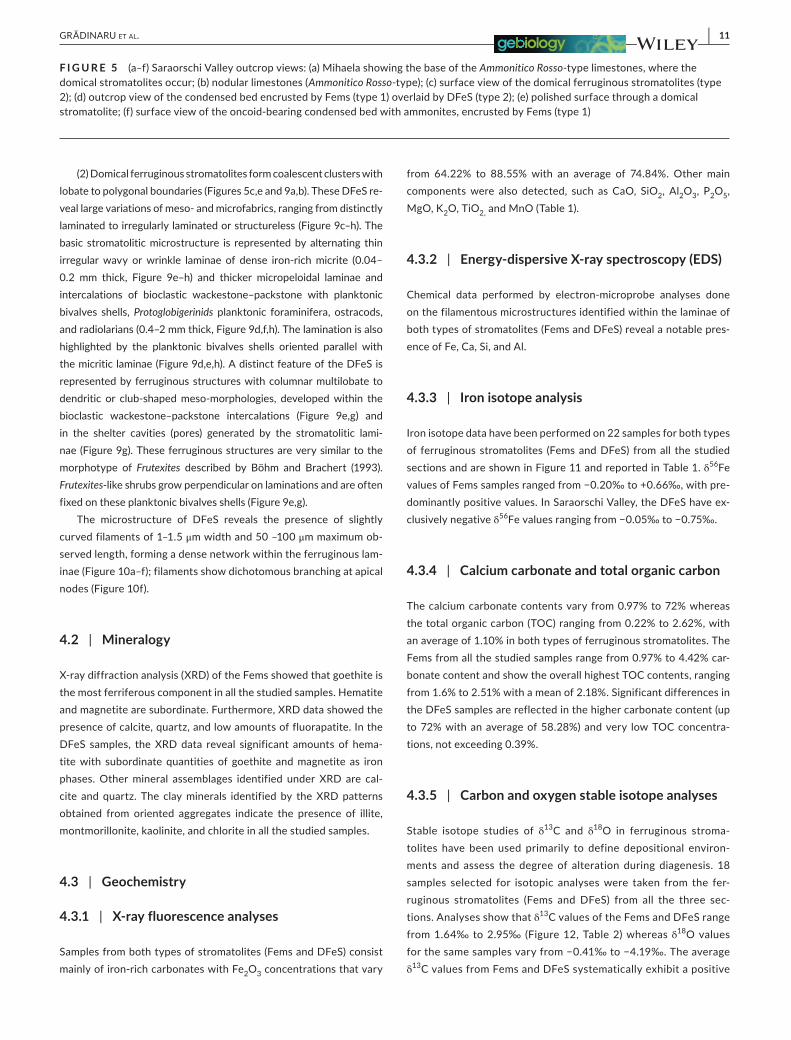
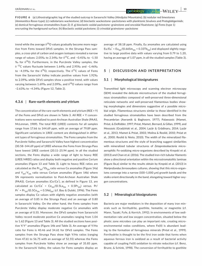
• The Middle Jurassic succession starts with whitish-gray quartz sandstones, assigned without paleontological data to the Aalenian by Răileanu (1953) and Popa et al. (1977). These are followed by Bajocian gray and cherry-red crinoidal grainstone–packstone (Figures 5d and 6i) and bioclastic ooidal packstone (Figure 6h) (al-most 5–10 m thick), followed by a condensed bed (0.15–0.50 m thick) of Fe-ooidal oncoidal bioclastic limestones (Figures 5d,f and 6f,g) very rich in ammonites belonging to different biozones. The ammonite assemblage indicates Bathonian–Early Callovian time interval (cf. Năstăseanu, Bercia, Iancu, Vlad, & Hârtopanu, 1981; Patrulius & Popa, 1971; Răileanu, 1953, 1960) or the Middle Bathonian (cf. Galácz, 1994). This bed contains ferruginous mac-ro-oncoids, and the top is marked by a hardground surface cov-ered by Fems of type 1 (1–3 cm thick, Figure 5d). The condensed bed reveals early lithification evidenced by the precipitation of early cements such as syntaxial overgrowths surrounding echi-noderm grains which are developed laterally and contempora-neously with the isopachous fibrous cement that surrounds the grains and intergranular spaces (Grădinaru, 2011). Just above the hardground encrusted with Fems (Figures 5d and 6g), other types of spectacular domical ferruginous stromatolites (DFeS) (type 2, Figures 5c,d,e and 6e) are developed, representing the base of the next unit composed of nodular red limestones (Figure 5a,b; Ammonitico Rosso-type, Middle-Upper Callovian, cf. Răileanu, 1953, Năstăseanu et al., 1981). These nodular limestones consist of bioclastic wackestone–packstones with planktonic bivalves, benthic (Lenticulina) and planktonic (Protoglobigerina) foramin-ifera, ostracods, radiolarians, peloids (Figure 6b,d). These are overlain by decimeter beds of grayish limestone and red or green-ish limetones with cherts (Figure 6c, Oxfordian, cf. Năstăseanu et al., 1981).
3 | SAMPLES AND METHODS
In the studied sections, more than 250 samples have been col-lected from the condensed unit and the adjacent strata. Microfacies types and the meso- and microstructures of the ferruginous stro-matolites were investigated in 120 thin sections under petrographic and binocular microscopes; 35 polished slabs were examined under cathodoluminescence (CL) microscopy. The chemical and mineralog-ical analyses were performed on 22 samples (Figure S1, supporting information) from all the studied sections.
The chemical and mineralogical composition was determined by X-ray fluorescence analysis (XRF), using a Horiba XGT 7000 device for major elements, and X-ray diffraction data, using a PANalytical's X'Pert PRO (microXRD) diffractometer. In addition, oriented clay ag-gregates were prepared and transferred on to a glass slide, dried at room temperature prior to X-ray scanning. Removal of calcite and separation of clay fractions were carried out according to Ostrum (1961). The samples have been also treated with ethylene glycol sol-vation and heated at 330 and 500°C to expand the smectite.
Rare-earth elements and yttrium (REE + Y) were determined in solution mode using a Thermo X-Series-2 Quadrupole Inductively Coupled Plasma-Mass Spectrometer at the University of Arizona (analytical details are presented in Rosel et al., 2013). Prior to anal-ysis, sample aliquots were dissolved in mixtures of HF-HNO3 ultra clean concentrated acids in a clean laboratory environment. Half of the sample aliquots were used for ICP-MS analyses and were re-dissolved in dilute HCl acid (1%) for introduction into the instru-ment nebulizer. The remainder of the sample was further processed for iron isotopic analysis. For REE + Y measurement, instrumen-tal drift was corrected using Ru, In, and Re as internal standards. Uncertainties, calculated by repeated analyses of external standards (BCR-2), are ± 2%.
Aliquots for iron isotope measurements were passed through standard anion columns for chemical separation/purification after dissolution. Samples and standards were diluted to 3 ppm total iron. A Cu standard (JMC-ICP solution Specpure Lot No. 200536E) was added to each sample before analysis to a concentration of 3 ppm (Arnold, Weyer, & Anbar, 2004). Measurement of iron isotopes was performed at the University of Arizona on an ISOPROBE multicollec-tor ICP-MS instrument. Measurements were performed in multicol-lector mode at the highest mass resolution following the procedures described elsewhere by Arnold et al. (2004). Each detector was po-sitioned individually to discriminate the polyatomic interferences (mainly 40Ar16O+, 40Ar16OH+, and 40Ar14N+) at the edge of the de-tector slits so that only the undisturbed iron beams were detected.
F I G U R E 3 (a) Lithostratigraphic log of the studied outcrop in Strunga Pass (Bucegi Mountains) (modified from Lazăr et al., 2013); (b–h) microphotographs of the representative microfacies in the studied section: (b) bioclastic wackestone–packstone; (c) marly limestone (bioclastic wackestone–packstone); (d) ferruginous microstromatolites growing downward from the walls of a cavity filled with bioclastic wackestone–packstone; note the calcite pinnacles grew on the base of the stromatolitic laminae; (e) oncoidal floatstone; (f, g) top of the condensed bed (here represented by bioclastic ooidal packstone–grainstone) affected by an uneven hardground surface encrusted with Fem; (h) ooidal bioclastic grainstone–packstone
8 | GRĂDINARU et Al.
| 9GRĂDINARU et Al.
With the ISOPROBE mass resolution entrance slit, a resolving power of 8,000–9,000 is achieved. This is sufficient to fully resolve poly-atomic interferences, which differ from the respective iron isotopes by less than 4,000 M/ΔM and to produce flat top peak sections en-abling high-precision measurements. This includes all likely interfer-ences except 56FeH+, which is not fully resolved from 57Fe+. Iron and Cu were measured in a dynamic mode, measuring all iron isotopes plus 53Cr+ and 60Ni+ in parallel during the first integration and the two Cu masses during the second integration. 53Cr+ and 60Ni+ were used to correct for potential isobaric interferences of 54Cr+ and 58Ni+ on 54Fe+ and 58Fe+, respectively. An assumed 63Cu/65Cu isotope ratio of 0.44563 of the Cu standard was used to correct for instru-mental mass bias. The IRMM-014 standard was measured between every sample measurement. A blank measurement was made be-tween every 4–5 samples. Other standards included a “gravimetric” iron standard. The gravimetric iron standard was prepared by the addition of a known amount of 54Fe tracer to a JMC-Fe standard. External precision on the δ56/54Fe values is ~0.1 permil (2σ).
The organic matter and carbonate content were quantified by calculation of the weight loss during the reactions, measured by weighing the samples before and after heating at 105°C over-night to remove water, at 550°C for two hours to remove organic matter, and at 950°C for two hours to remove carbonates. The organic matter and carbonate content have been calculated using the equations: LOI550 = [(DW105 − DW550)/DW105] × 100 and LOI950 = [(DW550 – DW950)/DW105] × 100 (Heiri, Lotter, & Lemcke, 2001). In addition, carbon and oxygen stable isotope analyses of 18 carbonate powder samples were performed at the isotope lab-oratories of the Iso-Analytical Limited, United Kingdom. All the samples have been tested prior to isotope analysis with respect to potential diagenetic overprinting, using petrographic and cathodo-luminescence microscopy as well as chemical data. Powdered sam-ples were taken from polished slabs using a Dremel drill equipped with precision bits. The powdered samples were treated with 100% phosphoric acid at 75°C, and the evolved CO2 was analyzed by Continuous Flow-Isotope Ratio Mass Spectrometry (CF-IRMS). All isotopic results are reported relative to the PDB (PeeDee Belemnite) standard (standard deviation smaller than 0.04%). Samples containing ferruginous stromatolites were examined di-rectly under the SEM using backscattered electron imaging and energy-dispersive X-ray (EDS) spectroscopy microanalysis. The 25 SEM samples were cut perpendicular to the bedding, mounted on stubs and polished. Subsequently, the samples for SEM analysis were etched in 5% HCl for 15–25 s, and then, these were washed with distilled water, dried, and coated with gold. Etching of the
sample after polishing was aimed to eliminate potential surface contamination.
4 | RESULTS
4.1 | Morphology of the ferruginous stromatolites
Two categories of ferruginous stromatolites were analyzed, based on their macrofabrics and accretion patterns, following the defini-tions and terminology proposed by Burkhalter (1995) and Lazăr et al. (2013): (1) Fems forming crusts with laminated macrofabric (3–30 mm thick) with planar to low-domal and columnar morphologies; (2) DFeS forming grouped coalescent clusters around 100–200 mm diameter and 20–60 mm high, with lobate to polygonal boundaries.
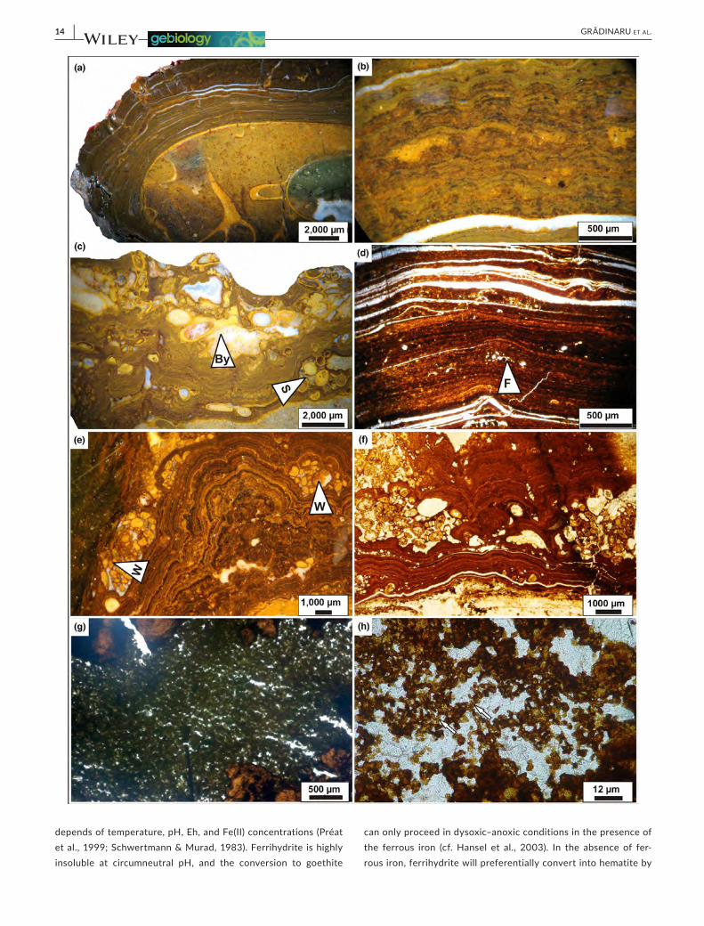
(1) Ferruginous microstromatolites show a general laminated macrofabric (10–30 mm thick on the hardground surfaces and 3–10 mm on the cortex of the macro-oncoids) (Figure 7a–c). The laminae are developed in several growth phases: from planar to low-domal, followed by columnar to wrinkled or wavy growth forms (Figure 7b–f). The planar to low-domal lamination consists of alter-nating light red to dark brown or green laminae (5 to 300 μm thick) and white to light yellow laminae (10–50 μm thick) (Figure 7d). The microstructure consists of dense iron-rich micrite and/or a mesh-work of iron-rich micropeloids variously arranged parallel to the laminae, forming clusters, or showing an irregular laminated fabric. The white calcite laminae (10–50 μm thick) are composed of sparite and occur at irregular intervals. The columnar to wrinkle or wavy morphology shows faint irregular laminated fabric, suggested by color and texture variations of dense ferruginous micrite, clusters of irregular micropeloids, and diffuse meshwork of microfilaments (Figure 7g,h). This type of Fe microstromatolites was described in detail in terms of their morphology, dimensions, macrofabrics, and microfabrics by Lazăr et al. (2013).
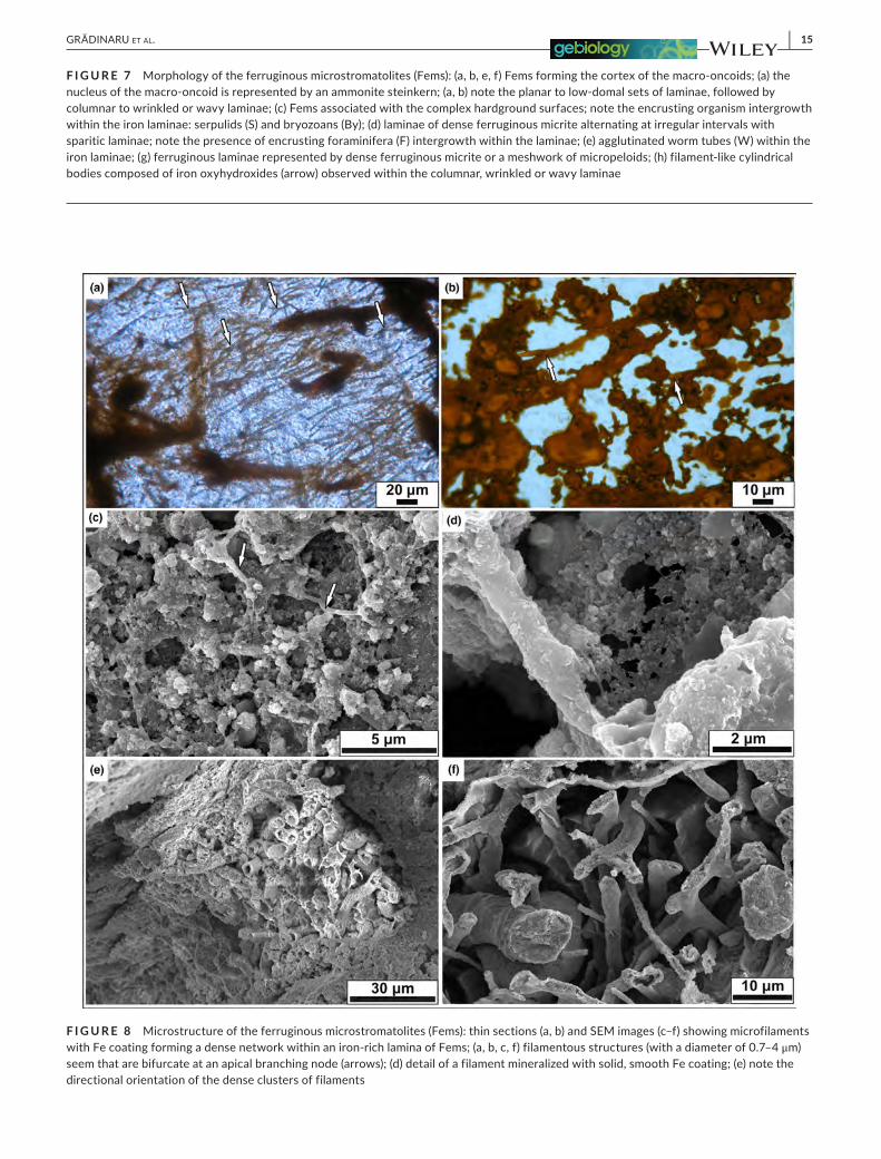
Petrographical microscopy and scanning electron microscopy (SEM) revealed the microstructure of the Fems. The iron-rich lam-inae contain slightly curved filaments, with a diameter of 0.7–4 μm and variable length, with complex branching, forming a net-like structure (Figures 7h and 8). The filamentous structures bifurcate at an apical branching node (Figure 8a–c,f). These filaments are abun-dant, locally forming a continuous film with Fe coating in the ferrugi-nous-sparitic laminae. In several samples, the directional orientation of the filaments could be observed (Figure 8e). Some filaments show evidence of a twisting morphology. However, this feature has been partially obscured by Fe coating.
F I G U R E 4 (a) Lithostratigraphic log of the studied outcrop in Purcărete Valley (Rucăr zone) (modified from Lazăr & Grădinaru, 2014); (b, c) microbial bioclastic packstone with ammonites and reworked metamorphic clasts; the top of this bed is affected by neptunian dikes filled with peloidal bioclastic packstone to grainstone with Favreina microcoprolites; (d, e) green-red bioclastic packstone with ammonites and belemnites; (f, g) oncoid-bearing condensed bed (bioclastic ooidal packstone–grainstone and oncoidal floatstone) encrusted with Fems (type 1); (h) ferruginous microstromatolites (Fems, type 1) and the agglutinated worm tubes (W) assemblages encrusting an ammonite steinkern; (i) packstone–grainstone mixed with quartz and feldspar grains surrounded by isopachous fibrous cement (IFC)
10 | GRĂDINARU et Al.
| 11GRĂDINARU et Al.
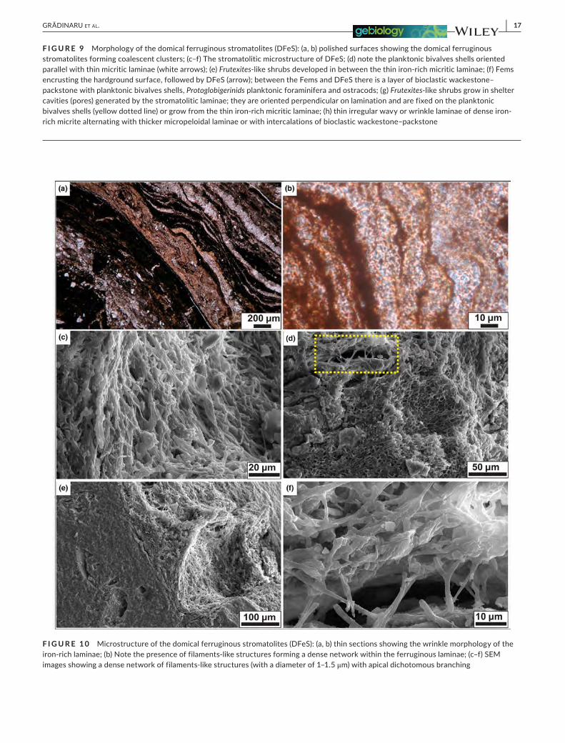
(2) Domical ferruginous stromatolites form coalescent clusters with lobate to polygonal boundaries (Figures 5c,e and 9a,b). These DFeS re-veal large variations of meso- and microfabrics, ranging from distinctly laminated to irregularly laminated or structureless (Figure 9c–h). The basic stromatolitic microstructure is represented by alternating thin irregular wavy or wrinkle laminae of dense iron-rich micrite (0.04–0.2 mm thick, Figure 9e–h) and thicker micropeloidal laminae and intercalations of bioclastic wackestone–packstone with planktonic bivalves shells, Protoglobigerinids planktonic foraminifera, ostracods, and radiolarians (0.4–2 mm thick, Figure 9d,f,h). The lamination is also highlighted by the planktonic bivalves shells oriented parallel with the micritic laminae (Figure 9d,e,h). A distinct feature of the DFeS is represented by ferruginous structures with columnar multilobate to dendritic or club-shaped meso-morphologies, developed within the bioclastic wackestone–packstone intercalations (Figure 9e,g) and in the shelter cavities (pores) generated by the stromatolitic lami-nae (Figure 9g). These ferruginous structures are very similar to the morphotype of Frutexites described by Böhm and Brachert (1993). Frutexites-like shrubs grow perpendicular on laminations and are often fixed on these planktonic bivalves shells (Figure 9e,g).
The microstructure of DFeS reveals the presence of slightly curved filaments of 1–1.5 μm width and 50 –100 μm maximum ob-served length, forming a dense network within the ferruginous lam-inae (Figure 10a–f); filaments show dichotomous branching at apical nodes (Figure 10f).
4.2 | Mineralogy
X-ray diffraction analysis (XRD) of the Fems showed that goethite is the most ferriferous component in all the studied samples. Hematite and magnetite are subordinate. Furthermore, XRD data showed the presence of calcite, quartz, and low amounts of fluorapatite. In the DFeS samples, the XRD data reveal significant amounts of hema-tite with subordinate quantities of goethite and magnetite as iron phases. Other mineral assemblages identified under XRD are cal-cite and quartz. The clay minerals identified by the XRD patterns obtained from oriented aggregates indicate the presence of illite, montmorillonite, kaolinite, and chlorite in all the studied samples.
4.3 | Geochemistry
4.3.1 | X-ray fluorescence analyses
Samples from both types of stromatolites (Fems and DFeS) consist mainly of iron-rich carbonates with Fe2O3 concentrations that vary
from 64.22% to 88.55% with an average of 74.84%. Other main components were also detected, such as CaO, SiO2, Al2O3, P2O5, MgO, K2O, TiO2, and MnO (Table 1).
4.3.2 | Energy-dispersive X-ray spectroscopy (EDS)
Chemical data performed by electron-microprobe analyses done on the filamentous microstructures identified within the laminae of both types of stromatolites (Fems and DFeS) reveal a notable pres-ence of Fe, Ca, Si, and Al.
4.3.3 | Iron isotope analysis
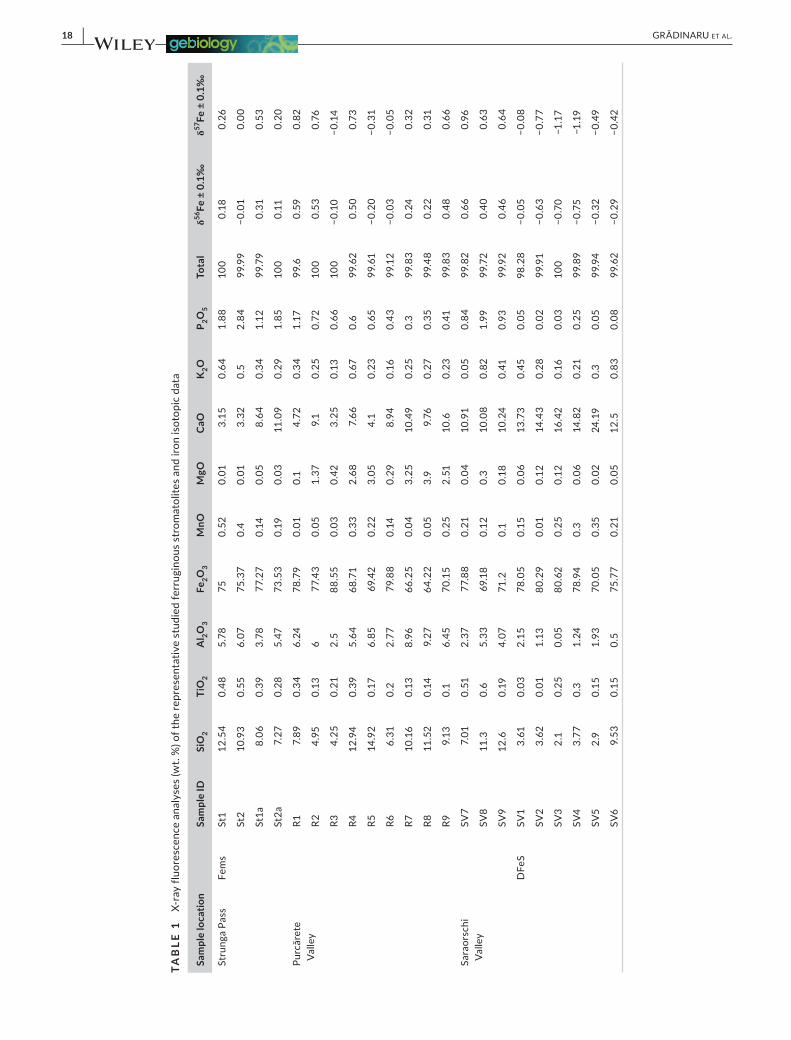
Iron isotope data have been performed on 22 samples for both types of ferruginous stromatolites (Fems and DFeS) from all the studied sections and are shown in Figure 11 and reported in Table 1. δ56Fe values of Fems samples ranged from −0.20‰ to +0.66‰, with pre-dominantly positive values. In Saraorschi Valley, the DFeS have ex-clusively negative δ56Fe values ranging from −0.05‰ to −0.75‰.
4.3.4 | Calcium carbonate and total organic carbon
The calcium carbonate contents vary from 0.97% to 72% whereas the total organic carbon (TOC) ranging from 0.22% to 2.62%, with an average of 1.10% in both types of ferruginous stromatolites. The Fems from all the studied samples range from 0.97% to 4.42% car-bonate content and show the overall highest TOC contents, ranging from 1.6% to 2.51% with a mean of 2.18%. Significant differences in the DFeS samples are reflected in the higher carbonate content (up to 72% with an average of 58.28%) and very low TOC concentra-tions, not exceeding 0.39%.
4.3.5 | Carbon and oxygen stable isotope analyses
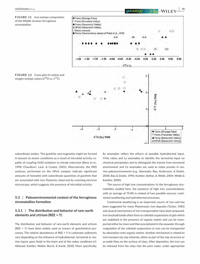
Stable isotope studies of δ13C and δ18O in ferruginous stroma-tolites have been used primarily to define depositional environ-ments and assess the degree of alteration during diagenesis. 18 samples selected for isotopic analyses were taken from the fer-ruginous stromatolites (Fems and DFeS) from all the three sec-tions. Analyses show that δ13C values of the Fems and DFeS range from 1.64‰ to 2.95‰ (Figure 12, Table 2) whereas δ18O values for the same samples vary from −0.41‰ to −4.19‰. The average δ13C values from Fems and DFeS systematically exhibit a positive
F I G U R E 5 (a–f) Saraorschi Valley outcrop views: (a) Mihaela showing the base of the Ammonitico Rosso-type limestones, where the domical stromatolites occur; (b) nodular limestones (Ammonitico Rosso-type); (c) surface view of the domical ferruginous stromatolites (type 2); (d) outcrop view of the condensed bed encrusted by Fems (type 1) overlaid by DFeS (type 2); (e) polished surface through a domical stromatolite; (f) surface view of the oncoid-bearing condensed bed with ammonites, encrusted by Fems (type 1)

12 | GRĂDINARU et Al.
| 13GRĂDINARU et Al.
trend while the average δ18O values gradually become more nega-tive from Fems toward DFeS samples. In the Strunga Pass sam-ples, a cross-plot of carbon and oxygen isotopes revealed a narrow range of values: 2.05‰ to 2.14‰ for δ13C, and −0.41‰ to −1.50 ‰ for δ18O. Furthermore, in the Purcărete Valley samples, the δ 13C values fluctuate between 1.64‰ and 2.95‰ and −1.46‰ to −4.19‰ for the δ18O, respectively. The δ13C values of Fems from the Saraorschi Valley indicate positive values from 1.92‰ to 2.09‰ while DFeS samples show a positive trend, with values varying between 1.69‰ and 2.09‰, and δ18O values range from −2.63‰ to −4.34‰ (Figure 12, Table 2).
4.3.6 | Rare-earth elements and yttrium
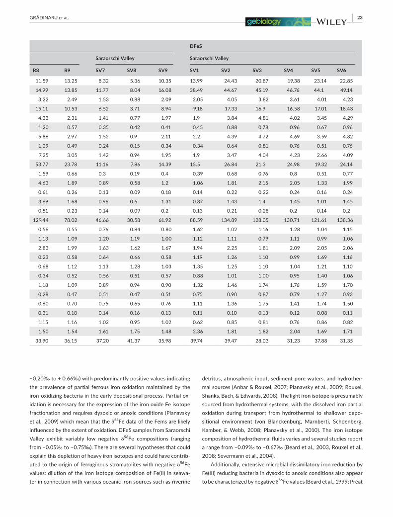
The concentration of the rare-earth elements and yttrium (REE + Y) of the Fems and DFeS are shown in Table 3. All REE + Y concen-trations were normalized to post-Archean Australian Shale (PAAS, McLennan, 1989). The total REE (ΣREE) contents for all samples range from 17.66 to 144.69 ppm, with an average of 79.89 ppm. Significant variations in ΣREE content are distinguished in differ-ent types of ferruginous stromatolites. The Fems samples from the Purcărete Valley and Saraorschi Valley have highest concentration (30.58–144.69 ppm) of ΣREE whereas the Fems from Strunga Pass have lowest ΣREE content (20.11–25.94 ppm). In all the studied sections, the Fems display a wide range of light to heavy REE (LREE/HREE) ratios and display both negative and positive Cerium anomalies (Figure 13 and Table 3). Light to heavy REE ratios are calculated as the PrSN/YbSN ratio versus Ce anomalies (Figure 14a) and YSN/YSN ratio versus Cerium anomalies (Figure 14b) where SN represents normalization to Post-Archean Australian Shale (PAAS). Cerium anomalies (Ce/Ce*), as defined in Figure 13, are calculated as Ce/Ce* = CeSN/(0.5LaSN + 0.5PrSN) versus. Pr/Pr* = PrSN/(0.5CeSN + 0.5NdSN; (cf. Bau & Dulski, 1996). The Fems samples display Ce values with slightly negative anomalies (with an average of 0.85 in the Strunga Pass) and an average of 0.80 in Saraorschi Valley. On the other hand, the Fems samples from Purcărete Valley display moderate negative Ce anomalies with an average of 0.55. Moreover, the DFeS samples from Saraorschi Valley record moderate positive Ce anomalies ranging from 1.04 to 1.62 (Figure 13 and Table 3). All the studied sections have posi-tive Y/Y* anomalies (Figure 14b and Table 3). An average of Y/Ho ratio for Fems is 40.46 and 34.61 for DFeS samples. The Fems samples from the Strunga Pass show high Y/Ho ratios (ranging from 47.61 to 56.75 with an average of 52.55) whereas the Fems samples from Purcărete Valley show an average of 35.85 ppm. In the Saraorschi Valley, the values for Fems samples display an
average of 38.18 ppm. Finally, Eu anomalies are calculated using Eu/Eu* = EuSN/(0.66SmSN + 0.33TbSN) and displayed slightly nega-tive to large positive data with values varying from 0.79 to 1.20, having an average of 1.07 ppm, in all the studied samples (Table 3).
5 | DISCUSSION AND INTERPRETATION
5.1 | Morphological biosignatures
Transmitted light microscopy and scanning electron microscopy (SEM) revealed the delicate microstructure of the studied ferrugi-nous stromatolites composed of well-preserved three-dimensional reticulate networks and well-preserved filamentous bodies show-ing morphologies and dimensions suggestive of a possible micro-bial origin. Filamentous structures similar to those identified in the studied ferruginous stromatolites have been described from the Precambrian (Awramik & Baghoorn, 1977), Palaeozoic (Mamet, Préat, & DeRidder, 1997; Préat, Mamet, Bernard, & Gillan, 1999), and Mesozoic (Gradziński et al., 2004; Lazăr & Grădinaru, 2014; Lazăr et al., 2013; Mamet & Préat, 2003; Molina & Reolid, 2010; Préat et al., 2000; Reolid & Nieto, 2010). The width of the mineralized fila-mentous structures and the style of branching suggest similarities with mineralized tubular structures of Zetaproteobacteria micro-aerophilic Fe-oxidizing micro-organisms described by Krepski et al. (2013) and Chan et al. (2016). The studied iron-rich tubular filaments show a directional orientation within the microstromatolitic laminae (Figure 8a,e) similar to the results obtain by Krepski et al. (2013) in Mariprofundus ferrooxydans cultures, showing that this micro-organ-isms converge into a narrow (100–1,000 μm) growth bands and the stalks orient directionally in the band, elongating toward higher oxy-gen concentrations.
5.2 | Mineralogical biosignatures
Bacteria are major mediators in the deposition of many iron min-erals such as ferrihydrite, goethite, hematite, or magnetite (cf. Mann, Tazaki, Fyfe, & Kerrich, 1992). In environments of low sedi-mentation rate and low oxygen concentration, situated below the photic zone microbes can play an important role, creating micro-environmental redox conditions, where Fe(II) is abundant lead-ing to the formation of ferruginous minerals (Préat et al., 1999). Ferrihydrite is thought to be the first iron oxide that forms when aqueous ferrous iron is oxidized as a result of bacterial activity capable of coupling Fe(II) oxidation to nitrate reduction (cf. Benz, Brune, & Schink, 1998). The conversion of ferrihydrite to goethite
F I G U R E 6 (a) Lithostratigraphic log of the studied outcrop in Saraorschi Valley (Almăjului Mountains); (b) nodular red limestones (Ammonitico Rosso-type); (c) radiolarians wackestone; (d) bioclastic wackestone–packstones with planktonic bivalves and Protoglobigerinids; (e) domical ferruginous stromatolites (type 2); (f, g) bioclastic ooidal packstone–grainstone and oncoidal floatstone; (g) Fems (type 1) encrusting the hardground surface; (h) Bioclastic ooidal packstone; (i) crinoidal grainstone–packstone
14 | GRĂDINARU et Al.
depends of temperature, pH, Eh, and Fe(II) concentrations (Préat et al., 1999; Schwertmann & Murad, 1983). Ferrihydrite is highly insoluble at circumneutral pH, and the conversion to goethite
can only proceed in dysoxic–anoxic conditions in the presence of the ferrous iron (cf. Hansel et al., 2003). In the absence of fer-rous iron, ferrihydrite will preferentially convert into hematite by
| 15GRĂDINARU et Al.
F I G U R E 7 Morphology of the ferruginous microstromatolites (Fems): (a, b, e, f) Fems forming the cortex of the macro-oncoids; (a) the nucleus of the macro-oncoid is represented by an ammonite steinkern; (a, b) note the planar to low-domal sets of laminae, followed by columnar to wrinkled or wavy laminae; (c) Fems associated with the complex hardground surfaces; note the encrusting organism intergrowth within the iron laminae: serpulids (S) and bryozoans (By); (d) laminae of dense ferruginous micrite alternating at irregular intervals with sparitic laminae; note the presence of encrusting foraminifera (F) intergrowth within the laminae; (e) agglutinated worm tubes (W) within the iron laminae; (g) ferruginous laminae represented by dense ferruginous micrite or a meshwork of micropeloids; (h) filament-like cylindrical bodies composed of iron oxyhydroxides (arrow) observed within the columnar, wrinkled or wavy laminae
F I G U R E 8 Microstructure of the ferruginous microstromatolites (Fems): thin sections (a, b) and SEM images (c–f) showing microfilaments with Fe coating forming a dense network within an iron-rich lamina of Fems; (a, b, c, f) filamentous structures (with a diameter of 0.7–4 μm) seem that are bifurcate at an apical branching node (arrows); (d) detail of a filament mineralized with solid, smooth Fe coating; (e) note the directional orientation of the dense clusters of filaments
16 | GRĂDINARU et Al.
dehydration (Schwertmann & Murad, 1983). In addition, dissimi-latory iron-reducing bacteria that can oxidize organic matter and reduce Fe(III) to Fe(II) under anoxic conditions, convert amorphous
ferric oxide to magnetite (Lovely, Stolz, Nord, & Phillips, 1987). In all the Fems samples, the XRD analyses revealed that ferrifer-ous component is goethite while hematite and magnetite are the
| 17GRĂDINARU et Al.
F I G U R E 9 Morphology of the domical ferruginous stromatolites (DFeS): (a, b) polished surfaces showing the domical ferruginous stromatolites forming coalescent clusters; (c–f) The stromatolitic microstructure of DFeS; (d) note the planktonic bivalves shells oriented parallel with thin micritic laminae (white arrows); (e) Frutexites-like shrubs developed in between the thin iron-rich micritic laminae; (f) Fems encrusting the hardground surface, followed by DFeS (arrow); between the Fems and DFeS there is a layer of bioclastic wackestone–packstone with planktonic bivalves shells, Protoglobigerinids planktonic foraminifera and ostracods; (g) Frutexites-like shrubs grow in shelter cavities (pores) generated by the stromatolitic laminae; they are oriented perpendicular on lamination and are fixed on the planktonic bivalves shells (yellow dotted line) or grow from the thin iron-rich micritic laminae; (h) thin irregular wavy or wrinkle laminae of dense iron-rich micrite alternating with thicker micropeloidal laminae or with intercalations of bioclastic wackestone–packstone
F I G U R E 1 0 Microstructure of the domical ferruginous stromatolites (DFeS): (a, b) thin sections showing the wrinkle morphology of the iron-rich laminae; (b) Note the presence of filaments-like structures forming a dense network within the ferruginous laminae; (c–f) SEM images showing a dense network of filaments-like structures (with a diameter of 1–1.5 μm) with apical dichotomous branching
18 | GRĂDINARU et Al.
TAB
LE 1
X-
ray
fluor
esce
nce
anal
yses
(wt.
%) o
f the
repr
esen
tativ
e st
udie
d fe
rrug
inou
s st
rom
atol
ites
and
iron
isot
opic
dat
a
Sam
ple
loca
tion
Sam
ple
IDSi
O2
TiO
2A
l 2O3
Fe2O
3M
nOM
gOC
aOK
2OP 2O
5To
tal
δ56Fe
± 0
.1‰
δ57Fe
± 0
.1‰
Stru
nga
Pass
Fem
sSt
112
.54
0.48
5.78
750.
520.
013.
150.
641.
8810
00.
180.
26
St2
10.9
30.
556.
0775
.37
0.4
0.01
3.32
0.5
2.84
99.9
9−0
.01
0.00
St1a
8.06
0.39
3.78
77.2
70.
140.
058.
640.
341.
1299
.79
0.31
0.53
St2a
7.27
0.28
5.47
73.5
30.
190.
0311
.09
0.29
1.85
100
0.11
0.20
Purc
ăret
e Va
lley
R17.
890.
346.
2478
.79
0.01
0.1
4.72
0.34
1.17
99.6
0.59
0.82
R24.
950.
136
77.4
30.
051.
379.
10.
250.
7210
00.
530.
76
R34.
250.
212.
588
.55
0.03
0.42
3.25
0.13
0.66
100
−0.1
0−0
.14
R412
.94
0.39
5.64
68.7
10.
332.
687.
660.
670.
699
.62
0.50
0.73
R514
.92
0.17
6.85
69.4
20.
223.
054.
10.
230.
6599
.61
−0.2
0−0
.31
R66.
310.
22.
7779
.88
0.14
0.29
8.94
0.16
0.43
99.1
2−0
.03
−0.0
5
R710
.16
0.13
8.96
66.2
50.
043.
2510
.49
0.25
0.3
99.8
30.
240.
32
R811
.52
0.14
9.27
64.2
20.
053.
99.
760.
270.
3599
.48
0.22
0.31
R99.
130.
16.
4570
.15
0.25
2.51
10.6
0.23
0.41
99.8
30.
480.
66
Sara
orsc
hi
Valle
ySV
77.
010.
512.
3777
.88
0.21
0.04
10.9
10.
050.
8499
.82
0.66
0.96
SV8
11.3
0.6
5.33
69.1
80.
120.
310
.08
0.82
1.99
99.7
20.
400.
63
SV9
12.6
0.19
4.07
71.2
0.1
0.18
10.2
40.
410.
9399
.92
0.46
0.64
DFe
SSV
13.
610.
032.
1578
.05
0.15
0.06
13.7
30.
450.
0598
.28
−0.0
5−0
.08
SV2
3.62
0.01
1.13
80.2
90.
010.
1214
.43
0.28
0.02
99.9
1−0
.63
−0.7
7
SV3
2.1
0.25
0.05
80.6
20.
250.
1216
.42
0.16
0.03
100
−0.7
0−1
.17
SV4
3.77
0.3
1.24
78.9
40.
30.
0614
.82
0.21
0.25
99.8
9−0
.75
−1.1
9
SV5
2.9
0.15
1.93
70.0
50.
350.
0224
.19
0.3
0.05
99.9
4−0
.32
−0.4
9
SV6
9.53
0.15
0.5
75.7
70.
210.
0512
.50.
830.
0899
.62
−0.2
9−0
.42
| 19GRĂDINARU et Al.
subordinate oxides. The goethite and magnetite might be formed in dysoxic to anoxic conditions as a result of microbial activity ca-pable of coupling Fe(II) oxidation to nitrate reduction (Benz et al., 1998; Chaudhuri, Lack, & Coates, 2001). Alternatively, the XRD analyses performed on the DFeS samples indicate significant amounts of hematite with subordinate quantities of goethite that are associated with iron filaments observed by scanning electron microscopy, which suggests the presence of microbial activity.
5.3 | Paleoenvironmental context of the ferruginous stromatolites formation
5.3.1 | The distribution and behavior of rare-earth elements and yttrium (REE + Y)
The distribution and behavior of rare-earth elements and yttrium (REE + Y) have been widely used as tracers of geochemical pro-cesses. The relative abundances of REE + Y in carbonate sediments vary depending on the influence of hydrothermal, terrestrial or ma-rine inputs upon fluids in the basin and of the redox conditions (cf. Allwood, Kamber, Walter, Burch, & Kanik, 2010). More specifically,
Eu anomalies reflect the effects of possible hydrothermal input, Y/Ho ratios and La anomalies to identify the terrestrial input on chemical precipitates and to distinguish the marine from terrestrial environment and Ce anomalies are used as redox proxies in ma-rine paleoenvironments (e.g., Alexander, Bau, Andersson, & Dulski, 2008; Bau & Dulski, 1996; Kamber, Bolhar, & Webb, 2004; Webb & Kamber, 2000).
The source of high iron concentrations: In the ferruginous stro-matolites studied here, the presence of high iron concentrations, with an average of 74.8% is related of two possible sources: conti-nental weathering and hydrothermal processes.
Continental weathering is an important source of iron and has been suggested for many Phanerozoic iron deposits (Tucker, 1981) and several mechanisms of iron transportation have been proposed. Iron (oxyhydr)oxide often form as colloidal suspensions of gels which are stabilized in the presence of organic matter and can be trans-ported either by rivers and then precipitated in the seawater through coagulation of the colloidal suspensions or iron can be transported by adsorption onto organic matter. Another mechanism is related to iron transport by clay minerals, either as part of the clay structure or as oxide films on the surface of clays. After deposition, the iron can be released from the clays into the pore water, under appropriate
F I G U R E 11 Iron isotope composition of the Middle Jurassic ferruginous stromatolites
F I G U R E 1 2 Cross-plot of carbon and oxygen isotope values (δ18O vs. δ13C)

20 | GRĂDINARU et Al.
Eh-pH conditions, and later converted by biologically induced min-eralization into crystalline oxyhydroxides as ferrihydrite to goethite and oxides like hematite or magnetite (Tucker, 1981). The oriented clay aggregates analyses reveal that the main mineral phases of the clay fraction are illite and montmorillonite. The SiO2/Al2O3 ratios of the studied samples reflect the presence of clay minerals and other intermixed detrital mineral phases which indicate that one of the sources of iron for the studied ferruginous stromatolites was the continental weathering.
The studied samples show the low LREE/HREE (Pr/Yb (SN) ˂1) and preferential oxidation/removal of Ce that was interpreted by Planavsky et al. (2010) as values to represent precipitation from sea-water which likely reflect lacking significant Mn and Ce cycling due to minimal dissolved O2 in the water column. On the other hand, the effects of light to heavy REE fractionation and preferential oxida-tion/removal of Ce in modern and ancient marine systems are rep-resented by the Er/Nd ratio (cf. De Baar, Bacon, Brewer, & Bruland, 1985; German & Elderfield, 1989). The Er/Nd ratio of carbonates reveals the seawater signature with 0.27 ppm for normal seawater and can be reduced up to 0.1 ppm due to preferential concentra-tion of Nd relative to Er when exist an addition of detrital material. The Er/Nd ratio of the Fems from Strunga Pass and Saraorschi Valley is ranging from 0.08 to 0.21 ppm that indicates the contribution of some detrital inputs. This observation is in concordance with min-eralogical results obtained which indicate the presence of quartz grains and clay minerals as illite, montmorillonite, kaolinite and chlo-rite. Furthermore, the (Dy/Yb)SN ratio in all the studied samples have an average of 1.14, being similar to the modern seawater ratio (e.g. ~ 0.8 to 1.2) and the positive La anomalies (with an average of 1.73)
are in agreement with deposition in seawaters (e.g. La/La* generally between 1.3 and 1.5).
Some authors have linked the genesis of banded iron formations to hydrothermal fluids released to the deep sea (Derry & Jacobsen, 1990). In this model, the hydrothermal signature was transported into shallow waters by upwelling and then deposited along a chemo-cline intersecting the basin (Isley, 1995). These arguments suggest that hydrothermal processes could have served as a source of the iron deposited in the formation of the studied ferruginous stromat-olites. Under reducing environments, Eu3+ is reduced to Eu2+ being the only one that may remain in the soluble state in hydrothermal fluids because it is stable at higher temperatures (cf. Bau, 1991). The presence of positive Eu anomaly in many banded iron formations was suggested to be the result of the input from hydrothermal solutions (cf. Bau & Dulski, 1996; Derry & Jacobsen, 1988; Kamber & Webb, 2001). In all the studied ferruginous stromatolites the presence of low positive Eu anomalies (with an average of 1.07) is consistent with reducing conditions and also could be correlated with a diffuse hy-drothermal input on the seawater during the accretion of the stud-ied stromatolites. The presence of diffuse hydrothermal input during the Bathonian-Callovian time interval may be documented for the first time in the studied areas and could be related to the opening of the adjacent Ceahlău–Severin Ocean during the Middle-Upper Jurassic interval.
Krepski et al. (2013) described filamentous Fe(III)-oxyhydroxide microfossils from a 170 Ma (Middle Jurassic, Early Bajocian) marine Fe-Si hydrothermal deposit. In the modern oceans, microbial mats of Fe(II) oxidizing micro-organisms are primarily associated with diffuse flow hydrothermal vents (Chan et al., 2016 and references therein).
Cerium is the most abundant REE and has two possible oxida-tion states, Ce3+ and Ce4+. According to Sholkovitz and Schneider (1991) the Ce3+ anomaly in seawater is the result of microbial oxi-dation followed by preferential scavenging of Ce4+. In oxygenated seawater, soluble Ce3+ is oxidized to insoluble Ce4+ that tends to be rapidly removed by scavenging on the surface of particles such as Fe/Mn (oxyhydr)oxides, organic matter and clay minerals. For this reason, oxygenated seawater is typically depleted in Ce (cf. Elderfield & Greaves, 1982) and shows a strong negative Ce/Ce* anomaly (cf. Webb & Kamber, 2000). In contrast, under suboxic and anoxic conditions, Ce+4 is reduced to Ce3+, resulting in mini-mal fractionation and smaller, absent or positive Ce anomalies due to reductive dissolution of settling iron-rich particles (cf. Byrne & Sholkovitz, 1996; German, Holliday, & Elderfield, 1991; Planavsky et al., 2010; Slack, Grenne, Bekker, Rouxel, & Lindberg, 2007). Positive Ce anomalies and light REE enrichment develop in anoxic and dysoxic waters from some basins, due to reductive dissolution (Bau, Moller, & Dulski, 1997).
The redox cycling of Ce is directed mainly by its selective re-moval onto Fe-Mn (oxyhydr)oxides. Iron (oxyhydr)oxides record Ce anomalies from the water column from which they precipitated qual-itatively, in contrast to Mn oxides that scavenge Ce and LREE prefer-entially (Bau, 1999). Dysoxic or anoxic waters lead to iron (oxyhydr)
TA B L E 2 Carbon and oxygen isotope ratios of the studied ferruginous stromatolites
Location Samples δ13C ‰ (PDB) δ18O ‰ (PDB)
Strunga Pass St1-Fems 2.08 −1.50
St2-Fems 2.05 −0.41
St1a-Fems 2.14 −0.56
Purcărete Valley
R1-Fems 2.22 −2.04
R2-Fems 2.24 −1.81
R3-Fems 2.28 −1.49
R4-Fems 2.95 −1.53
R5-Fems 2.62 −1.46
R6-Fems 1.64 −2.86
R7-Fems 1.76 −4.19
R8-Fems 1.65 −3.23
Saraorschi Valley
SV1-DFeS 1.93 −3.99
SV2-DFeS 2.04 −3.52
SV3-DFeS 2.03 −2.63
SV4-DFeS 1.96 −3.64
SV5-DFeS 1.69 −2.64
SV8-Fems 1.92 −3.53
SV9-Fems 2.09 −4.34

| 21GRĂDINARU et Al.
oxide precipitation and positive Ce anomalies would be transferred to the sedimentary record (Planavsky et al., 2010).
The combination of iron oxide enrichment with Ce anomalies, when applied to carbonate sedimentary rocks, can provide import-ant insights into paleoredox conditions (Tostevin et al., 2016).
In the studied samples corresponding to Fems (associated with the hardground surfaces) Ce anomalies display moderate negative value in the Purcărete Valley and small negative Ce anomalies in Strunga Pass and Saraorschi Valley, respectively. In the Fems sam-ples that form the cortex of the macro-oncoids from all the stud-ied sections, Ce anomalies indicate large to small negative values, showing fluctuation in oxygenated conditions. In the Purcărete Valley, the presence of moderately negative Ce anomalies related to Fems samples, show that oxidized phases alternate with poorly oxygenated conditions in specific micro-environments, located in an open-marine offshore transition depositional environment, re-sults that are in agreement with those obtained in previous stud-ies (Lazăr & Grădinaru, 2014). Moderate to very small negative Ce anomalies obtained in the Strunga Pass samples reflect the forma-tion of Fems under weakly oxygenated conditions, in slowly reduc-ing environment that is in concordance with the results achieved by Lazăr et al. (2013). These authors inferred a depositional setting corresponding to open-marine environments, below fair-weather wave-base or near to storm wave-base, within the deep euphotic/dysphotic zone. These environments were characterized by low sedimentation rates and early marine cementation, intervals of omission/ non-deposition, erosion and in situ reworking, pro-duced by bottom currents and storm events, generating dysoxic/anoxic interfaces (Lazăr et al., 2013). In the Saraorschi Valley, the Fems samples present small negative Ce anomalies being consis-tent with the formation under poorly oxygenated environment. Conversely, the DFeS developed at the base of Ammonitico Rosso-type limestones, show moderate positive Ce anomalies suggesting that these stromatolites were formed in deeper at the anoxic–sub-oxic interfaces. The Ammonitico Rosso-type limestones containing the DFeS suggest a transition from deep-subtidal to hemipelagic environments, corresponding to a submarine plateau character-ized by absence of light, scarcity of oxygen (indicated also by weak bioturbation), low sedimentation rate (indicated by ferruginous hardgrounds; cf. Préat et al., 2011; Préat, Morano, Loreau, Durlet, & Mamet, 2006 and references therein).
Yttrium has similar chemical properties to the REEs and it is considered analogous to holmium (Ho) because they both have identical ionic radii and valences, respectively (cf. Bau & Dulski, 1995). Yttrium, which is less particle reactive than Ho (Bau et al., 1997; Planavsky et al., 2010), has a distinct behavior in aque-ous solutions (Nozaki, Zhang, & Amakawa, 1997). The presence of positive Y anomalies (resulting in Y/Ho ratios) is common for marine seawaters with higher Y anomalies (~40–80) occurring in open-marine settings and smaller Y anomalies (~33–40) in near shore or restricted settings whereas terrigenous materials and volcanic ash have constant Y/Ho ratios of ~28 (Bau et al., 1997; Tostevin et al., 2016).
In the studied samples, the Y/Ho ratio is closer to those of sea-water (∼40–80), being variable with an average of 52.55 in Strunga Pass, 35.85 in Purcărete Valley and 35.80 in Saraorschi Valley, in-dicating that these ratios were influenced by REE + Y scavenging during iron (oxyhydr)oxide precipitation in ferruginous stromato-lites. The low Y/Ho ratios in all the studied samples suggest a detri-tal input influenced by high siliciclastic components represented by quartz grains. Generally, the Y anomaly is less pronounced in sam-ples with high iron concentration and higher clay minerals (Tostevin et al., 2016).
5.3.2 | Stratigraphic records of the stable isotope composition of δ13C
Stratigraphic records of the stable isotope composition of δ13C have been widely used as proxies for paleoenvironmental recon-struction (cf. Veizer et al., 1999). The carbon isotope ratios of fer-ruginous stromatolites are useful for the reconstruction of ancient environmental changes whereas the oxygen isotope ratios are mainly used here to constrain the diagenetic processes. Most of the carbonates that precipitate from seawater have an equilibrium isotopic composition, with low positive δ13C and low negative δ18O values (Marshall & Ashton, 1980; Veizer et al., 1999). The studied ferruginous stromatolites (Fems and DFeS) from all the studied sections have normal marine δ13C values varying between 1.64‰ and 2.95‰. These values are consistent with precipitation in equi-librium with marine waters (cf. Marshall & Ashton, 1980; Veizer et al., 1999). The oxygen isotopic composition is depleted in δ18O in all the studied samples, with values from −0.41‰ to −4.34‰. Oxygen isotopes of carbonate rocks become progressively more depleted in δ18O either due to elevated temperature, the altera-tion of the original isotopic compositions or with increasing age of the rocks (Veizer et al., 1999). However, the samples from Saraorschi Valley have the lowest δ18O values (from −2.64‰ to −4.34‰), suggesting that these samples were apparently slightly affected by thermal overprint during the burial diagenesis.
5.3.3 | Total organic carbon (TOC) contents
Total organic carbon (TOC) contents of the studied ferruginous stro-matolites samples range from 0.22% to 2.62% as a result of the low preservation of organic matter content due to its degradation which could have been influenced by redox oscillations. Significant differ-ences in the TOC concentrations of the analyzed stromatolites sam-ples are also reflected in the calcium carbonate content. The Fems present the overall highest TOC values (~2.18%) and an average of 3% carbonate content. Variations of the generally high calcium car-bonate content with an average of 58.28% and very low TOC con-tent (~0.39%) of DFeS samples mostly reflect fluctuations in the ratio of biogenic iron-rich carbonate to terrigenous sediment inputs such as illite and montmorillonite.
22 | GRĂDINARU et Al.
5.3.4 | Iron isotope ratios provide insights into biogeochemical processes
In modern and ancient environments, iron isotope fractionation can occur upon transformation of iron redox species (between aqueous ferrous and ferric species) and may reflect a combination of kinetic and equilibrium fractionations during rapid dissolution and precipi-tation (cf. Frierdich, Beard, Reddy, Scherer, & Johnson, 2014; Welch, Beard, Johnson, & Braterman, 2003).
The biologically induced iron mineralization processes that form diverse ferruginous stromatolites include microbial oxidation of fer-rous iron (Fe(II)) to ferric iron (Fe(III)), at near-neutral pH at the dys-oxic–anoxic interfaces (cf. Planavsky et al., 2009).
Iron isotope analysis performed on ferruginous stromatolites and banded iron formations have been widely used to support the interpreted activity of iron-oxidizing bacteria (e.g., Czaja et al., 2013; Czaja, Kranendonkc, Beard, & Johnson, 2018; Konhauser, Kappler, & Roden, 2011). Positive iron isotope fractionation occurs during redox changes and is connected with the partial oxidation of dis-solved ferrous iron (Dauphas, Cates, Mojzsis, & Busigny, 2007; Heimann et al., 2010; Johnson, Beard, Klein, Beukes, & Roden, 2008; Rouxel, Bekker, & Edwards, 2005). It has been shown that partial oxidation of ferrous iron to ferric iron precipitate either through abi-otic (e.g., Bullen, White, Childs, Vivit, & Schulz, 2001) or biogenic process at circumneutral pH (cf. Beard et al., 1999). The δ56Fe val-ues of the Fems samples show low negative to positive values (from
TA B L E 3 Rare earth element (REE-Y) concentrations (in ppm) in the studied ferruginous stromatolites
Sample location
Fems DFeS
Strunga Pass Purcărete Valley Saraorschi Valley Saraorschi Valley
Sample ID St1 St2 St1a St2a R1 R2 R3 R4 R5 R6 R7 R8 R9 SV7 SV8 SV9 SV1 SV2 SV3 SV4 SV5 SV6
La 3.4 2.69 2.81 3.6 9 8.29 11.20 21.18 8.62 5.90 6.97 11.59 13.25 8.32 5.36 10.35 13.99 24.43 20.87 19.38 23.14 22.85
Ce 4.36 4.74 3.97 6.46 5.61 5.87 15.05 28.13 10.46 6.95 8.44 14.99 13.85 11.77 8.04 16.08 38.49 44.67 45.19 46.76 44.1 49.14
Pr 0.55 0.48 0.46 0.66 1.21 1.15 2.89 4.01 2.45 1.55 1.90 3.22 2.49 1.53 0.88 2.09 2.05 4.05 3.82 3.61 4.01 4.23
Nd 2.33 2.05 1.95 2.77 5.17 4.91 13.27 17.10 11.52 7.33 8.93 15.11 10.53 6.52 3.71 8.94 9.18 17.33 16.9 16.58 17.01 18.43
Sm 0.46 0.47 0.4 0.63 1.19 1.15 3.79 4.16 3.38 2.03 2.54 4.33 2.31 1.41 0.77 1.97 1.9 3.84 4.81 4.02 3.45 4.29
Eu 0.11 0.11 0.09 0.15 0.3 0.29 1.06 1.08 0.91 0.55 0.69 1.20 0.57 0.35 0.42 0.41 0.45 0.88 0.78 0.96 0.67 0.96
Gd 0.55 0.56 0.48 0.74 1.6 1.55 5.20 5.30 4.32 2.71 3.36 5.86 2.97 1.52 0.9 2.11 2.2 4.39 4.72 4.69 3.59 4.82
Tb 0.09 0.1 0.08 0.13 0.28 0.28 0.98 0.99 0.80 0.48 0.62 1.09 0.49 0.24 0.15 0.34 0.34 0.64 0.81 0.76 0.51 0.76
Dy 0.54 0.62 0.49 0.82 1.89 1.88 6.59 6.57 5.25 3.16 4.03 7.25 3.05 1.42 0.94 1.95 1.9 3.47 4.04 4.23 2.66 4.09
Y 6.81 7.22 5.97 8.57 20.21 18.51 50.80 45.89 34.76 23.83 27.65 53.77 23.78 11.16 7.86 14.39 15.5 26.84 21.3 24.98 19.32 24.14
Ho 0.12 0.14 0.11 0.18 0.44 0.43 1.43 1.43 1.14 0.70 0.88 1.59 0.66 0.3 0.19 0.4 0.39 0.68 0.76 0.8 0.51 0.77
Er 0.36 0.43 0.35 0.55 1.43 1.4 4.08 4.19 3.33 2.04 2.61 4.63 1.89 0.89 0.58 1.2 1.06 1.81 2.15 2.05 1.33 1.99
Tm 0.05 0.06 0.05 0.08 0.22 0.22 0.51 0.56 0.46 0.27 0.36 0.61 0.26 0.13 0.09 0.18 0.14 0.22 0.22 0.24 0.16 0.24
Yb 0.33 0.43 0.39 0.53 1.63 1.59 2.97 3.61 3.00 1.70 2.30 3.69 1.68 0.96 0.6 1.31 0.87 1.43 1.4 1.45 1.01 1.45
Lu 0.05 0.06 0.06 0.07 0.24 0.23 0.40 0.50 0.42 0.24 0.32 0.51 0.23 0.14 0.09 0.2 0.13 0.21 0.28 0.2 0.14 0.2
Σ REE 20.11 20.16 17.66 25.94 50.42 47.75 120.22 144.69 90.82 59.44 71.61 129.44 78.02 46.66 30.58 61.92 88.59 134.89 128.05 130.71 121.61 138.36
Ce/Ce* 0.72 0.95 0.79 0.96 0.38 0.42 0.61 0.70 0.52 0.53 0.53 0.56 0.55 0.76 0.84 0.80 1.62 1.02 1.16 1.28 1.04 1.15
Eu/Eu* 1.09 1.03 1.02 1.06 1.06 1.05 1.12 1.09 1.13 1.15 1.13 1.13 1.09 1.20 1.19 1.00 1.12 1.11 0.79 1.11 0.99 1.06
Y/Y* 2.12 1.91 1.91 1.83 1.94 1.83 2.76 2.60 2.25 1.94 2.01 2.83 1.99 1.63 1.62 1.67 1.94 2.25 1.81 2.09 2.05 2.06
(La/Yb)SN 0.76 0.46 0.53 0.50 0.41 0.38 0.28 0.43 0.21 0.25 0.22 0.23 0.58 0.64 0.66 0.58 1.19 1.26 1.10 0.99 1.69 1.16
(La/Nd)SN 1.29 1.16 1.28 1.15 1.54 1.50 0.75 1.10 1.04 0.71 0.69 0.68 1.12 1.13 1.28 1.03 1.35 1.25 1.10 1.04 1.21 1.10
(Nd/Yb)SN 0.59 0.40 0.42 0.43 0.26 0.26 0.37 0.39 0.32 0.36 0.32 0.34 0.52 0.56 0.51 0.57 0.88 1.01 1.00 0.95 1.40 1.06
(Dy/Yb)SN 0.99 0.87 0.76 0.93 0.7 0.71 1.34 1.10 1.05 1.12 1.06 1.18 1.09 0.89 0.94 0.90 1.32 1.46 1.74 1.76 1.59 1.70
(Pr/Yb)SN 0.53 0.36 0.38 0.40 0.24 0.23 0.31 0.35 0.26 0.29 0.26 0.28 0.47 0.51 0.47 0.51 0.75 0.90 0.87 0.79 1.27 0.93
(Sm/Yb)SN 0.71 0.55 0.52 0.60 0.37 0.37 0.65 0.58 0.57 0.60 0.56 0.60 0.70 0.75 0.65 0.76 1.11 1.36 1.75 1.41 1.74 1.50
Er/Nd 0.15 0.21 0.18 0.20 0.28 0.28 0.31 0.24 0.29 0.28 0.29 0.31 0.18 0.14 0.16 0.13 0.11 0.10 0.13 0.12 0.08 0.11
Pr/Pr* 1.10 0.91 0.97 0.92 1.23 1.19 1.13 1.06 1.18 1.16 1.17 1.15 1.16 1.02 0.95 1.02 0.62 0.85 0.81 0.76 0.86 0.82
La/La* 1.80 1.67 1.78 1.55 2.22 2.15 1.46 1.56 1.48 1.64 1.53 1.50 1.54 1.61 1.75 1.48 2.36 1.81 1.82 2.04 1.69 1.71
Y/Ho 56.75 51.57 54.27 47.61 45.93 43.05 35.43 32.10 30.61 34.28 31.28 33.90 36.15 37.20 41.37 35.98 39.74 39.47 28.03 31.23 37.88 31.35
Note: All REE-Y concentrations were normalized to post-Archean Australian Shale (PAAS, cf. Taylor and McLennan, 1985).
| 23GRĂDINARU et Al.
−0.20‰ to + 0.66‰) with predominantly positive values indicating the prevalence of partial ferrous iron oxidation maintained by the iron-oxidizing bacteria in the early depositional process. Partial ox-idation is necessary for the expression of the iron oxide Fe isotope fractionation and requires dysoxic or anoxic conditions (Planavsky et al., 2009) which mean that the δ56Fe data of the Fems are likely influenced by the extent of oxidation. DFeS samples from Saraorschi Valley exhibit variably low negative δ56Fe compositions (ranging from −0.05‰ to −0.75‰). There are several hypotheses that could explain this depletion of heavy iron isotopes and could have contrib-uted to the origin of ferruginous stromatolites with negative δ56Fe values: dilution of the iron isotope composition of Fe(II) in seawa-ter in connection with various oceanic iron sources such as riverine
detritus, atmospheric input, sediment pore waters, and hydrother-mal sources (Anbar & Rouxel, 2007; Planavsky et al., 2009; Rouxel, Shanks, Bach, & Edwards, 2008). The light iron isotope is presumably sourced from hydrothermal systems, with the dissolved iron partial oxidation during transport from hydrothermal to shallower depo-sitional environment (von Blanckenburg, Marnberti, Schoenberg, Kamber, & Webb, 2008; Planavsky et al., 2010). The iron isotope composition of hydrothermal fluids varies and several studies report a range from −0.09‰ to −0.67‰ (Beard et al., 2003, Rouxel et al., 2008; Severmann et al., 2004).
Additionally, extensive microbial dissimilatory iron reduction by Fe(III) reducing bacteria in dysoxic to anoxic conditions also appear to be characterized by negative δ56Fe values (Beard et al., 1999; Préat
TA B L E 3 Rare earth element (REE-Y) concentrations (in ppm) in the studied ferruginous stromatolites
Sample location
Fems DFeS
Strunga Pass Purcărete Valley Saraorschi Valley Saraorschi Valley
Sample ID St1 St2 St1a St2a R1 R2 R3 R4 R5 R6 R7 R8 R9 SV7 SV8 SV9 SV1 SV2 SV3 SV4 SV5 SV6
La 3.4 2.69 2.81 3.6 9 8.29 11.20 21.18 8.62 5.90 6.97 11.59 13.25 8.32 5.36 10.35 13.99 24.43 20.87 19.38 23.14 22.85
Ce 4.36 4.74 3.97 6.46 5.61 5.87 15.05 28.13 10.46 6.95 8.44 14.99 13.85 11.77 8.04 16.08 38.49 44.67 45.19 46.76 44.1 49.14
Pr 0.55 0.48 0.46 0.66 1.21 1.15 2.89 4.01 2.45 1.55 1.90 3.22 2.49 1.53 0.88 2.09 2.05 4.05 3.82 3.61 4.01 4.23
Nd 2.33 2.05 1.95 2.77 5.17 4.91 13.27 17.10 11.52 7.33 8.93 15.11 10.53 6.52 3.71 8.94 9.18 17.33 16.9 16.58 17.01 18.43
Sm 0.46 0.47 0.4 0.63 1.19 1.15 3.79 4.16 3.38 2.03 2.54 4.33 2.31 1.41 0.77 1.97 1.9 3.84 4.81 4.02 3.45 4.29
Eu 0.11 0.11 0.09 0.15 0.3 0.29 1.06 1.08 0.91 0.55 0.69 1.20 0.57 0.35 0.42 0.41 0.45 0.88 0.78 0.96 0.67 0.96
Gd 0.55 0.56 0.48 0.74 1.6 1.55 5.20 5.30 4.32 2.71 3.36 5.86 2.97 1.52 0.9 2.11 2.2 4.39 4.72 4.69 3.59 4.82
Tb 0.09 0.1 0.08 0.13 0.28 0.28 0.98 0.99 0.80 0.48 0.62 1.09 0.49 0.24 0.15 0.34 0.34 0.64 0.81 0.76 0.51 0.76
Dy 0.54 0.62 0.49 0.82 1.89 1.88 6.59 6.57 5.25 3.16 4.03 7.25 3.05 1.42 0.94 1.95 1.9 3.47 4.04 4.23 2.66 4.09
Y 6.81 7.22 5.97 8.57 20.21 18.51 50.80 45.89 34.76 23.83 27.65 53.77 23.78 11.16 7.86 14.39 15.5 26.84 21.3 24.98 19.32 24.14
Ho 0.12 0.14 0.11 0.18 0.44 0.43 1.43 1.43 1.14 0.70 0.88 1.59 0.66 0.3 0.19 0.4 0.39 0.68 0.76 0.8 0.51 0.77
Er 0.36 0.43 0.35 0.55 1.43 1.4 4.08 4.19 3.33 2.04 2.61 4.63 1.89 0.89 0.58 1.2 1.06 1.81 2.15 2.05 1.33 1.99
Tm 0.05 0.06 0.05 0.08 0.22 0.22 0.51 0.56 0.46 0.27 0.36 0.61 0.26 0.13 0.09 0.18 0.14 0.22 0.22 0.24 0.16 0.24
Yb 0.33 0.43 0.39 0.53 1.63 1.59 2.97 3.61 3.00 1.70 2.30 3.69 1.68 0.96 0.6 1.31 0.87 1.43 1.4 1.45 1.01 1.45
Lu 0.05 0.06 0.06 0.07 0.24 0.23 0.40 0.50 0.42 0.24 0.32 0.51 0.23 0.14 0.09 0.2 0.13 0.21 0.28 0.2 0.14 0.2
Σ REE 20.11 20.16 17.66 25.94 50.42 47.75 120.22 144.69 90.82 59.44 71.61 129.44 78.02 46.66 30.58 61.92 88.59 134.89 128.05 130.71 121.61 138.36
Ce/Ce* 0.72 0.95 0.79 0.96 0.38 0.42 0.61 0.70 0.52 0.53 0.53 0.56 0.55 0.76 0.84 0.80 1.62 1.02 1.16 1.28 1.04 1.15
Eu/Eu* 1.09 1.03 1.02 1.06 1.06 1.05 1.12 1.09 1.13 1.15 1.13 1.13 1.09 1.20 1.19 1.00 1.12 1.11 0.79 1.11 0.99 1.06
Y/Y* 2.12 1.91 1.91 1.83 1.94 1.83 2.76 2.60 2.25 1.94 2.01 2.83 1.99 1.63 1.62 1.67 1.94 2.25 1.81 2.09 2.05 2.06
(La/Yb)SN 0.76 0.46 0.53 0.50 0.41 0.38 0.28 0.43 0.21 0.25 0.22 0.23 0.58 0.64 0.66 0.58 1.19 1.26 1.10 0.99 1.69 1.16
(La/Nd)SN 1.29 1.16 1.28 1.15 1.54 1.50 0.75 1.10 1.04 0.71 0.69 0.68 1.12 1.13 1.28 1.03 1.35 1.25 1.10 1.04 1.21 1.10
(Nd/Yb)SN 0.59 0.40 0.42 0.43 0.26 0.26 0.37 0.39 0.32 0.36 0.32 0.34 0.52 0.56 0.51 0.57 0.88 1.01 1.00 0.95 1.40 1.06
(Dy/Yb)SN 0.99 0.87 0.76 0.93 0.7 0.71 1.34 1.10 1.05 1.12 1.06 1.18 1.09 0.89 0.94 0.90 1.32 1.46 1.74 1.76 1.59 1.70
(Pr/Yb)SN 0.53 0.36 0.38 0.40 0.24 0.23 0.31 0.35 0.26 0.29 0.26 0.28 0.47 0.51 0.47 0.51 0.75 0.90 0.87 0.79 1.27 0.93
(Sm/Yb)SN 0.71 0.55 0.52 0.60 0.37 0.37 0.65 0.58 0.57 0.60 0.56 0.60 0.70 0.75 0.65 0.76 1.11 1.36 1.75 1.41 1.74 1.50
Er/Nd 0.15 0.21 0.18 0.20 0.28 0.28 0.31 0.24 0.29 0.28 0.29 0.31 0.18 0.14 0.16 0.13 0.11 0.10 0.13 0.12 0.08 0.11
Pr/Pr* 1.10 0.91 0.97 0.92 1.23 1.19 1.13 1.06 1.18 1.16 1.17 1.15 1.16 1.02 0.95 1.02 0.62 0.85 0.81 0.76 0.86 0.82
La/La* 1.80 1.67 1.78 1.55 2.22 2.15 1.46 1.56 1.48 1.64 1.53 1.50 1.54 1.61 1.75 1.48 2.36 1.81 1.82 2.04 1.69 1.71
Y/Ho 56.75 51.57 54.27 47.61 45.93 43.05 35.43 32.10 30.61 34.28 31.28 33.90 36.15 37.20 41.37 35.98 39.74 39.47 28.03 31.23 37.88 31.35
Note: All REE-Y concentrations were normalized to post-Archean Australian Shale (PAAS, cf. Taylor and McLennan, 1985).

24 | GRĂDINARU et Al.
et al., 2008; Severmann, Johnson, Beard, & McManus, 2006). In the studied ferruginous stromatolites, the lowest δ56Fe values may indi-cate a possible biofractionation, that probably depend on changes in microenvironmental conditions (Eh, pH, low oxygen content) as well as various iron sources. The presence of well-preserved filamentous bodies coated with iron (oxyhydr)oxide (Figure 10) indicates that the lower δ56Fe values in the ferruginous stromatolites could then be interpreted as reflecting a possible biogenic iron (oxyhydr)oxide formation. The iron bacteria have precipitated iron (oxyhydr)oxide (subsequently transformed in hematite and goethite) with preferen-tially lower iron isotopes in their tubular structures. Our results are in concordance with those of Préat et al. (2008) who documented that the iron oxides have formed in close association with bacte-ria and fungi and which have recorded a low negative Fe isotopic
value (with an average δ56Fe value of –0.84‰, Figure 11). In con-clusion, iron isotope compositions of microbial laminae from all the studied ferruginous stromatolites display a range between −0.75‰ and +0.66‰ which indicate that iron isotope compositions of hy-drothermal iron (oxyhydr)oxide precipitates are sensitive to local environmental conditions where they form. Rouxel, Toner, Germain, and Glazer (2018) proposed that both positive and negative δ56Fe values are likely the result of partial oxidation of iron formation near or below the seafloor during the circulation of low-temperature hy-drothermal source.
6 | CONCLUSIONS
Based on the morphological and geochemical results, we propose that microaerophilic Fe-oxidizing micro-organisms played a signifi-cant role in the genesis of the Middle Jurassic (Bathonian–Callovian) ferruginous stromatolites. The narrow range of widths, the biogenic branching patterns and the directional orientation of the filamentous structures observed within the iron-rich laminae, are consistent with the data previously obtains for recent microaerophilic Fe-oxidizing micro-organisms.
Considering the microfossils observed in Fems laminae and the iron isotopes data predominantly positive, near zero, or even low negative values (from −0.20‰ to +0.66‰), we infer that there was partial oxidation of Fe(II) to Fe(III) precipitation maintained by the Fe(II)-oxidizing bacteria. These data are consistent with moderate to small negative Ce anomalies indicating weakly oxygenated con-ditions suitable with slowly reducing environments at the site of the formation of Fems.
The lowest negative δ56Fe values (up to −0.75‰) present only in DFeS samples point to initial iron mobilization where the Fe(II) was produced by dissimilatory Fe(III) reduction of ferric oxides by Fe(III)-reducing bacteria. These data are in agreement with moder-ate positive Ce anomalies suggesting that they formed in deeper en-vironments, at anoxic–suboxic waters–sediment interfaces.
In all the ferruginous stromatolites, the presence of high iron concentrations with an average of 74.8% is related of two possible sources: continental weathering and hydrothermal processes. The continental weathering is suggested by the presence of clay fraction
F I G U R E 14 Cross-plots of REE + Y characteristics of the studied ferruginous stromatolites. (a) Light to heavy REE ratios calculated as the PrSN/YbSN ratio versus Ce anomalies. (b) YSN/YSN ratio versus Ce anomalies (CeSN/(0.5(PrSN + LaSN)
F I G U R E 1 3 Plot of post-Archean Australian shale (PAAS)-normalized Pr/Pr* = PrSN/(0.5CeSN + 0.5NdSN) versus (Ce/Ce* = CeSN/(0.5LaSN + 0.5PrSN). The plot discriminates between positive La and true negative Ce anomalies on the studied samples
| 25GRĂDINARU et Al.
represented by illite, montmorillonite, and Er/Nd ratio ranging from 0.08 to 0.21 ppm.
The presence of positive Eu anomalies (with an average of 1.07) and the negative iron isotope values show that local development of the DFeS could be related to diffuse hydrothermal input on the sites of their formation.
ACKNOWLEDG MENTSThis research was supported by the Research Institute of the University of Bucharest (ICUB) project for Young Researchers, 28544/2017. We thank our colleagues Mihai E. Popa (University of Bucharest) and Emanoil Săsăran (Babes-Bolyai University, Cluj-Napoca) for using their microscopy equipment. We are grateful to the reviewers Maxwell Lechte and Alain R. Préat and to the editor N. J. Planavsky for their corrections, comments, and suggestions that strongly improved the original manuscript.
AUTHOR CONTRIBUTIONSM.G. and I.L. conducted the design, field work, prepared samples, thin sections, polished slabs, light micrographs, and acquired ESEM/EDS data, performed XRF and CL analyses, microfacies analyses and wrote the paper. M.G. and M.N.D. performed REE + Y and iron iso-topes measurements. L.P. acquired XRD data.
ORCIDMihaela Grădinaru https://orcid.org/0000-0003-3669-0472
R E FE R E N C E SAbad, I., & Reolid, M. (2012). Hydrothermal and hydrogenetic origins of
the Middle Jurassic Fe and Mn crusts from Betic-Rifian Cordillera based on geochemical analyses. Revista De La Sociedad Española De Mineralogía, 16, 224–225.
Alexander, B. W., Bau, M., Andersson, P., & Dulski, P. (2008). Continentally-derived solutes in shallow Archean seawater: Rare earth element and Nd isotope evidence in iron formation from the 2.9Ga Pongola Supergroup, South Africa. Geochimica Et Cosmochimica Acta, 72, 378–394. https ://doi.org/10.1016/j.gca.2007.10.028
Allwood, A. C., Kamber, B. S., Walter, M. R., Burch, I. W., & Kanik, I. (2010). Trace elements record depositional history of an Early Archean stro-matolitic carbonate platform. Chemical Geology, 270, 148–163. https ://doi.org/10.1016/j.chemg eo.2009.11.013
Anbar, A. D., & Rouxel, O. (2007). Metal stable isotopes in paleocean-ography. Annual Review of Earth and Planetary Sciences, 35, 717–746. https ://doi.org/10.1146/annur ev.earth.34.031405.125029
Arnold, G. L., Weyer, S., & Anbar, A. D. (2004). Fe isotope variations in natural materials measured using high mass resolution multi-ple collector ICPMS. Analytical Chemistry, 76, 322–327. https ://doi.org/10.1021/ac034 601v
Awramik, S. M., & Baghoorn, E. S. (1977). The Gunflint mi-crobiota. Precambrian Research, 5, 121–142. https ://doi.org/10.1016/0301-9268(77)90025-0
Balintoni, I., Balica, C., Ducea, M. N., & Hann, H. P. (2014). Peri-Gondwanan terranes in the Romanian Carpathians: A review of their spatial distribution, origin, provenance and evolution. Geoscience Frontiers, 5, 395–411. https ://doi.org/10.1016/j.gsf.2013.09.002
Bau, M. (1991). Rare-earth element mobility during hydrothermal and metamorphic fluid-rock interaction and the significance of the
oxidation state of europium. Chemical Geology, 93, 219–230. https ://doi.org/10.1016/0009-2541(91)90115-8
Bau, M. (1999). Scavenging of dissolved yttrium and rare earths by pre-cipitating iron oxyhydroxide: Experimental evidence for Ce oxida-tion, Y-Ho fractionation, and lanthanide tetrad effect. Geochimica Et Cosmochimica Acta, 63, 67–77. https ://doi.org/10.1016/S0016-7037(99)00014-9
Bau, M., & Dulski, P. (1995). Comparative study of yttrium and ra-re-earth element behaviours in fluorine-rich hydrothermal fluids. Contributions to Mineralogy and Petrology, 119, 213–223. https ://doi.org/10.1007/BF003 07282
Bau, M., & Dulski, P. (1996). Distribution of yttrium and rare-earth el-ements in the Penge and Kuruman iron-formations, Transvaal Supergroup, South Africa. Precambrian Research, 79, 37–55. https ://doi.org/10.1016/0301-9268(95)00087-9
Bau, M., Moller, P., & Dulski, P. (1997). Yttrium and lanthanides in eastern Mediterranean seawater and their fractionation during redox-cy-cling. Marine Chemistry, 56, 123–131. https ://doi.org/10.1016/S0304-4203(96)00091-6
Beard, B. L., Johnson, C. M., Cocs, L., Sun, H., Nealson, K. H., & Aguilar, C. (1999). Iron isotope biosignatures. Science, 285, 1889–1892. https ://doi.org/10.1126/scien ce.285.5435.1889
Beccaro, P., & Lazăr, I. (2007). Oxfordian and Callovian radiolarians from the Bucegi Massif and Piatra Craiului Mountains (Southern Carpathians, Romania). Geologica Carpathica, 58(4), 305–320.
Benz, M., Brune, A., & Schink, B. (1998). Anaerobic and aerobic oxida-tion of ferrous iron at neutral pH by chemoheterotrophic nitrater-educing bacteria. Archives of Microbiology, 169, 159–165. https ://doi.org/10.1007/s0020 30050555
Böhm, F., & Brachert, T. C. (1993). Deep-water stromatolites and Frutexites Maslov from the Early and Middle Jurassic of S-Germany and Austria. Facies, 28, 145–168. https ://doi.org/10.1007/BF025 39734
Bullen, T. D., White, A. F., Childs, C. W., Vivit, D. V., & Schulz, M. S. (2001). Demonstration of significant abiotic iron isotope fractionation in na-ture. Geology, 29, 699–702. https ://doi.org/10.1130/0091-7613(2001)029<0699:DOSAI I>2.0.CO;2
Burkhalter, R. M. (1995). Ooidal ironstones and ferruginous microbial-ites: Origin and relation to sequence stratigraphy (Aalenian and Bajocian, Swiss Jura mountains). Sedimentology, 42, 57–74. https ://doi.org/10.1111/j.1365-3091.1995.tb012 71.x
Byrne, R. H., & Sholkovitz, E. R. (1996). Marine chemistry and geochem-istry of the lanthanides. In K. A. Gschneider, Jr., & E. LeRoy (Eds.), Handbook on the Physics and Chemistry of Rare Earths (vol. 23, pp. 497–593). Amsterdam, Netherlands: Elsevier.
Callomon, J. H., & Grădinaru, E. (2005). From the thesaurus of the mu-seum collections. I. Liassic ammonites from Munteana (Sviniţa zone, Southern Carpathians, Romania). Acta Paleontologica Romaniae, 5, 49–65.
Chan, C. S., Emerson, D., & Luther, G. W. (2016). The role of microaero-philic Fe-oxidizing micro-organisms in producing banded iron forma-tions. Geobiology, 14, 509–528. https ://doi.org/10.1111/gbi.12192
Chaudhuri, S. K., Lack, J. G., & Coates, J. D. (2001). Biogenic magnetite formation through anaerobic bio-oxidation of Fe(II). Applied and Environmental Microbiology, 67, 2844–2848. https ://doi.org/10.1128/AEM.67.6.2844-2848.2001
Codarcea, A., Răileanu, G. R., Liteanu, E., Savu, H., Ghenea, C., & Ghenea, A. (1966). Geological Map of Romania, Sheet 40 Turnu-Severin L-34-XXXV, map scale 1:200000, Geological Institute of Romania.
Czaja, A. D., Johnson, C. M., Beard, B. L., Roden, E. E., Li, W., & Moorbath, S. (2013). Biological Fe oxidation controlled deposition of banded iron formation in the ca. 3,770 Ma Isua Supracrustal Belt (West Greenland). Earth and Planetary Science Letters, 363, 192–203. https ://doi.org/10.1016/j.epsl.2012.12.025

26 | GRĂDINARU et Al.
Czaja, A. D., Van Kranendonkc, M. J., Beard, B. L., & Johnson, C. M. (2018). A multistage origin for Neoarchean layered hematite-mag-netite iron formation from the Weld Range, Yilgarn Craton, Western Australia. Chemical Geology, 488, 125–137. https ://doi.org/10.1016/j.chemg eo.2018.04.019
Dauphas, N., Cates, N. L., Mojzsis, S. J., & Busigny, V. (2007). Identification of chemical sedimentary protoliths using iron isotopes in the N3750 Ma Nuvvuagittuq supracrustal belt, Canada. Earth and Planetary Science Letters, 254, 358–376.
De Baar, H. J. W., Bacon, M. P., Brewer, P. G., & Bruland, K. W. (1985). Rare earth elements in the Atlantic and Pacific Oceans. Geochimica Et Cosmochimica Acta, 49, 1943–1959.
Derry, L. A., & Jacobsen, S. B. (1988). The Nd and Sr evolution of Proterozoic seawater. Geophysical Research Letters, 15, 397–400.
Derry, L. A., & Jacobsen, S. B. (1990). The chemical evolution of Precambrian seawater: Evidence from REEs in banded iron forma-tions. Geochimica Cosmochimica Acta, 54, 2965–2977. https ://doi.org/10.1016/0016-7037(90)90114-Z
Ducea, M. N., Negulescu, E., Profeta, L., Săbău, G., Jianu, D., Petrescu, L., & Hoffman, D. (2016). Evolution of the Sibişel Shear Zone (South Carpathians): A study of its type locality near Răşinari (Romania) and tectonic implications. Tectonics, 35, 1–27. https ://doi.org/10.1002/2016T C004193
Elderfield, H., & Greaves, M. J. (1982). The rare earth elements in seawa-ter. Nature, 296, 214–219. https ://doi.org/10.1038/296214a0
Frierdich, A. J., Beard, B. L., Reddy, T. R., Scherer, M. M., & Johnson, C. M. (2014). Iron isotope fractionation between aqueous Fe(II) and goethite revisited: New insights based on a multi-direction approach to equilibrium and isotopic exchange rate modification. Geochimica Et Cosmochimica Acta, 139, 383–398. https ://doi.org/10.1016/j.gca.2014.05.001
Fürsich, F. T. (1978). The influence of faunal condensation and mixing on the preservation of fossil benthic communities. Lethaia, 11(3), 243–250. https ://doi.org/10.1111/j.1502-3931.1978.tb012 31.x
Galácz, A. (1994). Ammonite stratigraphy of the Bathonian red, nodular marl in the Mecsek Mts (S Hungary). Annales Universitatis Scientiarum Budapestinensis De Rolando Eötvös Nominatae, Sectio Geologica, 30, 115–150.
German, C. R., & Elderfield, H. (1989). Rare earth elements in Saanich Inlet, British Columbia, a seasonally anoxic basin. Geochimica Et Cosmochimica Acta, 53, 2561–2571. https ://doi.org/10.1016/0016-7037(89)90128-2
German, C. R., Holliday, B. P., & Elderfield, H. (1991). Redox cy-cling of rare earth elements in the suboxic zone of the Black Sea. Geochimica Et Cosmochimica Acta, 55, 3553–3558. https ://doi.org/10.1016/0016-7037(91)90055-A
Gómez, J. J., & Fernández-López, S. (1994). Condensation processes in shallow platforms. Sedimentary Geology, 92, 147–159. https ://doi.org/10.1016/0037-0738(94)90103-1
Grădinaru, M. (2011). Geneza hardground-urilor jurasice din Munţii Bucegi (versantul vestic) şi din Munţii Almăjului (Sviniţa). Unpublished PhD thesis, University of Bucharest.
Gradziński, M., Tyszka, J., Uchman, A., & Jach, R. (2004). Large microbial foraminiferal oncoids from condensed Lower-Middle Jurassic depos-its: A case study from the Tatra Mountains, Poland. Palaeogeography, Palaeoclimatology, Palaeoecology, 213, 133–151. https ://doi.org/10.1016/j.palaeo.2004.07.010
Grigore, D., Lazăr, I., & Gheucă, I. (2015). New Middle and Upper Jurassic fossiliferous deposits mapped in the Rucăr area-Purcărețului Valley (South Carpathians-Romania). In I. Bucur, I. Lazăr, & E. Săsăran (Eds.), Tenth Romanian Symposium on Paleontology, Abstracts and Field TripGuide. Cluj-Napoca, Romania: Cluj University Press.
Haas, J., Kovács, S., Gawlick, H.-J., Grădinaru, E., Karamata, S., Sudar, M., … Buser, S. (2011). Jurassic evolution of the tectonostratigraphic
units of the Circum-Pannonian Region. Jahrbuch Der Geologischen Bundesanstalt, 151(3–4), 281–354.
Hansel, C. M., Benner, S. G., Neiss, J., Dohnalkova, A., Kukkadapu, R., & Fendorf, S. (2003). Secondary mineralization pathways induced by dissimilatory iron reduction of ferrihydrite under advective flow. Geochimica Et Cosmochimica Acta, 67, 2977–2992. https ://doi.org/10.1016/S0016-7037(03)00276-X
Heimann, A., Johnson, C. M., Beard, B. L., Valley, J. W., Roden, E. E., Spicuzza, M. J., & Beukes, N. J. (2010). Fe, C, and O isotope com-positions of banded iron formation carbonates demonstrate a major role for dissimilatory iron reduction in ~2.5 Ga marine environ-ments. Earth and Planetary Science Letters, 294, 8–18. https ://doi.org/10.1016/j.epsl.2010.02.015
Heiri, O., Lotter, A. F., & Lemcke, G. (2001). Loss on ignition as a method for estimating organic and carbonate content in sedi-ments: Reproducibility and comparability of results. Journal of Paleolimnology, 25, 101–110.
Isley, A. E. (1995). Hydrothermal plumes and the delivery of iron to banded iron formation. The Journal of Geology, 103(2), 169–185. https ://doi.org/10.1086/629734
Jenkyns, H. C. (1971). The genesis of condensed sequences in the Tethyan Jurassic. Lethaia, 4, p. 327–352. .https://doi.org/10.1111/ j.1502-3931.1971.tb01928.x
Johnson, C. M., Beard, B. L., Klein, C., Beukes, N. J., & Roden, E. E. (2008). Iron isotopes constrain biologic and abiologic processes in banded iron formation genesis. Geochimica Et Cosmochimica Acta, 72, 151–169. https ://doi.org/10.1016/j.gca.2007.10.013
Kamber, B. S., Bolhar, R., & Webb, G. E. (2004). Geochemistry of late Archaean stromatolites from Zimbabwe; evidence for microbial life in restricted epicontinental seas. Precambrian Research, 132, 379–399. https ://doi.org/10.1016/j.preca mres.2004.03.006
Kamber, B. S., & Webb, G. E. (2001). The geochemistry of late Archaean microbial carbonate: Implications for ocean chemistry and continen-tal erosion history. Geochimica Et Cosmochimica Acta, 65, 2509–2525. https ://doi.org/10.1016/S0016-7037(01)00613-5
Kazmierczak, J., & Kempe, S. (2006). Genuine modern analogues of Precambrian stromatolites from caldera lakes of Niuafoʾou, Tonga. Naturwissenschaften, 93, 119–126.
Konhauser, K. O., Hamade, T., Raiswell, R., Morris, R. C., Ferris, F. G., Southam, G., & Canfield, D. E. (2002). Could bacteria have formed the Precambrian banded iron formations? Geology, 30(12), 1079–1082. https ://doi.org/10.1130/0091-7613(2002)030<1079:CBHFT P>2.0.CO;2
Konhauser, K. O., Kappler, A., & Roden, E. E. (2011). Iron in microbial metabolisms. Elements, 7, 89–93. https ://doi.org/10.2113/gsele ments.7.2.89
Krepski, S. T., Emerson, D., Hredzak-Showalter, P. L., Luther, G. W., & Chan, C. S. (2013). Morphology of biogenic iron oxides records micro-bial physiology and environmental conditions: Toward interpreting iron microfossils. Geobiology, 11, 457–471. https ://doi.org/10.1111/gbi.12043
Lazăr, I. (2006). Jurasicul mediu din Bucegi-versantul vestic-Paleontologie şi paleoecologie. Bucureşti: Ars Docendi.
Lazăr, I., & Grădinaru, M. (2014). Paleoenvironmental context and paleo-ecological significance of unique agglutinated polychaete worm tube ferruginous microstromatolite assemblages from the Middle Jurassic of the Southern Carpathians (Romania). Facies, 60, 515–540. https ://doi.org/10.1007/s10347-013-0390-0
Lazăr, I., Grădinaru, M., & Petrescu, L. (2013). Ferruginous microstro-matolites related to Middle Jurassic condensed sequences and hardgrounds (Bucegi Mountains, Southern Carpathians, Romania). Facies, 59, 359–390. https ://doi.org/10.1007/s10347-012-0313-5
Lin, Y., Tang, D., Shi, X., Zhou, X., & Huang, K. (2019). Shallow-marine ironstones formed by microaerophilic iron-oxidizing bacteria in
| 27GRĂDINARU et Al.
terminal Paleoproterozoic. Gondwana Research, 76, 1–18. https ://doi.org/10.1016/j.gr.2019.06.004
Lovely, D. R., Stolz, J. F., Nord, G. L., & Phillips, E. J. P. (1987). Anaerobic production of magnetite dissimilatory iron-reducing microorganism. Nature, 330, 252–254.
Mamet, B., & Préat, A. (2003). Sur l’origine de la pigmentation de l’Ammo-nitico Rosso (Jurassique, région de Vérone, Italie du Nord). Revue De Micropaléontologie, 46, 35–46.
Mamet, B., Préat, A., & DeRidder, C. (1997). Bacterial origin of the red pigmentation in the Devonian Slivenec Limestone, Czech Republic. Facies, 36, 173–187. https ://doi.org/10.1007/BF025 36883
Mann, H., Tazaki, K., Fyfe, W. S., & Kerrich, R. (1992). Microbial accumu-lation of iron and manganese in different aquatic environments: An electron optical study. In H. C. W. Skinner, & R. W. Fitzpatrick (Eds.), Biominrealization: Processes of iron and manganese (pp. 115–132). Cremlingen, Germany: Catena Verlag.
Marshall, J. D., & Ashton, M. (1980). Isotopic and trace element evi-dence for submarine lithification of hardgrounds in the Jurassic of eastern England. Sedimentology, 27, 271–289. https ://doi.org/10.1111/j.1365-3091.1980.tb011 78.x
McLennan, S. M. (1989). Rare earth elements in sedimentary rocks: Influence of provenance and sedimentary processes. Review of Mineralogy, 21, 169–200.
Molina, J. M., & Reolid, M. (2010). Costras ferromanganesíferas pelági-cas inducidas por comunidades bentónicas microbianas en el límite Jurásico Medio-Superior (Subbético Externo, provincia de Córdoba). Geogaceta, 48, 59–62.
Năstăseanu, S. (1979). Geologie des Monts Cerna. Anuarul Institutului De Geologie Si Geofizică, 54, 155–280.
Năstăseanu, S., Bercia, I., Iancu, V., Vlad, S., & Hârtopanu, I. (1981). The structure of the South Carpathians (Mehedinți – Banat Area). Carpatho-Balkan Geological Association, Twelve Congress, Guide to excursion B2, Bucharest, Guidebook Series 22, 100 pp
Neagu, T., Manea, I., & Gavrilescu, N. (1983). Studiul microbiostra-tigrafic al Doggerului din versantul vestic al Masivului Bucegi-Pasul Strunguliţa. Ocrotirea naturii și mediului înconjurător, University of Bucharest Press, 27, 122–127.
Nozaki, Y., Zhang, J., & Amakawa, H. (1997). The fractionation between Y and Ho in the marine environment. Earth and Planetary Science Letters, 148, 329–340. https ://doi.org/10.1016/S0012-821X(97)00034-4
Ostrum, M. E. (1961). Separation of clay minerals from carbonate rocks by using acid. Journal of Sedimentary Research, 31, 123–129.
Palmer, T. J., & Wilson, M. A. (1990). Growth of ferruginous oncoliths in the Bajocian (Middle Jurassic) of Europe. Terra Nova, 2, 142–147. https ://doi.org/10.1111/j.1365-3121.1990.tb000 55.x
Patrulius, D. (1969) Geologia Masivului Bucegi şi a Culoarului Dîmbovicioara. Bucureşti: Editura Academiei Republicii Socialiste Romania.
Patrulius, D., & Popa, E. (1971). Lower and Middle Jurassic ammonite zones in the Roumanian Carpathians. Annales Instituti Geologici Publici Hungarici, 54, 131–147.
Patrulius, D., Popa, E., Avram, E., Baltreş, A., Pop, G., Iva, M., … Iordan, M. (1980). Studiul petrologic şi biostratigrafic complex al formaţi-unilor jurasice şi neocomiene din Carpaţii Româneşti şi Dobrogea în vederea evaluării potenţialului de resurse minerale. Sectorul Leaota-Braşov-Munţii Perşani. Raport IGG, Tema Nr. 47/1979.
Planavsky, N., Bekker, A., Rouxel, O. J., Kamber, B., Hofmann, A., Knudsen, A., & Lyons, T. W. (2010). Rare Earth Element and yttrium compositions of Archean and Paleoproterozoic Fe formations revis-ited: New perspectives on the significance and mechanisms of depo-sition. Geochimica Et Cosmochimica Acta, 74, 6387–6405. https ://doi.org/10.1016/j.gca.2010.07.021
Planavsky, N. J., Rouxel, O. J., Bekker, A., Hofmann, A., Little, C. T. S., & Lyons, T. W. (2012). Iron isotope composition of some Archean and Proterozoic iron formations. Geochimica Et Cosmochimica Acta, 80, 158–159. https ://doi.org/10.1016/j.gca.2011.12.001
Planavsky, N., Rouxel, O., Bekker, A., Shapiro, R., Fralick, P., & Knudsen, A. (2009). Iron-oxidizing microbial ecosystems thrived in late Paleoproterozoic redox-stratified oceans. Earth and Planetary Science Letters, 286, 230–242. https ://doi.org/10.1016/j.epsl.2009.06.033
Popa, E., Năstăseanu, S., & Antonescu, E. (1977). Nouvelles données con-cernant la biotratigraphie du Jurassique inférieur de la zone Sirinia (Banat). Dări De Seamă, Institutl De Geologie Și Geofizică, 63, 7–24.
Popa, E., & Patrulius, D. (1996). Lower Jurassic ammonites in the Romanian Carpathians. Memoriile Institutului Geologic Al României, 36, 53–63.
Préat, A. R., de Jong, J. T. M., Mamet, B. L., & Mattielli, N. (2008). Stable iron isotopes and microbial mediation in red pigmentation of the Rosso Ammonitico (Mid-Late Jurassic, Verona Area, Italy). Astrobiology, 8, 1–17. https ://doi.org/10.1089/ast.2006.0035
Préat, A., Mamet, B., Bernard, A., & Gillan, D. (1999). Bacterial media-tion, red matrices diagenesis, Devonian, Montagne Noire (south-ern France). Sedimentary Geology, 126, 223–242. https ://doi.org/10.1016/S0037-0738(99)00042-1
Préat, A., Mamet, B., De Ridder, C., Boulvain, F., & Gillan, D. (2000). Iron bacterial and fungal mats, Bajocian stratotype (Mid-Jurassic, north-ern Normandy, France). Sedimentary Geology, 137, 107–126. https ://doi.org/10.1016/S0037-0738(00)00101-9
Préat, A., Mamet, B., Di Stefano, P., Martire, L., & Kolo, K. (2011). Microbially-induced Fe and Mn oxides in condensed pelagic sedi-ments (Middle-Upper Jurassic, Western Sicily). Sedimentary Geology, 237, 179–188. https ://doi.org/10.1016/j.sedgeo.2011.03.001
Préat, A., Morano, S., Loreau, J. L., Durlet, C., & Mamet, B. (2006). Petrography and biosedimentology of the Rosso Ammonitico Veronese (middle-upper Jurassic, north eastern Italy). Facies, 52, 265–278. https ://doi.org/10.1007/s10347-005-0032-2
Răileanu, G. (1953). Cercetări geologice în regiunea Svinița-Fața Mare. Buletin Stiințific al Academiei RPR, 5(2), 307–409.
Răileanu, G. (1960). Recherches geologiques dans la region Sviniţa - Faţa Mare. Anuarul Comitetului de Stat al Geologiei România, 26(28), 347–383.
Reolid, M., & Abad, I. (2018). The Middle Upper Jurassic unconformity in the South Iberian Palaeomargin (Western Tethys): A history of carbonate platform fragmentation, emersion and subsequent drowning. Journal of Iberian Geology, 44, https ://doi.org/10.1007/s41513-018-0085-z
Reolid, M., Abad, I., & Martín-García, J. M. (2008). Palaeoenvironmental implications of ferruginous deposits related to a Middle-Upper Jurassic discontinuity (Prebetic Zone, Betic Cordillera, Southern Spain). Sedimentary Geology, 203, 1–16. https ://doi.org/10.1016/j.sedgeo.2007.10.001
Reolid, M., & Molina, J. M. (2010). Serpulid-Frutexites assemblage from shadow-cryptic environments in Jurassic marine caves, Betic Cordillera, southern Spain. Palaios, 25, 468–474. https ://doi.org/10.2110/palo.2009.p09-150r
Reolid, M., & Nieto, L. M. (2010). Jurassic Fe–Mn macro-oncoids from pe-lagic swells of the External Subbetic (Spain): Evidences of microbial or-igin. Geologica Acta, 8, 151–168. https ://doi.org/10.1344/105.00000 1525
Rosel, P., Oliveros, V., Ducea, M. N., Charrier, R., Scaillet, S., Retamala, L., & Figueroa, O. (2013). The Early Andean subduction system as an analogue to island arcs: Evidence from across-arc geochemical varia-tions in northern Chile. Lithos, 179, 211–230.
Rouxel, O., Bekker, A., & Edwards, K. (2005). Iron isotope constraints on the Archean and Paleoproterozoic Ocean redox state. Science, 307, 1088–1091. https ://doi.org/10.1126/scien ce.1105692
Rouxel, O., Shanks, W. C., Bach, W., & Edwards, K. (2008). Integrated Fe and S isotope study of seafloor hydrothermal vents at East Pacific Rise 9–10 N. Chemical Geology, 252, 214–227. https ://doi.org/10.1016/j.chemg eo.2008.03.009

28 | GRĂDINARU et Al.
Rouxel, O., Toner, B., Germain, Y., & Glazer, B. (2018). Geochemical and iron isotopic insights into hydrothermal iron oxyhydroxide deposit formation at loihi seamount. Geochimica Et Cosmochimica Acta, 220, 449–482. https ://doi.org/10.1016/j.gca.2017.09.050
Salama, W., El Aref, M. M., & Gaupp, R. (2013). Mineral evolution and pro-cesses of ferruginous microbialite accretion - an example from the Middle Eocene stromatolitic and ooidal ironstones of the Bahariya Depression, Western Desert, Egypt. Geobiology, 11, 15–28. https ://doi.org/10.1111/gbi.12011
Sandoval, J., Checa, A. et al (2002). Taphonomy of cephalopod concen-trations in the Jurassic of the Subbetic (Southern Spain). In M. De Renzi (Ed.), Current topics on taphonomy and fossilization (pp. 223–230). Ayuntamiento de Valencia: Valencia.
Săndulescu, M. (1984). Geotectonica României. Bucureşti: Editura Tehnică.Săndulescu, M. (1994). Overview on Romanian geology. Romanian Journal
of Tectonics and Regional Geology, 75, 3–15.Schwertmann, U., & Murad, E. (1983). Effect of pH on the formation of
goethite and hematite from ferrihydrite. Clays and Clay Minerals, 31, 277–284. https ://doi.org/10.1346/CCMN.1983.0310405
Severmann, S., Johnson, C. M., Beard, B. L., German, C. R., Edmonds, H. N., Chiba, H., & Green, D. R. H. (2004). The effect of plume pro-cesses on the Fe isotope composition of hydrothermally derived Fe in the deep ocean as inferred from the Rainbow vent site, Mid-Atlantic Ridge, 36 degrees 14' N. Earth and Planetary Science Letters, 225, 63–76.
Severmann, S., Johnson, C. M., Beard, B. L., & McManus, J. (2006). The effect of early diagenesis on the Fe isotope compositions of pore-waters and authigenic minerals in continental margin sediments. Geochimica Et Cosmochimica, Acta, 70, 2006–2022. https ://doi.org/10.1016/j.gca.2006.01.007
Shapiro, R. S., & Konhauser, K. O. (2015). Hematite-coated microfos-sils: Primary ecological fingerprint or taphonomic oddity of the Paleoproterozoic? Geobiology, 13, 209–224. https ://doi.org/10.1111/gbi.12127
Sholkovitz, E. R., & Schneider, D. L. (1991). Cerium redox cycles and rare earth elements in the Sargasso Sea. Geochimica Et Cosmochima Acta, 55, 2737–2743. https ://doi.org/10.1016/0016-7037(91)90440-G
Slack, J. F., Grenne, T., Bekker, A., Rouxel, O. J., & Lindberg, P. A. (2007). Suboxic deep seawater in the late Paleoproterozoic: Evidence from hematitic chert and Fe formation related to seafloor-hydrothermal sulfide deposits, central Arizona, USA. Earth and Planetary Science Letters, 255, 243–256.
Taylor, S. R., & McLennan, S. M. (1985). The continental crust: Its com-position and evolution (p. 312). Oxford, UK: Blackwell Scientific Publications.
Tostevin, R., Shields, G. A., Tarbuck, G. M., He, T., Clarkson, M. O., & Wood, R. A. (2016). Effective use of cerium anomalies as a redox proxy in carbonate-dominated marine settings. Chemical Geology, 438, 146–162. https ://doi.org/10.1016/j.chemg eo.2016.06.027
Tucker, M. E. (1981). Sedimentary petrology: An introduction (pp. 252). Oxford: Blackwell.
Veizer, J., Ala, D., Azmy, K., Bruckschen, P., Buhl, D., Bruhn, F., … Strauss, H. (1999). 87Sr/86Sr, δ13C and δ18O evolution of Phanerozoic seawater. Chemical Geology, 161, 59–88. https ://doi.org/10.1016/S0009-2541(99)00081-9
von Blanckenburg, F., Marnberti, M., Schoenberg, R., Kamber, B. S., & Webb, G. E. (2008). The iron isotope composition of microbial car-bonate. Chemical Geology, 249, 113–128. https ://doi.org/10.1016/j.chemg eo.2007.12.001
Webb, G. E., & Kamber, B. S. (2000). Rare earth elements in Holocene reefal microbialites: A new shallow seawater proxy. Geochimica Et Cosmochimica Acta, 64, 1557–1565. https ://doi.org/10.1016/S0016-7037(99)00400-7
Welch, S. A., Beard, B. L., Johnson, C. M., & Braterman, P. S. (2003). Kinetic and equilibrium Fe isotope fractionation between aqueous Fe(II) and Fe(III). Geochimica Et Cosmochima Acta, 67, 4231–4250. https ://doi.org/10.1016/S0016-7037(03)00266-7
SUPPORTING INFORMATIONAdditional supporting information may be found online in the Supporting Information section.
How to cite this article: Grădinaru M, Lazăr I, Ducea MN, Petrescu L. Microaerophilic Fe-oxidizing micro-organisms in Middle Jurassic ferruginous stromatolites and the paleoenvironmental context of their formation (Southern Carpathians, Romania). Geobiology. 2020;00:1–28. https ://doi.org/10.1111/gbi.12376