michael j. mcconnell hhs public access 1,*,† john v ... · somatic mosaicism network aims to...
TRANSCRIPT

Intersection of diverse neuronal genomes and neuropsychiatric disease: The Brain Somatic Mosaicism Network
Michael J. McConnell1,*,†, John V. Moran2,3,*,†, Alexej Abyzov4, Schahram Akbarian5, Taejeong Bae4, Isidro Cortes-Ciriano6, Jennifer A. Erwin7, Liana Fasching8, Diane A. Flasch2, Donald Freed9,10, Javier Ganz11,12, Andrew E. Jaffe13, Kenneth Y. Kwan2,14, Minseok Kwon6, Michael A. Lodato11,12, Ryan E. Mills2,15, Apua C. M. Paquola7, Rachel E. Rodin11,12, Chaggai Rosenbluh16, Nenad Sestan17, Maxwell A. Sherman6, Joo Heon Shin13, Saera Song18,19, Richard E. Straub13, Jeremy Thorpe9,10, Daniel R. Weinberger13,20,21, Alexander E. Urban22, Bo Zhou22, Fred H. Gage7, Thomas Lehner23, Geetha Senthil23, Christopher A. Walsh11,12, Andrew Chess16, Eric Courchesne24, Joseph G. Gleeson18,19, Jeffrey M. Kidd2,15, Peter J. Park6, Jonathan Pevsner9,10, Flora M. Vaccarino8,25, and Brain Somatic Mosaicism Network‡
1Department of Biochemistry and Molecular Genetics, Department of Neuroscience, Center for Brain Immunology and Glia, Children’s Health Research Center, and Center for Public Health Genomics, University of Virginia School of Medicine, 1340 Jefferson Park Avenue, Charlottesville, VA 22908, USA
2Department of Human Genetics, University of Michigan Medical School, 1241 East Catherine Street, Ann Arbor, MI 48109, USA
3Department of Internal Medicine, University of Michigan, 1500 East Medical Center Drive, Ann Arbor, MI 48109, USA
4Department of Health Sciences Research, Center for Individualized Medicine, Mayo Clinic, 200 1st Street S.W., Rochester, MN 55905, USA
5Department of Psychiatry, Friedman Brain Institute, Icahn School of Medicine at Mount Sinai Hess Center for Science and Medicine, 1470 Madison Avenue, New York, NY 10029, USA
6Department of Biomedical Informatics, Harvard Medical School, 10 Shattuck Street, Boston, MA 02115, USA
7The Salk Institute for Biological Studies, 10010 North Torrey Pines Road, La Jolla, CA 92037, USA
8Child Study Center, Yale School of Medicine, 333 Cedar Street, New Haven, CT 06520, USA
9Department of Neurology, Kennedy Krieger Institute, 707 North Broadway, Baltimore, MD 21205
†Corresponding author. [email protected] (M.J.M.); [email protected] (J.V.M.).*These authors contributed equally to this work.‡Full membership of the Brain Somatic Mosaicism Network is listed in the supplementary materials.
SUPPLEMENTARY MATERIALSwww.sciencemag.org/content/356/6336/eaal1641/suppl/DC1Brain Somatic Mosaicism Network Listing
HHS Public AccessAuthor manuscriptScience. Author manuscript; available in PMC 2017 August 16.
Published in final edited form as:Science. 2017 April 28; 356(6336): . doi:10.1126/science.aal1641.
Author M
anuscriptA
uthor Manuscript
Author M
anuscriptA
uthor Manuscript

10Program in Biochemistry, Cellular and Molecular Biology, Johns Hopkins School of Medicine, 725 North Wolfe Street, Baltimore, MD 21205, USA
11Division of Genetics and Genomics, Manton Center for Orphan Disease, and Howard Hughes Medical Institute, Boston Children’s Hospital, 3 Blackfan Circle, Boston, MA 02115, USA
12Departments of Neurology and Pediatrics, Harvard Medical School, 25 Shattuck Street, Boston, MA 02115, USA. Broad Institute of MIT and Harvard, 415 Main Street, Cambridge, MA 02142, USA
13Lieber Institute for Brain Development, 855 North Wolfe Street, Baltimore, MD 21205, USA
14Molecular and Behavioral Neuroscience Institute, University of Michigan Medical School, 109 Zina Pitcher Place, Ann Arbor, MI 48109, USA
15Department of Computational Medicine and Bioinformatics, University of Michigan Medical School, 100 Washtenaw Avenue, Ann Arbor, MI 48109, USA
16Department of Cell, Developmental and Regenerative Biology, Department of Genetics and Genomic Sciences, Department of Neuroscience, Friedman Brain Institute, Icahn Institute for Genomics and Multiscale Biology, Icahn School of Medicine at Mount Sinai, One Gustave L. Levy Place, New York, NY 10029, USA
17Department of Neuroscience and Kavli Institute for Neuroscience, Yale School of Medicine, 333 Cedar Street, New Haven, CT 06510, USA
18Howard Hughes Medical Institute, Laboratory of Pediatric Brain Disease, The Rockefeller University, 1230 York Avenue, New York, NY 10065
19Rady Institute of Genomic Medicine, University of California, 9500 Gilman Drive, San Diego, La Jolla, CA 92093, USA
20Departments of Psychiatry and Behavioral Sciences and Neuroscience, 600 North Wolfe Street, Baltimore, MD 21287, USA
21McKusick-Nathans Institute of Genetic Medicine, Johns Hopkins School of Medicine, 733 North Broadway, Baltimore, MD 21230, USA
22Department of Psychiatry and Behavioral Sciences and Department of Genetics, Stanford University School of Medicine, 3165 Porter Drive, Palo Alto, CA, 94304, USA
23Office of Genomics Research Coordination, National Institute of Mental Health, National Institutes of Health, 6001 Executive Boulevard, Rockville, MD 20852, USA
24Autism Center of Excellence, Department of Neuroscience, School of Medicine, University of California San Diego, 8110 La Jolla Shores Drive, La Jolla, CA 92037, USA
25Department of Neuroscience, Yale School of Medicine, 333 Cedar Street, New Haven, CT 06520, USA
Abstract
Neuropsychiatric disorders have a complex genetic architecture. Human genetic population-based
studies have identified numerous heritable sequence and structural genomic variants associated
McConnell et al. Page 2
Science. Author manuscript; available in PMC 2017 August 16.
Author M
anuscriptA
uthor Manuscript
Author M
anuscriptA
uthor Manuscript

with susceptibility to neuropsychiatric disease. However, these germline variants do not fully
account for disease risk. During brain development, progenitor cells undergo billions of cell
divisions to generate the ~80 billion neurons in the brain. The failure to accurately repair DNA
damage arising during replication, transcription, and cellular metabolism amid this dramatic
cellular expansion can lead to somatic mutations. Somatic mutations that alter subsets of neuronal
transcriptomes and proteomes can, in turn, affect cell proliferation and survival and lead to
neurodevelopmental disorders. The long life span of individual neurons and the direct relationship
between neural circuits and behavior suggest that somatic mutations in small populations of
neurons can significantly affect individual neurodevelopment. The Brain Somatic Mosaicism
Network has been founded to study somatic mosaicism both in neurotypical human brains and in
the context of complex neuropsychiatric disorders.
Graphical Abstract
Collectively, somatic SNVs, indels, structural variants (e.g., CNVs), and MEIs (e.g., L1 retrotransposition events) shape the genomic landscape of individual neurons. The Brain
Somatic Mosaicism Network aims to systematically generate pioneering data on the types and
frequencies of brain somatic mutations in both neurotypical individuals and those with
neuropsychiatric disease. The resulting data will be shared as a large community resource.
The human body reaches a steady-state level of approximately 1014 cells in adulthood.
Because DNA replication and DNA repair are imperfect processes (estimated at ~0.27 to
0.99 errors in ~109 nucleotides per cell division) (1), somatic cells within an individual must
differ in the presence of single-nucleotide variants (SNVs) and/or small insertion/deletion
(indel) mutations (2–4). In addition to SNVs and indels (5), subsets of neurons also harbor
structural variants [which include large (>1 Mb) copy number variants (CNVs), inversions,
translocations, and whole-chromosome gains or losses (6–10)] and smaller mobile genetic
element insertions (MEIs) (11–16). Here, we define somatic mosaicism as the existence of
different genomes within the cells of a monozygotic individual. Well-known examples of
somatic mosaicism include ichthyosis with confetti and lines of Blaschko (4).
Healthy neuronal development requires that neural stem cells and progenitor cells (NPCs)
undergo tens of billions of cell divisions, both before birth and during the first years of life,
McConnell et al. Page 3
Science. Author manuscript; available in PMC 2017 August 16.
Author M
anuscriptA
uthor Manuscript
Author M
anuscriptA
uthor Manuscript

to generate the ~80 billion neurons in the fully developed human brain (17). Because
neurons are among the longest-lived cells in the body, the accumulation of somatic
mutations (i.e., SNVs, indels, structural variants, and MEIs) within NPCs, or perhaps
postmitotic neurons (18), could influence neuronal development, complexity, and function
(19, 20). Indeed, mounting evidence indicates that somatic mutations in small populations of
neurons contribute to various neurodevelopmental disorders (Table 1).
Genomic studies implicitly assume that every cell within an individual has the same
genome. Family-based genetic studies, genome-wide association studies (GWAS), and
exome sequencing analyses have identified numerous common, rare, and de novo germline
SNVs and CNVs associated with an increased risk of autism spectrum disorder (ASD),
schizophrenia, and bipolar disorder, but each variant only represents a minor component of
population-level disease risk (21–24). In general, these approaches sequence the DNA from
available clinical samples (e.g., peripheral blood) to interrogate an individual’s germline
genome; they do not account for any additional disease risk brought about by somatic
mutations that occur during brain development. To address this knowledge gap, the National
Institute of Mental Health (NIMH) supported the formation of the Brain Somatic Mosaicism
Network (BSMN). Notably, several outstanding reviews have recently discussed how
somatic mutations within the brain may contribute to neurological disease [e.g., (2, 25, 26)].
Here, we build on these discussions and highlight how somatic mutations with in the brain
may contribute to neuronal diversity. We also evaluate emerging genomic approaches to
measure and validate somatic mosaicism and summarize BSMN efforts to generate a large
publicly available resource to evaluate the contribution of somatic mosaicism to
neuropsychiatric disease (Fig. 1).
Mechanisms of somatic mosaicism
DNA damage occurs constantly in every cell in our bodies, and many components of the
DNA damage response are essential for neurodevelopment. Single-strand and double-strand
DNA breaks, as well as base mutations, arise as a consequence of DNA replication,
transcription, epigenetic modification, cellular respiration, and environmental stressors. If
the resultant damage is not accurately repaired, DNA mutations can occur that can lead to
somatic variation among neurons and other cell types.
The nonhomologous end-joining (NHEJ) pathway of DNA repair is required for
neurodevelopment. Mice deficient in NHEJ proteins exhibit extensive NPC apoptosis and
often die prenatally (27). Intriguingly, the embryonic lethality and NPC apoptosis
phenotypes are rescued in a p53-null mouse background, suggesting that genotoxic stress
contributes to lethality (28). Consistent with these data, compound heterozygous mutations
in DNA damage response genes [e.g., ataxia telangiectasia mutated (ATM), ataxia
telangiectasia-related (ATR), and ATR-interacting protein (ATRIP)] can lead to increased
mutational loads, neurodevelopmental brain defects, and neuronal degeneration (29–31).
More broadly, deficits in other DNA repair pathways, such as transcription-coupled repair,
homologous recombination, and nucleotide excision repair, also can lead to human
neurodevelopmental phenotypes (32, 33).
McConnell et al. Page 4
Science. Author manuscript; available in PMC 2017 August 16.
Author M
anuscriptA
uthor Manuscript
Author M
anuscriptA
uthor Manuscript

Defects in different DNA repair pathways are associated with distinct somatic mutation
profiles. For example, SNVs and indels can arise from errors during base excision repair,
nucleotide excision repair, and transcription-coupled repair (33). Moreover, the action of the
apolipoprotein B mRNA editing enzyme, catalytic polypeptide-like-3 (APOBEC3) family of
cytosine deaminase proteins can lead to cytidine-to-uridine transition mutations on single-
strand DNA that, upon replication, lead to guanosine-to-adenosine mutations on the
opposing DNA strand (34). Errors made during DNA mismatch repair also can lead to either
interspersed SNVs or indels within microsatellite repeat sequences, whereas errors made
during double-strand break repair by homologous recombination, NHEJ, or alternative-
NHEJ can lead to CNVs (35, 36).
Errors incurred during DNA replication or transcription also can lead to the formation of
CNVs. Large, actively transcribed genes that undergo replication during late S-phase
correspond to chromosomal fragile sites and are hot spots for the generation of genomic
variants and translocations (37, 38). Because neuronal genes are overrepresented among the
longest genes in the human genome, transcription may predispose these genes to somatic
CNVs (39). Indeed, intragenic deletions within large, neuronally expressed genes (e.g.,
AUTS2, IMMP2L, NXRN1, and CNTNAP2) are associated with ASD, intellectual
disability, and other neurodevelopmental disorders (40, 41). Thus, if individuals harbor
somatic CNVs at these loci in many neurons or in neurons within specific functional brain
regions, they may be susceptible to neurological disease.
Long interspersed element-1s (LINE-1s or L1s) can mobilize (i.e., retrotranspose) within the
brain, leading to another form of somatic variation (42). Active L1s encode two proteins,
ORF1p and ORF2p, which are required for retrotransposition. ORF2p contains
endonuclease and reverse transcriptase activities that are needed to “copy-and-paste” L1
sequences into a new genomic location by a mechanism termed target-site primed reverse
transcription (TPRT) (42, 43). In addition to canonical TPRT, L1s occasionally can integrate
into endogenous DNA lesions (44). Moreover, recombination events that arise either during
(15, 45–47) or after L1 retrotransposition (48) can lead to the formation of structural
variants.
Somatic mutations in human disease
Mosaicism and structural brain abnormalities
One of the most common causes of medically refractory pediatric epilepsy is focal dysplasia
of the cerebral cortex. Until recently, the basis of this disorder remained a medical mystery.
Genetic studies of the most severe form of focal dysplasia, hemimegalencephaly, in which
one entire cerebral hemisphere is enlarged in size, led to the identification of gain-of-
function somatic mutations in the phosphatidylinositol-3-kinase (PI3K)–protein kinase B
(Akt) and mammalian target of rapamycin (mTOR) signaling pathways (Table 1, Fig. 2). We
now know that mutations in mTOR are the single largest contributor to focal dysplasia in
pediatric epilepsy (49–51). Similarly, germline mutations in one allele of the TSC1 or TSC2 gene confer susceptibility to tuberous sclerosis, a disease characterized by facial and skin
lesions, seizures, intellectual disability, cardiac and renal tumors, and cortical tubers (52).
Because the Tsc1 and Tsc2 proteins are negative regulators of the mTOR-signaling pathway,
McConnell et al. Page 5
Science. Author manuscript; available in PMC 2017 August 16.
Author M
anuscriptA
uthor Manuscript
Author M
anuscriptA
uthor Manuscript
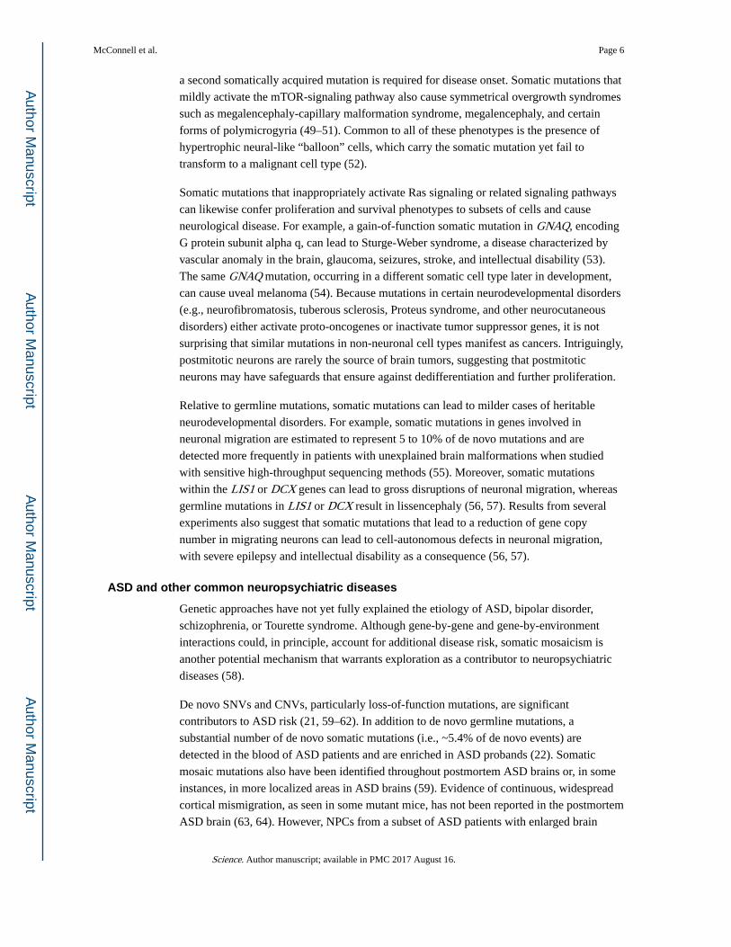
a second somatically acquired mutation is required for disease onset. Somatic mutations that
mildly activate the mTOR-signaling pathway also cause symmetrical overgrowth syndromes
such as megalencephaly-capillary malformation syndrome, megalencephaly, and certain
forms of polymicrogyria (49–51). Common to all of these phenotypes is the presence of
hypertrophic neural-like “balloon” cells, which carry the somatic mutation yet fail to
transform to a malignant cell type (52).
Somatic mutations that inappropriately activate Ras signaling or related signaling pathways
can likewise confer proliferation and survival phenotypes to subsets of cells and cause
neurological disease. For example, a gain-of-function somatic mutation in GNAQ, encoding
G protein subunit alpha q, can lead to Sturge-Weber syndrome, a disease characterized by
vascular anomaly in the brain, glaucoma, seizures, stroke, and intellectual disability (53).
The same GNAQ mutation, occurring in a different somatic cell type later in development,
can cause uveal melanoma (54). Because mutations in certain neurodevelopmental disorders
(e.g., neurofibromatosis, tuberous sclerosis, Proteus syndrome, and other neurocutaneous
disorders) either activate proto-oncogenes or inactivate tumor suppressor genes, it is not
surprising that similar mutations in non-neuronal cell types manifest as cancers. Intriguingly,
postmitotic neurons are rarely the source of brain tumors, suggesting that postmitotic
neurons may have safeguards that ensure against dedifferentiation and further proliferation.
Relative to germline mutations, somatic mutations can lead to milder cases of heritable
neurodevelopmental disorders. For example, somatic mutations in genes involved in
neuronal migration are estimated to represent 5 to 10% of de novo mutations and are
detected more frequently in patients with unexplained brain malformations when studied
with sensitive high-throughput sequencing methods (55). Moreover, somatic mutations
within the LIS1 or DCX genes can lead to gross disruptions of neuronal migration, whereas
germline mutations in LIS1 or DCX result in lissencephaly (56, 57). Results from several
experiments also suggest that somatic mutations that lead to a reduction of gene copy
number in migrating neurons can lead to cell-autonomous defects in neuronal migration,
with severe epilepsy and intellectual disability as a consequence (56, 57).
ASD and other common neuropsychiatric diseases
Genetic approaches have not yet fully explained the etiology of ASD, bipolar disorder,
schizophrenia, or Tourette syndrome. Although gene-by-gene and gene-by-environment
interactions could, in principle, account for additional disease risk, somatic mosaicism is
another potential mechanism that warrants exploration as a contributor to neuropsychiatric
diseases (58).
De novo SNVs and CNVs, particularly loss-of-function mutations, are significant
contributors to ASD risk (21, 59–62). In addition to de novo germline mutations, a
substantial number of de novo somatic mutations (i.e., ~5.4% of de novo events) are
detected in the blood of ASD patients and are enriched in ASD probands (22). Somatic
mosaic mutations also have been identified throughout postmortem ASD brains or, in some
instances, in more localized areas in ASD brains (59). Evidence of continuous, widespread
cortical mismigration, as seen in some mutant mice, has not been reported in the postmortem
ASD brain (63, 64). However, NPCs from a subset of ASD patients with enlarged brain
McConnell et al. Page 6
Science. Author manuscript; available in PMC 2017 August 16.
Author M
anuscriptA
uthor Manuscript
Author M
anuscriptA
uthor Manuscript

volumes are inherently more proliferative and display abnormal neurogenesis when
compared to controls (65, 66). Other ASD patients have focal cortical abnormalities,
including disorganized neurons and lamina, polymicrogyria, and other local surface
malformations (67). Thus, in addition to specific mutations, additional cell cycles may
further affect somatic mutational loads in patients.
Prenatal challenges to the immune system in animals (i.e., maternal immune activation) (68)
can also lead to many features like those present in ASD brains. Maternal immune activation
leads to increased cellular proliferation, brain size, and ASD-like behaviors in animal
models (69–72). Intriguingly, an elevated prevalence of MEIs was observed in a primate
model of maternal immune activation (73). Elevated MEI levels likewise are observed in
schizophrenia (73) and Rett syndrome patients (74), suggesting that somatic MEI burden
may play a role in the etiology of some neurodevelopmental and neuropsychiatric diseases.
Methods to detect somatic mutations
The difficulty in detecting a somatic mutation depends on its frequency within a cell
population. Whereas mutations affecting a large fraction (e.g., 50%) of cells are readily
detected in bulk tissue sequencing experiments and generally result in high-confidence calls,
mutations affecting one or a few cells are unlikely to be detected with bulk tissue sequencing
approaches. The identification and validation of rare somatic mutations requires sequencing
DNA derived from small pools of cells, single cells, or clonally reprogrammed cells
followed by robust computational data analyses (Fig. 1).
Bulk tissue approaches
Whole-genome sequencing (WGS) or whole-exome sequencing (WES) of DNA derived
from bulk brain tissue allows a straightforward approach to discovering somatic mosaicism
(26). WGS and WES minimize sequencing artifacts that can confound downstream analyses
and, in the case of WGS, provide an opportunity for identifying a wide range of structural
rearrangements, including inversions and translocations. However, WGS and WES using
standard sequencing depths have reduced statistical power to detect mutations that occur at
low frequencies (i.e., <10% of cells in a population at 30 to 100x coverage). Although
increasing sequence coverage allows detection of somatic variants at lower frequencies, it
quickly becomes cost prohibitive. Moreover, WGS and WES do not provide information on
how somatic variants are distributed across individual cell lineages within a bulk tissue
sample.
Sorted-pools approaches
Fluorescence-activated cell or nuclei sorting (FACS/FANS) can be used to isolate specific
neural populations (e.g., NeuN+ neurons versus NeuN− cells or cortical inhibitory
interneurons versus excitatory principal neurons). Analysis of sorted nuclei populations
(e.g., 5000 or 500,000 cells) from specific brain regions increases the power to detect
somatic mosaicism that arises in one lineage, because these genomes are no longer diluted
by genomes derived from other lineages. Independent pools of sorted nuclei can then be
subjected to RNA sequencing (RNA-seq) and quantitative reverse transcription polymerase
McConnell et al. Page 7
Science. Author manuscript; available in PMC 2017 August 16.
Author M
anuscriptA
uthor Manuscript
Author M
anuscriptA
uthor Manuscript

chain reaction (qRT-PCR) to confirm cell type–specific gene expression profiles (75). In
addition to increasing the power for detecting a somatic mutation, cell sorting before DNA
extraction could yield information about the embryological origin and developmental
trajectory of somatic variation across the brain. Large pools of sorted cells can yield enough
DNA for the direct examination of somatic variants by WGS or WES. However, smaller
pool sizes will only generate small amounts of DNA; thus, they are best suited for
generating PCR amplicon libraries (e.g., as used in MEI detection and other targeted
sequencing) or for subsequent whole-genome amplification (WGA).
Single-cell approaches
WGA can be used to analyze the genomes of single neurons (26). The spectrum of
mutations identified from the genomes of single neurons can then be compared to germline
variants in bulk tissue data derived from a non-neuronal control (e.g., brain dural fibroblasts
or heart) to identify candidate somatic mutations (5). WGA approaches already are used in
pre-implantation genetic screening of embryos (76, 77) and include (i) degenerate-
oligonucleotide-primed PCR (DOP-PCR), (ii) multiple displacement amplification (MDA),
and (iii) multiple annealing and looping-based amplification (MALBAC). Each method has
its advantages and drawbacks. In general, DOP-PCR provides coverage evenly across the
genome, which facilitates the detection of large CNVs and chromosomal aneuploidies.
However, DOP-PCR has a higher read duplication rate, lower mapping rate, and lower
recovery rate when compared with MDA and MALBAC (78) and is cost prohibitive for
SNV, indel, and MEI detection. By comparison, MDA yields a high rate of artificial
chimeric DNA molecules that can lead to false-positive calls in downstream analyses (79),
whereas MALBAC exhibits reduced coverage of certain genomic regions (14, 16, 80),
especially those rich in repetitive sequences (78). Considerable advances have recently been
made in detecting SNVs (81, 82), CNVs (83), and MEIs (16) in WGA samples; however,
best practices necessitate evaluating each WGA approach for the detection of specific types
of somatic mosaicism.
Clonal expansion of single cells using human-induced pluripotent stem cell (hiPSC)
technology or somatic cell nuclear transfer (SCNT) provides a biological alternative to
WGA (80, 84). Any variant uniformly identified in the clonal line, but not in controls,
represents a candidate somatic mutation that requires confirmation in the tissue of origin. In
contrast, mutations introduced during cell culture will be present in a lower frequency of
cells within a clonal cell line and can be discriminated from bona fide somatic mutations in
downstream computational analysis. Although the clonal isolation and expansion of primary
human neural stem and progenitor cells is possible, the analysis of human neuronal genomes
using clonal reprogramming has several limitations. Foremost among these is the availability
of live human neurons. Moreover, neither clonal reprogramming nor SCNT have been
reported using human neurons; SCNT is further limited by the expense and availability of
human oocytes. Finally, reprogramming approaches currently are only successful in ~10%
of cells; thus, any neurons harboring highly aberrant genomes may be refractory to
reprogramming. Despite these caveats, clonal reprogramming of human neurons is
theoretically possible. In addition, it is noteworthy that mouse neurons reprogrammed by
McConnell et al. Page 8
Science. Author manuscript; available in PMC 2017 August 16.
Author M
anuscriptA
uthor Manuscript
Author M
anuscriptA
uthor Manuscript

SCNT contain genomic rearrangements (e.g., kataegis and chromothripsis) that would be
very challenging to validate using current WGA approaches (84).
Computational methods for mutation detection
WGS and WES have been used successfully to detect somatic SNVs in family-based studies
of Mendelian disease and large-scale sequencing studies of human patient cohorts (2). To
identify SNVs, most computational approaches compare call sets generated from an affected
sample to those generated from a matched healthy/unaffected sample and/or a control
population. These comparisons allow the identification and subsequent exclusion of
germline polymorphisms from downstream analyses; however, care must be taken to ensure
that any candidate somatic mutations are not germline variants that were missed in the
matched control. In general, variant callers initially developed to detect mutations in cancer
offer higher sensitivity for detecting mosaic SNVs when compared with standard approaches
used to detect germline variants (85, 86).
Somatic CNVs can be detected by identifying deviations either from the expected depth of
sequence or in the expected distances between paired-end sequencing reads. Similarly,
inversions can be identified through differences in the orientations of paired-end sequencing
reads. Numerous approaches have been developed to identify CNVs from WGS (7, 87–89),
and most can be applied directly to identify somatic mutations. For example, recent studies
using WGA in conjunction with WGS have identified megabase-scale de novo CNVs in
human and mouse neurons based on differences in read-depth across genomic bins (6–9).
CNVs are more difficult to identify using WES due to the biases encountered during the
capture of target exons (90).
Somatic MEIs can be detected from bulk tissue, PCR amplicons generated from sorted-cell
fractions, or single-cell WGA DNA using split-read and paired-end information (e.g., one
paired-end read may map to the reference genome, whereas another may map to a MEI) (91,
92). Detecting low-frequency MEIs with fewer supporting reads requires careful
bioinformatic analyses that can distinguish signal from noise, followed by experimental
validation with orthogonal methods (14, 93). The analysis of single-cell data remains
challenging due to the presence of chimeras generated during WGA (14, 16, 94); thus, care
must be taken in calling MEIs.
Validation of somatic mutations
It is essential to validate all candidate somatic mutations. False-positive calls can arise from
DNA sequencing errors, contamination with germline variants, chimeric molecules
generated during single-cell WGA, PCR-induced nucleotide substitutions, and the failure to
amplify certain genomic regions. False-negative calls are dependent on the allele frequency
of the somatic mutation within the sample, the type of mutation, and the method of
detection. Orthologous experimental methods are required to eliminate false-positives and to
calibrate the confidence of detection for different types of somatic mutations. Validation
experiments can then be performed on either the tissue of origin or amplified material used
to discover the variant. The first approach represents a biological validation, which
establishes the presence of a variant call in unamplified DNA from the source sample. The
McConnell et al. Page 9
Science. Author manuscript; available in PMC 2017 August 16.
Author M
anuscriptA
uthor Manuscript
Author M
anuscriptA
uthor Manuscript

second approach represents a technical validation, which establishes the presence/absence of
variant calls in the DNA source material used for discovery.
Biological/primary validation in the tissue of origin
Validation on unamplified DNA from the tissue of origin provides confirmation that a
candidate call is a genuine somatic variant and rules out the possibility that it corresponds to
a DNA amplification artifact or a mutation that occurred during clonal expansion. Biological
validation requires a variant to be present in multiple cells in the tissue of origin at a
frequency above experimental detection limits. As such, the failure to validate a variant in
the tissue of origin does not necessarily represent a false call. For example, only ~50% of
CNVs manifested in hiPSC clones could be directly confirmed in the primary fibroblast cells
used to derive hiPSCs (80).
Somatic variants can be confirmed in unamplified cell source material by (i) targeted DNA
capture followed by high-coverage (>100x) DNA resequencing, (ii) high-coverage
sequencing of multiplexed PCR amplicons, and (iii) droplet digital PCR (ddPCR). These
approaches vary in throughput and sensitivity. Targeted DNA capture and resequencing can
require the creation of several thousand custom oligonucleotides designed to capture the
genomic DNA either including or surrounding the putative variants. The captured DNA then
is subjected to high-coverage paired-end DNA sequencing, yielding a typical sensitivity of
variant detection in greater than 1% of cells. Amplicon sequencing involves PCR
amplification of candidate loci followed by high-coverage paired-end DNA sequencing,
yielding a typical sensitivity of variant detection in greater than 0.1% of cells. Finally,
ddPCR involves partitioning a DNA sample into large numbers of individual droplets that
generally contain one copy of template DNA. PCR takes place within these droplets, leading
to the production of a fluorescent readout, either through the use of an intercalating dye or a
fluorescent oligomer probe, to indicate the presence or absence of the PCR target of interest.
Subsequent quantification of the fluorescent droplets allows a determination of the number
of copies of the target locus present in the sample, yielding a typical sensitivity of variant
detection in greater than 0.001% of cells (95). Although extremely sensitive, ddPCR requires
the optimization of primers, probes, and amplification conditions, which is time-consuming
and limits throughput.
The goal when employing biological validation procedures is to detect putative somatic
variants and to assess, as precisely as possible, the frequency of each variant in that tissue of
origin. Biological validation can (i) determine whether certain individuals in the population
are more prone to somatic variation than others, (ii) investigate whether different areas of the
brain and/or specific brain cell types have varying amounts and types of particular forms of
somatic variation, (iii) assess whether developmental timing contributes to somatic variation,
and (iv) reveal whether somatic variations increase as a function of the number of cell
divisions and/or a function of age in postmitotic neurons.
Technical validation on source/amplified material
If a somatic variant is only present in a single cell, it will be impossible to validate in bulk
tissue. Likewise, a variant present in very few cells may be difficult to validate in the tissue
McConnell et al. Page 10
Science. Author manuscript; available in PMC 2017 August 16.
Author M
anuscriptA
uthor Manuscript
Author M
anuscriptA
uthor Manuscript

of origin. Thus, technical validation in the source DNA used to discover a putative variant
can be used to determine whether a call is true or false. Technical validation typically
employs PCR, qPCR, and Sanger sequencing of the locus in the DNA source material (e.g.,
WGA DNA or DNA from a clonal cell population). Multiple true/false verdicts form the
basis for estimating false-discovery and false-negative rates in the resultant call sets.
Present understanding of the prevalence of somatic mutation in
neurotypical individuals
Recent studies revealed that mosaic neuronal genomes are the rule, rather than the
exception; every neuron probably has a different genome than the neurons with which it
forms synapses. Not unexpectedly, SNVs are the most prevalent somatic mutations. A “triple
calling” strategy was used to identify and validate clonal SNVs in MDA-amplified DNA
from single neurons isolated from a neurotypical brain, leading to estimates of ~1000 to
1500 SNVs per neuronal genome (5). By comparison to human cortical neurons, a SCNT
experiment in reprogrammed mouse olfactory neurons detected hundreds of SNVs per
neuron and a lower proportion of C-to-T transition mutations (84). Although the divergent
SNV rates between these two studies may arise from technical differences (as discussed
above), both approaches establish that SNVs represent an important form of somatic
mutation in both human and mouse neurons.
Brain somatic CNVs initially were identified by comparing the sequences of bulk DNA
derived from multicellular samples of different brain regions to the sequences of DNA
derived from somatic tissues (96, 97). The first single-cell study of neuronal CNVs analyzed
110 human frontal cortex neurons and found that 13 to 41% of the neurons contained at least
one megabase-scale de novo CNV (6). Additional studies, which analyzed fewer neuronal
genomes, confirmed that de novo CNVs occur in at least 10% of neurons (7, 8). CNVs can
be shared by multiple neurons and inherited in a clonal manner (8). Furthermore, megabase-
scale CNVs typically alter the copy number of 10 or more genes in individual neurons. In
addition to expression-level differences that can accompany gene copy number changes,
mosaic neuronal CNVs also are expected to reveal or abate pernicious alleles on a neuron-
by-neuron basis in every individual.
L1 retrotransposon insertions alter the transcriptional regulation of genes in myriad ways
(42). Initial studies used engineered L1s containing a retrotransposition indicator cassette to
discover MEI activity in mouse brain (98) and in human NPCs in vitro (99). Studies of
MDA-amplified NeuN-positive nuclei isolated from a neurotypical human brain, followed
by L1-transposon profiling (13) or WGS (15, 16), have since suggested that 0.2 to 1 L1
insertion occur per neuronal genome. Another report, which employed MALBAC WGA in
conjunction with L1 capture technology (RC-seq), reported an average of 13 L1 insertions in
every neuronal genome (11), although a subsequent study suggested a high false-positive
rate in these data (14). By comparison, SCNT experiments in mouse olfactory neurons
reported ≤1.3 MEI per neuronal genome (84). An extrapolation of these data indicates that
potentially billions of neurons in the neurotypical brain contain de novo MEIs. Additional
studies are required to determine whether L1s retrotranspose at varying rates in different
McConnell et al. Page 11
Science. Author manuscript; available in PMC 2017 August 16.
Author M
anuscriptA
uthor Manuscript
Author M
anuscriptA
uthor Manuscript

brain regions, in different individuals, or preferentially insert into expressed genes, and
whether other mobile elements [e.g., Alu retrotransposons (42)] also contribute to intra-
individual neuronal genetic diversity.
Generation of a community resource
The BSMN will generate comprehensive maps of somatic genomic variation in neurotypical
and diseased human brains, including a prioritized call set of confirmed somatic variants
(Box 1) that may contribute to neuropsychiatric disease and epilepsy. Functional validation
experiments will be performed using CRISPR/Cas9-mediated genome engineering, hiPSC-
based neurogenesis, and mosaic mouse models generated by in utero electroporation (Fig.
3). The BSMN is initially determining concordance among disparate sequencing and
bioinformatic approaches by performing a “common experiment” in which pulverized tissue
from one neurotypical individual in the Lieber brain repository has been distributed to all of
the working groups for independent assessment of mosaicism.
Box 1
Criteria used to prioritize somatic variants for functional characterization
Absence from the germ line
We will focus on variants with a definitive somatic origin.
Recurrence and frequency of somatic variation at the locus of interest
We will prioritize loci at which somatic variations, across all types, recur in multiple
disease samples but not in control samples.
Mutation severity
Highly deleterious variations will be prioritized for likely functional importance.
Intersection with known disease loci and biochemical pathways
Taking advantage of data on germline variations in brain disorders, we will prioritize loci
that have been previously implicated in disease.
Intersection with brain expression and epigenomic data
Taking advantage of large, publicly funded consortia of human brain spatiatemporal
expression data (e.g., BrainSpan) and epigenomic data (e.g., PsychENCODE and
Roadmap Epigenomics), we will select genes that are expressed in brain regions
associated with brain disorders and noncoding loci with potential regulatory function.
The BSMN will generate an estimated 10,000 sequencing data sets that comprise >600
terabytes of data and facilitate data-sharing through the BSMN Knowledge Portal
(www.synapse.org/bsmn) and the NIMH Data Archive (https://data-archive.nimh.nih.gov).
Coordinated analyses with data derived from some of the same brain samples by the
CommonMind (www.synapse.org/cmc) and PsychENCODE (www.synapse.org/pec)
initiatives may elucidate the effect of somatic mosaicism on tissue-wide gene expression.
Data generated though the BSMN initiative will be released to the broader research
McConnell et al. Page 12
Science. Author manuscript; available in PMC 2017 August 16.
Author M
anuscriptA
uthor Manuscript
Author M
anuscriptA
uthor Manuscript

community on an ongoing basis through a controlled-access mechanism that follows NIH
policies and regulatory requirements.
Supplementary Material
Refer to Web version on PubMed Central for supplementary material.
Acknowledgments
We thank T. Insel for initiating this project, L. Bingaman for ongoing administrative assistance, and N. Leff and M. L. Gage for copyediting assistance. J.M.K acknowledges support provided by the Pew Biomedical Scholars Award. J.V.M. is an inventor on patent application 6150160, held by the John Hopkins University and the Trustees of the University of Pennsylvania, which covers the compositions and methods of use of mammalian retrotransposons. We also acknowledge the support of NIH R01 MH100914, Genomic mosaicism in developing human brain (F.M.V.). Some figures use images from the Servier Medical Art PowerPoint Image Bank. All the work was supported by U01MH106883, U01MH106874, U01MH106893, U01MH106892, U01MH106882, U01MH106876, U01MH1068898, U01MH106891, and U01MH106884. We regret that space constraints limit the number of references and apologize to many colleagues whose very valuable contributions to this field are not cited.
REFERENCES AND NOTES
1. Lynch M. Rate, molecular spectrum, and consequences of human mutation. Proc Natl Acad Sci USA. 2010; 107:961–968. DOI: 10.1073/pnas.0912629107 [PubMed: 20080596]
2. Freed D, Stevens EL, Pevsner J. Somatic mosaicism in the human genome. Genes (Basel). 2014; 5:1064–1094. DOI: 10.3390/genes5041064 [PubMed: 25513881]
3. Frank SA. Evolution in health and medicine Sackler colloquium: Somatic evolutionary genomics: Mutations during development cause highly variable genetic mosaicism with risk of cancer and neurodegeneration. Proc Natl Acad Sci USA. 2010; 107(suppl 1):1725–1730. DOI: 10.1073/pnas.0909343106 [PubMed: 19805033]
4. Lupski JR. Genome mosaicism—One human, multiple genomes. Science. 2013; 341:358–359. DOI: 10.1126/science.1239503 [PubMed: 23888031]
5. Lodato MA, et al. Somatic mutation in single human neurons tracks developmental and transcriptional history. Science. 2015; 350:94–98. DOI: 10.1126/science.aab1785 [PubMed: 26430121]
6. McConnell MJ, et al. Mosaic copy number variation in human neurons. Science. 2013; 342:632–637. DOI: 10.1126/science.1243472 [PubMed: 24179226]
7. Knouse KA, Wu J, Amon A. Assessment of megabase-scale somatic copy number variation using single-cell sequencing. Genome Res. 2016; 26:376–384. DOI: 10.1101/gr.198937.115 [PubMed: 26772196]
8. Cai X, et al. Single-cell, genome-wide sequencing identifies clonal somatic copy-number variation in the human brain. Cell Reports. 2014; 8:1280–1289. DOI: 10.1016/j.celrep.2014.07.043 [PubMed: 25159146]
9. Rehen SK, et al. Constitutional aneuploidy in the normal human brain. J Neurosci. 2005; 25:2176–2180. DOI: 10.1523/JNEUROSCI.4560-04.2005 [PubMed: 15745943]
10. Yurov YB, et al. Aneuploidy and confined chromosomal mosaicism in the developing human brain. PLOS ONE. 2007; 2:e558.doi: 10.1371/journal.pone.0000558 [PubMed: 17593959]
11. Upton KR, et al. Ubiquitous L1 mosaicism in hippocampal neurons. Cell. 2015; 161:228–239. DOI: 10.1016/j.cell.2015.03.026 [PubMed: 25860606]
12. Baillie JK, et al. Somatic retrotransposition alters the genetic landscape of the human brain. Nature. 2011; 479:534–537. DOI: 10.1038/nature10531 [PubMed: 22037309]
13. Evrony GD, et al. Single-neuron sequencing analysis of L1 retrotransposition and somatic mutation in the human brain. Cell. 2012; 151:483–496. DOI: 10.1016/j.cell.2012.09.035 [PubMed: 23101622]
McConnell et al. Page 13
Science. Author manuscript; available in PMC 2017 August 16.
Author M
anuscriptA
uthor Manuscript
Author M
anuscriptA
uthor Manuscript

14. Evrony GD, Lee E, Park PJ, Walsh CA. Resolving rates of mutation in the brain using single-neuron genomics. eLife. 2016; 5:e12966.doi: 10.7554/eLife.12966 [PubMed: 26901440]
15. Erwin JA, et al. L1-associated genomic regions are deleted in somatic cells of the healthy human brain. Nat Neurosci. 2016; 19:1583–1591. [PubMed: 27618310]
16. Evrony GD, et al. Cell lineage analysis in human brain using endogenous retroelements. Neuron. 2015; 85:49–59. DOI: 10.1016/j.neuron.2014.12.028 [PubMed: 25569347]
17. Lui JH, Hansen DV, Kriegstein AR. Development and evolution of the human neocortex. Cell. 2011; 146:18–36. DOI: 10.1016/j.cell.2011.06.030 [PubMed: 21729779]
18. Macia A, et al. Engineered LINE-1 retrotransposition in nondividing human neurons. Genome Res. 2017; 27:335–348. [PubMed: 27965292]
19. Muotri AR, Gage FH. Generation of neuronal variability and complexity. Nature. 2006; 441:1087–1093. DOI: 10.1038/nature04959 [PubMed: 16810244]
20. Bushman DM, Chun J. The genomically mosaic brain: Aneuploidy and more in neural diversity and disease. Semin Cell Dev Biol. 2013; 24:357–369. DOI: 10.1016/j.semcdb.2013.02.003 [PubMed: 23466288]
21. Iossifov I, et al. The contribution of de novo coding mutations to autism spectrum disorder. Nature. 2014; 515:216–221. DOI: 10.1038/nature13908 [PubMed: 25363768]
22. Freed D, Pevsner J. The contribution of mosaic variants to autism spectrum disorder. PLOS Genet. 2016; 12:e1006245.doi: 10.1371/journal.pgen.1006245 [PubMed: 27632392]
23. Fromer M, et al. De novo mutations in schizophrenia implicate synaptic networks. Nature. 2014; 506:179–184. DOI: 10.1038/nature12929 [PubMed: 24463507]
24. Malhotra D, Sebat J. CNVs: Harbingers of a rare variant revolution in psychiatric genetics. Cell. 2012; 148:1223–1241. DOI: 10.1016/j.cell.2012.02.039 [PubMed: 22424231]
25. Poduri A, Evrony GD, Cai X, Walsh CA. Somatic mutation, genomic variation, and neurological disease. Science. 2013; 341:1237758.doi: 10.1126/science.1237758 [PubMed: 23828942]
26. Lee JH. Somatic mutations in disorders with disrupted brain connectivity. Exp Mol Med. 2016; 48:e239. [PubMed: 27282107]
27. Sekiguchi JM, et al. Nonhomologous end-joining proteins are required for V(D)J recombination, normal growth, and neurogenesis. Cold Spring Harb Symp Quant Biol. 1999; 64:169–181. DOI: 10.1101/sqb.1999.64.169 [PubMed: 11232282]
28. Frank KM, et al. DNA ligase IV deficiency in mice leads to defective neurogenesis and embryonic lethality via the p53 pathway. Mol Cell. 2000; 5:993–1002. DOI: 10.1016/S1097-2765(00)80264-6 [PubMed: 10911993]
29. McConnell MJ, et al. Failed clearance of aneuploid embryonic neural progenitor cells leads to excess aneuploidy in the Atm-deficient but not the Trp53-deficient adult cerebral cortex. J Neurosci. 2004; 24:8090–8096. DOI: 10.1523/JNEUROSCI.2263-04.2004 [PubMed: 15371510]
30. Coufal NG, et al. Ataxia telangiectasia mutated (ATM) modulates long interspersed element-1 (L1) retrotransposition in human neural stem cells. Proc Natl Acad Sci USA. 2011; 108:20382–20387. DOI: 10.1073/pnas.1100273108 [PubMed: 22159035]
31. Iourov IY, Vorsanova SG, Liehr T, Kolotii AD, Yurov YB. Increased chromosome instability dramatically disrupts neural genome integrity and mediates cerebellar degeneration in the ataxia-telangiectasia brain. Hum Mol Genet. 2009; 18:2656–2669. DOI: 10.1093/hmg/ddp207 [PubMed: 19414482]
32. Shen J, et al. Mutations in PNKP cause microcephaly, seizures and defects in DNA repair. Nat Genet. 2010; 42:245–249. DOI: 10.1038/ng.526 [PubMed: 20118933]
33. Carvalho CM, Lupski JR. Mechanisms underlying structural variant formation in genomic disorders. Nat Rev Genet. 2016; 17:224–238. DOI: 10.1038/nrg.2015.25 [PubMed: 26924765]
34. Chiu YL, Greene WC. The APOBEC3 cytidine deaminases: An innate defensive network opposing exogenous retroviruses and endogenous retroelements. Annu Rev Immunol. 2008; 26:317–353. DOI: 10.1146/annurev.immunol.26.021607.090350 [PubMed: 18304004]
35. Arlt MF, Wilson TE, Glover TW. Replication stress and mechanisms of CNV formation. Curr Opin Genet Dev. 2012; 22:204–210. DOI: 10.1016/j.gde.2012.01.009 [PubMed: 22365495]
McConnell et al. Page 14
Science. Author manuscript; available in PMC 2017 August 16.
Author M
anuscriptA
uthor Manuscript
Author M
anuscriptA
uthor Manuscript

36. Hastings PJ, Lupski JR, Rosenberg SM, Ira G. Mechanisms of change in gene copy number. Nat Rev Genet. 2009; 10:551–564. DOI: 10.1038/nrg2593 [PubMed: 19597530]
37. Wei PC, et al. Long neural genes harbor recurrent DNA break clusters in neural stem/progenitor cells. Cell. 2016; 164:644–655. DOI: 10.1016/j.cell.2015.12.039 [PubMed: 26871630]
38. Wilson TE, et al. Large transcription units unify copy number variants and common fragile sites arising under replication stress. Genome Res. 2015; 25:189–200. DOI: 10.1101/gr.177121.114 [PubMed: 25373142]
39. King IF, et al. Topoisomerases facilitate transcription of long genes linked to autism. Nature. 2013; 501:58–62. DOI: 10.1038/nature12504 [PubMed: 23995680]
40. Zylka MJ, Simon JM, Philpot BD. Gene length matters in neurons. Neuron. 2015; 86:353–355. DOI: 10.1016/j.neuron.2015.03.059 [PubMed: 25905808]
41. Gabel HW, et al. Disruption of DNA-methylation-dependent long gene repression in Rett syndrome. Nature. 2015; 522:89–93. DOI: 10.1038/nature14319 [PubMed: 25762136]
42. Richardson SR, et al. The influence of LINE-1 and SINE retrotransposons on mammalian genomes. Microbiol Spectr. 2015; 3:MDNA3-0061-2014.doi: 10.1128/microbiolspec.MDNA3-0061-2014
43. Luan DD, Korman MH, Jakubczak JL, Eickbush TH. Reverse transcription of R2Bm RNA is primed by a nick at the chromosomal target site: A mechanism for non-LTR retrotransposition. Cell. 1993; 72:595–605. DOI: 10.1016/0092-8674(93)90078-5 [PubMed: 7679954]
44. Morrish TA, et al. DNA repair mediated by endonuclease-independent LINE-1 retrotransposition. Nat Genet. 2002; 31:159–165. DOI: 10.1038/ng898 [PubMed: 12006980]
45. Gilbert N, Lutz S, Morrish TA, Moran JV. Multiple fates of L1 retrotransposition intermediates in cultured human cells. Mol Cell Biol. 2005; 25:7780–7795. DOI: 10.1128/MCB.25.17.7780-7795.2005 [PubMed: 16107723]
46. Symer DE, et al. Human l1 retrotransposition is associated with genetic instability in vivo. Cell. 2002; 110:327–338. DOI: 10.1016/S0092-8674(02)00839-5 [PubMed: 12176320]
47. Gilbert N, Lutz-Prigge S, Moran JV. Genomic deletions created upon LINE-1 retrotransposition. Cell. 2002; 110:315–325. DOI: 10.1016/S0092-8674(02)00828-0 [PubMed: 12176319]
48. Chen JM, Stenson PD, Cooper DN, Férec C. A systematic analysis of LINE-1 endonuclease-dependent retrotranspositional events causing human genetic disease. Hum Genet. 2005; 117:411–427. DOI: 10.1007/s00439-005-1321-0 [PubMed: 15983781]
49. Mirzaa GM, et al. Association of MTOR mutations with developmental brain disorders, including megalencephaly, focal cortical dysplasia, and pigmentary mosaicism. JAMA Neurol. 2016; 73:836–845. DOI: 10.1001/jamaneurol.2016.0363 [PubMed: 27159400]
50. Mirzaa GM, et al. Characterisation of mutations of the phosphoinositide-3-kinase regulatory subunit, PIK3R2, in perisylvian polymicrogyria: A next-generation sequencing study. Lancet Neurol. 2015; 14:1182–1195. DOI: 10.1016/S1474-4422(15)00278-1 [PubMed: 26520804]
51. Rivière JB, et al. De novo germline and postzygotic mutations in AKT3, PIK3R2 and PIK3CA cause a spectrum of related megalencephaly syndromes. Nat Genet. 2012; 44:934–940. DOI: 10.1038/ng.2331 [PubMed: 22729224]
52. Henske EP, JóŸwiak S, Kingswood JC, Sampson JR, Thiele EA. Tuberous sclerosis complex. Nat Rev Dis Primers. 2016; 2:16035.doi: 10.1038/nrdp.2016.35 [PubMed: 27226234]
53. Shirley MD, et al. Sturge-Weber syndrome and port-stains caused by somatic mutation in. GNAQ N Engl J Med. 2013; 368:1971–1979. DOI: 10.1056/NEJMoa1213507 [PubMed: 23656586]
54. Van Raamsdonk CD, et al. Frequent somatic mutations of GNAQ in uveal melanoma and blue naevi. Nature. 2009; 457:599–602. DOI: 10.1038/nature07586 [PubMed: 19078957]
55. Jamuar SS, et al. Somatic mutations in cerebral cortical malformations. N Engl J Med. 2014; 371:733–743. DOI: 10.1056/NEJMoa1314432 [PubMed: 25140959]
56. Sicca F, et al. Mosaic mutations of the LIS1 gene cause subcortical band heterotopia. Neurology. 2003; 61:1042–1046. DOI: 10.1212/WNL.61.8.1042 [PubMed: 14581661]
57. Gleeson JG. Classical lissencephaly and double cortex (subcortical band heterotopia): LIS1 and doublecortin. Curr Opin Neurol. 2000; 13:121–125. DOI: 10.1097/00019052-200004000-00002 [PubMed: 10987567]
McConnell et al. Page 15
Science. Author manuscript; available in PMC 2017 August 16.
Author M
anuscriptA
uthor Manuscript
Author M
anuscriptA
uthor Manuscript

58. Insel TR. Brain somatic mutations: The dark matter of psychiatric genetics? Mol Psychiatry. 2014; 19:156–158. DOI: 10.1038/mp.2013.168 [PubMed: 24342990]
59. D’Gama AM, et al. Targeted DNA sequencing from autism spectrum disorder brains implicates multiple genetic mechanisms. Neuron. 2015; 88:910–917. DOI: 10.1016/j.neuron.2015.11.009 [PubMed: 26637798]
60. Iossifov I, et al. De novo gene disruptions in children on the autistic spectrum. Neuron. 2012; 74:285–299. DOI: 10.1016/j.neuron.2012.04.009 [PubMed: 22542183]
61. Neale BM, et al. Patterns and rates of exonic de novo mutations in autism spectrum disorders. Nature. 2012; 485:242–245. DOI: 10.1038/nature11011 [PubMed: 22495311]
62. Sebat J, et al. Strong association of de novo copy number mutations with autism. Science. 2007; 316:445–449. DOI: 10.1126/science.1138659 [PubMed: 17363630]
63. Hutsler JJ, Casanova MF. Review: Cortical construction in autism spectrum disorder: Columns, connectivity and the subplate. Neuropathol Appl Neurobiol. 2016; 42:115–134. DOI: 10.1111/nan.12227 [PubMed: 25630827]
64. Donovan AP, Basson MA. The neuroanatomy of autism - A developmental perspective. J Anat. 2017; 230:4–15. DOI: 10.1111/joa.12542 [PubMed: 27620360]
65. Marchetto MC, et al. Altered proliferation and networks in neural cells derived from idiopathic autistic individuals. Mol Psychiatry. 2016
66. Mariani J, et al. FOXG1-dependent dysregulation of GABA/glutamate neuron differentiation in autism spectrum disorders. Cell. 2015; 162:375–390. DOI: 10.1016/j.cell.2015.06.034 [PubMed: 26186191]
67. Stoner R, et al. Patches of disorganization in the neocortex of children with autism. N Engl J Med. 2014; 370:1209–1219. DOI: 10.1056/NEJMoa1307491 [PubMed: 24670167]
68. Patterson PH. Maternal infection and immune involvement in autism. Trends Mol Med. 2011; 17:389–394. DOI: 10.1016/j.molmed.2011.03.001 [PubMed: 21482187]
69. Choi GB, et al. The maternal interleukin-17a pathway in mice promotes autism-like phenotypes in offspring. Science. 2016; 351:933–939. doi:3310.1126/science.aad0314. [PubMed: 26822608]
70. Smith SE, Elliott RM, Anderson MP. Maternal immune activation increases neonatal mouse cortex thickness and cell density. J Neuroimmune Pharmacol. 2012; 7:529–532. DOI: 10.1007/s11481-012-9372-1 [PubMed: 22570011]
71. Malkova NV, Yu CZ, Hsiao EY, Moore MJ, Patterson PH. Maternal immune activation yields offspring displaying mouse versions of the three core symptoms of autism. Brain Behav Immun. 2012; 26:607–616. DOI: 10.1016/j.bbi.2012.01.011 [PubMed: 22310922]
72. Soumiya H, Fukumitsu H, Furukawa S. Prenatal immune challenge compromises development of upper-layer but not deeper-layer neurons of the mouse cerebral cortex. J Neurosci Res. 2011; 89:1342–1350. DOI: 10.1002/jnr.22636 [PubMed: 21674566]
73. Bundo M, et al. Increased l1 retrotransposition in the neuronal genome in schizophrenia. Neuron. 2014; 81:306–313. DOI: 10.1016/j.neuron.2013.10.053 [PubMed: 24389010]
74. Muotri AR, et al. L1 retrotransposition in neurons is modulated by MeCP2. Nature. 2010; 468:443–446. DOI: 10.1038/nature09544 [PubMed: 21085180]
75. Lake BB, et al. Neuronal subtypes and diversity revealed by single-nucleus RNA sequencing of the human brain. Science. 2016; 352:1586–1590. DOI: 10.1126/science.aaf1204 [PubMed: 27339989]
76. Vermeesch JR, Voet T, Devriendt K. Prenatal and pre-implantation genetic diagnosis. Nat Rev Genet. 2016; 17:643–656. DOI: 10.1038/nrg.2016.97 [PubMed: 27629932]
77. Yan L, et al. Live births after simultaneous avoidance of monogenic diseases and chromosome abnormality by next-generation sequencing with linkage analyses. Proc Natl Acad Sci USA. 2015; 112:15964–15969. DOI: 10.1073/pnas.1523297113 [PubMed: 26712022]
78. Hou Y, et al. Comparison of variations detection between whole-genome amplification methods used in single-cell resequencing. Gigascience. 2015; 4:37.doi: 10.1186/s13742-015-0068-3 [PubMed: 26251698]
79. Lasken RS, Stockwell TB. Mechanism of chimera formation during the multiple displacement amplification reaction. BMC Biotechnol. 2007; 7:19.doi: 10.1186/1472-6750-7-19 [PubMed: 17430586]
McConnell et al. Page 16
Science. Author manuscript; available in PMC 2017 August 16.
Author M
anuscriptA
uthor Manuscript
Author M
anuscriptA
uthor Manuscript

80. Abyzov A, et al. Somatic copy number mosaicism in human skin revealed by induced pluripotent stem cells. Nature. 2012; 492:438–442. DOI: 10.1038/nature11629 [PubMed: 23160490]
81. Roth A, et al. Clonal genotype and population structure inference from single-cell tumor sequencing. Nat Methods. 2016; 13:573–576. DOI: 10.1038/nmeth.3867 [PubMed: 27183439]
82. Zafar H, Wang Y, Nakhleh L, Navin N, Chen K. Monovar: Single-nucleotide variant detection in single cells. Nat Methods. 2016; 13:505–507. DOI: 10.1038/nmeth.3835 [PubMed: 27088313]
83. Garvin T, et al. Interactive analysis and assessment of single-cell copy-number variations. Nat Methods. 2015; 12:1058–1060. DOI: 10.1038/nmeth.3578 [PubMed: 26344043]
84. Hazen JL, et al. The complete genome sequences, unique mutational spectra, and developmental potency of adult neurons revealed by cloning. Neuron. 2016; 89:1223–1236. DOI: 10.1016/j.neuron.2016.02.004 [PubMed: 26948891]
85. Cibulskis K, et al. Sensitive detection of somatic point mutations in impure and heterogeneous cancer samples. Nat Biotechnol. 2013; 31:213–219. DOI: 10.1038/nbt.2514 [PubMed: 23396013]
86. Koboldt DC, et al. VarScan: Variant detection in massively parallel sequencing of individual and pooled samples. Bioinformatics. 2009; 25:2283–2285. DOI: 10.1093/bioinformatics/btp373 [PubMed: 19542151]
87. Zhao X, Emery SB, Myers B, Kidd JM, Mills RE. Resolving complex structural genomic rearrangements using a randomized approach. Genome Biol. 2016; 17:126.doi: 10.1186/s13059-016-0993-1 [PubMed: 27287201]
88. Xi R, Lee S, Xia Y, Kim T-M, Park PJ. Copy number analysis of whole-genome data using BIC-seq2 and its application to detection of cancer susceptibility variants. Nucleic Acids Res. 2016; 44:6274–6286. DOI: 10.1093/nar/gkw491 [PubMed: 27260798]
89. Layer RM, Chiang C, Quinlan AR, Hall IM. LUMPY: A probabilistic framework for structural variant discovery. Genome Biol. 2014; 15:R84.doi: 10.1186/gb-2014-15-6-r84 [PubMed: 24970577]
90. Meynert AM, Ansari M, FitzPatrick DR, Taylor MS. Variant detection sensitivity and biases in whole genome and exome sequencing. BMC Bioinformatics. 2014; 15:247.doi: 10.1186/1471-2105-15-247 [PubMed: 25038816]
91. Lee E, et al. Landscape of somatic retrotransposition in human cancers. Science. 2012; 337:967–971. DOI: 10.1126/science.1222077 [PubMed: 22745252]
92. Stewart C, et al. A comprehensive map of mobile element insertion polymorphisms in humans. PLOS Genet. 2011; 7:e1002236.doi: 10.1371/journal.pgen.1002236 [PubMed: 21876680]
93. Richardson SR, Morell S, Faulkner GJ. L1 retrotransposons and somatic mosaicism in the brain. Annu Rev Genet. 2014; 48:1–27. DOI: 10.1146/annurev-genet-120213-092412 [PubMed: 25036377]
94. Zhang CZ, et al. Calibrating genomic and allelic coverage bias in single-cell sequencing. Nat Commun. 2015; 6:6822.doi: 10.1038/ncomms7822 [PubMed: 25879913]
95. Hindson BJ, et al. High-throughput droplet digital PCR system for absolute quantitation of DNA copy number. Anal Chem. 2011; 83:8604–8610. DOI: 10.1021/ac202028g [PubMed: 22035192]
96. Piotrowski A, et al. Somatic mosaicism for copy number variation in differentiated human tissues. Hum Mutat. 2008; 29:1118–1124. DOI: 10.1002/humu.20815 [PubMed: 18570184]
97. O’Huallachain M, Karczewski KJ, Weissman SM, Urban AE, Snyder MP. Extensive genetic variation in somatic human tissues. Proc Natl Acad Sci USA. 2012; 109:18018–18023. DOI: 10.1073/pnas.1213736109 [PubMed: 23043118]
98. Muotri AR, et al. Somatic mosaicism in neuronal precursor cells mediated by L1 retrotransposition. Nature. 2005; 435:903–910. DOI: 10.1038/nature03663 [PubMed: 15959507]
99. Coufal NG, et al. L1 retrotransposition in human neural progenitor cells. Nature. 2009; 460:1127–1131. DOI: 10.1038/nature08248 [PubMed: 19657334]
100. Kurek KC, et al. Somatic mosaic activating mutations in PIK3CA cause CLOVES syndrome. Am J Hum Genet. 2012; 90:1108–1115. DOI: 10.1016/j.ajhg.2012.05.006 [PubMed: 22658544]
101. Lee JH, et al. De novo somatic mutations in components of the PI3K-AKT3-mTOR pathway cause hemimegalencephaly. Nat Genet. 2012; 44:941–945. DOI: 10.1038/ng.2329 [PubMed: 22729223]
McConnell et al. Page 17
Science. Author manuscript; available in PMC 2017 August 16.
Author M
anuscriptA
uthor Manuscript
Author M
anuscriptA
uthor Manuscript

102. Lindhurst MJ, et al. Mosaic overgrowth with fibroadipose hyperplasia is caused by somatic activating mutations in PIK3CA. Nat Genet. 2012; 44:928–933. DOI: 10.1038/ng.2332 [PubMed: 22729222]
103. Rivière JB, et al. De novo germline and postzygotic mutations in AKT3, PIK3R2 and PIK3CA cause a spectrum of related megalencephaly syndromes. Nat Genet. 2012; 44:934–940. DOI: 10.1038/ng.2331 [PubMed: 22729224]
104. Samuels Y, Velculescu VE. Oncogenic mutations of PIK3CA in human cancers. Cell Cycle. 2004; 3:1221–1224. DOI: 10.4161/cc.3.10.1164 [PubMed: 15467468]
105. Lindhurst MJ, et al. A mosaic activating mutation in AKT1 associated with the Proteus syndrome. N Engl J Med. 2011; 365:611–619. DOI: 10.1056/NEJMoa1104017 [PubMed: 21793738]
106. Hussain K, et al. An activating mutation of AKT2 and human hypoglycemia. Science. 2011; 334:474.doi: 10.1126/science.1210878 [PubMed: 21979934]
107. Poduri A, et al. Somatic activation of AKT3 causes hemispheric developmental brain malformations. Neuron. 2012; 74:41–48. DOI: 10.1016/j.neuron.2012.03.010 [PubMed: 22500628]
108. Lim JS, et al. Brain somatic mutations in MTOR cause focal cortical dysplasia type II leading to intractable epilepsy. Nat Med. 2015; 21:395–400. DOI: 10.1038/nm.3824 [PubMed: 25799227]
109. Bar-Peled L, et al. A Tumor suppressor complex with GAP activity for the Rag GTPases that signal amino acid sufficiency to mTORC1. Science. 2013; 340:1100–1106. DOI: 10.1126/science.1232044 [PubMed: 23723238]
110. Baulac S, et al. Familial focal epilepsy with focal cortical dysplasia due to DEPDC5 mutations. Ann Neurol. 2015; 77:675–683. DOI: 10.1002/ana.24368 [PubMed: 25623524]
111. European Chromosome 16 Tuberous Sclerosis Consortium. Identification and characterization of the tuberous sclerosis gene on chromosome 16. Cell. 1993; 75:1305–1315. DOI: 10.1016/0092-8674(93)90618-Z [PubMed: 8269512]
112. van Slegtenhorst M, et al. Identification of the tuberous sclerosis gene TSC1 on chromosome 9q34. Science. 1997; 277:805–808. DOI: 10.1126/science.277.5327.805 [PubMed: 9242607]
113. Bourdeaut F, et al. Mosaicism for oncogenic G12D KRAS mutation associated with epidermal nevus, polycystic kidneys and rhabdomyosarcoma. J Med Genet. 2010; 47:859–862. DOI: 10.1136/jmg.2009.075374 [PubMed: 20805368]
114. Gantner S, et al. Absence of BRAF and HRAS mutations in eruptive Spitz naevi. Br J Dermatol. 2011; 164:873–877. DOI: 10.1111/j.1365-2133.2011.10210.x [PubMed: 21418173]
115. Hafner C, et al. Oncogenic PIK3CA mutations occur in epidermal nevi and seborrheic keratoses with a characteristic mutation pattern. Proc Natl Acad Sci USA. 2007; 104:13450–13454. DOI: 10.1073/pnas.0705218104 [PubMed: 17673550]
116. Hafner C, et al. Mosaicism of activating FGFR3 mutations in human skin causes epidermal nevi. J Clin Invest. 2006; 116:2201–2207. DOI: 10.1172/JCI28163 [PubMed: 16841094]
117. Papp T, et al. Mutational analysis of the N-ras, p53, p16INK4a, CDK4, and MC1R genes in human congenital melanocytic naevi. J Med Genet. 1999; 36:610–614. [PubMed: 10465111]
118. Pollock PM, et al. High frequency of BRAF mutations in nevi. Nat Genet. 2003; 33:19–20. DOI: 10.1038/ng1054 [PubMed: 12447372]
119. Rouleau GA, et al. Alteration in a new gene encoding a putative membrane-organizing protein causes neurofibromatosis type 2. Nature. 1993; 363:515–521. DOI: 10.1038/363515a0 [PubMed: 8379998]
120. Cawthon RM, et al. A major segment of the neurofibromatosis type 1 gene: cDNA sequence, genomic structure, and point mutations. Cell. 1990; 62:193–201. DOI: 10.1016/0092-8674(90)90253-B [PubMed: 2114220]
121. Garcia-Linares C, et al. Dissecting loss of heterozygosity (LOH) in neurofibromatosis type 1-associated neurofibromas: Importance of copy neutral LOH. Hum Mutat. 2011; 32:78–90. DOI: 10.1002/humu.21387 [PubMed: 21031597]
122. Messiaen L, et al. Mosaic type-1 NF1 microdeletions as a cause of both generalized and segmental neurofibromatosis type-1 (NF1). Hum Mutat. 2011; 32:213–219. DOI: 10.1002/humu.21418 [PubMed: 21280148]
McConnell et al. Page 18
Science. Author manuscript; available in PMC 2017 August 16.
Author M
anuscriptA
uthor Manuscript
Author M
anuscriptA
uthor Manuscript

123. Rasmussen SA, Friedman JM. NF1 gene and neurofibromatosis 1. Am J Epidemiol. 2000; 151:33–40. DOI: 10.1093/oxfordjournals.aje.a010118 [PubMed: 10625171]
124. Wallace MR, et al. Type 1 neurofibromatosis gene: Identification of a large transcript disrupted in three NF1 patients. Science. 1990; 249:181–186. DOI: 10.1126/science.2134734 [PubMed: 2134734]
125. Groesser L, et al. BRAF and RAS mutations in sporadic and secondary pyogenic granuloma. J Invest Dermatol. 2016; 136:481–486. DOI: 10.1038/JID.2015.376 [PubMed: 26802240]
126. Groesser L, et al. KRAS, HRAS and egfr mutations in sporadic sebaceous gland hyperplasia. Acta Derm Venereol. 2016; 96:737–741. [PubMed: 26804118]
127. Niemela JE, et al. Somatic KRAS mutations associated with a human nonmalignant syndrome of autoimmunity and abnormal leukocyte homeostasis. Blood. 2011; 117:2883–2886. DOI: 10.1182/blood-2010-07-295501 [PubMed: 21079152]
128. Takagi M, et al. Autoimmune lymphoproliferative syndrome-like disease with somatic KRAS mutation. Blood. 2011; 117:2887–2890. DOI: 10.1182/blood-2010-08-301515 [PubMed: 21063026]
129. Shirley MD, et al. Sturge-Weber syndrome and port-wine stains caused by somatic mutation in GNAQ. N Engl J Med. 2013; 368:1971–1979. DOI: 10.1056/NEJMoa1213507 [PubMed: 23656586]
130. Thomas AC, et al. Mosaic activating mutations in GNA11 and GNAQ are associated with phakomatosis pigmentovascularis and extensive dermal melanocytosis. J Invest Dermatol. 2016; 136:770–778. DOI: 10.1016/j.jid.2015.11.027 [PubMed: 26778290]
131. Couto JA, et al. A somatic MAP3K3 mutation is associated with verrucous venous malformation. Am J Hum Genet. 2015; 96:480–486. DOI: 10.1016/j.ajhg.2015.01.007 [PubMed: 25728774]
132. Patten JL, et al. Mutation in the gene encoding the stimulatory G protein of adenylate cyclase in Albright’s hereditary osteodystrophy. N Engl J Med. 1990; 322:1412–1419. DOI: 10.1056/NEJM199005173222002 [PubMed: 2109828]
133. Schwindinger WF, Francomano CA, Levine MA. Identification of a mutation in the gene encoding the alpha subunit of the stimulatory G protein of adenylyl cyclase in McCune-Albright syndrome. Proc Natl Acad Sci USA. 1992; 89:5152–5156. DOI: 10.1073/pnas.89.11.5152 [PubMed: 1594625]
134. Baxter EJ, et al. Acquired mutation of the tyrosine kinase JAK2 in human myeloproliferative disorders. Lancet. 2005; 365:1054–1061. DOI: 10.1016/S0140-6736(05)74230-6
135. Kralovics R, et al. A gain-of-function mutation of JAK2 in myeloproliferative disorders. N Engl J Med. 2005; 352:1779–1790. DOI: 10.1056/NEJMoa051113 [PubMed: 15858187]
136. Depienne C, et al. Mechanisms for variable expressivity of inherited SCN1A mutations causing Dravet syndrome. J Med Genet. 2010; 47:404–410. DOI: 10.1136/jmg.2009.074328 [PubMed: 20522430]
137. Tanaka N, et al. High incidence of NLRP3 somatic mosaicism in patients with chronic infantile neurologic, cutaneous, articular syndrome: Results of an International Multicenter Collaborative Study. Arthritis Rheum. 2011; 63:3625–3632. DOI: 10.1002/art.30512 [PubMed: 21702021]
138. Grzeschik KH, et al. Deficiency of PORCN, a regulator of Wnt signaling, is associated with focal dermal hypoplasia. Nat Genet. 2007; 39:833–835. DOI: 10.1038/ng2052 [PubMed: 17546031]
139. Takeda J, et al. Deficiency of the GPI anchor caused by a somatic mutation of the PIG-A gene in paroxysmal nocturnal hemoglobinuria. Cell. 1993; 73:703–711. DOI: 10.1016/0092-8674(93)90250-T [PubMed: 8500164]
McConnell et al. Page 19
Science. Author manuscript; available in PMC 2017 August 16.
Author M
anuscriptA
uthor Manuscript
Author M
anuscriptA
uthor Manuscript

Fig. 1. An overview of approaches employed by the BSMNThe general approach of the BSMN is to identify mosaic variants in primary human brain
tissue from large cohorts of neurotypical individuals and neuropsychiatric disease patients.
The methods include bulk sequencing of tissues or sorted neurons (top), sequencing of
single cells after whole-genome amplification (middle), or clonal expansion from single
cells followed by bulk sequencing (bottom). Each method offers a trade-off between
sensitivity and specificity.
McConnell et al. Page 20
Science. Author manuscript; available in PMC 2017 August 16.
Author M
anuscriptA
uthor Manuscript
Author M
anuscriptA
uthor Manuscript

Fig. 2. An example of brain somatic mosaicism that leads to a focal overgrowth condition(A) Axial brain magnetic resonance imaging (MRI) of focal overgrowth of one hemisphere
(arrows) from a 2-month-old child with intractable epilepsy and intellectual disability. MRI
showed poor differentiation between the gray and white matter with dysplasia of the cortical
gyri and sulci (arrows). (B) Brain mapping using high-resolution MRI or functional imaging
such as positron emission tomography (PET), together with electrocorticography to fine-
map specific epileptic foci, is followed by surgical resection of diseased brain tissue. (C)
Histological analysis with hematoxylin/eosin showing characteristic balloon cells (arrows)
consisting of large nuclei, distinct nucleoli, and glassy eosinophilic cytoplasm. (D)
Immunostained section for phospho-S6 (green), as evidence of increased mTOR pathway
activation. Arrows highlight large dysplastic cell showing strongest immunosignal. Scale
bar, 50 μm. Bulk tissue sequencing showed somatic activating mutation in the MTOR gene
c.6644C>T leading to p.S2215F in 15% of brain cells from the diseased hemisphere. After
surgery, the patient showed clinical improvement.
McConnell et al. Page 21
Science. Author manuscript; available in PMC 2017 August 16.
Author M
anuscriptA
uthor Manuscript
Author M
anuscriptA
uthor Manuscript

Fig. 3. A potential strategy to determine functional consequences of mosaic variantsIn utero electroporation (IUE) transfects a subpopulation of cortical neurons within a local
area and will be combined with genome editing to generate mosaic mouse models for
functional analysis. For example, a red fluorescent construct (CAG-TdTom) is shown
labeling a transfected subset of neurons, shown in the context of a coronal brain section in
which nuclei are stained blue with 4′,6-diamidino-2-phenylindole (DAPI). Scale bar, 500
μm.
McConnell et al. Page 22
Science. Author manuscript; available in PMC 2017 August 16.
Author M
anuscriptA
uthor Manuscript
Author M
anuscriptA
uthor Manuscript

Author M
anuscriptA
uthor Manuscript
Author M
anuscriptA
uthor Manuscript
McConnell et al. Page 23
Tab
le 1
Mos
aic
mut
atio
ns in
gen
es a
nd t
heir
ass
ocia
ted
sign
alin
g pa
thw
ays
and
dise
ases
Dis
ease
abb
revi
atio
ns: C
LO
VE
S, C
onge
nita
l lip
omat
ous
over
grow
th, v
ascu
lar
mal
form
atio
ns, a
nd e
pide
rmal
nev
i; FC
D, f
ocal
cor
tical
dys
plas
ia; G
PCR
,
G p
rote
in–c
oupl
ed r
ecep
tor;
HM
E, h
emim
egal
ence
phal
y; M
CA
P, m
egal
ence
phal
y-ca
pilla
ry m
alfo
rmat
ion-
poly
mic
rogy
ria
synd
rom
e; M
PPH
2,
meg
alen
ceph
aly-
poly
mic
rogy
ria-
poly
dact
yly-
hydr
ocep
halu
s sy
ndro
me-
2; N
F, n
euro
-fib
rom
atos
is; R
AL
D, R
as-a
ssoc
iate
d au
toim
mun
e le
ukop
rolif
erat
ive
diso
rder
; TSC
, tub
erou
s sc
lero
sis
com
plex
. Mos
aici
sm a
bbre
viat
ions
: G, g
erm
line;
S, s
omat
ic; O
S, o
blig
ator
y so
mat
ic; M
S, m
ilder
som
atic
; SH
S, s
econ
d-
hit s
omat
ic.
Gen
e(s)
Sign
alin
g pa
thw
ay(s
)D
isea
se(s
)C
ellu
lar
func
tion
(s)
Can
cer(
s)C
ance
r ro
leM
osai
cism
PIK
3CA
(10
0–10
4)PI
3K-A
KT-
mT
OR
HM
E, m
osai
c ov
ergr
owth
syn
drom
e, ty
pe 2
se
gmen
tal,
CL
OV
ES,
MC
AP
PI3K
sub
unit,
ser
ine/
thre
onin
e ki
nase
Cer
vica
l, va
riou
s ne
opla
sms,
co
lore
ctal
Onc
ogen
eO
S
AK
T1
(105
)PI
3K-A
KT-
mT
OR
Prot
eus
synd
rom
eSe
rine
/thre
onin
e ki
nase
Bre
ast,
ovar
ian,
col
orec
tal
Onc
ogen
eO
S
AK
T2
(106
)PI
3K-A
KT-
mT
OR
Dia
bete
s m
ellit
usSe
rine
/thre
onin
e ki
nase
Ova
rian
, pan
crea
tic, b
reas
t, co
lore
ctal
, lun
g ca
ncer
Onc
ogen
eG
/S
AK
T3
(101
, 103
, 13,
10
7)PI
3K-A
KT-
mT
OR
HM
E, M
CA
P, M
PPH
2Se
rine
/thre
onin
e ki
nase
Mel
anom
a, g
liom
a, o
vari
an c
ance
rO
ncog
ene
OS
MT
OR
(10
8)PI
3K-A
KT-
mT
OR
FCD
type
II
Seri
ne/th
reon
ine
kina
seC
arci
nom
a, g
liobl
asto
ma,
mel
anom
aO
ncog
ene
OS
DE
PDC
5 (1
09, 1
10)
PI3K
-AK
T-m
TO
RE
pile
psy
with
FC
Dm
TO
RC
1 re
pres
sor
Glio
blas
tom
a an
d ov
aria
n tu
mor
sT
umor
sup
pres
sor
G/S
TSC
1 (1
11, 1
12)
PI3K
-AK
T-m
TO
RT
SCN
egat
ive
regu
lato
r of
m
TO
RC
1R
enal
ang
iom
yolip
omas
Tum
or s
uppr
esso
rSH
S
TSC
2 (1
11, 1
12)
PI3K
-AK
T-m
TO
RT
SCN
egat
ive
regu
lato
r of
m
TO
RC
1R
enal
ang
iom
yolip
omas
Tum
or s
uppr
esso
rSH
S
NR
AS,
BR
AF,
FG
FR3,
PIK
3CA
(1
13–1
18)
RA
S, P
I3K
-AK
T-m
TO
RC
onge
nita
l mel
anoc
ytic
, oth
er n
evi;
sebo
rrhe
ic k
erat
osis
Cel
l cyc
le r
egul
atio
n(F
GFR
3) b
ladd
er, c
ervi
cal,
urot
helia
lO
ncog
ene
G/S
NF2
(11
9)R
AS,
PI3
K-A
KT-
mT
OR
NF
type
2N
egat
ive
regu
lato
r of
R
as, m
TO
R p
athw
ays
Neu
rofi
brom
asT
umor
sup
pres
sor
G/M
S
NF1
(12
0–12
4)R
AS
NF
type
1, W
atso
n sy
ndro
me
Neg
ativ
e re
gula
tor
of
Ras
pat
hway
Neu
rofi
brom
as, l
euke
mia
Tum
or s
uppr
esso
rSH
S
BR
AF,
NR
AS,
K
RA
S (1
25)
RA
SPy
ogen
ic g
ranu
lom
aC
ell c
ycle
reg
ulat
ion
(KR
AS)
bre
ast,
colo
rect
al, o
ther
; (N
RA
S) th
yroi
d, m
elan
oma,
oth
er;
(BR
AF)
mel
anom
a, c
olor
ecta
lO
ncog
ene
OS
HR
AS,
KR
AS
(126
)R
AS
Schi
mm
elpe
nnin
g-Fe
uers
tein
-Mim
s sy
ndro
me
Cel
l cyc
le r
egul
atio
n(K
RA
S) b
ladd
er, b
reas
t, co
lore
ctal
, pa
ncre
atic
, oth
er; (
HR
AS)
C
olor
ecta
l, bl
adde
r, ki
dney
, oth
erO
ncog
ene
OS
KR
AS
(127
, 128
)R
AS
RA
LD
Cel
l cyc
le r
egul
atio
nB
reas
t, bl
adde
r, ot
her
Onc
ogen
eO
S
GN
AQ
(12
9)G
PCR
, MA
PKSt
urge
-Web
er s
yndr
ome
G p
rote
in a
lpha
sub
unit
Mel
anom
aO
ncog
ene
OS
Science. Author manuscript; available in PMC 2017 August 16.

Author M
anuscriptA
uthor Manuscript
Author M
anuscriptA
uthor Manuscript
McConnell et al. Page 24
Gen
e(s)
Sign
alin
g pa
thw
ay(s
)D
isea
se(s
)C
ellu
lar
func
tion
(s)
Can
cer(
s)C
ance
r ro
leM
osai
cism
GN
AQ
, GN
A11
(13
0)G
PCR
, MA
PKD
erm
al m
elan
ocyt
osis
and
pha
kom
atos
is
pigm
ento
vasc
ular
isG
pro
tein
alp
ha s
ubun
itM
elan
oma
Onc
ogen
eO
S
MA
P3K
3 (1
31)
MA
PKV
erru
cous
ven
ous
mal
form
atio
nC
ell c
ycle
reg
ulat
ion
Bre
ast,
colo
n, r
ecta
l can
cers
Onc
ogen
eO
S
GN
AS
(132
, 133
)G
PCR
McC
une-
Alb
righ
t syn
drom
eG
pro
tein
alp
ha s
ubun
itA
deno
mas
, car
cino
mas
, ova
rian
ne
opla
sms
Onc
ogen
eO
S
JAK
2 (1
34, 1
35)
JAK
-STA
TM
yelo
fibr
osis
, pol
ycyt
hem
ia v
era,
and
es
sent
ial t
hrom
bocy
them
iaC
ell c
ycle
reg
ulat
ion
Leu
kem
iaO
ncog
ene
SHS
SCN
1A (
136)
Sodi
um c
hann
elD
rave
t syn
drom
eN
eura
l exc
itatio
n–
–G
/MS
NL
RP3
(13
7)C
aspa
se/in
flam
mas
ome
CIN
CA
syn
drom
eIn
flam
mas
ome
subu
nit
––
G/M
S
POR
CN
(13
8)W
ntFo
cal d
erm
al h
ypop
lasi
aO
-acy
ltran
sfer
ase
––
G/M
S
PIG
A (
139)
Hem
atop
oies
isPa
roxy
smal
noc
turn
al h
emog
lobi
nuri
aE
R p
rote
in p
roce
ssin
gL
euke
mia
–O
S
Science. Author manuscript; available in PMC 2017 August 16.