mhc class i related antigen-processing machinery
TRANSCRIPT
Trans la t iona l Onco logy Volume 5 Number 1 February 2012 pp. 48–55 48
www.transonc.com
MHC Class I–RelatedAntigen-Processing MachineryComponent Defects in FelineMammary Carcinoma1
Alessandra Favole*, Paolo Cascio†, Fulvia Cerruti†,Alessandra Sereno*, Massimiliano Tursi*,Alessandro Tomatis*, Cristina Della Beffa*,Soldano Ferrone‡,§,¶,#,** and Enrico Bollo*
*Department of Animal Pathology, University of Turin,Grugliasco, Turin, Italy; †Department of VeterinaryMorphophysiology, University of Turin, Grugliasco, Turin,Italy; ‡University of Pittsburgh Cancer Institute, University ofPittsburgh, Pittsburgh, PA, USA; §Department of Surgery,School of Medicine, University of Pittsburgh, Pittsburgh,PA, USA; ¶Department of Immunology, School of Medicine,University of Pittsburgh, Pittsburgh, PA, USA; #Departmentof Pathology, School of Medicine, University of Pittsburgh,Pittsburgh, PA, USA; **Cancer Immunology Program, Schoolof Medicine, University of Pittsburgh, Pittsburgh, PA, USA
AbstractDefects in HLA class I antigen-processing machinery (APM) component expression and/or function are frequent inhuman tumors. These defects may provide tumor cells with a mechanism to escape from recognition and destruc-tion by HLA class I antigen-restricted, tumor antigen–specific cytotoxic T cells. However, expression and functionalproperties of MHC class I antigens and APM components in malignant cells in other animal species have beeninvestigated to a limited extent. However, this information can contribute to our understanding of the mechanismsunderlying the association of MHC class I antigen and APM component defects with malignant transformation ofcells and to identify animal models to validate targeted therapies to correct these defects. To overcome this lim-itation in the present study, we have investigated the expression of the catalytic subunits of proteasome (Y, X, andZ) and of immunoproteasome (LMP2, LMP7, and LMP10) as well as of MHC class I heavy chain (HC) in 25 primaryfeline mammary carcinomas (FMCs) and in 23 matched healthy mammary tissues. We found a reduced expressionof MHC class I HC and of LMP2 and LMP7 in tumors compared with normal tissues. Concordantly, proteasomalcleavage specificities in extracts from FMCs were different from those in healthy tissues. In addition, correlationanalysis showed that LMP2 and LMP7 were concordantly expressed in FMCs, and their expression was signifi-cantly correlated with that of MHC class I HC. The abnormalities we have found in the APM in FMCs may cause adefective processing of some tumor antigens.
Translational Oncology (2012) 5, 48–55
Address all correspondence to: Alessandra Favole, BS, Department of Animal Pathology,School of Veterinary Medicine, University of Turin, Via Leonardo da Vinci 44, 10095Grugliasco, Turin, Italy. E-mail: [email protected] work was supported by PHS grants PO1CA109688 (S.F.), RO1CA110249 (S.F.),and RO1CA104947 (S.F.) awarded by the National Cancer Institute.Received 18 August 2011; Revised 6 November 2011; Accepted 16 November 2011
Copyright © 2012 Neoplasia Press, Inc. All rights reserved 1944-7124/12/$25.00DOI 10.1593/tlo.11247
IntroductionThe recognition of tumor cells by MHC class I antigen-restricted,tumor antigen (TA)–specific cytotoxic T lymphocytes is mediatedby β2-microglobulin (β2-m)-MHC class I heavy chains (HCs)–TA-derived peptide complexes. The generation and expression of thesetrimolecular complexes on the cell membrane requires the integrityof three essential pathways: 1) the degradation of proteins into peptidesin the cytoplasm, 2) the transport of the peptides into the endoplasmicreticulum, and 3) the peptide loading on nascent MHC class I mole-cules as well as their transport to the cell surface [1]. The peptides
Translational Oncology Vol. 5, No. 1, 2012 MHC Class I–Related APM Component Defects in FMCs Favole et al. 49
presented by MHC class I antigens are generated by the degradation ofubiquitin-marked intracellular proteins by the proteasome, a multi-meric proteolytic complex; its β-subunits delta (Y), MB1 (X), and Zare responsible for its catalytic activity [2,3]. When cells are incubatedwith interferon-γ (IFN-γ), the three catalytic subunits Y, X, and Z ofthe proteasome are replaced by the low-molecular-weight proteinsLMP2, LMP7, and LMP10, respectively, leading to the replacementof constitutive proteasome with the so-called immunoproteasome [4].It is well established that the presence of these IFN-γ–induced subunitschanges catalytic activity against model peptide substrates. They en-hance cleavages after hydrophobic, basic, and branched chain residuesbut suppress cleavages after acidic residues. Therefore, immunopro-teasomes generate a different spectrum of oligopeptides compared withproteasomes. This type of peptides has been proposed to enhanceantigen presentation [5] because the transporter associated with antigenprocessing (TAP) and MHC class I molecules preferentially bind pep-tides with carboxyl-terminal hydrophobic and basic residues over thosewith acidic residues.Convincing experimental evidence has shown that malignant
transformation of cells is frequently associated with defects in theexpression of antigen-processing machinery (APM) componentsand HLA class I antigens in humans [6–8]. These defects may havefunctional significance because they may provide tumor cells with amechanism to escape from recognition and destruction by HLA classI antigen-restricted, TA-specific cytotoxic T cells [9–16]. Further-more, they may have clinical significance because they are often asso-ciated with the histopathologic characteristics of the lesions and/orwith the clinical course of the disease [17–20]. Nevertheless, theexpression and functional properties of MHC class I antigens andAPM components in malignant cells in other animal species havebeen investigated to a limited extent. However, this informationcan contribute to our understanding of the mechanisms underlyingthe association of MHC class I antigen and APM component defectswith malignant transformation of cells and to identify animal modelsto validate targeted therapies to correct these defects. To overcomethis limitation in the present study, we have investigated the expres-sion of the catalytic subunits of proteasome (Y, X, and Z) and ofimmunoproteasome (LMP2, LMP7, and LMP10) as well as ofMHC class I HC in 25 primary feline mammary carcinomas (FMCs)and in 23 matched healthy mammary tissues. FMC has been selectedfor our studies because it is the third most common neoplasm in catsand is an informative model for the study of tumor biology in otherspecies, including humans. Furthermore, we have tested the func-tional properties of proteasome and immunoproteasome in extractsof FMC lesions.
Materials and Methods
Patients and Tissue Samples
Mammary tumors and normal tissues. Twenty-five primaryFMCs and 23 matched normal mammary epithelium were collectedin total. Paraffin wax blocks of 16 mammary tumor tissues with 14matched normal mammary epithelium were retrieved from thearchives of our laboratory. Each block was reviewed by a pathologist(T.M.) to confirm the diagnosis, and mammary tumors were catego-rized according to the type of carcinoma (complex, simple solid,simple tubulopapillary, anaplastic, or others). Mammary tumor sam-ples came from primary masses removed for therapeutic purposes at
our hospital or other referring hospitals. In the course of the describedstudies, an additional nine surgical samples of primary mammary tumorand corresponding normal mammary tissue were collected, with theowners’ consent. A portion of each tissue sample was fixed in 4% buff-ered formalin and embedded in paraffin following standard procedures,and a second portion was snap frozen for proteasome activity assay.Samples were stored at −80°C until use.
The mammary tumors collected included 16 simple tubular carci-nomas, 4 simple tubulopapillary carcinomas, 3 simple solid carcinomas,and 1 adenocarcinoma. Four of the mammary tumors examined wereknown to have regional lymph node metastasis. Of the 25 cats withmammary masses, 18 were sexually intact females, aged 8 to 18 years.
Monoclonal AntibodiesThe mouse monoclonal antibody (mAb) HC-10, which recognizes a
determinant expressed on all of the β2m-free HLA-B HCs and on β2m-free HLA-A10, -A28, -A29, -A30, -A31, -A32, and -A33 HCs [21,22];the LMP2-specific mAb SY-1 [23]; the LMP7-specific mAbHB-2 [23],the LMP10-specific mAbTO-7 [23]; the Y-specific mAb SY-5 [23]; theX-specific mAb SJJ-3 [23]; and the Z-specific mAb NB-1 [23], weredeveloped and characterized as described. mAbs were purified fromascitic fluid by sequential precipitation with ammonium sulfate andcaprylic acid [24]. Purity and activity of mAb preparations were mon-itored by SDS-PAGE and by reactivity with the corresponding antigensin Western blot analysis.
Immunohistochemical StainingImmunoperoxidase staining of tissue sections was performed using
the EnVision+ system (DakoCytomation, Carpinteria, CA). Briefly,5-μm-thick paraffin-embedded tissue sections were deparaffinizedwith xylene and rehydrated by passage through decreasing concentra-tions of ethanol. Tissue sections were incubated for 40 minutes at98°C in citrate solution, pH 6.0, for antigen retrieval. Then tissuesections were incubated with 3% H2O2 in methanol for 15 minutesat room temperature to block endogenous peroxidase activity. Afterrepeated rinsing with phosphate-buffered saline solution (PBS), tissuesections were incubated overnight at 4°C with an optimal amount ofmAb. To reduce the background staining, mAb preparations werediluted with PBS supplemented with 10% bovine serum albumin(BSA). After washing, tissue sections were incubated for 30 minutesat room temperature with the EnVision+ reagent. Staining was visualizedusing the liquid 3,3′-diaminobenzidine substrate-chromogen system(DakoCytomation). Tissue sections were counterstained for 2 minuteswith Mayer hematoxylin solution. The percentage of stained tumor cellsin each lesion was evaluated independently by two investigators (A.F.and M.T.). Variations in the percentage of stained cells enumeratedby the two investigators were within a 10% range. In consideration ofthe error, we evaluated the percentage of stained tumor cells at 10%levels. Normal lymphocytes and vessel endothelium were used in eachspecimen as internal positive controls. Negative controls were performedby omitting primary antibodies. Results were scored as (+), (±), and (−)when the percentage of stained cells in the tissue was more than 75%,25% to 75%, and less than 25%, respectively, according to the criterionestablished by the HLA and Cancer component of the 12th Interna-tional Histocompatibility Workshop [25].
Total ProteinsSamples of mammary carcinoma and healthy mammary gland were
collected from the same animal during surgery, frozen, homogenized in

50 MHC Class I–Related APM Component Defects in FMCs Favole et al. Translational Oncology Vol. 5, No. 1, 2012
ice-cold extraction buffer (50mMTris-HCl pH7.5, 1mMdithiothreitol,250 mM sucrose, 5 mMMgCl2, 0.5 mM EDTA, 2 mM ATP) using anultraturax DIAX900 homogenizer (Heidolph Instruments, Kelheim,Germany) and centrifuged at 3616g for 20 minutes at 4°C. The proteinconcentration in supernatants was determined using the QUICK STARTBradford Dye Reagent 1× (Biorad, Hercules, CA) using a standard curveconstructed with BSA. Samples were stored at −80°C until use.
Immunoblot AnalysesImmunoblot analyses of the immunoproteasomal catalytic β sub-
units, LMP2, LMP7, and LMP10 in both carcinomas and healthy
Figure 1. Immunohistochemical staining of MHC class I HCs and antigland tissue. Sections of formalin-fixed, paraffin-embedded, healthy mwith mouse anti–MHC class I HC (A), anti-LMP2- (B), anti-LMP7- (C),bodies. The sections were then counterstained withMayer hematoxylinantibodies. Whereas anti-MHC class I was localized at the cell surface,was localized in the cytoplasm and/or nuclei. Original magnification, ×
mammary tissues extracts were performed as already described [26].Briefly, 60 μg of total proteins was separated on a 12% SDS-PAGE gel,and proteins were transferred on a Hybond-P membrane (AmershamPharmacia, Buckinghamshire, United Kingdom). Membranes werestained with Ponceau Red before incubating with the primary antibodyto confirm that similar amounts of proteins had been transferred. Themembrane was then incubated in a blocking buffer (2% BSA in 1×PBS; 0.1% Tween-20), followed by incubation with rabbit antiseraagainst LMP2 and LMP7 (a kind gift from Dr K. Tanaka, TokyoMetropolitan Institute ofMedical Science, Tokyo, Japan) and withmousemonoclonal anti-LMP10 Ab TO-7 (a kind gift from Prof Ferrone).
gen-processing machinery (APM) components in normal mammaryammary gland tissue were stained in immunoperoxidase reactionanti-LMP10- (D), anti-Y- (E), anti-X- (F), and anti-Z- (G) specific anti-solution. Surface epithelia are homogeneously stained by the sevenantiproteasome and immunoproteasome catalytic subunits staining400.
aryFM
Cs.
YX
Z
Tum
orHealth
yTum
orHealth
yTum
orHealth
yTum
or
3)88%
(22/25)
95.7%
(22/23)
92%
(23/25)
100%
(23/23)
96%
(24/25)
95.7%
(22/23)
92%
(23/25)
)8%
(2/25)
4.3%
(1/23)
8%(2/25)
0(0/23)
4%(1/25)
4.3%
(1/23)
8%(2/25)
)4%
(1/25)
0(0/23)
0(0/25)
0(0/23)
0(0/25)
0(0/23)
0(0/25)
greaterthan
75%,between75%
and25%
inclusive,andlessthan
25%,respectively.
Translational Oncology Vol. 5, No. 1, 2012 MHC Class I–Related APM Component Defects in FMCs Favole et al. 51
Bound antibodies were visualized using the ECL technique (ECLPlusWestern Blotting Detection Reagents; Amersham Pharmacia).
Proteasome Activity AssaysPeptidase activities of proteasomes were assayed by monitoring the
production of 7-amino-4-methylcoumarin (amc) from fluorogenicpeptides as previously described [27]. Briefly, Suc-LLVY-amc (forthe chymotrypsin-like activity) and Z-YVAD-amc (for the caspase-like activity) (BACHEM, Bubendorf, Switzerland) were used at afinal concentration of 100 μM in 20 mM Tris-HCl pH 7.5, 1 mMATP, 2 mM MgCl2 and 0.2% BSA. Reactions were started by addingan aliquot of tissue extract and the fluorescence of released amc (exci-tation, 380 nm; emission, 460 nm) was monitored continuously at 37°Cwith a Carry Eclipse spectrofluorimeter (VARIAN, Palo Alto, CA).Background activity (caused by nonproteasomal degradation) wasdetermined by adding the proteasome inhibitor MG132 (BACHEM)at a final concentration of 10 μM. Specific proteasomal activity wastherefore calculated as the fraction of the total activity that could beinhibited by MG132.
Statistical AnalysisCorrelation of APM component expression among themselves and
with MHC class I HC expression was analyzed using the Spearmanrank correlation coefficient. Furthermore, the Wilcoxon test was usedto establish if the chymotrypsin-like and caspase-like activities differedsignificantly between mammary carcinoma and healthy mammarygland. Data were graphically visualized using box plots.P < .05 was considered statistically significant.
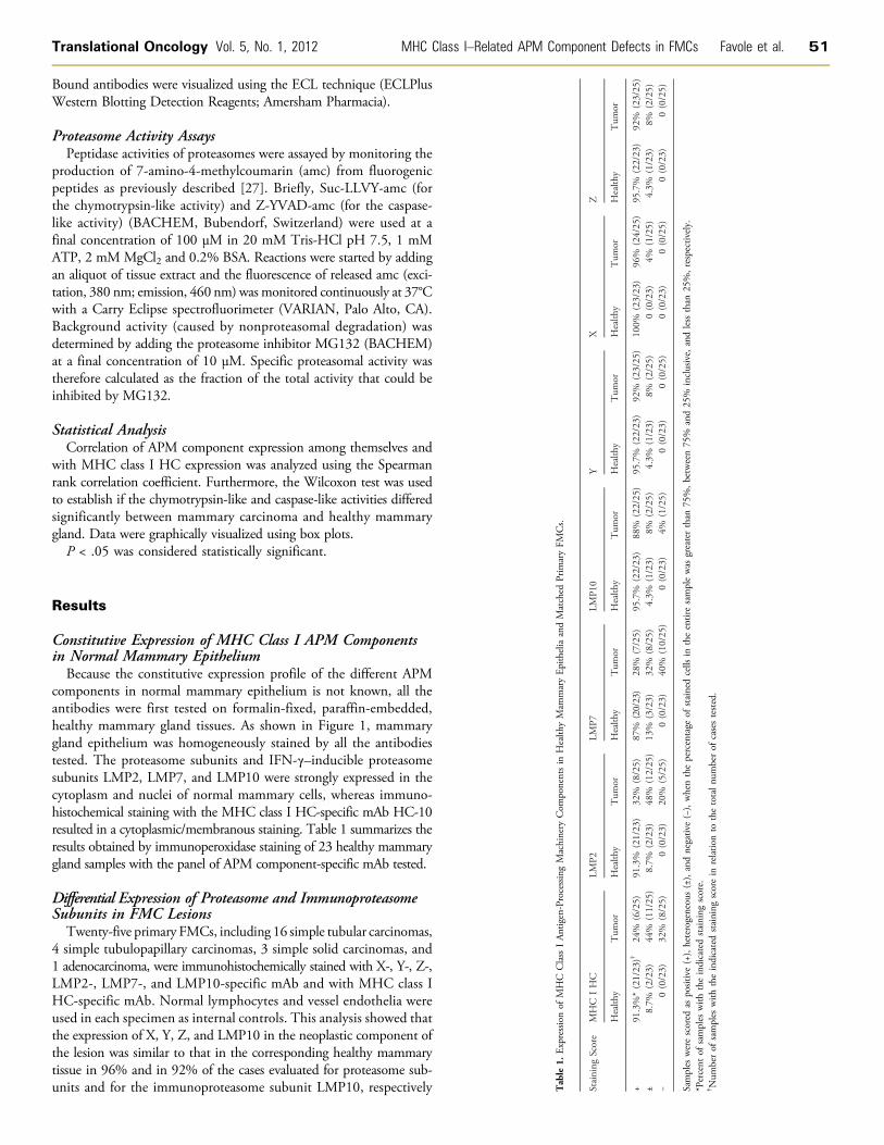
Table
1.Expressionof
MHC
ClassIAntigen-ProcessingMachinery
Com
ponentsin
Health
yMam
maryEpitheliaandMatched
Prim
Staining
Score
MHC
IHC
LMP2
LMP7
LMP1
0
Health
yTum
orHealth
yTum
orHealth
yTum
orHealth
y
+91.3%*(21/23)†
24%
(6/25)
91.3%
(21/23)
32%
(8/25)
87%
(20/23)
28%
(7/25)
95.7%
(22/2
±8.7%
(2/23)
44%
(11/25)
8.7%
(2/23)
48%
(12/25)
13%
(3/23)
32%
(8/25)
4.3%
(1/23
−0(0/23)
32%
(8/25)
0(0/23)
20%
(5/25)
0(0/23)
40%
(10/25)
0(0/23
Samples
werescored
aspositive(+),heterogeneous(±),andnegative(−),whenthepercentage
ofstainedcells
intheentiresamplewas
*Percent
ofsamples
with
theindicatedstaining
score.
†Num
berof
samples
with
theindicatedstaining
scorein
relatio
nto
thetotalnu
mberof
casestested.
Results
Constitutive Expression of MHC Class I APM Componentsin Normal Mammary EpitheliumBecause the constitutive expression profile of the different APM
components in normal mammary epithelium is not known, all theantibodies were first tested on formalin-fixed, paraffin-embedded,healthy mammary gland tissues. As shown in Figure 1, mammarygland epithelium was homogeneously stained by all the antibodiestested. The proteasome subunits and IFN-γ–inducible proteasomesubunits LMP2, LMP7, and LMP10 were strongly expressed in thecytoplasm and nuclei of normal mammary cells, whereas immuno-histochemical staining with the MHC class I HC-specific mAb HC-10resulted in a cytoplasmic/membranous staining. Table 1 summarizes theresults obtained by immunoperoxidase staining of 23 healthy mammarygland samples with the panel of APM component-specific mAb tested.
Differential Expression of Proteasome and ImmunoproteasomeSubunits in FMC LesionsTwenty-five primary FMCs, including 16 simple tubular carcinomas,
4 simple tubulopapillary carcinomas, 3 simple solid carcinomas, and1 adenocarcinoma, were immunohistochemically stained with X-, Y-, Z-,LMP2-, LMP7-, and LMP10-specific mAb and with MHC class IHC-specific mAb. Normal lymphocytes and vessel endothelia wereused in each specimen as internal controls. This analysis showed thatthe expression of X, Y, Z, and LMP10 in the neoplastic component ofthe lesion was similar to that in the corresponding healthy mammarytissue in 96% and in 92% of the cases evaluated for proteasome sub-units and for the immunoproteasome subunit LMP10, respectively

52 MHC Class I–Related APM Component Defects in FMCs Favole et al. Translational Oncology Vol. 5, No. 1, 2012
(Table 1). On the contrary, tumor lesions were found to have a highlyvariable expression profile ranging from a total loss, to heterogeneousbut decreased expression levels, to normal expression for LMP2, LMP7,and MHC class I HC (Table 1 and Figure 2). The expression of MHCclass I HC and IFN-γ–induced catalytic subunits LMP2 and LMP7was reduced in tumor lesions compared with matched healthy tissues.As shown in Table 1, among the seven proteins examined, MHC class IHC and LMP7 showed the greatest reduction in expression, being notdetectable in 32% and 40% of the lesions, respectively, and down-regulated in 44% and 32% of the lesions, respectively. X showed thesmallest reduction, being positive in 96% and downregulated in only 4%
Figure 2. Immunohistochemical staining patterns of formalin-fixed, pand APM component-specific mAbs. The staining with MHC class I HLMP7-specific mAb (E and F) andwith LMP10-specific mAb (G and H) wrespectively. Normal lymphocytes, which serve as the internal control
of the lesions. LMP2 was not detectable in 20% and downregulated in48% of the FMC lesions. Y, Z, and LMP10 were positive in 92% anddownregulated in 8% of the lesions. Taken together, these results clearlydemonstrate that, compared with healthy mammary tissues, mammarycarcinomas did not differ in the expression levels of the proteasome sub-units X, Y, and Z but displayed a down-regulation of the two immuno-proteasome subunits LMP2 and LMP7 and of MHC class I HC.
Additional analyses tested whether the expression level of APMcomponents is correlated to that of MHC class I HC. Analysis by theSpearman rank correlation coefficient showed that LMP2 and LMP7expression was significantly correlated with that of MHC class I HC
araffin-embedded primary FMC lesions with MHC class I antigenC-specific mAb (A and B), with LMP2-specific mAb (C and D), withas scored as positive (A, C, E, and G) and as negative (B, D, F, and H),, are stained as well (arrow). Original magnification, ×200.
Translational Oncology Vol. 5, No. 1, 2012 MHC Class I–Related APM Component Defects in FMCs Favole et al. 53
(r = 0.43, P = .017 and r = 0.70, P < .0001, respectively). Interestingly,LMP2 and LMP7 are concordantly expressed in FMCs (r = 0.58, P =.001), suggesting that they are regulated by similar mechanisms.
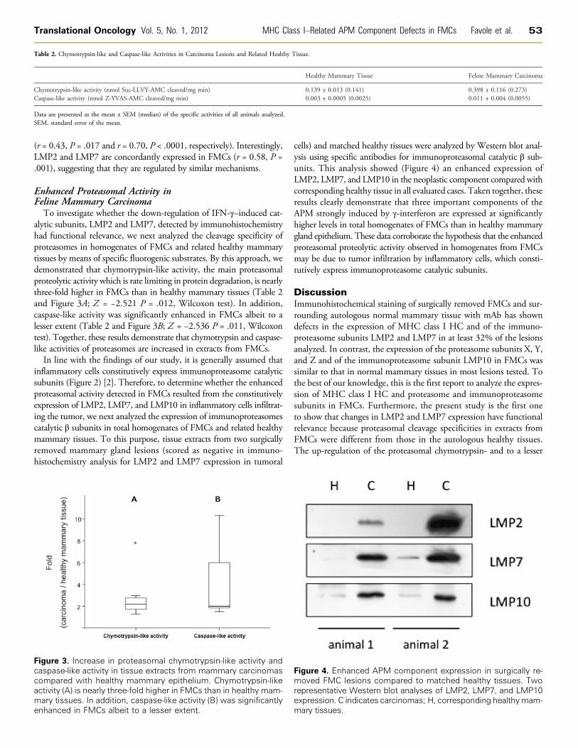
Enhanced Proteasomal Activity inFeline Mammary CarcinomaTo investigate whether the down-regulation of IFN-γ–induced cat-
alytic subunits, LMP2 and LMP7, detected by immunohistochemistryhad functional relevance, we next analyzed the cleavage specificity ofproteasomes in homogenates of FMCs and related healthy mammarytissues by means of specific fluorogenic substrates. By this approach, wedemonstrated that chymotrypsin-like activity, the main proteasomalproteolytic activity which is rate limiting in protein degradation, is nearlythree-fold higher in FMCs than in healthy mammary tissues (Table 2and Figure 3A; Z = −2.521 P = .012, Wilcoxon test). In addition,caspase-like activity was significantly enhanced in FMCs albeit to alesser extent (Table 2 and Figure 3B; Z = −2.536 P = .011, Wilcoxontest). Together, these results demonstrate that chymotrypsin and caspase-like activities of proteasomes are increased in extracts from FMCs.In line with the findings of our study, it is generally assumed that
inflammatory cells constitutively express immunoproteasome catalyticsubunits (Figure 2) [2]. Therefore, to determine whether the enhancedproteasomal activity detected in FMCs resulted from the constitutivelyexpression of LMP2, LMP7, and LMP10 in inflammatory cells infiltrat-ing the tumor, we next analyzed the expression of immunoproteasomescatalytic β subunits in total homogenates of FMCs and related healthymammary tissues. To this purpose, tissue extracts from two surgicallyremoved mammary gland lesions (scored as negative in immuno-histochemistry analysis for LMP2 and LMP7 expression in tumoral
Figure 3. Increase in proteasomal chymotrypsin-like activity andcaspase-like activity in tissue extracts from mammary carcinomascompared with healthy mammary epithelium. Chymotrypsin-likeactivity (A) is nearly three-fold higher in FMCs than in healthy mam-mary tissues. In addition, caspase-like activity (B) was significantlyenhanced in FMCs albeit to a lesser extent.
cells) and matched healthy tissues were analyzed by Western blot anal-ysis using specific antibodies for immunoproteasomal catalytic β sub-units. This analysis showed (Figure 4) an enhanced expression ofLMP2, LMP7, and LMP10 in the neoplastic component compared withcorresponding healthy tissue in all evaluated cases. Taken together, theseresults clearly demonstrate that three important components of theAPM strongly induced by γ-interferon are expressed at significantlyhigher levels in total homogenates of FMCs than in healthy mammarygland epithelium. These data corroborate the hypothesis that the enhancedproteasomal proteolytic activity observed in homogenates from FMCsmay be due to tumor infiltration by inflammatory cells, which consti-tutively express immunoproteasome catalytic subunits.
DiscussionImmunohistochemical staining of surgically removed FMCs and sur-rounding autologous normal mammary tissue with mAb has showndefects in the expression of MHC class I HC and of the immuno-proteasome subunits LMP2 and LMP7 in at least 32% of the lesionsanalyzed. In contrast, the expression of the proteasome subunits X, Y,and Z and of the immunoproteasome subunit LMP10 in FMCs wassimilar to that in normal mammary tissues in most lesions tested. Tothe best of our knowledge, this is the first report to analyze the expres-sion of MHC class I HC and proteasome and immunoproteasomesubunits in FMCs. Furthermore, the present study is the first oneto show that changes in LMP2 and LMP7 expression have functionalrelevance because proteasomal cleavage specificities in extracts fromFMCs were different from those in the autologous healthy tissues.The up-regulation of the proteasomal chymotrypsin- and to a lesser
Table 2. Chymotrypsin-like and Caspase-like Activities in Carcinoma Lesions and Related Healthy Tissue.
Figmorepexpma
Healthy Mammary Tissue
ure 4. Enhanced APM component expressved FMC lesions compared to matched hresentative Western blot analyses of LMP2ression. C indicates carcinomas; H, correspory tissues.
Feline Mammary Carcinoma
Chymotrypsin-like activity (nmol Suc-LLVY-AMC cleaved/mg min)
0.139 ± 0.013 (0.141) 0.398 ± 0.116 (0.273) Caspase-like activity (nmol Z-YVAS-AMC cleaved/mg min) 0.003 ± 0.0005 (0.0025) 0.011 ± 0.004 (0.0055)Data are presented as the mean ± SEM (median) of the specific activities of all animals analyzed.SEM, standard error of the mean.
ion in surgically re-ealthy tissues. Two, LMP7, and LMP10nding healthymam-

54 MHC Class I–Related APM Component Defects in FMCs Favole et al. Translational Oncology Vol. 5, No. 1, 2012
extent caspase-like activities found in homogenates from FMCs com-pared with healthy mammary tissues was unexpected because of thedown-regulation of two immunoproteasome subunits found in FMCs.This apparent paradox may reflect an up-regulation of the constitutiveproteasome catalytic subunits (X, Y, and Z) in the neoplastic tissue,perhaps to compensate for the down-regulation of the immunoprotea-some subunits. Expression of the X and Y genes is reciprocal to that ofthe LMP genes: X and Y are upregulated in mutant cell lines lackingLMPs [28]. In this regard, it is noteworthy that our immunohisto-chemical analysis evaluated the number of stained tumor cells in theentire lesion but did not evaluate the staining intensity at the level ofsingle-stained tumoral cells. Therefore, in tumor lesions, an increasedexpression of constitutive proteasome catalytic subunits cannot be ex-cluded. An additional mechanism contributing to the enhanced protea-somal proteolytic activity observed in homogenates from FMCs may berepresented by the infiltration of tumors with inflammatory cells, whichconstitutively express immunoproteasome catalytic subunits (Figure 2)[2]. Immunoblot analysis supports this hypothesis: tissue extracts fromtwo surgically removed mammary gland lesions, scored in immuno-histochemistry analysis as negative for LMP2 and LMP7 expressionin tumoral cells, on the contrary by Western blot analysis showed anenhanced expression of immunoproteasome catalytic subunits com-pared with healthy mammary tissues. In line with our results, defectiveexpression of MHC class I antigens and APM components has beendemonstrated in almost all works by immunohistochemistry analysisbecause this technique allows to discriminate between inflammatoryand tumoral cells [31,32,36].
The changes in the proteasomal cleavage specificities found in ex-tracts from FMCs argue in favor of the possibility that LMP2 andLMP7 down-regulation in FMCs affects the repertoire of TA-derivedpeptides expressed by tumor cells. Whether these changes influencethe recognition of mammary tumor cells by MHC class I antigen-restricted, TA-derived peptide-specific T cells remains to be investi-gated. Should this be the case, cats with mammary tumors representa useful model to investigate the contribution of immunoproteasomesubunit defects to escape mechanisms used by tumor cells to avoidrecognition and destruction by MHC class I antigen-restricted, TA-specific cytotoxic T lymphocyte. Furthermore, cats with mammarytumors may represent useful models to develop and test strategies tocounteract escape mechanisms caused by changes in the expressionand functional properties of immunoproteasome subunits.
The expression of immunoproteasome and proteasome subunitshas been investigated in human mammary tumors. The phenotypeof FMCs we have described resembles that of human breast carci-noma lesions as Gobbi et al. [29] found a down-regulation of LMP2and MHC class I HC in 51.4% and 40.0% of the lesions tested,respectively. Whereas all normal tissues and benign lesions were posi-tive for β2-m andHLA class I HC, total loss of HLA class I antigens wasfound in 43% of the breast primary tumors and in 70% of the lymphnode metastases [30]. To the best of our knowledge, the expressionof immunoproteasome subunits has been described in a number ofhuman solid tumors, whereas that of proteasome subunits has beeninvestigated in a much lower number of solid tumors. The frequencyof LMP2 and LMP7 down-regulation we have found in FMCs is sim-ilar to that described in renal, cervical, and head and neck carcinomas[15,20,31]. In addition, an unbalanced expression of LMP2, LMP7, andLMP10 has been described in many human solid tumors [20,34,35].Nevertheless, the expression of proteasome subunits has been describedonly in human brain and bladder carcinomas [32,33]. As we have
described in this study, the X, Y, and Z housekeeping proteasomal sub-units, the LMP10 immunoproteasomal subunit, were detected in mostmedulloblastoma and astrocytic tumor lesions [32].
It is generally assumed that immunoproteasome subunits expres-sion is constitutive only in immunologic cells, whereas it is induced inother cells on exposure to cytokines (i.e., IFN-γ), thereby increasingthe number of peptides capable of binding MHC class I antigens [5].At variance with this notion, we observed basal expression of LMP2,LMP7, and LMP10 in normal mammary epithelial cells. In fact,LMP2, LMP7, and LMP10 seem coordinately expressed in all healthymammary tissues tested in our study. It should be stressed that theexpression of immunoproteasome subunits in nonimmune cells underbasal conditions is not unique to feline mammary tissue because it hasalso been described in human mammary, renal, laryngeal, neural,bladder, prostate, and cervical tissues [15,18,20,29,32–36].
From a methodological view point, it is noteworthy that the pres-ent study is one of the very few ones that compare the expression ofMHC class I HC in malignant cells and autologous surrounding tis-sues. To the best of our knowledge, a similar comparison has beendone only in a study that analyzed the expression of APM compo-nents in bladder carcinoma cells and in surrounding normal cells.The conclusions from these studies about changes in the expressionof APM components in malignant cells are more meaningful thanthose derived from most of the published studies, including ourown, which assess the expression of these molecules in malignant cellsby comparison with the endothelial cells and lymphoid cells present inthe tissue section analyzed [36].
In summary, this study provides the first description of changes inthe expression and function of MHC class I–related APM componentsin FMCs. Characterization of the role of these changes in the interac-tions of FMCs with the host immune system may contribute to ourunderstanding of the molecular mechanisms used by tumor cells andmay suggest targeted strategies to counteract these escape mechanisms.
References[1] Gromme M and Neefjes J (2002). Antigen degradation or presentation by MHC
class I molecules via classical and non-classical pathways.Mol Immunol 39, 181–202.[2] Pardoll DM and Topalian SL (1998). The role of CD4+ T cell responses in
antitumor immunity. Curr Opin Immunol 10, 588–594.[3] Van den Eynde BJ and Morel S (2001). Differential processing of class-I–
restricted epitopes by the standard proteasome and the immunoproteasome.Curr Opin Immunol 13, 147–153.
[4] Tanaka K and Kasahara M (1998). The MHC class I ligand–generating system:roles of immunoproteasomes and the interferon-γ–inducible proteasome activatorPA28. Immunol Rev 163, 161–176.
[5] Goldberg AL, Cascio P, Saric T, and Rock KL (2002). The importance of theproteasome and subsequent proteolytic steps in the generation of antigenicpeptides. Mol Immunol 39, 147–164.
[6] Campoli M and Ferrone S (2008). HLA antigen changes in malignant cells:epigenetic mechanisms and biologic significance. Oncogene 27, 5869–5885.
[7] Chang CC and Ferrone S (2007). Immune selective pressure and HLA class Iantigen defects in malignant lesions. Cancer Immunol Immunother 56, 227–236.
[8] Cabrera T, López-Nevot MA, Gaforio JJ, Ruiz-Cabello F, and Garrido F(2003). Analysis of HLA expression in human tumor tissues. Cancer ImmunolImmunother 52, 1–9.
[9] Campoli M, Chang CC, Oldford SA, Edgecombe AD, Drover S, and Ferrone S(2004). HLA antigen changes in malignant tumors of mammary epithelial origin:molecular mechanisms and clinical implications. Breast Dis 20, 105–125.
[10] Seliger B, Maeurer MJ, and Ferrone S (2000). Antigen-processing machinerybreakdown and tumor growth. Immunol Today 21, 455–464.
[11] Seliger B (2008). Molecular mechanisms of MHC class I abnormalitiesand APM components in human tumors. Cancer Immunol Immunother 57,1719–1726.
Translational Oncology Vol. 5, No. 1, 2012 MHC Class I–Related APM Component Defects in FMCs Favole et al. 55
[12] Aptsiauri N, Cabrera T, Mendez R, Garcia-Lora A, Ruiz-Cabello F, andGarrido F (2007). Role of altered expression of HLA class I molecules in cancerprogression. Adv Exp Med Biol 601, 123–131.
[13] Cabrera T, Maleno I, Collado A, Lopez Nevot MA, Tait BD, and Garrido F(2007). Analysis of HLA class I alterations in tumors: choosing a strategy basedon known patterns of underlying mechanisms. Tissue Antigens 69, 264–268.
[14] Garrido F, Ruiz-Cabello F, Cabrera T, Perez-Villar JJ, Lopez-Botet M, Duggan-Keen M, and Stern PL (1997). Implications for immunosurveillance of alteredHLA class I phenotypes in human tumours. Immunol Today 18, 89–95.
[15] Atkins D, Ferrone S, Schmahl GE, Störkel S, and Seliger B (2004). Down-regulation of HLA class I antigen processing molecules: an immune escapemechanism of renal cell carcinoma? J Urol 171, 885–889.
[16] Mehling M, Simon P, Mittelbronn M, Meyermann R, Ferrone S, Weller M,and Wiendl H (2007). WHO grade associated downregulation of MHC class Iantigen-processing machinery components in human astrocytomas: does it reflect apotential immune escape mechanism? Acta Neuropathol 114, 111–119.
[17] Ferrone S and Marincola FM (1995). Loss of HLA class I antigens by melanomacells: molecular mechanisms, functional significance and clinical relevance.Immunol Today 16, 487–494.
[18] Ogino T, Shigyo H, Ishii H, Katayama A, Miyokawa N, Harabuchi Y, andFerrone S (2006). HLA class I antigen down-regulated in primary laryngeal squa-mous cell carcinoma lesions as a poor prognostic marker. Cancer Res 66, 9281–9289.
[19] Gudmundsdóttir I, Gunnlaugur Jónasson J, Sigurdsson H, Olafsdóttir K,Tryggvadóttir L, and Ogmundsdóttir HM (2000). Altered expression of HLAclass I antigens in breast cancer: association with prognosis. Int J Cancer 89,500–505.
[20] Mehta AM, Jordanova ES, Kenter GG, Ferrone S, and Fleuren GJ (2008).Association of antigen processing machinery and HLA class I defects withclinicopathological outcome in cervical carcinoma. Cancer Immunol Immunother57, 197–206.
[21] Stam NJ, Spits H, and Ploegh HL (1986). Monoclonal antibodies raised againstdenatured HLA-B locus heavy chains permit biochemical characterization ofcertain HLA-C locus products. J Immunol 137, 2299–2306.
[22] Perosa F, Luccarelli G, Prete M, Favoino E, Ferrone S, and Dammacco F(2003). β2-Microglobulin-free HLA class I heavy chain epitope mimicry bymonoclonal antibody HC-10–specific peptide. J Immunol 171, 1918–1926.
[23] Bandoh N, Ogino T, Cho HS, Hur SY, Shen J, Wang X, Kato S, Miyokawa N,Harabuchi Y, and Ferrone S (2005). Development and characterization ofhuman constitutive proteasome and immunoproteasome subunit-specific mono-clonal antibodies. Tissue Antigens 66, 185–194.
[24] Perosa F, Carbone R, Ferrone S, and Dammacco F (1990). Purification of humanimmunoglobulins by sequential precipitation with caprylic acid and ammoniumsulphate. J Immunol Methods 128, 9–16.
[25] Garrido F, Cabrera T, Accolla RS, Bensa JC, Bodmer W, Dohr G, Drouet M,Fauchet R, Ferrara GB, Ferrone S, et al. (1997). HLA and cancer. In HLA:Genetic Diversity of HLA. Functional and Medical Implication. D Charron(Ed). EDK, Paris, France. pp. 445–452.
[26] Cerruti F, Martano M, Morello E, Buracco P, and Cascio P (2010). Proteasomesare not a target for doxorubicin in feline injection-site sarcoma. J Comp Pathol143, 164–172.
[27] Santoni de Sio FR, Gritti A, Cascio P, Neri M, Sampaolesi M, Galli C, Luban J,and Naldini L (2008). Lentiviral vector gene transfer is limited by the proteasomeat postentry steps in various types of stem cells. Stem Cells 26, 2142–2152.
[28] Belich MP, Glynne RJ, Senger G, Sheer D, and Trowsdale J (1994). Protea-some components with reciprocal expression to that of the MHC-encoded LMPproteins. J Curr Biol 4, 769–776.
[29] Gobbi G, Mirandola P, Micheloni C, Solenghi E, Sponzilli I, Artico M, Soda G,Zanelli G, Pelusi G, Fiorini T, et al. (2004). Expression of HLA class I antigenand proteasome subunits LMP-2 and LMP-10 in primary vs. metastatic breastcarcinoma lesions. Int J Oncol 25, 1625–1629.
[30] Redondo M, García J, Villar E, Rodrigo I, Perea-Milla E, Serrano A, and MorellM (2003). Major histocompatibility complex status in breast carcinogenesis andrelationship to apoptosis. Hum Pathol 34, 1283–1289.
[31] Meissner M, Reichert TE, Kunkel M, Gooding W, Whiteside TL, Ferrone S,and Seliger B (2005). Defects in the human leukocyte antigen class I antigenprocessing machinery in head and neck squamous cell carcinoma: associationwith clinical outcome. Clin Cancer Res 11, 2552–2560.
[32] Raffaghello L, Nozza P, Morandi F, Camoriano M, Wang X, Garrè ML, Cama A,Basso G, Ferrone S, Gambini C, et al. (2007). Expression and functional analysisof human leukocyte antigen class I antigen-processing machinery in medullo-blastoma. Cancer Res 67, 5471–5478.
[33] Cathro HP, Smolkin ME, Theodorescu D, Jo VY, Ferrone S, and Frierson HF Jr(2010). Relationship between HLA class I antigen processing machinery compo-nent expression and the clinicopathologic characteristics of bladder carcinomas.Cancer Immunol Immunother 59, 465–472.
[34] Raffaghello L, Prigione I, Bocca P, Morandi F, Camoriano M, Gambini C,Wang X, Ferrone S, and Pistoia V (2005). Multiple defects of the antigen-processing machinery components in human neuroblastoma: immunotherapeuticimplications. Oncogene 24, 4634–4644.
[35] Facoetti A, Nano R, Zelini P, Morbini P, Benericetti E, Ceroni M, Campoli M, andFerrone S (2005). Human leukocyte antigen and antigen processing machinerycomponent defects in astrocytic tumors. Clin Cancer Res 11, 8304–8311.
[36] Seliger B, Stoehr R, Handke D, Mueller A, Ferrone S, Wullich B, Tannapfel A,Hofstaedter F, and Hartmann A (2010). Association of HLA class I antigenabnormalities with disease progression and early recurrence in prostate cancer.Cancer Immunol Immunother 59, 529–540.