method validation reports on rules proposals for the ... · om12-06 method validation reports on...
TRANSCRIPT

International Seed Testing Association Secretariat, Zürichstrasse 50, CH-8303 Bassersdorf, Switzerland Phone: +41 44 838 60 00 Fax: +41 44 838 60 01 Email: [email protected] - http://www.seedtest.org
Document OM12-06
INFORMATION DOCUMENT TO SUPPORT MAIN MOTIONS
PRESENTED IN DOCUMENT ‘OM12-05 RULES PROPOSALS FOR
THE INTERNATIONAL RULES FOR SEED TESTING 2013 EDITION’
OM12-06 Method Validation Reports on Rules Proposals for the ISTA Rules 2013 Edition.doc 2012-04-09 13:06 Approved by ECOM and RUL on March 23, 2012 Page 1/24
Method Validation Reports on Rules Proposals for the International Rules for Seed Testing 2013 Edition
Contents
PCR as a new identification method of Xanthomonas campestris pv. campestris on Brassica spp. seed 2
New method for the detection of infectious tobamoviruses on tomato (Lycopersicon esculentum) seed by local lesion assay (indexing) on Nicotiana tabacum plants 12

Grimault et al. New method of Xanthomonas campestris pv. campestris on Brassica spp. seed
OM12-06 Method Validation Reports on Rules Proposals for the ISTA Rules 2013 Edition.doc 2012-04-09 13:06 Approved by ECOM and RUL on March 23, 2012 Page 2/24
PCR as a new identification method of Xanthomonas campestris pv. campestris on Brassica spp. seed
V. Grimault1, C. Andro2 & A. Politikou1
1GEVES-SNES and
2BioGEVES, Rue Georges Morel, BP 90024, 49071 Beaucouzé Cedex, France
Summary
The efficiency of the PCR technique for the identification of Xanthomonas campestris pv. campestris (Xcc) on Brassica spp. seed was compared to the pathogenicity test described in ISTA Seed Health Method 7-019 in a peer validation study organized by the International Seed Health Initiative for Vegetables. Four laboratories from the Netherlands and France together tested 1472 suspect bacteria isolates of X. campestris pathovars by conducting in parallel a PCR and a pathogenicity test. Xcc was identified using the DLH primer sets by Berg et al. (2005) and the Zup primer sets by Rijlaarsdam et al. (2004), and the results of the PCR and pathogenicity test were summarized and compared. There is a negligible risk of a false-positive PCR result for Xcc, caused by primer sets targeting X. campestris pv. incanae. It is highly unlikely that X. campestris pv. incanae isolates are present on cultivated Brassica spp. seeds. The study showed comparable results for the PCR and pathogenicity tests for 97.21% of the total of suspect isolates. Compared to the pathogenicity test, the PCR produced a false-negative result in only 0.41% of the suspect isolates tested. The PCR technique was shown to provide complementary information in cases where the pathogenicity test did not show clear symptoms, and to give additional information on the suspected occurrence of X. campestris pv. armoraciae or X. campestris pv. raphani (Xca/Xcr). Similarly, the pathogenicity test would be valuable when an indeterminate PCR result appears. The risk of a final false-negative result on a seed lot is minimized by testing at least six suspect isolates per seed subsample, as instructed by ISTA Method 7-019. The use of a PCR technique is highly recommended as an alternative or complementary method for Xcc identification in seed health testing laboratories.
Introduction
Xanthomonas campestris pv. campestris (Xcc) is a seed-borne, pathogenic bacterium on cruciferous plants and the causal agent of black rot disease that can cause severe economic losses worldwide (Qian et al., 2005). Both ISTA Seed Health Method 7-019 for untreated seed, and the ISHI-Veg method (www.worldseed.org >> Trade Related Topics >> Phytosanitary Matters >> Seed Health >> ISHI-Veg) for disinfected seed comprise a pathogenicity test following dilution plating as a confirmation step of suspect Xcc colonies. Symptoms on inoculated bioassay plants are recorded 10 to 14 days after inoculation. Although this confirmation method is reliable, the duration of the test is of concern to laboratories which routinely test Brassica spp. seeds for the presence of Xcc. The PCR technique is faster than the pathogenicity test, and could be of use for these laboratories.
For several years, a PCR technique using pathogen-specific primers has been used to identify several pathogenic Xanthomonas species on various hosts (Pan et al., 1999; Fargier and Manceau (2007); Palacio-Bielsa et al., 2009). Comparative studies between the pathogenicity test and PCR for X. hortorum pv. carotae (Xhc) identification on carrot seed showed that PCR was a reliable and rapid confirmation tool (Asma et al., 2002). PCR as an alternative to the pathogenicity test for confirming suspect Xhc colonies is part of ISTA Seed Health Method 7-020 (ISTA, 2006).
Berg et al. (2005) developed the primer set DLH120-125, which is specific to all X. campestris pathovars (X. c. pv. campestris, X. c. pv. armoraciae, X. c. pv. raphani and X. c. pv. incanae), while Rijlaarsdam et al. (2004) and Zaccardelli et al. (2007) developed primer sets for identification of Xcc. Fargier and Manceau (2007) validated the specificity of the Zup2309-2310 and Zup2311-2312 primer sets developed by Rijlaarsdam et al. (2004) and the primer sets of Berg et al. (2005) on a collection of 47 X. campestris isolates by comparing PCR results to pathogenicity tests. In that study, the Rijlaarsdam et al. (2004) primer sets were found to amplify the DNA of Xcc and X. c. pv. incanae isolates. However, X. c. pv. incanae isolates were pathogenic only on Matthiola spp. and Erysimum cheiri (previous name Cheiranthus cheiri) plants. Given Fargier and Marceau‟s results, as well as the fact that it is highly unlikely to have X. c. pv. incanae on cultivated Brassica spp. seeds, the combination of primer sets by Berg et al. (2005) and Rijlaarsdam et al.
Grimault et al. New method of Xanthomonas campestris pv. campestris on Brassica spp. seed
OM12-06 Method Validation Reports on Rules Proposals for the ISTA Rules 2013 Edition.doc 2012-04-09 13:06 Approved by ECOM and RUL on March 23, 2012 Page 3/24
(2004) was considered appropriate in the present study for validation of the identification of Xcc on Brassica spp. seeds without posing any risk of a Xcc false-positive PCR result.
Vicente et al. (2006) did not confirm the existence of X. c. pv. armoraciae after testing one isolate that was received as such. Moreover, Fargier and Manceau (2007), after testing three isolates received as X. c. pv. armoraciae, didn‟t support the existence of another leaf spot disease caused by this pathogen. In further validation studies, Porcher et al. (2008) and Mathis et al. (2009) found a PCR result that was negative with Rijlaarsdam et al. (2004) primers and positive with Berg et al. (2005) primers. This PCR result, after taking into consideration that Xcc and X. c. pv. raphani (Xcr) are carried by Brassica spp. seed and are pathogenic on Brassica spp. plants, was interpreted as the suspected presence of Xcr. However, final conclusions on the presence of Xcr should be legitimate only after validation by epidemiological studies and identification of Xanthomonas sp. strains.
Despite the conclusions of Fargier and Manceau (2007) and Vicente et al. (2006) on X. c. pv. armoraciae (Xca) nomenclature, the names of both “armoraciae” and “raphani” pathovars are included in the list of Bull et al. (2010). Moreover, no change has been made to section 7.7 of ISTA Method 7-019 regarding the denomination of the causal agent (leaf spot Xanthomonas) of the leaf spot disease symptoms on the pathogenicity test. Thus, for consistency with ISTA Method 7-019, this study uses the names of both pathovars as Xca/Xcr wherever appropriate.
Scope and objective of the peer validation study
The scope of this peer validation study is to compare the efficiency of a PCR test with a pathogenicity test to identify suspect Xcc isolates among a large number of suspects. For this purpose, participating laboratories were called to provide data generated over the past years on the comparison of the two tests. This study was performed in addition to the work done on primer validation by Fargier and Manceau (2007). The objective of the study is to use PCR, if found to be efficient and comparable, as an alternative to the pathogenicity test described in ISTA Method 7-019 for the identification and confirmation of suspect Xcc colonies isolated from Brassica spp. seed.
Materials and methods
Bacterial isolates
A total of 1472 bacterial isolates of Xanthomonas campestris pathovars were identified by four laboratories: BioGEVES and SNES (Beaucouzé, France), Clause Tézier (Valence, France), Naktuinbouw (Roelofarendsveen, the Netherlands) and Rijk Zwaan (De Lier, the Netherlands). The isolates were from the collections or the participant laboratories or other company collections, or extracted from Brassica spp. seeds or plants. The numbers of isolates tested by each laboratory are provided in Table 3. The laboratories used Xcc and Xca/Xcr reference isolates as positive controls. Mock inoculation with sterile water and/or inoculation with isolates of other Xanthomonas species were used as negative controls.
Pathogenicity test and PCR protocols
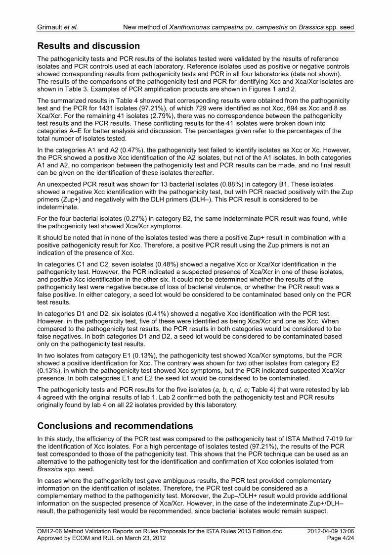
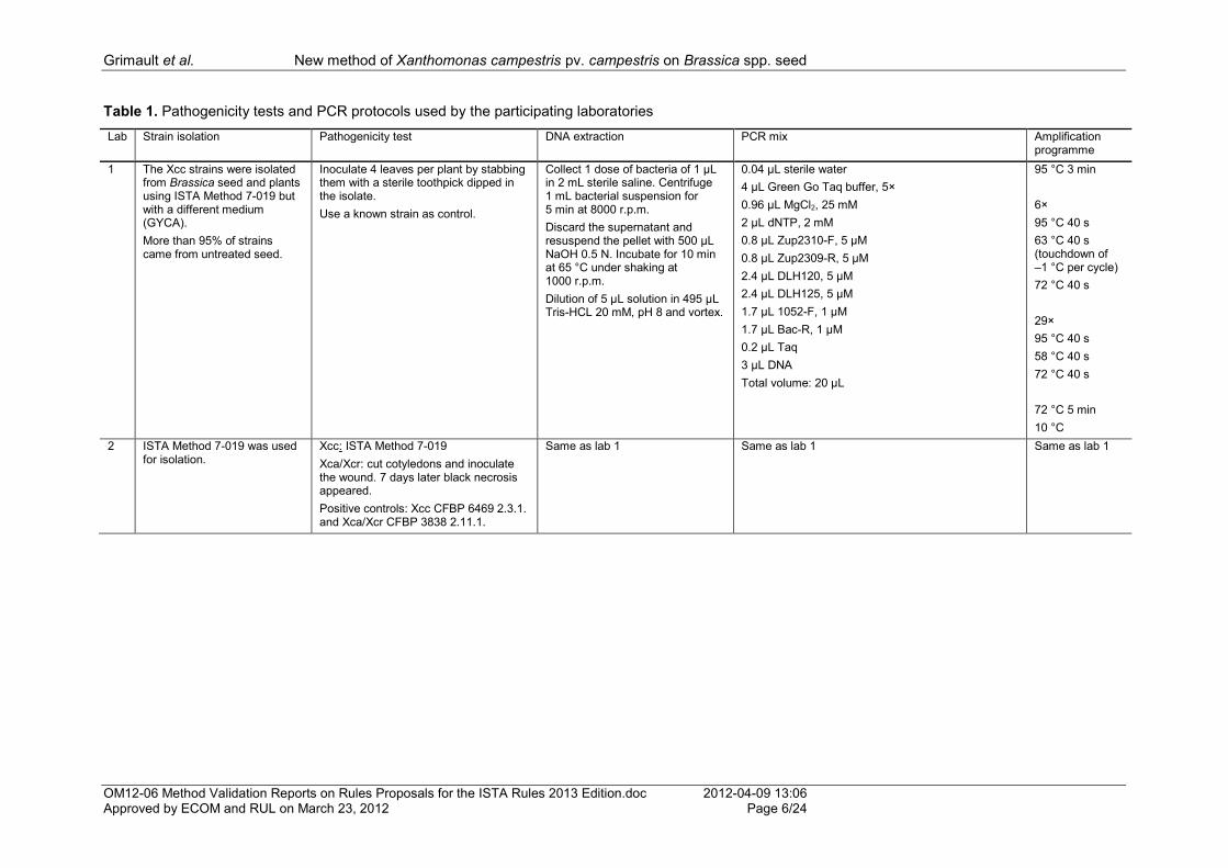
A detailed description of the pathogenicity test and PCR protocol used by each laboratory is provided in Table 1 (participating laboratories were given code numbers). All laboratories used the pathogenicity test described in ISTA Method 7-019 or a slightly modified version thereof. Lab 1 and lab 2 used the same PCR protocol. Lab 3 used a PCR mix to analyse 1145 isolates, with primers by Rijlaarsdam et al. (2004) and UpBacF/UpBacR universal primers adapted from Eden et al. (1991). Ten isolates showed Xcc false-positive identification with PCR compared to the pathogenicity test result, and 5 isolates showed Xca/Xcr symptoms in the pathogenicity test and didn‟t react with Rijlaarsdam et al. (2004) primers. These were retested by lab 3 using Berg et al. (2005) primers in the aforementioned PCR mix with the same amplification program. Lab 4 used an adapted version of the PCR protocol utilized by lab 3. The sequences of all primer sets used are presented in Table 2.
The 22 isolates that were initially tested by lab 4 were retested by lab 2 using a pathogenicity test and PCR to confirm the original results. Five isolates (Table 3) that had been originally tested by lab 1 were retested by lab 4 by PCR in parallel to a pathogenicity test. Three of these isolates had originally shown ambiguous results, while the other 2 had corresponding results and served as a control to lab 4.
Grimault et al. New method of Xanthomonas campestris pv. campestris on Brassica spp. seed
OM12-06 Method Validation Reports on Rules Proposals for the ISTA Rules 2013 Edition.doc 2012-04-09 13:06 Approved by ECOM and RUL on March 23, 2012 Page 4/24
Results and discussion
The pathogenicity tests and PCR results of the isolates tested were validated by the results of reference isolates and PCR controls used at each laboratory. Reference isolates used as positive or negative controls showed corresponding results from pathogenicity tests and PCR in all four laboratories (data not shown). The results of the comparisons of the pathogenicity test and PCR for identifying Xcc and Xca/Xcr isolates are shown in Table 3. Examples of PCR amplification products are shown in Figures 1 and 2.
The summarized results in Table 4 showed that corresponding results were obtained from the pathogenicity test and the PCR for 1431 isolates (97.21%), of which 729 were identified as not Xcc, 694 as Xcc and 8 as Xca/Xcr. For the remaining 41 isolates (2.79%), there was no correspondence between the pathogenicity test results and the PCR results. These conflicting results for the 41 isolates were broken down into categories A–E for better analysis and discussion. The percentages given refer to the percentages of the total number of isolates tested.
In the categories A1 and A2 (0.47%), the pathogenicity test failed to identify isolates as Xcc or Xc. However, the PCR showed a positive Xcc identification of the A2 isolates, but not of the A1 isolates. In both categories A1 and A2, no comparison between the pathogenicity test and PCR results can be made, and no final result can be given on the identification of these isolates thereafter.
An unexpected PCR result was shown for 13 bacterial isolates (0.88%) in category B1. These isolates showed a negative Xcc identification with the pathogenicity test, but with PCR reacted positively with the Zup primers (Zup+) and negatively with the DLH primers (DLH–). This PCR result is considered to be indeterminate.
For the four bacterial isolates (0.27%) in category B2, the same indeterminate PCR result was found, while the pathogenicity test showed Xca/Xcr symptoms.
It should be noted that in none of the isolates tested was there a positive Zup+ result in combination with a positive pathogenicity result for Xcc. Therefore, a positive PCR result using the Zup primers is not an indication of the presence of Xcc.
In categories C1 and C2, seven isolates (0.48%) showed a negative Xcc or Xca/Xcr identification in the pathogenicity test. However, the PCR indicated a suspected presence of Xca/Xcr in one of these isolates, and positive Xcc identification in the other six. It could not be determined whether the results of the pathogenicity test were negative because of loss of bacterial virulence, or whether the PCR result was a false positive. In either category, a seed lot would be considered to be contaminated based only on the PCR test results.
In categories D1 and D2, six isolates (0.41%) showed a negative Xcc identification with the PCR test. However, in the pathogenicity test, five of these were identified as being Xca/Xcr and one as Xcc. When compared to the pathogenicity test results, the PCR results in both categories would be considered to be false negatives. In both categories D1 and D2, a seed lot would be considered to be contaminated based only on the pathogenicity test results.
In two isolates from category E1 (0.13%), the pathogenicity test showed Xca/Xcr symptoms, but the PCR showed a positive identification for Xcc. The contrary was shown for two other isolates from category E2 (0.13%), in which the pathogenicity test showed Xcc symptoms, but the PCR indicated suspected Xca/Xcr presence. In both categories E1 and E2 the seed lot would be considered to be contaminated.
The pathogenicity tests and PCR results for the five isolates (a, b, c, d, e; Table 4) that were retested by lab 4 agreed with the original results of lab 1. Lab 2 confirmed both the pathogenicity test and PCR results originally found by lab 4 on all 22 isolates provided by this laboratory.
Conclusions and recommendations
In this study, the efficiency of the PCR test was compared to the pathogenicity test of ISTA Method 7-019 for the identification of Xcc isolates. For a high percentage of isolates tested (97.21%), the results of the PCR test corresponded to those of the pathogenicity test. This shows that the PCR technique can be used as an alternative to the pathogenicity test for the identification and confirmation of Xcc colonies isolated from Brassica spp. seed.
In cases where the pathogenicity test gave ambiguous results, the PCR test provided complementary information on the identification of isolates. Therefore, the PCR test could be considered as a complementary method to the pathogenicity test. Moreover, the Zup–/DLH+ result would provide additional information on the suspected presence of Xca/Xcr. However, in the case of the indeterminate Zup+/DLH–result, the pathogenicity test would be recommended, since bacterial isolates would remain suspect.

Grimault et al. New method of Xanthomonas campestris pv. campestris on Brassica spp. seed
OM12-06 Method Validation Reports on Rules Proposals for the ISTA Rules 2013 Edition.doc 2012-04-09 13:06 Approved by ECOM and RUL on March 23, 2012 Page 5/24
The PCR test gave false negative results compared to the pathogenicity test in 0.41% of all cases. Thus, using the PCR technique, in 99.6% of cases a laboratory will obtain an accurate result, or will get an indication to perform an additional pathogenicity test to confirm the PCR result.
When following the instructions of ISTA Method 7-019 to test at least six suspect isolates per subsample, the risk of a final false negative result in a seed lot is minimized.
Given that all 27 isolates showed the same PCR results when tested with the protocols of labs 2 and 4 (the latter being a slight variant of the lab 3 protocol), these protocols can be considered to be equivalent. Consequently, this report supports two PCR options, each comprising different primers and amplification regimes for identifying or confirming suspect Xcc isolates.
Acknowledgements
The participating laboratories BioGEVES and SNES (Beaucouzé, France), Clause Tézier (Valence, France), Naktuinbouw (Roelofarendsveen, the Netherlands) and Rijk Zwaan (De Lier, the Netherlands) are gratefully acknowledged for providing bacterial isolates and carrying out this peer-validation study.
References
Asma, M., de Vogel, R., Woudt, B. & Krause, D. (2002). Evaluation of pathogenicity testing, rep-fingerprinting and PCR for the identification of Xanthomonas campestris pv. carotae. ISHI Report Bejo Zaden BV, Research Report P9317-16.
Berg T., Tesoriero L. & Hailstones D. L. (2005). PCR-based detection of Xanthomonas campestris pathovars in Brassica seed. Plant Pathology, 54, 416–427.
Bull, C. T., De Boer, S. H., Denny, T. P., Firrao, G., Fischer-Le Saux, M., Saddler, G. S., Scortichini, M., Stead, D. E. & Takikawa, Y. (2010). Comprehensive List Of Names Of Plant Pathogenic Bacteria, 1980-2007. Journal of Plant Pathology, 92, 551–592.
Eden, P. A., Schmidt, T. M., Blackemore, R. P. & Pace, N. R. (1991). Phylogenetic Analysis of Aquaspirillum magnetotacticum using Polymerase Chain Reaction Amplified 16S rRNA specific DNA. International Journal of Systematic Bacteriology, 41, 324–325.
Fargier, E. & Manceau, C. (2007). Pathogenicity assays restrict the species Xanthomonas campestris into three pathovars and reveal nine races within X. campestris pv. campestris. Plant Pathology, 56, 805–818.
ISTA (2007). Detection of Xanthomonas campestris pv. campestris on Brassica spp. International Rules for Seed Testing. Annexe to Chapter 7: Seed Health Testing Methods 7-019, 16 pp.
ISTA (2006). Detection of Xanthomonas hortorum pv. carotae on Daucus carota. International Rules for Seed Testing. Annexe to Chapter 7: Seed health Testing Methods 7-020, 19 pp.
Mathis, R., Porcher, L., Fargier, E., Briand, B., Guillaumès, J., Andro, C., Grimault, V., Valette, N., Darrieutort, G. & Manceau, C. (2009). A new method to detect Xanthomonas campestris in cruciferous seeds by enrichment – PCR. The EPPO Conference on Diagnostics, York (UK).
Palacio-Bielsa, A., Cambra, M. A. & Lopez, M. M. (2009). PCR detection and identification of plant-pathogenic bacteria: Updated review of protocols (1989-2007). Journal of Plant Pathology, 91 (2), 249–297.
Pan, Y.-B., Grisham, M. P., Burner, D. M., Legendre, B. L. & Wei, Q. (1999). Development of polymerase chain reaction primers highly specific for Xanthomonas albilineans, the causal bacterium of sugarcane leaf scald disease. Plant Disease, 83, 218–222.
Porcher, L., Mathis, R., Fargier, E., Briand, B., Guillaumès, J., Grimault, V., Valette, N., Guyot, L., Darrieutort, G. & Manceau, C. (2008). Development of a new detection method of living Xanthomans campestris in cruciferous seed lots by Bio-PCR. 6th ISTA Seed Health Symposium, 14–18 April 2008, Kruger National Park (South Africa).
Qian, W., Jia, Y., Ren, S.-X., He, Y.-Q., Feng, J.-X., Lu, L.-F., Sun, Q., Ying, G., Tang, D.-J., Tang, H., Wu, W., Hao, P., Wang, L., Jiang, B.-L., Zeng, S., Gu, W.-Y., Lu, G., Rong, L., Tian, Y., Yao, Z., Fu, G., Chen, B., Fang, R., Qiang, B., Chen, Z., Zhao, G.-P., Tang, J.-L. & He, C. (2005). Comparative and functional genomic analyses of the pathogenicity of phytopathogen Xanthomonas campestris pv. campestris. Genome Research, 15, 757–767. ISSN 1088-9051/05; www.genome.org.
Rijlaarsdam, A., Woudt, B., Simons, G., Koenraadt, H., Oosterhof, J., Asma, M., Buddiger, P., Roorda, P., Grimault, V. & De Koning, J. (2004). Development of specific primer for the molecular detection of Xanthomonas campestris pv. campestris. EPPO Conference on Quality of Diagnosis and New Diagnostic Methods for Plant Pests. Noordwijkerhout, NL, 2004-04-09/22.
Vicente, J. C., Everett, B. & Roberts S. J. (2006). Identification of Isolates that Cause a Leaf Spot Disease of Brassicas as Xanthomonas campestris pv. raphani and Pathogenic and Genetic comparison with Related Pathovars. Phytopathology, 96 (7), 735–745.
Zacardelli, M., Campanile, F., Spasiano, A. & Merighi, M. (2007). Detection and identification of the crucifer pathogen, Xanthomonas campestris pv. campestris, by PCR amplification of the conserved Hrp/type III secretion system gene hrcC. European Journal of Plant Pathology, 118, 299–306.
Grimault et al. New method of Xanthomonas campestris pv. campestris on Brassica spp. seed
OM12-06 Method Validation Reports on Rules Proposals for the ISTA Rules 2013 Edition.doc 2012-04-09 13:06 Approved by ECOM and RUL on March 23, 2012 Page 6/24
Table 1. Pathogenicity tests and PCR protocols used by the participating laboratories
Lab Strain isolation Pathogenicity test DNA extraction PCR mix Amplification programme
1 The Xcc strains were isolated from Brassica seed and plants using ISTA Method 7-019 but with a different medium (GYCA).
More than 95% of strains came from untreated seed.
Inoculate 4 leaves per plant by stabbing them with a sterile toothpick dipped in the isolate.
Use a known strain as control.
Collect 1 dose of bacteria of 1 µL in 2 mL sterile saline. Centrifuge 1 mL bacterial suspension for 5 min at 8000 r.p.m.
Discard the supernatant and resuspend the pellet with 500 µL NaOH 0.5 N. Incubate for 10 min at 65 °C under shaking at 1000 r.p.m.
Dilution of 5 µL solution in 495 µL Tris-HCL 20 mM, pH 8 and vortex.
0.04 µL sterile water
4 µL Green Go Taq buffer, 5×
0.96 µL MgCl2, 25 mM
2 µL dNTP, 2 mM
0.8 µL Zup2310-F, 5 µM
0.8 µL Zup2309-R, 5 µM
2.4 µL DLH120, 5 µM
2.4 µL DLH125, 5 µM
1.7 µL 1052-F, 1 µM
1.7 µL Bac-R, 1 µM
0.2 µL Taq
3 µL DNA
Total volume: 20 µL
95 °C 3 min
6×
95 °C 40 s
63 °C 40 s (touchdown of –1 °C per cycle)
72 °C 40 s
29×
95 °C 40 s
58 °C 40 s
72 °C 40 s
72 °C 5 min
10 °C
2 ISTA Method 7-019 was used for isolation.
Xcc: ISTA Method 7-019
Xca/Xcr: cut cotyledons and inoculate the wound. 7 days later black necrosis appeared.
Positive controls: Xcc CFBP 6469 2.3.1. and Xca/Xcr CFBP 3838 2.11.1.
Same as lab 1 Same as lab 1 Same as lab 1

Grimault et al. New method of Xanthomonas campestris pv. campestris on Brassica spp. seed
OM12-06 Method Validation Reports on Rules Proposals for the ISTA Rules 2013 Edition.doc 2012-04-09 13:06 Approved by ECOM and RUL on March 23, 2012 Page 7/24
Lab Strain isolation Pathogenicity test DNA extraction PCR mix Amplification programme
3 ISTA Method 7-019 was used for isolation.
Strains came from treated and untreated seed lots.
Suspect colonies of Xcc and Xca/Xcr were inoculated into kohlrabi plants.
Symptoms were assessed after 2 and 3 weeks.
The following controls were used: Xcc pathogenic, Xcc weak pathogenic and Xca/Xcr (positive controls), Xhc and demineralized water (negative controls).
Take a pipette tip amount of bacterial slime and add it to 1 mL of sterile 0.5 mM NaOH.
Place bacterial suspension for 5 min at 100 °C and then on ice for at least 5 min.
Spin down for 10 s.
a) 1145 isolates were tested with UpBacF/UpBacR universal and Rijlaarsdam et al. (2004) primers. The PCR mix used is the same as the one described below, but the Berg primers were replaced by 2 µL milliQ.
b) 15 isolates were retested with the following PCR mix:
2.5 µL, Taq buffer 10x
1.0 µL dNTPs, 5 mM
0.2 µL UpBacF, 20 pmol/µL
0.2 µL UpBacR, 20 pmol/µL
1.0 µL XccF-ZUP2311, 20 pmol/µL
1.0 µL XccR-ZUP2312, 20 pmol/µL
1.0 µL DLH120, 20 pmol/µL
1.0 µL DLH125, 20 pmol/µL
15.9 µL milliQ
0.2 µL Taq polymerase ,5 U/µL
1 µL DNA
Total volume: 25 µL
94 °C 5 min
4×
94 °C 1 min
65 °C 1 min (touchdown of –1°C per cycle)
72 °C 1 min
30×
94 °C 1 min
60 °C 1 min
72 °C 1 min
72 °C 10 min
8 °C
Cooling and heating, all steps 1 °C/s
4 ISTA Method 7-019 was used for isolation.
Five cabbage plants „Lahn‟ were grown per pot in a greenhouse at about 25 °C.
Plants were inoculated with one strain per pot at the stage of two true leaves.
Each seedling was inoculated 4 times in the secondary veins of the first and second true leaf using a toothpick that had been dipped in a suspected colony.
Plants were incubated for two weeks in a greenhouse.
Xccam1 and Xca/Xcrrm2 were used as Xcc and Xca/Xcr references.
Cells from a suspected colony were transferred with a toothpick to a tube with 5 mM NaOH.
The cells were incubated at 100 °C
for 5 min and then cooled
on ice.
17.1 µL H2O PCR grade (ELGA)
2.5 µL PCR buffer,10×
0.5 µL dNTP, 10 mM
0.5 µL Zup2311, 10 µM
0.5 µL Zup2312, 10 µM
0.5 µL DLH120, 10 µM
0.5 µL DLH125, 10 µM
0.5 µL UpBacR, 10 µM
0.5 µM UpBacF, 10 µM
0.5 µL TAQ, 5 U/ml
2.0 µL DNA
Total volume: 25.6 µL
95 °C 5 min
35×
94 °C 15 s
58 °C 15 s
72 °C 15 s
72 °C 5 min
15 °C
Grimault et al. New method of Xanthomonas campestris pv. campestris on Brassica spp. seed
OM12-06 Method Validation Reports on Rules Proposals for the ISTA Rules 2013 Edition.doc 2012-04-09 13:06 Approved by ECOM and RUL on March 23, 2012 Page 8/24
Table 2. Sequences of universal and specific primer sets used for identifying the suspect isolates
Lab 2 1 Universal primers (adapted from Eden et al., 1991):
441 bp amplification product 1052 –F: 5' GCA TGG TTG TCG TCA GCT CGT 3' Bac –R: 5' TAC GGC TAC CTT GTT ACG ACT.T 3'
2 Specific primers (Berg et al., 2005):
619 bp amplification product DLH 120: 5' CCG TAG CAC TTA GTG CAA TG 3' DLH 125: 5' GCA TTT CCA TCG GTC ACG ATT G 3'
3 Specific primers (Rijlaarsdam et al., 2004):
370 bp amplification product Zup2309: 5' AAA TCA GGG GGA TGC GGT GG 3' Zup2310: 5' TCC GGC CAG GGT CGA TAC AGT G 3'
Lab 3 1 Universal primers (adapted from Eden et al., 1991):
1511 bp amplification product UpBacF: 5' TAC GGC TAC CTT GTT ACG ACT T 3' UpBacR: 5' GAA GAG TTT GAT CCT GGC TCA G 3'
2 Specific primers (Berg et al., 2005):
619 bp amplification product DLH 120: 5' CCG TAG CAC TTA GTG CAA TG 3' DLH 125: 5' GCA TTT CCA TCG GTC ACG ATT G 3'
3 Specific primers (Rijlaarsdam et al., 2004):
445 bp amplification product Zup2311: 5' GCA AAG CCC TCG TTC ACG CAT 3' Zup2312: 5' GGT GGT GTG GCC GCT CTT CTC AT 3'
1 Primer sets detecting bacterial presence
2 Primer sets specific to all Xanthomonas campestris pathovars
3 Primer sets specific to Xanthomonas campestris pv. campestris and Xanthomonas campestris pv. incanae
Grimault et al. New method of Xanthomonas campestris pv. campestris on Brassica spp. seed
OM12-06 Method Validation Reports on Rules Proposals for the ISTA Rules 2013 Edition.doc 2012-04-09 13:06 Approved by ECOM and RUL on March 23, 2012 Page 9/24
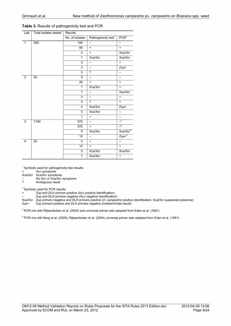
Table 3. Results of pathogenicity test and PCR
Lab Total isolates tested Results
No. of isolates Pathogenicity test1 PCR
2
1 240 144 – –
84 + +
2 + Xca/Xcr
1 Xca/Xcr Xca/Xcr
3 – +
3 – Zup+
3 ? –
2 50 5 – –
26 + +
1 Xca/Xcr +
1 – Xca/Xcr
3 – +
4 ? +
4 Xca/Xcr Zup+
5 Xca/Xcr –
1 + –
3 1160 575 – –3
570 + +3
5 Xca/Xcr Xca/Xcr4
10 – Zup+4
4 22 5 – –
14 + +
2 Xca/Xcr Xca/Xcr
1 Xca/Xcr +
1 Symbols used for pathogenicity test results:
+ Xcc symptoms Xca/Xcr Xca/Xcr symptoms – No Xcc or Xca/Xcr symptoms ? Ambiguous result 2 Symbols used for PCR results:
+ Zup and DLH primers positive (Xcc positive identification) – Zup and DLH primers negative (Xcc negative identification) Xca/Xcr Zup primers negative and DLH primers positive (X. campestris positive identification, Xca/Xcr suspected presence) Zup+ Zup primers positive and DLH primers negative (Indeterminate result) 3 PCR mix with Rijlaardsdam et al. (2004) and universal primer sets adapted from Eden et al. (1991)
4 PCR mix with Berg et al. (2005), Rijlaardsdam et al. (2004) universal primer sets adapted from Eden et al. (1991)

Grimault et al. New method of Xanthomonas campestris pv. campestris on Brassica spp. seed
OM12-06 Method Validation Reports on Rules Proposals for the ISTA Rules 2013 Edition.doc 2012-04-09 13:06 Approved by ECOM and RUL on March 23, 2012 Page 10/24
Table 4. Summary of the pathogenicity test and PCR results
Number of tested isolates
Pathogenicity test result
1
PCR result2 Discussion No. of tested
isolates per lab Isolates retested by lab 4
Isolates with correspondence in results
729 – – (a, b)
694 + +
8 Xca/Xcr Xca/Xcr
Total 1431
Isolates with differing results
3 ? – A1 3 (lab 1)
4 ? + A2 4 (lab 2)
13 – Zup+ B1 3 (lab 1), 10 (lab 3) (c, d)
4 Xca/Xcr Zup+ B2 4 (lab 2)
1 – Xca/Xcr C1 1 (lab 2)
6 – + C2 3 (lab 1), 3 (lab 2) (e)
5 Xca/Xcr – D1 5 (lab 2)
1 + – D2 1 (lab2)
2 Xca/Xcr + E1 1 (lab 2), 1 (lab 4)
2 + Xca/Xcr E2 2 (lab1)
Total 41 1 Symbols used for pathogenicity test results:
+ Xcc symptoms Xca/Xcr Xca/Xcr symptoms – No Xcc or Xca/Xcr symptoms ? Ambiguous result 2 Symbols used for PCR results:
+ Zup and DLH primers positive (Xcc positive identification) – Zup and DLH primers negative (Xcc negative identification) Xca/Xcr Zup primers negative and DLH primers positive (X. campestris positive identification, Xca/Xcr suspected presence) Zup+ Zup primers positive and DLH primers negative (Indeterminate result) 3 PCR mix with Rijlaardsdam et al. (2004) and universal primer sets adapted from Eden et al. (1991)
4 PCR mix with Berg et al. (2005), Rijlaardsdam et al. (2004) universal primer sets adapted from Eden et al. (1991)
Grimault et al. New method of Xanthomonas campestris pv. campestris on Brassica spp. seed
OM12-06 Method Validation Reports on Rules Proposals for the ISTA Rules 2013 Edition.doc 2012-04-09 13:06 Approved by ECOM and RUL on March 23, 2012 Page 11/24
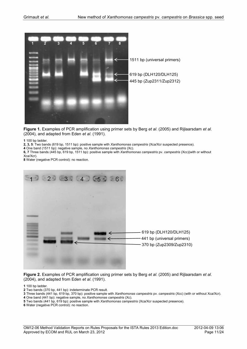
Figure 1. Examples of PCR amplification using primer sets by Berg et al. (2005) and Rijlaarsdam et al. (2004), and adapted from Eden et al. (1991).
1 100 bp ladder. 2, 3, 5: Two bands (619 bp, 1511 bp): positive sample with Xanthomonas campestris (Xca/Xcr suspected presence). 4 One band (1511 bp): negative sample, no Xanthomonas campestris (Xc). 6, 7 Three bands (445 bp, 619 bp, 1511 bp): positive sample with Xanthomonas campestris pv. campestris (Xcc)(with or without Xca/Xcr). 8 Water (negative PCR control): no reaction.
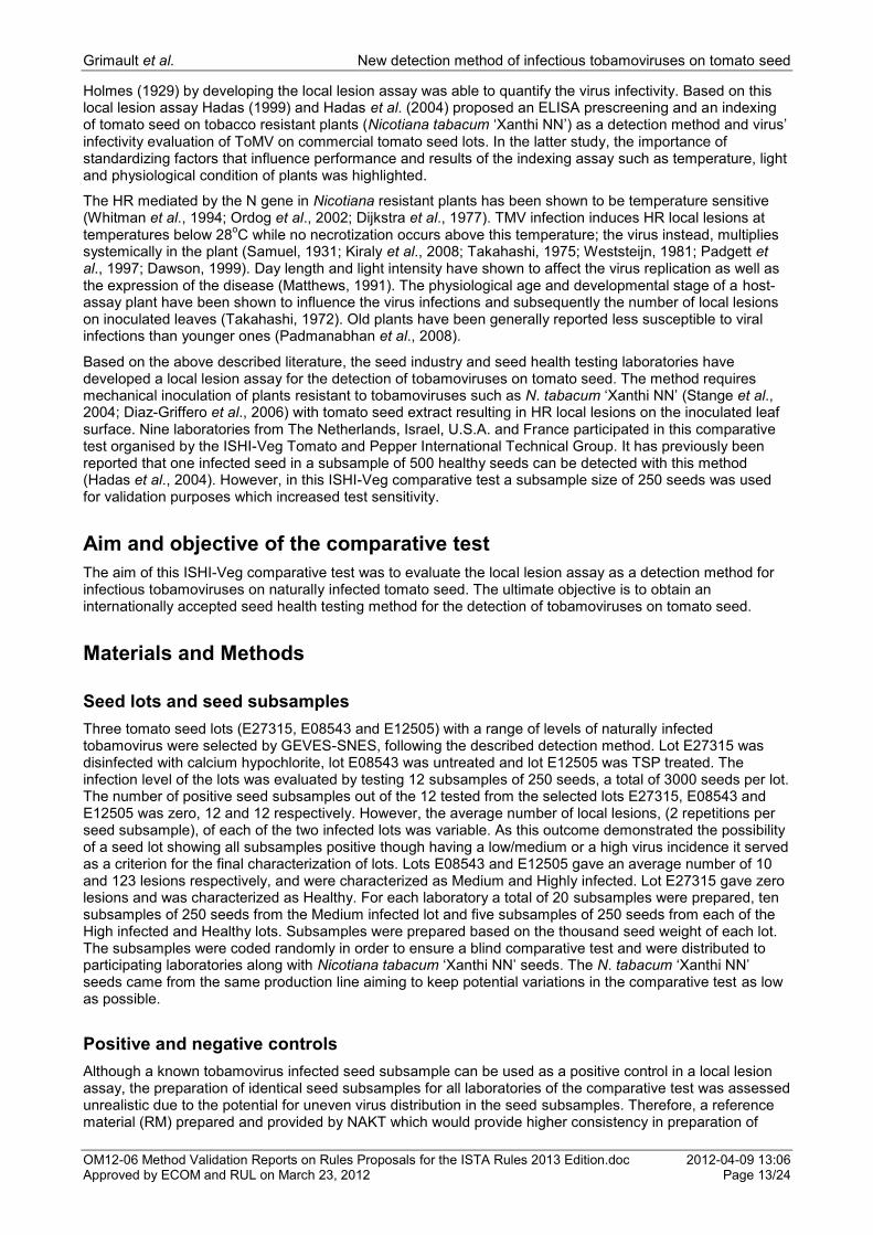
Figure 2. Examples of PCR amplification using primer sets by Berg et al. (2005) and Rijlaarsdam et al. (2004), and adapted from Eden et al. (1991).
1 100 bp ladder. 2 Two bands (370 bp, 441 bp): indeterminate PCR result. 3 Three bands (441 bp, 619 bp, 370 bp): positive sample with Xanthomonas campestris pv. campestris (Xcc) (with or without Xca/Xcr). 4 One band (441 bp): negative sample, no Xanthomonas campestris (Xc). 5 Two bands (441 bp, 619 bp): positive sample with Xanthomonas campestris (Xca/Xcr suspected presence). 6 Water (negative PCR control): no reaction.
1 2 3 4 5 6 7 8
1511 bp (universal primers)
445 bp (Zup2311/Zup2312)
619 bp (DLH120/DLH125)
619 bp (DLH120/DLH125)
441 bp (universal primers)
370 bp (Zup2309/Zup2310)

Grimault et al. New detection method of infectious tobamoviruses on tomato seed
OM12-06 Method Validation Reports on Rules Proposals for the ISTA Rules 2013 Edition.doc 2012-04-09 13:06 Approved by ECOM and RUL on March 23, 2012 Page 12/24
New method for the detection of infectious tobamoviruses on tomato (Lycopersicon esculentum) seed by local lesion assay (indexing) on Nicotiana tabacum plants
V. Grimault1, H. M. S. Koenraaadt2, A. Politikou1
1GEVES/SNES, Rue Georges Morel, BP 90024, 49071 Beaucouzé Cedex, France ([email protected],
[email protected]) 2Naktuinbouw, P.O. Box 40, 2370 AA Roelofarendsveen, The Netherlands ([email protected])
Summary
The local lesion assay (indexing) with Nicotiana tabacum „Xanthi NN‟ resistant plants was evaluated as a detection method for infectious tobamoviruses on tomato (Lycopersicon esculentum) seed in a comparative test between nine laboratories, organised by the International Seed Health Initiative Vegetable group (ISHI-Veg). Two naturally infected seed lots with medium and high infection levels, one virus-free seed lot and two infection levels of reference material (RM) were used. Subsamples of the seed lots and RM samples were distributed to laboratories who inoculated their extracts onto the leaf surface of two assay plants. For each (sub)sample x plant x leaf combination, the number of necrotic local lesions was recorded 5-7 days post inoculation under specific incubation conditions by each laboratory. No false positives were recorded and most laboratories were able to detect the expected number of positive seed subsamples and RM samples and to distinguish between the various infection levels. The growth stage of tobacco assay plants and the incubation temperature were found to be critical factors for the detection of positive seed and RM samples. The local lesion assay was found repeatable and reproducible for both seed subsamples and RM samples. The use of a known infected seed sample or a reference material sample is indispensable for the validation of the results. The local lesion assay with Nicotiana resistant plants to tobamoviruses is considered a reliable detection method of infectious tobamoviruses on tomato seed and is highly recommended in routine tomato seed testing.
Introduction
Tobacco mosaic virus (TMV) and Tomato mosaic virus (ToMV) belong to the genus Tobamovirus (Lewandowski and Dawson 1998). TMV and ToMV are seed-borne and seed transmitted viruses (Hadas et al., 2004), commonly found on the tomato (Lycopersicon esculentum) seed, localizing the seed coat and sometimes the endosperm (Huttinga and Rast, 1995). They are able to survive for long periods outside the plant tissue and can be easily transmitted mechanically on tomato plants causing significant economic yield losses. (Averre and Gooding, 2000; Demski, 1981; Huttinga and Rast, 1995). A seed health program certifying virus-free seed is considered an important tool for the control of tomato plants‟ infection (Hadas et al., 2004).
Several serological methods based on ELISA tests have been broadly used for the detection and identification of various plant viruses (Clark and Adams, 1977; Maury et al., 1987). It is known however, that ELISA tests detect both infectious and non-infectious virus particles, therefore it does not allow for the evaluation of virus seed transmission and yields false positive results (Maury et al., 1987; Nolan and Campbell, 1984).
TMV and other tobamoviruses have played a significant role in the virology research in studies on plant-virus interactions (Dawson, 1999). The infection mode, pathway, replication and expression of tobamoviruses have been extensively studied on the TMV-Tobacco (Nicotiana spp.) plants model (Rhee et al., 2000; Diaz-Griffero et al., 2006; Bawden, 1964; Padmanabhan et al., 2008; Demski, 1981). Many references are available in the literature about the induction of hypersensitive response (HR) reaction in tobacco plants from tobamoviruses; a plant defence mechanism against the attack of viruses (Ehrenfeld et al., 2008; Takahashi, 1956; Erickson et al., 1999b; Whitman et al., 1994; Taliansky et al., 1994). The HR reaction is the outcome of the gene-for-gene resistance (Flor, 1971; Kiraly et al., 2007), mediated in tobacco plants carrying the dominant N resistant gene (Holmes 1938; Hammond-Kosack and Jones 1996; Erickson et al., 1999a; Whitman et al., 1994; Boovaraghan et al., 2007). The HR is characterised by the virus confinement on the initial infection site through death-cell and the development of local necrotic lesions (Holmes 1938; Takahashi, 1956; Dawson, 1999).
Grimault et al. New detection method of infectious tobamoviruses on tomato seed
OM12-06 Method Validation Reports on Rules Proposals for the ISTA Rules 2013 Edition.doc 2012-04-09 13:06 Approved by ECOM and RUL on March 23, 2012 Page 13/24
Holmes (1929) by developing the local lesion assay was able to quantify the virus infectivity. Based on this local lesion assay Hadas (1999) and Hadas et al. (2004) proposed an ELISA prescreening and an indexing of tomato seed on tobacco resistant plants (Nicotiana tabacum „Xanthi NN‟) as a detection method and virus‟ infectivity evaluation of ToMV on commercial tomato seed lots. In the latter study, the importance of standardizing factors that influence performance and results of the indexing assay such as temperature, light and physiological condition of plants was highlighted.
The HR mediated by the N gene in Nicotiana resistant plants has been shown to be temperature sensitive (Whitman et al., 1994; Ordog et al., 2002; Dijkstra et al., 1977). TMV infection induces HR local lesions at temperatures below 28
oC while no necrotization occurs above this temperature; the virus instead, multiplies
systemically in the plant (Samuel, 1931; Kiraly et al., 2008; Takahashi, 1975; Weststeijn, 1981; Padgett et al., 1997; Dawson, 1999). Day length and light intensity have shown to affect the virus replication as well as the expression of the disease (Matthews, 1991). The physiological age and developmental stage of a host-assay plant have been shown to influence the virus infections and subsequently the number of local lesions on inoculated leaves (Takahashi, 1972). Old plants have been generally reported less susceptible to viral infections than younger ones (Padmanabhan et al., 2008).
Based on the above described literature, the seed industry and seed health testing laboratories have developed a local lesion assay for the detection of tobamoviruses on tomato seed. The method requires mechanical inoculation of plants resistant to tobamoviruses such as N. tabacum „Xanthi NN‟ (Stange et al., 2004; Diaz-Griffero et al., 2006) with tomato seed extract resulting in HR local lesions on the inoculated leaf surface. Nine laboratories from The Netherlands, Israel, U.S.A. and France participated in this comparative test organised by the ISHI-Veg Tomato and Pepper International Technical Group. It has previously been reported that one infected seed in a subsample of 500 healthy seeds can be detected with this method (Hadas et al., 2004). However, in this ISHI-Veg comparative test a subsample size of 250 seeds was used for validation purposes which increased test sensitivity.
Aim and objective of the comparative test
The aim of this ISHI-Veg comparative test was to evaluate the local lesion assay as a detection method for infectious tobamoviruses on naturally infected tomato seed. The ultimate objective is to obtain an internationally accepted seed health testing method for the detection of tobamoviruses on tomato seed.
Materials and Methods
Seed lots and seed subsamples
Three tomato seed lots (E27315, E08543 and E12505) with a range of levels of naturally infected tobamovirus were selected by GEVES-SNES, following the described detection method. Lot E27315 was disinfected with calcium hypochlorite, lot E08543 was untreated and lot E12505 was TSP treated. The infection level of the lots was evaluated by testing 12 subsamples of 250 seeds, a total of 3000 seeds per lot. The number of positive seed subsamples out of the 12 tested from the selected lots E27315, E08543 and E12505 was zero, 12 and 12 respectively. However, the average number of local lesions, (2 repetitions per seed subsample), of each of the two infected lots was variable. As this outcome demonstrated the possibility of a seed lot showing all subsamples positive though having a low/medium or a high virus incidence it served as a criterion for the final characterization of lots. Lots E08543 and E12505 gave an average number of 10 and 123 lesions respectively, and were characterized as Medium and Highly infected. Lot E27315 gave zero lesions and was characterized as Healthy. For each laboratory a total of 20 subsamples were prepared, ten subsamples of 250 seeds from the Medium infected lot and five subsamples of 250 seeds from each of the High infected and Healthy lots. Subsamples were prepared based on the thousand seed weight of each lot. The subsamples were coded randomly in order to ensure a blind comparative test and were distributed to participating laboratories along with Nicotiana tabacum „Xanthi NN‟ seeds. The N. tabacum „Xanthi NN‟ seeds came from the same production line aiming to keep potential variations in the comparative test as low as possible.
Positive and negative controls
Although a known tobamovirus infected seed subsample can be used as a positive control in a local lesion assay, the preparation of identical seed subsamples for all laboratories of the comparative test was assessed unrealistic due to the potential for uneven virus distribution in the seed subsamples. Therefore, a reference material (RM) prepared and provided by NAKT which would provide higher consistency in preparation of
Grimault et al. New detection method of infectious tobamoviruses on tomato seed
OM12-06 Method Validation Reports on Rules Proposals for the ISTA Rules 2013 Edition.doc 2012-04-09 13:06 Approved by ECOM and RUL on March 23, 2012 Page 14/24
identical samples was used as a positive control. This RM was consisted of freeze-dried Pepper mild mottle virus (PMMoV) infected Nicotiana occidentalis leaves diluted at two different levels with dry pea flour. The RM that was 50x diluted and gave an average of 40 local lesions was characterized as a High infected level whereas the RM that was 500x diluted and gave an average of 10 local lesions was characterized as a Medium infected level. Participating laboratories received 5 randomly coded samples of 0.5 g of each of the two RM levels. For the negative control, Phosphate Buffer Saline (PBS) was prepared by dissolving 8.0 g/L NaCl, 1.15 g/L Na2HPO4 and 0.2 g/L KH2PO4 in distilled water. The pH was adjusted to between 7.2-7.4 and the solution was autoclaved at 121 °C at 15 psi for 15 min.
Local lesion assay
Production of N. tabacum ‘Xanthi NN’ plants
Plants of N. tabacum „Xanthi NN‟ were grown from the supplied seeds by individual laboratories at 20-28 C under sufficient light intensity for 6-8 weeks until they reached the 4-5 true leaves growth stage. Two tobacco plants per tomato seed subsample, two per RM sample and two plants for the negative control were selected resulting in 62 plants in total. Plants were watered the day before the mechanical inoculation to ensure leaves with high turgor. Two consecutive leaves of each pair of plants were labelled with the corresponding code of the sample for inoculation.
Virus extraction from seed and reference material
In each laboratory the 20 tomato seed subsamples of 250 seeds were each ground in 10 ml PBS seed extraction buffer with a grinder. The 10 samples of 0.5 g of each RM were similarly ground in 5 ml PBS seed extraction buffer.
Mechanical inoculation of plants
The two labelled leaves of each pair of plants were dusted with a fine layer of carborundum powder and 100 µL of the corresponding ground extract was placed on each leaf‟s surface. The 100 µL of extract was smeared in the entire leaf surface by applying light pressure with fingers. Plastic finger tips or gloves were used and changed between (sub) samples. Special care was taken to avoid leaf damage that might be caused by too much pressure. Two consecutive leaves of one pair of plants were inoculated in a similar manner with 100 µL of PBS and served as negative control plants. All inoculated leaves were rinsed with tap water a few minutes after inoculation and plants were incubated at 20-25 °C with alternating 12 h light-12 h dark regime for 5-7 days. Plants were examined for the development of typical local necrotic lesions by comparison to positive and negative control plants and the number of local lesions developed on each leaf per plant and per (sub)sample was counted.
Data analysis
Following the seed lots‟ characterization based on the average of developed lesions, another local lesion assay was conducted for each infected lot with a smaller number of subsamples per lot and seeds per subsample. The aim of this assay was to calculate the infection rate of each lot with the spreadsheet application Seedcalc version 8 (http://www.seedtest.org/en/statistical-tools-for-seed-testing-_content---1--1143--279.html) and to calculate the probability of expected positive subsamples in the comparative test at a confidence level above 99% with the spreadsheet application developed by J.L. Laffont. Two positive subsamples of 32 seeds (1/8 of 250) out of 6 subsamples tested revealed 1.26% infection rate and the number of 8-10 expected positive subsamples out of the 10 total distributed to laboratories for a confidence level above 99% for the Medium level lot. Likewise, 3 positive subsamples of 62 seeds (1/4 of 250) out of 6 subsamples revealed 1.11% infection rate and the number of 4-5 expected positive subsamples out of the 5 total distributed to laboratories for a confidence level above 99% for the High level lot.
For each combination of laboratory X infection level X (sub) sample X plant X leaf the number of developed local lesions was recorded. A seed subsample or a RM sample was considered positive when at least one local lesion developed in at least one leaf. Values of the Medium level seed subsamples of laboratory 1 were excluded from the analysis as they were all recorded zero as well as values of High level seed subsamples of laboratory 9 were also excluded as the values of 4 out the 5 subsamples were zero.
The average number of local lesions was calculated for each combination of laboratory X infection level X (sub) sample. The number of local lesions of positive seed subsamples from both Medium and High infected

Grimault et al. New detection method of infectious tobamoviruses on tomato seed
OM12-06 Method Validation Reports on Rules Proposals for the ISTA Rules 2013 Edition.doc 2012-04-09 13:06 Approved by ECOM and RUL on March 23, 2012 Page 15/24
level seed lots, was analysed with the use of the Analysis of Variance (ANOVA) implemented in Statgraphics Plus 5.0 statistical program. Likewise, an ANOVA on the number of local lesions from positive RM samples – and excluding the missing values- from both Medium and High infected levels was performed. Prior to the analysis, the original values of seed subsamples and RM samples were transformed with the natural logarithm plus one and with the natural logarithm respectively which allowed for their normal distribution based on Levene‟s test. The number of lesions from positive seed subsamples and from positive RM samples were also analysed with an ANOVA for each individual laboratory. Prior to the analysis, the original values of seed subsamples and RM samples were transformed with the natural logarithm which allowed for their normal distribution based on Levene‟s test.
The repeatability (within laboratory variability) and reproducibility (between laboratory variability) was evaluated using the original values of the above mentioned set of data for seed subsamples and RM samples of all laboratories with the use of ISO 5725 (http://www.seedtest.org/en/statistical-tools-for-seed-testing-_content---1--1143--279.html).
Results
Seed subsamples
No local lesions were recorded by laboratories on plants inoculated with seed extract from the Healthy lot subsamples or on plants inoculated with the negative control (PBS seed extraction buffer) (data not shown).
The number of the detected and the expected positive seed subsamples out of the total tested per infection level lot and per laboratory is presented in Table 1. All laboratories except laboratories 1 and 4, detected the expected number of positive seed subsamples from the Medium infected seed lot. Regarding the Highly infected seed lot, all laboratories except laboratory 9, detected the expected number of positive seed subsamples (Table 1).
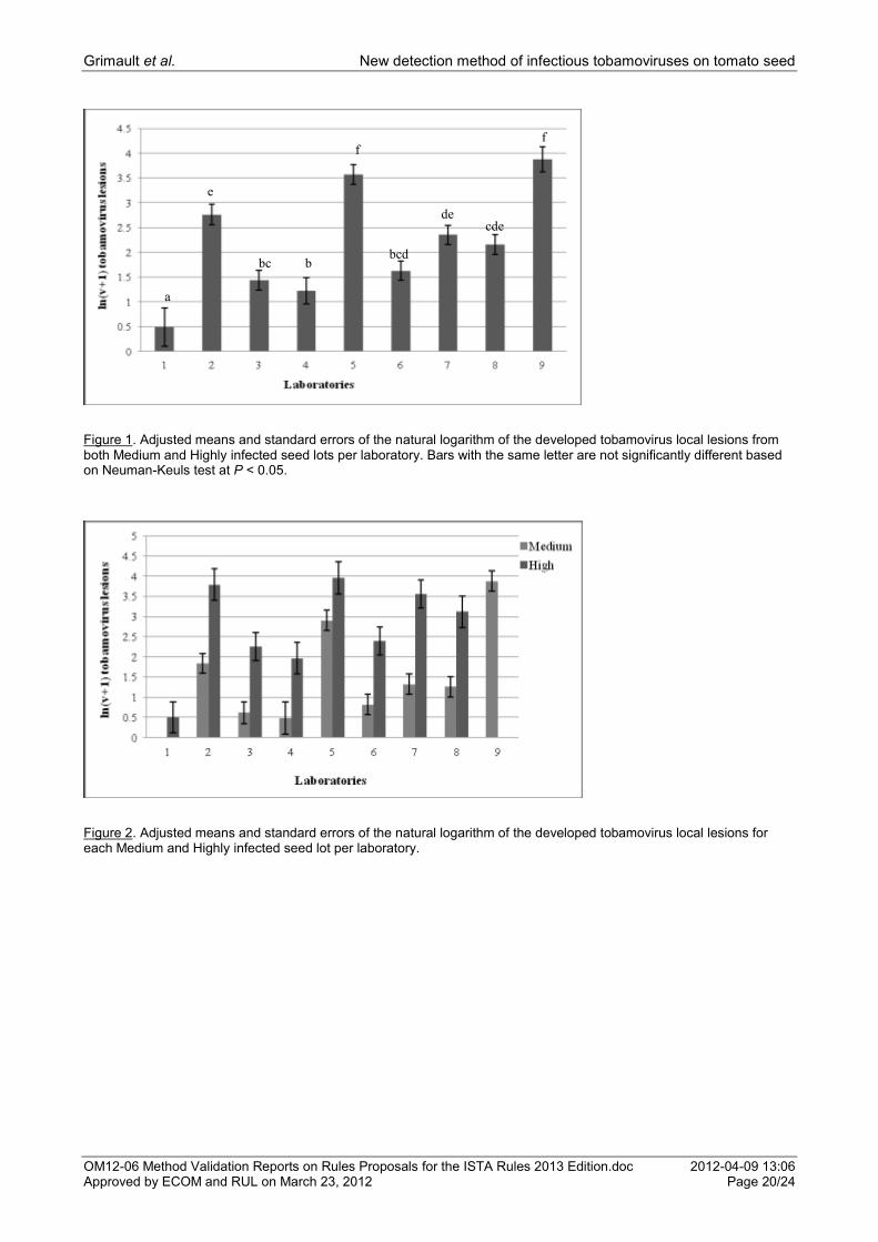
The ANOVA for the average number of local lesions from both the Medium and High level lots showed a significant difference between laboratories (P = 0.0000) and infection levels (seed lots) (P = 0.0000). No test on laboratory x infection level interaction was reported by Statgraphics program as not all combinations between the values of these two factors were possible (e.g. Lab1 Medium level seed subsamples were excluded). The average number of the natural logarithm plus one of local lesions from positive seed subsamples of both Medium and High level lots for all nine laboratories is presented in Figure 1. The highest number of local lesions was recorded by laboratories 5 and 9 and the lowest by laboratory 4 (Fig. 1).
The ANOVA for the average number of local lesions per infection level for each laboratory showed an infection level effect in each analysed laboratory: Lab 2 (P = 0.000), Lab 3 (P = 0.015), Lab 4 (P = 0.006), Lab 5 (P = 0.008), Lab 6 (P = 0.002), Lab 7 (P = 0.026) and Lab 8 (P = 0.000). The average number of the natural logarithm plus one of local lesions from positive seed subsamples for each of the Medium and High level lots per laboratory is depicted in Figure 2. All laboratories detected a higher number of local lesions in the High than in the Medium level lot (Fig. 2).
The standard deviation of repeatability and reproducibility based on the mean of local lesions from positive seed subsamples of Medium and High infected lots are shown in Table 2. No (h) critical values were reported while there were (k) critical values at 5% confidence level for Lab 5 for both Medium and High levels, for Lab 7 for High level and for Lab 9 for Medium level (Figs. 5, 6).
Reference Material (RM) samples
The number of detected and the expected positive RM samples out of the total tested per infection level, per laboratory is indicated in Table 1. In both Medium and High infection levels, all laboratories detected five positive samples out of five tested except laboratory 1 where one negative RM sample was recorded. Laboratory 6 inoculated only two RM samples due to the lack of sufficient tobacco assay plants. However, both RM samples were recorded positive.
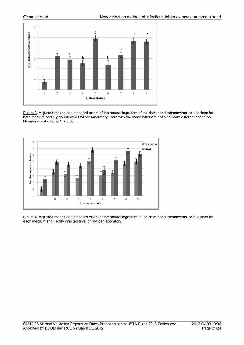
An ANOVA on the average number of lesions for Medium and High infected RM showed a significant difference between laboratories (P = 0.000) and between infection levels (P = 0.000). No significant laboratory x infection level interaction was shown (P = 0.901). The average number of the natural logarithm plus one of local lesions from positive RM samples of both Medium and High infected RM levels per laboratory is presented in Figure 3. The highest number of local lesions was detected by laboratories 5, 8 and 9 and the lowest by laboratory 1 (Fig. 3).

Grimault et al. New detection method of infectious tobamoviruses on tomato seed
OM12-06 Method Validation Reports on Rules Proposals for the ISTA Rules 2013 Edition.doc 2012-04-09 13:06 Approved by ECOM and RUL on March 23, 2012 Page 16/24
An ANOVA for this number of lesions showed an infection level effect for the following laboratories: Lab 1 (P = 0.001), Lab 4 (P = 0.004), Lab 5 (P = 0.000), Lab 7 (P = 0.001), Lab 8 (P = 0.000) and Lab 9 (P = 0.000). There was no infection level effect shown for Laboratories 2, 3 and 6 . The average number of the natural logarithm plus one of local lesions from positive RM samples for each of the Medium and High infected levels per laboratory is depicted in Figure 4. All laboratories detected a higher number of lesions in the High infection level than in the Medium (Fig. 4).
The standard deviation of repeatability and reproducibility based on the mean of local lesions from positive RM samples of Medium and High infection are shown in Table 3. No (h) critical values were reported while there were (k) critical values at 5% confidence level for Lab 5 for Medium and High level and for Lab 8 for High level (Figs. 7, 8).
Discussion
No false positives were reported in the local lesion assay and no cross contamination occurred as all laboratories found all seed subsamples from the Healthy lot negative and did not record any lesions on the negative control plants. This finding is in agreement with the Hadas et al. (2004) findings.
Most laboratories detected the expected number of positive seed subsamples from the Medium and High infection level lots. Laboratories 1, 4 and 9 that detected less than the expected number of positive seed subsamples reported assay performance under conditions considerably variable to the optimal ones (e.g. plants under or over the optimal growth stage, suboptimal or fluctuating incubation temperature). The growth stage of plants is well known to affect the infection of tobamoviruses and the number of lesions on the inoculated leaves. In older plants less severe virus infections are generally reported than in younger plants (Takahashi 1972; Padmanabhan et al., 2007). In temperatures higher than 28
oC tobacco plants inoculated
with tobamoviruses do not onset a hypersensitive reaction (Whitman et al., 1994; Ordog et al., 2002). Generally, the desired optimal temperature cannot be easily retained in greenhouse conditions, resulting in deviations. Thereafter, it is postulated that the uneven virus distribution in the seed subsamples shown in the preliminary tests for seed lots‟ characterization, the suboptimal incubation conditions and the potential mishandling of the subsamples played individually or in combination a significant role in the detection of positive seed subsamples by these laboratories.
The laboratory effect that was shown on the number of local lesions from seed subsamples of Medium and High infection level lots is also attributed to the variations in plants‟ growth stage and in incubation temperature factors applied by each laboratory. This statistical outcome was expected by the organizers of this comparative test who were aware that the number of local lesions developed as a HR product of tobamovirus-inoculated tobacco plants from seed lots with variable infection levels cannot be considered an absolute number that is expected to be reproduced by the participants.
However, as no (h) critical values for the reproducibility were reported by ISO 5725-2, no lab X level interaction was shown in the ANOVA and most of the laboratories detected the expected number of positive seed subsamples, the local lesion assay was evaluated as being reproducible for the seed subsamples.
The Medium and High infection level of the seed lots were distinguishable by the laboratories as it was indicated by the level effect that was shown on the number of local lesions from the seed subsamples. All laboratories recorded a higher average number of lesions in seed subsamples from the High level lot than from the Medium. Moreover, this difference in infection level of the lots was demonstrated within each laboratory by the level effect that was shown in each of them. The (k) critical values shown for Labs 5, 7, and 9 indicate that the uneven virus distribution in the subsamples and/or the variations from the optimal incubation conditions may result in variations in the number of developed local lesions between subsamples. However, as the majority of the laboratories detected the expected number of positive seed subsamples the local lesion assay has been considered repeatable for seed subsamples.
All laboratories were able to detect all RM samples from the Medium and High infection levels, with Laboratory 1 being the only exception in the Medium level, as it recorded one negative sample. Previously mentioned factors that can influence the assay and/or suboptimal manipulation of the sample can explain the results of Laboratory 1.
The significant difference shown between laboratories on the number of lesions from RM samples of the Medium and High infection levels is also attributed to the same influencing factors mentioned for the seed subsamples. However, as no (h) critical values for the reproducibility were reported by the ISO 5725-2, and no lab x level interaction was shown in the ANOVA, the local lesion assay was evaluated as being reproducible for the RM samples.
Grimault et al. New detection method of infectious tobamoviruses on tomato seed
OM12-06 Method Validation Reports on Rules Proposals for the ISTA Rules 2013 Edition.doc 2012-04-09 13:06 Approved by ECOM and RUL on March 23, 2012 Page 17/24
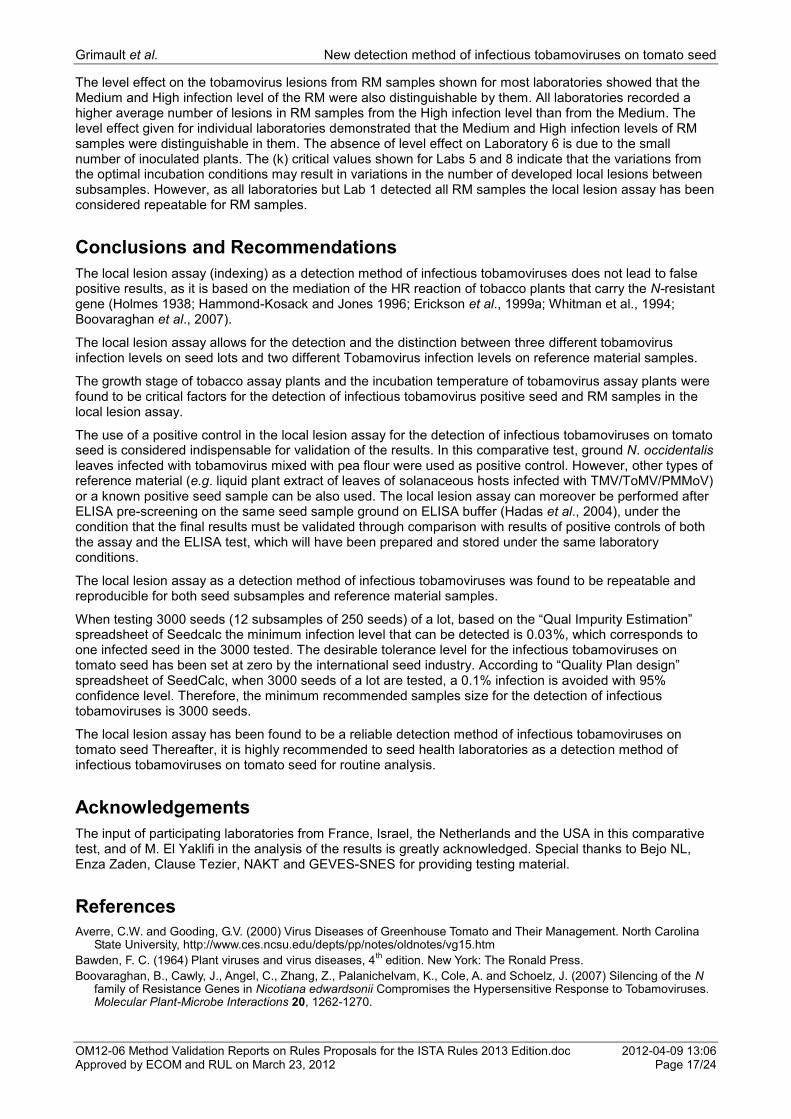
The level effect on the tobamovirus lesions from RM samples shown for most laboratories showed that the Medium and High infection level of the RM were also distinguishable by them. All laboratories recorded a higher average number of lesions in RM samples from the High infection level than from the Medium. The level effect given for individual laboratories demonstrated that the Medium and High infection levels of RM samples were distinguishable in them. The absence of level effect on Laboratory 6 is due to the small number of inoculated plants. The (k) critical values shown for Labs 5 and 8 indicate that the variations from the optimal incubation conditions may result in variations in the number of developed local lesions between subsamples. However, as all laboratories but Lab 1 detected all RM samples the local lesion assay has been considered repeatable for RM samples.
Conclusions and Recommendations
The local lesion assay (indexing) as a detection method of infectious tobamoviruses does not lead to false positive results, as it is based on the mediation of the HR reaction of tobacco plants that carry the N-resistant gene (Holmes 1938; Hammond-Kosack and Jones 1996; Erickson et al., 1999a; Whitman et al., 1994; Boovaraghan et al., 2007).
The local lesion assay allows for the detection and the distinction between three different tobamovirus infection levels on seed lots and two different Tobamovirus infection levels on reference material samples.
The growth stage of tobacco assay plants and the incubation temperature of tobamovirus assay plants were found to be critical factors for the detection of infectious tobamovirus positive seed and RM samples in the local lesion assay.
The use of a positive control in the local lesion assay for the detection of infectious tobamoviruses on tomato seed is considered indispensable for validation of the results. In this comparative test, ground N. occidentalis leaves infected with tobamovirus mixed with pea flour were used as positive control. However, other types of reference material (e.g. liquid plant extract of leaves of solanaceous hosts infected with TMV/ToMV/PMMoV) or a known positive seed sample can be also used. The local lesion assay can moreover be performed after ELISA pre-screening on the same seed sample ground on ELISA buffer (Hadas et al., 2004), under the condition that the final results must be validated through comparison with results of positive controls of both the assay and the ELISA test, which will have been prepared and stored under the same laboratory conditions.
The local lesion assay as a detection method of infectious tobamoviruses was found to be repeatable and reproducible for both seed subsamples and reference material samples.
When testing 3000 seeds (12 subsamples of 250 seeds) of a lot, based on the “Qual Impurity Estimation” spreadsheet of Seedcalc the minimum infection level that can be detected is 0.03%, which corresponds to one infected seed in the 3000 tested. The desirable tolerance level for the infectious tobamoviruses on tomato seed has been set at zero by the international seed industry. According to “Quality Plan design” spreadsheet of SeedCalc, when 3000 seeds of a lot are tested, a 0.1% infection is avoided with 95% confidence level. Therefore, the minimum recommended samples size for the detection of infectious tobamoviruses is 3000 seeds.
The local lesion assay has been found to be a reliable detection method of infectious tobamoviruses on tomato seed Thereafter, it is highly recommended to seed health laboratories as a detection method of infectious tobamoviruses on tomato seed for routine analysis.
Acknowledgements
The input of participating laboratories from France, Israel, the Netherlands and the USA in this comparative test, and of M. El Yaklifi in the analysis of the results is greatly acknowledged. Special thanks to Bejo NL, Enza Zaden, Clause Tezier, NAKT and GEVES-SNES for providing testing material.
References
Averre, C.W. and Gooding, G.V. (2000) Virus Diseases of Greenhouse Tomato and Their Management. North Carolina State University, http://www.ces.ncsu.edu/depts/pp/notes/oldnotes/vg15.htm
Bawden, F. C. (1964) Plant viruses and virus diseases, 4th edition. New York: The Ronald Press.
Boovaraghan, B., Cawly, J., Angel, C., Zhang, Z., Palanichelvam, K., Cole, A. and Schoelz, J. (2007) Silencing of the N family of Resistance Genes in Nicotiana edwardsonii Compromises the Hypersensitive Response to Tobamoviruses. Molecular Plant-Microbe Interactions 20, 1262-1270.

Grimault et al. New detection method of infectious tobamoviruses on tomato seed
OM12-06 Method Validation Reports on Rules Proposals for the ISTA Rules 2013 Edition.doc 2012-04-09 13:06 Approved by ECOM and RUL on March 23, 2012 Page 18/24
Clark, M.F. and Adams, A.N. (1977) Characteristics of the microplate method of enzyme-linked immunosorbent assay for the detection of plant viruses. Journal of General Virology 34, 475-483.
Dawson, W.O. (1999) Tobacco mosaic virus virulence and avirulence, Philosophical Transactions of the Royal Society of London Biological Sciences 354, 645-651.
Demski, J.W. 1981. Tobacco mosaic virus is seed borne in pimiento peppers. Plant Disease 65, 723-724.
Diaz-Griffero, F., Espinoza Cancino, C., Medina Arevalo, C., and Arce-Jonhson, P. (2006) Expression of the crucifer-infecting TMV-Cg movement protein in tobacco plants complements in trans a TMV-U1 trafficking-deficient mutant. Biology Resistance 39, 269-279.
Dijkstra, J., Bruin, G.C.A., Burgers, A.C., Van Loon, L.C., Ritter, P., Van de Sanden, P. A.C.M. and Wieringa-Brants, D.H. (1977) Systemic infection of some N-gene-carrying Nicotiana species after inoculation with tobacco mosaic virus. Netherlands Journal of Plant Pathology 83, 41-59.
Ehrenfeld, N., Gonzalez, A., Canon, P., Medina, C., Perez-Acle, T. and Arce-Johnson, P. (2008) Structure-function relationship between the tobamovirus TMV-Cg coat protein and the HR-like response. Journal of General Virology 89, 809-817.
Erickson, F. L., Dinesh-Kumar, S. P., Holzberg, S., Ustach, C.V., Dutton, M., Handley, V., Corr, C. and Bakker, B. J. (1999a) Interactions between tobacco mosaic virus and the tobacco N gene, Philosophical Transactions of the Royal Society of London Biological Sciences, 354, 653-658.
Erickson, L. F., Holzberg, S., Calderon-Urea, A., Handley, V., Axtell, M., Corr, C., and Baker, B. (1999b) The helicase domain of the TMV replicase proteins induces the N-mediated defence response in tobacco. The Plant Journal 18, 67-75.
Flor, H. (1971) Current status of the gene-for-gene concept. Annual Review of Phytopathology 9, 275-296.
Hadas, R. (1999) A report of comparative test for Tobamoviruses in tomato seeds. ISHI-Veg Research Report 1-1999-Tomato-Tobamo. Nyon, Switzerland: International Seed Federation.
Hadas, R., Pearlsman, M., Gefen, T., Lachman, O., Hadar, E., Sharabany, G. and Antignus, Y. (2004) Indexing system for Tomato mosaic virus (ToMV) in commercial tomato seed lots. Phytoparasitica 32 (4), 421-424.
Hammond-Kosack, K. E. and Jones, J. D. G. (1996) Resistance Gene-Dependent Plant Defense Responses. The Plant Cell 8, 1773-1791.
Holmes, F.O. (1929) Local lesions in tobacco mosaic. Botanical Gazette (Chicago) 87, 39-55.
Huttinga, H. and Rast, A. Th.B. (1995) Tomato mosaic tobamovirus. In: Brunt, A.A., Crabtree, K., Dallwitz, M.J., Gibbs, A.J. and Watson, L. (Eds.) Viruses of Plants. CAB International, Wallingford, UK. pp. 1302-130
ISO 5725 (http://www.seedtest.org/en/statistical-tools-for-seed-testing-_content---1--1143--279.html)
Kiraly, L., Barna, B. and Kiraly, Z. (2007) Plant Resistance to Pathogen Infection: Forms and Mechanisms of Innate and Acquired Resistance. Journal of Phytopathology 155, 385-396.
Kiraly, L., Hafez, Y. M., Fodor, J. and Kiraly, Z. (2008) Suppression of tobacco mosaic virus-induced hypersensitive-type necrotization in tobacco at high temperature is associated with downregulation of NADPH oxidase and superoxide and stimulation of dehydroascorbate reductase. Journal of General Virology 89, 799-808.
Lewandowski, D. J. and Dawson, W. O. (1998). Tobamoviruses. In Encyclopedia of Virology, second edition, Granoff, A. and Webster, R. G. (Eds.), Academic Press 3, 1780-1783.
Matthews, R. E. F. (1991) Plant Virology, 3rd
ed. Academic Press, Inc. New York, N.Y.
Maury, Y., Bossennec, J.M., Boudazin, G., Hampton, R.O., Pietersen, G. and Maguire, J. (1987) Factors influencing ELISA evaluation of transmission of pea seed-borne mosaic virus in infected pea seed: seed-group size and seed decortication. Agronomie 7, 225-230.
Nolan, P.A. and Campbell, R.N. (1984) Squash Mosaic Virus Detection in Individual Seeds and Seed Lots of Cucurbits by Enzyme-Linked Immunosorbent Assay. Plant Disease 68, 971-975.
Ordog, S. H., Higgins, V. J. and Vanlerberghe, G. C. (2002) Mitochondrial Alternative Oxidase Is Not a Critical Component of Plant Viral Resistance But May Play a Role in the Hypersensitive Response. Plant Physiology 129, 1858-1865.
Padgett, H. S. Watanabe, Y. and Beachy, R. N. (1997) Identification of the TMV Replicase Sequence That Activates the N Gene-Mediated Hypersensitive Response. Molecular Plant-Microbe Interactions 6, 709-715.
Padmanabhan, M. S., Kramer, S. R., Wang, X. and Culver, J. N. (2008) Tobacco Mosaic Virus Replicase-Auxin/Indole Acetic Acid Protein Interactions: Reprogramming the Auxin Response Pathway To Enhance Virus Infection. Journal of Virology 82, 2477-2485.
Rhee, Y., Tzfira, T., Chen, M.-H., Waigmann, E., and Citovsky, V. (2000) Cell-to-cell movement of tobacco mosaic virus: enigmas and explanations. Molecular Plant Pathology 1, 33-39.
Samuel., G. (1931) Some experiments on inoculating methods with plant viruses and on local lesions. Annals of Applied Biology 18, 494-507.
Seedcalc version 8 (http://www.seedtest.org/en/statistical-tools-for-seed-testing-_content---1--1143--279.html)
Stange, C., Matus, J. T., Elorza A. and Arce-Johnson, P. (2004)Identification and characterisation of a novel tobacco mosaic virus resistance N gene homologue in Nicotiana tabacum plants. Functional Plant Biology 31 (2), 149-158.
Takahashi, W.N. (1956) Increasing the sensitivity of the local-lesion method of virus assay. Phytopathology 46, 654-656.
Takahashi, T. (1972) Studies on viral pathogenesis in plant hosts. III. Leaf age-dependent susceptibility to tobacco mosaic virus infection in “Samsun NN” tobacco plants. Phytopathology, Z. 75,140-155.
Takahashi, T. (1975) Studies on viral pathogenesis in plant hosts VIII. Systemic virus invasion and localization of infection on “Samsun NN” tobacco plants resulting from tobacco virus infection. Phytopathology, Z. 84, 75-87.
Taliansky, M., Aranda, M. A. and Garcia-Arenal, F. (1994) Differential Invasion by Tobamoviruses of Nicotiana megalosiphon Following the Hypersensitive Response. Phytopathology 84, 812-815.
Grimault et al. New detection method of infectious tobamoviruses on tomato seed
OM12-06 Method Validation Reports on Rules Proposals for the ISTA Rules 2013 Edition.doc 2012-04-09 13:06 Approved by ECOM and RUL on March 23, 2012 Page 19/24
Weststeijn, E. A. (1981) lesion growth and virus localization in leaves of Nicotiana tabacum cv. Xanthi nc. after inoculation with tobacco mosaic virus and incubation alternately at 22
oC and 32
oC. Physiological Plant Pathology 18,
357-368.
Whitham, S., Dinesh-Kumar, S. P., Choi, D., Hehl, R., Corr, C., and Baker, B. (1994) The Product of the Tobacco Mosaic Virus Resistancec Gene N: Similarity to Toll and the interleukin-1 Receptor, Cell 78, 1101-1115.
Grimault et al. New detection method of infectious tobamoviruses on tomato seed
OM12-06 Method Validation Reports on Rules Proposals for the ISTA Rules 2013 Edition.doc 2012-04-09 13:06 Approved by ECOM and RUL on March 23, 2012 Page 20/24
Figure 1. Adjusted means and standard errors of the natural logarithm of the developed tobamovirus local lesions from both Medium and Highly infected seed lots per laboratory. Bars with the same letter are not significantly different based on Neuman-Keuls test at P < 0.05.
Figure 2. Adjusted means and standard errors of the natural logarithm of the developed tobamovirus local lesions for each Medium and Highly infected seed lot per laboratory.
a
e
bc b bcd
f
de cde
f
Grimault et al. New detection method of infectious tobamoviruses on tomato seed
OM12-06 Method Validation Reports on Rules Proposals for the ISTA Rules 2013 Edition.doc 2012-04-09 13:06 Approved by ECOM and RUL on March 23, 2012 Page 21/24
Figure 3. Adjusted means and standard errors of the natural logarithm of the developed tobamovirus local lesions for both Medium and Highly infected RM per laboratory. Bars with the same letter are not significant different based on Neuman-Keuls test at P < 0.05.
Figure 4. Adjusted means and standard errors of the natural logarithm of the developed tobamovirus local lesions for each Medium and Highly infected level of RM per laboratory.
a
c
b b
b b
b
c c

Grimault et al. New detection method of infectious tobamoviruses on tomato seed
OM12-06 Method Validation Reports on Rules Proposals for the ISTA Rules 2013 Edition.doc 2012-04-09 13:06 Approved by ECOM and RUL on March 23, 2012 Page 22/24
Table 1. Number of detected and expected positive seed subsamples and RM samples, incubation temperature and growth stage of assay plants per laboratory.
No. of detected
positive
subsamples/tota
l tested
Expected
number to be
detected with a
99%
confidence
No. of detected
positive
subsamples/total
tested
Expected
number to be
detected with a
99%
confidence
No. of
detected
positive RM
samples/total
tested
Detected
positive RM
samples/tota
l tested
No. of
Expected
number to be
detected for
both
Medium/High
RM samples
Temperature
(oC)
Growth
stage (No.
true
leaves)
Laboratories Medium seed
lot
Medium seed
lot
High seed lot High seed
lot
Medium RM High RM
1 0/10 8-10/10 4/5 4-5/5 4/5 5/5 5/5 **24 3-4
2 10/10 8-10/10 4/5 4-5/5 5/5 5/5 5/5 20 4-5
3 9/10 8-10/10 5/5 4-5/5 5/5 5/5 5/5 25 +/-2 4-5
4 4/10 8-10/10 4/5 4-5/5 5/5 5/5 5/5 26 +/-3 5-6
5 10/10 8-10/10 4/5 4-5/5 5/5 5/5 5/5 **25 4-5
6 10/10 8-10/10 5/5 4-5/5 *2/2 *2/2 5/5 24 +/-2 7-8
7 10/10 8-10/10 5/5 4-5/5 5/5 5/5 5/5 28 3-4
8 10/10 8-10/10 4/5 4-5/5 5/5 5/5 5/5 **27 4-5
9 10/10 8-10/10 1/5 4-5/5 5/5 5/5 5/5 **29 4-5
*Inoculation of 2 samples of each RM1 and RM2 due to not enough assay plants. **Incubation of inoculated assay plants in a greenhouse.

Grimault et al. New detection method of infectious tobamoviruses on tomato seed
OM12-06 Method Validation Reports on Rules Proposals for the ISTA Rules 2013 Edition.doc 2012-04-09 13:06 Approved by ECOM and RUL on March 23, 2012 Page 23/24
Table 2. Repeatability and reproducibility for all 9 laboratories for positive Medium and High level seed subsamples. Original values were used in the ISO 5725.
Figures 5-6. Reproducibility (h) and repeatability (k) graphs and critical values.

Grimault et al. New detection method of infectious tobamoviruses on tomato seed
OM12-06 Method Validation Reports on Rules Proposals for the ISTA Rules 2013 Edition.doc 2012-04-09 13:06 Approved by ECOM and RUL on March 23, 2012 Page 24/24
Table 3. Repeatability and reproducibility for all 9 labs for positive RM samples of Medium and High level. Original values were used in the ISO 5725.
Figures 7-8. Reproducibility (h) and repeatability (k) graphs and critical values.