methionine depletion enhances the antitumoral efficacy of
TRANSCRIPT

Methionine Depletion Enhances the Antitumoral Efficacy ofCytotoxic Agents in Drug-resistant HumanTumor Xenografts1
F. Poirson-Bichat, R. A. Bras Goncalves,L. Miccoli, B. Dutrillaux, and M. F. Poupon 2
Institut Curie, UMR 147 CNRS-Institut Curie, 75231 Paris Cedex 05,France
ABSTRACTEfficacy of chemotherapy is limited in numerous tu-
mors by specific cellular mechanisms that inactivate cyto-toxic antitumoral drugs, such as ATP-dependent drug effluxand/or drug detoxification by glutathione. In reducing ATPpools and/or glutathione synthesis, it might be possible toenhance the efficacy of drugs affected by such resistancemechanisms. Reduction of the ATP pool and glutathionecontent is achievable in cancer cells by depleting the exog-enous methionine (Met) supply and ethionine. Thus, therationale for the present study was to use Met depletion todecrease the ATP and glutathione pools so as to sensitizetumors refractory to cytotoxic anticancer drugs. Met deple-tion was achieved by feeding mice a methionine-free dietsupplemented with homocysteine. The effects of Met deple-tion combined with ethionine and/or chemotherapeuticagents were studied using human solid cancers xenograftedinto nude mice. TC71-MA (a colon cancer) SCLC6 (a smallcell lung cancer), and SNB19 (a glioma) were found to berefractory to cisplatin, doxorubicin, and carmustine, respec-tively. These three drugs are used to treat such tumors andare dependent for their activity on the lack of cellular ATP-or glutathione-dependent mechanisms of resistance. TC71-MA, SCLC6, and SNB19 were Met dependent because theirproliferation in vitro and growth in vivo were reduced byMet depletion. Cisplatin was inactive in the treatment ofTC71-MA colon cancer, whereas a methionine-free diet,alone or in combination with ethionine, prolonged the sur-vival of mice by 2-fold and 2.8-fold, respectively. When allthree approaches were combined, survival was prolonged by3.3-fold. Doxorubicin did not affect the growth of SCLC6, aMDR1-MRP-expressing tumor. A Met-deprived diet and
ethionine slightly decreased SCLC6 growth and, in combi-nation with doxorubicin, an inhibition of 51% was obtained,with survival prolonged by 1.7-fold. Combined treatmentproduced greater tumor growth inhibition (74%) in SCLC6-Dox, a SCLC6 tumor pretreated with doxorubicin. Growthof SNB19 glioma was not inhibited by carmustine, but whenit was combined with Met depletion, survival duration wasprolonged by 2-fold, with a growth inhibition of 80%. Theseresults indicate the potential of Met depletion to enhance theantitumoral effects of chemotherapeutic agents on drug-refractory tumors.
INTRODUCTIONMetabolic anomalies are commonly found in solid tumors,
and some of them have been known for decades (1). Among themetabolic abnormalities recurrently found in cancers, Met3 de-pendency and alterations of Met metabolism have been found inmany, if not in all, types of human tumors (2–11). Met depend-ency leads to the inability of cells to proliferate in culture whenMet is absent and replaced by Hcy, one of its metabolic precur-sors, whereas normal cells grow in such a medium (2). Thisdifference in the growth of normal cells and Met-dependenttumor cells in Met2Hcy1 medium might be due to the differentrequirement for transmethylation reactions necessary to main-tain their proliferation rate. Methods for reducingin vivo Metintake are to decrease the Met supply in food either using aMet-deprived diet (6), a methioninase infusion (12, 13), orMet-free total parenteral nutrition (14, 15). Met-depleted dietseffectively decreased metastatic potential and tumorogenicity inan experimental rat model, as previously shown by us (16) andby Guoet al. (8, 9).
In recent studies, we have obtained a potentiation of theantitumoral effects of a Met-deprived diet by treating tumor-bearing mice simultaneously with ethionine, a Met analogue.Ethionine might act by inhibiting most methyltransferases andhence lead to DNA hypomethylation, which can affect geneactivity, as shown by Razin and Riggs (17). Ethionine combinedwith a Met-deprived diet can augment the effects of Met deple-tion alone in reducing the growth of human prostate tumor (11)and glioma (10) xenografted into nude mice and of Yoshidasarcoma grafted into the rat (9, 18).In vitro, we also observedthat when tumor cells were cultured in a Met2Hcy1 mediumcontaining ethionine, their ATP and glutathione pools were
Received 5/24/99; revised 11/1/99; accepted 11/1/99.The costs of publication of this article were defrayed in part by thepayment of page charges. This article must therefore be hereby markedadvertisementin accordance with 18 U.S.C. Section 1734 solely toindicate this fact.1 Supported by the Association sur les Tumeurs Ce´rebrales (ARTC), theAssociation pour la Recherche sur le Cancer (ARC), and Luxembourggovernment Grant R/D BFR 95/035.2 To whom requests for reprints should be addressed, at Institut Curie,UMR 147 CNRS-Institut Curie, 26 rue d’Ulm, 75231 Paris Cedex 05,France. Phone: 33-1-42-34-66-67; Fax: 33-1-42-34-66-74; E-mail:[email protected].
3 The abbreviations used are: Met, methionine; Hcy, homocysteine;RTV, relative tumor volume; Pgp, P-glycoprotein; dNTP, deoxynucleo-side triphosphate; SCLC, small cell lung cancer; MRP, multidrug relatedprotein.
643Vol. 6, 643–653, February 2000 Clinical Cancer Research
Research. on April 3, 2019. © 2000 American Association for Cancerclincancerres.aacrjournals.org Downloaded from

decreased, and the cell cycle was blocked in S phase-G2, induc-ing an irreversible arrest of DNA replication.
Current chemotherapy of cancer is based on the use ofcytostatic and/or cytotoxic agents acting on components andcellular functions that control growth and cellular division. Thefailure of chemotherapy is often due to direct or indirect targetalterations that induce resistance. Various parameters contributeto drug resistance, and some of them are due to the increasedexpression of ATP-dependent mechanisms such as P-glycopro-tein (19, 20) and MRP proteins (21, 22). Drug resistance canalso be related to a high glutathione content and overexpressionof some enzymes, such as glutathione transferases (23, 24).Therefore, a decrease of ATP or of the glutathione pools mightcounteract the ATP- or glutathione-dependent mechanisms ofresistance.
Experiments combining Met depletion with doxorubicinalone, doxorubicin and vincristine, or 5-fluorouracil using Met-free parenteral nutrition were conducted using the Yoshidasarcoma grafted into the rat (14, 25, 26). They showed a poten-tiation of the antitumoral effect of cytotoxic drugs. Similarassays were conducted using a human gastric cancer grafted intonude mice, combining Met-depletion with 5-fluorouracil (27).In patients with gastrointestinal tract cancers, it was shown thatMet depletion might play a role as a modulator of 5-fluorouracilby decreasing the free-thymidylate synthetase activity (27).
Met is an essential amino acid, and we have shown thatMet depletion is not compatible with long-term survival. Con-sequently, Met is substituted by Hcy in the diet used in ourexperiments, thereby allowing animals to survive (6).
In the present study, Met depletion was induced by aMet-deprived diet and ethionine to decrease the cellular ATPand glutathione content and to sensitize drug-resistant tumors tothe effect of chemotherapeutic drugs. This was tested usingdrug-resistant human tumors xenografted into nude mice ex-pressing Pgp and/or MRP, both ATP-dependent molecular de-terminants of resistance, and/or a high level of glutathione. Wehave treated xenografted human cancers, a glioblastoma, a smallcell lung cancer, and a colon cancer with a combination of aMet-deprived diet and ethionine with carmustine, doxorubicin,and cisplatin, three drugs used for therapy of such tumors.
MATERIALS AND METHODSCell Culture and Proliferation Assay
SNB19, TC71-MA, and SCLC6 were established as mono-layer cell lines as described previously (10, 11). For assays, cellswere maintained in RPMI 1640 (Sigma) supplemented with10% FCS (Dutscher, Brumath, France). Briefly, cells wereplated in 10% FCS-RPMI 1640 for 24 h in a 5% CO2 atmo-sphere at 37°C at 105 cells/well in 24-well plates (ATGC,Noisy-le-Grand, France) in triplicate. The medium was replacedby a Met-free medium supplemented with dialyzed FCS, 100mM folic acid, and 1.5mM hydrocobalamin with 100mM of Hcy(Met2Hcy1) or Met (Met1Hcy1), with or without 0.5 mg/mlethionine (all products from Sigma). Cell monolayers were fixedwith methanol and then stained with methylene blue. Methyleneblue incorporated in fixed cells was solubilized in hydrochloricacid (0.1 M), and the absorbance of each well solution wasmeasured (wavelength, 620 nm) with a spectrophotometer
(LP500; J Bio, les Ulis, France). The cell proliferation index wascalculated as the ratio of absorbances corresponding to the assaymedium to that of controls3 100. Means and SDs were calculated,and statistical analyses were performed with the Student’st test.
Measurement of ATP ContentATP content was quantified by a bioluminescence assay
using a Lumac biocounter M1500 (Lumac Perstop Analytical,Bezons, France), as described previously (10). Cells were platedas described above for 24 and 48 h, in Met2Hcy1 or Met1Hcy1
medium with or without ethionine (0.5 mg/ml). Cell extractswere obtained using 1% trichloroacetic acid (Sigma) in water.ATP was measured in a 450-ml mixture of luciferin-luciferase(20 ml of a 40 mg/ml stock solution), ATP standard (both fromSigma) (10ml; 5.04 3 1028
M), and 10ml of the sample toanalyze in distilled water. Results are expressed as percentagesof ATP nmol/1026 treated cell extracts divided by ATP nmol/1026 control cell extracts. Experiments were repeated fivetimes. Means of data were calculated.
PCR for MDR-1 and MRP mRNA ExpressionTotal RNA of xenografted tumor was extracted with TRIZOL
reagent (Life Technologies, Inc.), and RNA was kept snap-frozenin liquid nitrogen and stored until use.MDR-1 and MRP geneexpression was analyzed by reverse transcription-PCR (28). Levelsof human-specific actin were measured as an endogenous controlfor cDNA synthesis. The primers listed below were selected fortheir specificity and selectivity for human gene sequences and pur-chased from Oligo Express (Paris, France): (a) mdr1-1, antisense,59-ATATGTTCAAACTTCTGCTCCTGA-39; (b) mdr1-2, sense,59-TGTACCCATCATTGCAATAGCAGG-39(29); (c) mrp-1, an-tisense, 59-GTACACGGAAAGCTTGAC-39; (d) mrp-2, sense, 59-GGTCACGCACAGCATG-39 (29); (e) b2 microglobulin-1,antisense, 59-GACAAGTCTGAATGCTCCAC-39; and (f) b2microglobulin-2, sense, 59-TATCCAGCGTACTCCAAAGA-39.
PCR Conditions. The final PCR reaction volume was 50ml. The cDNA solution (2.5ml) was pipetted into a sterile0.2-ml tube, and the following mixture containing 5ml of 103Taq buffer (Appligene-Oncor, Illkirch, France), 1ml of dNTP(final concentration, 2.5 mM each), 1ml of each of the 59and 39primers (100 ng/ml), 38.5ml of water, and 0.5ml (2.5 units) ofTaq polymerase (Appligene-Oncor) was added. After preheatingat 94°C (hot start), the tubes were placed in a Perkin-Elmer 2400thermocycler (Yvelines, France) for 5 min at 94°C followed by:(a) for theMDR-1gene, 2 cycles (1 min at 94°C, 1 min at 62°C,and 1 min at 72°C) and 38 cycles (1 min at 94°C, 1 min at 60°C,and 1 min at 72°C), and a final elongation at 72°C for 10 min; (b)for theMRPgene, 35 cycles (1 min at 95°C, 1 min at 52°C, and 1.5min at 72°C) and a final elongation at 72°C for 10 min; and (c) forthe b2 microglobulin gene, 3 min at 94°C, 35 cycles (1 min at94°C, 30 s at 60°C, and 30 s at 72°C) and 10 min at 72°C.
Glutathione AssayThe glutathione assay was performed for the SNB19 (gli-
oma) xenograft in nude mice that were fed a Met2Hcy1 dietcombined with ethionine treatment for 5 days. The tumor sam-ples (50 mg) were pulverized with 20% salicylic acid andcentrifuged. Tumor cells (106) cultured 24 h in Met2Hcy1
644 Potentiated Antitumoral Drug Effect by Methionine Depletion
Research. on April 3, 2019. © 2000 American Association for Cancerclincancerres.aacrjournals.org Downloaded from
medium without ethionine, as detailed above, were similarlytreated. For each assay, 500ml of the supernatant were mixedwith 500ml of mixture buffer (0.3M Na2HPO4 (pH 7.5), 10 mM
EDTA, and 0.2 mM DTNB) before reading absorbance at 412nm. The level of absorbance is proportional to the glutathionepool and is expressed relative to the cultured cell number (invitro) or to the weight (in grams) of tissue (in vivo).
In Vivo StudiesMice. Eight- to 10-week-old, Swissnu/numale or female
mice (20–25 g body weight) bred in the animal facilities of theCurie Institute (Paris, France) were used in these assays. Theanimals were maintained under specified pathogen-free condi-tions. Their care and housing were in accordance with theinstitutional guidelines of the French Ethical Committee (Min-istere de l’Agriculture et de la Foret, Direction de la Sante´ et dela Protection Animale, Paris, France) and supervised by autho-rized investigators.
Tumors and Xenotransplantation. Xenografts wereestablished by implantation of tumor samples into the scap-ular area of the nude mice. These samples were taken eitherfrom a tumor obtained previously by a s.c. injection of 103106 SNB19 glioma cells [obtained from Grosset al. (30) andsubcultured in our laboratory] into the flank of nude mice orfrom human tumor fragments obtained from the clinicalsamples and established in serial transplantation, such asTC71-MA, a colon cancer (31), and SCLC6, a SCLC (32).Tumors were maintained by successive passages from mouseto mouse. A SCLC6-Dox tumor derived from SCLC6 wasestablished in nude mice by i.p. injection of 30 mg/kg/bodyweight of doxorubicin followed by transplantation 2 h laterinto a new nude mouse. This procedure was repeated threetimes before use in thein vivo assays (32).
Tumor Growth Inhibition Studies. Mice were graftedwith tumor fragments of approximately 15 mm3 in volume.Tumors appeared at the graft site 2–5 weeks later. Mice bearinggrowing tumors with a volume of 60–100 mm3 were individu-ally identified and randomly assigned to the control or treatmentgroup, and the treatment was started. The animals bearingtumors were sacrificed when their tumor volume reached 2500mm3, the level defined as ethical sacrifice. Volumes of individ-ual tumors were calculated from the measurements of twoperpendicular diameters using a caliper, performed every 2days. Each tumor volume (V) was calculated according to thefollowing formula (33, 34):V 5 a2 3 b/2, wherea andb are thesmallest and largest perpendicular tumor diameters. RTVs werecalculated from the formula: RTV5 (Vx/V1), whereVx 5 thevolume on dayx, andV1 is the tumor volume at the initiation oftherapy (day 1). Growth curves were obtained for each individ-ual tumor by plotting values of RTV against time (expressed asdays after the start of treatment). The antitumor activity wasevaluated according to three criteria: (a) the tumor growthinhibition, which was calculated according to the followingformula: 1002 (RTVt/RTVc) 3 100 where RTVt is the meanRTV of the treated group and RTVc is the mean RTV of thecontrol group at the time of optimal response; (b) the tumorgrowth delay, calculated as the time in days required for thetumors to reach a 15-fold increase in RTV, corresponding to thesurvival time of treated mice; prolongation of survival was
calculated as the ratio between the survival time in the treatedgroup and that of controls; (c) the tumor doubling time wascalculated as the delay in days required to double an initialtumor volume of 200 mm3 (size in exponential growth phase).
The statistical significance of the differences between thetumor volumes reached in each group was calculated with theANOVA test (GraphPad InStat, San Diego, CA) and the Stu-dent’s t test.
Formulation and Administration of Diets and Drugs.Mice were fed either a regular diet (UAR, Villemoisson, France)or a Met-deprived Hcy-supplemented diet in which the proteinswere replaced by an amino acid mixture without Met(Met2Hcy1; Refs. 10 and 11). Hcy (Sigma, Grenoble, France)was added at 0.4 g/100 g of diet. Ethionine (Sigma) was chlo-rohydrated and solubilized extemporaneously in water at a con-centration of 25 mg/ml, and 0.2 ml (200 mg/kg) of this solutionwas injected daily by the i.p. route. Cisplatin (Rhone-Poulenc-Rorer, Vitry-sur Seine, France) was solubilized in water at aconcentration of 0.15 mg/ml, and 0.2 ml (1 mg/kg) of thissolution was injected by the i.p. route for 5 consecutive daysevery 2 weeks.
Carmustine (Bristol Myers Squibb, Paris, France) was sol-ubilized in NaCl 0.9% at a concentration of 0.33 mg/ml, and 0.1ml of this solution (1 mg/kg) was injected by the i.p. route for3 consecutive days. Doxorubicin (Pharmacia, Saint-Quentin-en-Yvelines, France) was solubilized extemporaneously in water ata concentration of 0.5 mg/ml, and 0.25 ml (5 mg/kg) of thissolution was injected by the i.p. route once a week for 2 or 3weeks, as specified in the text.
Combinations of Met Depletion with Anticancer Drugs.For all combinations, mice were fed a Met-deprived diet(Met2Hcy1) throughout the experiment. For doxorubicin com-bined treatment, ethionine (200 mg/kg/day, diluted in water)was administered i.p. on days 1 and 2; two h after the secondinjection of ethionine, doxorubicin (5 mg/kg, diluted in water)was injected i.p. This treatment was repeated weekly for 2 or 3weeks, as reported in Fig. 3. Different control groups were used,namely, mice treated with Met2Hcy1 diet or ethionine alone(i.e., Met depletion), doxorubicin alone, or 0.9% NaCl. Forcisplatin combined treatment, mice fed a Met2Hcy1 diet andreceiving ethionine daily were treated with cisplatin (1 mg/kg,diluted in water) for 5 consecutive days/week, every 2 weeks.Four treatment cycles were performed. Different control groupswere used, namely, mice treated with ethionine, a Met2Hcy1
diet, or cisplatin alone, a combination of a Met2Hcy1 diet withethionine or cisplatin or with NaCl. For carmustine combinedtreatment, mice fed either a Met-deprived diet (Met2Hcy1) andethionine daily or a regular diet were treated with carmustine(6 mg/kg in 0.9% NaCl) for 3 consecutive days/week for 3weeks. Different control groups were used, namely, mice treatedonly with carmustine or with Met2Hcy1 diet-ethionine or withNaCl.
RESULTSBiological Characteristics of the Xenografted Human
Tumors Used. All tumors originated from patients before anytreatment, and their human origin was checked by karyotypeanalysis (data not shown).MDR-1 and MRP mRNA were ex-
645Clinical Cancer Research
Research. on April 3, 2019. © 2000 American Association for Cancerclincancerres.aacrjournals.org Downloaded from

pressed in TC71-MA tumor, a colon cancer, and in SCLC6 andSCLC6-Dox, both SCLCs, but not in SNB19, a xenograftedglioma, as shown by reverse transcription-PCR (Fig. 1).MDR-1andMRP mRNA expression was not significantly increased inSCLC6-Dox, despite pretreatment with a high dosage of doxo-rubicin.
In Vitro and in Vivo Effects of Met Depletion in HumanTumor Xenografts. The in vitro proliferation rate of SNB19cells decreased after several hours of culture in Met-free Hcy-supplemented medium (Fig. 2). Addition of ethionine to theculture medium reduced the cell proliferation rate of SNB19,and inhibition of cell proliferation by ethionine was greater inthe absence of Met. Measurement of ATP pools, performed inparallel with the proliferation assay, showed a drop in ATP after48 h of culture in Met-free medium and ethionine. Glutathionecontent was decreased in cells after a 24-h culture in Met-freemedium, with or without ethionine, as shown in Fig. 2. Whengrafted into nude mice, growth of SNB19 glioma was reducedby 26% as compared to the control group (P. 0.05); noprolongation of survival was observed (Table 1).In vivo Metdepletion was obtained in nude mice bearing xenografts byfeeding them a Met-free diet for 3 weeks. Treatment started assoon as the tumor size reached a volume of 60–100 mm3. Metdepletion with a Met-free diet and ethionine inhibited tumorgrowth by 53% (P, 0.01) and induced a prolongation insurvival of 16 days (1.6-fold;P , 0.05; greater than the controlgroup). This was associated with a reduction in the glutathionecontent of 500 mm3 SNB19 xenografts after treatment with aMet-free diet and ethionine. A 4-fold decrease in glutathionecontent was observed after 5 days of treatment in comparisonwith untreated tumors (Fig. 2).
The in vitro proliferation rate of TC71-MA cells decreasedafter several hours of culture in Met-free Hcy-supplementedmedium (Table 1). Addition of ethionine to the culture mediumdid not affect the proliferation rate of TC71-MA, which wasalready very low. Growth of the TC71-MA colon cancer wasinhibited (46%;P , 0.05), prolonging the survival of mice by12 days, which represents a 2.1-fold improvement over controls(Table 2). The combination of a Met-free diet with ethionine,leading to a more complete Met depletion, inhibited tumor
growth by 56% (P, 0.01), prolonging the survival of mice by2.8-fold. The doubling time of the tumor was 13 and 26 daysafter feeding mice with a Met-free diet, without or with ethio-nine, respectively.
The in vitro proliferation rate of SCLC6 cells decreasedafter several hours of culture in Met-free Hcy-supplementedmedium (Table 1). Addition of ethionine to the culture mediumdid not change that of SCLC6 (Table 1). Growth of both SCLC6and SCLC6-Dox was not significantly reduced by Met depletion(Table 1), and administration of ethionine had no effect onSCLC6-Dox, whereas it moderately slowed the growth ofSCLC6 (see Fig. 4).
Combination of Met Depletion with Cisplatin for theTreatment of TC71-MA Xenograft, a Colon Cancer. Cis-platin did not inhibit the growth of TC71-MA (Table 2 and Fig.3). Ethionine alone or combined with cisplatin given to mice feda regular diet had no effect. When cisplatin was administered tomice fed a Met-free diet, the growth inhibition was 56% (46%with the diet alone), and the survival was prolonged by 2.8-fold(2.1-fold with the diet alone). A similar effect was obtained inmice fed a Met-free diet and treated with ethionine (Fig. 3). Asuperior effect was obtained by treating the tumor-bearing micewith the combination of a Met-free diet, ethionine, and cisplatin,which resulted in a growth inhibition of 61% and prolongedsurvival by 3.3-fold, although this was only slightly differentfrom the antitumoral effects of the Met-free diet with ethionineor the Met-free diet with cisplatin. It was clear that the Met-freediet enhanced the antitumoral efficacy of the two other com-pounds used.
Combination of Met Depletion with Doxorubicin for theTreatment of SCLC6 Xenograft, a Small Cell Lung Cancer.SCLC6, a SCLC expressing Pgp, MRP, and glutathione, wasrefractory to the effect of doxorubicin. SCLC6-Dox tumors,pretreated with doxorubicin, expressed the same determi-nants of resistance and were also resistant to doxorubicinwhen tumor-bearing mice were fed a regular diet (Table 3and Fig. 4). Doxorubicin reduced the growth of SCLC6tumors when mice were fed a Met-free diet and receivedethionine. Indeed, a 51% growth inhibition (P , 0.01) wasseen, and survival of mice was prolonged by 1.7-fold,whereas SCLC6 growth was slowed slightly by the diet plusethionine. Similar observations were made with SCLC6-Doxtumors; an antitumoral effect of doxorubicin was obtained bythe combination of doxorubicin, a Met-free diet, and ethio-nine, leading to a growth inhibition of 74% (P , 0.01) and aprolonged survival (1.7-fold; Fig. 4). These results were allthe more striking with SCLC6-Dox because it was not inhib-ited at all by the Met depletion.
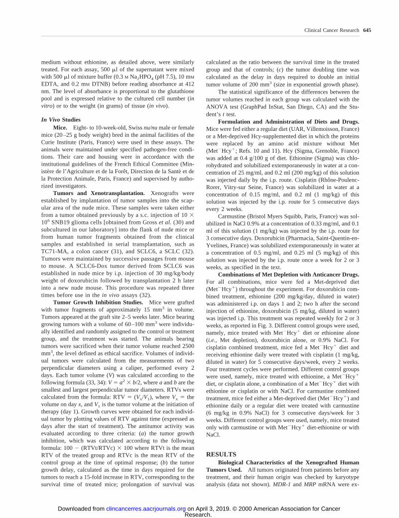
Combination of Depletion with Carmustine for theTreatment of SNB19 Xenograft, a Glioblastoma. A limitedantitumoral effect of carmustine alone was obtained in SNB19glioma (30% tumor growth inhibition; Table 4 and Fig. 5). Thecombination of carmustine with the Met-deprived diet and ethi-onine enhanced the antitumoral effect of the Met depriveddiet-ethionine association (80% and 46% tumor growth inhibi-tion, respectively), leading to a prolongation of mice survival of2-fold in the group receiving the triple therapy.
Fig. 1 MDR-1andMRPmRNA expression of the xenografted humantumors used.
646 Potentiated Antitumoral Drug Effect by Methionine Depletion
Research. on April 3, 2019. © 2000 American Association for Cancerclincancerres.aacrjournals.org Downloaded from

DISCUSSIONMet depletion of cells inducedin vitro by substitution of
Met by Hcy in the culture medium and the addition of ethioninereduced cell proliferation of cancer lines and decreased ATP andglutathione pools. ATP is required for the function of drugefflux pumps, such as Pgp encoded by theMDR-1 gene andMRP, which are involved in the resistance to numerous com-pounds including doxorubicin. Glutathione and glutathionetransferases, which are known to be constantly elevated intumors as compared to normal tissues, detoxify xenobiotics
including the drugs doxorubicin, cisplatin, and carmustine usedin this study. Met depletion might allow recovery of the antitu-moral efficacy of these cytotoxic agents in drug-refractory tu-mors by decreasing their resistance potential.In vivo, animalscan be starved of Met by replacing the standard diet with aMet-free regimen and simultaneous administration of ethionine.Our results confirm this hypothesis.
We and others have previously described the Met depen-dency of tumor cells (3, 11, 35, 36). This led us to design atherapeutic approach using a Met-deprived diet and ethionine, a
Fig. 2 Effects of Met depriva-tion and ethionine on cell pro-liferation, ATP pools, and glu-tathione content of SNB19, ahuman glioma.Top, in vitrocell proliferation assay in twodifferent media with Hcy, withand without Met, with andwithout ethionine, added at aconcentration of 0.5 mg/ml.Middle,relative content in ATPof SNB19 cultured in Met-con-taining medium (left) or inMet-free medium (right), as afunction of time.Bottom,glu-tathione content in cells (left)and in tumor tissue (right), incontrols, Met-free medium, ordiet and ethionine.
647Clinical Cancer Research
Research. on April 3, 2019. © 2000 American Association for Cancerclincancerres.aacrjournals.org Downloaded from

Met analogue, both contributing to Met depletion in animals.The Met-deprived diet was well tolerated by nude mice whenHcy was added to substitute for Met. Hcy is a precursor inendogenous Met synthesis and is used efficiently by normalcells to maintain their metabolism, but not by tumor cells.Ethionine treatment, combined with a Met-deprived diet, wasnot toxic up to a daily dose of 1 g/kg body weight. In all tumorstested from various origins, tumor growth inhibition was ob-served when ethionine treatment was associated with the Met-deprived diet (11), reaching a value of 80% with significanttumor growth delay, prolonging survival times by 2–3-fold intumor-bearing animals. Such an antitumoral effect was con-firmed in the present study in nude mice bearing TC71-MA, ahuman colon cancer, and SNB19, a glioblastoma.
Met deprivation acts by reducing proliferation of tumorcells, and this inhibition is not counteracted by Hcy but isreinforced by ethionine. Met depletion decreases the glutathione
content, irreversibly blocks cells in S phase and G2 of the cellcycle, and induces apoptosis (10). Combined with ethionine, itinduced a drop in ATP pools. Decreases in glutathione inducedin the absence of Met have been described previously (37, 38).In the present study, we show that the combination of a Met-deprived diet and ethionine induced a depletion of the glutathi-one pool in tumors xenografted into nude mice.
The fundamental basis of the antiproliferative effect of Metdepletion in tumor cells might be partly due to dNTP imbalanceinduced by folate deviation toward endogenous Met synthesis,which is responsible for the S-phase blockade. The intracellularMet concentration determines the metabolic priority of folate(39, 40). Under normal conditions, folates are used for endog-enous Met synthesis and purine and pyrimidine bases. Thymi-dine, which is essential for DNA reparation and replication, isthe methylated analogue of uracil. This methylation reaction,which is catalyzed by thymidylate synthetase, specifically re-
Table 1 In vitroand in vivo antitumoral effects of Met-free medium or Met-free diets with and without ethionine (Eth)
Treatment
SNB19 TC71-MA SCLC6SCLC6-
Dox
Met2Hcy1Met2Hcy1
1 Etha Met2Hcy1Met2Hcy1
1 Eth Met2Hcy1Met2Hcy1
1 EthMet2Hcy1
1 Eth
Cell proliferation index(% of control)b 63c 6 5 16d 6 2 17d 6 0.1 2d 6 0.1 52c 6 4 50c 6 7 NDe
Tumor growth inhibition(Mean %6 SD)
26f 6 4 53c 6 8 46c 6 13 56c 6 10 12f 6 10 28c 6 16 4f 6 2
Tumor doubling time indays (control)
6 (5) 8 (5) 13 (11) 26 (11) 5 (4) 6 (4) 4 (4)
Tumor growth delay indays
26 41 36 51 12 13 11
Survival prolongation ratiog 1 1.6 1.5 2.1 1 1.1 1a Ethionine was injected i.p. daily, at a dose of 200 mg/kg. Mice were fed a Met2Hcy1 diet as soon as the tumors reached a 60–100 mm3.b Percentage of surviving cell number after a 3-day culture in Met-free, Hcy-supplemented medium, with or without ethionine (0.5 mg/ml), as
compared to the number of cells cultured in Met-containing medium.c 0.05, P , 0.01.d P , 0.01.e ND, not determined.f P . 0.05, statistically nonsignificant.g Ratio to the survival time of untreated mice.
Table 2 Antitumoral effects of cisplatin, Met-free diet, and ethionine on the growth of TC71-MA, a human colon cancer grafted into nude mice
Diet TreatmentRTV at day 33Mean6 SD
Tumor growthinhibition (%)Mean6 SD
Tumor doublingtime (days)Mean6 SD
Survivalprolongation (days)
Mean6 SD(ratio)a
Regular diet None 216 4 11 6 1Regular diet Ethionineb 236 2 66 7 126 1 1Met2Hcy11c None 116 1d 466 13d 136 1 12d (2.1)Met2Hcy1 Ethionine 9 6 1e 566 10e 266 2e 27e (2.8)Regular diet Cisplatinf 186 3 176 8 146 2 6Regular diet Cisplatin1 ethionine 196 3 116 8 96 1 3Met2Hcy1 Cisplatin 9 6 2e 566 16e 236 1e 27e (2.8)Met2Hcy1 Cisplatin1 ethionine 86 1e 616 15e 286 1e 33e (3.3)
a Ratio to the survival time of untreated mice.b Ethionine was injected i.p. daily, at a dose of 200 mg/kg.c Mice were fed a Met2Hcy1 diet as soon as the tumors reached 60–100 mm3.d 0.05, P , 0.01.e P , 0.01.f Cisplatin was injected i.p., 1 mg/kg, for 5 consecutive days, every 2 weeks.
648 Potentiated Antitumoral Drug Effect by Methionine Depletion
Research. on April 3, 2019. © 2000 American Association for Cancerclincancerres.aacrjournals.org Downloaded from

quires 5,10-tetrahydrofolate as a methyl donor. When the reg-ular diet is replaced by a Met-deprived diet, folate is divertedaway from DNA synthesis to the resynthesis of Met. As a result,
there is an imbalance in the dNTP pool. This, in turn, is knownto promote an accumulation of DNA strand breaks (41), impairDNA repair (42), and lead to apoptotic death (43, 44). DNA
Fig. 3 Tumor growth curves of TC71-MA, ahuman colon cancer xenografted into nudemice. Top, mice (10 mice/group) were treatedwith ethionine (200 mg/kg/day) daily; cisplatin(1 mg/kg/day) for 5 consecutive days weeklyfor 3 weeks; cisplatin1 ethionine; or with aMet2Hcy1 diet. Bottom,control mice were feda regular diet; mice were treated withMet2Hcy1 diet 1 cisplatin; Met2Hcy1 diet 1ethionine; or Met2Hcy1 diet 1 ethionine 1cisplatin.
Table 3 Antitumoral effects of doxorubicin, Met-free diet, and ethionine on the growth of SCLC6, a human SCLC established in nude mice,and on the growth of SCLC6-Dox, derived from SCLC6 after treatment with doxorubicin, as detailed in “Materials and Methods”
Tumors Diet Treatment
RTV atday 14
Mean6 SD
Tumor growthinhibition (%)Mean6 SD
Tumor doublingtime (days)Mean6 SD
Survivalprolongation (days)
Mean6 SD(ratio)a
SCLC6 Regular diet None 226 3 66 1Regular diet Doxorubicinb 216 4 16 3 66 0.8 0Met2Hcy1c Ethionined 166 2 286 16e 6 6 0.7 2Met2Hcy1 Doxorubicin1 ethionine 106 1f 516 13f 8 6 2 8 (1.7)
SCLC6-Dox Regular diet None 256 6 46 0.6 0Regular diet Doxorubicinb 256 3 0 46 0.5 0Met2Hcy1 Ethionined 236 8 46 2 46 2 0Met2Hcy1 Doxorubicin1 ethionine 76 3f 746 12f 8 6 1 8 (1.7)
a Ratio to the survival time of untreated mice.b Doxorubicin was injected i.p. 2 h after ethionine injection at a dose of 5 mg/kg, weekly.c Mice were fed a Met2Hcy1 diet as soon as the tumors reached 60–100 mm3.d Ethionine was injected i.p. daily, at a dose of 200 mg/kg.e 0.05, P , 0.01.f P , 0.01.
649Clinical Cancer Research
Research. on April 3, 2019. © 2000 American Association for Cancerclincancerres.aacrjournals.org Downloaded from

strand breaks and replication arrest (45) could also be inducedby ethionine treatment. Ethionine, the Met analogue used, ismetabolized to S-adenosylethionine and thus might transethyl-ate DNA, RNA, and phospholipids. The DNA ethylation byS-adenosylethionine could prevent polymerase recognition ofthe ethylated cytosine and induce the S-phase blockade by areplication arrest or DNA strand breaks. DNA hypomethylationinduced by Met depletion (46, 47) can also induce an accumu-lation of DNA strand breaks (41).
We decided to combine metabolism-targeted therapy andMet depletion, which induces a decrease in glutathione and ATPcontent, with chemotherapeutic agents, whose efficacy couldinversely depend on the expression of Pgp, MRP, or glutathione.The choice of the cytotoxic agent to be combined with Metdepletion was dependent on the type of tumor. For SCLC6, asmall cell lung cancer, the association of doxorubicin with Metdepletion was based on the drug-refractory phenotype ofSCLC6, which expresses Pgp and MRP proteins (48). The
activity of Pgp could be responsible for chemoresistance, andwe showed previously that it could be reversed by verapamil(49). Expression of MRP and of glutathione S-transferasepprobably contributes to the resistance of SCLC6. Doxorubicin isused for treating patients (50), even if it is not the referencetreatment of SCLC. SCLC6, a doxorubicin-resistant tumor, wasa suitable model to show the capacity of increasing the sensi-tivity to doxorubicin by Met depletion. Indeed, cell chemosen-sitivity to doxorubicin is dependent on its intracellular accumu-lation, with doxorubicin resistance being induced by thealteration of membrane transport such as ATP-dependent mech-anisms of efflux or by a high glutathione concentration (51). Thedecrease of ATP and glutathione pools induced by Met deple-tion in the presence of ethionine might lead to the recovery ofdoxorubicin chemosensitivity. Both SCLCs (SCLC6 andSCLC6-Dox) were very resistant to doxorubicin and expressedMDR1,MRP, and GSTp, yet they responded to the combinationof doxorubicin with Met depletion by a significant inhibition oftumor growth and prolonged survival, whereas doxorubicinalone had little or no activity. The results observed could also beexplained by the DNA intercalation and topoisomerase II inhi-bition of doxorubicin, which could potentiate the effects of thehypomethylation induced by the Met depletion.
Cisplatin has been used in the treatment of colon cancer(52, 53). TC71-MA, a human colon cancer, was poorly sensitiveto cisplatin. Several arguments led us to combine cisplatin withMet depletion. Cisplatin interferes with Met transport and actsas an inhibitor of amino acid entry. This was demonstrated inbrain tissue (54), and this could contribute to augment Metdepletion in tumor tissue. Alternatively, cisplatin detoxificationwas found to require glutathione, which forms complexes withthis heavy metal (55), and furthermore, efflux of glutathione-cisplatin complexes is driven by MRP, an ATP-dependent trans-porter (22) that could be reduced by Met depletion. TheTC71-MA tumor displayed a high level of glutathione andexpressed MRP, like the majority of colon cancers, and thiscould explain the lack of efficacy of cisplatin. The association ofa Met-deprived diet with cisplatin enhanced the antitumoralefficacy of cisplatin. We hypothesize that the simultaneousadministration of a Met-deprived diet decreases the glutathionepool, thereby decreasing the formation of cisplatin-glutathionecomplexes and hence sensitizing TC71-MA to cisplatin.
Carmustine is the reference drug for the treatment of gli-oma (56, 57). However, its efficacy is limited, like all nitro-soureas, by glutathione detoxification, and a relationship be-tween the response of tumors to carmustine and their glutathionecontent has been described previously (58). We hypothesize thatthe effect of Met depletion and ethionine in decreasing theamount of glutathione could be responsible for the potentiationof the antitumoral efficacy of carmustine observed in SNB19xenografts.
In conclusion, the potentiation of the antitumoral effect ofa Met-deprived diet, ethionine, and chemotherapy could beexplained, at least in part, by a decrease in glutathione andavailable ATP pools induced by Met depletion and ethionineadministration, which in turn diminished the resistance potentialof cancer cells to the cytotoxic agents. The three tumors arerepresentative of solid tumors refractory to conventional ther-
Fig. 4 Tumor growth curves of SCLC6, a human SCLC xenograftedinto nude mice and its variant, SCLC6-Dox, obtained after three suc-cessive injections of 30 mg/kg i.p. of doxorubicin followed by trans-plantation 2 h later.Top, control mice with SCLC6 (10 mice/group)were fed a regular diet; mice were treated with doxorubicin (5 mg/kg/day) once a week; Met2Hcy1 diet 1 ethionine; or Met2Hcy1 diet 1ethionine1 doxorubicin. Bottom, mice with SCLC6-Dox (10 mice/group) were treated with doxorubicin (5 mg/kg/day) once a week;Met2Hcy1 diet 1 ethionine; or Met2Hcy1 diet 1 ethionine1 doxo-rubicin.
650 Potentiated Antitumoral Drug Effect by Methionine Depletion
Research. on April 3, 2019. © 2000 American Association for Cancerclincancerres.aacrjournals.org Downloaded from
apy, yet they displayed significant responses to chemotherapywhen combined with Met depletion.
ACKNOWLEDGMENTSWe are grateful to V. Bordier and C. Alberti for excellent technical
assistance in animal experimentation. We thank Dr. S. Agrawal forhelpful critical review and reviewing the English used in this report.
REFERENCES1. Warburg, O. On the origin of cancer cells. Science (Washington DC),123: 309–314, 1956.2. Hoffman, R. M. Altered methionine metabolism, DNA methylationand oncogene expression in carcinogenesis. Biochem. Biophys. Acta,738: 49–87, 1984.3. Hoffman, R. M. Altered methionine metabolism and transmethyla-tion in cancer. Anticancer Res.,5: 1–30, 1985.4. Breillout, F., Poupon, M. F., Blanchard, P., Lascaux, V., Echinard-Garin, P., and Robert-Gero, M. Association of SIBA treatment and aMet-depleted diet inhibitsin vitro growth andin vivo metastatic spreadof experimental tumor cell lines. Clin. Exp. Metastasis,6: 3–16, 1988.5. Breillout, F., Antoine, E., Lascaux, V., Rolland, Y., and Poupon,M. F. Promotion of micrometastasis proliferation in a rat rhabdomyo-sarcoma model by epidermal growth factor. J. Natl. Cancer Inst.,81:702–705, 1989.
6. Breillout, F., Antoine, E., and Poupon, M. F. Methionine dependencyof malignant tumors: a possible approach for therapy. J. Natl. CancerInst., 82: 1628–1632, 1990.
7. Guo, H. Y., Herrera, H., Groce, A., and Hoffman, R. M. Expressionof the biochemical defect of methionine dependence in fresh patienttumors in primary histoculture. Cancer Res.,53: 2479–2483, 1993.
8. Guo, H., Lishko, V. K., Herrera, H., Groce, A., Kubota, T., andHoffman, R. M. Therapeutic tumor-specific cell cycle block induced bymethionine starvationin vivo. Cancer Res.,53: 5676–5679, 1993.
9. Guo, H., Tan, Y., Kubota, T., Moossa, A. R., and Hoffman, R. M.Methionine depletion modulates the antitumor and antimetastatic effi-cacy of ethionine. Anticancer Res.,16: 2719–2723, 1996.
10. Poirson-Bichat, F., Lopez, R., Bras Goncalves, R. A., Miccoli, L.,Bourgeois, Y., Demerseman, P., Poisson, M., Dutrillaux, B., and Pou-pon, M. F. Methionine deprivation and methionine analogs inhibit cellproliferation and growth of human xenografted gliomas. Life Sci.,60:919–931, 1997.
11. Poirson-Bichat, F., Gonfalone, G., Bras-Goncalves, R. A., Dutril-laux, B., and Poupon, M. F. Growth of methionine-dependent humanprostate cancer (PC-3) is inhibited by ethionine combined with methi-onine starvation. Br. J. Cancer,75: 1605–1612, 1997.12. Tan, Y., Xu, M., Guo, H., Sun, X., Kubota, T., and Hoffman, R. M.Anticancer efficacy of methioninasein vivo.Anticancer Res.,16: 3931–3936, 1996.13. Tan, Y., Zavala, J., Sr., Han, Q., Xu, M., Sun, X., Tan, X., Tan, X.,Magana, R., Geller, J., and Hoffman, R. M. Recombinant methioninase
Table 4 Antitumoral effects of carmustine, Met-free diet, and ethionine on the growth of SNB19, a human glioma established in nude mice
Diet Treatment
RTV atday 24
Mean6 SD
Tumor growthinhibition (%)Mean6 SD
Tumor doublingtime (days)Mean6 SD
Survivalprolongation (days)
Mean6 SD(ratio)a
Regular diet None 246 4 11 6 1Regular diet Carmustineb 176 2 306 7 126 1 2Met2Hcy1c Ethionined 136 1e 466 13e 136 1 4Met2Hcy1 Carmustine1 ethionine 56 1f 806 10f 266 2f 27f (2)
a Ratio to the survival time of untreated mice.b Carmustine was injected i.p. 6 mg/kg, for 3 consecutive days, for 3 weeks.c Mice were fed a Met2Hcy1 diet as soon as the tumors reached 60–100 mm3.d Ethionine was injected i.p. daily, at a dose of 200 mg/kg.e 0.05, P , 0.01.f P , 0.01.
Fig. 5 Tumor growth curves of SNB19, a humanglioma xenografted into nude mice. Tumor-bear-ing mice (10 mice/group) were treated with car-mustine (6 mg/kg/day) for 3 consecutive daysweekly, for 3 weeks; Met2Hcy1 diet 1 ethionine;or Met2Hcy1 diet 1 ethionine 1 carmustine;control mice were fed a regular diet.
651Clinical Cancer Research
Research. on April 3, 2019. © 2000 American Association for Cancerclincancerres.aacrjournals.org Downloaded from

infusion reduces the biochemical endpoint of serum methionine withminimal toxicity in high-stage cancer patients. Anticancer Res.,17:3857–3860, 1997.
14. Goseki, N., Yamasaki, S., Endo, M., Onodera, T., Kosaki, G.,Hibino, Y., and Kuwahata, T. Antitumor effect of methionine-depletingtotal parenteral nutrition with doxorubicin administration on Yoshida-sarcoma-bearing rats. Cancer (Phila.),69: 1865–1872, 1992.
15. Nagahama, T., Goseki, N., and Endo, M. Doxorubicin and vincris-tine with methionine depletion contributed to survival in the Yoshidasarcoma bearing rats. Anticancer Res.,18: 25–31, 1998.
16. Breillout, F., Hadida, F., Echinard-Garin, P., Lascaux, V., andPoupon, M. F. Decreased rat rhabdomyosarcoma pulmonary metastasesin response to a low methionine diet. Anticancer Res.,7: 861–867,1987.
17. Razin, A., and Riggs, A. D. DNA methylation and gene function.Science (Washington DC),210: 604–610, 1980.
18. Tan, Y., Xu, M., Guo, H., Sun, X., Kubota, T., and Hoffman, R. M.Anticancer efficacy of methioninasein vivo.Anticancer Res.,16: 3931–3936, 1996.
19. Abraham, E. H., Prat, A. G., Gerweck, L., Seneveratne, T., Arceci,R. J., Kramer, R., Guidotti, G., and Cantiello, H. F. The multidrugresistance (mdr1) gene product functions as an ATP channel. Proc. Natl.Acad. Sci. USA,90: 312–316, 1993.
20. Broxterman, H. J., and Pinedo, H. M. Energy metabolism in mul-tidrug resistant tumor cells: a review. J. Cell. Pharmacol.,2: 239–247,1991.
21. Flens, M. J., Scheffer, G. L., van der Valk, P., Broxterman, H. J.,Eijdems, E. W., Huysmans, A. C., Izquierdo, M. A., and Scheper, R. J.Identification of novel drug resistance-associated proteins by a panel ofrat monoclonal antibodies. Int. J. Cancer,73: 249–257, 1997.
22. Cole, S. P., and Deeley, R. G. Multidrug resistance mediated by theATP-binding cassette transporter protein MRP. Bioessays,20: 931–940,1998.
23. Tew, K. D. Glutathione-associated enzymes in anticancer drugresistance. Cancer Res.,54: 4313–4320, 1994.
24. Shen, H., Kauvar, L., and Tew, K. D. Importance of glutathione andassociated enzymes in drug response. Oncol. Res.,9: 295–302, 1997.
25. Goseki, N., Nagahama, T., Maruyama, M., and Endo, M. Enhancedanticancer effect of vincristine with methionine infusion after methi-onine-depleting total parenteral nutrition in tumor-bearing rats. Jpn. J.Cancer Res.,87: 194–199, 1996.
26. Nagahama, T., Goseki, N., and Endo, M. Doxorubicin and vincris-tine with methionine depletion contributed to survival in the Yoshidasarcoma bearing rats. Anticancer Res.,18: 25–31, 1998.
27. Goseki, N., Yamazaki, S., Shimojyu, K., Kando, F., Maruyama, M.,Endo, M., Koike, M., and Takahashi, H. Synergistic effect of methi-onine-depleting total parenteral nutrition with 5-fluorouracil on humangastric cancer: a randomized, prospective clinical trial. Jpn. J. CancerRes.,86: 484–489, 1995.
28. Bichat, F., Mouawad, R., Solis-Recendez, G., Khayat, D., andBastian, G. Cytoskeleton alteration in MCF7R cells, a multidrug resist-ant human breast cancer cell line. Anticancer Res.,17: 3393–3401,1997.
29. Chevillard, S., Pouillart, P., Beldjord, C., Asselain, B., Beuzeboc,P., Magdelenat, H., and Vielh, P. Sequential assessment of multidrugresistance phenotype and measurement of S-phase fraction as predictivemarkers of breast cancer response to neoadjuvant chemotherapy. Cancer(Phila.),77: 292–300, 1996.
30. Gross, J. L., Behrens, D. L., Mullins, D. E., Kornblith, P. L., andDexter, D. L. Plasminogen activator and inhibitor activity in humanglioma cells and modulation by sodium butyrate. Cancer Res.,48:291–296, 1988.
31. Lefrancois, D., Olschwang, S., Delattre, O., Muleris, M., Dutrillaux,A. M., Thomas, G., and Dutrillaux, B. Preservation of chromosome andDNA characteristics of human colorectal adenocarcinomas after passagein nude mice. Int. J. Cancer,44: 871–878, 1989.
32. Arvelo, F., Poupon, M. F., Goguel, A. F., Lizard, G., Bourgeois, Y.,Arriagada, R., and Le Chevalier, T. Response of a multidrug-resistanthuman small-cell lung cancer xenograft to chemotherapy. J. Cancer Res.Clin. Oncol.,120: 17–23, 1993.
33. Poupon, M. F., Arvelo, F., Goguel, A. F., Bourgeois, Y., Jacrot, M.,Hanania, N., Arriagada, R., and Le Chevalier, T. Response of small-celllung cancer xenografts to chemotherapy: multidrug resistance and directclinical correlates. J. Natl. Cancer Inst.,85: 2023–2029, 1993.34. Houghton, P. J., Cheshire, P. J., Hallman, J. D., II, Lutz, L.,Friedman, H. S., Danks, M. K., and Houghton, J. A. Efficacy oftopoisomerase I inhibitors, topotecan and irinotecan, administered atlow dose levels in protracted schedules to mice bearing xenografts ofhuman tumors. Cancer Chemother. Pharmacol.,36: 393–403, 1995.35. Tisdale, M. J. Utilization of performed and endogenously synthe-sized methionine by cells in tissue culture. Br. J. Cancer,49: 315–320,1984.36. Hoshiya, Y., Guo, H., Kubota, T., Inada, T., Asanuma, F., Yamada,Y., Koh, J., Kitajima, M., and Hoffman, R. Human tumors are methi-onine dependentin vivo. Anticancer Res.,15: 717–718, 1995.37. Hunter, E., and Grimble, R. Dietary sulphur amino acid adequacyinfluences glutathione synthesis and glutathione-dependent enzymesduring the inflammatory response to endoxin and tumour necrosisfactor-a in rats. Clin. Sci.,92: 297–305, 1997.38. Morand, C., Rios, L., Moundras, C., Besson, C., Remesy, C., andDemigne, C. Influence of methionine availability on glutathione syn-thesis and delivery by the liver. Nutr. Biochem.,8: 246–255, 1997.39. Scott, J., and Weir, D. The methyl folate trap: a physiologicalresponse in man to prevent methyl group deficiency in kwashiorkor(methionine deficiency) and an explanation for folic-acid-induced ex-acerbation of subacute combined degeneration in pernicious anemia.Lancet,2: 337–340, 1981.40. James, S., Miller, B., McGarrity, L., and Morris, S. The effect offolic acid and/or methionine deficiency on deoxyribonucleotide poolsand cell cycle distribution in mitogen-stimulated rat lymphocytes. CellProlif., 27: 395–406, 1994.41. Li, J. C., and Kaminskas, E. Accumulation of DNA strand breaksand methotrexate cytotoxicity. Proc. Natl. Acad. Sci. USA,81: 5694–5698, 1984.42. Holliday, R. Aspects of DNA repair and nucleotide pool imbalance.Basic Life Sci.,31: 453–460, 1985.43. Yoshioka, A., Tanaka, S., Hiraoka, O., Koyama, Y., Hirota, Y.,Ayusawa, D., Seno, T., Garrett, C., and Wataya, Y. Deoxyribonucleo-side triphosphate imbalance. 5-Fluorodeoxyuridine-induced DNA dou-ble strand breaks in mouse FM3A cells and the mechanism of cell death.J. Biol. Chem.,262: 8235–8241, 1987.44. Oliver, F. J., Collins, M. K., and Lopez-Rivas, A. dNTP poolsimbalance as a signal to initiate apoptosis. Experientia,52: 995–1000,1996.45. Thomas, T., Faaland, C. A., Adhikarakunnathu, S., and Thomas,T. J. Structure-activity relations of S-adenosylmethionine decarboxylaseinhibitors on the growth of MCF-7 breast cancer cells. Breast CancerRes. Treat.,39: 293–306, 1996.46. Cox, R., and Irving, C. C. Inhibition of DNA Methylation byS-adenosylethionine with the production of methyl-deficient DNA inregenerating Rat Liver. Cancer Res.,37: 222–225, 1977.47. Christman, J., Sheikhnejad, G., Dizik, M., Abileah, S., and Wain-fan, E. Reversibility of changes in nucleic acid methylation and geneexpression induced in rat liver by severe dietary methyl deficiency.Carcinogenesis (Lond.), 551–557, 1993.48. Canitrot, Y., Bichat, F., Cole, S. P., Deeley, R. G., Gerlach, J. H.,Bastian, G., Arvelo, F., and Poupon, M. F. Multidrug resistance genes(MRP) and MDR1 expression in small cell lung cancer xenografts:relationship with response to chemotherapy. Cancer Lett.,130: 133–141, 1998.49. Arvelo, F., Poupon, M. F., Bichat, F., Grossin, F., Bourgeois, Y.,Jacrot, M., Bastian, G., and Le Chevalier, T. Adding a reverser (vera-pamil) to combined chemotherapy overrides resistance in small cell lungcancer xenografts. Eur. J. Cancer,31A: 1862–1868, 1995.
652 Potentiated Antitumoral Drug Effect by Methionine Depletion
Research. on April 3, 2019. © 2000 American Association for Cancerclincancerres.aacrjournals.org Downloaded from

50. Pujol, J. L., Douillard, J. Y., Riviere, A., Quoix, E., Lagrange, J. L.,Berthaud, P., Bardonnet-Comte, M., Polin, V., Gautier, V., Milleron, B.,Chomy, F., Chomy, P., Spaeth, D., and Le Chevalier, T. Dose-intensityof a four-drug chemotherapy regimen with or without recombinanthuman granulocyte-macrophage colony-stimulating factor in extensive-stage small-cell lung cancer: a multicenter randomized Phase III study.J. Clin. Oncol.,15: 2082–2089, 1997.51. Kisara, S., Furusawa, S., Takayanagi, Y., and Sasaki, K. Effect ofglutathione depletion by buthionine sulfoximine on doxorubicin toxicityin mice. Res. Com. Mol. Pathol. Pharmacol.,89: 401–410, 1995.52. Scheithauer, W., Depisch, D., Kornek, G., Pidlich, J., Rosen, H.,Karall, M., Prochaska, M., Ernst, A., Sebesta, C., and Eckhardt, S.Randomized comparison of fluorouracil and leucovorin therapyversusfluorouracil, leucovorin, and cisplatin therapy in patients with advancedcolorectal cancer. Cancer (Phila.),73: 1562–1568, 1994.53. Sagaster, P., Essl, R., Teich, G., Fritz, E., Wasilewski, M., Umek,H., Dunser, E., Mascher, H., and Micksche, M. Treatment of advancedcolorectal cancer with folinic acid and 5-fluorouracil in combinationwith cisplatinum. Eur. J. Cancer,30A: 1250–1254, 1994.
54. Mineura, K., Sasajima, T., Sasajima, H., and Kowada, M. Inhibitionof methionine uptake bycis-diamminedichloroplatinum (II) in experi-mental brain tumors. Int. J. Cancer,67: 681–683, 1996.
55. Chen, Z., Muto, M., Sumizawa, T., Furukawa, T., Haraguchi, M.,Tani, A., Saijo, N., Kondo, T., and Akiyama, S. An active efflux systemfor heavy metals in cisplatin-resistant human KB carcinoma cells. Exp.Cell Res.,240: 312–320, 1998.
56. Brandes, A. A., Scelzi, E., Zampieri, P., Rigon, A., Rotilio, A.,Amista, P., Berti, F., and Fiorentino, M. V. Phase II trial with BCNUplus a-interferon in patients with recurrent high-grade gliomas. Am. J.Clin. Oncol.,20: 364–367, 1997.
57. Gundersen, S., Lote, K., and Watne, K. A retrospective study of thevalue of chemotherapy as adjuvant therapy to surgery and radiotherapyin grade 3 and 4 gliomas. Eur. J. Cancer,34: 1565–1569, 1998.
58. Allalunis-Turner, M., Day, R., McKean, J., Petruk, K., Allen, P.,Aronyk, K., Weir, B., Huyser-Wierenga, D., Fulton, D., and Urtasun, R.Glutathione levels and chemosensitizing effects of buthionine sulfoximinein human malignant glioma cells. J. Neurooncol.,11: 157–164, 1991.
653Clinical Cancer Research
Research. on April 3, 2019. © 2000 American Association for Cancerclincancerres.aacrjournals.org Downloaded from
2000;6:643-653. Clin Cancer Res F. Poirson-Bichat, R. A. Bras Gonçalves, L. Miccoli, et al. Cytotoxic Agents in Drug-resistant Human Tumor XenograftsMethionine Depletion Enhances the Antitumoral Efficacy of
Updated version
http://clincancerres.aacrjournals.org/content/6/2/643
Access the most recent version of this article at:
Cited articles
http://clincancerres.aacrjournals.org/content/6/2/643.full#ref-list-1
This article cites 52 articles, 10 of which you can access for free at:
Citing articles
http://clincancerres.aacrjournals.org/content/6/2/643.full#related-urls
This article has been cited by 13 HighWire-hosted articles. Access the articles at:
E-mail alerts related to this article or journal.Sign up to receive free email-alerts
Subscriptions
Reprints and
To order reprints of this article or to subscribe to the journal, contact the AACR Publications
Permissions
Rightslink site. Click on "Request Permissions" which will take you to the Copyright Clearance Center's (CCC)
.http://clincancerres.aacrjournals.org/content/6/2/643To request permission to re-use all or part of this article, use this link
Research. on April 3, 2019. © 2000 American Association for Cancerclincancerres.aacrjournals.org Downloaded from