metals in medicine - download.e-bookshelf.de file2. metals–therapeutic use. i. title....
TRANSCRIPT

Metals in Medicine
JAMES C. DABROWIAKDepartment of Chemistry, Syracuse University, New York, USA


Metals in Medicine


Metals in Medicine
JAMES C. DABROWIAKDepartment of Chemistry, Syracuse University, New York, USA
This edition first published 2009� 2009 John Wiley & Sons, Ltd
Registered officeJohn Wiley & Sons Ltd, The Atrium, Southern Gate, Chichester, West Sussex, PO19 8SQ, United Kingdom
For details of our global editorial offices, for customer services and for information about how to apply for permission to reuse theco pyright material in th is book pl ease see our website at www.wiley.com.
The right of the author to be identified as the author of this work has been asserted in accordance with the Copyright, Designs andPatents Act 1988.
All rights reserved. No part of this publication may be reproduced, stored in a retrieval system, or transmitted, in any form or by anymeans, electronic, mechanical, photocopying, recording or otherwise, except as permitted by the UK Copyright, Designs andPatents Act 1988, without the prior permission of the publisher.
Wiley also publishes its books in a variety of electronic formats. Some content that appears in print may not be available inelectronic books.
Designations used by companies to distinguish their products are often claimed as trademarks. All brand names and product names used inthis book are trade names, service marks, trademarks or registered trademarks of their respective owners. The publisher is not associatedwith any product or vendor mentioned in this book. This publication is designed to provide accurate and authoritative information in regardto the subject matter covered. It is sold on the understanding that the publisher is not engaged in rendering professional services.If professional advice or other expert assistance is required, the services of a competent professional should be sought.
The publisher and the author make no representations or warranties with respect to the accuracy or completeness of the contents of thiswork and specifically disclaim all warranties, including without limitation any implied warranties of fitness for a particular purpose.This work is sold with the understanding that the publisher is not engaged in rendering professional services. The advice and strategiescontained herein may not be suitable for every situation. In view of ongoing research, equipment modifications, changes in governmentalregulations, and the constant flow of information relating to the use of experimental reagents, equipment, and devices, the reader is urgedto review and evaluate the information provided in the package insert or instructions for each chemical, piece of equipment, reagent, or devicefor, among other things, any changes in the instructions or indication of usage and for added warnings and precautions. The fact that anorganization or Website is referred to in this work as a citation and/or a potential source of further information does not mean that theauthor or the publisher endorses the information the organization or Website may provide or recommendations it may make. Further,readers should be aware that Internet Websites listed in this work may have changed or disappeared between when this work was writtenand when it is read. No warranty may be created or extended by any promotional statements for this work. Neither the publisher nor theauthor shall be liable for any damages arising herefrom.
Library of Congress Cataloging-in-Publication Data
Dabrowiak, James C.Metals in medicine / James C. Dabrowiak.
p. cm.Includes bibliographical references and index.ISBN 978-0-470-68196-1 (cloth) – ISBN 978-0-470-68197-8 (pbk.) 1. Metals in medicine.
2. Metals–Therapeutic use. I. Title.RM666.M513D33 2009615’.231–dc22
2009028763
A catalogue record for this book is available from the British Library.Typeset in 10/12pt Times by Thomson Digital, Noida, India.Printed and bound in Great Britain by CPI Antony Rowe Ltd, Chippenham, Wiltshire.

To my wife, Tatiana, ‘Tati’without whose love, support and understanding
this book would not have been possible
Barnett (Barney) Rosenberg, the discoverer of cisplatin, was a remarkable scientist. Hetransformed a laboratory discovery into one of the most important drugs for treating cancerand he did it in an era with no previous success of a metal-based anticancer drug. The storyof cisplatin will always be inspirational to the budding scientist, sustaining to those doingresearch in the field and motivational to all pursuing the unknown. Barney Rosenberg
passed away on August 8, 2009. He will be greatly missed.


Contents
Feature Boxes xiiiPreface xvAcknowledgments xvii
1 Inorganic Chemistry Basics 1
1.1 Crystal field theory 11.1.1 Octahedral crystal field 21.1.2 Other crystal fields 51.1.3 Factors affecting the crystal field splitting parameter, D 71.1.4 High- and low-spin complexes 9
1.2 Molecular orbital theory 111.2.1 MO diagram of molecular hydrogen 111.2.2 MO diagram for [Co(NH3)6]
3þ 131.3 Absorption spectra of metal complexes 16
1.3.1 Band intensity/selection rules 191.3.2 Spectroscopic and crystal field terms 201.3.3 Band assignments and D 24
1.4 Magnetic properties of metal complexes 281.5 Reactions of metal complexes 29
1.5.1 Forward and reverse rates and equilibrium 291.5.2 Water exchange rates for metal ions 311.5.3 Transition State Theory, the kinetic rate constant and equilibrium 331.5.4 Trans effect and substitution reactions 371.5.5 Stability of metal complexes 401.5.6 Chelate effect 411.5.7 Macrocyclic effect 421.5.8 Hard–soft acids–bases 44
Problems 45References 47Further reading 47
2 Metallo-Drugs and Their Action 49
2.1 Introduction 492.2 Proteins as targets for metallo-drugs 49
2.2.1 Protein structure 502.2.2 Metal binding sites on proteins 51
2.3 DNA as a target for metallo-drugs 582.3.1 Structure of DNA and RNA 582.3.2 Metal binding sites on DNA 62
2.4 Reaction of metal complexes in the biological milieu 632.4.1 Reactions with chloride 632.4.2 Reactions with phosphate 642.4.3 Reactions with carbonate 64
2.5 Evaluating the pharmacological effects of agents 652.5.1 Measuring the cytotoxicity of a drug 652.5.2 Measuring drug uptake 662.5.3 Animal studies 67
2.6 From discovery to the marketplace 672.6.1 Drug approval process 672.6.2 Profits 69
Problems 69References 70Further reading 71
3 Cisplatin 73
3.1 Physical and chemical properties of cisplatin 753.2 Formulation, administration and pharmacokinetics 803.3 Reaction of cisplatin in biological media 813.4 Uptake, cytotoxicity and resistance 83
3.4.1 Influx and efflux of cisplatin 833.4.2 Modification by glutathione and metallothionein 893.4.3 DNA repair 903.4.4 Other resistance mechanisms 91
3.5 Interaction of cisplatin with cellular targets 913.5.1 DNA as a target 913.5.2 Non-DNA targets 98
Problems 101References 102Further reading 107
4 Platinum Anticancer Drugs 109
4.1 Carboplatin 1094.1.1 Synthesis and properties of carboplatin 1094.1.2 Formulation, administration and pharmacokinetics of carboplatin 1134.1.3 Reactions of carboplatin in the biological milieu 1144.1.4 Cytotoxicity and uptake of carboplatin 1174.1.5 Interaction of carboplatin with cellular targets 119
4.2 Oxaliplatin 1224.2.1 Stereochemistry of oxaliplatin 1224.2.2 Synthesis and properties of oxaliplatin 126
Contents viii

4.2.3 Formulation, administration and pharmacokinetics of oxaliplatin 1274.2.4 Reaction of oxaliplatin in the biological milieu 1284.2.5 Cytotoxicity and uptake of oxaliplatin 1304.2.6 Interaction of oxaliplatin with cellular targets 130
4.3 New platinum agents 1344.3.1 Nedaplatin 1354.3.2 Lobaplatin 1364.3.3 Heptaplatin 1364.3.4 Satraplatin (JM216) 1364.3.5 Picoplatin (AMD473, ZD473) 1394.3.6 BBR3464 140
Problems 141References 143Further reading 147
5 Ruthenium, Titanium and Gallium for Treating Cancer 149
5.1 Ruthenium compounds for treating cancer 1495.1.1 Chemistry of ruthenium in the biological milieu 1495.1.2 Structure, synthesis and properties of ruthenium antitumor agents 1505.1.3 Synthesis and biological properties of NAMI-A 1535.1.4 Control of tumor growth by NAMI-A 1565.1.5 Clinical trials with NAMI-A 1575.1.6 Interaction of NAMI-A with potential biological targets 1575.1.7 Synthesis and biological properties of KP1019 1585.1.8 Biological activity of KP1019 1585.1.9 Clinical trials with KP1019 1595.1.10 Interaction of KP1019 with potential biological targets 1595.1.11 Synthesis and properties of ruthenium arene compounds 1615.1.12 Biological activity of the arene complexes 1635.1.13 Targets of the ruthenium arene compounds 164
5.2 Titanium compounds for treating cancer 1675.2.1 Structure, synthesis and properties of titanocene dichloride 1685.2.2 Interaction of titanocene dichloride with potential biological targets 1705.2.3 Structure, synthesis and properties of budotitane 1705.2.4 Antitumor activity and clinical trials with budotitane 1745.2.5 Biological interactions of budotitane 1755.2.6 Titanium anticancer drugs in development 175
5.3 Gallium for treating cancer 1775.3.1 Chemistry of gallium in biological media 1775.3.2 Structures, synthesis and properties of gallium anticancer agents 1795.3.3 Uptake, cytotoxicity and reactivity of gallium agents in the biological system 1805.3.4 Gallium anticancer agents in development 181
Problems 182References 184Further reading 189
ix Contents

6 Gold Compounds for Treating Arthritis, Cancerand Other Diseases 191
6.1 Chemistry of gold in biological media 1916.2 Gold compounds for treating arthritis 192
6.2.1 Structures, synthesis and properties of gold antiarthritic drugs 1936.2.2 Formulation, administration and pharmacokinetics of gold antiarthritic drugs 1956.2.3 Reactions in biological media, uptake and cytotoxicity of gold drugs 1966.2.4 Interactions with cellular targets 199
6.3 Gold complexes for treating cancer 2056.3.1 Structures, synthesis and properties of gold anticancer agents 2056.3.2 Reactions in biological media, uptake and cytotoxicity of gold anticancer agents 2076.3.3 Reactions with cellular targets 208
6.4 Gold complexes for treating AIDS and other diseases 210Problems 212References 214Further reading 217
7 Vanadium, Copper and Zinc in Medicine 219
7.1 Vanadium for treating diabetes 2197.1.1 Diabetes mellitus (DM) 2207.1.2 Bis(ethylmaltolato)oxovanadium(IV), BEOV and
bis(maltolate)oxovanadium(IV), BMOV 2217.1.3 Bis(acetylacetonato)oxovanadium(IV), VO(acac)2, bis(picolinato)oxovanadium(IV),
VO(pic)2 and oxodiperoxo(1,10-phenanthroline)vanadium(V), [bpV(phen)]� 2267.1.4 Anticancer effects of vanadium compounds 228
7.2 Role of copper and other metal ions in Alzheimer’s disease 2307.2.1 Metal chelating agents for treating Alzheimer’s disease 2307.2.2 Complexes of the amyloid beta (Ab) peptide 2327.2.3 Radical production in AD 234
7.3 Copper in Wilson’s and Menkes diseases 2357.4 Zinc–bicyclam: a chemokine receptor antagonist 238Problems 242References 244Further reading 249
8 Metal Complexes for Diagnosing Disease 251
8.1 Technetium in diagnostic nuclear medicine 2528.1.1 Clinically used 99mTc imaging agents 2558.1.2 Technetium imaging agents for selective targeting 2608.1.3 Innovations in synthesis, the [Tc(CO)3]
þ core 2628.2 Metal compounds as contrast agents for MRI 263
8.2.1 Clinically used MRI contrast agents 266
Contents x

8.2.2 Contrast agents in development 2708.2.3 Iron oxide particles 273
8.3 Radionuclides for palliative care and cancer treatment 274Problems 277References 279Further reading 282
9 Nanomedicine 283
9.1 Nanoscience for treating disease 2839.1.1 Single walled carbon nanotubes 2839.1.2 Metal–organic frameworks 2869.1.3 Mesoporous silica 2889.1.4 Encapsulation 2909.1.5 Gold nanoparticles 293
9.2 Nanomedicine in diagnosing disease 2959.2.1 Computed tomography 2959.2.2 Magnetic resonance imaging 2969.2.3 Quantum dots 300
9.3 Potential health risks of nanoparticles 3049.3.1 Cell culture assays and nanomaterials 3049.3.2 Cytotoxicity of nanoparticles 305
Problems 306References 307Further reading 310
Index 311
xi Contents


Feature Boxes
Box 1.1 Absorption spectra of metal complexes 17Box 1.2 Tanabe–Sugano diagrams 23Box 2.1 Cellular guardians 56Box 3.1 Discovery of cisplatin 73Box 3.2 Heteronuclear single quantum coherence (HSQC) NMR 77Box 3.3 Influx, efflux and drug transport 86Box 3.4 Supercoiled DNA as a drug substrate 94Box 4.1 Area under curve (AUC) 113Box 4.2 Optical activity, ORD/CD 123Box 4.3 Thioredoxin reductase as a target for metallo-drugs 133Box 4.4 Synthesis of satraplatin 137Box 5.1 Organometallic chemistry 151Box 5.2 Transferrin 161Box 5.3 Geometric and optical isomerism of metal complexes 171Box 5.4 Ribonucleotide reductase as a drug target 180Box 6.1 Zinc finger proteins 201Box 7.1 Electron paramagnetic resonance 223Box 8.1 Single photon emission computed tomography (SPECT) 253Box 8.2 Magnetic resonance imaging, MRI, and contrast agents 264Box 9.1 Photodynamic therapy 294Box 9.2 Quantum dots 302


Preface
Metals in Medicine is a textbook for undergraduate and graduate students in chemistry, biochemistry, biologyand the related areas of biophysics, pharmacology and bioengineering. The first chapter of the book presentsbasic bonding concepts in inorganic chemistry and provides a brief overview of the physical and chemicalproperties of metal complexes using concepts and ideas presented in general chemistry. The more demandingconcept of quantummechanics, although not generally discussed in beginning level chemistry courses, is alsobriefly covered at an easy-to-understand level in Chapter 1. Chapter 2 emphasizes the nature and structure ofbiological targets, the reactivity of metal complexes in the biological milieu, and methods for measuring theefficacy and toxicity of agents. The steps from drug discovery to marketplace are also briefly outlined anddiscussed in this chapter.
The remaining six chapters ofMetals in Medicine focus on individual metallo-drugs, drug candidates andmetal-containing agents used to treat and diagnose disease, their synthesis, structures, formulations,pharmacokinetics and known mechanisms of action, and important physical and chemical principles thatapply, while the last chapter addresses the role of inorganic chemistry in the emerging and exciting field ofnanomedicine.
No attempt was made to cover all of the metal-containing compounds that are actively being used or beingconsidered for use in medicine, but rather select topics were focused upon, in order to present a brief overviewof the area, stressing important chemical, physical and biological principles, and pointing out where the areamight be headed. It was felt that this was the best way to prevent ‘saturation’ and leave room for the motivatedstudent to discover more on their own.
Chapter 3 of the text covers cisplatin, which is arguably the most important metal-containing agent used inmedicine and without which this book would not be possible. In this chapter, the student is introduced to howcisplatin was discovered, its physical and chemical properties, and possible methods by which it may killcancer cells, covering mechanisms that involve DNA as well as protein targets in the cell. Chapter 4 discussesthe later-generation cisplatin analogues, carboplatin and oxaliplatin, which are in worldwide use for treatingcancer, aswell as other platinumdrugs that havegained regional approval and a select number of platinumdrugcandidates that are in clinical trails. The huge success of cisplatin prompted the search for other metalcomplexes with antitumor properties and Chapter 5 addresses compounds of ruthenium, titanium and galliumthat exhibit anticancer activity. In order to emphasize the breadth of application of inorganic chemistry inmedicine, the use of gold complexes for treating arthritis, cancer and other diseases is presented in Chapter 6,while the use of vanadium for treating diabetes, copper in Wilson’s, Menkes and Alzheimer’s diseases, andzinc-bicyclam as a stem-cell mobilizing agent and for potentially treating AIDS are covered in Chapter 7.
Althoughmetal-containing agents have had amajor impact on treating disease, they are alsowidely used fordisease detection. Chapter 8 outlines the importance of radioactive technetium complexes in diagnosticnuclear medicine and discusses the use of paramagnetic gadolinium compounds as contrast enhancing agentsin magnetic resonance imaging, MRI. This chapter also briefly discusses the use of radioactive agents forpalliative care and cancer treatment in radioimmunotherapy.
Chapter 9 covers the design and construction of nano-size structures for biomedical applications innanomedicine. Since nanomedicine is one of the most dynamic areas of science, presenting all of thedevelopments in the fieldwith an inorganic theme proved impossible, so a limited number of examples, chosen
largely because they extend the information in previous chapters, are presented and discussed. Althoughnanomedicine has enormous potential, the fact that nanomaterials are totally alien to the biological system hasraised serious questions about the health risks that they may pose to humans. This topic is also discussed inChapter 9.
Throughout the book are Feature Boxes that expand important concepts in metals in medicine, includingrelevant physical techniques, structures of biological targets and transport molecules, the discovery ofcisplatin, synthesis of compounds, special assays and principles behind medical techniques. Although theFeature Boxes were not intended to be comprehensive, they provide sufficient information to show ‘howthings work’, with additional information being found in the extensive list of references at the end of eachchapter. Following each chapter are specifically designed problems, with solutions, that allow the student toapply the laws of thermodynamics and the principles of equilibrium and kinetics to problem solving in thetopic being addressed.
While this textbook is designed for teaching a one-semester course on the role of metal complexes inmedicine, it could also be used to teach basic coordination chemistry against the exciting backdrop ofmetals inmedicine. It has been the author’s experience that students with no previous background in inorganicchemistry and with sights on careers in medicine will easily accept learning some of the most challengingaspects of inorganic chemistry as long as the ultimate goal is learning howmetal-containing agents are used inmedicine. It is also clear that those students who are committed to chemistry and its related disciplines find thesubject matter totally intoxicating and easily acclimatize to the many biochemical and medical aspects thatmetals in medicine involves.
In reading the extensive volume of literature needed to write this book, the author was impressed, indeedhumbled, by the huge body and quality of work produced by investigators in the field. Clearly, many decisionsneeded to be made about what or what not to include, but in the end the guiding principle was on what to tellstudents wishing to gain an overview of an exciting area of science and, most importantly, to let them see howtheymight fit into the area and ultimately how they could help tomove it forward. Since selectionwas of coursecarried out by the author, he accepts full responsibility for the emphasis of the book and any omissions andinaccuracies that it may contain.
James C. DabrowiakMay 2009
Web Site
PowerPoint slides of all figures from this book, along with the solutions to the problems, can be found athttp://www.wiley.com/go/dabrowiak.
Preface xvi

Acknowledgments
It is difficult towrite any bookwithout the help, encouragement and support of many people. Special thanks toa professional colleague and personal friend, Jerry Goodisman, who read and commented on various parts ofthe manuscript for its substance and technical accuracy, and thanks also to many professional colleagues andsupport staff at Syracuse University for their insight and suggestions. Dr Matthew D. Hall at the NationalInstitutes of Health suggested many important concepts and ideas in the development stage of the book thatwere ultimately incorporated into the finished manuscript. Through the years a number of outstandinggraduate students, because they often focused thinking in unexpected directions, have helped shape the viewsof the author, and thus their influence is also in this work. Gratitude is also expressed to the graduate andundergraduate students in the 2009 edition of the course ‘Metals in Medicine’, who helped to make manysections of the book stronger as the manuscript was being written. Spending countless hours reading andwriting is a strain on a relationship and my wife, Tatiana, deserves enormous credit for understanding whatneeded to be done and, most importantly, for ‘being there’ when things were not going as well as they might.


1
Inorganic Chemistry Basics
No description of the metal-containing compounds that have found their way into medicine would be usefulwithout first providing basic information on the bonding in metal complexes, their spectral and magneticproperties and, most importantly, the manner in which they react with water and biological targets in the cell.The approach taken in this chapter assumes background knowledge of general and organic chemistry with noprevious exposure to inorganic chemistry, as would occur in a junior- or senior-level course at mostuniversities. The concepts presented are for the most part intuitive, requiring basic knowledge of chemistryand physics, but sometimes more abstract issues like quantum mechanics – which explains the spectralproperties of metal complexes – will also need to be covered. The overall goal of this chapter is to bring allreaders to a common level, providing them with the ‘core’ of information needed to understand how and why,from the chemical perspective, metal complexes play important roles in medicine.
1.1 Crystal field theory
The bonding that exists in metal complexes, their spectral and magnetic properties and their chemicalreactivity are not easily explained using a single theory. However, one approach that is often used in a basicpresentation of bonding concepts in transition metal chemistry is crystal field CF theory, which because it isbased on simple electrostatic arguments, is relatively easy to understand. In CF theory and MO theory theinteractions between the metal ion (M) and the groups attached to it (called ligands and denoted by L) areconsidered to be electrostatic in nature and the bonding in the compound is described as being salt-like incharacter. The metal ion, a cation, electrostatically interacts with a series of surrounding ligands, which areusually negatively charged or, if they are uncharged, have the negative end of a dipole directed toward themetalion. Barring any serious steric interactions between the ligands, the arrangements about themetal ion generallyhave high-symmetry geometries. For example, a 6-coordinte complex – that is, a compound with six ligandsattached to the metal ion – has an octahedral arrangement of ligands, while five-coordinate complexes havesquare or trigonal bipyramidal arrangements, four-coordinate structures are tetrahedral and square planar, andso on. These geometries, alongwith compounds and intermediates commonly encountered inmetal complexesused in medicine, are shown in Figure 1.1.
Metals in Medicine James C. Dabrowiak� 2009 John Wiley & Sons, Ltd

1.1.1 Octahedral crystal field
The first-row transition metal series, which begins with scandium, Sc, fills the 3d level of the atom, while thesecond- and third-row transition metal series, which begin with yttrium, Y, and lanthanum, La, respectively,fill the 4d (second row) and 5d (third row) orbitals of the atom. The transition metal ions and the electronicconfigurations of common oxidation states are shown in Figure 1.2. Since ions of these elements have electronoccupancies in the d level, which is considered the ‘valence’ level of the ion, CF theory focuses on the changein energy of the d-orbitals when charges representing the ligands approach the metal ion and form salt-likebonds.
Figure 1.1 Common geometries of metal complexes and intermediates found in inorganic chemistry
Metals in Medicine 2

The spatial arrangements of the five d-orbitals on a Cartesian coordinate system are shown in Figure 1.3.The shapes shown represent the probability of finding an electron in a volume of space about the nucleus ofthe metal ion. If the metal ion has no bonded ligands – this is referred to as a free ion – the energies of all fived-orbitals will be the same and are said to be five-fold degenerate in energy. This situation is shown on theleft side of Figure 1.4. Let’s suppose that instead of existing as a free ion, the metal ion is part of a stablecomplex consisting of six negatively-charged ligands bound to the metal ion in an octahedral array. The waythat crystal field theory approaches this situation is to consider what happens to the five d-orbitals in the
Figure 1.2 Transition metal ions and their electronic configurations for various oxidation states
Figure 1.3 Boundary surfaces of the five d-orbitals
3 Inorganic Chemistry Basics

electrostatic field set that is up by the ligands. The first thing that the theory does is to consider a situation inwhich the total negative charge of the ligands is ‘smeared’ equally over the surface of a sphere with a radiusequal to the metal–ligand bond distance and with the metal ion at its center. Since the d-orbitals haveelectrons in them and the surface of the sphere is negatively charged, the energies of the d-orbitals will beraised; that is, they will become less stable relative to the free ion, due to electrostatic repulsion between thed-electrons and the negatively-charged surface of the sphere. Since the charge on the sphere has no‘directionality’ – that is, the negative charges are equally distributed over the entire surface of the sphere – allfive d-orbitals must experience the same electrostatic perturbation from the sphere and move as a group to anew energy, Eo (see Figure 1.4). The next step is to redistribute the charge on the surface of the sphere andconcentrate it at the six points where the axes penetrate the sphere. If the charge at each of the six points isidentical, this will produce a perfect octahedral crystal field about the central metal ion and simulate what thed-orbitals experience in an octahedral metal complex. It should be evident that since dx2�y2 and dz2 are pointeddirectly at the charges (ligands), they must experience a different perturbation than the three orbitals, dxz, dyz,dxy, that are directed between the charges.While itmaynot be obvious that bothdx2�y2 anddz2 should experiencean identical perturbation from the octahedral field, quantum mechanics shows that dz2 , which has a ring ofelectron density in the xy plane (Figure 1.3), is actually a composite of two orbitals that are identical to dx2�y2except that they lie in the yz and xz planes. Thus, since dz2 is a composite of two orbitals that look like dx2�y2 , itmakes sense that the crystal field will affect dz2 and dx2�y2 identically, as shown in Figure 1.4. It should also beevident that since these orbitals are pointed directly at the ligands, they feel the electrostatic repulsion directly,and thus their energies are raised relative to the energy of the spherical field,Eo. It is possible to show that if thetotal charge on the sphere is simply rearranged or ‘localized’ to certain positions on the sphere, the energy of thesystem cannot change; that is, Eo for the sphere and the octahedral field must be the same. This is the center ofgravity rule, which applies to electrostatic models of this type. The consequences of this is that if two orbitals,
Figure 1.4 Generation of the octahedral crystal field from the free ion
Metals in Medicine 4

dx2�y2 and dz2 , are raised by a certain amount, the remaining three, dxz, dyz, dxy, must be lowered by a certainamount. Inspection of the shapes and orientations of dxz, dyz, dxy shows that since these orbitals are directed 45
�
to the axes of the system, and each is related to the others by a simple rotation, all must experience exactly thesame perturbation from the charges which are on the axes of the system. This set of orbitals, which are ‘triplydegenerate’, is often referred to as the ‘t2g’ set due to its symmetry properties. In a similar fashion, the orbitals,dx2�y2 and dz2 which are ‘doubly degenerate’ are referred to as the ‘eg’ set. The labels t2g and eg are products ofthe application of group theory, a mathematical tool for characterizing the symmetry properties of molecules.
Simple electrostatic arguments show that the spacing between the t2g and eg levels depends on the distancethat the charge is from the origin of the system and the magnitude of the charge. If the distance is decreased orif the magnitude of the negative charge is increased, the splitting between t2g and eg will increase. As we willsee, metal complexes can be made with a wide variety of attached ligands, some of which are negativelycharged, for example, Cl�, CN� and so on, and some of which are electrically neutral, for example, H2O, NH3
and so on. However, one thing that all ligands have in common is that they direct electrons, usually a lone pair,toward the metal ion, and these electrons become the ‘point charges’ in the crystal field model describing theelectronic structure of the complex. Since the ability of different ligands to perturb the d-orbitals variesconsiderably, the spitting between the t2g and eg sets of orbitals can be quite different for different complexes.In order to address this, crystal field theory denotes the splitting between the t2g and eg sets as Do, which is thecrystal field splitting parameter. The subscript ‘o’ inDo indicates that a crystal field of octahedral symmetry isbeing addressed. If there are no attached ligands – that is, in the free ion case – there can be no crystal field andDo is zero. Since the splitting between the levels is different for different metal complexes, Do, which carriesunits of energy usually expressed in wavenumbers (cm�1), varies over a wide range. However, the relativedisplacement of the t2g and eg levels in terms ofDo from the center of gravity, Eo, is the same for all octahedralcomplexes with the eg level at 0.6Do and the t2g level at�0.4Do. These values arise because (2 orbitals)� (0.6Do) þ (3 orbitals)� (�0.4Do): 0, which satisfies the center of gravity rule. It should be evident thatEo is theaverage crystal field.
1.1.2 Other crystal fields
Numerous anticancer drugs containingPtþ2 have a squareplanargeometry inwhich four ligands at the corners ofa square are bonded to themetal ion (Figure 1.1). Thebestway togenerate the square planar crystal field splittingpattern for the d-orbitals is to first consider an intermediate field called the tetragonal crystal field. Suppose thatthe charge on each of the two point charges on the plus andminus z-axis of the octahedral crystal field is slightlyreduced in magnitude relative to the other four charges in the plane or, the equivalent situation, wherein themagnitude of the charges on the plus and minus z-axis remain unchanged but the charges are moved to greaterdistance from the metal ion than the four charges in the plane. In this case, the electrostatic field on the z-axis isless than the field seen by themetal ion on thex- and y-axes of the system.As a consequence of thisasymmetry ornon-equivalence in the field, all orbitals with z-components – that is, dxz, dyz and dz2 – will have their energieslowered; that is, theywill becomemore stable in the applied field (Figure 1.5). Since dz2 is pointed directly at theweaker charges on the z-axis, it must experience greater stabilization – that is, more lowering – than dxz, dyz,which are directed away from the point charges. As a consequence of the center of gravity rule, if some levels godown in energy, others – that is, dx2�y2 and dxy – must become less stable and their energies must be raised.
The limiting case of the tetragonal distortion is the square planar geometry in which the two charges on thez-axis have been reduced to zero; that is, there are only four charges in the plane of the system. The removal ofthe axial charges causes a significant stabilization in dz2 , which moves downward in the energy diagram andpasses below (becomes more stable than) the dxy orbital. Since dxz, dyz also have z-components, they are alsostabilized by the loss of the axial charges, but to a lesser extent than dz2 . The resulting crystal field splittingdiagram, sometime called the square planar limit, is shown in Figure 1.5.
5 Inorganic Chemistry Basics

Asecond very common structure formetal complexeswith four groups bonded to the central metal ion is thetetrahedral geometry (Figure 1.1). Compared to the previous examples, rationalizing the d-orbital splittingpattern for the tetrahedral geometry is less straightforward. Figure 1.6 shows a Cartesian coordinate system inthe center of a cube. Placement of charges at opposite corners of opposite faces of the cube and hypotheticallyconnecting them to the metal ion in the center of the cube generates the tetrahedral geometry; that is, allcharge–metal–charge angles are 109.5�. It should be evident from the figure that none of the d-orbitals pointdirectly at the charges, and although other relative arrangements of the cube on the d-orbital coordinate systemare possible, all lead to the conclusion given for the splitting pattern shown in Figure 1.6. The tetrahedralcrystal field has a doubly degenerate set of orbitals, the dz2 and dx2�y2 , termed for symmetry reasons the ‘e’ set,which is lowest in energy, and a triply degenerate set, dxz, dyz and dxy, called the ‘t2’ set, which is highest inenergy. While this pattern is exactly the opposite of the octahedral case, the labels e and t2, which also comefrom group theory, aremissing the subscript ‘g’. This is because the octahedron has a symmetry element calledthe center of inversion (i), which is associated with a mathematical operation in which each point charge of the
Figure 1.5 Octahedral, tetragonal and square planar crystal field
Figure 1.6 Tetrahedral crystal field
Metals in Medicine 6
structure can be passed along a straight line through the central metal ion to reach an identical point charge(Figure 1.5). Since i is not present in the tetrahedron, the subscript ‘g’ is missing from the labels. Although thetetrahedral pattern is the exact opposite of the splitting pattern for the octahedron, the magnitude of thesplitting between the e and t2 levels for the tetrahedral geometry, denoted as Dt, is only 4/9 the value of thesplitting between t2g and eg of the octahedron; that is,Dt¼ 4/9Do or 0.445 Do. Thus, for the tetrahedron, the t2orbital set is at 0.178 Do and the e orbital set is at �0.267 Do.
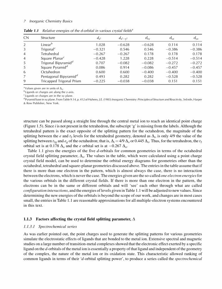
Table 1.1 gives the energies of the five d-orbitals for common geometries in terms of the octahedralcrystal field splitting parameter, Do. The values in the table, which were calculated using a point chargecrystal field model, can be used to determine the orbital energy diagrams for geometries other than theoctahedral, tetrahedral and square-planar geometries discussed above. The entries in the table assume that ifthere is more than one electron in the pattern, which is almost always the case, there is no interactionbetween the electrons, which is never the case. The energies given are the so-called one electron energies forthe various orbitals in the different crystal fields. If there is more than one electron in the pattern, theelectrons can be in the same or different orbitals and will ‘see’ each other through what are calledconfiguration interactions, and the energies of levels given in Table 1.1will be adjusted to newvalues. Sincedetermining the new energies of the orbitals is beyond the scope of our work, and changes are in most casessmall, the entries in Table 1.1 are reasonable approximations for all multiple-electron systems encounteredin this text.
1.1.3 Factors affecting the crystal field splitting parameter, D
1.1.3.1 Spectrochemical series
As was earlier pointed out, the point charges used to generate the splitting patterns for various geometriessimulate the electrostatic effects of ligands that are bonded to the metal ion. Extensive spectral and magneticstudies on a large number of transition-metal complexes showed that the electronic effect exerted by a specificligand on the d-orbitals of themetal ion is essentially a property of that ligand and independent of the geometryof the complex, the nature of the metal ion or its oxidation state. This characteristic allowed ranking ofcommon ligands in terms of their ‘d-orbital splitting power’, to produce a series called the spectrochemical
Table 1.1 Relative energies of the d-orbital in various crystal fieldsa
CN Structure dz2 dx2�y2 dxy dxz dyz
2 Linearb 1.028 �0.628 �0.628 0.114 0.1143 Trigonalc �0.321 0.546 0.546 �0.386 �0.3864 Tetrahedral �0.267 �0.267 0.178 0.178 0.1784 Square Planarc �0.428 1.228 0.228 �0.514 �0.5145 Trigonal Bipyramidd 0.707 �0.082 �0.082 �0.272 �0.2725 Square Pyramidd 0.086 0.914 �0.086 �0.457 �0.4576 Octahedron 0.600 0.600 �0.400 �0.400 �0.4007 Pentagonal Bipyramidd 0.493 0.282 0.282 �0.528 �0.5289 Tricapped Trigonal Prism �0.225 �0.038 �0.038 0.151 0.151
aValues given are in units of Do.bLigands or charges are along the z-axis.cLigands or charges are in the xy plane.dPyramid base in xy plane. FromTable 9.14, p. 412ofHuheey, J.E. (1983) Inorganic Chemistry: Principles of Structure andReactivity, 3rd edn,Harper& Row Publisher, New York.
7 Inorganic Chemistry Basics

series (Table 1.2). Ligands on the left of the series, which are referred to as weak field ligands, for exampleI� and Br�, cause a small splitting in the d-orbitals, while ligands on the right of the series, for example CN�
(cyanide), CO (carbon monoxide) – strong field ligands – cause a large splitting in the orbitals. While there islittle doubt that the order of the ligands in the series is correct (the order is obtained from experiment), the seriesdoes not seem to follow our intuitive feeling about which ligands should be high in the series and which shouldbe low. For example, CO, which is uncharged, is highest in the series but iodide, I�, which is negativelycharged, is lowest in the series. Based on the electrostatic arguments put forth in connection with the crystalfield thismakes little sense: I� should have agreater perturbation on the d-orbitals than unchargedCO.Clearly,factors other than simple electrostatic effects must influence Do. While the crystal field model works well formost of the cases encountered in this text, complexes which have considerable overlap between the orbitals onthemetal and ligand – that is, when covalent bonding is present – cause the theory to ‘bend’ but not completelybreak down. How basic crystal field theory needs to be modified to accommodate this will be addressed in alater section.
1.1.3.2 Principal quantum number, n
While the spectrochemical series rank orders the experimentally-measured effects of ligands on the splittingof the d-orbitals, it is also possible to make some general statements concerning the effects of the metal ionon the magnitude of D. If one moves down a given column in the periodic chart, the quantum numbern, which is called the principal quantum number, increases. For example, the first-row transition metal serieselements have electrons in the 3d (n¼ 3) level, the second-row in the 4d (n¼ 4) level and the third-row in the5d (n¼ 5) level of the atom. Experimentally, it has been found that the magnitude of the crystal field splittingparameter D increases in the order 3d< 4d< 5d, with D 4d� 1.5 (D 3d) and D 5d� (1.75 D 3d) (Table 1.2).The effects of this increase with n can easily be seen for the series [Co(NH3)6]
3þ, 3d6, [Rh(NH3)6]3þ, 4d6 and
[Ir(NH3)6]3þ, 5d6, which have identical geometries (octahedral), ligands (ammonia) and metal ion oxidation
states (þ3), and belong to the same family (column) of the periodic chart. The values of Do for thesecomplexes are �22 000 cm�1, �34 000 cm�1 and �41 000 cm�1, respectively, which shows that movingdown a given column in the periodic chart does indeed cause the values of the crystal field splittingparameter to increase by the approximate amounts given. Since atoms, and ions, become larger with atomicnumber, M–L bond lengths increase in moving from the first to the second and third rows of the transitionmetal series. Simple point-charge arguments would predict that if the M–L distance were increased, themagnitude of D would decrease, not increase as observed. The fact that the opposite is found is further proofthat the simple point-charge model cannot be entirely correct and that other factors are important indetermining the magnitude of D.
Table 1.2 Factors affecting the crystal field splitting parameter, D
The Liganda Spectrochemical Series, Increasing DI�<Br�< S�2<NCS�<Cl�<NO3
�<N3�< F�<OH�<C2O4
�2�H2O<NCS�
<CH3CN<NH3< en< bipy< phen<NO2�< PPh3<CN�<CO
The Metal Ion Principal Quantum Number, nFirst-row transition metal ion, 3d level, D3d
Second-row transition metal ion, 4d level, D4d� 1.5 D3d
Third-row transition metal ion, 5d level, D5d� 1.75 D3d
The Metal Ion Oxidation State, Increasing DMþ<Mþ2<Mþ3<Mþ4<Mþ5
aThe underscored atom is the donor atom to the metal ion. en, ethylenediamine, 1, 2 diaminoethane; bipy, 2, 20 bipyridine; phen, 1, 10phenanthroline.
Metals in Medicine 8

1.1.3.3 Metal ion oxidation state
Experimentally, it can be shown that increasing the charge on the metal ion – that is, increasing its oxidationstate – causes the d-d absorption bands of the complex to shift toward the UV region of the spectrum, whichmeans that D has increased (Table 1.2). Since the ionic radius of any ion decreases with an increase in the netpositive charge on the ion, the distance between the metal ion and its bonded ligands must decrease whenoxidation state is increased. Since decreased distance would lead to greater electrostatic repulsions betweenelectrons on the metal ion and the ligands, the observed trends in D (with changes in oxidation state on themetal ion) are predicted by simple crystal field arguments.
1.1.4 High- and low-spin complexes
When considering the ways in which electrons can occupy energy levels of an atom, ion or molecule,Hund’srule states that the electronic configuration with the lowest overall energy is one for which the spins for theelectrons are unpaired, even if it means placing electrons in a nearby less-stable orbital (level) in order to doso. For the free ion, the five d-orbitals are degenerate in energy and electrons are added to the orbitals bymaximizing the number of unpaired spins. If, for example, there are four electrons in the d-level of a free ion,it is possible to place the electrons in the level in a number of different ways, some of which are shown inFigure 1.7. Experimentally, Figure 1.7a, which has the maximum amount of spin unpairing, is known to bethe lowest-energy (most stable) configuration. When describing the electron spin of any system, it is best touse the value of the magnetic spin quantum number, ms, associated with the spin angular momentum of theelectron. Each electron has spin angular momentum of �1/2 in units of h/2p. With n electrons, the maximumpossible value of the total magnetic spin quantum number S is n/2 (all electrons unpaired) and the minimumpossible value of S is zero (if n is even) or 1/2 (if n is odd). For simplicity, the term h/2p, where h is Planck’sconstant, is usually dropped. Thus, for the configuration shown in Figure 1.7a, S¼ (4)(1/2)¼ 2, while S forFigure 1.7b is (þ1/2 �1/2 þ 1/2þ 1/2)¼ 1 and for Figure 1.7c is (þ1/2 �1/2 þ 1/2 �/2)¼ 0. Two factorsassociated with electronic configurations, coulombic interactions and spin correlations, form the basis forHund’s rule. Since placement of two electrons in the same orbital forces them to occupy the same regions ofspace, the coulombic repulsion between the electrons will be high, thus destabilizing the system. Thisobvious electrostatic repulsion makes it easy to see why maximum spin unpairing, maximum S, is desirable.While columbic considerations are important, the ability to exchange one electron with another in a givenconfiguration without changing S is even more important. This aspect of Hund’s rule, which is a product ofquantum mechanics, in called spin correlation or exchange energy. Both of these factors – coulombic(electrostatic) and spin correlation (exchange energy) – drive the system to obtain maximum spin unpairing,
Figure 1.7 Some possible electronic configurations for the d4 free ion and their respective values of S
9 Inorganic Chemistry Basics
and for the free ion case, where all of the d-orbitals have the same energy, the configuration with the largestvalue of S always has the lowest, most negative, energy.
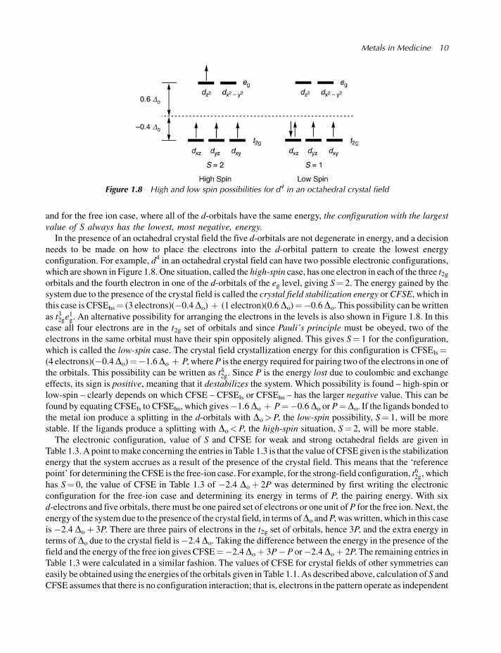
In the presence of an octahedral crystal field the five d-orbitals are not degenerate in energy, and a decisionneeds to be made on how to place the electrons into the d-orbital pattern to create the lowest energyconfiguration. For example, d4 in an octahedral crystal field can have two possible electronic configurations,which are shown in Figure 1.8. One situation, called the high-spin case, has one electron in each of the three t2gorbitals and the fourth electron in one of the d-orbitals of the eg level, giving S¼ 2. The energy gained by thesystem due to the presence of the crystal field is called the crystal field stabilization energy orCFSE, which inthis case is CFSEhs¼ (3 electrons)(�0.4Do) þ (1 electron)(0.6Do)¼�0.6Do. This possibility can bewrittenas t32ge
1g. An alternative possibility for arranging the electrons in the levels is also shown in Figure 1.8. In this
case all four electrons are in the t2g set of orbitals and since Pauli’s principle must be obeyed, two of theelectrons in the same orbital must have their spin oppositely aligned. This gives S¼ 1 for the configuration,which is called the low-spin case. The crystal field crystallization energy for this configuration is CFSEls¼(4 electrons)(�0.4Do)¼�1.6Do þ P, whereP is the energy required for pairing two of the electrons in one ofthe orbitals. This possibility can be written as t42g. Since P is the energy lost due to coulombic and exchangeeffects, its sign is positive, meaning that it destabilizes the system. Which possibility is found – high-spin orlow-spin – clearly depends on which CFSE – CFSEls or CFSEhs – has the larger negative value. This can befound by equating CFSEls to CFSEhs, which gives�1.6Do þ P¼�0.6Do or P¼Do. If the ligands bonded tothe metal ion produce a splitting in the d-orbitals with Do>P, the low-spin possibility, S¼ 1, will be morestable. If the ligands produce a splitting with Do<P, the high-spin situation, S¼ 2, will be more stable.
The electronic configuration, value of S and CFSE for weak and strong octahedral fields are given inTable 1.3. A point tomake concerning the entries in Table 1.3 is that thevalue of CFSEgiven is the stabilizationenergy that the system accrues as a result of the presence of the crystal field. This means that the ‘referencepoint’ for determining theCFSE is the free-ion case. For example, for the strong-field configuration, t62g, whichhas S¼ 0, the value of CFSE in Table 1.3 of �2.4 Doþ 2P was determined by first writing the electronicconfiguration for the free-ion case and determining its energy in terms of P, the pairing energy. With sixd-electrons and five orbitals, there must be one paired set of electrons or one unit ofP for the free ion. Next, theenergy of the systemdue to the presence of the crystal field, in terms ofDo andP, waswritten, which in this caseis �2.4 Doþ 3P. There are three pairs of electrons in the t2g set of orbitals, hence 3P, and the extra energy interms of Do due to the crystal field is�2.4 Do. Taking the difference between the energy in the presence of thefield and the energy of the free ion gives CFSE¼�2.4Doþ 3P�P or�2.4Doþ 2P. The remaining entries inTable 1.3 were calculated in a similar fashion. The values of CFSE for crystal fields of other symmetries caneasily be obtained using the energies of the orbitals given in Table 1.1. As described above, calculation of S andCFSE assumes that there is no configuration interaction; that is, electrons in the pattern operate as independent
Figure 1.8 High and low spin possibilities for d4 in an octahedral crystal field
Metals in Medicine 10