metagenome derived carbohydrate active enzymes for ... · in gt1 werden enzyme klassifiziert, die...
TRANSCRIPT

Metagenome derived carbohydrate active enzymes
for directed modification of polyphenols
Development of a TLC-based functional screening method for identification of
flavonoid modifying enzymes and the isolation and characterization of flavonoid
modifying glycosyltransferases and glycoside hydrolases
Zur Erlangung des Doktorgrades der Naturwissenschaften (Dr. rer. nat.)
im Fachgebiet Mikrobiologie und Biotechnologie des Fachbereichs Biologie
der Fakultät für Mathematik, Informatik und Naturwissenschaften,
der Universität Hamburg
vorgelegt von
Ulrich Rabausch, geb. Köhler
aus Hamburg
Hamburg 2013

Genehmigt vom Fachbereich Biologie der Fakultät für Mathematik, Informatik und Naturwissenschaften an der Universität Hamburg auf Antrag von Professor Dr. W. STREIT Weiterer Gutachter der Dissertation : Professor Dr. B. BISPING Tag der Disputation : 7. Juni 2013
Hamburg , den 18. Juni 2013
Professor Dr. C. Lohr Vorsitzender des
F ach-Promotionsausschusses Biologie
2
Vorwort zum Stand der Wissenschaft
Polyphenole und Flavonoide
Flavonoide gehören zu den Polyphenolen und sind sekundäre Pflanzeninhaltsstoffe, die über
den Shikimisäureweg biosynthetisiert werden (1). Von vielen Polyphenolen sind für den Men-
schen gesundheitsfördernde Wirkungen bekannt (2). Neben der antioxidativen und Radikal-
bindenden Funktion können die Verbindungen auf den Organismus antiallergen, antibakteriell,
antifungal, antiviral, entzündungshemmend, schmerzstillend, gefäßstabilisierend, kreislauf-
fördernd, hormonell und sogar krebsvorbeugend wirken (3). Aufgrund der vielfältigen Wirk-
samkeiten werden Flavonoide daher zunehmend in der Kosmetik-, der Nahrungs- und Nah-
rungsergänzungsmittelindustrie und der Pharmazie eingesetzt und unterliegen deshalb einer
stark wachsenden Nachfrage (4-6).
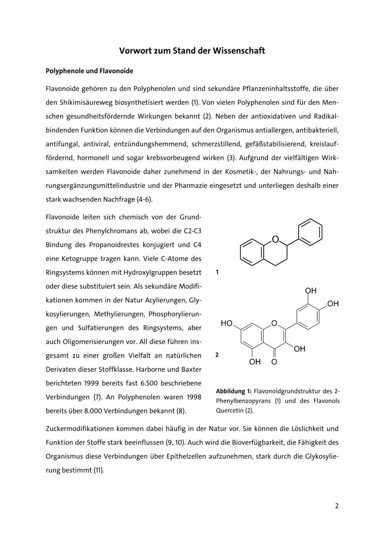
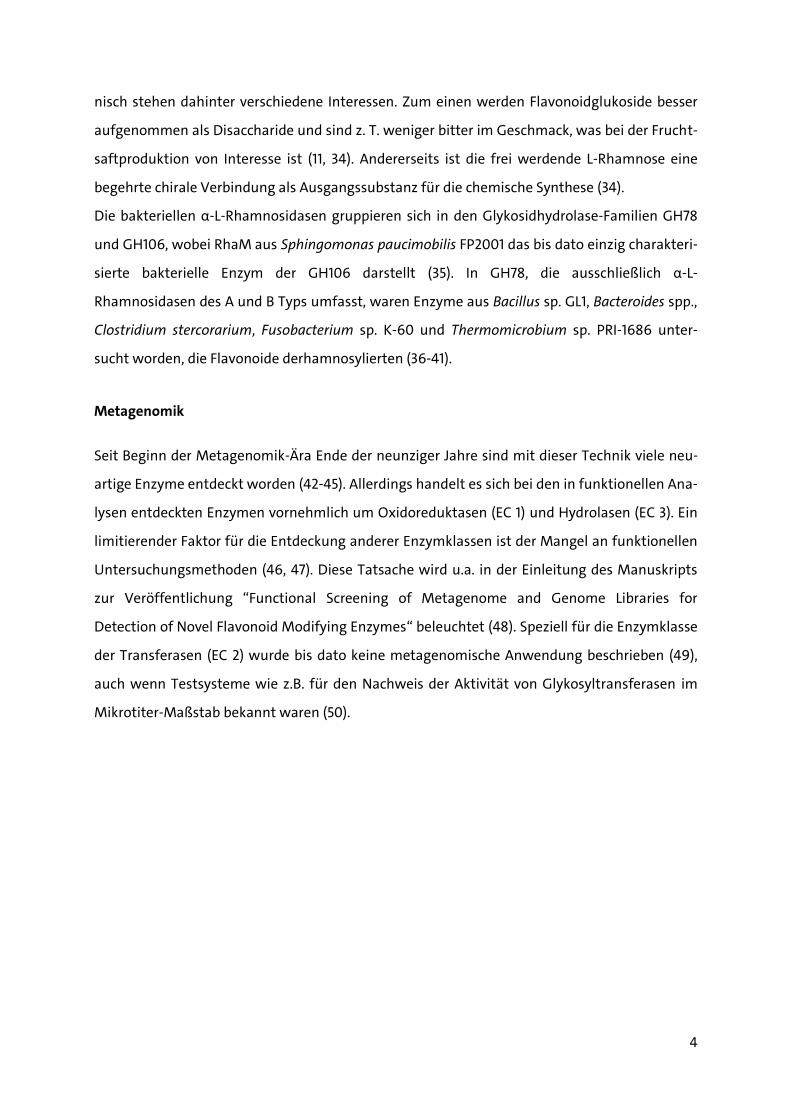
Flavonoide leiten sich chemisch von der Grund-
struktur des Phenylchromans ab, wobei die C2-C3
Bindung des Propanoidrestes konjugiert und C4
eine Ketogruppe tragen kann. Viele C-Atome des
Ringsystems können mit Hydroxylgruppen besetzt
oder diese substituiert sein. Als sekundäre Modifi-
kationen kommen in der Natur Acylierungen, Gly-
kosylierungen, Methylierungen, Phosphorylierun-
gen und Sulfatierungen des Ringsystems, aber
auch Oligomerisierungen vor. All diese führen ins-
gesamt zu einer großen Vielfalt an natürlichen
Derivaten dieser Stoffklasse. Harborne und Baxter
berichteten 1999 bereits fast 6.500 beschriebene
Verbindungen (7). An Polyphenolen waren 1998
bereits über 8.000 Verbindungen bekannt (8).
Zuckermodifikationen kommen dabei häufig in der Natur vor. Sie können die Löslichkeit und
Funktion der Stoffe stark beeinflussen (9, 10). Auch wird die Bioverfügbarkeit, die Fähigkeit des
Organismus diese Verbindungen über Epithelzellen aufzunehmen, stark durch die Glykosylie-
rung bestimmt (11).
1
Abbildung 1: Flavonoidgrundstruktur des 2-
Phenylbenzopyrans (1) und des Flavonols
Quercetin (2).
2

3
Bekannte Flavonoid-modifizierende Enzyme
Als Flavonoid-modifizierende Enzyme waren Glykosyltransferasen (EC 2.4.1) von besonderem
Interesse. An pflanzlichen Enzymen, die für die sekundäre Modifikation von Flavonoiden ver-
antwortlich sind, wurden speziell Glykosyltransferasen gut untersucht. Auch war die Kristall-
struktur einiger pflanzlichen Glykosyltransferasen, teilweise in Bindung mit ihren Donor- und
Akzeptorsubstraten, bereits aufgeklärt (12-14). Die Flavonoid-Glykosyltransferasen gehören zur
Familie 1 der Glykosyltransferasen (GT1) (15). In GT1 werden Enzyme klassifiziert, die Zuckerreste
von aktivierten Donorsubstraten, meist UDP-Glukose, auf lipophile Akzeptorverbindungen
übertragen (16). Sie katalysieren einheitlich eine Transferreaktion von Zuckerresten bei der die
Bindung des anomerischen C-Atoms invertiert wird und besitzen stets eine als GT-B Faltung
bezeichnete Tertiärstruktur (17). In der Primärstruktur lässt sich das so genannte PSPG-Motiv,
ein konserviertes Sequenzmotiv, aufzeigen, das im C-terminalen Bereich der Enzyme lokalisiert
ist und maßgeblich die Donorbinde-Domäne konstituiert (18, 19).
Bei Bakterien war die Fähigkeit Flavonoide zu glykosylieren in einem Bacillus cereus Stamm
bereits Anfang der achtziger Jahren des letzten Jahrhunderts gezeigt worden (20). Bis zu Be-
ginn dieser Dissertation waren allerdings nur wenige weitere Studien zur Glykosylierung von
Flavonoiden durch bakterielle Enzyme publiziert worden. Bakterielle Glykosyltransferasen, de-
nen eine Rolle in der Detoxifizierung von Xenobiotika zugeschrieben wird, waren als Flavonoid-
modifizierende Enzyme bekannt aus B. cereus, Xanthomonas campestris und Streptomyces spp.
(21-23). Von diesen fallen BcGT-1 aus B. cereus ATCC10987, OleD aus Str. antibioticus und Mgt
aus Str. lividans in die Subfamilie der sog. Makrosid-Glykosyltransferasen (TIGR01426) eine Un-
terfamilie der UDP-Glykosyltransferasen (PFAM00201) innerhalb GT1.
Zudem waren einige bakterielle Enzyme aus der Glykosidhydrolase Familie GH13 bekannt (24),
die Zuckerreste von Di- und Oligosacchariden auf polyphenolische Verbindungen übertragen
können. Zu ihnen zählen die Cyclomaltodextrin-Glukanotransferasen, α-Amylasen aus Bacillus
spp. und Thermotoga maritima (25-28), sowie Sucrasen aus den Lactobacillaceae, Leuconostoc
mesenteroides und Streptococcus-Stämmen (29-33).
Biotechnisch bedeutende Flavonoid-modifizierende Enzyme sind die α-L-Rhamnosidasen (34).
Die bakteriellen α-L-Rhamnosidasen sind Glykosidhydrolasen (EC 3.2.1.40), die L-Rhamnose von
Flavonoidrutinosiden (-6-O-α-L-Rhamnopyranosyl-β-D-Glukopyranoside) und Flavonoidneo-
hesperidosiden (-2-O-α-L-Rhamnopyranosyl-β-D-Glukopyranoside) abspalten können. Biotech-
4
nisch stehen dahinter verschiedene Interessen. Zum einen werden Flavonoidglukoside besser
aufgenommen als Disaccharide und sind z. T. weniger bitter im Geschmack, was bei der Frucht-
saftproduktion von Interesse ist (11, 34). Andererseits ist die frei werdende L-Rhamnose eine
begehrte chirale Verbindung als Ausgangssubstanz für die chemische Synthese (34).
Die bakteriellen α-L-Rhamnosidasen gruppieren sich in den Glykosidhydrolase-Familien GH78
und GH106, wobei RhaM aus Sphingomonas paucimobilis FP2001 das bis dato einzig charakteri-
sierte bakterielle Enzym der GH106 darstellt (35). In GH78, die ausschließlich α-L-
Rhamnosidasen des A und B Typs umfasst, waren Enzyme aus Bacillus sp. GL1, Bacteroides spp.,
Clostridium stercorarium, Fusobacterium sp. K-60 und Thermomicrobium sp. PRI-1686 unter-
sucht worden, die Flavonoide derhamnosylierten (36-41).
Metagenomik
Seit Beginn der Metagenomik-Ära Ende der neunziger Jahre sind mit dieser Technik viele neu-
artige Enzyme entdeckt worden (42-45). Allerdings handelt es sich bei den in funktionellen Ana-
lysen entdeckten Enzymen vornehmlich um Oxidoreduktasen (EC 1) und Hydrolasen (EC 3). Ein
limitierender Faktor für die Entdeckung anderer Enzymklassen ist der Mangel an funktionellen
Untersuchungsmethoden (46, 47). Diese Tatsache wird u.a. in der Einleitung des Manuskripts
zur Veröffentlichung “Functional Screening of Metagenome and Genome Libraries for
Detection of Novel Flavonoid Modifying Enzymes“ beleuchtet (48). Speziell für die Enzymklasse
der Transferasen (EC 2) wurde bis dato keine metagenomische Anwendung beschrieben (49),
auch wenn Testsysteme wie z.B. für den Nachweis der Aktivität von Glykosyltransferasen im
Mikrotiter-Maßstab bekannt waren (50).
5
Literaturverweise
1. Andersen, Ø. M., and Markham, K. R. 2006. Flavonoids: Chemistry, biochemistry, and
applications. CRC, Taylor & Francis, Boca Raton, FL.
2. Ross, J. A., and Kasum, C. M. 2002. DIETARY FLAVONOIDS: Bioavailability, Metabolic
Effects, and Safety. Annu Rev Nutr 22:19-34.
3. Ververidis, F., Trantas, E., Douglas, C., Vollmer, G., Kretzschmar, G., and Panopoulos, N.
2007. Biotechnology of flavonoids and other phenylpropanoid-derived natural products.
Part II: Reconstruction of multienzyme pathways in plants and microbes. Biotechnol J
2:1235-1249.
4. Schütz, K., Muks, E., Carle, R., and Schieber, A. 2006. Quantitative determination of
phenolic compounds in artichoke-based dietary supplements and pharmaceuticals by
high-performance liquid chromatography. J Agric Food Chem 54:8812-8817.
5. Wang, M., Liang, C., Pei, Wu, Q., Li, Simon, J., E., and Ho, C., (ed.) 2006. Instrumental
analysis of popular botanical products in the U.S. market, p. 25–38. In Herbs: challenges
in chemistry and biology. American Chemical Society,Washington, DC.
6. Leonard, E., Yan, Y., Fowler, Z. L., Li, Z., Lim, C. G., Lim, K. H., and Koffas, M. A. 2008.
Strain improvement of recombinant Escherichia coli for efficient production of plant
flavonoids. Mol Pharm 5:257-265.
7. Harborne, J. B., and Baxter, H. 1999. The handbook of natural flavonoids. Volume 1 and
Volume 2. Wiley, Chichester, UK.
8. Bravo, L. 1998. Polyphenols: chemistry, dietary sources, metabolism, and nutritional
significance. Nutr Rev 56:317-333.
9. Graefe, E. U., Wittig, J., Mueller, S., Riethling, A. K., Uehleke, B., Drewelow, B., Pforte,
H., Jacobasch, G., Derendorf, H., and Veit, M. 2001. Pharmacokinetics and bioavailability
of quercetin glycosides in humans. J Clin Pharmacol 41:492-499.
10. Kren, V., and Martinkova, L. 2001. Glycosides in medicine: The role of glycosidic residue
in biological activity. Curr Med Chem 8:1303-1328.
11. Hollman, P. C. H., Bijsman, M. N. C. P., van Gameren, Y., Cnossen, E. P. J., de Vries, J. H.
M., and Katan, M. B. 1999. The sugar moiety is a major determinant of the absorption
of dietary flavonoid glycosides in man. Free Radic Res 31:569-573.
12. Li, L., Modolo, L. V., Escamilla-Trevino, L. L., Achnine, L., Dixon, R. A., and Wang, X. 2007.
Crystal structure of Medicago truncatula UGT85H2--insights into the structural basis of
a multifunctional (iso)flavonoid glycosyltransferase. J Mol Biol 370:951-963.
13. Offen, W., Martinez-Fleites, C., Yang, M., Kiat-Lim, E., Davis, B. G., Tarling, C. A., Ford, C.
M., Bowles, D. J., and Davies, G. J. 2006. Structure of a flavonoid glucosyltransferase
reveals the basis for plant natural product modification. Embo J 25:1396-1405.
14. Shao, H., He, X., Achnine, L., Blount, J. W., Dixon, R. A., and Wang, X. 2005. Crystal
structures of a multifunctional triterpene/flavonoid glycosyltransferase from Medicago
truncatula. Plant Cell 17:3141-3154.
15. Coutinho, P. M., Deleury, E., Davies, G. J., and Henrissat, B. 2003. An evolving
hierarchical family classification for glycosyltransferases. J Mol Biol 328:307-317.
16. Bowles, D., Lim, E. K., Poppenberger, B., and Vaistij, F. E. 2006. Glycosyltransferases of
lipophilic small molecules. Annu Rev Plant Biol 57:567-597.
17. Lairson, L. L., Henrissat, B., Davies, G. J., and Withers, S. G. 2008. Glycosyltransferases:
structures, functions, and mechanisms. Annu Rev Biochem 77:521-555.
18. Paquette, S., Moller, B. L., and Bak, S. 2003. On the origin of family 1 plant
glycosyltransferases. Phytochemistry 62:399-413.
6
19. Hughes, J., and Hughes, M. A. 1994. Multiple secondary plant product UDP-glucose
glucosyltransferase genes expressed in cassava (Manihot esculenta Crantz) cotyledons.
DNA Seq 5:41-49.
20. Rao, K. V., and Weisner, N. T. 1981. Microbial Transformation of Quercetin by Bacillus
cereus. Appl Environ Microbiol 42:450-452.
21. Ko, J. H., Gyu Kim, B., and Joong-Hoon, A. 2006. Glycosylation of flavonoids with a
glycosyltransferase from Bacillus cereus. FEMS Microbiol Lett 258:263-268.
22. Kim, H. J., Kim, B. G., Kim, J. A., Park, Y., Lee, Y. J., Lim, Y., and Ahn, J. H. 2007.
Glycosylation of flavonoids with E. coli expressing glycosyltransferase from
Xanthomonas campestris. J Microbiol Biotechnol 17:539-542.
23. Yang, M., Proctor, M. R., Bolam, D. N., Errey, J. C., Field, R. A., Gilbert, H. J., and Davis, B.
G. 2005. Probing the breadth of macrolide glycosyltransferases: in vitro remodeling of a
polyketide antibiotic creates active bacterial uptake and enhances potency. J Am Chem
Soc 127:9336-9337.
24. Henrissat, B., and Davies, G. 1997. Structural and sequence-based classification of
glycoside hydrolases. Current Opinion in Structural Biology 7:637-644.
25. Shimizu, R., Shimabayashi, H., and Moriwaki, M. 2006. Enzymatic production of highly
soluble myricitrin glycosides using beta-galactosidase. Biosci Biotechnol Biochem
70:940-948.
26. Suzuki, Y., and Suzuki, K. 1991. Enzymatic formation of 4G-alpha-D-glucopyranosyl-
rutin. Agric Biol Chem 55:181-187.
27. Kometani, T., Nishimura, T., Nakae, T., Takii, H., and Okada, S. 1996. Synthesis of
neohesperidin glycosides and naringin glycosides by cyclodextrin glucanotransferase
from an alkalophilic Bacillus species. Biosci Biotechnol Biochem 60:645-649.
28. Li, D., Park, J. H., Park, J. T., Park, C. S., and Park, K. H. 2004. Biotechnological production
of highly soluble daidzein glycosides using Thermotoga maritima maltosyltransferase. J
Agric Food Chem 52:2561-2567.
29. Kitao, S., Ariga, T., Matsudo, T., and Sekine, H. 1993. The Syntheses of Catechin-
Glucosides by Transglycosylation with Leuconostoc mesenteroides Sucrose
Phosphorylase. Biosci Biotechnol Biochem 57:2010-2015.
30. Moon, Y.-H., Jin-Ha Lee, Deok-Young Jhon, Woo-Jin Jun, Seong-Soo Kang, Jeonggu Sim,
Heungsic Choi, Jae-Hak Moon and Doman Kim. 2007. Synthesis and characterization of
novel quercetin-α-d-glucopyranosides using glucansucrase from Leuconostoc
mesenteroides. Enzyme Microbial Technol 40:1124-1129.
31. Meulenbeld, G. H., Zuilhof, H., van Veldhuizen, A., van den Heuvel, R. H., and
Hartmans, S. 1999. Enhanced (+)-catechin transglucosylating activity of Streptococcus
mutans GS-5 glucosyltransferase-D due to fructose removal. Appl Environ Microbiol
65:4141-4147.
32. Nakahara, K., Kontani, M., Ono, H., Kodama, T., Tanaka, T., Ooshima, T., and Hamada,
S. 1995. Glucosyltransferase from Streptococcus sobrinus Catalyzes Glucosylation of
Catechin. Appl Environ Microbiol 61:2768-2770.
33. Sato, T., Nakagawa, H., Kurosu, J., Yoshida, K., Tsugane, T., Shimura, S., Kirimura, K.,
Kino, K., and Usami, S. 2000. Alpha-anomer-selective glucosylation of (+)-catechin by
the crude enzyme, showing glucosyl transfer activity, of Xanthomonas campestris WU-
9701. J Biosci Bioeng 90:625-630.
34. Manzanares, P., Vallés, S., Ramòn, D., and Orejas, M. 2007. α-L-rhamnosidases: Old and
New Insights, p. 117-140. In Industrial Enzymes. Springer Netherlands.

7
35. Miake, F., Satho, T., Takesue, H., Yanagida, F., Kashige, N., and Watanabe, K. 2000.
Purification and characterization of intracellular α-L-rhamnosidase from Pseudomonas
paucimobilis FP2001. Arch Microbiol 173:65-70.
36. Hashimoto, W., Miyake, O., Nankai, H., and Murata, K. 2003. Molecular identification of
an alpha-L-rhamnosidase from Bacillus sp strain GL1 as an enzyme involved in complete
metabolism of gellan. Arch Biochem Biophys 415:235-244.
37. Zverlov, V. V., Hertel, C., Bronnenmeier, K., Hroch, A., Kellermann, J., and Schwarz, W.
H. 2000. The thermostable alpha-L-rhamnosidase RamA of Clostridium stercorarium:
biochemical characterization and primary structure of a bacterial alpha-L-rhamnoside
hydrolase, a new type of inverting glycoside hydrolase. Mol Microbiol 35:173-179.
38. Jang, I. S., and Kim, D. H. 1996. Purification and characterization of alpha-L-
rhamnosidase from Bacteroides JY-6, a human intestinal bacterium. Biol Pharm Bull
19:1546-1549.
39. Bokkenheuser, V. D., Shackleton, C. H., and Winter, J. 1987. Hydrolysis of dietary
flavonoid glycosides by strains of intestinal Bacteroides from humans. Biochem J
248:953-956.
40. Park, S.-Y., Kim, J.-H., and Kim, D.-H. 2005. Purification and Characterization of
Quercitrin-Hydrolyzing alpha-L-Rhamnosidase from Fusobacterium K-60, a Human
Intestinal Bacterium. J Microbiol Biotechnol 15:519-524.
41. Birgisson, H., Hreggvidsson, G. O., Fridjónsson, O. H., Mort, A., Kristjánsson, J. K., and
Mattiasson, B. 2004. Two new thermostable alpha-L-rhamnosidases from a novel
thermophilic bacterium. Enzyme Microb Technol 34:561-571.
42. Ferrer, M., Beloqui, A., Timmis, K. N., and Golyshin, P. N. 2009. Metagenomics for
mining new genetic resources of microbial communities. J Mol Microbiol Biotechnol
16:109-123.
43. Tuffin, M., Anderson, D., Heath, C., and Cowan, D. A. 2009. Metagenomic gene
discovery: How far have we moved into novel sequence space? Biotechnol J 4:1671-1683.
44. Steele, H. L., Jaeger, K. E., Daniel, R., and Streit, W. R. 2009. Advances in recovery of
novel biocatalysts from metagenomes. J Mol Microbiol Biotechnol 16:25-37.
45. Simon, C., and Daniel, R. 2009. Achievements and new knowledge unraveled by
metagenomic approaches. Appl Microbiol Biotechnol 85:265-276.
46. Uchiyama, T., and Miyazaki, K. 2009. Functional metagenomics for enzyme discovery:
challenges to efficient screening. Curr Opin Biotechnol 20:616-622.
47. Taupp, M., Mewis, K., and Hallam, S. J. 2011. The art and design of functional
metagenomic screens. Curr Opin Biotechnol 22:465-472.
48. Rabausch, U., Juergensen, J., Ilmberger, N., Böhnke, S., Fischer, S., Schubach, B., Schulte,
M., and Streit, W. R. 2013. Functional Screening of Metagenome and Genome Libraries
for Detection of Novel Flavonoid Modifying Enzymes. Appl Environ Microbiol 79:4551-
4563.
49. Perner, M., Ilmberger, N., Köhler, H. U., Chow, J., and Streit, W. R. 2011. Emerging Fields
in Functional Metagenomics and Its Industrial Relevance: Overcoming Limitations and
Redirecting the Search for Novel Biocatalysts, p. 481-498. In Handbook of Molecular
Microbial Ecology II. John Wiley & Sons, Inc.
50. Aharoni, A., Thieme, K., Chiu, C. P., Buchini, S., Lairson, L. L., Chen, H., Strynadka, N. C.,
Wakarchuk, W. W., and Withers, S. G. 2006. High-throughput screening methodology
for the directed evolution of glycosyltransferases. Nat Methods 3:609-614.

8
Publikationsliste
Aus den Forschungsergebnissen der Arbeit sind folgende Publikationen hervorgegangen, die als
Nachweis einer kumulativen Dissertation zusammen mit einem Forschungsbericht eingereicht
wurden.
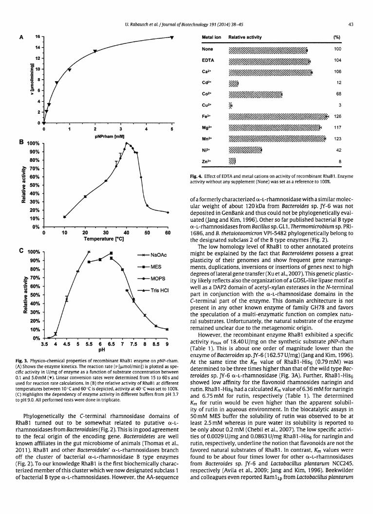
1. Rabausch, U., Ilmberger, N., and Streit, W.R. 2014. The metagenome-derived enzyme RhaB
opens a new subclass of bacterial B type α-L-rhamnosidases. J Biotechnol 191: 38-45.
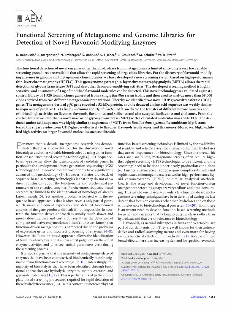
2. Rabausch, U., Juergensen, J., Ilmberger, N., Böhnke, S., Fischer, S., Schubach, B., Schulte, M.,
and Streit, W. R. 2013. Functional Screening of Metagenome and Genome Libraries for
Detection of Novel Flavonoid Modifying Enzymes. Appl Environ Microbiol 79: 4551-4563.
3. Wiegand, S., Rabausch, U., Chow, J., Daniel, R., Streit, W.R., and Liesegang, H. 2013. Complete
Genome Sequence of Geobacillus sp. GHH01, a Thermophilic Lipase-secreting Bacterium.
Genome Announc March/April 2013 1 (2) e00092-13
4. Ilmberger, N., Meske, D., Juergensen, J., Schulte, M., Barthen, P., Rabausch, U., Angelov, A.,
Mientus, M., Liebl, W., Schmitz, R. A., and Streit, W. R. 2012. Metagenomic cellulases highly
tolerant towards the presence of ionic liquids-linking thermostability and halotolerance.
Appl Microbiol Biotechnol 95: 135-146
5. Perner, M., Ilmberger, N., Köhler, H. U., Chow, J., and Streit, W. R. 2011. Emerging Fields in
Functional Metagenomics and Its Industrial Relevance: Overcoming Limitations and
Redirecting the Search for Novel Biocatalysts, p 481-498, In Handbook of Molecular Microbial
Ecology II. John Wiley & Sons, Inc.
Nachfolgend sind beiden erstgenannten Publikationen angefügt. Sie enthalten die wesentli-
chen Ergebnisse und Erkenntnisse der Arbeit.
Ulrich Rabausch


Functional Screening of Metagenome and Genome Libraries forDetection of Novel Flavonoid-Modifying Enzymes
U. Rabausch,a J. Juergensen,a N. Ilmberger,a S. Böhnke,a S. Fischer,b B. Schubach,b M. Schulte,b W. R. Streita
Abteilung für Mikrobiologie und Biotechnologie, Biozentrum Klein Flottbek, Universität Hamburg, Hamburg, Germanya; Merck KGaA, Darmstadt, Germanyb
The functional detection of novel enzymes other than hydrolases from metagenomes is limited since only a very few reliablescreening procedures are available that allow the rapid screening of large clone libraries. For the discovery of flavonoid-modify-ing enzymes in genome and metagenome clone libraries, we have developed a new screening system based on high-performancethin-layer chromatography (HPTLC). This metagenome extract thin-layer chromatography analysis (META) allows the rapiddetection of glycosyltransferase (GT) and also other flavonoid-modifying activities. The developed screening method is highlysensitive, and an amount of 4 ng of modified flavonoid molecules can be detected. This novel technology was validated against acontrol library of 1,920 fosmid clones generated from a single Bacillus cereus isolate and then used to analyze more than 38,000clones derived from two different metagenomic preparations. Thereby we identified two novel UDP glycosyltransferase (UGT)genes. The metagenome-derived gtfC gene encoded a 52-kDa protein, and the deduced amino acid sequence was weakly similarto sequences of putative UGTs from Fibrisoma and Dyadobacter. GtfC mediated the transfer of different hexose moieties andexhibited high activities on flavones, flavonols, flavanones, and stilbenes and also accepted isoflavones and chalcones. From thecontrol library we identified a novel macroside glycosyltransferase (MGT) with a calculated molecular mass of 46 kDa. The de-duced amino acid sequence was highly similar to sequences of MGTs from Bacillus thuringiensis. Recombinant MgtB trans-ferred the sugar residue from UDP-glucose effectively to flavones, flavonols, isoflavones, and flavanones. Moreover, MgtB exhib-ited high activity on larger flavonoid molecules such as tiliroside.
For more than a decade, metagenome research has demon-strated that it is a powerful tool for the discovery of novel
biocatalysts and other valuable biomolecules by using either func-tion- or sequence-based screening technologies (1–3). Sequence-based approaches allow the identification of candidate genes. Inparticular, the development of next-generation sequencing (NGS)technology and improved bioinformatic tools have significantlyadvanced this methodology (4). However, a major drawback ofsequence-based screening technologies is that they do not allowdirect conclusions about the functionality and biochemical pa-rameters of the encoded enzymes. Furthermore, sequence-basedsearches are limited to the identification of homologs of alreadyknown motifs (5). Yet another problem associated with the se-quence-based approach is that it often reveals only partial genes,which make subsequent expression and detailed biochemicalanalysis of the gene products difficult if not impossible. In con-trast, the function-driven approach is usually much slower andmore labor-intensive and costly but results in the detection ofcomplete and active enzyme clones. It is of course well known thatfunction-driven metagenomics is hampered due to the problemsof expressing genes and incorrect processing of enzymes (6–8).However, the function-based approach allows the identificationof truly novel enzymes, and it allows a first judgment on the actualenzyme activities and physicochemical parameters even duringthe screening process.
It is not surprising that the majority of metagenome-derivedenzymes that have been characterized biochemically mainly orig-inated from function-based screenings (9, 10). Interestingly, themajority of biocatalysts that have been identified through func-tional approaches are hydrolytic enzymes, mainly esterases andglycoside hydrolases (11, 12). This is perhaps linked to the simpleplate-based screening procedures required for rapid detection ofthese hydrolytic enzymes (13). In this context it is noteworthy that
function-based screening technology is limited by the availabilityof sensitive and reliable assays for enzymes other than hydrolasesthat are of importance for biotechnology. Since the overall hitrates are usually low, metagenome screens often require high-throughput screening (HTS) technologies to be efficient, and thescreenings need to be done under nearly production conditions(6). Further, enzyme screens often require complex substrates andsophisticated chromogenic assays as well as high-performance liq-uid chromatography (HPLC) or similar analytical methods.Clearly, the setup and development of novel function-drivenmetagenome screening assays are very tedious and time-consum-ing. This may be one reason why only a few function-based meta-genome screening techniques have been developed during the lastdecade that focus on enzymes other than hydrolases and on thosewith relevance to biotechnological processes (14–20). Thus, thereis an urgent need to develop function-based screening methodsfor genes and enzymes that belong to enzyme classes other thanhydrolases and that are of relevance to biotechnology.
Flavonoids, as natural substances in fruits and vegetables, arepart of our daily nutrition. They are well known for their antioxi-dative and radical scavenging nature and even more for havingvarious beneficial effects on human health (21). Because of thesebroad effects, there is an increasing demand for specific flavonoids
Received 5 April 2013 Accepted 13 May 2013
Published ahead of print 17 May 2013
Address correspondence to W. R. Streit, [email protected].
Supplemental material for this article may be found at http://dx.doi.org/10.1128/AEM.01077-13.
Copyright © 2013, American Society for Microbiology. All Rights Reserved.
doi:10.1128/AEM.01077-13
August 2013 Volume 79 Number 15 Applied and Environmental Microbiology p. 4551–4563 aem.asm.org 4551
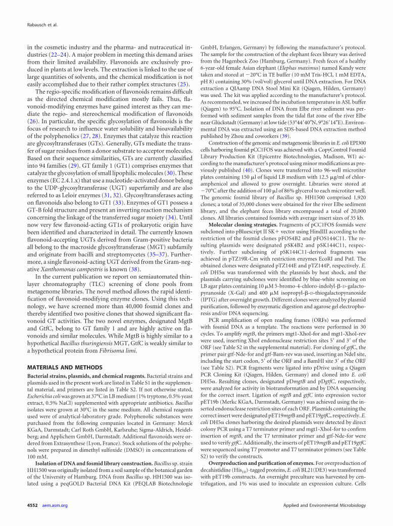
in the cosmetic industry and the pharma- and nutraceutical in-dustries (22–24). A major problem in meeting this demand arisesfrom their limited availability. Flavonoids are exclusively pro-duced in plants at low levels. The extraction is linked to the use oflarge quantities of solvents, and the chemical modification is noteasily accomplished due to their rather complex structures (25).
The regio-specific modification of flavonoids remains difficultas the directed chemical modification mostly fails. Thus, fla-vonoid-modifying enzymes have gained interest as they can me-diate the regio- and stereochemical modification of flavonoids(26). In particular, the specific glycosylation of flavonoids is thefocus of research to influence water solubility and bioavailabilityof the polyphenolics (27, 28). Enzymes that catalyze this reactionare glycosyltransferases (GTs). Generally, GTs mediate the trans-fer of sugar residues from a donor substrate to acceptor molecules.Based on their sequence similarities, GTs are currently classifiedinto 94 families (29). GT family 1 (GT1) comprises enzymes thatcatalyze the glycosylation of small lipophilic molecules (30). Theseenzymes (EC 2.4.1.x) that use a nucleotide-activated donor belongto the UDP-glycosyltransferase (UGT) superfamily and are alsoreferred to as Leloir enzymes (31, 32). Glycosyltransferases actingon flavonoids also belong to GT1 (33). Enzymes of GT1 possess aGT-B fold structure and present an inverting reaction mechanismconcerning the linkage of the transferred sugar moiety (34). Untilnow very few flavonoid-acting GT1s of prokaryotic origin havebeen identified and characterized in detail. The currently knownflavonoid-accepting UGTs derived from Gram-positive bacteriaall belong to the macroside glycosyltransferase (MGT) subfamilyand originate from bacilli and streptomycetes (35–37). Further-more, a single flavonoid-acting UGT derived from the Gram-neg-ative Xanthomonas campestris is known (38).
In the current publication we report on semiautomated thin-layer chromatography (TLC) screening of clone pools frommetagenome libraries. The novel method allows the rapid identi-fication of flavonoid-modifying enzyme clones. Using this tech-nology, we have screened more than 40,000 fosmid clones andthereby identified two positive clones that showed significant fla-vonoid GT activities. The two novel enzymes, designated MgtBand GtfC, belong to GT family 1 and are highly active on fla-vonoids and similar molecules. While MgtB is highly similar to ahypothetical Bacillus thuringiensis MGT, GtfC is weakly similar toa hypothetical protein from Fibrisoma limi.
MATERIALS AND METHODSBacterial strains, plasmids, and chemical reagents. Bacterial strains andplasmids used in the present work are listed in Table S1 in the supplemen-tal material, and primers are listed in Table S2. If not otherwise stated,Escherichia coli was grown at 37°C in LB medium (1% tryptone, 0.5% yeastextract, 0.5% NaCl) supplemented with appropriate antibiotics. Bacillusisolates were grown at 30°C in the same medium. All chemical reagentsused were of analytical-laboratory grade. Polyphenolic substances werepurchased from the following companies located in Germany: MerckKGaA, Darmstadt; Carl Roth GmbH, Karlsruhe; Sigma-Aldrich, Heidel-berg; and Applichem GmbH, Darmstadt. Additional flavonoids were or-dered from Extrasynthese (Lyon, France). Stock solutions of the polyphe-nols were prepared in dimethyl sulfoxide (DMSO) in concentrations of100 mM.
Isolation of DNA and fosmid library construction. Bacillus sp. strainHH1500 was originally isolated from a soil sample of the botanical gardenof the University of Hamburg. DNA from Bacillus sp. HH1500 was iso-lated using a peqGOLD Bacterial DNA Kit (PEQLAB Biotechnologie
GmbH, Erlangen, Germany) by following the manufacturer’s protocol.The sample for the construction of the elephant feces library was derivedfrom the Hagenbeck Zoo (Hamburg, Germany). Fresh feces of a healthy6-year-old female Asian elephant (Elephas maximus) named Kandy weretaken and stored at �20°C in TE buffer (10 mM Tris-HCl, 1 mM EDTA,pH 8) containing 30% (vol/vol) glycerol until DNA extraction. For DNAextraction a QIAamp DNA Stool Mini Kit (Qiagen, Hilden, Germany)was used. The kit was applied according to the manufacturer’s protocol.As recommended, we increased the incubation temperature in ASL buffer(Qiagen) to 95°C. Isolation of DNA from Elbe river sediment was per-formed with sediment samples from the tidal flat zone of the river Elbenear Glückstadt (Germany) at low tide (53°44=40�N, 9°26=14�E). Environ-mental DNA was extracted using an SDS-based DNA extraction methodpublished by Zhou and coworkers (39).
Construction of the genomic and metagenomic libraries in E. coli EPI300cells harboring fosmid pCC1FOS was achieved with a CopyControl FosmidLibrary Production Kit (Epicentre Biotechnologies, Madison, WI) ac-cording to the manufacturer’s protocol using minor modifications as pre-viously published (40). Clones were transferred into 96-well microtiterplates containing 150 �l of liquid LB medium with 12.5 �g/ml of chlor-amphenicol and allowed to grow overnight. Libraries were stored at�70°C after the addition of 100 �l of 86% glycerol to each microtiter well.The genomic fosmid library of Bacillus sp. HH1500 comprised 1,920clones; a total of 35,000 clones were obtained for the river Elbe sedimentlibrary, and the elephant feces library encompassed a total of 20,000clones. All libraries contained fosmids with average insert sizes of 35 kb.
Molecular cloning strategies. Fragments of pCC1FOS fosmids weresubcloned into pBluescript II SK� vector using HindIII according to therestriction of the fosmid clones pFOS4B2 and pFOS144C11. The re-sulting plasmids were designated pSK4B2 and pSK144C11, respec-tively. Further subcloning of pSK144C11-derived fragments wasachieved in pTZ19R-Cm with restriction enzymes EcoRI and PstI. Theobtained clones were designated pTZ144E and pTZ144P, respectively. E.coli DH5� was transformed with the plasmids by heat shock, and theplasmids carrying subclones were identified by blue-white screening onLB agar plates containing 10 �M 5-bromo-4-chloro-indolyl-�-D-galacto-pyranoside (X-Gal) and 400 �M isopropyl-�-D-thiogalactopyranoside(IPTG) after overnight growth. Different clones were analyzed by plasmidpurification, followed by enzymatic digestion and agarose gel electropho-resis and/or DNA sequencing.
PCR amplification of open reading frames (ORFs) was performedwith fosmid DNA as a template. The reactions were performed in 30cycles. To amplify mgtB, the primers mgt1-XhoI-for and mgt1-XhoI-revwere used, inserting XhoI endonuclease restriction sites 5= and 3= of theORF (see Table S2 in the supplemental material). For cloning of gtfC, theprimer pair gtf-Nde-for and gtf-Bam-rev was used, inserting an NdeI site,including the start codon, 5= of the ORF and a BamHI site 3= of the ORF(see Table S2). PCR fragments were ligated into pDrive using a QiagenPCR Cloning Kit (Qiagen, Hilden, Germany) and cloned into E. coliDH5�. Resulting clones, designated pDmgtB and pDgtfC, respectively,were analyzed for activity in biotransformation and by DNA sequencingfor the correct insert. Ligation of mgtB and gtfC into expression vectorpET19b (Merkc KGaA, Darmstadt, Germany) was achieved using the in-serted endonuclease restriction sites of each ORF. Plasmids containing thecorrect insert were designated pET19mgtB and pET19gtfC, respectively. E.coli DH5� clones harboring the desired plasmids were detected by directcolony PCR using a T7 terminator primer and mgt1-XhoI-for to confirminsertion of mgtB, and the T7 terminator primer and gtf-Nde-for wereused to verify gtfC. Additionally, the inserts of pET19mgtB and pET19gtfCwere sequenced using T7 promoter and T7 terminator primers (see TableS2) to verify the constructs.
Overproduction and purification of enzymes. For overproduction ofdecahistidine (His10)-tagged proteins, E. coli BL21(DE3) was transformedwith pET19b constructs. An overnight preculture was harvested by cen-trifugation, and 1% was used to inoculate an expression culture. Cells
Rabausch et al.
4552 aem.asm.org Applied and Environmental Microbiology

carrying pET19mgtB were grown at 22°C until an optical density at 600nm (OD600) of 0.7. The culture was transferred to 17°C and induced by100 �M IPTG. After 16 h, the culture was harvested by centrifugation at7,500 � g at 4°C. Cells were resuspended in 50 mM phosphate-bufferedsaline (PBS) with 0.3 M NaCl at pH 7.4 and disrupted by ultrasonicationwith an S2 Sonotrode in a UP200S instrument (Hielscher, Teltow, Ger-many) at a cycle of 0.5 and an amplitude of 75%.
The overproduction of decahistidine-tagged GtfC was induced at 37°Cat an OD600 of 0.6 with 100 �M IPTG. Cells were then incubated f for 4 h,harvested, and lysed as stated above for MgtB.
Crude cell extracts were centrifuged at 15,000 � g and 4°C to sedimentthe cell debris. The clarified extracts were loaded on 1-ml HisTrap FFCrude columns using an ÄKTAprime Plus system (GE Healthcare). Theenzymes were purified according to the manufacturer’s protocol for gra-dient elution of His-tagged proteins. Eluted protein solutions were dia-lyzed twice against 1,000 volumes of 50 mM PBS, pH 7.4, with 0.3 M NaClat 4°C. The purification was analyzed by 12% SDS-PAGE. The concentra-tion of protein was determined by the Bradford method using Roti-Quant(Carl Roth GmbH, Karlsruhe, Germany).
Biotransformations and biocatalyses. For the detection of flavonoidmodifications in bacteria, we used a biotransformation approach. Cul-tures were grown in LB medium with appropriate antibiotics overnight.Expression cultures were prepared as stated above for overproduction ofenzymes. The cells were sedimented by centrifugation at 4,500 � g andresuspended in 50 mM sodium phosphate buffer, pH 7, supplementedwith 1% (wt/vol) �-D-glucose. Biotransformations with a final concentra-tion of 100 �M flavonoid inoculated from stock solutions of 100 mM inDMSO (i.e., 0.1%) were incubated in Erlenmeyer flasks at 30°C and 175rpm up to 24 h. Samples of 4 ml were withdrawn and acidified with 100 �lof 1 M H3PO4 aqueous (aq) for extraction in 2 ml of ethyl acetate (EtOAc).They were shaken for 1 min and phase separated by centrifugation at2,000 � g and 4°C. The supernatant was applied in TLC analysis. Forquantification, samples of 100 �l were taken and dissolved 1/10 in ethylacetate-acetic acid (3:1). These acidified ethyl acetate samples were cen-trifuged at 10,000 � g. The supernatant was used for quantitative TLCanalysis as stated below.
Fosmid clones were grown in 96-deep-well plates overnight. Cloneswere joined in 96, 48, 8, or 6 clones per pool. The pools were harvested bycentrifugation at 4,500 � g and resuspended in 50 ml of LB mediumcontaining 12.5 �g/ml chloramphenicol (CopyControl AutoinductionSolution; Epicentre, Madison, WI) (5 mM arabinose final concentration)and 100 �M flavonoid for biotransformation. As an alternative to deep-well plates, clones were precultured on agar plates. After overnight incu-bation, the colonies where washed off with 50 mM sodium phosphatebuffer, pH 7, harvested by centrifugation, and resuspended as outlinedabove. The biotransformations were incubated in 300-ml Erlenmeyerflasks at 30°C with shaking at 175 rpm. Single clones were tested analo-gously but precultured in 5 ml of LB medium and resuspended in 20 ml ofbiotransformation medium in 100-ml flasks. Samples of 4 ml were takenfrom the reaction products after 16, 24, and 48 h, acidified with 40 �l HClaq, and prepared for TLC analysis as stated above. Positive pools wereverified in a second biotransformation and then systematically downsizedto detect the corresponding hit in a smaller pool until the responsiblesingle clone was identified.
Biocatalytic reaction mixtures of 1 ml contained 5 �g of purified His-tagged enzyme, and reactions were performed in 50 mM sodium phos-phate buffer, pH 7, at 37°C. UDP-�-D-glucose or UDP-�-D-galactose wasadded to a final concentration of 500 �M as a donor substrate from 50mM stock solutions in 50 mM sodium phosphate buffer, pH 7. Acceptorsubstrates were used in concentrations of 100 �M and were added fromstock solutions of 100 mM in DMSO, leading to a final content of 0.1% inthe reaction mixture. The reaction was stopped by dissolving 100 �l ofreaction mixture 1/10 in ethyl acetate-acetic acid (3:1). These sampleswere used directly for quantitative TLC analysis.
TLC analyses. The extracts transferred into HPLC flat-bottom vialswere used for TLC analysis. Samples of 20 �l were applied on 20- by10-cm2 high-performance thin-layer chromatograph (HPTLC) silica gel60 F254 plates (Merck KGaA, Darmstadt, Germany) versus 200 pmol ofreference flavonoids. To avoid carryover of substances, i.e., to preventfalse positives, samples were spotted with double syringe rinses in betweenby an Automatic TLC Sampler 4 instrument (ATS 4; Camag, Muttenz,Switzerland). The sampled TLC plates were developed in ethyl acetate-acetic acid-formic acid-water (100:11:11:27) (Universal Pflanzenlaufmit-tel, or universal plant solvent) (41). After band separation, the TLC plateswere dried in an oven at 80°C for 5 min. The absorbance of the separatedbands was determined densitometrically depending on the absorbancemaximum of the applied educts at 285 to 370 nm using a deuterium lampin a TLC Scanner 3 (Camag, Muttenz, Switzerland). Subsequently, thesubstances on developed TLC plates were stained by either dipping orspraying the plates in a 1% (wt/vol) methanolic solution of Naturstoffreagent A, containing diphenyl boric acid �-aminoethyl ester (42), avail-able from Carl Roth GmbH, Karlsruhe, Germany. After immediate dryingwith a hot air fan, the TLC plates were dipped in or sprayed with a 5%(wt/vol) solution of polyethylene glycol 4000 in ethanol (70%, vol/vol).For dipping, a chromatogram immersion device (Camag, Muttenz, Swit-zerland) was used. After complete drying the bands were visualized at 365nm with a UV hand lamp and photographed. Alternatively, fluorescenceof the bands was determined densitometrically by the TLC Scanner 3depending on the absorbance maxima of the applied substances at 320 to370 nm.
Quantification of flavonoids by TLC. To quantify flavonoids in bio-transformation and biocatalytic reactions, samples were diluted 1/10 inethyl acetate-acetic acid (3:1) and subsequently centrifuged. Samples of 20�l were sprayed by an ATS 4 (Camag, Muttenz, Switzerland) on HPTLCsilica gel 60 F254 plates (Merck KGaA, Darmstadt, Germany) versus dif-ferent amounts of respective standard educt and product substances. TLCplates were developed, dried, derivatized, and analyzed as stated above.Regression curves were calculated from the peak area of the applied ref-erence substances to determine the amounts of produced and residualflavonoids.
HPLC-ESI-MS analysis. HPLC was carried out on a Purospher StarRP-18e 125-4 column (particle size of 3 �m; Merck, Darmstadt, Ger-many) with a Rheos 2000 pump (Flux Instruments, Suisse) and set pres-sure limits, with a minimum of 0 Pa and a maximum of 400 � 105 Pa.Injection volumes of 10 �l were separated with solvent A (water supple-mented with 0.1% trifluoroacetic acid [TFA]) and solvent B (acetonitrilewith 0.1% TFA) under the following HPLC gradient conditions: from 0min, 0.6 ml/min of 90% A and 10% B; from 14 min, 0.6 ml/min of 75% Aand 25% B; from 18 min, 0.6 ml/min of 5% A and B � 95%; from 22 min,0.6 ml/min of 5% A and 95% B; from 22.1 min, 0.6 ml/min of 90% A and10% B; and from 28.1 min, 0.6 ml/min of 90% A and 10% B. Elution wasmonitored with a Finnigan Surveyor photodiode array (PDA) detector,and fractions were collected by an HTC PAL autosampler (CTC Analyt-ics). Mass spectrometry (MS) was performed on a Thermo LCQ Deca XPPlus with an electrospray ionization (ESI) interface in positive ionization.
Sequence analysis. Automated DNA sequencing of small insertedplasmids was performed using an ABI377 instrument and dye terminatorchemistry according to the manufacturer’s instructions. Large fosmid se-quences were established by 454 sequencing technology. The sequenceswere assembled by using Gap, version 4, software. ORF finding was per-formed with Clone Manager Professional, version 9, software.
Nucleotide sequence accession numbers. All sequences mentioned inthis work were deposited in GenBank. The DNA sequence of the Bacillussp. HH1500 16S rRNA gene was deposited in GenBank under accessionnumber KC145729. The fosmid-derived genes from Bacillus sp. HH1500identified on subclone pSK4B2 are bspA (JX157885), mgtB (JX157886),and bspC (JX157887), and their sequences have been deposited underaccession numbers AGH18135 to AGH18137, respectively. The Elbe sed-iment metagenome-derived fosmid subclone pSK144C11 comprised
Metagenome Screening of Flavonoid-Modifying Enzymes
August 2013 Volume 79 Number 15 aem.asm.org 4553
genes esmA (JX157626), gtfC (JX157627), esmB (JX157628), and esmC(JX157629). The sequences of the deduced proteins have been depositedunder GenBank accession numbers AGH18138 to AGH18141.
RESULTSScreening method: setup of a TLC-based screening method forthe detection of flavonoid-modifying enzyme clones. Since it isknown that Bacillus cereus and Bacillus subtilis encode glycosyl-transferases mediating the glucosylation of flavonoids (36), weinitially tested several single bacterial isolates from our strain col-lections with respect to their flavonoid-modifying activities. Bio-transformations using whole cells of wild-type isolates confirmedthe presence of flavonoid-modifying enzymes in one of thestrains. This strain was originally isolated from a soil sample of thebotanical garden in Hamburg, Germany, and was designated Bacillussp. HH1500. Sequence analysis of a 16S rRNA gene (GenBank entryKC145729) showed 100% identity to members of the B. cereusgroup (data not shown). In order to use this strain as a positivecontrol, we constructed a fosmid library of its genomic DNA inpCC1FOS. The obtained library contained 1,920 clones with anaverage insert size of 35 kb. Thus, the library encompassed ap-proximately 67 Mb of cloned genomic DNA (gDNA), or about 10times the average size of a genome from B. cereus group members(43). Further, the sensitivity of the (HP)TLC-based assay was ver-
ified using a serial dilution of isoquercitrin, the 3-O-�-D-gluco-side of quercetin, by spraying 10 �l of a solution of 0.78 �M up to100 �M isoquercitrin on TLC plates and measuring the absor-bance at 365 nm (see Table S3 in the supplemental material). Inaddition, 10-�l samples of other glycosylated flavonoids were as-sayed at 10 �M concentrations and could be detected as clearpeaks on the absorbance chromatograms (see Table S3; also datanot shown).
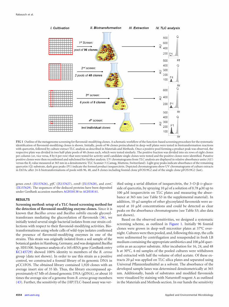
Based on the observed sensitivities, we designed a systematicscreening scheme, as outlined in Figure 1. Initially 96 fosmidclones were grown in deep-well microtiter plates at 37°C over-night. Cultures were then pooled, and, following this step, the cellswere sedimented by centrifugation and resuspended in fresh LBmedium containing the appropriate antibiotics and 100 �M quer-cetin as an acceptor substrate. After incubation for 16, 24, and 48h at 30°C, 4-ml samples of the pooled cultures were withdrawnand extracted with half the volume of ethyl acetate. Of these ex-tracts 20 �l was applied on TLC silica plates and separated usingUniversal Pflanzenlaufmittel as a solvent. The absorbance of thedeveloped sample lanes was determined densitometrically at 365nm. Additionally, bands of substrates and modified flavonoidswere visualized by staining with Naturstoff reagent A as outlinedin the Materials and Methods section. In our hands the sensitivity
FIG 1 Outline of the metagenome screening for flavonoid-modifying clones. A schematic workflow of the function-based screening procedure for the systematicidentification of flavonoid-modifying clones is shown. Initially, pools of 96 clones preincubated in deep-well plates were tested in biotransformation reactionswith quercetin, followed by culture extract TLC analysis as described in Materials and Methods. Once a positive pool forming a product peak was observed, therespective plate was divided in two half-plate pools of 48 clones each, which were tested similarly. The positive fraction was divided into six rows of eight clonesper column (or, vice versa, 8 by 6 per row) that were tested for activity until candidate single clones were tested and the positive clones were identified. Putativepositive clones were then reconfirmed and subcloned for further analysis. UV chromatograms from TLC analysis are displayed in relative absorbance units (AU)versus the Rf value measured at 365 nm in a densitometric TLC Scanner 3 (Camag, Muttenz, Switzerland). Light gray peaks indicate absorbance of the remainingquercetin (Q) substrate, dark gray peaks (P1) indicate the formed product isoquercitrin. Depicted chromatograms show UV chromatograms of culture extractsin EtOAc after 24-h biotransformations of pools with 96, 48, and 8 clones including fosmid clone pFOS19G2 and of the single clone pFOS19G2 (last).
Rabausch et al.
4554 aem.asm.org Applied and Environmental Microbiology

of the assay was high enough to detect a single flavonoid-modify-ing enzyme clone in a mixture of 96 clones (Fig. 1). After thedetection of a positive signal, we divided the 96 fosmid clones intopools of 48 to locate the same peak in one of the resulting twomicrotiter half-plates. Following this procedure, we divided the 48clones into six groups of eight clones (Fig. 1) and finally analyzedthe eight individual clones. This strategy was applied successfullyto identify six overlapping positive clones in the Bacillus sp.HH1500 fosmid library testing all 20 microtiter plates with a totalof 1,920 clones.
Of these six fosmid clones, one clone, pFOS4B2, of approxi-mately 46 kb was subcloned using the HindIII restriction site ofthe pBluescript II SK� vector. The obtained subclones were ana-lyzed using the above-mentioned TLC screening technology.Thereby, a positive subclone designated pSK4B2 was identifiedand completely sequenced (GenBank entries JX157885 toJX157887). Subclone pSK4B2 carried an insert of 3,225 bp (seeFig. S1A in the supplemental material) and harbored a gene, des-ignated mgtB, encoding a protein of 402 amino acids (aa). Theidentified ORF was subcloned, creating plasmid pDmgtB, andagain assayed for activity. TLC analysis clearly confirmed the gly-cosylation activity of the MgtB enzyme in this construct as well.The deduced amino acid sequence of MgtB (GenBank accessionnumber AGH18136) was highly similar to a predicted B. thurin-giensis macroside glycosyltransferase (Table 1). The mgtB-sur-rounding DNA sequences in plasmid pSK4B2 represented twotruncated genes that consistently were almost identical to genesfrom B. thuringiensis (Table 1). This phylogenetic relation was inaccordance to the preliminary sequence analysis of the 16S rRNAgene of Bacillus sp. HH1500 (see above).
These tests suggested that the screening procedure was suitablefor the functional screening of large-insert metagenome libraries.For the function-based screening of metagenomes, we termed thismethodology META, for metagenome extract TLC analysis. Al-though it is not a fully automated high-throughput screening(HTS) technology, META allows screening of about 1,200 clonesper TLC plate within a time of 48 h for preculture, biotransforma-tion, and analysis. This number of clones appeared to be feasible ifa single person did the screening. Generally, the sampling of aboutone TLC plate per hour by the ATS 4 is the time-limiting step ofthe method. But this still allows the pooled screening of severalplates a day and, hence, throughput of several thousand clones aday by META.
Identification of a novel glycosyltransferase from a meta-genome library. To further apply the screening for enzyme dis-
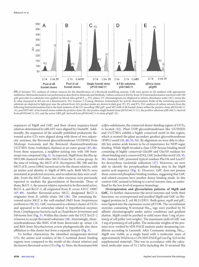
covery in metagenome libraries, we tested two fosmid librariesconstructed in our laboratory. One library was constructed fromDNA isolated from river Elbe sediment; the other was from DNAisolated from fresh elephant feces. Altogether both libraries en-compassed approximately 50,000 clones with an average insertsize of 35 kb. Both libraries were screened using quercetin as asubstrate. Using the described strategy, we discovered one positivemicrotiter plate pool in the river Elbe sediment library. Furtherscreening of this pool resulted in the identification of a singlepositive fosmid clone, designated pFOS144C11 (Fig. 2). Biotrans-formations of quercetin (Fig. 2A, Q) with 48 clone pools presentedone product peak (P2) by TLC separation with an Rf value com-parable to that of quercitrin, the quercetin-3-O-�-L-rhamnoside(Fig. 2A). A second peak (P3) with an Rf value higher than valuesfor the available reference quercetin glycones was observed in con-versions with the six-clone pool and the single fosmid clone(Fig. 2B and C, respectively). Clone pFOS144C11 carried a fosmidof approximately 40 kb. Subsequent restriction fragment subclon-ing into pBluescript II SK� with HindIII yielded the identificationof the positive E. coli DH5� subclone pSK144C11. However, bio-transformations with pSK144C11 showed two product peaks, amajor one (Fig. 2D, P2) with an Rf value comparable to that ofquercitrin and a minor one (P1) similar to isoquercitrin (Fig. 2D).The subclone pSK144C11 still had an insert of approximately 8.5kb in size. Further sequencing and subcloning of pSK144C11 fi-nally identified the gene putatively responsible for the modifica-tions, which we designated gtfC (see Fig. S1B in the supplementalmaterial). The deduced 459-aa sequence of the corresponding en-zyme revealed motif similarities to UDP-glucuronosyltransferase/UDP-glucosyltransferases. GtfC (GenBank entry AGH18139)showed a similarity of 71% to the putative glycosyltransferase ofthe Gram-negative bacterium Fibrisoma limi covering 92% of theprotein (Table 1). Further cloning of the gtfC ORF into the pDrivevector and biotransformation with E. coli DH5� carrying the re-spective construct pDgtfC confirmed the flavonoid-modifying ac-tivity of GtfC (Fig. 2E).
In summary, these results demonstrated that the developedscreening procedure, META, is sufficiently sensitive to allow theidentification of large-insert clones from individual bacterial ge-nomes (i.e., Bacillus sp. HH1500) and complex metagenome li-braries (i.e., the river Elbe sediment library) showing flavonoid-modifying activities.
Sequence-based classification of MgtB and GtfC. To analyzethe affiliation of MgtB and GtfC, we constructed a phylogenetictree using the MEGA, version 5, software (44). The amino acid
TABLE 1 ORFs identified on subclones pSK4B2, derived from the active Bacillus sp. HH1500 fosmid clone, and pSK144C11, derived from the riverElbe sediment active fosmid clone
Subcloneand ORF
Position(aa) Homolog (accession no.) Coverage (%) % identity % similarity
pSK4B2bspA 221 Putative protein kinase from Bacillus thuringiensis (EEM66464) 100 99 99mgtB 402 Macrolide glycosyltransferase from Bacillus thuringiensis (EEM96628) 100 98 99mgtC 261 Hypothetical membrane protein from Bacillus thuringiensis (EAO54527) 100 99 100
pSK144C11esmA 80 Putative UDP-N-acetylmuramate–L-alanine ligase from Niabella soli (EHP51575) 99 69 80gtfC 459 Putative UDP-glucosyltransferase from Fibrisoma limi (CCH52088) 92 51 71esmB 170 Hypothetical protein from Niastella koreensis (YP005009630) 95 63 77esmC 150 Putative membrane protein from Solitalea canadensis (YP006258217) 98 68 81
Metagenome Screening of Flavonoid-Modifying Enzymes
August 2013 Volume 79 Number 15 aem.asm.org 4555
sequences of MgtB and GtfC and their closest sequence-basedrelatives determined by pBLAST were aligned by ClustalW. Addi-tionally, the sequences of the actually published prokaryotic fla-vonoid-active GTs were aligned along with those of two eukary-otic enzymes, the flavonoid glucosyltransferase UGT85H2 fromMedicago truncatula and the flavonoid rhamnosyltransferaseUGT78D1 from Arabidopsis thaliana as an outer group (45, 46).From these sequences, a neighbor-joining tree with 100 boot-straps was computed (Fig. 3). As expected, MgtB from Bacillus sp.HH1500 clustered with other MGTs from the B. cereus group. Atthe time of writing, the MGT of B. thuringiensis IBL 200 and theMGT of B. cereus G9842 turned out to be the closest relatives, withan amino acid identity to MgtB of 98% each. Both MGTs wereannotated as predicted enzymes, and no substrate data were avail-able. From the MGT cluster, five other enzymes were previouslyreported to mediate the glucosylation of flavonoids. Three ofthem, BcGT-1, the nearest relative reported to be flavonoid active,BcGT-4, and BcGT-3, all originated from B. cereus ATCC 10987(47–49). Another flavonoid-active MGT, designated BsGT-3,originates from B. subtilis strain 168 (36). The remaining fla-vonoid-active MGT is the well-studied OleD from Streptomycesantibioticus (50, 51). GtfC was located in a distinct cluster of UGTsand appeared to be somewhat related to hypothetical enzymesfrom Cytophagaceae bacteria such as Dyadobacter fermentans andFibrisoma limi (Fig. 3). Within this cluster only the UGT XcGT-2is known to accept flavonoid substrates (38). Interestingly, rham-nosyltransferases like BSIG 4748 from Bacteroides sp. strain 116and RtfA from Mycobacterium avium phylogenetically also showaffiliation to this cluster but form a separate branch (Fig. 3).
To further characterize the identified metagenome-derivedGTs, the amino acid residues of the C-terminal donor bindingregions were compared to the motifs of the closest relatives andthe known flavonoid-active GTs (Fig. 4). Here, the Rossmann fold
�/�/� subdomain, the conserved donor-binding region of UGTs,is located (52). Plant UDP-glycosyltransferases like UGT85H2and UGT78D1 exhibit a highly conserved motif in this region,which is termed the plant secondary product glycosyltransferase(PSPG) motif (45, 46, 53, 54). By alignment we were able to iden-tify key amino acids known to be of importance for NDP-sugarbinding. While MgtB revealed a clear UDP-hexose binding motifconsisting of highly conserved Gln289 and Glu310 residues forribose binding and a conserved DQ, GtfC lacked this motif (45, 55,56). Instead, GtfC presented typical residues Phe336 and Leu357for deoxyribose nucleotide utilization (57). Moreover, we wereable to identify the pyrophosphate binding sites in the MgtBamino acid sequence (Fig. 4). However, GtfC does not possessthese conserved phosphate binding residues, suggesting that GtfCand related enzymes have another donor binding mode. In thiscontext GtfC seemed to belong to a novel enzyme class, as under-lined by the low level of sequence homology.
Overexpression and glycosylation patterns of MgtB andGtfC. To further characterize the novel enzymes and verify theirfunctions, we overexpressed and purified MgtB and GtfC as His-tagged proteins in E. coli BL21(DE3). Both genes, mgtB and gtfC,were ligated into the expression vector pET19b. The recombinantenzymes containing N-terminal His10 tags were purified by Niaffinity chromatography under native conditions and gradientelution. MgtB could be purified to yield more than 5 mg of pro-tein/g of cell pellet (wet weight). The maximum yield of GtfC was3 mg of protein/g of cell pellet. The molecular weights of the pro-teins were verified by SDS-PAGE analysis under denaturing con-ditions according to Laemmli. After Coomassie staining, His10-MgtB was visible as a single band with a molecular mass ofapproximately 50 kDa on a 12% SDS-PAGE gel (see Fig. S2A in thesupplemental material). This was in accordance with the calcu-lated molecular mass of 51.2 kDa including the N-terminal His
FIG 2 Iterative TLC analyses of culture extracts for the identification of a flavonoid-modifying enzyme. Cells were grown in LB medium with appropriateantibiotics. Biotransformation was performed as described in Materials and Methods. Culture extracts in EtOAc from 24-h biotransformation reactions with 100�M quercetin as a substrate were applied on Merck silica gel 60 F254 TLC plates. UV chromatograms are displayed in relative absorbance units (AU) versus theRf value measured at 365 nm on a densitometric TLC Scanner 3 (Camag, Muttenz, Switzerland) for activity determination. Peaks of the remaining quercetinsubstrate are depicted in light gray near the solvent front (Q); product peaks are shown in dark gray (P1, P2, and P3). TLC analyses of culture extracts from thefollowing biotransformations led to the final isolation of the GT-encoding ORF gtfC: pool MT144R of 48 fosmid clones within the positive clone pFOS144C11(A), pool MT144C of six fosmid clones within the positive clone (B), the positive single fosmid clone pFOS144C11 (C), the positive subclone pSK144C11 derivedfrom pFOS144C11 (D), and the active ORF gtfC derived from pFOS144C11 in clone pDgtfC (E).
Rabausch et al.
4556 aem.asm.org Applied and Environmental Microbiology

tag. His10-GtfC revealed a molecular mass of about 55 kDa on a12% SDS-PAGE gel, which was well in accordance with the calcu-lated molecular mass of 54.7 kDa including the N-terminal Histag. While virtually no additional bands were visible on SDS-PAGE gels with purified recombinant MgtB protein, some minorcontaminating bands were still visible on the SDS-PAGE gelloaded with purified GtfC (see Fig. S2B in the supplemental ma-terial). In summary, both proteins could be purified to allow fur-ther biochemical characterization.
The purified His10-MgtB protein was able to use UDP-�-D-glucose as a donor substrate. The recombinant enzyme catalyzedthe transfer of �-D-glucose residues to various polyphenols. Bio-catalytic reactions were performed with 500 �M UDP-�-D-glu-cose as a donor and 100 �M acceptor substrate. The followingflavonoids served as acceptor substrates and were modified withhigh yields: luteolin, quercetin, kaempferol, tiliroside, naringenin,and genistein (Table 2). Flavonols turned out to be the best accep-tor molecules. Generally, the conversion during a 2-h assay rangedfrom 52% for naringenin to approximately 100% for quercetinand kaempferol. Interestingly, in the presence of quercetin andkaempferol, no residual educts could be monitored by HPTLCanalysis. The specific educts and their observed glycones of the
biocatalytic reactions are summarized in Table 2 together with therespective Rf values. MgtB favored the glucosylation at the C-3hydroxy group, if accessible, as in the aglycone flavonols quercetinand kaempferol. Further, the C-7-OH was attacked and gluco-sylated by the enzyme, which could be shown for not only theflavone luteolin but also the flavanone naringenin and the isofla-vone genistein (Table 2). MgtB glucosylated luteolin also at theC-3= hydroxy group forming the 3=,7-di-O-glucoside of luteolin ifthe C-7-OH was glucosylated previously. Furthermore, MgtB cat-alyzed catalyzed the conversion of the kaempferol derivative tili-roside [kaempferol-3-(O-6�trans-p-coumaryl)-glucoside]. Oneglucosylated product with an Rf value of 0.54 was detected.
Finally, the chalcone xanthohumol and the stilbene t-resvera-trol were tested in biotransformation reactions with E. coli ex-pressing mgtB, but conversions were not quantified (data notshown). Xanthohumol yielded three detectable products, whereasthe biotransformation of t-resveratrol yielded one observed prod-uct by absorbance TLC analysis.
Tests with recombinant and purified GtfC using UDP-�-D-glu-cose and UDP-�-D-galactose and quercetin as an acceptor moleculesuggested that dTDP-activated sugar moieties were transferred bythis enzyme. This finding was confirmed by HPLC-ESI-MS analyses
FIG 3 Phylogenetic dendrogram of glycosyltransferases (GTs) related to the two GTs (MgtB and GtfC in black boxes) identified in this study. GTs known to acton flavonoids as acceptors are highlighted in gray. Phylogenetic analysis was conducted using MEGA, version 5 (44), with ClustalW sequence alignment in aBLOSUM protein weight matrix. The neighbor-joining tree was calculated using the bootstrap method with the Poisson model; bootstrap values higher than 75are indicated next to the branches. The scale represents the number of amino acid changes per residue. All GTs shown belong to the family of Gtf-like GT1 andare further subclassified into MGTs (macroside glycosyltransferases; TIGR01426), NGTs (N-glycosyltransferases), and UGT (a UDP-glucuronosyltransferase/-glucosyltransferase; PF00201) as indicated on the right. GenBank numbers were retrieved from NCBI as follows: YP002445489, MGT from B. cereus G9842;ZP04071678, MGT from B. thuringiensis IBL 200; AGH18136, MgtB from Bacillus sp. HH1500; AAP25969 Bacillus anthracis strain Ames; ABY43166, MGT fromBacillus weihenstephanensis KBAB4; NP978481, BcGT-1 from B. cereus ATCC 10987; NP979441, BcGT-4 from B. cereus ATCC 10987; AAS41737, BcGT-3 fromB. cereus ATCC 10987; NP389104, BsGT-3 from B. subtilis strain 168; ABA42119, OleD from S. antibioticus; AAM41712, XcGT-2 from X. campestris ATCC 33913;YP003086330, Dfer 1940 from Dyadobacter fermentans DSM 18053; AGH18139, GtfC from Elbe river sediment metagenome; CCH52088, UGT from Fibrisomalimi BUZ 3; YP003388759, Slin3970 from Spirosoma linguale DSM 74; ACV78946, Namu2594 from Nakamurella multipartita DSM 44233; YP712191,FRAAL1959 from Frankia alni ACN14a; AAN01207, RebG from Lechevalieria aerocolonigenes; ZP09941874, BSIG 4748 from Bacteroides sp. 116; AAC71702, RtfAfrom Mycobacterium avium; AFK05536, Emtol 0266 from Emticicia oligotrophica DSM 17448; 2PQ6A, UGT85H2 from Medicago truncatula; and NP564357,UGT78D1 from Arabidopsis thaliana.
Metagenome Screening of Flavonoid-Modifying Enzymes
August 2013 Volume 79 Number 15 aem.asm.org 4557

of biotransformation assays (see the following paragraph). Unfortu-nately, deoxyribose nucleotide-activated hexoses, e.g., dTDP-rham-noside, were commercially not available to further analyze the ob-tained reaction products in more detail (58).
Biotransformations with the E. coli strain expressing GtfC andusing various polyphenols as substrates yielded conversions rang-ing from 52% for xanthohumol up to almost 100% turnover formost flavonols tested (Table 3). Quercetin was transformed al-most completely after 4-h biotransformations and yielded threedetectable products (P1 to P3). To further characterize theseproducts, UV absorbance spectra were recorded and compared tothe reference glycones of quercetin isoquercitrin and quercitrin(59). P1 revealed an Rf value identical to the value of isoquercitrin.Further, the UV absorbance spectrum of P1 matched the spec-trum of isoquercitrin (see Fig. S3A in the supplemental material).P2 revealed an Rf value identical to the one known for quercitrin.P2 also exhibited the same UV absorbance spectrum as quercitrin(see Fig. S3B). P3 revealed an Rf value of 0.82, which clearly dif-fered from the Rf values of known and available quercetin gly-cones. Compared to isoquercitrin, P3 showed a similar hypso-chromic shift of band I to a maximum wavelength (max) of 363nm (see Fig. S3C); however, it revealed a less hypsochromic shiftin band II of only 5 nm to 272 nm with a shoulder at 280 nm. It isfurther notable that the HPLC-ESI-MS analysis of biotransforma-tion products of quercetin consistently identified three distinct
reaction products (see Fig. S4). P1 had a retention time (RT) of17.93 min in the HPLC analysis and revealed a molecular massof 464 Da, which is equivalent to isoquercitrin. P2 revealed an RTof 18.06 min and had a molecular mass of 448 Da. This masscorresponds well with the molecular mass of quercitrin. Finally,P3 with a RT of 18.31 min revealed a molecular mass of 446 Da,indicating the formation of a novel, not further characterized,quercetin glycoside.
Glycosylation patterns of GtfC on quercetin suggested a pref-erence to act on the C-3 hydroxy group mediating the transfer ofdifferent sugar residues. However, if a C-3-OH group was notavailable, GtfC efficiently catalyzed the glycosylation of other po-sitions. Flavones lacking the hydroxy function at C-3 were con-verted depending on the availability of other hydroxy groups. Pra-tol, which possesses only a single free C-7 hydroxy group, wasconverted weakly and yielded a single detectable product. Further,the biotransformation of 3=,4=-dihydroxyflavone yielded three de-tectable glycones, and 5-methoxy-eupatorin yielded two products(see Table S4 in the supplemental material; also data not shown);the biotransformation of the mono-4=-hydroxyflavanone yieldedone glycosylated product, and the glycosylation of naringeninyielded two products. The major biotransformation product ofnaringenin revealed the same Rf values and absorbance spectra asprunin, the naringenin-7-O-glucoside (Table 3). The second nar-ingenin glycone could not be further specified due to the lack of
FIG 4 ClustalW alignment of the MgtB and GtfC (black boxes) amino acid sequences, their nearest sequence-based relatives, and other flavonoid-active GTs(gray boxes). The C-terminal region of the Rossmann fold �/�/� subdomain, the conserved donor-binding region of UGTs, is shown. Plant UDP-glycosyltrans-ferases like UGT85H2 (2PQ6A) from Medicago truncatula and UGT78D1 (NP564357) from Arabidopsis thaliana exhibit the highly conserved plant secondaryproduct glycosyltransferase (PSPG) motif in this region. Amino acids are boxed according to their roles in donor nucleoside (blue), phosphate group (green), andhexose (red) binding, as reported in the literature (45, 52, 55–57, 73, 74). Dark and light green boxes refer to beta- and alpha-phosphate group binding of thedonor molecule, respectively. Analogous binding of the nucleoside part is indicated for base positions (light and full bright blue) and ribose positions (dark blue);for the latter, lighter variants of blue boxes differentiate deoxyribose-specific and ribose-specific positions. GenBank numbers were retrieved from NCBI (see thelegend of Fig. 3 for species identifications).
Rabausch et al.
4558 aem.asm.org Applied and Environmental Microbiology

commercially available reference substances. Altogether these re-sults suggested that GtfC acts on the C-3, C-3=, C-4=, and C-7hydroxy groups of the flavonoid backbone.
In summary these data demonstrated that MgtB and GtfC pos-
sess interesting biocatalytic properties. While MgtB specificallymediated the transfer of glucose residues, GtfC transferred differ-ent hexose moieties. MgtB was capable of catalyzing the gluco-sylation of already glycosylated flavonoids to form diglycosides
TABLE 2 Flavonoid substrates converted by recombinant MgtB in bioassays
Substrate
Conversion (%)a Rf valueb ProductbName Structure
Quercetin 100 0.79 —c
0.64 Isoquercitrin0.27 —0.25 —
Kaempferol 100 0.74 Astragalin0.35 —
Luteolin 82 0.65 Cynaroside
0.32 -3=,7-Di-O-Glc
Naringenin 52 0.76 Prunin
Genistein 72 0.69 Genistin
Tiliroside 83 0.54 —
a Reactions were carried out at 37°C for 2 h in triplicate using 1 ml of reaction mixture consisting of 500 �M UDP-glucose, a 100 �M concentration of the respective flavonoid, and5 �g/ml of purified and recombinant MgtB.b Rf values and products in bold indicate the main products of the biocatalytic reactions.c Products were not specified due to the lack of available reference substances.
Metagenome Screening of Flavonoid-Modifying Enzymes
August 2013 Volume 79 Number 15 aem.asm.org 4559

TABLE 3 Flavonoid substrates and products of biotransformation assays with recombinant GtfC
Substrate
Conversion (%)a Rf valueb ProductbName Structure
Luteolin 86 0.81 —c
0.73 —
0.68 —0.58 —
Quercetin 100 0.82 —
0.75 Quercitrin
0.64 Isoquercitrin
Kaempferol 100 0.85 —
0.80 —
0.68 Astragalin
Naringenin 76 0.87 —0.84 —
0.77 Prunin
Genistein 68 0.83 —0.76 —0.68 Genistin
t-Resveratrol 96 0.83 —0.77 —0.64 —0.58 —0.51 —0.46 —
Xanthohumol 52 0.85 —0.48 —
a Quantification of the reaction products was performed as stated in Materials and Methods. Triplicate reactions using 50 ml of reaction mixture were performed in 50 mM sodiumphosphate buffer, pH 7.0, containing 1% (wt/vol) glucose and 200 �M flavonoid at 30°C.b Rf values and products in bold indicate the main products of the biotransformation reactions.c Products were not specified due to the lack of available reference substances.
Rabausch et al.
4560 aem.asm.org Applied and Environmental Microbiology

(e.g., formation of luteolin-3=,7-di-O-glucoside) and even tiliro-side to generate novel glucosides not available from natural re-sources. In contrast, the glycosylation pattern of GtfC suggestedthe transfer of single sugar residues to only aglycone flavonoidforms. Interestingly, GtfC seemed to be very variable with respectto its activity at various positions on the flavonoid backbone. Thismay lead to the formation of truly novel flavonoids not availablenaturally. Hence, both enzymes might be helpful in the generationof new natural compounds.
DISCUSSION
Within the manuscript, we report on the development of a semi-automated TLC-based detection system for flavonoid-modifyingenzyme clones. The screening assay was highly reproducible andhighly sensitive. It allowed the detection of micromolar concen-trations of glycosylated flavonoids. Isoquercitrin was detectable ata 0.78 �M concentration in the assay (see Table S3 in the supple-mental material). Using this assay we were able to systematicallyidentify one positive clone out of pools of 96 metagenome clones(Fig. 1 and 2). To our knowledge this is the first published TLC-based screening method for functional searches in metagenomelibraries. We speculate that slight modifications of this screeningsystem could easily allow the detection of other flavonoid-modi-fying enzyme clones, for example, through acylation or methyl-ation reactions.
Using this novel screening technology, we identified a macro-side glycosyltransferase, MgtB, from a soil isolate (i.e., Bacillus sp.HH1500). A fosmid library established with DNA from this strain,which had been isolated from the local botanical garden only re-cently, was initially used to develop and verify the outlined screen-ing technology; and using the novel screening technology, MgtBwas quickly identified from a pool of almost 2,000 clones. Isola-tion and purification of recombinant MgtB revealed a novel MGT.MgtB shared 89% amino acid identity with BcGT-1 from B. cereusATCC 10987, the closest relative reported to act on flavonoids.BcGT-1 was reported to catalyze the glucosylation of flavones,flavonols, flavanones, and isoflavones (47). On flavonols BcGT-1acted on C-3, C-7, and C-4= hydroxy groups creating triglucosidesof kaempferol (48). In contrast, biocatalysis of kaempferol withMgtB yielded just two detectable glucosylated products. However,reactions with quercetin resulted in three detectable glycones.These data suggested that MgtB acted at the C-3=-OH group. Thishypothesis also was supported by the observation that recombi-nant MgtB converted luteolin to luteolin-3=,7-di-O-glucoside as aby-product. These results were in accordance with the glucosyla-tion pattern of BcGT-3, yet another MGT from B. cereus ATCC10987 (49). Interestingly, BcGT-3 shares only 40% amino acididentity with MgtB, but both enzymes act on the same flavonoids,forming diglucosides from flavones and flavonols at the same po-sitions and only monoglucosides from naringenin. The most spec-tacular conversion observed for MgtB was that of tiliroside. Theproduct is likely to be the 7-O-glucoside, taking the glycosylationpattern of MgtB into account. Tiliroside glycosides, however, havenot been reported in scientific literature. This raises the possibilityof the generation of new natural compounds. The natural sub-strates of Bacillus MGTs still have not been reported. Other MGTslike OleD usually detoxify macroside antibiotics but often possessbroad acceptor tolerance (35, 60).
The metagenome-derived GtfC turned out to be a completelynovel enzyme. Only seven flavonoid-active UGTs have been re-
ported so far that originate from five different prokaryotes (35, 36,38, 47, 49). With the exception of XcGT-2 from the Gram-nega-tive X. campestris ATCC 33913, all are MGT enzymes from Gram-positive Bacilli and Streptomycetes. MGTs play an important rolein xenobiotic defense mechanisms of prokaryotes and thus showbroad acceptor specificities (55, 60). This also applies to eukary-otic UGTs, pointing to a biological principle of detoxification(61). To our knowledge GtfC is the first metagenome-derived GTacting on flavonoids. Moreover, it is also the first bacterial enzymereported to transfer various dTDP-activated hexose sugars topolyphenols (see below), in contrast to the usually stringent donorspecificities of Gtf-like enzymes such as GtfD (57). With respect tothe notion that many NDP-sugars in prokaryotes are dTDP andnot UDP activated, GtfC might be a promising biocatalyst inglyco-diversification approaches (58, 62, 63). GtfC is similar topredicted GTs from Cytophagaceae bacteria (64–66). These Gram-negative bacteria have large genomes, suggesting extensive sec-ondary metabolic pathways, and they are well known for the pres-ence of resistance mechanisms to antibiotics such as trimethoprimand vancomycin (67, 68). As commonly known, glycosylation ofxenobiotics is a ubiquitous detoxification process in all kingdomsof life. The phylogenetically diverse members of Cytophagaceaehave only recently become an object of research, and concreteestimation of the phylogenetic breadth of this family and exacttaxonomic ranking still remain unclear (65, 69). Thus, the identi-fication of the metagenome-derived GtfC and its partial charac-terization suggest that this group of microorganisms is perhaps ahighly promising resource for novel GTs and also other enzymes.
A ClustalW alignment of the donor-binding region of GtfCsuggested that the activated donor substrates are of deoxy-thymi-dine nucleoside origin. GtfC possesses the typical amino acid res-idues Phe336 for thymine base stacking and hydrophobic Leu357for deoxyribose fitting (57). Concerning the donor binding ofGTs, GtfC appears not to exhibit the known amino acid residuesfor pyrophosphate binding (Fig. 4). Instead of the conserved res-idue His/Arg in the current solved protein structures, GtfC con-tains an Asn at amino acid position 349 (52, 70). This applies alsofor the nearest GtfC relatives Dfer1940, UGT of F. limi BUZ 3, andSlin3970 as well as the NGTs RebG and BSIG4748. Further, GtfCdoes not show the conserved Ser/Thr residue responsible for�-phosphate binding. Instead, the Gly354 appears to be of impor-tance for the �-phosphate binding, similar to structure of theOleD transferase (55).
The assumption of dTDP-activated cosubstrates used by GtfCwas supported by the observation that glucose, rhamnose, and athird sugar residue with a molecular weight of 446 were trans-ferred by GtfC in biotransformations using intact E. coli cells.Moreover, biocatalytic approaches with purified GtfC and eitherUDP-�-D-glucose or -galactose as a donor substrate failed. In bac-teria, the activated sugars, dTDP-�-D-glucose, -4-keto-6-deoxy-�-D-glucose or -4-keto-�-L-rhamnose, and -�-L-rhamnose, arepart of the dTDP-sugar biosynthesis pathway and are present in E.coli (71). Moreover, levels of dTDP-sugars are allosterically regu-lated by dTDP-rhamnose levels through activity of RmlA (72).
In summary, the screening protocol described in this report isa very helpful tool for the identification of truly novel enzymes forthe modifications of flavonoids and related substrates. In thisstudy, we have used the technology to identify two novel fla-vonoid-modifying enzymes. Both of these enzymes would per-haps not have been detected without the above-developed screen-
Metagenome Screening of Flavonoid-Modifying Enzymes
August 2013 Volume 79 Number 15 aem.asm.org 4561

ing technology. The partial biochemical characterizations usingeither biocatalysis or biotransformation suggest that MgtB andGtfC are both very interesting enzymes with high potential forbiotechnological applications with respect to flavonoid modifica-tions on an industrial scale. Thus, future work will now refine thistechnology to also identify other enzymes linked to flavonoidmodifications.
ACKNOWLEDGMENTS
This work was funded by the Ministry of Education and ResearchBundesministerium für Bildung und Forschung (BMBF) within the net-work program Biokatalyse2021 as project P18 (FKZ 0315166C).
The Bacillus Genetic Stock Center (Columbus, OH, USA) kindly pro-vided Bacillus cereus ATCC 10987 used as a positive control.
REFERENCES1. Perner M, Ilmberger N, Köhler HU, Chow J, Streit WR. 2011.
Emerging fields in functional metagenomics and its industrial rele-vance: overcoming limitations and redirecting the search for sovel bio-catalysts, p 483– 498. In de Bruijn FJ (ed), Handbook of molecularmicrobial ecology II: metagenomics in different habitats. John Wiley &Sons, Inc., Hoboken, NJ.
2. Schmeisser C, Steele H, Streit WR. 2007. Metagenomics, biotechnologywith non-culturable microbes. Appl. Microbiol. Biotechnol. 75:955–962.
3. Iqbal HA, Feng Z, Brady SF. 2012. Biocatalysts and small moleculeproducts from metagenomic studies. Curr. Opin. Chem. Biol. 16:109 –116.
4. Simon C, Daniel R. 2011. Metagenomic analyses: past and future trends.Appl. Environ. Microbiol. 77:1153–1161.
5. Daniel R. 2005. The metagenomics of soil. Nat. Rev. Microbiol. 3:470 –478.
6. Streit WR, Daniel R, Jaeger K-E. 2004. Prospecting for biocatalysts anddrugs in the genomes of non-cultured microorganisms. Curr. Opin. Bio-technol. 15:285–290.
7. Warren RL, Freeman JD, Levesque RC, Smailus DE, Flibotte S, HoltRA. 2008. Transcription of foreign DNA in Escherichia coli. Genome Res.18:1798 –1805.
8. Uchiyama T, Miyazaki K. 2009. Functional metagenomics for enzymediscovery: challenges to efficient screening. Curr. Opin. Biotech. 20:616 –622.
9. Ferrer M, Beloqui A, Timmis KN, Golyshin PN. 2009. Metagenomics formining new genetic resources of microbial communities. J. Mol. Micro-biol. Biotechnol. 16:109 –123.
10. Tuffin M, Anderson D, Heath C, Cowan DA. 2009. Metagenomic genediscovery: how far have we moved into novel sequence space? Biotechnol.J. 4:1671–1683.
11. Simon C, Daniel R. 2009. Achievements and new knowledge unraveledby metagenomic approaches. Appl. Microbiol. Biotechnol. 85:265–276.
12. Steele HL, Jaeger KE, Daniel R, Streit WR. 2009. Advances in recovery ofnovel biocatalysts from metagenomes. J. Mol. Microbiol. Biotechnol. 16:25–37.
13. Taupp M, Mewis K, Hallam SJ. 2011. The art and design of functionalmetagenomic screens. Curr. Opin. Biotechnol. 22:465– 472.
14. Beloqui A, Pita M, Polaina J, Martinez-Arias A, Golyshina OV, Zumar-raga M, Yakimov MM, Garcia-Arellano H, Alcalde M, Fernandez VM,Elborough K, Andreu JM, Ballesteros A, Plou FJ, Timmis KN, Ferrer M,Golyshin PN. 2006. Novel polyphenol oxidase mined from a metagenomeexpression library of bovine rumen: biochemical properties, structuralanalysis, and phylogenetic relationships. J. Biol. Chem. 281:22933–22942.
15. Collier AC, Tingle MD, Keelan JA, Paxton JW, Mitchell MD. 2000. Ahighly sensitive fluorescent microplate method for the determination ofUDP-glucuronosyl transferase activity in tissues and placental cell lines.Drug Metab. Dispos. 28:1184 –1186.
16. DeSantis G, Zhu Z, Greenberg WA, Wong K, Chaplin J, Hanson SR,Farwell B, Nicholson LW, Rand CL, Weiner DP, Robertson DE, BurkMJ. 2002. An enzyme library approach to biocatalysis: development ofnitrilases for enantioselective production of carboxylic acid derivatives. J.Am. Chem. Soc. 124:9024 –9025.
17. Knietsch A, Waschkowitz T, Bowien S, Henne A, Daniel R. 2003.Metagenomes of complex microbial consortia derived from different soils
as sources for novel genes conferring formation of carbonyls from short-chain polyols on Escherichia coli. J. Mol. Microbiol. Biotechnol. 5:46 –56.
18. Schipper C, Hornung C, Bijtenhoorn P, Quitschau M, Grond S, StreitWR. 2009. Metagenome-derived clones encoding two novel lactonasefamily proteins involved in biofilm inhibition in Pseudomonas aeruginosa.Appl. Environ. Microbiol. 75:224 –233.
19. Uchiyama T, Abe T, Ikemura T, Watanabe K. 2005. Substrate-inducedgene-expression screening of environmental metagenome libraries forisolation of catabolic genes. Nat. Biotechnol. 23:88 –93.
20. Williamson LL, Borlee BR, Schloss PD, Guan C, Allen HK, HandelsmanJ. 2005. Intracellular screen to identify metagenomic clones that induce orinhibit a quorum-sensing biosensor. Appl. Environ. Microbiol. 71:6335–6344.
21. Ververidis F, Trantas E, Douglas C, Vollmer G, Kretzschmar G, Pano-poulos N. 2007. Biotechnology of flavonoids and other phenylpropanoid-derived natural products. Part II: reconstruction of multienzyme path-ways in plants and microbes. Biotechnol. J. 2:1235–1249.
22. Schütz K, Muks E, Carle R, Schieber A. 2006. Quantitative determina-tion of phenolic compounds in artichoke-based dietary supplements andpharmaceuticals by high-performance liquid chromatography. J. Agric.Food Chem. 54:8812– 8817.
23. Leonard E, Yan Y, Fowler ZL, Li Z, Lim CG, Lim KH, Koffas MA. 2008.Strain improvement of recombinant Escherichia coli for efficient produc-tion of plant flavonoids. Mol. Pharm. 5:257–265.
24. Wang M, Liang CP, Wu QL, Simon JE, Ho CT (ed.). 2006. Instrumentalanalysis of popular botanical products in the U.S. market, p. 25–38. InHerbs: challenges in chemistry and biology. American Chemical Society,Washington, DC.
25. Manach C, Scalbert A, Morand C, Remesy C, Jimenez L. 2004. Poly-phenols: food sources and bioavailability. Am. J. Clin. Nutr. 79:727–747.
26. Das S, Rosazza JP. 2006. Microbial and enzymatic transformations offlavonoids. J. Nat. Prod. 69:499 –508.
27. Graefe EU, Wittig J, Mueller S, Riethling AK, Uehleke B, Drewelow B,Pforte H, Jacobasch G, Derendorf H, Veit M. 2001. Pharmacokineticsand bioavailability of quercetin glycosides in humans. J. Clin. Pharmacol.41:492– 499.
28. Kren V, Martinkova L. 2001. Glycosides in medicine: the role of glyco-sidic residue in biological activity. Curr. Med. Chem. 8:1303–1328.
29. Coutinho PM, Deleury E, Davies GJ, Henrissat B. 2003. An evolvinghierarchical family classification for glycosyltransferases. J. Mol. Biol. 328:307–317.
30. Bowles D, Lim EK, Poppenberger B, Vaistij FE. 2006. Glycosyltrans-ferases of lipophilic small molecules. Annu. Rev. Plant Biol. 57:567–597.
31. Mackenzie PI, Owens IS, Burchell B, Bock KW, Bairoch A, Belanger A,Fournel-Gigleux S, Green M, Hum Iyanagi DWT, Lancet D, Louisot P,Magdalou J, Chowdhury JR, Ritter JK, Schachter H, Tephly TR, TiptonKF, Nebert DW. 1997. The UDP glycosyltransferase gene superfamily:recommended nomenclature update based on evolutionary divergence.Pharmacogenetics 7:255–269.
32. Lairson LL, Henrissat B, Davies GJ, Withers SG. 2008. Glycosyltrans-ferases: structures, functions, and mechanisms. Annu. Rev. Biochem. 77:521–555.
33. Osmani SA, Bak S, Møller BL. 2009. Substrate specificity of plant UDP-dependent glycosyltransferases predicted from crystal structures and ho-mology modeling. Phytochemistry 70:325–347.
34. Breton C, Snajdrova L, Jeanneau C, Koca J, Imberty A. 2006. Structuresand mechanisms of glycosyltransferases. Glycobiology 16:29R–37R.
35. Yang M, Proctor MR, Bolam DN, Errey JC, Field RA, Gilbert HJ, DavisBG. 2005. Probing the breadth of macrolide glycosyltransferases: in vitroremodeling of a polyketide antibiotic creates active bacterial uptake andenhances potency. J. Am. Chem. Soc. 127:9336 –9337.
36. Jeon Y, Kim B, Kim J, Cheong Y, Ahn J-H. 2009. Enzymatic glycosyla-tion of phenolic compounds using BsGT-3. J. Korean Soc. Appl. Biol.Chem. 52:98 –101.
37. Rao KV, Weisner NT. 1981. Microbial transformation of quercetin byBacillus cereus. Appl. Environ. Microbiol. 42:450 – 452.
38. Kim HJ, Kim BG, Kim JA, Park Y, Lee YJ, Lim Y, Ahn JH. 2007.Glycosylation of flavonoids with E. coli expressing glycosyltransferasefrom Xanthomonas campestris. J. Microbiol. Biotechnol. 17:539 –542.
39. Zhou J, Bruns MA, Tiedje JM. 1996. DNA recovery from soils of diversecomposition. Appl. Environ. Microbiol. 62:316 –322.
40. Ilmberger N, Meske D, Juergensen J, Schulte M, Barthen P, RabauschU, Angelov A, Mientus M, Liebl W, Schmitz RA, Streit WR. 2012.
Rabausch et al.
4562 aem.asm.org Applied and Environmental Microbiology

Metagenomic cellulases highly tolerant towards the presence of ionic liq-uids-linking thermostability and halotolerance. Appl. Microbiol. Biotech-nol. 95:135–146.
41. Wagner H, Bladt S, Zgainski EM. 1983. Drogenanalyse, Dünnschichtchro-matographische Analyse von Arzneidrogen. Springer, Berlin, Germany.
42. Neu R. 1957. Chelate von Diarylborsäuren mit aliphatischen Oxyalkyl-aminen als Reagenzien für den Nachweis von Oxyphenyl-benzo-ã-pyronen. Naturwissenschaften 44:181–182.
43. Tourasse NJ, Helgason E, Oslash OA, Hegna IK, Kolstø AB. 2006. TheBacillus cereus group: novel aspects of population structure and genomedynamics. J. Appl. Microbiol. 101:579 –593.
44. Tamura K, Peterson PDN, Stecher G, Nei M, Kumar S. 2011. MEGA5:molecular evolutionary genetics analysis using maximum likelihood, evo-lutionary distance, and maximum parsimony methods. Mol. Biol. Evol.28:2731–2739.
45. Li L, Modolo LV, Escamilla-Trevino LL, Achnine L, Dixon RA, WangX. 2007. Crystal structure of Medicago truncatula UGT85H2-insights intothe structural basis of a multifunctional (iso)flavonoid glycosyltrans-ferase. J. Mol. Biol. 370:951–963.
46. Jones P, Messner B, Nakajima J, Schäffner AR, Saito K. 2003. UGT73C6and UGT78D1, glycosyltransferases involved in flavonol glycoside biosyn-thesis in Arabidopsis thaliana. J. Biol. Chem. 278:43910 – 43918.
47. Hyung Ko JH, Gyu Kim B, Joong-Hoon A. 2006. Glycosylation offlavonoids with a glycosyltransferase from Bacillus cereus. FEMS Micro-biol. Lett. 258:263–268.
48. Jung NR, Joe EJ, Kim BG, Ahn BC, Park JC, Chong Y, Ahn JH. 2010.Change of Bacillus cereus flavonoid O-triglucosyltransferase into fla-vonoid O-monoglucosyltransferase by error-prone polymerase chain re-action. J. Microbiol. Biotechnol. 20:1393–1396.
49. Ahn BC, Kim BG, Jeon YM, Lee EJ, Lim Y, Ahn JH. 2009. Formation offlavone di-O-glucosides using a glycosyltransferase from Bacillus cereus. J.Microbiol. Biotechnol. 19:387–390.
50. Choi SH, Ryu M, Yoon YJ, Kim DM, Lee EY. 2012. Glycosylation ofvarious flavonoids by recombinant oleandomycin glycosyltransferasefrom Streptomyces antibioticus in batch and repeated batch modes. Bio-technol. Lett. 34:499 –505.
51. Williams GJ, Zhang C, Thorson JS. 2007. Expanding the promiscuity ofa natural-product glycosyltransferase by directed evolution. Nat. Chem.Biol. 3:657– 662.
52. Hu Y, Walker S. 2002. Remarkable structural similarities between diverseglycosyltransferases. Chem. Biol. 9:1287–1296.
53. Hughes J, Hughes MA. 1994. Multiple secondary plant product UDP-glucose glucosyltransferase genes expressed in cassava (Manihot esculentaCrantz) cotyledons. DNA Seq. 5:41– 49.
54. Paquette S, Moller BL, Bak S. 2003. On the origin of family 1 plantglycosyltransferases. Phytochemistry 62:399 – 413.
55. Bolam DN, Roberts S, Proctor MR, Turkenburg JP, Dodson EJ, Mar-tinez-Fleites C, Yang M, Davis BG, Davies GJ, Gilbert HJ. 2007. Thecrystal structure of two macrolide glycosyltransferases provides a blue-print for host cell antibiotic immunity. Proc. Natl. Acad. Sci. U. S. A.104:5336 –5341.
56. Offen W, Martinez-Fleites C, Yang M, Kiat-Lim E, Davis BG, TarlingCA, Ford CM, Bowles DJ, Davies GJ. 2006. Structure of a flavonoidglucosyltransferase reveals the basis for plant natural product modifica-tion. EMBO J. 25:1396 –1405.
57. Mulichak AM, Lu W, Losey HC, Walsh CT, Garavito RM. 2004. Crystalstructure of vancosaminyltransferase GtfD from the vancomycin biosyn-thetic pathway: interactions with acceptor and nucleotide ligands. Bio-chemistry 43:5170 –5180.
58. Lim E-K, Ashford DA, Bowles DJ. 2006. The synthesis of small-molecule
rhamnosides through the rational design of a whole-cell biocatalysis sys-tem. ChemBioChem 7:1181–1185.
59. Mabry TJ, Markham KR, Thomas MB. 1970. The systematic identifica-tion of flavonoids. Springer-Verlag, New York, NY.
60. Gantt RW, Goff RD, Williams GJ, Thorson JS. 2008. Probing theaglycon promiscuity of an engineered glycosyltransferase. Angew. Chem.Int. Ed. Engl. 47:8889 – 8892.
61. Vogt T, Jones P. 2000. Glycosyltransferases in plant natural productsynthesis: characterization of a supergene family. Trends Plant Sci. 5:380 –386.
62. Williams GJ, Gantt RW, Thorson JS. 2008. The impact of enzyme engi-neering upon natural product glycodiversification. Curr. Opin. Chem.Biol. 12:556 –564.
63. Yoon JA, Kim BG, Lee WJ, Lim Y, Chong Y, Ahn JH. 2012. Productionof a novel quercetin glycoside through metabolic engineering of Esche-richia coli. Appl. Environ. Microbiol. 78:4256 – 4262.
64. Filippini M, Kaech A, Ziegler U, Bagheri HC. 2011. Fibrisoma limi gen.nov., sp. nov., a filamentous bacterium isolated from tidal flats. Int. J. Syst.Evol. Microbiol. 61:1418 –1424.
65. Lail K, Sikorski J, Saunders E, Lapidus A, Glavina Del Rio T, CopelandA, Tice H, Cheng JF, Lucas S, Nolan M, Bruce D, Goodwin L, PitluckS, Ivanova N, Mavromatis K, Ovchinnikova G, Pati A, Chen A,Palaniappan K, Land M, Hauser L, Chang YJ, Jeffries CD, Chain P,Brettin T, Detter JC, Schutze A, Rohde M, Tindall BJ, Goker M, BristowJ, Eisen JA, Markowitz V, Hugenholtz P, Kyrpides NC, Klenk HP, ChenF. 2010. Complete genome sequence of Spirosoma linguale type strain (1).Stand. Genomic Sci. 2:176 –185.
66. Lang E, Lapidus A, Chertkov O, Brettin T, Detter JC, Han C, CopelandA, Glavina Del Rio T, Nolan M, Chen F, Lucas S, Tice H, Cheng JF,Land M, Hauser L, Chang YJ, Jeffries CD, Kopitz M, Bruce D, GoodwinL, Pitluck S, Ovchinnikova G, Pati A, Ivanova N, Mavrommatis K,Chen A, Palaniappan K, Chain P, Bristow J, Eisen JA, Markowitz V,Hugenholtz P, Goker M, Rohde M, Kyrpides NC, Klenk HP. 2009.Complete genome sequence of Dyadobacter fermentans type strain(NS114). Stand. Genomic Sci. 1:133–140.
67. Chelius MK, Triplett EW. 2000. Dyadobacter fermentans gen. nov., sp.nov., a novel gram-negative bacterium isolated from surface-sterilized Zeamays stems. Int. J. Syst. Evol. Microbiol. 50:751–758.
68. Finster KW, Herbert RA, Lomstein BA. 2009. Spirosoma spitsbergense sp.nov. and Spirosoma luteum sp. nov., isolated from a high Arctic permafrostsoil, and emended description of the genus Spirosoma. Int. J. Syst. Evol.Microbiol. 59:839 – 844.
69. Filippini M, Svercel M, Laczko E, Kaech A, Ziegler U, Bagheri HC.2008. 2011. Fibrella aestuarina gen. nov., sp. nov., a filamentous bacteriumof the family Cytophagaceae isolated from a tidal flat, and emended de-scription of the genus Rudanella Weon et al. 2008. Int. J. Syst. Evol. Mi-crobiol. 61:184 –189.
70. Ha S, Gross B, Walker S. 2001. E. coli MurG: a paradigm for a superfamilyof glycosyltransferases. Curr. Drug Targets Infect. Disord. 1:201–213.
71. Samuel G, Reeves P. 2003. Biosynthesis of O-antigens: genes and path-ways involved in nucleotide sugar precursor synthesis and O-antigen as-sembly. Carbohydr. Res. 338:2503–2519.
72. Giraud MF, Naismith JH. 2000. The rhamnose pathway. Curr. Opin.Struct. Biol. 10:687– 696.
73. Modolo LV, Li L, Pan H, Blount JW, Dixon RA, Wang X. 2009. Crystalstructures of glycosyltransferase UGT78G1 reveal the molecular basis forglycosylation and deglycosylation of (iso)flavonoids. J. Mol. Biol. 392:1292–1302.
74. Shao H, He X, Achnine L, Blount JW, Dixon RA, Wang X. 2005. Crystalstructures of a multifunctional triterpene/flavonoid glycosyltransferasefrom Medicago truncatula. Plant Cell 17:3141–3154.
Metagenome Screening of Flavonoid-Modifying Enzymes
August 2013 Volume 79 Number 15 aem.asm.org 4563