metabolomics and microbiome profiling as biomarkers in
TRANSCRIPT

Metabolomics and microbiome profiling as biomarkers inobstructive sleep apnoea: a comprehensive review
Xiaoman Zhang1,2,3,4, Shengming Wang1,2,3,4, Huajun Xu1,2,3, Hongliang Yi1,2,3, Jian Guan1,2,3 andShankai Yin1,2,3
1Dept of Otolaryngology Head and Neck Surgery & Center of Sleep Medicine, Shanghai Jiao Tong University Affiliated Sixth People’sHospital, Shanghai, China. 2Shanghai Key Laboratory of Sleep Disordered Breathing, Shanghai, China. 3Otolaryngological Institute ofShanghai Jiao Tong University, Shanghai, China. 4Both authors contributed equally.
Corresponding author: Huajun Xu ([email protected])
Shareable abstract (@ERSpublications)Unique alterations in metabolism and the microbiome play an integral role in the pathophysiologyof OSA and OSA-induced cardiovascular complications https://bit.ly/3mW2rD5
Cite this article as: Zhang X, Wang S, Xu H, et al. Metabolomics and microbiome profiling asbiomarkers in obstructive sleep apnoea: a comprehensive review. Eur Respir Rev 2021; 30: 200220[DOI: 10.1183/16000617.0220-2020].
AbstractIntroduction Obstructive sleep apnoea (OSA) is a common sleep disorder with a high social andeconomic burden. Thus, early prediction and diagnosis of OSA are important. Changes in metabolism andthe microbiome may serve as biomarkers for OSA. Herein, we review the literature on the metabolomicand microbiome changes associated with OSA, and identify the metabolites and microorganisms involved.Methods We searched the PUBMED and EMBASE electronic databases using the following terms:“obstructive sleep apnea”, “OSA”, “sleep disordered breathing”, “SDB”, “intermittent hypoxia”, “sleepfragmentation”, and either “metabolomics” or “microbiome”. In total, 273 papers were identified, of which28 were included in our study.Results Changes in the levels of certain metabolites related to fatty acid, carbohydrate and amino acidmetabolism were associated with the incidence of OSA. The diversity and abundance of microflora,particularly Firmicutes and Bacteroidetes, were altered in humans and rodents with OSA.Conclusions Certain changes in metabolism and the microbiota play an integral role in thepathophysiology of OSA and OSA-induced cardiovascular complications. Metabolomic and microbiomebiomarkers shed light on the pathogenesis of OSA, and facilitate early diagnosis and treatment.
IntroductionObstructive sleep apnoea (OSA) is the most common form of sleep disordered breathing and is caused bycollapse and obstruction of the upper airway during sleep. It has been estimated that 425 million adults(aged 30–69 years) worldwide have moderate to severe OSA [1]. Patients with OSA are more likely tosnore, and show sleep structure disorder and daytime sleepiness, which decrease sleep quality and reduceperformance at work. OSA has been associated with an increased risk of cardiovascular, metabolic andcognitive disorders [2–4]. Some evidence indicates that OSA is associated with patients with impaired lipidmetabolism; in particular, hyper-low-density lipoprotein cholesterol levels are independently correlatedwith OSA [5, 6]. In summary, OSA has a significant impact on individual health and imposes a largeburden on society.
The gold standard for diagnosing OSA is overnight polysomnography (PSG) [7]. However, PSG isexpensive and time consuming, which hinders its utility for the prevention, early diagnosis and treatmentof OSA. Thus, identifying biomarkers for OSA has been the focus of research for more than a decade. Thedevelopment of metabolomics and metagenomics technologies has drawn more attention to OSA.Metabolomics involves qualitative and quantitative analysis of small metabolites (<1500 Da) [8], andprovides metabolic information. Metabolomics directly reflects the activity of metabolic pathwaysoccurring at a particular moment, and can amplify small changes controlled at the level of the genome and/
Copyright ©The authors 2021
This version is distributed underthe terms of the CreativeCommons AttributionNon-Commercial Licence 4.0.For commercial reproductionrights and permissions [email protected]
Received: 8 July 2020Accepted: 3 Dec 2020
https://doi.org/10.1183/16000617.0220-2020 Eur Respir Rev 2021; 30: 200220
EUROPEAN RESPIRATORY REVIEWREVIEW
X. ZHANG ET AL.

or proteome [9]. Metabolomics studies can be divided into targeted and nontargeted types. In targetedmetabolomics, predetermined components of one or several biofluids are analysed, whereas nontargetedmetabolomics analyses all metabolic components in a specific biological sample, which is usually used tofind new biomarkers [10]. There are three platforms for nontargeted metabolomics: nuclear magneticresonance (NMR) imaging; gas chromatography-mass spectrometry; and liquid chromatography-massspectrometry. The role of the microbiome in the development of disease should also not be underestimated.The activities of the microbiota are readily affected by environmental stimuli and have been linked to hostimmunity and metabolism [11]. Progress in the microbiome and metabolomics studies could reveal thepathological processes underlying OSA and thus inform diagnostic methods. In this study, we reviewstudies on the metabolism and microflora of OSA in humans and rodents.
MethodsThe PUBMED and EMBASE electronic databases were searched for relevant studies using the keywords“obstructive sleep apnea”, “OSA”, “sleep disordered breath”, “SDB”, “intermittent hypoxia” and “sleepfragmentation” together with “metabolomics”. This search identified 115 articles. After a selection process,10 articles were finally included.
Similarly, the same keywords “obstructive sleep apnea”, “OSA”, “sleep disordered breath”, “SDB”,“intermittent hypoxia” and “sleep fragmentation” together with “microbiome” were used for searching anda total of 162 papers were identified. After selection, 18 articles were finally adopted from this search.
ResultsThe search identified 273 articles which was reduced to 228 after removing duplicates. After reviewing thetitles and abstracts, a further 150 studies that did not address the topic of our review were excluded. Thefull texts of the remaining 78 studies were screened, and the studies that did not meet the search criteriawere excluded. The exclusion criteria were: 1) any nonoriginal research articles (e.g. evaluation articles andconference reports); 2) any review; 3) ongoing research; and 4) research articles that used a repeated studypopulation. The final number of original articles included in this review was 28. A flowchart of the studyselection process is presented in figure 1.
OSA and metabolomic biomarkersAnimal modelsIntermittent hypoxia (IH) plays a pivotal role in the pathophysiology of OSA. In fact, most rodent studieshave used IH as a model of OSA. CONOTTE et al. [12] used NMR imaging for urinary metabolomicprofiling in a mouse model of IH. They found increased levels of lactate and trans-aconitate, and decreasedlevels of pyruvate, citrate, succinate and acetoacetate, indicating that the energy metabolic pathways of IHmice were predominately anaerobic metabolism. Oxidative stress is another pathological feature of OSA[13] and has been confirmed in IH mice (reflected in increased levels of oxidation products, such asallantoin and trimethylamine oxide (TMAO)) [12]. CONOTTE et al. [12] suggested that hypoxia-inducedoxidative stress might promote changes in metabolism in OSA.
Records identified through database searching
(n=111): PubMed: n=55; EMBASE: n= 56
Records excluded after reviewing
titles and abstracts (n=62)
Records identified through database searching (n=162):
PubMed: n=76; EMBASE: n=86
Records after duplicates removed
(n=135)
Records excluded after reviewing
titles and abstracts (n=88)
Review n=17
Overlapping population n=4
Ongoing research n=1
Not original article n=7
Full-text studies assessed for
eligibility (n=47)
18 studies were included in
Microbiome part
Review n=18
Ongoing research n=1
Not original article n=2
Records after duplicates removed
(n=93)
Full-text studies assessed for
eligibility (n=31)
10 studies were included in
Metabolomics part
Inclu
de
dE
lig
ibil
ity
Scre
en
ing
Ide
nti
fica
tio
n
Inclu
de
dE
lig
ibil
ity
Scre
en
ing
Ide
nti
fica
tio
n
a) b)
FIGURE 1 Flowchart of selecting literature in a) metabolomics and b) microbiome.
https://doi.org/10.1183/16000617.0220-2020 2
EUROPEAN RESPIRATORY REVIEW OBSTRUCTIVE SLEEP APNOEA | X. ZHANG ET AL.

Sleep fragmentation (i.e. frequent brief awakenings), is another key feature of OSA. YOON et al. [14]explored hippocampal metabolite profiles in rats with sleep fragmentation to understand the mechanism ofthe associated neurocognitive dysfunction. Metabolites in the hippocampus related to excitatoryneurotransmitters, such as glutamate and aspartate, were present in significantly smaller quantities in thechronic sleep fragmentation group, possibly as a protective mechanism against the excitatory toxicityassociated with frequent awakening. Precursors of acetylcholine (methionine and choline), an importantneurotransmitter in brain function, were also below normal levels in the chronic sleep fragmentation rats.These findings may explain why sleep fragmentation affects memory and learning ability.
Human studiesSerum/plasma, urine, saliva and faecal extract can be analysed in human metabolomics studies. Analysis ofdifferent biofluids is useful for understanding different metabolic changes, as summarised in the followingsections.
UrineAn NMR imaging study by ZĄBEK et al. [15] revealed that 10 urinary metabolites were better able todistinguish between OSA and COPD than analysis of two or three different biofluids, although theseresults have been questioned in terms of specificity [16]. Also, data on smoking status, disease severity andCOPD as a comorbidity are needed to allow comparison among OSA studies.
XU et al. [17] compared metabolite levels among simple snorers, OSA patients and healthy subjects. Atotal of 21 and 31 metabolites in simple snorers and OSA subjects showed differences, respectively, fromnormal controls. Most fatty acids and fatty acid-related compounds (e.g. 2,4-dihydroxybutyric acid,3,4-dihydrxoybutyric acid, pentanoic acid and glyceraldehyde), which are associated with abnormallipolysis in cases of IH, were elevated in OSA subjects compared with controls. Glycolytic intermediatesassociated with ATP production, and branch chain amino acids (BCAAs) associated with mitochondrialdysfunction, were also elevated in OSA subjects compared with controls. BCAAs, such as leucine,isoleucine and valine are important biomarkers of insulin resistance, and accumulation is associated withthe risk of type 2 diabetes mellitus (T2DM) [18]. Hence, metabolic profiles overlap between chronicinflammatory and metabolic diseases. Intestinal flora-related metabolites, such as TMAO andglycochenodeoxycholate-3-sulfate, show significantly increased expression in subjects with OSA [17].Individuals with OSA can be distinguished from healthy individuals based on the expression levels of sixmetabolites (4-hydroxypentenoic acid, arabinose, glycochenodeoxycholate-3-sulfate, isoleucine, serine andxanthine) with 75% sensitivity and 78% specificity [17].
Meta-analyses have shown that the risk of adverse cardiovascular effects increases in those with moderateto severe OSA [19, 20]. Stroke and coronary artery disease have a dose–response relationship with OSAseverity [21, 22]. Although the mechanism of the association between OSA and cardiovascular disease isnot fully understood, neurohormonal dysregulation, metabolic abnormalities, and inflammation may beresponsible for cardiovascular events in patients with OSA [23]. There is a complex but strong associationbetween OSA and cardiovascular disease and certain biomarkers of cardiovascular disease could assist inthe clinical diagnosis and prediction of OSA-induced cardiovascular diseases, especially through cell androdent experiments. Targeted metabolomics identified three urinary metabolites, namely long-chainacylcarnitine (C14:1) and the biogenic amines of symmetric dimethylarginine (SDMA) and sphingomyelin(C18:1), which could be used as predictive biomarkers of OSA [24]. Changes in acylcarnitine levelsindicate disordered lipid metabolism and an imbalance of β-oxidation of fatty acids in OSA. Fluctuationsin SDMA, associated with nitric oxide metabolism, and in sphingomyelin, associated with lipoproteinformation, drove high risk of OSA-related heart disease.
XU et al. [25] reported that 57 metabolites in paediatric OSA patients were significantly different fromthose in control patients. Of the metabolites, 52 were associated with amino acid, carbohydrate, microbial,vitamin, nucleic acid, fatty acid, butanoate, bilirubin metabolism or the ornithine cycle. Paediatric OSAsubjects presented with more metabolites associated with abnormal carbohydrate and amino acidmetabolism than adult subjects with OSA, possibly due to different pathogenic mechanisms; adult OSAseems to be more related to obesity, whereas paediatric OSA correlates with adenoid and tonsilhypertrophy [26, 27]. This could explain why abnormal lipid metabolism is more prevalent in adult thanpaediatric OSA patients.
Serum or plasmaTwo studies [28, 29] used blood-derived biofluids to characterise the metabolomic profiles of patients withOSA. The first of these studies [28] reported that 14 significant metabolites were altered in subjects with
https://doi.org/10.1183/16000617.0220-2020 3
EUROPEAN RESPIRATORY REVIEW OBSTRUCTIVE SLEEP APNOEA | X. ZHANG ET AL.

more versus less severe OSA, including porphyrins, glycerophospholipids (GPLs), fatty acids, eicosanoids,amino acids, and peptides. Porphyrins are endogenous pigments that protect against oxidative stress.Among the GPLs, lysophospholipids and O-alkyl GPLs are thought to influence phospholipase A2 familymembers, which could affect the production of inflammatory mediators and platelet aggregation [28].Thus, the decrease in GPL expression seen in patients with severe OSA may increase the risk ofcardiovascular events by promoting inflammation and platelet aggregation, in accordance with therelationship between the severity of OSA and cardiovascular events. The decrease in porphyrin and GPLexpression in severe OSA patients indicates that oxidative processes induce inflammation, which could bea causal or contributing factor in severe OSA [28]. An increase in piperic acid, which reflects thecatabolism of lysine by intestinal flora, was seen in severe patients [28]. Although the study was notadjusted for sex or smoking, and OSA was not confirmed by PSG, it clearly distinguished between mildand severe patients through nontargeted metabolomics.
LEBKUCHEN et al. [29] found that the expression of four metabolites (deoxy sugar; 2,6-diphenyl-1,7-dihydrodipyrrolo[2,3-b:3′,2′-e] pyridine; 9-hexadecenoic acid; and arachidonic acid) related to glucose andinflammation increased markedly in patients with OSA, whereas two metabolites (5,5′-biphthalide andL-glutamine) showed decreased levels. That study recommended that these biomarkers be used to predictthe risk of cardiovascular complications and metabolic disorders in patients with OSA. The results of OSAmetabolomic studies in human and rodents are summarised in table 1, and the metabolic changesassociated with OSA are summarised in figure 2a.
Summary of metabolomics studiesDisrupted amino acid, carbohydrate, fatty acid, xanthine and bilirubin metabolism has been described inOSA patients and rodent models [12, 17, 24, 25, 28, 29]. Changes in glycolytic intermediates correspondwith altered energy production in cases of IH [12]. Porphyrin and xanthine-related metabolites indicate arole of oxidative stress in the pathology of OSA [12, 28]. Metabolites associated with lipid metabolismmay be especially important, and an increase in carnitine has been confirmed in human OSA studies [17,24, 28]. These results indicate that changes in the intermediate products of aerobic and anaerobicmetabolism are related to the hypoxia seen in OSA. Allantoin, an end product of xanthine metabolism andreactive oxygen species (ROS) marker, is produced through a uricase-catalysed reaction or a simple redoxprocess [30]. Allantoin is only present in the urine when ROS are expressed and reflects the oxidativestress status of patients with OSA, making it a suitable biomarker for the disorder. Dietary choline isdecomposed by intestinal flora into trimethylamine, which is catalysed by flavin-containingmonooxygenase 3 in the liver to produce TMAO [31]. TMAO is a modulator of cholesterol and sterolmetabolism that contributes to foam-cell formation from macrophages, which directly play a direct role inatherosclerosis and cardiovascular disease [32]. Animal models of dietary choline and TMAO haveconfirmed the role of the latter in platelet aggregation and thrombosis [33]. In addition, TMAO may reduceadverse effects of oxidative stress, serving as an electron acceptor [34]. TMAO has the potential to predictthe risk of OSA-induced cardiovascular disease, but the effects of TMAO in OSA patients are nonspecific.TMAO increases in diseases associated with a high risk of cardiovascular events, such as T2DM. Thus, itis not suitable as a biomarker of OSA, but could be used to predict the risk of cardiovascular events inOSA patients. Although metabolomics has proven ability to reflect the pathophysiological state at a giventime, some limitations must be addressed. The reliability of the results described above is limited due tosmall sample sizes, cross-sectional designs, and a lack of adjustment for confounding variables.
OSA and the microbiomeAnimal modelsA complex microbial community exists in mammals, which is mainly controlled by obligate anaerobicbacteria (Firmicutes and Bacteroidetes). The dynamic balance between Firmicutes and Bacteroidetes is adefining feature of the human gut [35]. When animals are in a state of IH (characteristic of OSA), thearterial oxygen tension (PaO2
) in the intestinal lumen fluctuates and its average level drops. Significantchanges in intestinal microorganism expression are seen in a hypoxic environment, mainly manifested asan increase in Prevotella and Desulfovibrio and a decrease in Bacteroides species [36]. The change inPaO2
provides an ecological advantage for obligate anaerobic Gram-negative bacteria, which disrupt thestructure of the intestinal microbial community. After 6 weeks of normoxic recovery, the abundance ofFirmicutes increases, and that of Bacteroidetes decreases, compared with healthy controls, and circulatinglipopolysaccharides (LPS) are highly correlated with the abundance of Desulfovibrio [37]. Thus, 6 weeks isinsufficient to reverse flora disorders in OSA. Prevotella and Desulfovibrio play an important role inmaintaining mucosa. Prevotella is involved in the degradation of mucin and Desulfovibrio preventsinterference by sulfate during the degradation process [38]. Therefore, the increased abundance of
https://doi.org/10.1183/16000617.0220-2020 4
EUROPEAN RESPIRATORY REVIEW OBSTRUCTIVE SLEEP APNOEA | X. ZHANG ET AL.
Prevotella and Desulfovibrio promotes changes in mucosal permeability, which may result in high bloodlevels of LPS.
Similar microbial changes are observed in sleep fragmentation models at the phylum level; these includeincreased abundance of species in the Ruminococcaceae family and decreased abundance of species in theLactobacillaceae family [39, 40]. POROYKO et al. [39] reported that 4 weeks of sleep fragmentation led toinflammation of the mesenteric adipose tissue in mice, suggesting that microbial translocation and invasionelicited a bacteria-derived inflammatory load. However, TRIPLETT et al. [40] reported that the abundance ofintestinal bacteria was significantly changed by prolonged sleep fragmentation, but intestinal permeabilityand inflammation did not occur. There were differences in sleep fragmentation between these two studies,in terms of the frequency of awakening, which may affect the extent of pathology. In addition, these twostudies used different animal interference periods (e.g. in the rat experiment, there was a safety time of 3 h
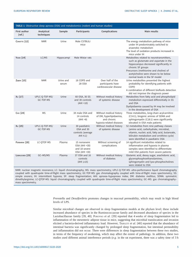
TABLE 1 Obstructive sleep apnoea (OSA) and metabolomics (rodent and human studies)
First author[ref.]
Analyticaltechniques
Sample Participants Complications Main results
CONOTTE [12] NMR Urine Male C57BL6Jmice
The energy metabolism pathway of miceunder IH predominately switched toanaerobic metabolism
The level of oxidation products increased inmice under IH
YOON [14] LC/MS Hippocampi Male Wistar rats Metabolites related to neurotransmitters,such as glutamate and aspartate in thehippocampus decreased significantly inchronic SF groups
Precursors (methionine and choline) ofacetylcholine were shown to be belownormal levels in the SF model
ZĄBEK [15] NMR Urine andserum
18 COPD and28 OSA
Over half of theparticipants have
cardiovascular disease
Urine metabolites presented the highestprobability for identifying patients withCOPD
A combination of different biofluids detectiondid not improve the diagnosis power
XU [17] UPLC-Q-TOF-MS/GC-TOF-MS
Urine 60 OSA, 30 SSand 30 controls
(AHI >10)
Without medical historyof systemic disease
Metabolites from fatty acid and phospholipidmetabolism expressed differentially in SSand OSA
Dyslipidaemia caused by IH may be involvedin the development of OSA
CHO [24] MS Urine 34 male OSA and14 controls(AHI >8)
Without medical historyof DM, hyperlipidaemia,
and chronichypoxia-related diseases
Three metabolites, long-chain acylcarnitine(C14:1), biogenic amines of SDMA andsphingomyelin (C18:1) were significantlyelevated in OSA male patients
XU [25] UPLC-Q-TOF-MS/GC-TOF-MS
Urine 30 paediatricOSA and 30
controls (averageAHI 4.1)
Without medical historyof systemic disease
52 metabolites from nine pathway classes(amino acid, carbohydrate, microbial,vitamin, nucleic acid, fatty acid, butanoate,bilirubin metabolism and ornithine cycle)were identified in paediatric OSA
FERRARINI [28] LC-QTOF-MS Plasma 15 nonsevereOSA (AHI <30)and 18 severeOSA (AHI >30)
Without screening ofcomplications
14 significant metabolites related toinflammation and hypoxia in plasmasamples were identified to differentiatemild OSA patients from severe patients
LEBKUCHEN [29] GC–MS/MS Plasma 37 OSA and 16controls(AHI ⩾15)
Without medical historyof diabetes
Glutamic acid, deoxy sugar, arachidonic acid,glycerophosphoethanolamines,sphingomyelin and lyso-phosphocholineswere related to OSA
NMR: nuclear magnetic resonance; LC: liquid chromatography; MS: mass spectrometry; UPLC-Q-TOF-MS: ultra-performance liquid chromatographycoupled with quadrupole time-of-flight mass spectrometry; GC-TOF-MS: gas chromatography coupled with time-of-flight mass spectrometry; SS:simple snorers; IH: intermittent hypoxia; SF: sleep fragmentation; AHI: apnoea–hypopnoea index; DM: diabetes mellitus; SDMA: symmetricdimethylarginine; LC-QTOF-MS: liquid chromatography coupled with quadrupole time-of-flight mass spectrometry; GC–MS: gas chromatography–mass spectrometry.
https://doi.org/10.1183/16000617.0220-2020 5
EUROPEAN RESPIRATORY REVIEW OBSTRUCTIVE SLEEP APNOEA | X. ZHANG ET AL.

during sleep). There is evidence that the endogenous circadian rhythm is a key modulator of the gutmicroflora [41]. Thus, pathological manifestations may be related to a disturbance of the circadian rhythm,which affects the microbiota profile in animal models and increases the release of inflammatory factors.
OSA is also an independent risk factor for hypertension. Several epidemiological studies have reported arelationship between OSA severity, defined based on apnoea–hypopnoea index (AHI) and hypertension [42–44].However, support for a causal relationship between OSA and hypertension comes from animal experiments; therelationship between OSA severity and the intestinal flora in patients with OSA-induced hypertension has notyet been explored. However, some biomarkers of hypertension could assist the diagnosis and prediction ofOSA-induced hypertension, which would help clarify the underlying mechanisms in cell and rodentexperiments. Many studies [45, 46] have confirmed that, over a short period, OSA alone is insufficient to causehypertension, except in the presence of a high fat or high salt diet. Moreover, repeated awakenings did not leadto blood pressure elevations in the absence of hypoxia [47]. The combination of OSA and a high saltdiet promotes hypertension by raising the level of TMAO in the blood and disrupting the regulation ofinflammation [46]. Transplanting caecal contents from OSA-induced hypertensive rats into OSA rats on anormal diet increases blood pressure in the recipient animals [45]. Thus, dysbiosis is involved in OSA-induced
d)Glutamic acid
Bile acids
Glycogen
Cholesterol
a)Hypoxanthine
Xanthine
Glucose Fat
Lipolysis
Pyruvate
BCAAsLactate
Glyceraldehyde
+GlycerolFree fatty acids and
derivatives
Acetyl CoA + NADH
Ketone bodies
TCA cycle
Acetyl CoAPorphyrin metabolism
IH
Haemoglobin
BiliverdinBilirubinOxidation
Urate
PathogenFlora imbalance
oxLDLNO
TMAOPLA2
Microbiota dysbiosis
SCFAs↓
IL-6, IL-8, IFN-γ, TGF-β1↑
OSA (intermittent
hypoxia)
c)
Kingdom
Firmicutes
Bacteria
Clostridia Erysipelotrichia Bacilli Bacteroidia
BacteroidalesLactobacillalesErysipelo-
trichalesClostridiales
Clostri-
diaceae ↑Lachno-
spiraceae
Erysipelo-
trichaceae
Lacto-
bacillaceae
Prevo-
tellaceaeBacteroid-
aceae
Porphyro-
monadaceae
Bifido-
bacteriaceae
Holdemania
↑
Allobaculum
↑
Lactobacillus
↑
Bifido-
bacterium ↑
Bacteroidetes Actinobacteria
Not assigned
Bifido-
bacteriales
Phylum
Class
Order
Family
Genus
b)
Kingdom
Phylum
Class
Order
Family
Genus
Bacteria
Firmicutes
Erysipelo-
trichia
Erysipelo-
trichales
Clostridia
Clostridiales
Peptocicc-
aceae ↓
Lachnospi-
raceae ↑
Allobaculum
↓
Turicibacter
↓
Bacteroides
↓
Odoribacter
↓
Porphyro-
monadaceaeBacteroid-
aceae ↓Erysipelo-
trichaceae ↓
Prevotell-
aceae ↑
Paraprevotella
↑
Prevotella
↑
Bacteroidales
Bacteroida
BacteroidetesProteo-
bacterias
FIGURE 2 a) Summary of metabolic alterations in obstructive sleep apnoea (OSA). The red arrows represent an increased level, while the greenarrows represent a decreased level. IH: intermittent hypoxia; BCAAs: branch chain amino acids; TCA: tricarboxylic acid; NADH: nicotinamide adeninedinucleotide. b) Alterations of gut microbiota in OSA rodent models. Red represents an increase in relative abundance, while green represents adecrease. c) Alterations of gut microbiota in OSA-induced hypertension models. Red represents an increase in relative abundance.d) Cardiovascular, hepatic, pulmonary and cerebral complications caused by OSA and influenced by microbiota dysbiosis. The dotted arrows referto the unclear mechanisms and pathophysiological pathways. SCFA: short-chain fatty acids; IL: interleukin; TGF: transforming growth factor; oxLDL:oxidised low-density lipoprotein; TMAO: trimethylamine oxide; PLA2: phospholipase A2; NO: nitric oxide.
https://doi.org/10.1183/16000617.0220-2020 6
EUROPEAN RESPIRATORY REVIEW OBSTRUCTIVE SLEEP APNOEA | X. ZHANG ET AL.

hypertension, and dysbiosis and hypertension seems to be linked by bacterially produced short-chain fatty acids(SCFAs) (primarily acetic, propionic and butyric acids). SCFAs originate from microbial metabolism of dietaryfibre in the intestine, where they exert effects on intestinal health and homeostasis by stabilising theintestinal epithelial barrier and regulating cytokine and antibody secretion [48]. Several gut microfloralanalyses [45, 49, 50] have shown that bacteria associated with SCFA production decrease in OSA-inducedhypertension models. SCFAs are involved in fatty acid synthesis and gluconeogenesis and can increase livertriglyceride levels, thus promoting intestinal energy metabolism and obesity [51]. However, elsewhere it wasreported that SCFAs may also increase energy consumption, and the production of satiety hormones, thuspreventing obesity [52]. SCFAs increase the number and suppressive function of colonic anti-inflammatoryregulatory T (Treg) cells by acting on G protein-coupled receptor (GPR)-43 [53]. There is some evidence thatSCFA-producing bacteria, such as Faecalibacterium, Oscillibacter and Howardella, are decreased in patientswith OSA compared with controls, indicating that a decrease in the anti-inflammatory effect of SCFAs mightpromote the occurrence and progression of OSA [54]. In addition, SCFAs regulate blood pressure by activatingGpr-41 expressed in the smooth muscle cells of small resistance vessels [55]. Therefore, the decrease of SCFAsin patients with OSA may promote obesity, inhibiting the anti-inflammatory effect and worsening hypertension.Prebiotic and probiotic treatments have been applied to further verify the role of SCFAs in OSA-inducedhypertension [49]. After Clostridium butyricum and Hylon probiotic treatments, the bacteria associated withSCFAs increase significantly in rats, and these treatments lessen the influence of decreasing acetateconcentrations in the caecum by inhibiting interleukin-1α and interleukin-6 mRNA expression. Similarly, in anOSA model, gut dysbiosis was confirmed to be associated with the risk of cardiac consequences. The expressionof Clostridiaceae and Coriobacteriaceae family members decreased significantly, while the expression ofLachnospiraceae family members increased, in OSA-induced models of hypertension compared with controls[56, 57]. However, a high fat or high salt diet, was used to simulate the cardiovascular complications in theseexperiments, which also used transgenic rodents with high susceptibility to obesity. Accordingly, the results mayhave been confounded by dietary and genetic factors. Nevertheless, changes in the relative abundance ofSCFA-producing bacteria are specific to OSA-induced hypertension, which sheds new light on the pathogenesisof the complications. The differences in microbes between OSA and OSA-induced hypertension models aresummarised in figure 2b and c.
Some similarities exist in the changes of intestinal flora among rodent models of OSA, where thesechanges are related to hallmarks of OSA, intermittent anoxia and reoxygenation. These similarities hint atpotential biomarkers for early diagnosis and treatment of OSA-induced complications. In the studiesanalysed herein, a decrease in the relative abundance of SCFA-producing bacteria manifested as changes inthe gut microbiome in OSA-induced hypertension models. Thus, modulating dietary SCFA intake couldinfluence SCFA-producing bacteria and their metabolic pathways, potentially affecting host metabolism.
Human studiesGut microbiomeFluctuations in serum microbial metabolites in the gut reflect intestinal floral disorders [25, 28]. The gutcontains a dense population of bacteria. The structure of the gut microbiome is relatively stable under normalphysiological conditions and appears to be nonpathogenic to the host. OSA changes markers of gut epithelialbarrier function and increases intestinal permeability, further disrupting the bacterial environment [58]. Inhumans, three enterotypes can be distinguished based on the abundance of specific bacteria genera:Bacteroides, Ruminococcus and Prevotella [38]. The enterotype is determined by the species composition,but the principal function is not necessarily determined by the dominant species. Enterotype is notsignificantly associated with body mass index, age, country of residence or other host characteristics [38].Sleep is significantly disrupted in OSA patients with an AHI ⩾15 and a Prevotella enterotype. In addition,obstructive, central and mixed apnoea indices, and mean heart rate are also significantly elevated in AHI ⩾15patients with a Prevotella enterotype [59]. Studies have shown that patients with the Prevotella enterotype aresusceptible to OSA [59]. The Prevotella enterotype is associated with diets high in carbohydrate (fibre) andsimple sugars [60]. Simple carbohydrate consumption promotes inflammation of epithelial cells and obesity,which are strongly associated with OSA [61]. Furthermore, increased abundance of Prevotella is associatedwith mucosal inflammation, mediated by type 17 T-helper (Th17) cells. Prevotella stimulatesantigen-presenting cells to produce Th17-polarising cytokines, mainly by activating Toll-like receptor 2, andcan also stimulate epithelial cells to produce inflammatory mediators [62], which may participate in OSA bypromoting chronic inflammation. However, another study indicated that a diet rich in fibre can protect thehost from OSA by promoting microbial fermentation and SCFA production and that the Prevotella enterotypeis related to a much higher risk for OSA [54]. These pathophysiological processes provide new insight intohow the enterotype might affect OSA susceptibility.
https://doi.org/10.1183/16000617.0220-2020 7
EUROPEAN RESPIRATORY REVIEW OBSTRUCTIVE SLEEP APNOEA | X. ZHANG ET AL.

Oral microbiomeThe human oral cavity contains a diverse community of trillions of microbiota. Streptococcus, Gemella,Abiotrophia, Granulicatella, Rothia, Neisseria and Prevotella are predominant among healthy oralbacteria [63]. Changes in the oral microbiome have been discovered in various systemic diseases, such asT2DM, pancreatic cancer and cardiovascular disease [64–66]. In OSA patients, airway pressure changesdramatically reduce air flow, which affects the moisture and oxygen content in the upper respiratory tract andcould explain disorders of the oropharyngeal flora in middle-aged men with moderate-to-severe OSA [67].
Oral bacteria have been confirmed to be involved in the pathogenesis of coronary heart disease andhypertension [68–70]. Haematogenous dissemination of periodontal bacteria is thought to be a cause ofcardiovascular disease [71]. ZHONG et al. [69] observed Porphyromonas gingivalis in coronaryatherosclerotic plaques. However, KO et al. [72] reported that Porphyromonas genes were not found inblood samples of patients with OSA. In addition, the relative abundance of Porphyromonas is higher inbuccal mucosa, and the levels of proinflammatory cytokines are also higher in patients with OSA. Theseresults suggest that oral microorganisms might affect the likelihood of cardiovascular events throughinflammatory mediators rather than direct invasion of gum tissue.
Nasal microbiomeBacterial communities colonise all areas of the respiratory tract and resist opportunistic pathogens. The nasalmicrobiome is characterised by Corynebacterium, Propionibacterium and Staphylococcus species [35]. Thenasal microbiome profile changes according to illness severity; in patients with severe OSA, enrichmentwith some commensal organisms (e.g. Streptococcus, Prevotella, Pseudomonas and Haemophilus) is seencompared with nonsevere OSA patients [73].This could be due to recurrent obstruction during sleep causingreflux of oropharyngeal secretions. Surprisingly, the nasal microbiome did not show significant differencesin α or β diversity after continuous positive airway pressure treatment, which could be explained by therelatively short (3-month) treatment course [37].
OthersIn addition to the upper respiratory tract, the microbiome profile of the lower airway has been studiedthrough bronchoscopy and bronchoalveolar lavage analyses. The levels of Proteobacteria and Fusobacteriawere significantly higher, whereas the level of Firmicutes was significantly lower in patients with OSAcompared with control patients [74]. A healthy airway microbiota is essential to maintain airway immunityand reduce the risk of infection. Changes in the airway microbiota may contribute to the growth ofpotential pathogens and airway inflammation in patients with OSA [75]. Studies of the microbiome inpatients with OSA and animal models are summarised in table 2.
Summary of microbiome studiesThe microbiota plays a pathophysiological role in OSA and OSA-induced hypertension. Dysbiosis wascharacterised by a changed Firmicutes to Bacteroidetes ratio in an IH mimicking model [45, 50]. Decreasedabundance of SCFA-producing bacteria increased abundance of mucin-degrading bacteria, and increasedlevels of proinflammatory cytokines appeared to be the major mechanisms underlying cardiac complicationsof OSA in an OSA-related hypertension model [36, 37, 45, 49, 50]. Overall, however, few studies haveinvestigated the microbiome in OSA patients and animal models and few studies have investigated microbialcommunities in OSA patients in different parts of the body. Therefore, the consistency in microbiomeprofiles among OSA patients cannot be easily determined. Importantly, SCFA-producing bacteria and LPSsare involved in obesity and metabolic changes, including in T2DM [76, 77]. Some bacteria, such asLactobacillus, Bacteroides, Bifidobacterium and Prevotella, which are found in animal models and patientswith OSA, have also been implicated in obesity and T2DM [78]. There is large overlap in the diseasemanifestations and contributing factors for OSA and metabolic disorders, and some microbiome signatures ofOSA might be shared with those already observed in obesity and T2DM. Therefore, OSA patients withdifferent complications and different diet or other external factors will have a great impact on intestinal flora,that is to say different OSA phenotypes or combination of comorbidities might be associated with differentmetabolic and gut microbiota profiles. The unique signatures of OSA awaits further elucidation. There aresome other limitations to studies of the microbiome in the context of OSA. First, the sample sizes aregenerally small, so the results are not reliable. Secondly, few studies have investigated whether intestinal florachanges occur with disease progression; longitudinal metabolomics and multi-omics studies using advancedapproaches are therefore needed to increase the number of microbiome biomarkers.
Metabolomics combined with the microbiomeOnly three studies [25, 56, 57] performed a joint analysis of metabolomics and the microbiome in associationwith OSA. XU et al. [25] explored the relationship between the oral microbiome and urine metabolism in
https://doi.org/10.1183/16000617.0220-2020 8
EUROPEAN RESPIRATORY REVIEW OBSTRUCTIVE SLEEP APNOEA | X. ZHANG ET AL.

TABLE 2 Obstructive sleep apnoea (OSA) and the microbiome (rodent and human studies)
First author[ref.]
Model OSA model Methods Biofluid Relative microbial abundance Results summary
MORENO-INDIAS [36] Germ-free C57BL/6 male mice
6 weeks IH 16 S rRNApyrosequencing
Faecalsamples
IH (Family):Lachnospiraceae↑,Prevotellaceae↑,Peptococcaceae↓,Erysipelotrichaceae↓,Bacteroidaceae↓
IH (Genus):Paraprevotella↑, Prevotella↑,Allobaculum↓, Turicibacter↓,Bacteroides↓, Odoribacter↓
Under IH, the intestinalPaO2
fluctuatedintermittently and theaverage level decreased
The evenness of themicrobial communityunder IH was increased inthe intestine
MORENO-INDIAS [37] Germ-free C57BL/6 male mice
6 weeks IHfollowed by6 weeksnormoxicrecovery
16 S rRNApyrosequencing
Faecalsamples
IH (Phylum):Firmicutes↑, Deferribacteres↑,Bacteroidetes↓
IH (Family):Ruminococcaceae↑,Desulfovibrionaceae↑,Helicobacteraceae↑,Rikenellaceae↑,Odoribacteraceae↑,Enterobacteriaceae↑,Erysipelotrichaceae↑,Clostridiaceae↑, S24-7↓,Lactobacillaceae↓
IH (Genus):Clostridium↑, Desulfovibrio↑,Sutterella↑, Allobaculum↑,Rikenella↑, Odoribacter↑,Mucispirillum↑, Lactobacillus↓,Ruminococcus↓, Oscillospira↓,Prevotella↓
Decrease of SCFA-producingbacteria and increase ofmucin-degrading bacteriacould induce a high levelof LPS in blood in IHgroup
DURGAN [45] RatsHFD
Endotrachealobstruction
deviceimplantation
16 S rRNAsequencing
Faecalsamples
IH+HFD:Clostridiaceae↑, Holdemania↑
In the OSA model, thecoexistence of OSA and aHFD could lead tohypertension
The ratio of F/B was lower innormal diet OSA groupthan in the HFD alonegroup
GANESH [49] RatsHFD
HFD+HylonHFD+Clostridium
butyricum
Endotrachealobstruction
deviceimplantation
16 S rRNAsequencing
Faecalsamples
OSA+HFD (Genus):Lactobacillus↑, Allobaculum↑,Bifidobacterium↑
OSA+HFD+C. butyricum (Genus):Dehalobacterium↓,
Anaerostipes↓
C. butyricum and Hylon canreduce the effects of OSAon intestinal flora, such asthinning mucosa and adecrease of cecum acetate
C. butyricum and Hyloncould alleviateOSA-induced goblet cellloss and microgliaactivation
Supplementary acetate inthe cecum could preventOSA-induced hypertension
TRIPATHI [56] C57BL/6J maleLdlr−/− mice
HFD
6 weeks IHH 16S rRNAsequencing
Faecalsamples
IHH (Family):Clostridiaceae↓,Coriobacteriaceae↓,Lachnospiraceae↑
IHH (Genus):Oscillospira↑
Under the condition of IHHand HFD, mice sufferedfrom intestinal dysbiosis,which led to changes inmetabolic profiling, mainlychanges in bile acids,phytoestrogens and fattyacids
Continued
https://doi.org/10.1183/16000617.0220-2020 9
EUROPEAN RESPIRATORY REVIEW OBSTRUCTIVE SLEEP APNOEA | X. ZHANG ET AL.

TABLE 2 Continued
First author[ref.]
Model OSA model Methods Biofluid Relative microbial abundance Results summary
LUCKING [50] 1. Guinea pig2. HFD
IH 16 S rRNAsequencing
Faecalsamples
IH (Phylum):Firmicutes↓, Bacteroidetes↑
In guinea pigs, thecharacteristics ofcardiopulmonary disease(mainly apnoea andhypertension) in ratsinduced by CIH exposurewere not obvious, but itchanged brain stemneurochemistry, reducedthe richness of intestinalflora species and changedthe composition of flora
TRIPATHI [57] C57BL/6J maleLdlr−/−mice
+HFDC57BL/6J maleApoE−/− mice
+HFD
6 weeks(Ldlr−/−) or10 weeks
(ApoE−/−) IHH
16S rRNAsequencing
Faecalsamples
IHH (Family):Clostridiaceae↓,Lachnospiraceae↑
Under the intervention ofOSA, atherosclerosismodel involved commonmicrobial and metabolicchanges
LIU [46] Sprague–DawleyratsNDHSD
CIH + NDCIH + HSD
6 weeks IH 16S rRNAsequencing
Faecalsamples
IH:Lactobacillus rhamnosus↓
Because of the synergisticeffect of OSA and HSD, theabundance of lactobacilliin intestine decreasedsignificantly, and theblood pressure increasedsignificantly
LGG could prevent theaggravation ofhypertension by reducingthe level of TMAO,regulating the imbalanceof Th1/Th2 cytokines, andinhibiting thephosphorylation of ERK1/2, Akt and mTOR
POROYKO [39] C57BL/6J mice 4 weeks SF 16S rRNAsequencing
Faecalsamples
SF (Phylum):Firmicutes↑, Bacteroidetes↑
SF (Families):Lachnospiraceae↑,Ruminococcaceae↑,Lactobacillaceae↓
SF could lead to increasedvisceral fat mass and fattissue inflammation, aswells as systemic insulinresistance and increasedleptin plasma levels
SF had no effect on aerobiccommunity metabolism,but suppressed anaerobicsubstrate utilisation
TRIPLETT [40] Sprague–Dawleyrats
6 weeks SF 16S rRNAsequence
Intestinalcontent
SF ileum (Family):Enterobacteriaceae↓,Lactobacillaceae↓,Turicibacteraceae↑,Clostridiaceae↑
SF caecum (Family):Ruminococcaceae↑,Turicibacteraceae↑
SF colon (Family):Turicibacteraceae,Clostridiaceae,Erysipelotrichaceae↑
Chronic SF significantlyincreased crypt depth
Chronic SF did not triggersystemic inflammation inrats, and microbialextraintestinaltranslocation and invasiondid not occur
LU [74] 11 OSA and 8controls (withoutmedical historyof systemicdisease)
Fibreopticbronchoscopy
16S rRNAsequencing
BALF OSA (Phylum):Proteobacteria andFusobacteria↑, Firmicutes↓
The disruption of pulmonarymicrobiota in OSA mightbe the cause of pulmonaryinflammation andsensitivity to injury
Continued
https://doi.org/10.1183/16000617.0220-2020 10
EUROPEAN RESPIRATORY REVIEW OBSTRUCTIVE SLEEP APNOEA | X. ZHANG ET AL.

paediatric OSA patients; the overall diversity of oral microorganisms was lower in these patients, but thelevels of members of the Veillonellaceae, Campylobacteraceae and C111 Paraprevotellaceae families werehigher. Correlations between metabolic changes (in urine samples) and disturbances in the oral microbiomewere found, but causal relationships were not explored. Metabolism was also shown to be related to gutdysbiosis in rodent models [56, 57]. These studies described a role for microbial-related metabolic disordersin the initiation of cardiac complications in OSA. However, how microbial disturbances affect metabolicpathways remains to be explored.
TABLE 2 Continued
First author[ref.]
Model OSA model Methods Biofluid Relative microbial abundance Results summary
XU [25] 30 OSA childrenand 30 controls(aged 3–11 yearsold; without
medical historyof systemicdisease)
AHI >1 16S rRNAsequencing
Metabolomics
Buccalmucosa
OSA (Family):Veillonellaceae,Campylobacteraceae, C111Paraprevotellaceae↑(Genus):Veillonella, Prevotella,Mogibacterium,Campylobacter andButyrivibrio↑
The oral microbial diversityof paediatric OSA patientsdecreased, and themicrobial structure wasdifferent from that of thecontrol group
KO [72] 126 OSA and 13controls (without
screening ofcomplications)
AHI >5 16 S rRNApyrosequencing
Buccalmucosa
OSA (Genus):Porphyromonas andAggregatibacter↑
Compared with non-OSApatients, the relativeabundance ofPorphyromonas, and theproinflammatory cytokineswere higher in patientswith OSA
YANG [67] 26 male OSA and25 male controls
(aged 30–65 years; withoutmedical historyof systemicdisease)
AHI ⩾15 16S rRNAsequencing
Oropharynx Glaciecola was not detected inmoderate and severe OSAgroups
Severe OSA:Tannerella, Anaerovorax and
Halomonas ↓
The diversity and abundanceof bacteria in the OSAgroup decreasedsignificantly, and the mostsignificant diversity declineoccurred in the moderateOSA group
WU [73] 472 subjects fromthe WTCSNOREand 93 subjects
from theZaragoza sleep
cohort
AHI ⩾5 16S rRNAsequencing
Nasallavages
Severe OSA (Genus):Streptococcus, Prevotella,Veillonella and Granulicatella↑
The inflammatory mediatorsin nasal lavage fluid ofpatients with OSAincreased
KO [54] 93 OSA and 20controls (without
screening ofcomplications)
AHI >5 16S rRNAsequencing
Faecalsamples
OSA (Genus):Faecalibacterium↓,Megamonas↓,Ruminococcaceae↓,Clostridiales↓, Alistipes↓,Bifidobacterium↑
Ruminococcus enterotypewas associated withincreased risk of OSA
The level of Lactobacillus wasrelated to the level ofhomocysteine
KO [59] 52 OSA and 61controls (without
medicalhistory of
gastrointestinaldiseases,infection,
unexplaineddiarrhoea, andantibiotics or
probiotics used)
AHI ⩾15 16 S rRNApyrosequencing
Faecalsamples
The parameters related tosleep were significantlydamaged in patients withintestinal type ofPrevotella, whoseobstructive, central andmixed apnoea index andmean heart rate were alsosignificantly increased
IH: intermittent hypoxia; PaO2: arterial oxygen tension; SCFA: short-chain fatty acids; LPS: lipopolysaccharide; HFD: high fat diet; F/B: Firmicutes/
Bacteroidetes ratio; IHH: IH and hypercapnia; CIH: chronic intermittent hypoxia; ND: normal diet; HSD: high salt diet; LGG: Lactobacillus rhamnosusGG strain; TMAO: trimethylamine oxide; Th: helper T-cell; SF: sleep fragmentation; BALF: bronchoalveolar lavage fluid; AHI: apnoea–hypopnoea index.
https://doi.org/10.1183/16000617.0220-2020 11
EUROPEAN RESPIRATORY REVIEW OBSTRUCTIVE SLEEP APNOEA | X. ZHANG ET AL.
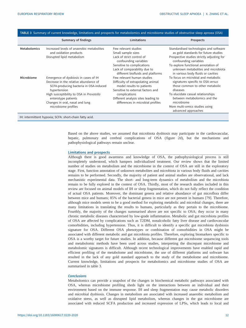
Based on the above studies, we assumed that microbiota dysbiosis may participate in the cardiovascular,hepatic, pulmonary and cerebral complications of OSA (figure 2d), but the mechanisms andpathophysiological pathways remain unclear.
Limitations and prospectsAlthough there is good awareness and knowledge of OSA, the pathophysiological process is stillincompletely understood, which hampers individualised treatment. Our review shows that the limitednumber of studies on metabolism and the microbiome in the context of OSA are still in the exploratorystage. First, function annotation of unknown metabolites and microbiota in various body fluids and cavitiesremains to be performed. Secondly, the majority of patient and animal studies are observational, and lackmechanistic experimental data. The short- and long-term dynamics of metabolism and the microbiotaremain to be fully explored in the context of OSA. Thirdly, most of the research studies included in thisreview are focused on animal models of IH or sleep fragmentation, which do not fully reflect the conditionof actual OSA patients. Moreover, the dominant genera and relative abundance of gut microflora differbetween mice and humans; 85% of the bacterial genera in mice are not present in humans [79]. Therefore,although mice models seem to be a good method for exploring metabolic and microbial changes, there aremany limitations in translating the results to humans, particularly as they pertain to the microbiome.Fourthly, the majority of the changes summarised above are not specific to OSA; they occur in manychronic metabolic diseases characterised by low-grade inflammation. Metabolic and gut microbiota profilesof OSA are affected by complications such as T2DM, nonalcoholic fatty liver disease and cardiovascularcomorbidities, including hypertension. Thus, it is difficult to identify a specific gut microbiota dysbiosissignature for OSA. Different OSA phenotypes or combination of comorbidities in OSA might beassociated with different metabolic and gut microbiota profiles. Therefore, exploring biomarkers specific toOSA is a worthy target for future studies. In addition, because different gut microbiome sequencing toolsand metabolomic methods have been used across studies, interpreting the discrepant microbiome andmetabolomic signatures is difficult. Although recent technological improvements have enabled rapid andefficient profiling of the metabolome and microbiome, the use of different platforms and software hasresulted in the lack of any gold standard approach to the study of the metabolome and microbiome.Current knowledge, limitations and prospects for metabolomics and microbiome studies of OSA aresummarised in table 3.
ConclusionsMetabolomics can provide a snapshot of the changes in biochemical metabolic pathways associated withOSA, whereas microbiome profiling sheds light on the interactions between an individual and theirenvironment based on the immune response. IH and sleep fragmentation may cause metabolic disordersand microbial dysbiosis. Changes in metabolism are associated with increased anaerobic metabolism andoxidative stress, as well as disrupted lipid metabolism, whereas changes in the gut microbiome areassociated with reduced SCFA production and increased expression of LPSs, which leads to local and
TABLE 3 Summary of current knowledge, limitations and prospects for metabolomics and microbiome studies of obstructive sleep apnoea (OSA)
Summary of findings Limitations Prospects
Metabolomics Increased levels of anaerobic metabolitesand oxidation products
Disrupted lipid metabolism
Few relevant studiesSmall sample sizesLack of strict control of
confounding variablesSensitive to complicationsLack of comparability due to
different biofluids and platforms
Standardised technologies and softwareas gold standards for future studies
Prospective studies strictly adjusting forconfounding variables
To explore functional annotation ofunknown metabolites and microbiotain various body fluids or cavities
To focus on microbial and metabolicsignatures specific to OSA versusthose common to other metabolicdiseases
To elucidate casual relationshipsbetween metabolomics and themicrobiome
More multi-omics studies usingadvanced approaches
Microbiome Emergence of dysbiosis in cases of IHDecrease in the relative abundance of
SCFA-producing bacteria in OSA-inducedhypertension
High susceptibility to OSA in Prevotellaenterotype patients
Changes in oral, nasal and lungmicrobiome profiles
Few relevant human studiesDifficulty of extrapolating animal
model results to patientsSensitive to external factors and
complicationsDifferent analysis sites leading to
differences in microbial profiles
IH: intermittent hypoxia; SCFA: short-chain fatty acid.
https://doi.org/10.1183/16000617.0220-2020 12
EUROPEAN RESPIRATORY REVIEW OBSTRUCTIVE SLEEP APNOEA | X. ZHANG ET AL.

systemic inflammatory responses and metabolic comorbidities. Importantly, it has recently become evidentthat the metabolomic and microbiota signatures of OSA might be those already observed in metabolicdisorders, such as obesity and T2DM [28]. A lack of consideration of the impact of metabolic disorders on“omics” analyses may have complicated the changes in the microbiome and metabolism seen in OSApatients, as well as animal models. Although current research methods are somewhat limited, moremulti-omics studies should facilitate the early diagnosis and treatment of OSA.
Provenance: Submitted article, peer reviewed.
Authors’ contributions: The authors take responsibility and vouch for the accuracy and completeness of the dataand analyses. S. Yin, J. Guan and H. Xu had full access to all of the data in the study and took responsibility forthe integrity of the data and the accuracy of the data analysis. Study design: H. Xu, J. Guan and S. Yin. Datacollection: H. Xu, J. Guan, H. Yi, S. Wang and X. Zhang. Statistical analysis: H. Xu and X. Zhang. Manuscript draft:H. Xu and X. Zhang. All authors have seen and approved the manuscript.
Conflict of interest: None declared.
Support statement: This study was supported by grants-in-aid from Shanghai Municipal Commission of Science andTechnology (grant no.18DZ2260200); Innovative research team of high-level local universities in Shanghai; InnovationProgram of Shanghai Municipal Education Commission (2017-01-07-00-02-E00047); National Natural ScienceFoundation of China (82071030, 81970870, 81700896, 81770987, 81701306, 81770988); National Key R&D Program ofChina (2017YFC0112500) and a multicentre clinical research project from the School of Medicine, Shanghai JiaoTong University (DLY201502). Funding information for this article has been deposited with the Crossref FunderRegistry.
References1 Benjafield AV, Ayas NT, Eastwood PR, et al. Estimation of the global prevalence and burden of obstructive
sleep apnoea: a literature-based analysis. Lancet Respir Med 2019; 7: 687–698.2 McNicholas WT, Bassetti CL, Ferini-Strambi L, et al. Challenges in obstructive sleep apnoea. Lancet Respir Med
2018; 6: 170–172.3 Ryan S, Cummins EP, Farre R, et al. Understanding the pathophysiological mechanisms of cardiometabolic
complications in obstructive sleep apnoea: towards personalised treatment approaches. Eur Respir J 2020;56: 1902295.
4 Yaffe K, Laffan AM, Harrison SL, et al. Sleep-disordered breathing, hypoxia, and risk of mild cognitiveimpairment and dementia in older women. JAMA 2011; 306: 613–619.
5 Qian Y, Yi H, Zou J, et al. Independent association between sleep fragmentation and dyslipidemia in patientswith obstructive sleep apnea. Sci Rep 2016; 6: 26089.
6 Xu H, Guan J, Yi H, et al. Elevated low-density lipoprotein cholesterol is independently associated withobstructive sleep apnea: evidence from a large-scale cross-sectional study. Sleep Breath 2016; 20: 627–634.
7 Semelka M, Wilson J, Floyd R. Diagnosis and treatment of obstructive sleep apnea in adults. Am FamPhysician 2016; 94: 355–360.
8 Dunn WB, Broadhurst D, Begley P, et al. Procedures for large-scale metabolic profiling of serum and plasma usinggas chromatography and liquid chromatography coupled to mass spectrometry. Nat Protoc 2011; 6: 1060–1083.
9 Newgard CB. Metabolomics and metabolic diseases: where do we stand? Cell Metab 2017; 25: 43–56.10 Bujak R, Struck-Lewicka W, Markuszewski MJ, et al. Metabolomics for laboratory diagnostics. J Pharm Biomed
Anal 2015; 113: 108–120.11 Barko PC, McMichael MA, Swanson KS, et al. The gastrointestinal microbiome: a review. J Vet Intern Med
2018; 32: 9–25.12 Conotte S, Tassin A, Conotte R, et al. Metabonomic profiling of chronic intermittent hypoxia in a mouse
model. Respir Physiol Neurobiol 2018; 256: 157–173.13 Ryan S. Adipose tissue inflammation by intermittent hypoxia: mechanistic link between obstructive sleep
apnoea and metabolic dysfunction. J Physiol 2017; 595: 2423–2430.14 Yoon DW, Kwon HN, Jin X, et al. Untargeted metabolomics analysis of rat hippocampus subjected to sleep
fragmentation. Brain Res Bull 2019; 153: 74–83.15 Ząbek A, Stanimirova I, Deja S, et al. Fusion of the H NMR data of serum, urine and exhaled breath
condensate in order to discriminate chronic obstructive pulmonary disease and obstructive sleep apneasyndrome. Metabolomics 2015; 11: 1563–1574.
16 Maniscalco M, Motta A. Metabolomics of chronic obstructive pulmonary disease and obstructive sleep apneasyndrome: a comment. Metabolomics 2016; 12: 29.
17 Xu H, Zheng X, Qian Y, et al. Metabolomics profiling for obstructive sleep apnea and simple snorers. Sci Rep2016; 6: 30958.
https://doi.org/10.1183/16000617.0220-2020 13
EUROPEAN RESPIRATORY REVIEW OBSTRUCTIVE SLEEP APNOEA | X. ZHANG ET AL.

18 Wang TJ, Larson MG, Vasan RS, et al. Metabolite profiles and the risk of developing diabetes. Nat Med 2011;17: 448–453.
19 Dong J-Y, Zhang Y-H, Qin L-Q. Obstructive sleep apnea and cardiovascular risk: meta-analysis of prospectivecohort studies. Atherosclerosis 2013; 229: 489–495.
20 Zhou M, Guo B, Wang Y, et al. The association between obstructive sleep apnea and carotid intima–mediathickness: a systematic review and meta-analysis. Angiology 2017; 68: 575–583.
21 Sorajja D, Gami AS, Somers VK, et al. Independent association between obstructive sleep apnea andsubclinical coronary artery disease. Chest 2008; 133: 927–933.
22 Yaggi HK, Concato J, Kernan WN, et al. Obstructive sleep apnea as a risk factor for stroke and death. N Engl JMed 2005; 353: 2034–2041.
23 Bauters F, Rietzschel ER, Hertegonne KBC, et al. The link between obstructive sleep apnea andcardiovascular disease. Curr Atheroscler Rep 2015; 18: 1.
24 Cho K, Yoon DW, Lee M, et al. Urinary metabolomic signatures in obstructive sleep apnea through targetedmetabolomic analysis: a pilot study. Metabolomics 2017; 13: 88.
25 Xu H, Li X, Zheng X, et al. Pediatric obstructive sleep apnea is associated with changes in the oralmicrobiome and urinary metabolomics profile: a pilot study. J Clin Sleep Med 2018; 14: 1559–1567.
26 Marcus CL, Brooks LJ, Draper KA, et al. Diagnosis and management of childhood obstructive sleep apneasyndrome. Pediatrics 2012; 130: 576–584.
27 Tagaya M, Nakata S, Yasuma F, et al. Relationship between adenoid size and severity of obstructive sleepapnea in preschool children. Int J Pediatr Otorhinolaryngol 2012; 76: 1827–1830.
28 Ferrarini A, Rupérez FJ, Erazo M, et al. Fingerprinting-based metabolomic approach with LC–MS to sleepapnea and hypopnea syndrome: a pilot study. Electrophoresis 2013; 34: 2873–2881.
29 Lebkuchen A, Carvalho VM, Venturini G, et al. Metabolomic and lipidomic profile in men with obstructivesleep apnoea: implications for diagnosis and biomarkers of cardiovascular risk. Sci Rep 2018; 8: 11270.
30 Dall’Acqua S, Stocchero M, Boschiero I, et al. New findings on the in vivo antioxidant activity of Curcuma longaextract by an integrated (1)H NMR and HPLC-MS metabolomic approach. Fitoterapia 2016; 109: 125–131.
31 Aron-Wisnewsky J, Clément K. The gut microbiome, diet, and links to cardiometabolic and chronic disorders.Nat Rev Nephrol 2016; 12: 169–181.
32 Randrianarisoa E, Lehn-Stefan A, Wang X, et al. Relationship of serum trimethylamine N-oxide (TMAO) levelswith early atherosclerosis in humans. Sci Rep 2016; 6: 26745.
33 Zhu W, Gregory JC, Org E, et al. Gut microbial metabolite TMAO enhances platelet hyperreactivity andthrombosis risk. Cell 2016; 165: 111–124.
34 Lupachyk S, Watcho P, Stavniichuk R, et al. Endoplasmic reticulum stress plays a key role in the pathogenesisof diabetic peripheral neuropathy. Diabetes 2013; 62: 944–952.
35 Human Microbiome Project Consortium. Structure, function and diversity of the healthy human microbiome.Nature 2012; 486: 207–214.
36 Moreno-Indias I, Torres M, Montserrat JM, et al. Intermittent hypoxia alters gut microbiota diversity in amouse model of sleep apnoea. Eur Respir J 2015; 45: 1055–1065.
37 Moreno-Indias I, Torres M, Sanchez-Alcoholado L, et al. Normoxic recovery mimicking treatment of sleepapnea does not reverse intermittent hypoxia-induced bacterial dysbiosis and low-grade endotoxemia in mice.Sleep 2016; 39: 1891–1897.
38 Arumugam M, Raes J, Pelletier E, et al. Enterotypes of the human gut microbiome. Nature 2011; 473:174–180.
39 Poroyko VA, Carreras A, Khalyfa A, et al. Chronic sleep disruption alters gut microbiota, induces systemic andadipose tissue inflammation and insulin resistance in mice. Sci Rep 2016; 6: 35405.
40 Triplett J, Ellis D, Braddock A, et al. Temporal and region-specific effects of sleep fragmentation on gutmicrobiota and intestinal morphology in Sprague–Dawley rats. Gut Microbes 2020; 11: 706–720.
41 Voigt RM, Forsyth CB, Green SJ, et al. Circadian rhythm and the gut microbiome. In: Cryan JF, Clarke G, eds.International Review of Neurobiology, 131. Cambridge, MA, Academic Press, 2016; pp. 193–205.
42 Nieto FJ, Young TB, Lind BK, et al. Association of sleep-disordered breathing, sleep apnea, and hypertensionin a large community-based study. Sleep Heart Health Study. JAMA 2000; 283: 1829–1836.
43 Durán J, Esnaola S, Rubio R, et al. Obstructive sleep apnea–hypopnea and related clinical features in apopulation-based sample of subjects aged 30 to 70 yr. Am J Respir Crit Care Med 2001; 163: 685–689.
44 Peppard PE, Young T, Palta M, et al. Prospective study of the association between sleep-disordered breathingand hypertension. N Engl J Med 2000; 342: 1378–1384.
45 Durgan DJ, Ganesh BP, Cope JL, et al. Role of the gut microbiome in obstructive sleep apnea-inducedhypertension. Hypertension 2016; 67: 469–474.
46 Liu J, Li T, Wu H, et al. Lactobacillus rhamnosus GG strain mitigated the development of obstructive sleepapnea-induced hypertension in a high salt diet via regulating TMAO level and CD4 T cell induced-type Iinflammation. Biomed Pharmacother 2019; 112: 108580.
https://doi.org/10.1183/16000617.0220-2020 14
EUROPEAN RESPIRATORY REVIEW OBSTRUCTIVE SLEEP APNOEA | X. ZHANG ET AL.

47 Brooks D, Horner RL, Kozar LF, et al. Obstructive sleep apnea as a cause of systemic hypertension. Evidencefrom a canine model. J Clin Invest 1997; 99: 106–109.
48 Tan J, McKenzie C, Potamitis M, et al. The role of short-chain fatty acids in health and disease. Adv Immunol2014; 121: 91–119.
49 Ganesh BP, Nelson JW, Eskew JR, et al. Prebiotics, probiotics, and acetate supplementation preventhypertension in a model of obstructive sleep apnea. Hypertension 2018; 72: 1141–1150.
50 Lucking EF, O’Connor KM, Strain CR, et al. Chronic intermittent hypoxia disrupts cardiorespiratoryhomeostasis and gut microbiota composition in adult male guinea pigs. EBioMedicine 2018; 38: 191–205.
51 Samuel BS, Shaito A, Motoike T, et al. Effects of the gut microbiota on host adiposity are modulated by theshort-chain fatty-acid binding G protein-coupled receptor, Gpr41. Proc Natl Acad Sci USA 2008; 105:16767–16772.
52 Canfora EE, Jocken JW, Blaak EE. Short-chain fatty acids in control of body weight and insulin sensitivity. NatRev Endocrinol 2015; 11: 577–591.
53 Smith PM, Howitt MR, Panikov N, et al. The microbial metabolites, short-chain fatty acids, regulate colonicTreg cell homeostasis. Science 2013; 341: 569–573.
54 Ko CY, Liu QQ, Su HZ, et al. Gut microbiota in obstructive sleep apnea–hypopnea syndrome: disease-relateddysbiosis and metabolic comorbidities. Clin Sci (Lond) 2019; 133: 905–917.
55 Pluznick JL, Protzko RJ, Gevorgyan H, et al. Olfactory receptor responding to gut microbiota-derived signalsplays a role in renin secretion and blood pressure regulation. Proc Natl Acad Sci USA 2013; 110: 4410–4415.
56 Tripathi A, Melnik AV, Xue J, et al. Intermittent hypoxia and hypercapnia, a hallmark of obstructive sleepapnea, alters the gut microbiome and metabolome. mSystems 2018; 3: e00020-18.
57 Tripathi A, Xu ZZ, Xue J, et al. Intermittent hypoxia and hypercapnia reproducibly change the gutmicrobiome and metabolome across rodent model systems. mSystems 2019; 4: e00058-19.
58 Barceló A, Esquinas C, Robles J, et al. Gut epithelial barrier markers in patients with obstructive sleep apnea.Sleep Med 2016; 26: 12–15.
59 Ko CY, Fan JM, Hu AK, et al. Disruption of sleep architecture in Prevotella enterotype of patients withobstructive sleep apnea–hypopnea syndrome. Brain Behav 2019; 9: e01287.
60 Conlon MA, Bird AR. The impact of diet and lifestyle on gut microbiota and human health. Nutrients 2014; 7:17–44.
61 Orlando A, Cazzaniga E, Giussani M, et al. Hypertension in children: role of obesity, simple carbohydrates,and uric acid. Front Public Health 2018; 6: 129.
62 Larsen JM. The immune response to Prevotella bacteria in chronic inflammatory disease. Immunology 2017;151: 363–374.
63 Bik EM, Long CD, Armitage GC, et al. Bacterial diversity in the oral cavity of 10 healthy individuals. ISME J2010; 4: 962–974.
64 Fan X, Alekseyenko AV, Wu J, et al. Human oral microbiome and prospective risk for pancreatic cancer: apopulation-based nested case-control study. Gut 2018; 67: 120–127.
65 Koren O, Spor A, Felin J, et al. Human oral, gut, and plaque microbiota in patients with atherosclerosis. ProcNatl Acad Sci USA 2011; 108: Suppl. 1, 4592–4598.
66 Long J, Cai Q, Steinwandel M, et al. Association of oral microbiome with type 2 diabetes risk. J Periodont Res2017; 52: 636–643.
67 Yang W, Shao L, Heizhati M, et al. Oropharyngeal microbiome in obstructive sleep apnea: decreased diversityand abundance. J Clin Sleep Med 2019; 15: 1777–1788.
68 Ueno M, Izumi Y, Kawaguchi Y, et al. Prediagnostic plasma antibody levels to periodontopathic bacteria andrisk of coronary heart disease. Int Heart J 2012; 53: 209–214.
69 Zhong LJ, Zhang YM, Liu H, et al. [Detection of periodontal pathogens in coronary atherosclerotic plaques].Zhonghua Kou Qiang Yi Xue Za Zhi 2008; 43: 4–7.
70 Pietropaoli D, Del Pinto R, Ferri C, et al. Definition of hypertension-associated oral pathogens in NHANES.J Periodontol 2019; 90: 866–876.
71 Klarström Engström K, Khalaf H, Kälvegren H, et al. The role of Porphyromonas gingivalis gingipains inplatelet activation and innate immune modulation. Mol Oral Microbiol 2015; 30: 62–73.
72 Ko C-Y, Hu A-K, Chou D, et al. Analysis of oral microbiota in patients with obstructive sleep apnea-associatedhypertension. Hypertens Res 2019; 42: 1692–1700.
73 Wu BG, Sulaiman I, Wang J, et al. Severe obstructive sleep apnea is associated with alterations in the nasalmicrobiome and an increase in inflammation. Am J Respir Crit Care Med 2019; 199: 99–109.
74 Lu D, Yao X, Abulimiti A, et al. Profiling of lung microbiota in the patients with obstructive sleep apnea.Medicine (Baltimore) 2018; 97: e11175.
75 Segal LN, Rom WN, Weiden MD. Lung microbiome for clinicians. New discoveries about bugs in healthy anddiseased lungs. Ann Am Thorac Soc 2014; 11: 108–116.
76 Cani PD, Amar J, Iglesias MA, et al. Metabolic endotoxemia initiates obesity and insulin resistance. Diabetes2007; 56: 1761–1772.
https://doi.org/10.1183/16000617.0220-2020 15
EUROPEAN RESPIRATORY REVIEW OBSTRUCTIVE SLEEP APNOEA | X. ZHANG ET AL.

77 Schwiertz A, Taras D, Schäfer K, et al. Microbiota and SCFA in lean and overweight healthy subjects. Obesity(Silver Spring) 2010; 18: 190–195.
78 Aron-Wisnewsky J, Vigliotti C, Witjes J, et al. Gut microbiota and human NAFLD: disentangling microbialsignatures from metabolic disorders. Nat Rev Gastroenterol Hepatol 2020; 17: 279–297.
79 Nguyen TL, Vieira-Silva S, Liston A, et al. How informative is the mouse for human gut microbiota research?Dis Model Mech 2015; 8: 1–16.
https://doi.org/10.1183/16000617.0220-2020 16
EUROPEAN RESPIRATORY REVIEW OBSTRUCTIVE SLEEP APNOEA | X. ZHANG ET AL.