metabolic regulation as a consequence of anaerobic 5 … · metabolic regulation as a consequence...
TRANSCRIPT

Metabolic Regulation as a Consequence of Anaerobic 5-Methylthioadenosine Recycling in Rhodospirillum rubrum
Justin A. North,a Jaya Sriram,a Karuna Chourey,b Christopher D. Ecker,a Ritin Sharma,c* John A. Wildenthal,a Robert L. Hettich,b
F. Robert Tabitaa
Department of Microbiology, The Ohio State University, Columbus, Ohio, USAa; Chemical Sciences Division, Oak Ridge National Laboratory, Oak Ridge, Tennessee, USAb;University of Tennessee-ORNL Graduate School of Genome Science and Technology, Knoxville, Tennessee, USAc
* Present address: Ritin Sharma, Department of Molecular Oncology, H. Lee Moffitt Cancer Center and Research Institute, Tampa, Florida, USA.
J.A.N., J.S., and K.C. contributed equally to this work.
ABSTRACT Rhodospirillum rubrum possesses a novel oxygen-independent, aerobic methionine salvage pathway (MSP) for re-cycling methionine from 5-methylthioadenosine (MTA), the MTA-isoprenoid shunt. This organism can also metabolize MTA asa sulfur source under anaerobic conditions, suggesting that the MTA-isoprenoid shunt may also function anaerobically as well.In this study, deep proteomics profiling, directed metabolite analysis, and reverse transcriptase quantitative PCR (RT-qPCR)revealed metabolic changes in response to anaerobic growth on MTA versus sulfate as sole sulfur source. The abundance of pro-tein levels associated with methionine transport, cell motility, and chemotaxis increased in the presence of MTA over that in thepresence of sulfate. Purine salvage from MTA resulted primarily in hypoxanthine accumulation and a decrease in protein levelsinvolved in GMP-to-AMP conversion to balance purine pools. Acyl coenzyme A (acyl-CoA) metabolic protein levels for lipidmetabolism were lower in abundance, whereas poly-�-hydroxybutyrate synthesis and storage were increased nearly 10-fold. Theknown R. rubrum aerobic MSP was also shown to be upregulated, to function anaerobically, and to recycle MTA. This suggestedthat other organisms with gene homologues for the MTA-isoprenoid shunt may also possess a functioning anaerobic MSP. Insupport of our previous findings that ribulose-1,5-carboxylase/oxygenase (RubisCO) is required for an apparently purely anaer-obic MSP, RubisCO transcript and protein levels both increased in abundance by over 10-fold in cells grown anaerobically onMTA over those in cells grown on sulfate, resulting in increased intracellular RubisCO activity. These results reveal for the firsttime global metabolic responses as a consequence of anaerobic MTA metabolism compared to using sulfate as the sulfur source.
IMPORTANCE In nearly all organisms, sulfur-containing byproducts result from many metabolic reactions. Unless these com-pounds are further metabolized, valuable organic sulfur is lost and can become limiting. To regenerate the sulfur-containingamino acid methionine, organisms typically employ one of several variations of a “universal” methionine salvage pathway(MSP). A common aspect of the universal MSP is a final oxygenation step. This work establishes that the metabolically versatilebacterium Rhodospirillum rubrum employs a novel MSP that does not require oxygen under either aerobic or anaerobic condi-tions. There is also a separate, dedicated anaerobic MTA metabolic route in R. rubrum. This work reveals global changes in cellu-lar metabolism in response to anaerobic MTA metabolism compared to using sulfate as a sulfur source. We found that cell mo-bility and transport were enhanced, along with lipid, nucleotide, and carbohydrate metabolism, when cells were grown in thepresence of MTA.
Received 12 May 2016 Accepted 7 June 2016 Published 12 July 2016
Citation North JA, Sriram J, Chourey K, Ecker CD, Sharma R, Wildenthal JA, Hettich RL, Tabita FR. 2016. Metabolic regulation as a consequence of anaerobic 5-methylthioadenosine recycling in Rhodospirillum rubrum. mBio 7(4):e00855-16. doi:10.1128/mBio.00855-16.
Editor Colleen M. Cavanaugh, Harvard University
Copyright © 2016 North et al. This is an open-access article distributed under the terms of the Creative Commons Attribution 4.0 International license.
Address correspondence to F. Robert Tabita, [email protected].
This article is a direct contribution from a Fellow of the American Academy of Microbiology. External solicited reviewers: Donald Bryant, The Pennsylvania State University;Alfred Spormann, Stanford University.
5-Methylthioadenosine (MTA) is a critical metabolic byprod-uct resulting from multiple essential cellular processes, in-
cluding polyamine synthesis for cell proliferation, homoserinelactone production in quorum sensing, and ethylene hormoneproduction for fruit ripening in plants (1–3). The inability tofurther metabolize MTA results in a reduction of available sulfurpools, and intracellular buildup of MTA can lead to cytotoxicity(4–6). In a wide variety of organisms, from microbes to plants andanimals, MTA is detoxified and converted into usable
L-methionine by the “universal” methionine salvage pathway(MSP) (Fig. 1, black arrows) (2). The importance of a functionalMSP is underscored by the fact that many cancers develop a de-fective MSP, wherein the first gene of the universal MSP, MTAphosphorylase (Fig. 1, letter D), is deleted or downregulated, lead-ing to an intercellular buildup of MTA that facilitates carcinomaaggression (7–9).
In the universal MSP, MTA is recycled to methionine by sixconsecutive reactions: nucleosidase/phosphorylase, isomerase,
RESEARCH ARTICLE
crossmark
July/August 2016 Volume 7 Issue 4 e00855-16 ® mbio.asm.org 1
on May 11, 2020 by guest
http://mbio.asm
.org/D
ownloaded from

dehydratase, enolase/phosphatase, dioxygenase, and transam-ination (Fig. 1, letters D to F and I to K). However, recentstudies have revealed a mechanistic diversity for MTA metab-olism in the microbial community, resulting in variations on theuniversal MSP theme (10–12). In Bacillus subtilis, the MTA phos-phorylase is replaced by a separate MTA nucleosidase and5-methylthioribose (MTR) kinase (Fig. 1, letters B and C) (13, 14).Additionally, the bifunctional 2,3-diketo-5-methylthiopentyl-1-phosphate (DK-MTP-1P) enolase/phosphatase (Fig. 1, letter I) is
replaced by a DK-MTP-1P enolase and subsequent 2-hydroxy-3-keto-5-methylthiopente(1)ene-1-phosphate (HK-MTPene-1P)phosphatase (Fig. 1, letters G and H) (15, 16). In Tetrahymena sp.,a multifunctional fusion enzyme catalyzes the dehydratase, eno-lase/phosphatase, and dioxygenase reactions to generate 2-keto-4-methylthiobutyrate (KMTB) from 5-methylthioribulose-1-phosphate (MTRu-1P) (Fig. 1, letter L) (11). Some of thesevariations employ a RubisCO-like protein (RLP) (16–21). InB. subtilis, a Ykr class RLP (Fig. 1, letter G, mtnW) functions as the
FIG 1 Variations on aerobic methionine salvage pathway. K. pneumoniae is shown with black arrows as a reference, B. subtilis variations are shown in blue, theR. rubrum MTA-isoprenoid shunt is shown in green, and the Tetrahymena sp. mtnBD fusion gene is shown in orange. Gene designations are given in italics foreach enzyme (letters) in the pathways; numbers correspond to pathway compounds.
North et al.
2 ® mbio.asm.org July/August 2016 Volume 7 Issue 4 e00855-16
on May 11, 2020 by guest
http://mbio.asm
.org/D
ownloaded from
DK-MTP-1P enolase. RLPs, or form IV RubisCOs, have highstructural similarity to bona fide forms of RubisCO, which carryout carboxylation/oxygenation of ribulose-1,5-bisphosphate(RuBP). However, key active site substitutions have left RLPs in-capable of catalyzing RubisCO-dependent carboxylation or oxy-genation (22–25).
The mechanistic variations described thus far are ultimatelyoxygen dependent due to the requirement of the dioxygenase(Fig. 1, letter J). Indeed, under anaerobic conditions, MTA metab-olism is not supported in eukaryotes, B. subtilis, Klebsiella pneu-moniae, Escherichia coli, and many facultative anaerobes (2). Re-cently, the first aerobic MSP that does not employ adioxygenase to metabolize MTA was described in Rhodospiril-lum rubrum. Rather, a novel MTA-isoprenoid shunt links MTAmetabolism to methanethiol (MT) release for methionine regen-eration and 1-deoxyxylulose-5-phosphate (DXP) synthesis forisoprenoid metabolism (Fig. 1, green arrows) (12). This is per-formed by a Deep-Ykr class RLP, which catalyzes the isomeriza-tion of MTRu-1P to 1-methylthioxylulose-5-phosphate (MTXu-5P) (Fig. 1, letter M, Rru_A1998) (12, 17). MTXu-5P issubsequently metabolized by a cupin-type MTXu-5P sulfurylaseto generate MT and DXP (Fig. 1, letter N, Rru_A2000). Finally,MT as a glutathione adduct is coupled with O-acetylhomoserineby O-acetylhomoserine sulfhydrylase to regenerate methionine(Fig. 1, letter O, Rru_A0774 and Rru_A0784) (12, 26, 27). More-over, this same oxygen-independent MTA-isoprenoid shunt ap-pears to also function under anaerobic conditions in R. rubrum,along with an apparently separate, purely anaerobic MTA routeinvolving RubisCO (28), representing the first potential anaerobicMSPs described in any organism.
In the current study, we employed a combination of deep pro-teomics profiling, reverse transcriptase quantitative PCR (RT-qPCR), and directed metabolite detection to examine metabolicchanges resulting from anaerobic growth in the presence of MTAinstead of sulfate as sole sulfur source in R. rubrum (Fig. 2). Wefound that numerous proteins associated with cell motility andmethionine transport increased in abundance. De novo purinebiosynthesis proteins decreased in abundance due to purine sal-vage of adenine from MTA. There was also a decrease in numerousacyl coenzyme A (acyl-CoA) metabolic enzyme levels involved infatty acid metabolism, whereas poly-�-hydroxybutyrate (PHB)synthesis and phasin protein synthesis for PHB granule storageincreased nearly 10-fold. Expression of cbbM transcript andRubisCO protein levels both increased by over 10-fold, resultingin a 2-fold increase in in vivo RubisCO carboxylase activity, con-sistent with the requirement for RubisCO during anaerobic MTAmetabolism (21, 28). Last, during anaerobic growth on MTA, weobserved an increased abundance of proteins known to functionin the R. rubrum aerobic MSP (12, 27), with these genes also ap-pearing to be transcriptionally upregulated in the presence ofMTA. Subsequent knockout strain analysis and directed metabo-lite detection under anaerobic growth conditions revealed thesame metabolites being involved in the R. rubrum aerobic MSP.This established that the R. rubrum MTA-isoprenoid shunt wasfunctional for both aerobic and anaerobic MTA metabolism and,based on gene sequence homology, suggested that other organ-isms may possess a functioning MTA-isoprenoid shunt for anaer-obic MSP. Moreover, these results reveal that R. rubrum metabo-lism is globally regulated under anaerobic conditions to supportgrowth on MTA.
RESULTSGlobal proteome response to anaerobic growth on MTA versussulfate. To determine the cellular pathways affected as a conse-quence of growth on MTA versus sulfate, we compared the pro-teome profiles of R. rubrum cultures grown in the presence ofMTA and those grown in the presence of sulfate as sole sulfursource (Table 1; see also Table S1 in the supplemental material).R. rubrum contains a 4,352,825-bp chromosome and a 53,732-bpmegaplasmid with the total possibility of 3,838 protein-codinggenes and 83 RNA genes (29). A total of 1,501 proteins were ob-served by proteomic measurements across all data sets, represent-ing 38.5% of the genome. Of these, 326 proteins showed changesin abundance levels in response to anaerobic growth on MTAversus sulfate as sulfur source (Table 1; see also Table S2 in thesupplemental material). A majority of the observed proteins (929)were detected in both cells grown on MTA and cells grown onsulfate, while 304 proteins were observed only in MTA-growncells, and 268 proteins were observed only in sulfate-grown cells(see Fig. S1A and Table S2).
To verify consistency in sample preparation between the twodifferent growth conditions, we analyzed the cellular localizationpatterns of the identified proteins using PSORT v3.0, a subcellularlocalization prediction tool (30). Although the total number ofproteins identified in MTA- versus sulfate-grown cells differed,the distribution patterns of proteins per their predicted cellularlocations were similar for the two groups (see Fig. S1B in thesupplemental material). These data point toward efficient cellularlysis and protein extraction from cells grown under each condi-tion.
To visualize global proteome changes, the proteins identifiedfrom MTA- and sulfate-grown cells were compared based on thefunctional category distribution of the identified proteins (seeFig. S1C in the supplemental material). In MTA-grown cells, pro-teins involved in cell maintenance and repair, including transcrip-tion (category K); DNA replication, recombination, and repair(category L); cell wall and membrane synthesis (category M); sig-nal transduction (category T); and defense mechanisms (categoryV), along with several proteins of unknown function, were moreabundant than those in sulfate-grown cells. Conversely, proteinsinvolved in cell growth and proliferation, including energy pro-duction and conversion (category C); cell cycle control and divi-sion (category D); amino acid (category E), nucleotide (categoryF), and lipid (category I) transport; and secondary metabolite bio-synthesis (category Q) were more abundant in sulfate-grown cells.These differences in proteome profiles in cells grown anaerobi-cally on MTA compared with those grown on sulfate represent anobservable shift in global metabolism as a result of growth onMTA instead of sulfate as sole sulfur source.
Methionine transport system. Methionine transport in manyorganisms is attributed to members of the MetINQ complex. Thiscomplex consists of an ATP-binding cassette transporter (MetN),permease (MetI), and periplasmic solute-binding protein (MetQ)to facilitate the transport of methionine across the cell membraneat the expense of ATP (31, 32). R. rubrum homologous methio-nine transport proteins were analyzed by STRING v10 analysis(33) to identify interacting partners forming a putative MetINQcomplex (see Fig. S2 in the supplemental material). BothR. rubrum orthologues of MetN (Rru_0788 and Rru_A2419) in-creased in response to growth on MTA versus sulfate (Table 1).
Methylthioadenosine Regulates R. rubrum Metabolism
July/August 2016 Volume 7 Issue 4 e00855-16 ® mbio.asm.org 3
on May 11, 2020 by guest
http://mbio.asm
.org/D
ownloaded from

Additionally, one of the two MetQ orthologues (Rru_A2417), an-other MetQ-like periplasmic substrate binding protein (Rru_A0779), and an annotated inner membrane lipoprotein (NlpA,Rru_A0791), similar to MetQ in other organisms, were also ob-served in increased abundance.
Chemotaxis is essential for growth on MTA. The role of cel-lular chemotaxis in the presence of MTA is underpinned by a10-fold increase in abundance of the motor switch control proteinCheY (Rru_A3206) (Table 1) and is further corroborated by anobserved increase in multiple proteins of the flagellar motor com-plex, including FliG and FliN (see Fig. S3 in the supplementalmaterial). Binding of phosphorylated CheY to FliN of the C-ring
switches flagellar rotation from the default counterclockwise ro-tation (forward swimming) to clockwise rotation (tumbling mo-tion) (34). Repellants, such as MTA, that bind to the methyl-accepting chemotaxis proteins (MCPs) result in increased MCPphosphorylase activity. Activated MCP phosphorylates CheY,which in turn increases the flagellar rotor switching frequencyfrom forward to tumbling swimming to avoid the repellant (35,36). Requirement for proper chemotaxis in the presence of MTAis bolstered by an R. rubrum strain bearing a Tn5 disruption of theCheB (Rru_A1406) gene (Fig. 3; see also Fig. S3). This mutant iscapable of growing on sulfate but not MTA as a sulfur source.Increased MCP phosphorylase activity in the presence of repel-
FIG 2 Map of metabolic pathways regulated by anaerobic growth on MTA compared to sulfate observed by deep proteomics. Gene designations are given besideeach reaction. Gray, protein not identified; black, protein identified; green, protein increased in abundance; red, protein decreased in abundance. The name ofeach numbered chemical is listed below the figure.
North et al.
4 ® mbio.asm.org July/August 2016 Volume 7 Issue 4 e00855-16
on May 11, 2020 by guest
http://mbio.asm
.org/D
ownloaded from

lants results in phosphorylated CheB, which in turn demethylatesthe MCP to reduce MCP sensitivity and thus phosphorylase activ-ity in response to the repellant (37, 38). This functions to preventunproductive, continuous tumbling motion. In B. subtilis, dele-
tion of CheB resulted in the inability to properly respond andswim in the presence of repellants (39). Collectively, these re-sults point to a specific cellular chemotaxis response in thepresence of MTA.
Acylhomoserine and PHB metabolism. During R. rubrum an-aerobic growth on MTA versus sulfate, the levels of numerousenzymes utilizing acetyl-CoA as a substrate were observed inlower abundance (Fig. 2; Table 1) (23). However, abundance lev-els were not affected in either acetyl-CoA hydrolase (Rru_A1927)or succinyl-CoA synthetase (Rru_A1211/Rru_A1212), which reg-ulate acetyl-CoA and succinyl-CoA pools, respectively (Fig. 2; seealso Table S2 in the supplemental material) (23, 40). Furthermore,our previous in vitro characterization of the R. rubrumO-acetylhomoserine sulfhydrylase (Rru_A0774) showed similarreaction kinetics using O-acetylhomoserine (kcat � 3.5 s�1) andO-succinylhomoserine (kcat � 1.3 s�1) (12). This is consistentwith the conclusion that O-acetylhomoserine and O-succinyl-homoserine pools are important for anaerobic MTA metabolism(Fig. 2) (18). Surprisingly, the R. rubrum MetX (Rru_A3266) or-thologue, which catalyzes the synthesis of O-acetylhomoserinefrom acetyl-CoA and serine, was not observed by deep proteom-ics. Additionally, R. rubrum does not appear to possess a MetAorthologue (EC 2.3.1.46, homoserine O-succinyltransferase) forsynthesis of O-succinylhomoserine (29).
The regulation of other acyl-CoA pools during anaerobicgrowth on MTA was further implied by decreased abundance offatty acid degradation and poly-3-hydroxybutyrate (PHB) bio-synthesis protein levels (Fig. 2; Table 1). The observed 4-fold de-crease in PHB synthase levels (Rru_A2413) apparently results inpart from a 1.7-fold increase in the PHA operon repressor, PhaR(Rru_A0276). Surprisingly, the most distinctively abundant pro-tein (14.5-fold) in MTA- versus sulfate-fed cells was a phasin(Rru_2817) from the phasin 2 family, which includes Ralstoniaeutropha PhaP1 (41). This increase in phasin protein levels corre-lated with a 45- � 19-fold increase in mRNA transcript, as deter-mined by RT-qPCR. In addition, the phasin-like activator of PHBdegradation (ApdA, Rru_A3283) (42) slightly increased (1.5-fold). PHB granules are formed primarily under anaerobic condi-tions for carbon and energy storage, and phasin expression is ob-served to coincide with PHB accumulation (43, 44). In R. rubrumand other organisms, phasins interact with PHB granules to reg-ulate granule synthesis, morphology, and coalescence and to pre-vent nonspecific protein binding (44, 45).
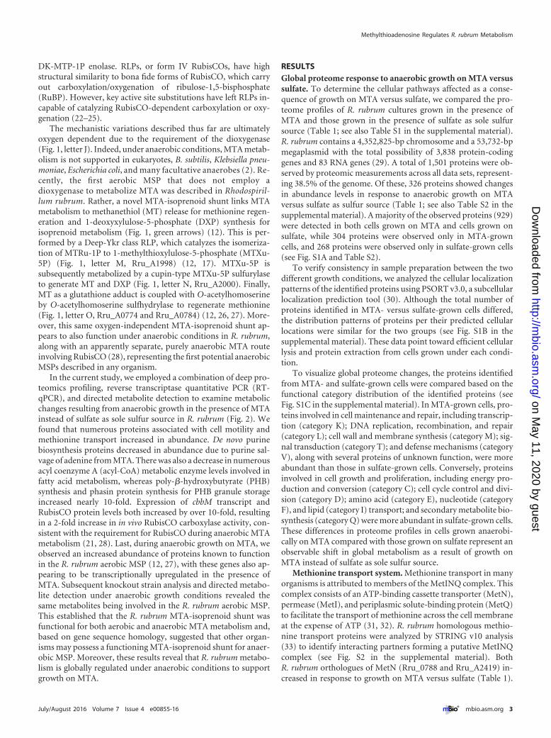
In order to determine how PHB accumulation is affected as aconsequence of growth on MTA as sole sulfur source, we quanti-fied PHB levels in cells grown on acetate or malate as a carbonsource and sulfate or MTA as a sulfur source (Fig. 4). Malate-grown cells with MTA as a sulfur source accumulated 3- to 7-foldmore PHB than did those with sulfate during exponential growth.Moreover, cells grown on malate and MTA produced PHB at thesame rate as did cells grown on acetate and sulfate, the latter ofwhich is known to stimulate PHB production (42). This increasein PHB production in the presence of MTA correlates with theobserved increase in both phasin protein and transcript levels.Clearly, even though PHB synthase (Rru_A2413) levels decreasein the presence of MTA, there is an increased flux of (R)-3-hydroxybutyryl-CoA toward PHB synthesis.
Purine salvage from MTA. During aerobic MTA metabolism,the first enzymatic step hydrolyzes MTA to produce MTR-1P andadenine (12). Presumably, adenine is salvaged to generate other
TABLE 1 Proteins regulated by growth on MTAa
Gene identifier Protein name Fold change
A0274 Acetyl-CoA C-acetyltransferase �2.30A0276 PHB synthesis repressor, PhaR 1.69A0774 O-Acetylhomoserine sulfhydrylase 3.14A0779 MetQ-like solute-binding protein 6.32A0784 O-Acetylhomoserine sulfhydrylase 3.88A0786 Cystathionine gamma-synthase 10.88A0788 MetN ABC transporter 9.44A0791 NlpAb lipoprotein 6.38A1142 Glycine hydroxymethyltransferase �3.09A1308 Acyl-CoA dehydrogenase �11.41A1309 Acetoacetyl-CoA reductase �4.92A1310 Acetyl-CoA C-acyltransferase �4.38A1334 Ribose-5-phosphate isomerase B 9.82A1823 3,4-Dihydroxy-2-butanone-4P synthase �1.80A1958 GMP reductase �12.04A1964 Adenylosuccinate synthase �12.18A1998 RubisCO-like MTRu-1P isomerase 8.13A2000 Cupin-like MTXu-5P sulfurylase 0.96A2400 RubisCO, form II 10.57A2404 Phosphoribulokinase 9.50A2413 PHB synthase, class I �3.61A2417 MetQ periplasmic solute-binding protein 2.63A2419 MetN ABC transporter 2.28A2465 Pyruvate kinase �1.27A2817 Phasin 14.52A3206 CheY response regulator protein 8.83A3283 Phasin-like activator of PHB degradation 1.51A3575 Acetate-CoA ligase �2.33a Protein name and gene correspond to annotated R. rubrum ATCC 11170 genome(NCBI reference sequence NC_007643.1). Fold change is the increase (�) or decrease(�) in protein abundance observed in MTA- versus sulfate-grown cells by deepproteomics.b NlpA, inner membrane lipoprotein A.
FIG 3 Anaerobic growth of R. rubrum wild-type (WT) strain (black) andtransposon mutant with disrupted CheB methylesterase gene (�Rru_A1406;see Fig. S4 in the supplemental material) (white) in malate minimal mediumwithout sulfur source (diamonds) or supplemented with 500 �M MTA (cir-cles) or 500 �M sulfate (squares). Error bars represent standard deviations forn � 3 independent growth experiments.
Methylthioadenosine Regulates R. rubrum Metabolism
July/August 2016 Volume 7 Issue 4 e00855-16 ® mbio.asm.org 5
on May 11, 2020 by guest
http://mbio.asm
.org/D
ownloaded from
nucleosides and nucleotides for cellular metabolism. This is sub-stantiated by the observation that the GMP-to-AMP conversionpathway, which balances A and G levels in the cell, is stronglydownregulated. GMP reductase (Rru_A1958), which irreversibly
converts GMP to IMP, and adenylosuccinate synthase(Rru_A1964), which generates AMP from IMP, each show a 12-fold decrease in protein levels (Fig. 2; Table 1). This is evidently inresponse to the excess adenine flux from MTA metabolism.
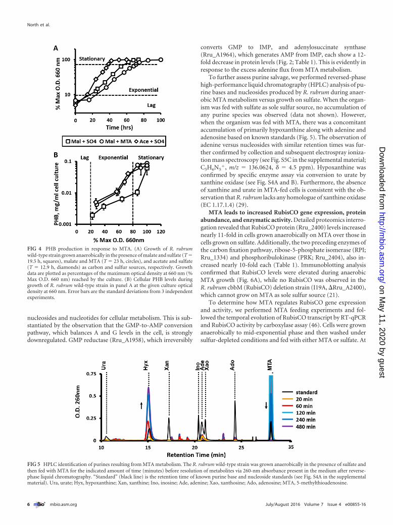
To further assess purine salvage, we performed reversed-phasehigh-performance liquid chromatography (HPLC) analysis of pu-rine bases and nucleosides produced by R. rubrum during anaer-obic MTA metabolism versus growth on sulfate. When the organ-ism was fed with sulfate as sole sulfur source, no accumulation ofany purine species was observed (data not shown). However,when the organism was fed with MTA, there was a concomitantaccumulation of primarily hypoxanthine along with adenine andadenosine based on known standards (Fig. 5). The observation ofadenine versus nucleosides with similar retention times was fur-ther confirmed by collection and subsequent electrospray ioniza-tion mass spectroscopy (see Fig. S5C in the supplemental material;C5H6N5
�, m/z � 136.0624, � � 4.5 ppm). Hypoxanthine wasconfirmed by specific enzyme assay via conversion to urate byxanthine oxidase (see Fig. S4A and B). Furthermore, the absenceof xanthine and urate in MTA-fed cells is consistent with the ob-servation that R. rubrum lacks any homologue of xanthine oxidase(EC 1.17.1.4) (29).
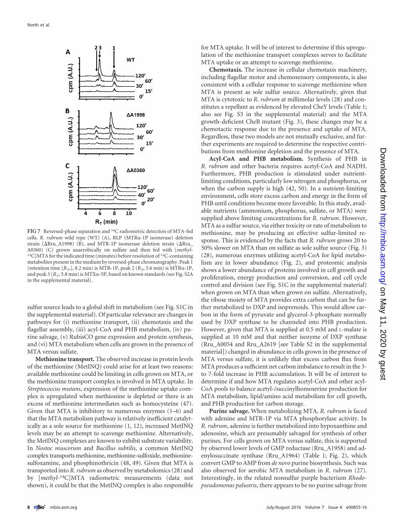
MTA leads to increased RubisCO gene expression, proteinabundance, and enzymatic activity. Detailed proteomics interro-gation revealed that RubisCO protein (Rru_2400) levels increasednearly 11-fold in cells grown anaerobically on MTA over those incells grown on sulfate. Additionally, the two preceding enzymes ofthe carbon fixation pathway, ribose-5-phosphate isomerase (RPI;Rru_1334) and phosphoribulokinase (PRK; Rru_2404), also in-creased nearly 10-fold each (Table 1). Immunoblotting analysisconfirmed that RubisCO levels were elevated during anaerobicMTA growth (Fig. 6A), while no RubisCO was observed in theR. rubrum cbbM (RubisCO) deletion strain (I19A, �Rru_A2400),which cannot grow on MTA as sole sulfur source (21).
To determine how MTA regulates RubisCO gene expressionand activity, we performed MTA feeding experiments and fol-lowed the temporal evolution of RubisCO transcript by RT-qPCRand RubisCO activity by carboxylase assay (46). Cells were grownanaerobically to mid-exponential phase and then washed undersulfur-depleted conditions and fed with either MTA or sulfate. At
FIG 4 PHB production in response to MTA. (A) Growth of R. rubrumwild-type strain grown anaerobically in the presence of malate and sulfate (T �19.5 h, squares), malate and MTA (T � 23 h, circles), and acetate and sulfate(T � 12.9 h, diamonds) as carbon and sulfur sources, respectively. Growthdata are plotted as percentages of the maximum optical density at 660 nm (%Max O.D. 660 nm) reached by the culture. (B) Cellular PHB levels duringgrowth of R. rubrum wild-type strain in panel A at the given culture opticaldensity at 660 nm. Error bars are the standard deviations from 3 independentexperiments.
FIG 5 HPLC identification of purines resulting from MTA metabolism. The R. rubrum wild-type strain was grown anaerobically in the presence of sulfate andthen fed with MTA for the indicated amount of time (minutes) before resolution of metabolites via 260-nm absorbance present in the medium after reverse-phase liquid chromatography. “Standard” (black line) is the retention time of known purine base and nucleoside standards (see Fig. S4A in the supplementalmaterial). Ura, urate; Hyx, hypoxanthine; Xan, xanthine; Ino, inosine; Ade, adenine; Xao, xanthosine; Ado, adenosine; MTA, 5-methylthioadenosine.
North et al.
6 ® mbio.asm.org July/August 2016 Volume 7 Issue 4 e00855-16
on May 11, 2020 by guest
http://mbio.asm
.org/D
ownloaded from

the point of feeding sulfate or MTA, transcript and RubisCO ac-tivity levels were identical in the two populations (Fig. 6B and C).During the first 60 min postfeeding, there was an ~5-fold increasein RubisCO transcript levels in MTA-fed cells over those insulfate-fed cells, but the levels then returned to initial levels at latertimes (Fig. 6B). This spike in transcript and concomitant proteinlevels resulted in RubisCO activity levels increasing by 2- to 3-fold(Fig. 6C). Clearly, MTA metabolism upregulates RubisCO expres-sion, resulting in increased cellular RubisCO activity. This furtherunderpins the requirement for RubisCO during anaerobic MTAmetabolism (21, 28).
Methionine salvage pathway under anaerobic growth condi-tions. Previous studies of R. rubrum aerobic MTA metabolism bymetabolomics and transcriptomics revealed that its entire aerobicMSP (Fig. 1, green arrows) was upregulated during growth onMTA versus sulfate (27). Here, while the same set of enzymes wasobserved by proteomics under anaerobic growth on both sulfateand MTA (see Table S2 in the supplemental material), only theR. rubrum RLP (Fig. 2; Rru_A1998), cupin (Fig. 2; Rru_A2000),and O-acetylhomoserine sulfhydrylase (Fig. 2; Rru_A0774/0784)showed changes in protein levels in response to growth on MTAversus sulfate (Table 1). The first two enzymes of the known aer-obic MSP, MTA phosphorylase (Fig. 1, letter D, and Fig. 2;
Rru_A0361) and MTR-1P isomerase (Fig. 1, letter E, and Fig. 2;Rru_A0360), were measured at similar levels in cells grown anaer-obically on MTA and on sulfate. To further identify potential in-volvement and regulation of these enzymes in response to anaer-obic MTA metabolism, we performed RT-qPCR on the MTAphosphorylase and RLP genes. Consistent with a lack of MTAphosphorylase regulation at the protein level, transcript levels didnot change in cells grown on MTA compared with those grown onsulfate (threshold cycle [��CT], 1.3 � 0.4). This suggests thatexpression of MTA phosphorylase (Rru_A0361) and of the pre-ceding gene, that for MTR-1P isomerase (Rru_A0360), is not reg-ulated by MTA during anaerobic growth, whereas it is regulatedby MTA under aerobic conditions (12, 27). However, there was a7- � 3-fold increase in RLP transcript levels in cells grown anaer-obically on MTA over that in cells grown on sulfate, similar toaerobic growth. This shows that the RLP is regulated by MTAunder both anaerobic and aerobic conditions.
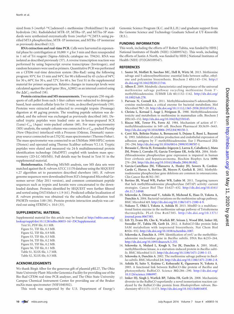
Our recent metabolomics studies of R. rubrum anaerobic MTAmetabolism showed increased levels of MTR-1P, MTRu-1P, MT,DXP, and subsequent isoprenoid precursors in cells fed with MTAover those in cells fed with sulfate as a sulfur source (28). Thissuggested that the known R. rubrum aerobic MSP (Fig. 1, greenarrows) may also function during anaerobic growth. To deter-mine if the MTA phosphorylase, MTR-1P isomerase, and RLP(Fig. 1, letters D, E, and M) are functionally involved in anaerobicMTA metabolism, we performed 14C radiochromatography byHPLC of knockout strains fed with [methyl-14C]MTA. In thewild-type strain, MTR-1P, MTRu-1P, and MTXu-5P/MTRu-5P(Fig. 7A; also see Fig. S5A in the supplemental material) and MT(28) levels increased upon feeding cells anaerobically with MTA.Inactivation of the R. rubrum RLP resulted in loss of MTXu-5P/MTRu-5P (Fig. 7B) and concomitant loss of MT generation (28).When the MTR-1P isomerase was inactivated, only MTR-1P wasobserved (Fig. 7C). Coordinately, when the MTA phosphorylasewas inactivated, MTR-1P was no longer observed as well (seeFig. S5B). Furthermore, production of each metabolite is specificto a single corresponding gene. Previously, under aerobic condi-tions, it was demonstrated that complementation of each knock-out strain with the corresponding protein restored production ofthe specific metabolite (12). Taken together, these results establishthat the R. rubrum aerobic MSP (MTA-isoprenoid shunt) coordi-nately functions anaerobically and that it is transcriptionally up-regulated via the RLP during growth on MTA.
DISCUSSION
Recycling MTA, a dead-end byproduct from polyamine, homo-serine lactone, and ethylene biosynthesis, is necessary for mostorganisms to maintain usable sulfur pools in a low-sulfur environ-ment and to prevent MTA cytotoxicity. In eukaryotes and manyprokaryotes, MTA is recycled into usable methionine by the uni-versal methionine salvage pathway, which requires oxygen for thefinal metabolic step to occur (Fig. 1, black arrows). Many anaero-bic organisms cannot metabolize MTA into usable sulfur, pre-sumably due to the oxygen requirement of the universal MSP, ordue to the lack of an MSP altogether (2). However, we have ob-served that Rhodospirillum rubrum (21) and Rhodopseudomonaspalustris (40) are able to metabolize MTA anaerobically, indicat-ing the existence of an anaerobic MSP(s). Through deep proteom-ics, qRT-PCR, and directed metabolite detection, we have ob-served that anaerobic growth on MTA instead of sulfate as the
FIG 6 RubisCO regulation by MTA. (A) Immunoblotting analysis forRubisCO proteins. Lane 1, purified R. rubrum RubisCO; lane 2, molecularmass standards; lane 3, RubisCO deletion strain (I19A) grown anaerobicallyon sulfate; lane 4, wild-type strain grown anaerobically on sulfate; lane 5,wild-type strain grown anaerobically on MTA. (B) RT-qPCR of RubisCO gene(Rru_A2400) levels in wild-type strain fed anaerobically with MTA (circles) orsulfate (squares). (C) RubisCO specific carboxylase activity in nanomoles ofCO2 fixed per minute per milligram of total cell protein in wild-type strain fedanaerobically with MTA (circles) or sulfate (squares). Error bars represent thestandard deviations for n � 3 independent feeding experiments.
Methylthioadenosine Regulates R. rubrum Metabolism
July/August 2016 Volume 7 Issue 4 e00855-16 ® mbio.asm.org 7
on May 11, 2020 by guest
http://mbio.asm
.org/D
ownloaded from
sulfur source leads to a global shift in metabolism (see Fig. S1C inthe supplemental material). Of particular relevance are changes inpathways for (i) methionine transport, (ii) chemotaxis and theflagellar assembly, (iii) acyl-CoA and PHB metabolism, (iv) pu-rine salvage, (v) RubisCO gene expression and protein synthesis,and (vi) MTA metabolism when cells are grown in the presence ofMTA versus sulfate.
Methionine transport. The observed increase in protein levelsof the methionine (MetINQ) could arise for at least two reasons:available methionine could be limiting in cells grown on MTA, orthe methionine transport complex is involved in MTA uptake. InStreptococcus mutans, expression of the methionine uptake com-plex is upregulated when methionine is depleted or there is anexcess of methionine intermediates such as homocysteine (47).Given that MTA is inhibitory to numerous enzymes (1–6) andthat the MTA metabolism pathway is relatively inefficient catalyt-ically as a sole source for methionine (1, 12), increased MetINQlevels may be an attempt to scavenge methionine. Alternatively,the MetINQ complexes are known to exhibit substrate variability.In Nostoc muscorum and Bacillus subtilis, a common MetINQcomplex transports methionine, methionine-sulfoxide, methionine-sulfoxamine, and phosphinothricin (48, 49). Given that MTA istransported into R. rubrum as observed by metabolomics (28) andby [methyl-14C]MTA radiometric measurements (data notshown), it could be that the MetINQ complex is also responsible
for MTA uptake. It will be of interest to determine if this upregu-lation of the methionine transport complexes serves to facilitateMTA uptake or an attempt to scavenge methionine.
Chemotaxis. The increase in cellular chemotaxis machinery,including flagellar motor and chemosensory components, is alsoconsistent with a cellular response to scavenge methionine whenMTA is present as sole sulfur source. Alternatively, given thatMTA is cytotoxic to R. rubrum at millimolar levels (28) and con-stitutes a repellant as evidenced by elevated CheY levels (Table 1;also see Fig. S3 in the supplemental material) and the MTAgrowth-deficient CheB mutant (Fig. 3), these changes may be achemotactic response due to the presence and uptake of MTA.Regardless, these two models are not mutually exclusive, and fur-ther experiments are required to determine the respective contri-butions from methionine depletion and the presence of MTA.
Acyl-CoA and PHB metabolism. Synthesis of PHB inR. rubrum and other bacteria requires acetyl-CoA and NADH.Furthermore, PHB production is stimulated under nutrient-limiting conditions, particularly low nitrogen and phosphorus, orwhen the carbon supply is high (42, 50). In a nutrient-limitingenvironment, cells store excess carbon and energy in the form ofPHB until conditions become more favorable. In this study, avail-able nutrients (ammonium, phosphorus, sulfate, or MTA) weresupplied above limiting concentrations for R. rubrum. However,MTA as a sulfur source, via either toxicity or rate of metabolism tomethionine, may be producing an effective sulfur-limited re-sponse. This is evidenced by the facts that R. rubrum grows 20 to50% slower on MTA than on sulfate as sole sulfur source (Fig. 3)(28), numerous enzymes utilizing acetyl-CoA for lipid metabo-lism are in lower abundance (Fig. 2), and proteomic analysisshows a lower abundance of proteins involved in cell growth andproliferation, energy production and conversion, and cell cyclecontrol and division (see Fig. S1C in the supplemental material)when grown on MTA than when grown on sulfate. Alternatively,the ribose moiety of MTA provides extra carbon that can be fur-ther metabolized to DXP and isoprenoids. This would allow car-bon in the form of pyruvate and glycerol-3-phosphate normallyused by DXP synthase to be channeled into PHB production.However, given that MTA is supplied at 0.5 mM and L-malate issupplied at 10 mM and that neither isozyme of DXP synthase(Rru_A0054 and Rru_A2619 [see Table S2 in the supplementalmaterial]) changed in abundance in cells grown in the presence ofMTA versus sulfate, it is unlikely that excess carbon flux fromMTA produces a sufficient net carbon imbalance to result in the 3-to 7-fold increase in PHB accumulation. It will be of interest todetermine if and how MTA regulates acetyl-CoA and other acyl-CoA pools to balance acetyl-/succinylhomoserine production forMTA metabolism, lipid/amino acid metabolism for cell growth,and PHB production for carbon storage.
Purine salvage. When metabolizing MTA, R. rubrum is facedwith adenine and MTR-1P via MTA phosphorylase activity. InR. rubrum, adenine is further metabolized into hypoxanthine andadenosine, which are presumably salvaged for synthesis of otherpurines. For cells grown on MTA versus sulfate, this is supportedby observed lower levels of GMP reductase (Rru_A1958) and ad-enylosuccinate synthase (Rru_A1964) (Table 1; Fig. 2), whichconvert GMP to AMP from de novo purine biosynthesis. Such wasalso observed for aerobic MTA metabolism in R. rubrum (27).Interestingly, in the related nonsulfur purple bacterium Rhodo-pseudomonas palustris, there appears to be no purine salvage from
FIG 7 Reversed-phase separation and 14C radiometric detection of MTA-fedcells. R. rubrum wild type (WT) (A), RLP (MTRu-1P isomerase) deletionstrain (�Rru_A1998) (B), and MTR-1P isomerase deletion strain (�Rru_A0360) (C) grown anaerobically on sulfate and then fed with [methyl-14C]MTA for the indicated time (minutes) before resolution of 14C-containingmetabolites present in the medium by reversed-phase chromatography. Peak 1(retention time [RT,], 8.2 min) is MTR-1P, peak 2 (RT, 5.6 min) is MTRu-1P,and peak 3 (RT, 5.8 min) is MTXu-5P, based on known standards (see Fig. S2Ain the supplemental material).
North et al.
8 ® mbio.asm.org July/August 2016 Volume 7 Issue 4 e00855-16
on May 11, 2020 by guest
http://mbio.asm
.org/D
ownloaded from

MTA, as only adenine accumulates and in stoichiometric amountsforms MTA (F. R. Tabita, unpublished observations). Surpris-ingly, based on homology, R. rubrum does not appear to possessmany of the conventional purine salvage enzymes, particularlypurine nucleosidase (EC 2.4.2.1), adenine deaminase (EC 3.5.4.2),and xanthine oxidase (EC 1.17.1.4) (29). As with many organisms,the R. rubrum MTA phosphorylase exhibits many similarities toother purine nucleosidases (2, 12). However, our previous studieswith the R. rubrum enzyme indicate that the catalytic rate of hy-drolysis for adenosine is ~30 times lower (�0.2 s�1) than for MTAand 5-deoxyadenosine (12). Moreover, neither the annotatedR. rubrum orthologue of adenosine deaminase (Rru_A0766) northat of purine phosphoribosyltransferase (Rru_A0607) was de-tected via proteomics. Further work is required to determine theprecise purine salvage pathway in R. rubrum.
Anaerobic MTA metabolism. For metabolism of MTA to us-able sulfur, proteomics and knockout metabolomics (Fig. 2 and 7)have confirmed our previous indications (28) that the R. rubrumMTA-isoprenoid shunt functions as an MSP anaerobically as wellas aerobically. In this MSP, the RLP and cupin (Fig. 1, letters Mand N) convert MTRu-1P into MT and DXP for methionine andisoprenoid synthesis, respectively, without the requirement foroxygen. Such an anaerobic MSP may be present in other bacteria(see Fig. S6 in the supplemental material), including Rhodopseu-domonas palustris, Meiothermus sp., Nitrococcus mobilis, and Ha-lorhodospira halophila, which possess homologues of theR. rubrum RLP and cupin in a similar gene organization.
Moreover, there is evidence for an additional anaerobic MSP aswell. Our previous reports showed that while the R. rubrum RLP isnot explicitly required to support anaerobic MTA metabolism, abona fide RubisCO is, leading to production of methylthiolactateand S-methylcysteine (21, 28). These MTA-derived metabolitesare not part of the MTA-isoprenoid shunt established as an anaer-obic MSP in this study. RubisCO’s essential role in an anaerobicMSP is further underpinned by its 10-fold-increased abundancein transcript and protein levels and 2- to 3-fold increase in activityin response to MTA. One possible role of RubisCO in anaerobicMSP is direct participation in the metabolism of MTR-1P intoS-methylcysteine or methylthiolactate. This is consistent with ourobservation that mutant RubisCOs devoid of carbon fixation ac-tivity are still able to support MTA metabolism in R. rubrum (28).An alternate model is that ribose-5-phosphate or ribulose-5-phosphate is generated during metabolism of MTR-1P intoS-methylcysteine. This is supported by an observed 10-fold in-crease in RPI (Rru_1334) and PRK (Rru_2404), the latter of whichconverts ribose-5-phosphate to RuBP, RubisCO’s substrate.While RuBP buildup does lead to toxicity and slow growth in anR. rubrum RubisCO deletion strain (51), our previous studies in-dicate that toxic RuBP buildup is not the cause of the no-growthphenotype in RubisCO deletion strains grown anaerobically onMTA as sole sulfur source (28). Experiments are under way toisolate RubisCO’s specific role in anaerobic MTA metabolism andto elucidate additional steps in anaerobic MSPs.
MATERIALS AND METHODSBacterial strains. The R. rubrum wild-type strain Str-2 is a spontaneousstreptomycin-resistant derivative of type strain S1 (ATCC 11170) (29).The RubisCO deletion strain I19A (�cbbM) contains a kanamycin resis-tance gene inserted into the cbbM gene (Rru_A2400) (52). The MTAphosphorylase (Rru_A0361) and MTR-1P isomerase (Rru_A0360) genes
were deleted as described in Text S1 in the supplemental material. The5-methylthioribulose-1-phosphate isomerase gene (Rru_A1998) was de-leted exactly as previously described (21).
Growth conditions. All R. rubrum strains were grown anaerobically at30°C in sulfur-free Ormerod’s minimal medium using modifications asdescribed previously (21) and supplemented with 20 mM sodium acetate(J. T. Baker) or 20 mM DL-malate (Sigma) and either 0.5 mM5-methylthioadenosine (Sigma) or ammonium sulfate (Sigma). Forgrowth, immunoblotting, RT-qPCR, PHB, and RubisCO activity analysis,photoheterotrophic cultures were grown in anaerobic test tubes under anitrogen atmosphere. For proteomics analysis, cells were grown in 1-literbottles fitted with anaerobic septa. All cultures were grown in duplicate.
Tn5 library screening. A 2,000-colony partial mini-Tn5 transposonlibrary was created by conjugating R. rubrum with E. coli DH5�-�pirtransformed with pRL27 (53) and screening for kanamycin-resistantR. rubrum transconjugants. Two-hundred-microliter photohetero-trophic cultures of each transconjugant were grown in 96-well plates un-der a 95:5 nitrogen-hydrogen atmosphere in thermally sealed bags(Scotchpak; 3M). Cultures were transferred by 48-pin stamp in an anaer-obic chamber (Coy Laboratories) to 12% (wt/vol) sulfur-free solid agarplates prepared as described elsewhere (54) containing Ormerod’s mini-mal medium with either 500 �M MTA or ammonium sulfate. Plates wereresealed in the same bag system and grown photoheterotrophically at30°C to screen for MTA metabolic mutants. The location of the mini-Tn5insertion was isolated and sequenced as previously described (53).
Protein purification and Western blot analysis. The R. rubrumRubisCO, MTA phosphorylase, MTR-1P isomerase, and MTRu-1Pisomerase (RLP) were purified as described previously (12). For immu-noblotting analysis of RubisCO, purified protein and crude extracts wereassayed as previously described (28). Immunoblots were developed withthe Attophos (Amersham, Buckinghamshire, England) detection reagentaccording to the manufacturer’s instructions and visualized with a Mo-lecular Dynamics Storm 840 imaging system.
Feeding experiments for RubisCO transcript, carboxylase, andMTA metabolism studies. The wild-type strain of R. rubrum was grownin the presence of sulfate to mid-exponential phase (optical density at 660nm [OD660] of ~0.5), washed anaerobically three times with sulfur-freeOrmerod’s minimal medium, and then resuspended to a final OD600 of~0.3 in 10 ml Ormerod’s minimal medium with 0.25 mM MTA, [methyl-14C]MTA, or ammonium sulfate. Feedings proceeded anaerobically at30°C for the designated amount of time, culture samples were centrifugedat 5,000 � g for 5 min at 20°C, and the spent medium and cell pellets wereseparately flash-frozen at �80°C for further analysis. Pellets for RT-qPCRwere extracted as described below.
PHB assay and RubisCO carboxylase activity. Pellets for PHB quan-tification were resuspended in 10 ml of 5% sodium hypochlorite (Clorox)per 1-OD660 unit of original cell culture. PHB was extracted and quanti-fied spectrophotometrically at 235 nm (Cary; Varian) according to themethod of Law and Slepecky (55). Pellets for RubisCO carboxylase activ-ity were resuspended in 0.5 ml 100 mM Bicine buffer (pH 8; Sigma),sonicated by a Branson Sonifier on ice for 3 min at a 50% duty cycle, andcentrifuged at 15,000 � g at 4°C for 10 min to generate cell extract.RubisCO carboxylase activity was monitored by [14C]bicarbonate(PerkinElmer) carboxylation of ribulose-1,5-bisphosphate (Sigma) andwas assayed as previously described (46).
HPLC metabolite analysis. Detection of R. rubrum metabolites due toMTA metabolism present in the spent medium was performed by reverse-phase HPLC on a Zorbax C18 (Agilent) column connected to a ShimadzuProminence HPLC system with dual-wavelength detection (215 nm and260 nm) and an inline �-RAM radiochromatography scintillation detec-tor (IN/US Systems). Metabolites were eluted on a linear gradient from 0to 50% acetonitrile (J. T. Baker) in 20 mM ammonium acetate over30 min at 30°C. All purine base and nucleoside standards were fromSigma-Aldrich. Spent medium was treated with xanthine oxidase (Sigma)according to the manufacturer’s protocol. [methyl-14C]MTA was synthe-
Methylthioadenosine Regulates R. rubrum Metabolism
July/August 2016 Volume 7 Issue 4 e00855-16 ® mbio.asm.org 9
on May 11, 2020 by guest
http://mbio.asm
.org/D
ownloaded from
sized from S-[methyl-14C]adenosyl-L-methionine (PerkinElmer) by acidhydrolysis (56). Radiolabeled MTR-1P, MTRu-1P, and MTXu-5P stan-dards were synthesized enzymatically from [methyl-14C]MTA using pu-rified MTA phosphorylase, MTR-1P isomerase, and MTRu-1P isomeraseas previously described (12).
RNA extraction and real-time PCR. Cells were harvested in exponen-tial phase by centrifugation at 10,000 � g for 3 min and then resuspendedin 1 ml of Tri reagent (Sigma-Aldrich; catalogue no. T9424). RNA wasisolated as described previously (57). A reverse transcription reaction wasperformed by using Superscript reverse transcriptase (Invitrogen), andrandom hexamers were used as primers. Quantitative PCR was performedon a CFX96 real-time detection system (Bio-Rad) using the followingprogram: 95°C for 15 min and 94°C for 90 s followed by 45 cycles of 94°Cfor 30 s, 60°C for 30 s, and 72°C for 60 s. See Text S1 in the supplementalmaterial for primer sequences. Relative changes in transcript levels werecalculated against the rpoD gene (Rru_A2882) as an internal control usingthe ��CT method (58).
Protein extraction and MS measurements. Two separate 250-mg ali-quots of cell pellet from each 1-liter culture were subjected to detergent-based, heat-assisted cellular lysis for 15 min, as described previously (59).Proteins were extracted and subjected to overnight trypsin (Promega)digestion at 40 �g/mg protein. The resulting peptide solution was de-salted, and the solvent was exchanged as previously described (60). De-salted tryptic peptides were loaded onto an in-house-prepared SCX(Luna)-C18 (Aqua) resin-packed column (60). For mass spectrometry(MS) analysis, the sample column was connected to a C18-packed Picotip(New Objective) interfaced with a Proxeon (Odense, Denmark) nanos-pray source connected to an LTQ XL mass spectrometer (Thermo Fisher).The mass spectrometer was connected to an UltiMate 3000 HPLC system(Dionex) and operated using Thermo Xcalibur software V2.1.0. Trypticpeptides were eluted and measured via 24-h multidimensional proteinidentification technology (MuDPIT) coupled with tandem mass spec-trometry (2D-LC-MS/MS). Full details may be found in Text S1 in thesupplemental material.
Bioinformatics. Following MS/MS analysis, raw MS data sets werematched against the R. rubrum ATCC 11170 database using the SEQUESTv.27 algorithm set to parameters described elsewhere (60). R. rubrumgenome sequences were downloaded from JGI’s Integrated Microbial Ge-nomes server (May 2011 version), and common contaminant peptidesequences such as trypsin and keratin were concatenated to the down-loaded database. Proteins identified by SEQUEST were further filteredand sorted using DATASelect v.1.9 (61). Predicted cellular localization ofR. rubrum proteins was obtained via the subcellular localization toolPSORTb version 3.00 (30). Protein-protein interaction analysis was car-ried out using STRING v. 10.0 (33).
SUPPLEMENTAL MATERIALSupplemental material for this article may be found at http://mbio.asm.org/lookup/suppl/doi:10.1128/mBio.00855-16/-/DCSupplemental.
Text S1, PDF file, 0.1 MB.Figure S1, TIF file, 0.3 MB.Figure S2, TIF file, 0.9 MB.Figure S3, TIF file, 0.5 MB.Figure S4, TIF file, 0.2 MB.Figure S5, TIF file, 0.2 MB.Figure S6, TIF file, 0.1 MB.Table S1, XLSX file, 1.7 MB.Table S2, XLSX file, 0.3 MB.
ACKNOWLEDGMENTS
We thank Birgit Alber for the generous gift of plasmid pRL27, The OhioState University Plant-Microbe Genomics Facility for providing use of theBio-Rad CFX96 real-time PCR analyzer, and The Ohio State UniversityCampus Chemical Instrument Center for providing use of the BrukermaXis mass spectrometer (NSF1040302).
This work was supported by the U.S. Department of Energy’s
Genomic Science Program (K.C. and R.L.H.) and graduate support fromthe Genome Science and Technology Graduate School at UT-Knoxville(R.S.).
FUNDING INFORMATIONThis work, including the efforts of F Robert Tabita, was funded by HHS |National Institutes of Health (NIH) (GM095742). This work, includingthe efforts of Justin A North, was funded by HHS | National Institutes ofHealth (NIH) (F32GM109547).
REFERENCES1. Sauter M, Moffatt B, Saechao MC, Hell R, Wirtz M. 2013. Methionine
salvage and S-adenosylmethionine: essential links between sulfur, ethyl-ene and polyamine biosynthesis. Biochem J 451:145–154. http://dx.doi.org/10.1042/BJ20121744.
2. Albers E. 2009. Metabolic characteristics and importance of the universalmethionine salvage pathway recycling methionine from 5=-methylthioadenosine. IUBMB Life 61:1132–1142. http://dx.doi.org/10.1002/iub.278.
3. Parveen N, Cornell KA. 2011. Methylthioadenosine/S-adenosylhomo-cysteine nucleosidase, a critical enzyme for bacterial metabolism. MolMicrobiol 79:7–20. http://dx.doi.org/10.1111/j.1365-2958.2010.07455.x.
4. Christa L, Kersual J, Augé J, Pérignon JL. 1988. Methylthioadenosinetoxicity and metabolism to methionine in mammalian cells. Biochem J255:145–152. http://dx.doi.org/10.1042/bj2550145.
5. Riscoe MK, Tower PA, Ferro AJ. 1984. Mechanism of action of 5=-methylthioadenosine in S49 cell. Biochem Pharmacol 33:3639 –3643.http://dx.doi.org/10.1016/0006-2952(84)90150-3.
6. Cerri MA, Beltrán-Nuñez A, Bernasconi S, Dejana E, Bassi L, BazzoniG. 1993. Inhibition of cytokine production and endothelial expression ofadhesion antigens by 5=-methylthioadenosine. Eur J Pharmacol 232:291–294. http://dx.doi.org/10.1016/0014-2999(93)90787-I.
7. Berasain C, Hevia H, Fernández-Irigoyen J, Larrea E, Caballería J, MatoJM, Prieto J, Corrales FJ, García-Trevijano ER, Avila MA. 2004. Meth-ylthioadenosine phosphorylase gene expression is impaired in humanliver cirrhosis and hepatocarcinoma. Biochim Biophys Acta 1690:276 –284. http://dx.doi.org/10.1016/j.bbadis.2004.08.002.
8. García-Castellano JM, Villanueva A, Healey JH, Sowers R, Cordon-Cardo C, Huvos A, Bertino JR, Meyers P, Gorlick R. 2002. Methylth-ioadenosine phosphorylase gene deletions are common in osteosarcoma.Clin Cancer Res 8:782–787.
9. Bertino JR, Waud WR, Parker WB, Lubin M. 2011. Targeting tumorsthat lack methylthioadenosine phosphorylase (MTAP) activity: currentstrategies. Cancer Biol Ther 11:627– 632. http://dx.doi.org/10.4161/cbt.11.7.14948.
10. Sekowska A, Dénervaud V, Ashida H, Michoud K, Haas D, Yokota A,Danchin A. 2004. Bacterial variations on the methionine salvage pathway.BMC Microbiol 4:9. http://dx.doi.org/10.1186/1471-2180-4-9.
11. Nakano T, Ohki I, Yokota A, Ashida H. 2013. MtnBD is a multifunc-tional fusion enzyme in the methionine salvage pathway of Tetrahymenathermophila. PLoS One 8:e67385. http://dx.doi.org/10.1371/journal.pone.0067385.
12. Erb TJ, Evans BS, Cho K, Warlick BP, Sriram J, Wood BM, Imker HJ,Sweedler JV, Tabita FR, Gerlt JA. 2012. A RubisCO-like protein linksSAM metabolism with isoprenoid biosynthesis. Nat Chem Biol8:926 –932. http://dx.doi.org/10.1038/nchembio.1087.
13. Sekowska A, Danchin A. 1999. Identification of yrrU as the methylthio-adenosine nucleosidase gene in Bacillus subtilis. DNA Res 6:255–264.http://dx.doi.org/10.1093/dnares/6.5.255.
14. Sekowska A, Mulard L, Krogh S, Tse JK, Danchin A. 2001. MtnK,methylthioribose kinase, is a starvation-induced protein in Bacillus subti-lis. BMC Microbiol 1:15. http://dx.doi.org/10.1186/1471-2180-1-15.
15. Sekowska A, Danchin A. 2002. The methionine salvage pathway in Bacil-lus subtilis. BMC Microbiol 2:8. http://dx.doi.org/10.1186/1471-2180-2-8.
16. Ashida H, Saito Y, Kojima C, Kobayashi K, Ogasawara N, Yokota A.2003. A functional link between RuBisCO-like protein of Bacillus andphotosynthetic RuBisCO. Science 302:286 –290. http://dx.doi.org/10.1126/science.1086997.
17. Imker HJ, Singh J, Warlick BP, Tabita FR, Gerlt JA. 2008. Mechanisticdiversity in the RuBisCO superfamily: a novel isomerization reaction cat-alyzed by the RuBisCO-like protein from Rhodospirillum rubrum. Bio-chemistry 47:11171–11173. http://dx.doi.org/10.1021/bi801685f.
North et al.
10 ® mbio.asm.org July/August 2016 Volume 7 Issue 4 e00855-16
on May 11, 2020 by guest
http://mbio.asm
.org/D
ownloaded from

18. Hanson TE, Tabita FR. 2001. A ribulose-1,5-bisphosphate carboxylase/oxygenase (RubisCO)-like protein from Chlorobium tepidum that is in-volved with sulfur metabolism and the response to oxidative stress. ProcNatl Acad Sci U S A 98:4397– 4402. http://dx.doi.org/10.1073/pnas.081610398.
19. Hanson TE, Tabita FR. 2003. Insights into the stress response and sulfurmetabolism revealed by proteome analysis of a Chlorobium tepidum mu-tant lacking the Rubisco-like protein. Photosynth Res 78:231–248. http://dx.doi.org/10.1023/B:PRES.0000006829.41444.3d.
20. Imker HJ, Fedorov AA, Fedorov EV, Almo SC, Gerlt JA. 2007. Mech-anistic diversity in the RuBisCO superfamily: the “enolase” in the methi-onine salvage pathway in Geobacillus kaustophilus. Biochemistry 46:4077– 4089. http://dx.doi.org/10.1021/bi7000483.
21. Singh J, Tabita FR. 2010. Roles of RubisCO and the RubisCO-like proteinin 5-methylthioadenosine metabolism in the nonsulfur purple bacteriumRhodospirillum rubrum. J Bacteriol 192:1324 –1331. http://dx.doi.org/10.1128/JB.01442-09.
22. Tabita FR, Hanson TE, Satagopan S, Witte BH, Kreel NE. 2008. Phy-logenetic and evolutionary relationships of RubisCO and the RubisCO-like proteins and the functional lessons provided by diverse molecularforms. Philos Trans R Soc Lond B Biol Sci 363:2629 –2640. http://dx.doi.org/10.1098/rstb.2008.0023.
23. Tabita FR, Hanson TE, Li H, Satagopan S, Singh J, Chan S. 2007.Function, structure, and evolution of the RubisCO-like proteins and theirRubisCO homologs. Microbiol Mol Biol Rev 71:576 –599. http://dx.doi.org/10.1128/MMBR.00015-07.
24. Saito Y, Ashida H, Sakiyama T, de Marsac NT, Danchin A, Sekowska A,Yokota A. 2009. Structural and functional similarities between a ribulose-1,5-bisphosphate carboxylase/oxygenase (RuBisCO)-like protein fromBacillus subtilis and photosynthetic RuBisCO. J Biol Chem 284:13256 –13264. http://dx.doi.org/10.1074/jbc.M807095200.
25. Ashida H, Saito Y, Nakano T, Tandeau de Marsac N, Sekowska A,Danchin A, Yokota A. 2008. RuBisCO-like proteins as the enolase enzymein the methionine salvage pathway: functional and evolutionary relation-ships between RuBisCO-like proteins and photosynthetic RuBisCO. J ExpBot 59:1543–1554. http://dx.doi.org/10.1093/jxb/ern104.
26. Warlick BP, Evans BS, Erb TJ, Ramagopal UA, Sriram J, Imker HJ,Sauder JM, Bonanno JB, Burley SK, Tabita FR, Almo SC, Sweedler JS,Gerlt JA. 2012. 1-Methylthio-D-xylulose 5-phosphate methylsulfurylase: anovel route to 1-deoxy-D-xylulose 5-phosphate in Rhodospirillumrubrum. Biochemistry 51:8324 – 8326. http://dx.doi.org/10.1021/bi301215g.
27. Cho K, Evans BS, Wood BM, Kumar R, Erb TJ, Warlick BP, Gerlt JA,Sweedler JV. 2015. Integration of untargeted metabolomics with tran-scriptomics reveals active metabolic pathways. Metabolomics 11:503–517.http://dx.doi.org/10.1007/s11306-014-0713-3.
28. Dey S, North JA, Sriram J, Evans BS, Tabita FR. 2015. In vivo studies inRhodospirillum rubrum indicate that ribulose-1,5-bisphosphatecarboxylase/oxygenase (RubisCO) catalyzes two obligatorily required andphysiologically significant reactions for distinct carbon and sulfur meta-bolic pathways. J Biol Chem 290:30658 –30668. http://dx.doi.org/10.1074/jbc.M115.691295.
29. Munk AC, Copeland A, Lucas S, Lapidus A, Del Rio TG, Barry K,Detter JC, Hammon N, Israni S, Pitluck S, Brettin T, Bruce D, Han C,Tapia R, Gilna P, Schmutz J, Larimer F, Land M, Kyrpides NC,Mavromatis K, Richardson P, Rohde M, Göker M, Klenk HP, Zhang Y,Roberts GP, Reslewic S, Schwartz DC. 2011. Complete genome sequenceof Rhodospirillum rubrum type strain (S1). Stand Genomic Sci4:293–302. http://dx.doi.org/10.4056/sigs.1804360.
30. Yu NY, Wagner JR, Laird MR, Melli G, Rey S, Lo R, Dao P, SahinalpSC, Ester M, Foster LJ, Brinkman FS. 2010. PSORTb 3.0: improvedprotein subcellular localization prediction with refined localization sub-categories and predictive capabilities for all prokaryotes. Bioinformatics26:1608 –1615. http://dx.doi.org/10.1093/bioinformatics/btq249.
31. Johnson E, Nguyen PT, Yeates TO, Rees DC. 2012. Inward facingconformations of the MetNI methionine ABC transporter: implicationsfor the mechanism of transinhibition. Protein Sci 21:84 –96. http://dx.doi.org/10.1002/pro.765.
32. Merlin C, Gardiner G, Durand S, Masters M. 2002. The Escherichia colimetD locus encodes an ABC transporter which includes Abc (MetN), YaeE(MetI), and YaeC (MetQ). J Bacteriol 184:5513–5517. http://dx.doi.org/10.1128/JB.184.19.5513-5517.2002.
33. Szklarczyk D, Franceschini A, Wyder S, Forslund K, Heller D, Huerta-
Cepas J, Simonovic M, Roth A, Santos A, Tsafou KP, Kuhn M, Bork P,Jensen LJ, von Mering C. 2015. STRING v10: protein–protein interactionnetworks, integrated over the tree of life. Nucleic Acids Res 43:D447–D452. http://dx.doi.org/10.1093/nar/gku1003.
34. Sarkar MK, Paul K, Blair D. 2010. Chemotaxis signaling protein CheYbinds to the rotor protein FliN to control the direction of flagellar rotationin Escherichia coli. Proc Natl Acad Sci U S A 107:9370 –9375. http://dx.doi.org/10.1073/pnas.1000935107.
35. Manson MD. 2010. Dynamic motors for bacterial flagella. Proc Natl AcadSci U S A. 107:11151–11152. http://dx.doi.org/10.1073/pnas.1006365107.
36. Morimoto YV, Minamino T. 2014. Structure and function of the bi-directional bacterial flagellar motor. Biomolecules 4:217–234. http://dx.doi.org/10.3390/biom4010217.
37. Djordjevic S, Stock AM. 1998. Structural analysis of bacterial chemotaxisproteins: components of a dynamic signaling system. J Struct Biol 124:189 –200. http://dx.doi.org/10.1006/jsbi.1998.4034.
38. Meir Y, Jakovljevic V, Oleksiuk O, Sourjik V, Wingreen NS. 2010.Precision and kinetics of adaptation in bacterial chemotaxis. Biophys J99:2766 –2774. http://dx.doi.org/10.1016/j.bpj.2010.08.051.
39. Kirby JR, Niewold TB, Maloy S, Ordal GW. 2000. CheB is required forbehavioural responses to negative stimuli during chemotaxis in Bacillussubtilis. Mol Microbiol 35:44 –57. http://dx.doi.org/10.1046/j.1365-2958.2000.01676.x.
40. Singh J. 2008. Functional relationships among Rubisco family members.Ph.D. thesis. The Ohio State University, Columbus, OH.
41. Mezzina MP, Wetzler DE, Catone MV, Bucci H, Di Paola M, PettinariMJ. 2014. A phasin with many faces: structural insights on PhaP fromAzotobacter sp. FA8. PLoS One 9:e103012. http://dx.doi.org/10.1371/journal.pone.0103012.
42. Handrick R, Reinhardt S, Schultheiss D, Reichart T, Schüler D, Jen-drossek V, Jendrossek D. 2004. Unraveling the function of the Rhodospi-rillum rubrum activator of polyhydroxybutyrate (PHB) degradation: theactivator is a PHB-granule-bound protein (phasin). J Bacteriol 186:2466 –2475. http://dx.doi.org/10.1128/JB.186.8.2466-2475.2004.
43. York GM, Junker BH, Stubbe JA, Sinskey AJ. 2001. Accumulation of thePhaP phasin of Ralstonia eutropha is dependent on production of polyhy-droxybutyrate in cells. J Bacteriol 183:4217– 4226. http://dx.doi.org/10.1128/JB.183.14.4217-4226.2001.
44. Yoshida K, Takemoto Y, Sotsuka T, Tanaka K, Takenaka S. 2013. PhaPphasins play a principal role in poly-�-hydroxybutyrate accumulation infree-living Bradyrhizobium japonicum. BMC Microbiol 13:290. http://dx.doi.org/10.1186/1471-2180-13-290.
45. Pötter M, Steinbüchel A. 2005. Poly(3-hydroxybutyrate) granule-associated proteins: impacts on poly(3-hydroxybutyrate) synthesis anddegradation. Biomacromolecules 6:552–560. http://dx.doi.org/10.1021/bm049401n.
46. Satagopan S, Scott SS, Smith TG, Tabita FR. 2009. A RubisCO mutantthat confers growth under a normally “inhibitory” oxygen concentration.Biochemistry 48:9076 –9083. http://dx.doi.org/10.1021/bi9006385.
47. Sperandio B, Gautier C, McGovern S, Ehrlich DS, Renault P, Martin-Verstraete I, Guédon E. 2007. Control of methionine synthesis and up-take by MetR and homocysteine in Streptococcus mutans. J Bacteriol 189:7032–7044. http://dx.doi.org/10.1128/JB.00703-07.
48. Singh AK, Syiem MB, Singh RS, Adhikari S, Rai AN. 2008. A commontransport system for methionine, L-methionine-DL-sulfoximine (MSX),and phosphinothricin (PPT) in diazotrophic cyanobacterium Nostoc mus-corum. Curr Microbiol 56:436 – 441. http://dx.doi.org/10.1007/s00284-008-9111-2.
49. Hullo MF, Auger S, Dassa E, Danchin A, Martin-Verstraete I. 2004. ThemetNPQ operon of Bacillus subtilis encodes an ABC permease transport-ing methionine sulfoxide, D- and L-methionine. Res Microbiol 155:80 – 86. http://dx.doi.org/10.1016/j.resmic.2003.11.008.
50. Guevara-Martínez M, Sjöberg Gällnö K, Sjöberg G, Jarmander J, Perez-Zabaleta M, Quillaguamán J, Larsson G. 2015. Regulating the produc-tion of (R)-3-hydroxybutyrate in Escherichia coli by N or P limitation.Front Microbiol 6:844. http://dx.doi.org/10.3389/fmicb.2015.00844.
51. Wang D, Zhang Y, Pohlmann EL, Li J, Roberts GP. 2011. The poorgrowth of Rhodospirillum rubrum mutants lacking RubisCO is due to theaccumulation of ribulose-1,5-bisphosphate. J Bacteriol 193:3293–3303.http://dx.doi.org/10.1128/JB.00265-11.
52. Falcone DL, Tabita FR. 1993. Complementation analysis and regulationof CO2 fixation gene expression in a ribulose-1,5-bisphosphate
Methylthioadenosine Regulates R. rubrum Metabolism
July/August 2016 Volume 7 Issue 4 e00855-16 ® mbio.asm.org 11
on May 11, 2020 by guest
http://mbio.asm
.org/D
ownloaded from

carboxylase-oxygenase deletion strain of Rhodospirillum rubrum. J Bacte-riol 175:5066 –5077.
53. Larsen RA, Wilson MM, Guss AM, Metcalf WW. 2002. Genetic analysisof pigment biosynthesis in Xanthobacter autotrophicus Py2 using a new,highly efficient transposon mutagenesis system that is functional in a widevariety of bacteria. Arch Microbiol 178:193–201. http://dx.doi.org/10.1007/s00203-002-0442-2.
54. Russell B, Mead TH, Polson A. 1964. A method of preparing agarose.Biochim Biophys Acta 86:169 –174. http://dx.doi.org/10.1016/0304-4165(64)90171-0.
55. Law JH, Slepecky RA. 1961. Assay of poly-�-hydroxybutyric acid. J Bac-teriol 82:33–36.
56. Schlenk F, Ehninger DJ. 1964. Observations on the metabolism of 5=-methylthioadenosine. Arch Biochem Biophys 106:95–100. http://dx.doi.org/10.1016/0003-9861(64)90161-4.
57. Chomczynski P. 1993. A reagent for the single-step simultaneous isola-
tion of RNA, DNA, and proteins from cell and tissue samples. Biotech-niques 15:532–537.
58. Schmittgen TD, Livak KJ. 2008. Analyzing real-time PCR data by thecomparative C(T) method. Nat Protoc 3:1101–1108. http://dx.doi.org/10.1038/nprot.2008.73.
59. Chourey K, Jansson J, VerBerkmoes N, Shah M, Chavarria KL, TomLM, Brodie EL, Hettich RL. 2010. Direct cellular lysis/protein extractionprotocol for soil metaproteomics. J Proteome Res 9:6615– 6622. http://dx.doi.org/10.1021/pr100787q.
60. Sharma R, Dill BD, Chourey K, Shah M, VerBerkmoes NC, Hettich RL.2012. Coupling a detergent lysis/cleanup methodology with intact proteinfractionation for enhanced proteome characterization. J Proteome Res11:6008 – 6018. http://dx.doi.org/10.1021/pr300709k.
61. Tabb DL, McDonald WH, Yates JR, III. 2002. DTASelect and contrast: toolsfor assembling and comparing protein identifications from shotgun pro-teomics. J Proteome Res 1:21–26. http://dx.doi.org/10.1021/pr015504q.
North et al.
12 ® mbio.asm.org July/August 2016 Volume 7 Issue 4 e00855-16
on May 11, 2020 by guest
http://mbio.asm
.org/D
ownloaded from