meta-analysis of 50 phase iii clinical trials in ... file(sgpt), serum alkaline phosphatase (sap),...
TRANSCRIPT
Medicine Update (2004): 12(2), 51-61
Meta-analysis of 50 Phase III clinical trials in evaluation of efficacy and safety of Liv.52 in infective hepatitis
Kolhapure, S.A., M.D., Senior Medical Advisor,
and Mitra, S.K., M.D., Executive Director,Research and Technical Service,
R&D Center, The Himalaya Drug Company, Bangalore, India ABSTRACT Hepatitis A (HA) has a worldwide distribution occurring in epidemic and sporadic patterns. Hepatitis A is an acute, but benign form of viral hepatitis and early renormalizations of hepatic functions with symptomatic and clinical recovery are the objectives in the clinical management of HA. meta-analysis is the term used to describe quantitative methods for combining information across different studies and this study was planned for meta-analysis of the efficacy and safety of Liv.52 tablet and syrup, in HA, as reported in 50 published study reports. All study reports evaluating efficacy and safety of Liv.52, were included for the meta-analysis, regardless of the study design, but Phase I and II clinical, experimental and preclinical studies were excluded from the meta-analysis. Each study was abstracted for the number and ages of enrolled patients, changes in the biochemical parameters [serum bilirubin (SB), serum glutamic oxaloacetic transaminase (SGOT), serum glutamic pyruvic transaminase (SGPT), serum alkaline phosphatase (SAP), serum albumin (SA) and serum globulin (SG), and prothrombin time (PT)] from baseline to values at the end of study and total duration of clinical recovery were recorded. Incidence of adverse events during the study period and patient compliance to the drug treatment was noted. The predefined primary endpoints were to determine level of statistical significance for symptomatic improvement, renormalization of biochemical parameters and total duration of clinical recovery. The predefined secondary endpoints were incidence of adverse events during the study period and compliance to the drug treatment. Total 50 clinical studies conducted over a span of 30 years were considered for this meta-analysis and the mean duration of these studies was 6.62 months. Out of total fifty studies, 3 were double-blind placebo-controlled studies, 21 were placebo-controlled studies, 22 were non-comparative studies and 4 studies were case reports. Total 4490 patients were enrolled in these studies and 233 children were part of study population. Cumulative data analysis showed a significant reduction in the mean SB, SGOT, SGPT, AP levels, PT and mean period required for total (symptomatic, clinical and biochemical) recovery. The decreased SA and SG levels were also increased significantly, when compared to the pre-treatment values, in all studies. There were no reported or observed significant adverse events in all trials and the overall drug compliance was excellent. Therefore, this metaanalysis concludes that, Liv.52 tablets and syrup are effective and safe in the management of hepatitis. INTRODUCTION Meta-analysis (also referred as ‘quantitative synthesis’ or ‘overview analysis’) is the term used to describe quantitative methods for combining information across different studies. Glass coined the term `meta-analysis’ in 19761, to describe this idea of utilizing information in many studies of the same effect, although the concept itself is much older (dating back to the 1930s studies by Fisher and Pearson). Today, the term `meta-analysis' is reserved for a
situation where one combines numerical effects from a collection of studies, rather than giving a more general non-quantitative overview. The term “Hepatitis A (HA)” has replaced all previous designations like ‘type A viral hepatitis’, ‘infectious hepatitis’, ‘epidemic hepatitis’, ‘epidemic jaundice’, ‘catarrhal jaundice’, ‘infectious icterus’, ‘Botkin’s disease’, and ‘MS-1 hepatitis’. HA has a worldwide distribution and occurs in both epidemic and sporadic patterns. In developing countries, the incidence of HA in adults is relatively low due to the exposure to the virus in childhood and most adults demonstrate an immunity that provides lifelong protection against reinfection2. Hepatitis A is an acute but benign form of viral hepatitis caused by an RNA virus that does not persist in the blood serum. Hepatitis A virus (HAV) is of the enterovirus group of the Picornaviridae family and HAV has a single molecule of RNA surrounded by a small (27 nm diameter) protein capsid. HA is a food/waterborne disease, with feco-oral transmission and the incubation period is variable (from 15 to 25 days). Incubation period is inversely proportional to infective dose and the infectious dose presumably is 10-100 virus particles2. Hepatitis A is usually a mild illness characterized by sudden onset of fever, malaise, nausea, anorexia and abdominal discomfort, followed after several days by jaundice. The dark urine (which precedes jaundice by 2/3 days), indicates the onset of the disease. The risk of transmission is generally low amongst household contacts, but outbreaks occur in nurseries and institutions with attack rates around 10%-15%. The duration of viral shedding is upto 14 days; with a sharp fall off after 5 days, but the period of infectiousness is about 8 to 17 days (the stool remains infectious upto 15 days). Asymptomatic infections are frequent. The serial interval between index and secondary cases is either shorter or equal to the incubation period, indicating that transmission usually occurs before or around the onset of jaundice 2. Hepatitis A is diagnosed by finding IgM anti-HAV in serum during the acute or early convalescent phases of disease. A moderate increase in conjugated bilirubin, with marked elevation of SGOT, SGPT and Lactate Dehydrogenase isoenzymes (LDH) are noted in HA (low Na+ and Cl- levels may be seen due to severe vomiting). Marginal elevation of alkaline phosphatase is seen due to compression of intrahepatic biliary canaliculi by swollen parenchymatous cells. Triglycerides and cholesterol may be temporarily and marginally lowered2. Hepatitis A is the least serious of the hepatitis viruses, as it does not kill liver cells and also, there is no risk for a chronic form. Hepatitis A is a self-limiting disease and no specific treatment is available. The primary goals for managing acute viral hepatitis are to provide adequate nutrition (with restricted protein and fat intake), to prevent further damage to the liver, and transmission of infection to others. Therefore, early renormalizations of hepatic functions with symptomatic and clinical recovery are the objectives in the clinical management of HA. Liv.52 tablet and syrup are polyherbal formulations, used extensively in the management of HA. Liv.52 syrup contains powders of Capparis spinosa, Cichorium intybus, Solanum nigrum, Terminalia arjuna, Cassia occidentalis, Achillea millefolium and Tamarix gallica (in Liv.52 tablet, Mandur bhasma is added). This study was planned for meta-analysis of the efficacy and safety of Liv.52 tablet and syrup, in HA, as reported in 50 published study reports.
AIM OF STUDY The aim of the study was to meta-analyze the efficacy and short- and long-term safety of Liv.52 in HA, as reported in fifty published study reports. STUDY DESIGN This study was a cumulative meta-analysis of fifty published study reports of Liv.52 in HA. MATERIALS AND METHODS Inclusion criteria All published study reports evaluating efficacy and safety of Liv.52 were included for the meta-analysis, regardless of the study design (either double-blind placebo-controlled studies, placebo-controlled studies, open, non-comparative studies, or case reports with less than ten patients). Exclusion criteria Phases I and II clinical, experimental and preclinical studies were excluded from the meta-analysis.
Study procedures All studies were categorized into four subgroups as per the design of the study. Group I included all double-blind placebo-controlled studies, group II included placebo-controlled studies, group III included all open (non-comparative) studies and group IV included case reports, in which less than 10 patients were enrolled. Each study was abstracted with emphasis on the number and ages of enrolled patients. Changes in the biochemical parameters (serum bilirubin (SB), serum glutamic oxaloacetic transaminase (SGOT), serum glutamic pyruvic transaminase (SGPT), serum alkaline phosphatase (SAP), serum albumin (SA), serum globulin (SG), and prothrombin time (PT)) from baseline to values at the end of study, and total duration of clinical recovery were recorded. Incidence of adverse events during the study period and patient compliance to the drug treatment was noted. Adverse events All adverse events either reported or observed by patients were recorded with information about severity, duration and action taken regarding the study drug. Relation of adverse events to study medication was predefined as “Unrelated” (a reaction that does not follow a reasonable temporal sequence from the administration of the drug), “Possible” (follows a known response pattern to the suspected drug, but could have been produced by the patient’s clinical state or other modes of therapy administered to the patient), and “Probable” (follows a known response pattern to the suspected drug that could not be reasonably explained by the known characteristics of the patient’s clinical state). For patients recorded as withdrawing from the study, efforts were made to ascertain the reason for dropout. Non-compliance (defined as failure to take less than 80% of the medication) was not regarded as treatment failure, and reasons for non-compliance were recorded.
Primary and secondary endpoints The predefined primary endpoints were to determine level of statistical significance for the following parameters: symptomatic improvement, renormalization of biochemical parameters and total duration of clinical recovery. The predefined secondary endpoints were incidence of adverse events during the study period and compliance to the drug treatment. Statistical analysis Statistical analysis was done according to intention-to-treat principles. The data was evaluated for normality by the Shapiro-Wilk normality test, while Gaussian approximation and status of exactness was evaluated by Wilcoxon Signed-Rank Test. Changes in various parameters from baseline values and values at the end of the study were pooled and analyzed cumulatively by “paired ‘t’ test”. The minimum level of significance was fixed at 99% confidence limit and a 2-sided p value of <0.001 was considered significant.
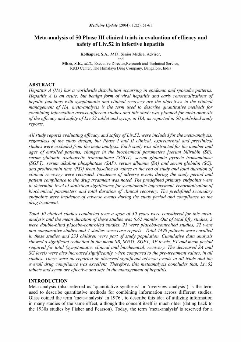
Figure 1: Distribution of studies as per study design
RESULTS Total fifty clinical studies that evaluated the efficacy and safety of Liv.52 in HA, conducted over a span of 30 years (from 1967 to 1997) were considered for this meta-analysis. The mean duration of these studies was 6.62 months and the total study duration of all trials was 331 months (minimum period=1.0 month, maximum period=36.00 months, mean of means (M)=6.62, Std. deviation (SD)=6.645, Std. error of mean (SEM)=0.9398, lower 99% confidence interval (CI) of mean=4.097, upper 99% CI of mean=9.143, W=0.6497 (Shapiro-Wilk normality test), p<0.0001, significant (S)) (Table 1). Out of fifty studies, three were double-blind placebo-controlled studies, twenty-one were placebo-controlled studies, twenty-two were open (non-comparative)
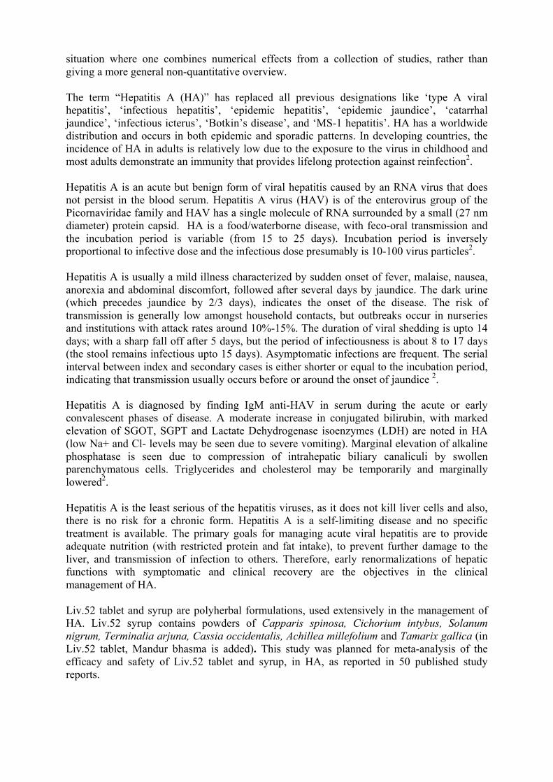
Figure 2: Distribution of patients as per received treatment
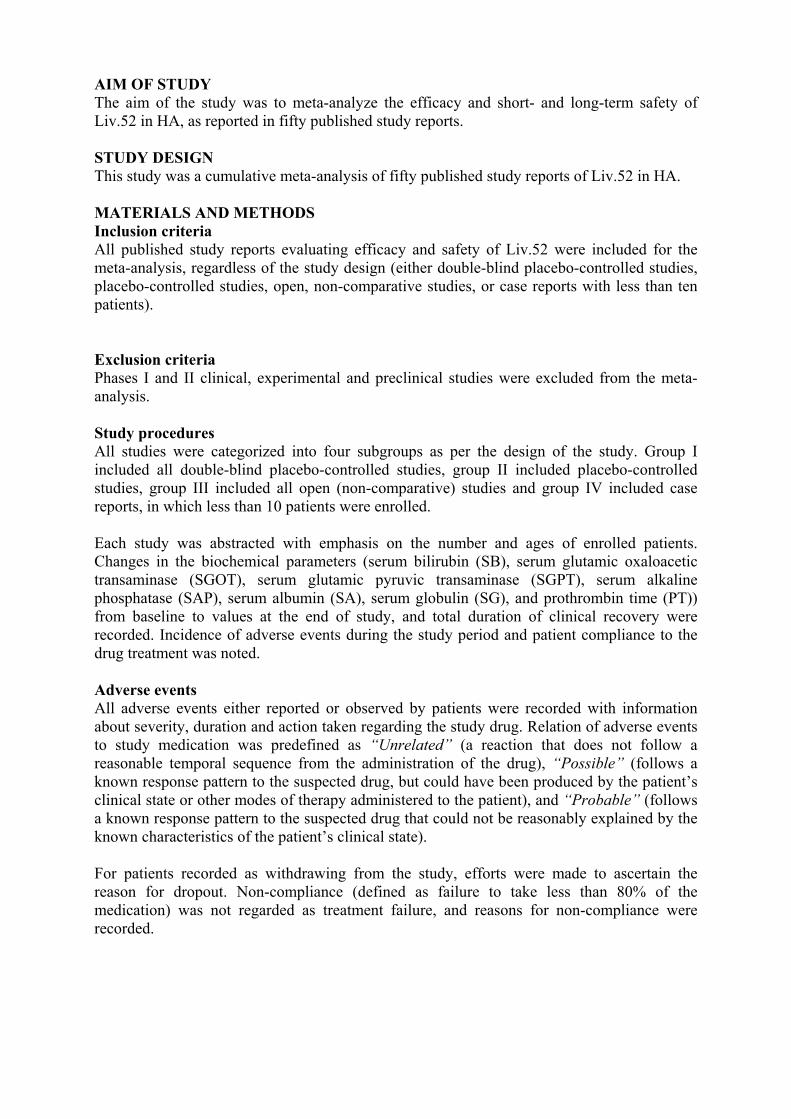
Figure 3: Agewise distribution of children enrolled invarious trials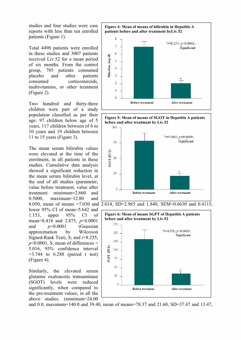
studies and four studies were case reports with less than ten enrolled patients (Figure 1). Total 4490 patients were enrolled in these studies and 3007 patients received Liv.52 for a mean period of six months. From the control group, 785 patients consumed placebo and other patients consumed corticosteroids, multivitamins, or other treatment (Figure 2). Two hundred and thirty-three children were part of a study population classified as per their age: 97 children below age of 5 years, 117 children between of 6 to 10 years and 19 children between 11 to 15 years (Figure 3).
Figure 4: Mean of means of bilirubin in Hepatitis A patients before and after treatment byLiv.52
The mean serum bilirubin values were elevated at the time of the enrolment, in all patients in these studies. Cumulative data analysis showed a significant reduction in the mean serum bilirubin level, at the end of all studies (parameter, value before treatment, value after treatment: minimum=2.660 and 0.5000, maximum=12.80 and 8.050, mean of means =7.030 and 2lower 95% CI of mean=5.642 and 1.153, upper 95% CI of mean=8.418 and 2.875, p<0.0001 and p<0.0001 (Gaussian approximation by Wilcoxon Signed-Rank Test), S; and t=8.255, p<0.0001, S, mean of differences = 5.016, 95% confidence interval =3.744 to 6.288 (paired t test) (Figure 4). Similarly, the elevated serum glutamic oxaloacetic transaminase (SGOT) levels were reduced significantly, when compared to the pre-treatment values, in all the above studies. (minimum=24.00 and 0.0, maximum=140.0 and 39.40,
Figure 5: Mean of means of SGOT in Hepatitis A patients before and after treatment by Liv.52
.014, SD=2.965 and 1.840, SEM=0.6630 and 0.4115,
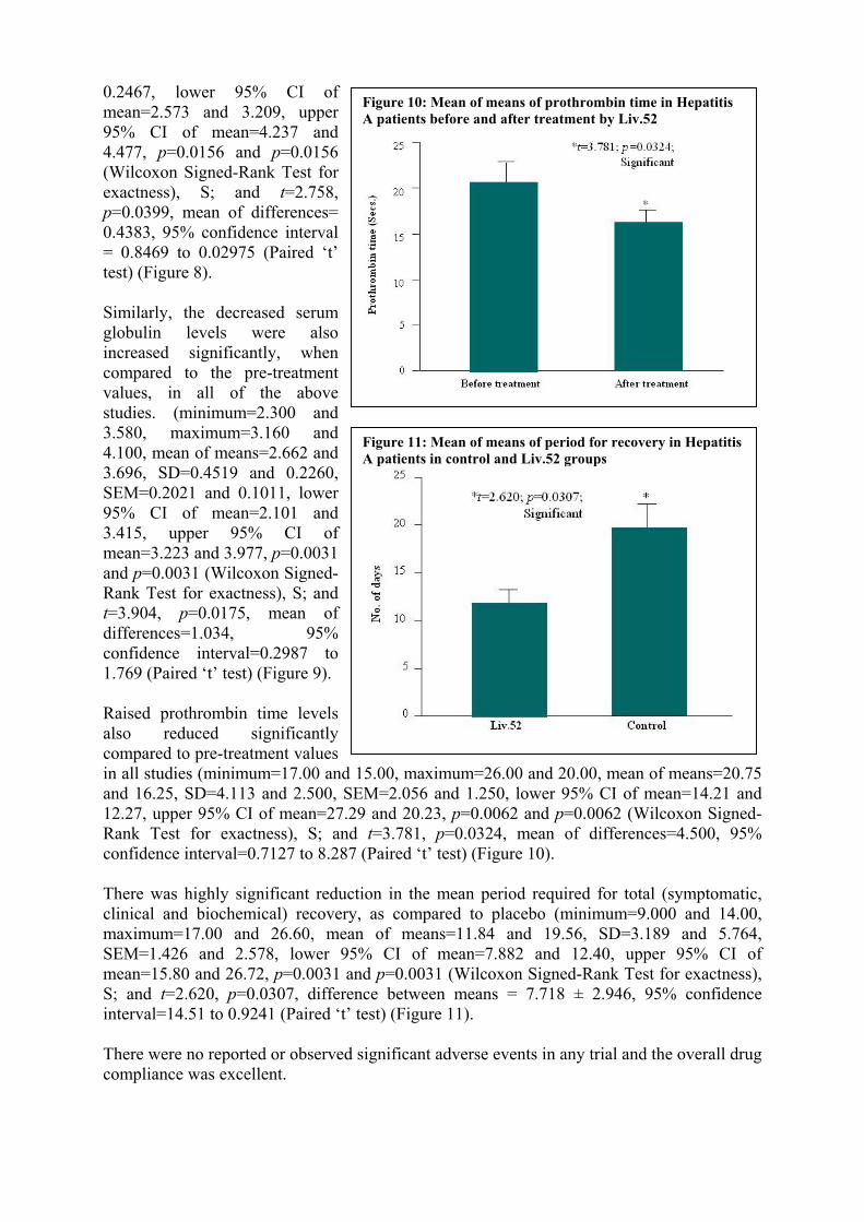
mean of means=78.37 and 21.60, SD=37.47 and 13.47,
Figure 6: Mean of means SGPT of Hepatitis A patients before and after treatment by Liv.52
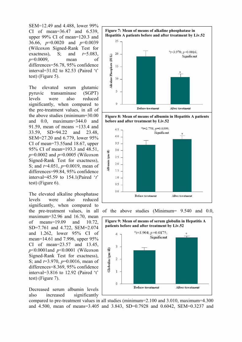
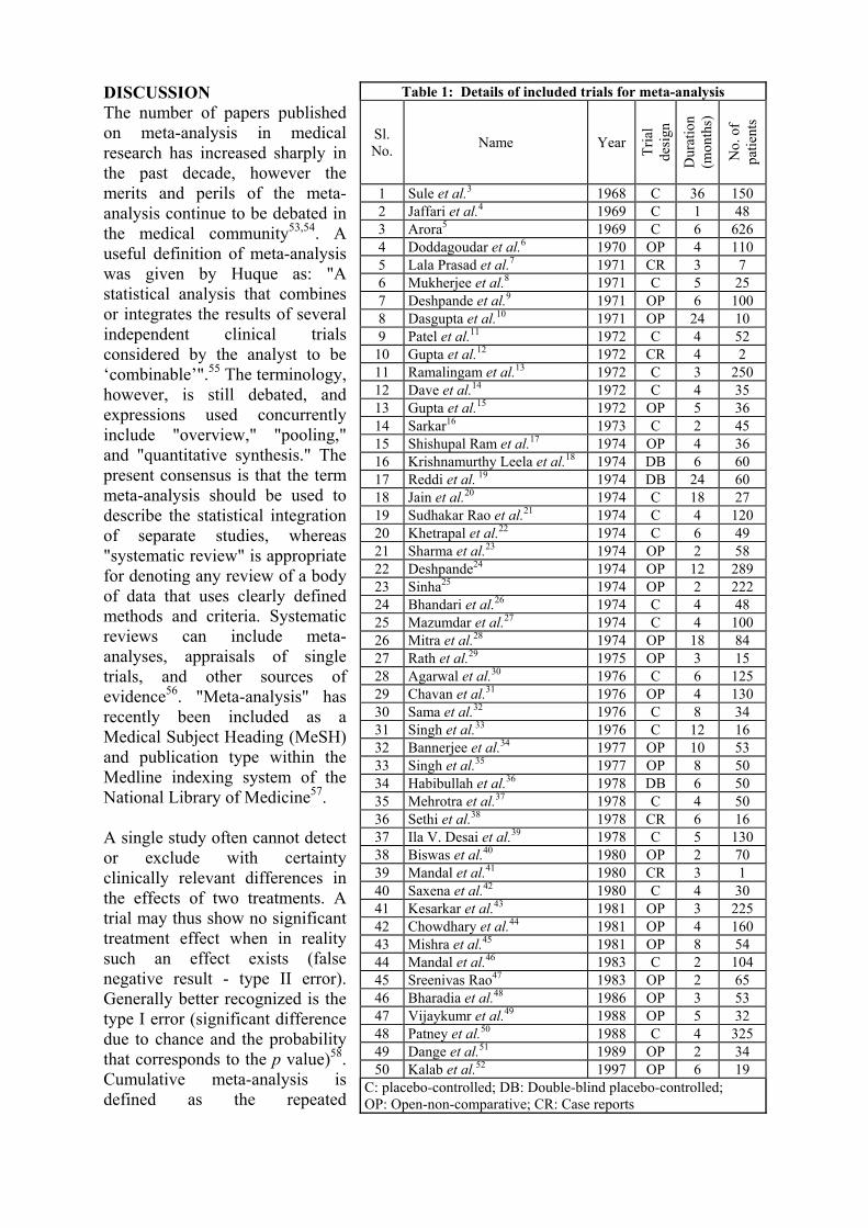
SEM=12.49 and 4.488, lower 99% CI of mean=36.47 and 6.539, upper 99% CI of mean=120.3 and 36.66, p=0.0020 and p=0.0039 (Wilcoxon Signed-Rank Test for exactness), S; and t=5.083, p=0.0009, mean of differences=56.78, 95% confidence interval=31.02 to 82.53 (Paired ‘t’ test) (Figure 5). The elevated serum glutamic pyruvic transaminase (SGPT) levels were also reduced significantly, when compared to the pre-treatment values, in all of the above studies (minimum=30.00 and 0.0, maximum=344.0 and 91.59, mean of means =133.4 and 33.59, SD=94.22 and 23.48, SEM=27.20 and 6.779, lower 95% CI of mean=73.55and 18.67, upper 95% CI of mean=193.3 and 48.51, p=0.0002 and p=0.0005 (Wilcoxon Signed-Rank Test for exactness), S; and t=4.051, p=0.0019, mean of differences=99.84, 95% confidence interval=45.59 to 154.1(Paired ‘t’ test) (Figure 6). The elevated alkaline phosphatase levels were also reduced significantly, when compared to the pre-treatment values, in all of the above studies (Minimum= 9.540 and 0.0, maximum=32.96 and 16.70, mean of means=19.09 and 10.72, SD=7.761 and 4.722, SEM=2.074 and 1.262, lower 95% CI of mean=14.61 and 7.996, upper 95% CI of mean=23.57 and 13.45, p<0.0001and p<0.0001 (Wilcoxon Signed-Rank Test for exactness), S; and t=3.970, p=0.0016, mean of differences=8.369, 95% confidence interval=3.816 to 12.92 (Paired ‘t’ test) (Figure 7).
Figure 7: Mean of means of alkaline phosphatase in Hepatitis A patients before and after treatment by Liv.52
Figure 8: Mean of means of albumin in Hepatitis A patientsbefore and after treatment by Liv.52
Decreased serum albumin levels also increased significantly compared to pre-treatment values in all studies (minimum=2.100 and 3.010, maximum=4.300 and 4.500, mean of means=3.405 and 3.843, SD=0.7928 and 0.6042, SEM=0.3237 and
Figure 9: Mean of means of serum globulin in Hepatitis A patients before and after treatment by Liv.52
0.2467, lower 95% CI of mean=2.573 and 3.209, upper 95% CI of mean=4.237 and 4.477, p=0.0156 and p=0.0156 (Wilcoxon Signed-Rank Test for exactness), S; and t=2.758, p=0.0399, mean of differences= 0.4383, 95% confidence interval = 0.8469 to 0.02975 (Paired ‘t’ test) (Figure 8). Similarly, the decreased serum globulin levels were also increased significantly, when compared to the pre-treatment values, in all of the above studies. (minimum=2.300 and 3.580, maximum=3.160 and 4.100, mean of means=2.662 and 3.696, SD=0.4519 and 0.2260, SEM=0.2021 and 0.1011, lower 95% CI of mean=2.101 and 3.415, upper 95% CI of mean=3.223 and 3.977, p=0.0031 and p=0.0031 (Wilcoxon Signed-Rank Test for exactness), S; and t=3.904, p=0.0175, mean of differences=1.034, 95% confidence interval=0.2987 to 1.769 (Paired ‘t’ test) (Figure 9).
Figure 10: Mean of means of prothrombin time in Hepatitis A patients before and after treatment by Liv.52
Raised prothrombin time levels also reduced significantly compared to pre-treatment values in all studies (minimum=17.00 and 15.00, maximum=26.00 and 20.00, mean of means=20.75 and 16.25, SD=4.113 and 2.500, SEM=2.056 and 1.250, lower 95% CI of mean=14.21 and 12.27, upper 95% CI of mean=27.29 and 20.23, p=0.0062 and p=0.0062 (Wilcoxon Signed-Rank Test for exactness), S; and t=3.781, p=0.0324, mean of differences=4.500, 95% confidence interval=0.7127 to 8.287 (Paired ‘t’ test) (Figure 10).
Figure 11: Mean of means of period for recovery in Hepatitis A patients in control and Liv.52 groups
There was highly significant reduction in the mean period required for total (symptomatic, clinical and biochemical) recovery, as compared to placebo (minimum=9.000 and 14.00, maximum=17.00 and 26.60, mean of means=11.84 and 19.56, SD=3.189 and 5.764, SEM=1.426 and 2.578, lower 95% CI of mean=7.882 and 12.40, upper 95% CI of mean=15.80 and 26.72, p=0.0031 and p=0.0031 (Wilcoxon Signed-Rank Test for exactness), S; and t=2.620, p=0.0307, difference between means = 7.718 ± 2.946, 95% confidence interval=14.51 to 0.9241 (Paired ‘t’ test) (Figure 11). There were no reported or observed significant adverse events in any trial and the overall drug compliance was excellent.
DISCUSSION The number of papers published on meta-analysis in medical research has increased sharply in the past decade, however the merits and perils of the meta-analysis continue to be debated in the medical community53,54. A useful definition of meta-analysis was given by Huque as: "A statistical analysis that combines or integrates the results of several independent clinical trials considered by the analyst to be ‘combinable’".55 The terminology, however, is still debated, and expressions used concurrently include "overview," "pooling," and "quantitative synthesis." The present consensus is that the term meta-analysis should be used to describe the statistical integration of separate studies, whereas "systematic review" is appropriate for denoting any review of a body of data that uses clearly defined methods and criteria. Systematic reviews can include meta-analyses, appraisals of single trials, and other sources of evidence56. "Meta-analysis" has recently been included as a Medical Subject Heading (MeSH) and publication type within the Medline indexing system of the National Library of Medicine57. A single study often cannot detect or exclude with certainty clinically relevant differences in the effects of two treatments. A trial may thus show no significant treatment effect when in reality such an effect exists (false negative result - type II error). Generally better recognized is the type I error (significant difference due to chance and the probability that corresponds to the p value)58. Cumulative meta-analysis is defined as the repeated
Table 1: Details of included trials for meta-analysis
Sl. No. Name Year Tr
ial
desi
gn
Dur
atio
n (m
onth
s)
No.
of
patie
nts
1 Sule et al.3 1968 C 36 150 2 Jaffari et al.4 1969 C 1 48 3 Arora5 1969 C 6 626 4 Doddagoudar et al.6 1970 OP 4 110 5 Lala Prasad et al.7 1971 CR 3 7 6 Mukherjee et al.8 1971 C 5 25 7 Deshpande et al.9 1971 OP 6 100 8 Dasgupta et al.10 1971 OP 24 10 9 Patel et al.11 1972 C 4 52
10 Gupta et al.12 1972 CR 4 2 11 Ramalingam et al.13 1972 C 3 250 12 Dave et al.14 1972 C 4 35 13 Gupta et al.15 1972 OP 5 36 14 Sarkar16 1973 C 2 45 15 Shishupal Ram et al.17 1974 OP 4 36 16 Krishnamurthy Leela et al.18 1974 DB 6 60 17 Reddi et al. 19 1974 DB 24 60 18 Jain et al.20 1974 C 18 27 19 Sudhakar Rao et al.21 1974 C 4 120 20 Khetrapal et al.22 1974 C 6 49 21 Sharma et al.23 1974 OP 2 58 22 Deshpande24 1974 OP 12 289 23 Sinha25 1974 OP 2 222 24 Bhandari et al.26 1974 C 4 48 25 Mazumdar et al.27 1974 C 4 100 26 Mitra et al.28 1974 OP 18 84 27 Rath et al.29 1975 OP 3 15 28 Agarwal et al.30 1976 C 6 125 29 Chavan et al.31 1976 OP 4 130 30 Sama et al.32 1976 C 8 34 31 Singh et al.33 1976 C 12 16 32 Bannerjee et al.34 1977 OP 10 53 33 Singh et al.35 1977 OP 8 50 34 Habibullah et al.36 1978 DB 6 50 35 Mehrotra et al.37 1978 C 4 50 36 Sethi et al.38 1978 CR 6 16 37 Ila V. Desai et al.39 1978 C 5 130 38 Biswas et al.40 1980 OP 2 70 39 Mandal et al.41 1980 CR 3 1 40 Saxena et al.42 1980 C 4 30 41 Kesarkar et al.43 1981 OP 3 225 42 Chowdhary et al.44 1981 OP 4 160 43 Mishra et al.45 1981 OP 8 54 44 Mandal et al.46 1983 C 2 104 45 Sreenivas Rao47 1983 OP 2 65 46 Bharadia et al.48 1986 OP 3 53 47 Vijaykumr et al.49 1988 OP 5 32 48 Patney et al.50 1988 C 4 325 49 Dange et al.51 1989 OP 2 34 50 Kalab et al.52 1997 OP 6 19
C: placebo-controlled; DB: Double-blind placebo-controlled; OP: Open-non-comparative; CR: Case reports
performance of meta-analysis whenever a new trial becomes available for inclusion. Such cumulative meta-analysis can retrospectively identify the point in time when a treatment effect first reached conventional levels of significance59. Meta-analysis thus not only consists of the combination of data but also includes the epidemiological exploration and evaluation of results ("epidemiology of results")60. Therefore, new hypotheses that were not posed in single studies can be tested in meta-analyses61. The number of patients included in clinical trials is often inadequate as in some cases the required sample size may be difficult to achieve62. Meta-analysis can, nevertheless, lead to the identification of the most promising or urgent research question, and may permit a more accurate calculation of the sample sizes needed in future studies63. The goals of meta-analysis are to enable the overall significance of an effect to be evaluated, based on the multiple studies available; to estimate an overall effect size by combining the individual estimates in multiple studies. The problem of comparability of different study designs and effects of a difference in quality in multiple studies is still not resolved and there are no guidelines for the same64. While evaluating the efficacy of Liv.52 in HA, the primary endpoints (quantitative comparison of pooled data for biochemical parameters by ‘before’ and ‘after’ tests) were selected by their specific significance in natural history of HA. Most adult-acquired liver disease causes impairment in bilirubin secretion from the liver, which causes elevation of SB in the blood. In acute liver disease, SB is usually elevated relative to the severity of the acute process. Blood levels of SGOT (also referred as aspartate aminotransferase (AST)) and SGPT (also referred as Alanine aminotransferase (ALT)) are elevated in all types of hepatitis (viral, alcoholic, drug-induced, etc.). An elevation in the level of SAP suggests disease of the bile ducts (bile duct obstruction, primary biliary cirrhosis or primary sclerosing cholangitis). Albumin is the major protein synthesized in the liver, which circulates in the bloodstream and low SA concentrations indicate poor liver function. The PT is prolonged when the blood concentrations of some of the clotting factors made by the liver are low and in acute liver diseases, the PT can be prolonged. This meta-analysis included data of 50 clinical studies (24 placebo-controlled studies and 26 open non-comparative studies) conducted over a 30 years, in 4490 patients (including 117 children) and this sample size is large enough for calculating the intervention (drug) effect. The recommended method for calculating sample size is by the formula: n=4pq/L2
(p=prevalence, q=100 – p, L=1.96) and by this formula, this sample size is large enough for a disease, with a very high prevalence. There was significant symptomatic control observed in a week’s time. The cumulative analysis revealed significant reduction in the levels of mean SB, SGOT, SGPT and SAP and there was renormalization of protein levels and PT. There was significant reduction in the mean period required for total recovery, as compared to placebo. There were no reported or observed significant adverse events in all trials and the overall patient compliance to the treatment was excellent. Therefore, as per the predefined primary and secondary endpoints the efficacy and safety of Liv.52 in HA is clinically, biochemically and statistically well proved. These significant effects might be due to the synergistic properties and actions of the ingredients of Liv.52. Khanfar et al. isolated and identified active ingredients of Capparis spinosa as beta-sitosterylglucoside-6'-octadecanoate and 3-methyl-2-butenyl-beta-glucoside65. p-Methoxy benzoic acid isolated from Capparis spinosa was found to possess potent hepatoprotective activity against CCl4, paracetamol (in vivo) and in thioacetamide, galactosamine (in vitro)-induced hepatotoxicity66. Al-Said et al. demonstrated strong anti-inflammatory activity
of Capparis spinosa, which was comparable to oxyphenbutazone67,68. Bonina et al. documented significant antioxidant activity of Capparis spinosa and also identified flavonols (Kaempferol and Quercetin derivatives) and hydroxycinnamic acids (caffeic acid, ferulic acid, p-cumaric acid, and cinnamic acid) as major antioxidants from Capparis spinosa69. In another study, Germano et al. observed the antioxidant activity of Capparis spinosa using tests like lipid peroxidation, bleaching of free radicals and autoxidation of iron ions70. Mahasneh et al. observed potent antimicrobial and antifungal activity of Capparis spinosa71,72. He et al. isolated 2,3,4,9-tetrahydro-1H-pyrido- (3,4-b) indole-3-carboxylic acid, azelaic acid and daucosterol as the major constituents of Cichorium intybus73 and Du et al. identified the other chemical constituents as alpha-amyrin, taraxerone, baurenyl acetate and beta-sitosterol74. Aktay et al. and Zafar et al. observed hepatoprotective effect (confirmed by histopathological examination) of Cichorium intybus against CCl4-induced hepatotoxicity and reported significant prevention of the elevation of malondialdehyde formation (plasma and hepatic) and enzyme levels (AST and ALT) 75,76. Ahmed et al. screened Cichorium intybus for antihepatotoxic activity and measured the degree of protection using biochemical parameters (AST, ALT, ALKP and TP). Potent antihepatotoxic activity (comparable to Silymarin) was observed with almost complete normalization of the tissues (as neither fatty accumulation nor necrosis was observed on histopathological study)77. Kim et al. studied the effects of Cichorium intybus on the immunotoxicity of ethanol and reported significant increase in the number of circulating leukocytes, the weights of concerned organs (liver, spleen and thymus), number of splenic plaque forming cells, hemagglutination titers and the secondary IgG antibody response. There were also significant increases in delayed-type hypersensitivity reaction, phagocytic activity, natural killer cell activity, cell proliferation and interferon gamma secretion78. Sultana et al. reported that, the presence of Cichorium intybus in the reaction mixture (containing calf thymus DNA and free radical generating system) protects DNA against oxidative damage to its deoxyribose sugar moiety. All these studies suggest that the observed hepatoprotective effect of Cichorium intybus might be due to its ability to suppress the oxidative degradation of DNA in the tissue debris79. El et al. and Papetti et al. documented antioxidative activity (radical scavenging effects, inhibition of hydrogen peroxide, and iron chelation) of Cichorium intybus80,81. Gurbuz et al. observed significant cytoprotection against ethanol-induced damage and these results were further confirmed by using histopathological techniques82. Amirghofran et al. reported the capacity of Cichorium intybus to enhance the proliferation of lymphocytes after stimulation with the allogenic cells83. Kim et al. investigated the effect of Cichorium intybus on mast cell-mediated immediate type allergic reactions and observed inhibition of systemic anaphylactic reaction, reduction in the plasma histamine level84. Ikeda et al. identified saponins (nigrumnins I and II) as the active ingredients of Solanum nigrum85. Solanum nigrum was investigated for its hepatoprotective activity against CCl4-induced hepatic damage and Raju et al. observed remarkable hepatoprotective activity confirmed by evaluated biochemical parameters (AST, ALT, ALP and TB)86. Sultana et al. demonstrated that Solanum nigrum protect DNA against oxidative damage and the results suggest that the observed hepatoprotective effect of Solanum nigrum might be due to the ability to suppress the oxidative degradation of DNA in the tissue debris79. Moundipa et al. studied the effects of Solanum nigrum on hepatotoxicity and reported increased activity of aminopyrine N-demethylase, uridine diphosphate glucuronyltransferase and glutathione S-transferase, without any alteration in levels of alkaline phosphatase, aspartate aminotransferase and gamma-glutamyltransferase levels in the serum87. Son et al. reported Solanum nigrum as a potent scavenger of hydroxyl radicals and DPPH radicals88. Prashanth Kumar et al. tested in vitro Solanum nigrum for its cytoprotection (against gentamicin-
induced toxicity) and observed significant inhibition of cytotoxicity, alongwith hydroxyl radical scavenging potential, which might be the mechanism of cytoprotection89. Similarly, Akhtar et al. observed gastric mucosal cytoprotection offered by Solanum nigrum against aspirin-induced gastric ulcers90. Qureshi et al. reported antifungal activity of Solanum nigrum91. Upadhyay et al. identified arjunetoside, oleanolic and arjunic acids as active ingredients from Terminalia arjuna92. Munasinghe et al. reported potent antioxidant activity of Terminalia arjuna, which might be due to its effects on lipid peroxidation93. Ali et al. demonstrated that arjunaphthanoloside from Terminalia arjuna inhibits nitric oxide (NO) production94 and terminoside A isolated from Terminalia arjuna, decreases inducible nitric oxide synthase (iNOS) levels in LPS-stimulated peritoneal macrophages95. Cheng et al. observed potent antiviral activity by virtue of inhibition of viral attachment and penetration by Terminalia arjuna96. Samy et al. demonstrated potent antibacterial activity of Terminalia arjuna97. Jafri et al. reported significant hepatoprotective effects of Cassia occidentalis in chemically induced liver damage98. Bin-Hafeez et al. showed that Cassia occidentalis modulates hepatic enzymes and provides hepatoprotection against induced immunosuppression99. Samy et al. reported antimicrobial properties of Cassia occidentalis comparable with standard reference antibiotics100. Perez et al. reported strong antibacterial activity of Cassia occidentalis against Salmonella typhi101. Tona et al. reported inhibitory effect of Cassia occidentalis on P. falciparum growth102. Caceres et al. and Graham et al. observed antifungal activity of Cassia occidentalis103,104. Harnyk et al. documented clinically beneficial effects of Achillea millefolium in the treatment of chronic hepatitis105. Krivenko et al. reported similar clinical improvements in chronic hepatocholecystitis and angiocholitis with Achillea millefolium106. Lin et al. observed anti-hepatoma activity of Achillea millefolium107. Candan et al. and Bezic et al. reported antioxidant and antimicrobial activities of Achillea millefolium108,109. Devarshi et al. studied Mandur bhasma for the hepatoprotective property in hepatitis induced by CCl4 and observed prevention of CCl4 mediated changes in the enzyme activities, which suggests the hepatoprotective role of Mandur bhasma110. Therefore, as discussed above these synergistic actions (hepatoprotective, antimicrobial, antioxidant and anti-inflammatory) exhibited by the ingredients of Liv.52 might provide the mechanism of action of Liv.52 in hepatitis.
CONCLUSION In this study, meta-analysis of 50 clinical studies conducted over 30 years in 4490 patients, was performed to evaluate the efficacy and short- and long-term safety of Liv.52 in HA. The cumulative data analysis revealed clinical and biochemical improvements with significant symptomatic control. In addition, there was highly significant reduction in the mean recovery period. There were no reported or observed significant adverse events in all trials and the overall drug compliance was excellent. Therefore, it may be concluded that Liv.52 tablets and syrup are effective and safe in the management of hepatitis A.
REFERENCES 1. Glass, G.V. Primary, secondary and meta-analysis of research. Educational
Researcher 1976; 5: 351-379. 2. Krugman, S., Ward, R., Giles, J.P. The natural history of infectious hepatitis. Am J
Med 1962; 32: 717-728. 3. Sule, C.R, Pai, V.R., Damania, R.F., Joshi, V.S. (Miss). Studies with Liv.52 therapy in
infective hepatitis. J Ind Med Prof 1968; 14: 6391-6397. 4. Jaffari, S.M.H., Shyam Raj. Liv.52 in infective hepatitis. The Antiseptic 1969; 5: 353. 5. Arora, A.K. Role of various types of treatment in infectious hepatitis. Armed Forces
Med. J 1969; 25(3): 362. 6. Doddagoudar. Infective hepatitis - Survey and study of 110 cases. Probe 1970; IX(3):
122–123. 7. Lala Surajnandan Prasad, Kaleshwer Prasad. Some observations on Liv.52 in the
treatment of infective hepatitis and cirrhosis of liver. Probe 1971; X(3): 114-116. 8. Mukerjee, A.B., Dasgupta, M. Chronic active hepatitis (a preliminary observation of
10 cases). J Ind Med Prof 1971; 18: 8097. 9. Deshpande, R.S., Shantilal C. Sheth, Joykutty. Infections hepatitis - Study of 100
cases. Curr Med Pract 1971; 6: 810. 10. Dasgupta, M., Mukerjee, A.B. Chronic active hepatitis (a preliminary observation of
10 cases). J Ind Med Prof 1971; 18: 8097. 11. Patel, G.T., Mruthyunjayanna, B.P., Seetharam, T.D., Channe Gowda, A.C. Liv.52
therapy in viral hepatitis. Probe 1972; XI(2): 112-119. 12. Gupta, S., Khatri, R.L., Srivastava, G. Therapeutic effects of Liv.52 in post-necrotic
hepatitis. Probe 1972; XII(1): 15-20. 13. Ramalingam, V., Sundarvalli, N., Balagopal Raju, V. Liv.52 studies in acute hepatitis.
Indian Pediatrics 1971; 12: 839. 14. Dave, D.S., Rajput, V.J., Gupta, M.R. Clinico-biochemical study of infective hepatitis
with special reference to Liv.52 therapy. Probe 1972; XI(4): 214-220. 15. Gupta, S., Khatri, R.L., Srivastava, G. Therapy of infectious hepatitis and other liver
disorders. Probe 1972; XI(2): 93-99. 16. Sarkar, M.K. Liv.52 in the treatment of acute infective hepatitis. The Ind Pract 1973;
August: 395. 17. Shishupal Ram, Kumar, P. Observations on the therapy of acute infectious hepatitis
with Liv.52. Probe 1974; XIV(1): 66-67. 18. Krishnamurthy Leela, Sama, S.K. and Ramachandra, K. Liv.52 in acute virus: A hepatitis - a
double-blind trial. XV Annual Conference, Indian Society of Gastroenterology, Institute of Medical Sciences, Banaras Hindu University, Varanasi, 8-10 Novermber, 1974, p.2.
19. Reddi, Y.R., Rohini, K., Kusuma, G., Sudhakar Rao, V. Role of Liv.52 and steroids in the management of viral hepatitis in children. Probe 1974; XIV(1): 35.
20. Jain, K.C., Khanijo, S.K., Bisarya, B.N. Studies with Liv.52 in infective hepatitis. Probe 1974; XIV(1): 64-65.
21. Reddi, Y.R., Sudhakar Rao, V., Rohini, K. The management of viral hepatitis in children. Indian Pediatrics 1975; XII(8): 659.
22. Khetarpal, S.K., Veera Kumar, E. Liv.52 therapy in infective hepatitis. Probe 1974; XIV(1): 59-63.
23. Sharma, N.L., Lahori, U.C., Mehta, S.K. Studies on Liv.52 in hepatitis disorders (Part I). Probe 1974; XIV(1): 54-58.
24. Deshpande, R.S. Acute infectious hepatitis with fulminant hepatic failure – a study of 289 cases. Probe 1974; XIV(1): 1-13.
25. Sinha, B.B., Studies on 222 cases of acute infective hepatitis during an epidemic. Probe 1974; XIII(3): 130-133.
26. Bhandari, N.R., Shashi Gupta (Miss.). Liv.52 in infective hepatitis. Probe 1974; XIV(1): 68-72.
27. Mazumdar, H., Mazumdar, M. Trial of Liv.52 in infections hepatitis in children in Goa. Probe 1974; 1: 82.
28. Mitra, D.K., Ashrafuddin, S., Talib, S.H. Liv.52 in viral hepatitis with special reference to its use in precoma and coma. Probe 1974; XIV(1): 14-22.
29. Rath, B.B., Sengupta, S.N., Bose, S.N. Preliminary observation on the role of Liv.52 in infective hepatitis with persistent jaundice. Capsule 1975; XV(8): 170-175.
30. Agarwal, V.K., Meena Sareen. Clinical trial of Liv.52 drops in infective hepatitis – a comparative study with broad spectrum antibiotics and corticosteroids. Probe 1976; XV(2): 112-120.
31. Chawhan, R.N., Talib, S.H., Talib, V.H., Sengupta, S.R., Patil, S.D. Viral hepatitis in children. The Med & Sur 1976; 3: 9.
32. Sama, S.K., Krishnamurthy, L., Ramachnadran, K., Krishnan Lal. Efficacy of an indigenous compound preparation Liv.52 in acute viral hepatitis – A double-blind study. Ind J Med Res 1976; 5: 738.
33. Singh, D.S., Sama, S.K.,Singh, G. A study of chronic hepatitis in northern India. Ind J Med Res 1976; 64(12):1728-1734.
34. Banerjee, L.K., Sahu, N.N.,Ray, S.S., Daljit Singh. Liv.52 therapy in viral hepatitis (A clinico-biochemical study). Capsule 1977; 8: 170.
35. Singh, K.K., Singh, Y.K., Sinha, S.K., Sharma, V., Mishra, B.N. Observations on the treatment of infective hepatitis with an indigenous drug Liv.52. Ind Med Jl 1977; 5: 69.
36. Habibullah, C.M. Vindod Chandra, Padmanaban, C.G., Ramakrishna, R. Liv.52 in acute viral hepatitis – Results of a double blind study. The Antiseptic 1978; 75(8): 491.
37. Mehrotra, M.P., Tandan, S. Liv.52, a clinicobiochemical trial in hepatic cirrhosis. Curr Med Pract 1973; 17(4): 185-188.
38. Sethi, J.P., Madhulika Sharma. Clinical management of severe acute hepatic failure with special reference to Liv.52 in therapy. Probe 1978; XVII(2): 155-158.
39. Ila V. Desai, Dudhia, M.V., Gandhi, V.K. A clinical study of infective hepatitis treated with Liv.52. Indian Pediatrics 1977; 3: 197.
40. Biswas, P. Role of Liv.52 in the treatment of infective hepatitis. Capsule 1980; 4: 74. 41. Mandal, U.S. Viral hepatitis and Liv.52. Capsule 1980; 3: 50. 42. Shakuntala Saxena (Mrs.), Ashok Kumar Garg, Ashok Jain. Study of Liv.52 therapy
in infective hepatitis in children. Curr Med Pract 1980; 24(5): 194. 43. Kesarkar, A.S. Liv.52 therapy in acute infective hepatitis. Capsule 1981; 4: 74. 44. Chowdhury, A.B. Role of Liv.52 in the treatment of infective hepatitis. Capsule 1981;
3: 50. 45. Misra, D.C. Infective hepatitis and Liv.52 drops. Capsule 1981; 6: 122. 46. Mandal, J.N., Roy, B.K. Studies with Liv.52 in the treatment of infective hepatitis,
chronic active hepatitis and cirrhosis of the liver. Probe 1983; XXII(4): 217-242. 47. Sreenivasa Rao, Y. Management of infective hepatitis. Probe 1983; XXII(2): 107-109. 48. Bharadia, G.R., Kukade, A.L., Kulkarni, A.S., Rathod, I.M. Liv.52 in the management
of various hepatic disorders – A study of 53 cases. Probe 1986; XXV(3): 277-280. 49. Vijaykumar, S. Karchi. A trial with Liv.52 in infective hepatitis. Capsule 1988;
Feb./Mar.: 126. 50. Patney, N.L., Sumanbala Pachori. A study of serum glycolytic enzymes and serum B
hepatitis in relation to Liv.52 therapy. The Med & Sur 1986; XXVI(4): 9. 51. Dange, S.V., Pawar, S.S., Phadke, S.A., Shrotri, D.S. Comparative efficacy of five
indigenous compound formulations in patients of acute viral hepatitis. Maharashtra Med J 1989; XXXVI(5): 75.
52. Kalab, M., Krechler, T. Effect of the hepatoprotective drug Liv.52 on liver damage. The J Czech Physicians 1997; 136(24): 758-760.
53. Naylor, C.D. Meta-analysis and the meta-epidemiology of clinical research. BMJ 1997; 315: 617-619.
54. Bailar, J.C. The promise and problems of meta-analysis [editorial]. N Engl J Med 1997; 337: 559-561.
55. Huque, M.F. Experiences with meta-analysis in NDA submissions. Proceedings of the Biopharmaceutical Section of the American Statistical Association 1988; 2: 28-33.
56. Chalmers, I., Altman, D.G. Foreword. In: Chalmers I, Altman DG (Eds.) Systematic reviews, London: BMJ Publishing, 1995.
57. Dickersin, K., Berlin, J.A. Meta-analysis: state-of-the-science. Epidemiol Rev 1992; 14: 154-176.
58. Freiman, J.A., Chalmers, T.C., Smith, H., Kuebler, R.R. The importance of beta, the type II error, and sample size in the design and interpretation of the randomized controlled trial. In: Bailar JC, Mosteller F, (Eds.) Medical uses of statistics. Boston, MA: NEJM Books, 1992: p. 357.
59. Lau, J., Antman, E.M., Jimenez-Silva, J., Kupelnick, B., Mosteller, F., Chalmers, T.C. Cumulative meta-analysis of therapeutic trials for myocardial infarction. N Engl J Med 1992; 327: 248-254.
60. Jenicek, M. Meta-analysis in medicine. Where we are and where we want to go. J Clin Epidemiol 1989; 42: 35-44.
61. Gelber, R.D., Goldhirsch, A. Interpretation of results from subset analyses within overviews of randomized clinical trials. Stat Med 1987; 6: 371-378.
62. Collins, R., Keech, A., Peto, R., Sleight, P., Kjekshus, J., Wilhelmsen, L., et al. Cholesterol and total mortality: need for larger trials. BMJ 1992; 304: 1689.
63. Chalmers, I. Randomised controlled trials of fetal monitoring 1973-1977. In: Thalhammer O, Baumgarten K, Pollak A, (Eds.) Perinatal medicine. Stuttgart: Thieme, 1979: p. 260.
64. Smith, Glass, Miller. Does psychotherapy benefit neurotic patients? Archives of General Psychiatry 1981; 36: 1203-1208.
65. Khanfar, M.A., Sabri, S.S., Zarga, M.H., Zeller. K.P. The chemical constituents of Capparis spinosa of Jordanian origin. Nat Prod Res 2003; 17(1): 9-14.
66. Gadgoli, C., Mishra, S.H. Antihepatotoxic activity of p-methoxy benzoic acid from Capparis spinosa. J Ethnopharmacol 1999; 66(2): 187-192.
67. Al-Said, M.S., Abdelsattar, E.A., Khalifa, S.I., el-Feraly, F.S. Isolation and identification of an anti-inflammatory principle from Capparis spinosa Pharmazie 1988; 43(9): 640-641.
68. Ageel, A.M., Parmar, N.S., Mossa, J.S., Al-Yahya, M.A., Al-Said, M.S., Tariq, M. Anti-inflammatory activity of some Saudi Arabian medicinal plants. Agents Actions 1986; 17(3-4): 383-384.
69. Bonina, F., Puglia, C., Ventura, D., Aquino, R., Tortora, S., Sacchi, A., Saija, A., Tomaino, A., Pellegrino, M.L., de Caprariis, P. In vitro antioxidant and in vivo photoprotective effects of a lyophilized extract of Capparis spinosa L buds. J Cosmet Sci 2002; 53(6): 321-335.
70. Germano, M.P., De Pasquale, R., D'Angelo, V., Catania, S., Silvari, V., Costa, C. Evaluation of extracts and isolated fraction from Capparis spinosa L. buds as an antioxidant source. J Agric Food Chem 2002; 50(5): 1168-1171.
71. Mahasneh, A.M. Screening of some indigenous Qatari medicinal plants for antimicrobial activity. Phytother Res 2002; 16(8): 751-753.
72. Ali-Shtayeh, M.S., Abu Ghdeib, S.I. Antifungal activity of plant extracts against dermatophytes. Mycoses 1999; 42(11-12): 665-672.
73. He, Y., Guo, Y.J., Gao, Y.Y. Studies on chemical constituents of root of Cichorium intybus. Zhongguo Zhong Yao Za Zhi. 2002; 27(3): 209-210.
74. Du, H., Yuan, S., Jiang, P. Chemical constituents of Cichorium intybus L. Zhongguo Zhong Yao Za Zhi. 1998; 23(11): 682-683, 704.
75. Aktay, G., Deliorman, D., Ergun, E., Ergun, F., Yesilada, E., Cevik, C. Hepatoprotective effects of Turkish folk remedies on experimental liver injury. J Ethnopharmacol 2000; 73(1-2): 121-129.
76. Zafar, R., Mujahid Ali, S. Anti-hepatotoxic effects of root and root callus extracts of Cichorium intybus L. J Ethnopharmacol 1998; 63(3): 227-231.
77. Ahmed, B., Al-Howiriny, T.A., Siddiqui, A.B. Antihepatotoxic activity of seeds of Cichorium intybus. J Ethnopharmacol 2003; 87(2-3): 237-240.
78. Mun, J.H., Woo, Y.J., Jeon, W.H., An, N.H., Park, J.S. Effects of the ethanol extract of Cichorium intybus on the immunotoxicity by ethanol in mice. Int Immunopharmacol 2002; 2(6): 733-744.
79. Sultana, S., Perwaiz, S., Iqbal, M., Athar, M. Crude extracts of hepatoprotective plants, Solanum nigrum and Cichorium intybus inhibit free radical-mediated DNA damage. J Ethnopharmacol 1995; 45(3): 189-192.
80. El, S.N., Karakaya, S. Radical scavenging and iron-chelating activities of some greens used as traditional dishes in Mediterranean diet. Int J Food Sci Nutr 2004; 55(1): 67-74.
81. Papetti, A., Daglia, M., Gazzani, G. Anti- and pro-oxidant activity of water soluble compounds in Cichorium intybus var. silvestre (Treviso red chicory). J Pharm Biomed Anal 2002; 30(4): 939-945.
82. Gurbuz, I., Ustun, O., Yesilada, E., Sezik, E., Akyurek, N. In vivo gastroprotective effects of five Turkish folk remedies against ethanol-induced lesions. J Ethnopharmacol 2002; 83(3): 241-244.
83. Amirghofran, Z., Azadbakht, M., Karimi, M.H. Evaluation of the immunomodulatory effects of five herbal plants. J Ethnopharmacol 2000; 72(1-2): 167-172.
84. Kim, H.M., Kim, H.W., Lyu, Y.S., Won, J.H., Kim, D.K., Lee, Y.M., Morii, E., Jippo, T., Kitamura, Y., An, N.H. Inhibitory effect of mast cell-mediated immediate-type allergic reactions by Cichorium intybus. Pharmacol Res 1999; 40(1): 61-65.
85. Ikeda, T., Tsumagari, H., Nohara, T. Steroidal oligoglycosides from Solanum nigrum. Chem Pharm Bull (Tokyo). 2000; 48(7): 1062-1064.
86. Raju, K., Anbuganapathi, G., Gokulakrishnan, V., Rajkapoor, B., Jayakar, B., Manian, S. Effect of dried fruits of Solanum nigrum LINN against CCl4-induced hepatic damage in rats. Biol Pharm Bull 2003; 26(11): 1618-1619.
87. Moundipa, P.F., Domngang, F.M. Effect of the leafy vegetable Solanum nigrum on the activities of some liver drug-metabolising enzymes after aflatoxin B1 treatment in female rats. Br J Nutr 1991; 65(1): 81-91.
88. Son, Y.O., Kim, J., Lim, J.C., Chung, Y., Chung, G.H., Lee, J.C. Ripe fruit of Solanum nigrum L. inhibits cell growth and induces apoptosis in MCF-7 cells. Food Chem Toxicol 2003; 41(10): 1421-1428.
89. Prashanth Kumar, V., Shashidhara, S., Kumar, M.M., Sridhara BY. Cytoprotective role of Solanum nigrum against gentamicin-induced kidney cell (Vero cells) damage in vitro. Fitoterapia 2001; 72(5): 481-486.
90. Akhtar, M.S., Munir, M. Evaluation of the gastric antiulcerogenic effects of Solanum nigrum, Brassica oleracea and Ocimum basilicum in rats. J Ethnopharmacol 1989; 27(1-2): 163-176.
91. Qureshi, S., Rai, M.K., Agrawal, S.C. In vitro evaluation of inhibitory nature of extracts of 18-plant species of Chhindwara against 3-keratinophilic fungi. Hindustan Antibiot Bull 1997; 39(1-4): 56-60.
92. Upadhyay, R.K., Pandey, M.B., Jha, R.N., Singh, V.P., Pandey, V.B. Triterpene glycoside from Terminalia arjuna. J Asian Nat Prod Res 2001; 3(3): 207-212.
93. Munasinghe, T.C., Seneviratne, C.K., Thabrew, M.I., Abeysekera, A.M. Antiradical and antilipoperoxidative effects of some plant extracts used by Sri Lankan traditional medical practitioners for cardioprotection. Phytother Res 2001; 15(6): 519-523.
94. Ali, A., Kaur, G., Hayat, K., Ali, M., Ather, M. A novel naphthanol glycoside from Terminalia arjuna with antioxidant and nitric oxide inhibitory activities. Pharmazie. 2003; 58(12): 932-934.
95. Ali, A., Kaur, G., Hamid, H., Abdullah, T., Ali, M., Niwa, M., Alam, M.S., Terminoside A a new triterpene glycoside from the bark of Terminalia arjuna inhibits nitric oxide production in murine macrophages. J Asian Nat Prod Res 2003; 5(2): 137-142.
96. Cheng, H.Y., Lin, C.C., Lin, T.C. Antiherpes simplex virus type 2 activity of casuarinin from the bark of Terminalia arjuna Linn. Antiviral Res 2002; 55(3): 447-455.
97. Perumal Samy, R., Ignacimuthu, S., Sen, A. Screening of 34 Indian medicinal plants for antibacterial properties. J Ethnopharmacol 1998; 62(2): 173-182.
98. Jafri, M.A., Jalis Subhani, M., Javed, K., Singh, S. Hepatoprotective activity of leaves of Cassia occidentalis against paracetamol and ethyl alcohol intoxication in rats. J Ethnopharmacol 1999; 66(3): 355-361.
99. Bin-Hafeez, B., Ahmad, I., Haque, R., Raisuddin, S. Protective effect of Cassia occidentalis L. on cyclophosphamide-induced suppression of humoral immunity in mice. J Ethnopharmacol 2001; 75(1): 13-18.
100. Samy, R.P., Ignacimuthu, S. Antibacterial activity of some folklore medicinal plants used by tribals in Western Ghats of India. J Ethnopharmacol 2000; 69(1): 63-71.
101. Perez, C., Anesini, C. In vitro antibacterial activity of Argentine folk medicinal plants against Salmonella typhi. J Ethnopharmacol 1994; 44(1): 41-46.
102. Tona, L., Ngimbi, N.P., Tsakala, M., Mesia, K., Cimanga, K., Apers, S., De Bruyne, T., Pieters, L., Totte, J., Vlietinck, A.J. Antimalarial activity of 20 crude extracts from nine African medicinal plants used in Kinshasa, Congo. J Ethnopharmacol 1999; 68(1-3): 193-203.
103. Caceres, A., Lopez, B., Juarez, X., del Aguila, J., Garcia, S. Plants used in Guatemala for the treatment of dermatophytic infections. 2. Evaluation of antifungal activity of seven American plants. J Ethnopharmacol 1993; 40(3): 207-213.
104. Graham, J.G., Zhang, H., Pendland, S.L., Santarsiero, B.D., Mesecar, A.D., Cabieses, F., Farnsworth, N.R. Antimycobacterial Naphthopyrones from Senna obliqua. J Nat Prod 2004; 67(2): 225-227.
105. Harnyk, T.P. The use of preparations of plant origin in treating and rehabilitating elderly patients with chronic hepatitis. Lik Sprava. 1999; 7-8: 168-170.
106. Krivenko, V.V., Potebnia, G.P., Loiko, V.V. Experience in treating digestive organ diseases with medicinal plants. Vrach. Delo. 1989; 3: 76-78.
107. Lin, L.T., Liu, L.T., Chiang, L.C., Lin, C.C. In vitro anti-hepatoma activity of fifteen natural medicines from Canada. Phytother Res 2002; 16(5): 440-444.
108. Candan, F., Unlu, M., Tepe, B., Daferera, D., Polissiou, M., Sokmen, A., Akpulat, H.A. Antioxidant and antimicrobial activity of the essential oil and methanol extracts of Achillea millefolium subsp. millefolium Afan. (Asteraceae). J Ethnopharmacol 2003; 87(2-3): 215-220.
109. Bezic, N., Skocibusic, M., Dunkic, V., Radonic, A. Composition and antimicrobial activity of Achillea clavennae L. essential oil. Phytother Res 2003; 17(9): 1037-1040.
110. Devarshi, P., Kanase, A., Kanase, R., Mane, S., Patil, S., Varute, A.T. Effect of Mandura bhasma on lipolytic activities of liver, kidney and adipose tissue of albino rat during CCl4 induced hepatic injury. J Biosciences 1986; 10(2): 227-234.