mesoporous silica: an alternative diffusion controlled ... · pdf filej. andersson*, j....
TRANSCRIPT
J. Andersson*, J. Rosenholm and M. Lindén
Summary
M esoscopically ordered mesoporous silica materials have attracted a wide interest since their
discovery in the early 1990’s due to their diverse potential applications areas; including catalysis,
filtration and chromatography. Supramolecular surfactant aggregates are used as structure directing
agents for inorganics during condensation, leading to mesoscopically ordered surfactant-inorganic
composites. Porosity can be induced in the inorganic part by removal of the surfactant portion
through thermal or chemical means. By controlling the synthesis conditions and choice of structure
directing agents, the system can be engineered to fit numerous functions of preference. As the purely
siliceous mesoporous materials have been shown to be biocompatible, or sometimes even bioactive,
there is an increasing interest in this class of materials for applications in the field of bioceramics,
especially as bone substitute materials. Furthermore, the highly organized porous silica matrix could
be used as a potential controlled drug release system. Another attractive advantage is that amorphous
silica is degradable in an aqueous solution, and thus problems related to the removal of the material
after use can be avoided. In this article, a few of the most common forms of drug delivery
mechanism from ceramic drug carrier systems are investigated. Furthermore, some examples are
given on different mesoporous silica systems as drug delivery devices and how factors such as pore
size, pore connectivity and surface affect the drug loading and the release (diffusion) rate.
KEYWORDS: bioceramics’, mesoporous silica, drug delivery, ibuprofen, silica, diffusion controlled
Mesoporous Silica: An Alternative Diffusion
Controlled Drug Delivery System
C H A P T E R 6
� *Correspondence to: J. Andersson, Department of Physical Chemistry, Åbo Akademi University, Porthansgatan 3-5, FIN-20500 Turku, Finland. E-mail: [email protected]
Topics in Multifunctional Biomaterials and Devices, Ed. N Ashammakhi © 2008.
Andersson et al. Mesoporous silica
2Topics in Multifunctional Biomaterials and Devices, Ed. N Ashammakhi © 2008.
INTRODUCTION
During the last decades a diversity of polymer based pharmaceutical carrier systems have
been developed as new means of controlling temporal or distributional (site-specific) drug
delivery (1). Pharmaceutical controlling delivery systems offer numerous advantages
compared to conventionally administrated drugs in dosage forms, such as improved
efficiency and reduced toxicity (1). Conventional dosage forms have the disadvantage not to
be able to control either the rate of drug delivery or the target area of drug administration and
provide a rapid and an immediate drug release. Thus, frequent administration is necessary in
order to maintain a therapeutic level, which in turn causes drug concentration in blood and
tissues to fluctuate widely.
Polymeric cross-linked carrier matrices, such as hydrogels and supra-molecular polymer
aggregates as well as different types of microencapsulation vehicles, are typical examples of
common drug delivery devices (1-4). Controlled release systems are generally classified
based on their physicochemical, pharmaceutical or clinical aspect. They can also be classified
according to their release mechanism and preparation methods as follows (5):
• Physical systems, including diffusion controlled systems such as monolithic porous
systems and biodegradable/bioerodable systems.
• Chemical systems, including immobilization of drugs.
• Biological systems, including gene therapy
Depending on the delivery system and the pharmaceutical in use, different release
mechanisms are applied. However, there are three primary ways by which active agents can
be released from such system: diffusion, degradation, and swelling followed by diffusion.
Any or all of these mechanisms may occur in a given release system. Diffusion occurs when
a drug or other active agent passes through the system (a ceramic or polymer based matrix)
that forms the controlled-release device. The diffusion can occur on a macroscopic scale—as
through pores in the matrix—or on a molecular level, by passing between, for instance,
polymer chains. The diffusion controlled release could be activated by several means,
including ionic strength, pH and thermal, magnetic or chemical changes. Controlled release
based on an eroding matrix (surface or bulk) allow diffusion of drug from the degrading
system (5).
Andersson et al. Mesoporous silica
3Topics in Multifunctional Biomaterials and Devices, Ed. N Ashammakhi © 2008.
Temporal or distributional drug delivery could be beneficial when handling numerous classes
of dugs, such as anti-inflammatory agents, antibiotics, chemotherapeutic drugs, steroids,
hormones and vaccines, to mention a few (5,6). The ability to control over the drug delivery
can be an important factor especially at times when traditional oral or injectable drug
formulations are difficult to distribute. In some cases there might be a need of a slow release
of a water soluble drug or a fast release of low-solubility drugs. It might also be convenient
for drug delivery to specific sites, drug delivery using nanoparticulate systems, delivery of
two or more agents with the same formulation, and also systems based on carriers that can
dissolve or degrade and be readily eliminated. The ideal drug delivery system should be inert
or biodegradable, biocompatible, mechanically strong, comfortable for the patient, capable of
achieving high drug loading, safe from accidental release, simple to administer and remove,
and easy to fabricate and sterilize.
MULTIFUNCTIONAL DRUG DELIVERY DEVICES - CERAMICS
The carrier system controlling the release of an active agent does not necessarily remain inert
to the conditions surrounding the device. An active carrier system can sometimes be a part of
an additional treatment in terms of contribution to the healing of the surrounding environment
(tissue). In this category we find bioactive devices such as certain bone substitute materials
used in the form of implants (screws, nails, plates etc.), fillers (beads, granules) or pastes. A
desirable aspect of many bone-substitute materials is that there will be a possibility to
simultaneously introduce in situ drug release in combination with the implant. The advantage
of localized biodegradable therapy includes high, local drug concentration at the site of
infection, as well as, obviation of the need for removal of the implant after treatment. Also, a
controlled drug release from the implant would help minimize toxic side effects and further
compliance problems that might occur due to prolonged oral antibiotic therapy (7,8).
A good example of this is when treating bone diseases due to osteomyelitis or osteosarcoma.
It is then necessary to maintain a high concentration of drug at the site of infection for a
sufficient period of time. An effective local concentration may not be achieved with a
systemic administration of a drug (e.g. chemotherapeutic, antibiotic or chemobiotic), and
surgical treatment as well as local administration of drug are often required. Local
administration limits the adverse effects of systemic administration, and higher concentration
Andersson et al. Mesoporous silica
4Topics in Multifunctional Biomaterials and Devices, Ed. N Ashammakhi © 2008.
of medication reach to the targeted site, and the surgical procedure results in producing bone
defects.
Some novel ceramic-based particulate systems for delivery and application of drug, such as
antibiotics, growth factors and anticancer drug are claimed to have enhanced bioavailability,
predictable therapeutic response and prolonged drug release time. Of particular interest are
the biodegradable systems where controlled biodegradation ability is being biocompatible
with tissues. Today’s implant drug delivery devices are based on biodegradable polymers,
such as polylactic acid (PLA) and polyglycolic acid (PGA) (9). At the same time, antibiotic-
impregnated acrylic bone cement beads have been widely used for local administration of
antibiotic and dead space management. Walenkamp et al. (10) reported that healing of
osteomyelitis was achieved in 92 of 100 patients using gentamicin-impregnated PMMA bone
cement beads. However, this implant material showed various problems, including the
requirement for subsequent removal of the beads, thermal damage of the antibiotic, and poor
antibiotic elution property. The amount of gentamicin, which is heat stable and released by
the beads, does not exceed 25 % of the total amount implanted. The collagen-gentamicin
sponge appears to be equally as popular as acrylic bone cement beads for the treatment of
osteomyelitis. Although the sponge is biodegradable and does not need surgical removal,
antibiotic release may last only four days. Thus, drug delivery system based on porous
bioceramics in form of granules has been developed to fill the bone defect with antibiotic
(11,12). However, the problem still remains of matching of drug release rate and
biodegradation kinetics (13,14). In order to obtain the ultimate implant material, the chemical
and phase compositions, the microstructure (pores size distribution and interconnectivity) and
their biological behavior with a cellular component needs to be systematically studied. The
objectives will be the sustainable for production in industry of new “smart” multifunctional
materials with tailor-made functionalities, where these materials may be used to restore the
viability of damaged tissues and/or organs in humans and drug delivery systems.
One new set of materials that have the potential to fulfill many of the criteria required for
such a multifunctional system are sol-gel-derived silicon oxides (15-21). However, before
going into further details about an example of these systems, models to describe the
mechanism behind the drug release are discussed in brief.
Andersson et al. Mesoporous silica
5Topics in Multifunctional Biomaterials and Devices, Ed. N Ashammakhi © 2008.
DRUG DELIVERY MECHANISMS
There are several mathematical models to describe release mechanism of an active agent
from a drug delivery system. To be able to describe or predict the release profile, knowledge
of the type of system, and rate controlling mechanism is crucial. Amongst the typical
controlled release system we find diffusion controlled devices (reservoir, membrane, or
monolithic devices), chemically controlled (monolithic) systems, water penetration controlled
(osmotic or swelling) systems and regulated (magnetic or chemical) systems (5). As we will
concentrate on drug release from a porous ceramic matrix, the diffusion controlled release is
of greatest interest and will be discussed in further detail.
DIFFUSION CONTROLLED DELIVERY SYSTEMS
A diffusion controlled release can be obtained by two different systems: monolithic devices
and reservoir devices (membrane controlled devices).
In a monolithic device the drug is dispersed in a matrix and the release is controlled by
diffusion from the system. The diffusion can occur on a macroscopic scale, either through
pores in the ceramic (or polymer) matrix, or on a molecular level, by passing between
polymer chains.
The solubility of a drug and its dissolution rate can significant influence on the drug release
kinetics. If the drug is present in the matrix below its solubility limit it can be dissolved in a
polymer matrix and if it is present above its solubility limit, it is dispersed. For a dispersed
drug in a matrix it is assumed that the dissolution rate of the drug is minimal compared to the
diffusion rate of the drug. When the dissolution rate is slower than the diffusion rate of the
drug, the former will determine the release rate. These kinds of systems will follow a two-
step process.
The release of a drug from a monolithic system is based on Fickian diffusion (5). The
fractional release from a one-dimensional porous system can be described using Fick’s
second law (I). Fick’s second law describes how the concentration within the diffusion
volume changes with respect to time, and D is the diffusion coefficient and c is the
concentration.
Andersson et al. Mesoporous silica
6Topics in Multifunctional Biomaterials and Devices, Ed. N Ashammakhi © 2008.
cDt
c∆⋅=
∂∂
(I)
In a one-dimensional system, the release rate is proportional to the square root of time. For
the first 60 % of released drug, the release correspond to the early time approximation of
Fick’s second law,
=∂
∂
tl
DM
t
M t
202π
(II)
Thereafter, the release kinetics follows the late time approximation (first order kinetics)
according to:
=
∂
∂2
2
2
0 exp8
l
Dt
l
DM
t
M t π (III)
where l is the thickness of a slab, Mo is the total amount of drug dissolved in the system, and
Mt is the amount released at time t. Here, the release rate is dependent on the diffusion length.
A first order linear release profile is generally obtained for a pharmaceutical released from a
porous matrix. There are several mathematical models that can describe the various aspects of
such systems. However, usually a simplified model is used to describe the systems in a first
approximation. A good example of this is the Higuchi model (22,23). The Higuchi model
could be applied for a spherical system or a planar surface. Studies showed that the time
required to release 50% of the drug appeared to take 10% of the time required to dissolve the
last trace of solid drug in the center of a spherical pellet (22). Higuchi discovered that the
release of a drug from an insoluble matrix could be described as a square root of time
dependent process based on Fickian diffusion as (5,23)
ss DtCCAQ )2( −= (V)
where Q is the amount of drug released per unit of exposed area at time t, from a planar
system having a homogeneous matrix, D is the diffusitivity of the drug in the homogeneous
Andersson et al. Mesoporous silica
7Topics in Multifunctional Biomaterials and Devices, Ed. N Ashammakhi © 2008.
matrix, A, the total amount of drug in the matrix per unit volume, and Cs, the solubility of the
drug in the matrix. Equation (V) was originally applied for release of a dispersed drug from
an ointment base. To avoid Higuchi’s approximation of the “pseudo-steady-state”, for which
the limit of A > Cs, does not satisfactory predict the release kinetics for A < Cs or even A = Cs,
other models were developed, including the “exact analysis” (unsteady state) by Paul and
McSpadden (VI).
DtCCAQ ss
−=3
22 (VI)
The kinetics of a drug released from porous carrier materials can be described using a
modification of the Higuchi model (22,23). According to this model, the release of a drug
from an insoluble, porous carrier matrix can be described as a square root of time-dependent
process based on Fickian diffusion. The release, Q, is related to following parameters as
Q = f (ε, τ, t, Cs, A, D) (IV)
where ε is the fractional porosity (pore volume/total material volume), τ relates to the
tortuosity factor that accounts for, or corrects for the additional distance a particle must travel
due to its circuitous path within the matrix, t is the immersion time, Cs the solubility of the
drug in the permeating fluid, A is the initial drug loading in the matrix, and D is describing
the diffusion of the drug in the medium inside which the carrier is immersed.
For a leaching type mechanism, where there is diffusion through a porous system, factors that
affect the diffusion path-length (τ) and the effective volume (ε ) are considered, as
( ) tktCCAD
Q hss =
−= ετε
2 (VII)
where kH is the release rate constant for the Higuchi model. Thus, for a purely diffusion
controlled process, the amount of drug released exhibit a linear relationship if plotted against
the square root of time. The Higuchi equation is based on ideal diffusion conditions and is not
Andersson et al. Mesoporous silica
8Topics in Multifunctional Biomaterials and Devices, Ed. N Ashammakhi © 2008.
able to describe the true in vitro release situation for a degrading device. Here, the Higuchi
relation provides merely a measure of the release rate constant.
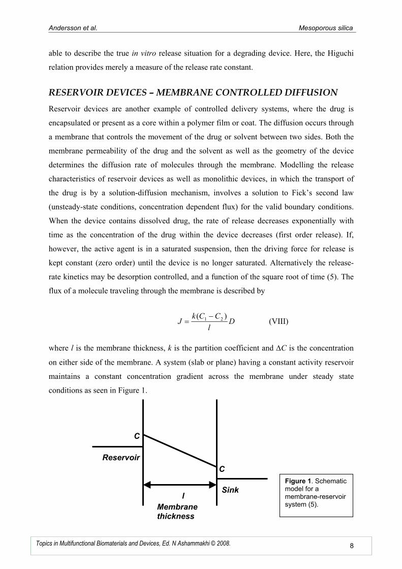
RESERVOIR DEVICES – MEMBRANE CONTROLLED DIFFUSION
Reservoir devices are another example of controlled delivery systems, where the drug is
encapsulated or present as a core within a polymer film or coat. The diffusion occurs through
a membrane that controls the movement of the drug or solvent between two sides. Both the
membrane permeability of the drug and the solvent as well as the geometry of the device
determines the diffusion rate of molecules through the membrane. Modelling the release
characteristics of reservoir devices as well as monolithic devices, in which the transport of
the drug is by a solution-diffusion mechanism, involves a solution to Fick’s second law
(unsteady-state conditions, concentration dependent flux) for the valid boundary conditions.
When the device contains dissolved drug, the rate of release decreases exponentially with
time as the concentration of the drug within the device decreases (first order release). If,
however, the active agent is in a saturated suspension, then the driving force for release is
kept constant (zero order) until the device is no longer saturated. Alternatively the release-
rate kinetics may be desorption controlled, and a function of the square root of time (5). The
flux of a molecule traveling through the membrane is described by
Dl
CCkJ
)( 21 −= (VIII)
where l is the membrane thickness, k is the partition coefficient and ∆C is the concentration
on either side of the membrane. A system (slab or plane) having a constant activity reservoir
maintains a constant concentration gradient across the membrane under steady state
conditions as seen in Figure 1.
C
C
Reservoir
Sink l
Membrane thickness
Figure 1. Schematic model for a membrane-reservoir system (5).
Andersson et al. Mesoporous silica
9Topics in Multifunctional Biomaterials and Devices, Ed. N Ashammakhi © 2008.
CONTROLLED DRUG RELEASE FROM ORDERED MICRO- AND
MESOPOROUS SILICA MATRICES
How a porous ceramic will function as a carrier system for a pharmaceutical when it comes to
both adsorption from and dissolution to a surrounding media, depends on several issues.
Factors, such as adsorption properties (interactions between drug and matrix), pore size, pore
connectivity, pore geometry and matrix reactions with surrounding media (dissolution
properties) are just a few of the things to take into account when designing a controlled drug
delivery system.
Mesoscopically ordered mesoporous silica materials have attracted a wide interest since their
discovery in the early 1990’s (24-28) due to their diverse potential applications areas;
including catalysis, filtration and chromatography. Supramolecular surfactant aggregates are
used as structure directing agents for inorganics during condensation, leading to
mesoscopically ordered surfactant-inorganic composites. Porosity can be induced in the
inorganic part by removal of the surfactant portion through thermal or chemical means
(24,26,29,30). By controlling the synthesis conditions and choice of structure directing
agents, the system can be engineered to fit numerous functions of preference.
The new group of mesoporous ordered materials was discovered independently by a Japanese
group (27,28) and a group from Mobil Oil, USA (24-26). The Mobil Oil family of
mesoporous materials were designated M41S, where the hexagonal MCM-41 was similar to
the Japanese material designated FSM-16.
The first MCM-41 materials prepared by the Mobil Oil group were derived under alkaline
synthesis conditions using an ionic surfactant (25,26). Characteristic of the Mobil Oil
materials were the well defined ordered and large pores ranging from 1.5 to 10.0 nm with
specific surface areas up to 1500m2/g. By varying the synthesis conditions they obtained
three different structures of the mesophase; a hexagonal phase (MCM-41, Mobil Composition
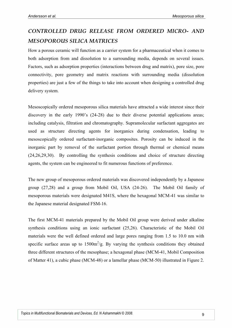
of Matter 41), a cubic phase (MCM-48) or a lamellar phase (MCM-50) illustrated in Figure 2.
Andersson et al. Mesoporous silica
10 Topics in Multifunctional Biomaterials and Devices, Ed. N Ashammakhi © 2008.
Figure 2. Mesophase structures of M41S: a) MCM-41, b) MCM-4831
and c) MCM-50.
The formation of mesoporous materials with a variety of crystallographically well-defined
frameworks has been realized via cooperative self-assembly including co-assembly of
surfactants and silica into liquid crystalline structures.
Later, synthesis of mesoporous silica under acidic conditions was reported by Stucky and co-
workers (pH < 2.0) (32,33). A similar material to M41S materials are the SBA materials,
developed at the University of Santa Barbara (34,35). The SBA-15 material has similar
features at that of MCM-41, but the pore walls are thicker, and the specific surface areas and
specific pore volumes are usually smaller than of the M41S materials. Therefore, the
mechanical and hydrothermal stability of these materials are better. Unlike the M41S
materials, the SBA-15 material has microporous pore walls. However, SBA-15 has a well
defined hexagonal symmetry similar to MCM-41.
As the purely siliceous mesoporous materials have been shown to be biocompatible, or
sometimes even bioactive, there is an increasing interest in this class of materials for
applications in the field of bioceramics, especially as bone substitute materials. Furthermore,
the highly organized porous silica matrix could be used as a potential controlled drug release
system. Another attractive advantage is that amorphous silica is degradable in an aqueous
solution, and thus problems related to the removal of the material after use can be avoided.
Andersson et. al. (36) describes the use of calcined mesoporous silica materials as
pharmaceutical carrier systems for controlled drug release. The drug release abilities were
investigated on a series of M41S and SBA materials (Table 1). There are important
a) b) c)
Andersson et al. Mesoporous silica
11 Topics in Multifunctional Biomaterials and Devices, Ed. N Ashammakhi © 2008.
differences in the pore structure and pore connectivity between the studied materials. The
MCM-41 and the SBA-3 type materials have straight one-dimensional cylindrical pores,
while the SBA-1 materials have a 3D interconnected porosity, where close to spherical pores
are connected through smaller windows. The c-MCM-41a material has also one-dimensional
pores, but the cylinders are not straight but corrugated, which essentially is equal to a
situation where larger pores are connected through smaller pore openings.
Ibuprofen, a common nonsteroidal anti-inflammatory pharmaceutical, was used as a model
drug due to its size and possibility to interact with silanol groups present in the pore walls of
the siliceous materials.
DRUG LOADING PROCEDURE
The impregnation process was performed according to one of the procedures described by
Vallet-Regi et al. (15). Ibuprofen was dissolved in hexane (33mg/mL), and a pellet of the
silica material (Table1) was soaked in the solution (33mg/mL silica/hexane) in a closed batch
to prevent evaporation of the liquid. The impregnation time was set to 3 days. The drug-
loaded disks were carefully washed with hexane to remove any adsorbed ibuprofen on the
exterior surface. RAMAN spectroscopy (Bruker IFS66, Germany) and TGA-FT-IR (Bruker
Equinox, Germany) were used in order to confirm complete evaporation of the hexane from
the impregnated materials. The amount of incorporated ibuprofen was determined
gravimetrically but also spectroscopically using a Perkin-Elmer UV-VIS-NIR spectrometer
(Germany) reading at 273 nm measuring the amount absorbed from solution.
DRUG RELEASE PROCEDURES
The drug-loaded tablets were dried and immersed in simulated body fluid, SBF, for in vitro
drug release studies (1mg ibuprofen/mL SBF) using static volumes. The release processes
were followed spectroscopically.
The silica contents from the degrading matrices were measured using molybdenum blue as a
tracer and the concentration were monitored by means of UV-VIS spectrophotometry (37).
Andersson et al. Mesoporous silica
12 Topics in Multifunctional Biomaterials and Devices, Ed. N Ashammakhi © 2008.
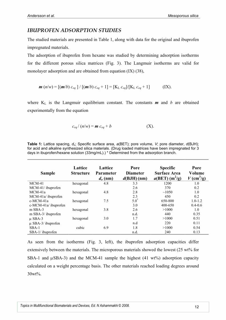
IBUPROFEN ADSORPTION STUDIES
The studied materials are presented in Table 1, along with data for the original and ibuprofen
impregnated materials.
The adsorption of ibuprofen from hexane was studied by determining adsorption isotherms
for the different porous silica matrices (Fig. 3). The Langmuir isotherms are valid for
monolayer adsorption and are obtained from equation (IX) (38),
m (n/w) = [(m/b) ceq ] / [(m/b) ceq + 1] = [KL ceq]/[KL ceq + 1] (IX).
where KL is the Langmuir equilibrium constant. The constants m and b are obtained
experimentally from the equation
ceq / (n/w) = m ceq + b (X).
Table 1: Lattice spacing, do; Specific surface area, a(BET); pore volume, V; pore diameter, d(BJH); for acid and alkaline synthesized silica materials. (Drug loaded matrices have been impregnated for 3 days in ibuprofen/hexane solution (33mg/mL).) * Determined from the adsorption branch.
Sample
Lattice
Structure
Lattice
Parameter
do (nm)
Pore
Diameter
d(BJH) (nm)
Specific
Surface Area
a(BET) (m2/g)
Pore
Volume
V (cm3/g)
MCM-41 hexagonal 4.8 3.3 1200 1.0 MCM-41/ ibuprofen 2.6 370 0.2 MCM-41a hexagonal 4.8 2.8 ~1050 1.0 MCM-41a/ ibuprofen 2.3 450 0.2 c-MCM-41a hexagonal 7.5 5.0* 650-800 1.0-1.2 c-MCM-41a/ ibuprofen 3.0 400-650 0.4-0.6 m SBA-3 hexagonal 3.8 2.6 >1000 1.0 m SBA-3/ ibuprofen n.d. 440 0.35 µ SBA-3 hexagonal 3.0 1.7 >1000 0.51 µ SBA-3/ ibuprofen n.d 220 0.11 SBA-1 cubic 6.9 1.8 >1000 0.54 SBA-1/ ibuprofen n.d. 240 0.13
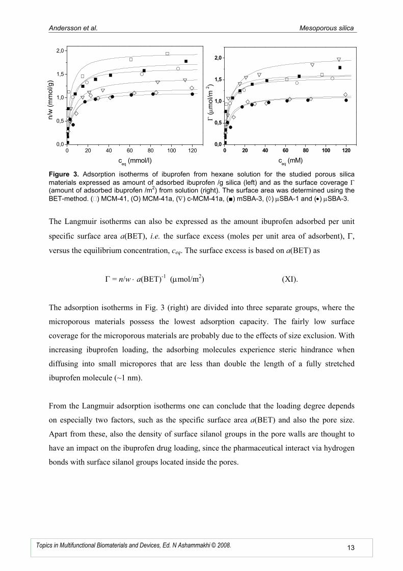
As seen from the isotherms (Fig. 3, left), the ibuprofen adsorption capacities differ
extensively between the materials. The microporous materials showed the lowest (25 wt% for
SBA-1 and µSBA-3) and the MCM-41 sample the highest (41 wt%) adsorption capacity
calculated on a weight percentage basis. The other materials reached loading degrees around
30wt%.
Andersson et al. Mesoporous silica
13 Topics in Multifunctional Biomaterials and Devices, Ed. N Ashammakhi © 2008.
0 20 40 60 80 100 1200,0
0,5
1,0
1,5
2,0n
/w (
mm
ol/g)
ceq
(mmol/l)
0 20 40 60 80 100 1200,0
0,5
1,0
1,5
2,0
Γ (µm
ol/m
2)
ceq
(mM)
Figure 3. Adsorption isotherms of ibuprofen from hexane solution for the studied porous silica
materials expressed as amount of adsorbed ibuprofen /g silica (left) and as the surface coverage Γ (amount of adsorbed ibuprofen /m
2) from solution (right). The surface area was determined using the
BET-method. () MCM-41, (O) MCM-41a, (∇) c-MCM-41a, (■) mSBA-3, (◊) µSBA-1 and (•) µSBA-3.
The Langmuir isotherms can also be expressed as the amount ibuprofen adsorbed per unit
specific surface area a(BET), i.e. the surface excess (moles per unit area of adsorbent), Γ,
versus the equilibrium concentration, ceq. The surface excess is based on a(BET) as
Γ = n/w ⋅ a(BET)-1 (µmol/m2) (XI).
The adsorption isotherms in Fig. 3 (right) are divided into three separate groups, where the
microporous materials possess the lowest adsorption capacity. The fairly low surface
coverage for the microporous materials are probably due to the effects of size exclusion. With
increasing ibuprofen loading, the adsorbing molecules experience steric hindrance when
diffusing into small micropores that are less than double the length of a fully stretched
ibuprofen molecule (~1 nm).
From the Langmuir adsorption isotherms one can conclude that the loading degree depends
on especially two factors, such as the specific surface area a(BET) and also the pore size.
Apart from these, also the density of surface silanol groups in the pore walls are thought to
have an impact on the ibuprofen drug loading, since the pharmaceutical interact via hydrogen
bonds with surface silanol groups located inside the pores.
Andersson et al. Mesoporous silica
14 Topics in Multifunctional Biomaterials and Devices, Ed. N Ashammakhi © 2008.
The difference in surface concentration of ibuprofen has important implications for the
release process, because the higher the ibuprofen surface loading the more hydrophobic the
pores will become, which could affect the kinetics of water diffusion into the matrix.
IN VITRO DRUG RELEASE FROM MESOPOROUS AND
MICROPOROUS SILICA
In vitro release studies were performed on the mesoporous siliceous materials and calcined
pellets of the materials were immersed in SBF solutions (39,40). The ibuprofen
concentrations were monitored in the solutions until all of the ibuprofen had diffused from
the pellets.
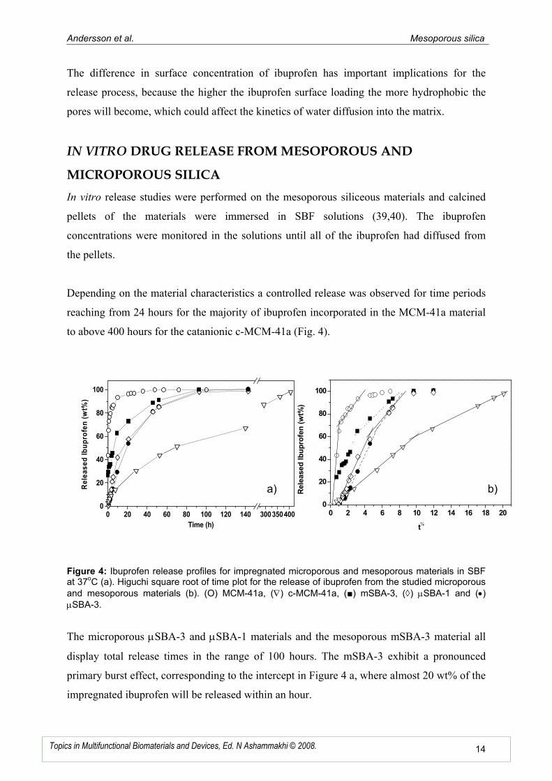
Depending on the material characteristics a controlled release was observed for time periods
reaching from 24 hours for the majority of ibuprofen incorporated in the MCM-41a material
to above 400 hours for the catanionic c-MCM-41a (Fig. 4).
Figure 4: Ibuprofen release profiles for impregnated microporous and mesoporous materials in SBF at 37
oC (a). Higuchi square root of time plot for the release of ibuprofen from the studied microporous
and mesoporous materials (b). (O) MCM-41a, (∇) c-MCM-41a, (■) mSBA-3, (◊) µSBA-1 and (•) µSBA-3.
The microporous µSBA-3 and µSBA-1 materials and the mesoporous mSBA-3 material all
display total release times in the range of 100 hours. The mSBA-3 exhibit a pronounced
primary burst effect, corresponding to the intercept in Figure 4 a, where almost 20 wt% of the
impregnated ibuprofen will be released within an hour.
0 20 40 60 80 100 120 140 3003504000
20
40
60
80
100
Released Ibuprofen (wt%
)
Time (h)
0 2 4 6 8 10 12 14 16 18 200
20
40
60
80
100
Released Ibuprofen (wt%
)
t½
b) a)
Andersson et al. Mesoporous silica
15 Topics in Multifunctional Biomaterials and Devices, Ed. N Ashammakhi © 2008.
The drug release kinetics of these systems can be described using the Highuchi model (22,23)
since the release is a diffusion controlled process and the adsorbed are relatively small and
uniformly distributed over the matrix. For a purely diffusion controlled process, the amount
of drug released exhibits a linear relationship if plotted against the square root of time.
However, the MCM-41a, mSBA-3, and µSBA-1 matrixes all display a two-step release (Fig.
4 b). The deviation from overall linearity is probably related to different dissolution rates of
the silica matrixes.
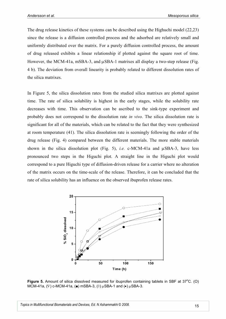
In Figure 5, the silica dissolution rates from the studied silica matrixes are plotted against
time. The rate of silica solubility is highest in the early stages, while the solubility rate
decreases with time. This observation can be ascribed to the sink-type experiment and
probably does not correspond to the dissolution rate in vivo. The silica dissolution rate is
significant for all of the materials, which can be related to the fact that they were synthesized
at room temperature (41). The silica dissolution rate is seemingly following the order of the
drug release (Fig. 4) compared between the different materials. The more stable materials
shown in the silica dissolution plot (Fig. 5), i.e. c-MCM-41a and µSBA-3, have less
pronounced two steps in the Higuchi plot. A straight line in the Higuchi plot would
correspond to a pure Higuchi type of diffusion-driven release for a carrier where no alteration
of the matrix occurs on the time-scale of the release. Therefore, it can be concluded that the
rate of silica solubility has an influence on the observed ibuprofen release rates.
Figure 5. Amount of silica dissolved measured for ibuprofen containing tablets in SBF at 37oC. (O)
MCM-41a, (∇) c-MCM-41a, (■) mSBA-3, (◊) µSBA-1 and (•) µSBA-3.
Andersson et al. Mesoporous silica
16 Topics in Multifunctional Biomaterials and Devices, Ed. N Ashammakhi © 2008.
Other factors that are thought to influence on the ibuprofen diffusion rate is the primary and
secondary particle size of the substrate, and also that the silica matrix is unlikely to dissolve
in a homogeneous manner. The silica would preferentially dissolve from regions, from which
the ibuprofen has been released, since the rate of silica dissolution was clearly slower for
materials containing ibuprofen as compared to the pure substrates.
In order to exclude any influence of apatite formed on the silica surface during the in vitro
drug release (42) also the calcium concentration was monitored from the SBF solution. It was
found that no, or negligible, precipitation was observed for any of the ibuprofen loaded
samples during the release period, which is why this effect can be excluded in this
investigation. Although, for the pure matrix, before or after the release, a calcium-loss from
SBF-solution was detected, indicating bioactivity and nucleation of hydroxyapatite.
CONCLUSION
In conclusion, the major factor contributing to the release of a drug from a porous ceramic
matrix, such as the mesoporous silica matrix, is the pore size. This was especially seen for the
materials exhibiting a 2D hexagonal structure with cylindrical pores, where the release rate
increased in the order of µSBA-3 < mSBA-3 < MCM-41. The slow release rate seen from the
c-MCM-41 material can be explained from its pore structure, as the material has corrugated,
cage like pores, where the pore opening is very small. The slow release kinetics could also be
explained by the more hydrophobic nature of the pore system due to the high drug loading,
which leads to slower water penetration rates (43,44) in this sample.
Andersson et al. Mesoporous silica
17 Topics in Multifunctional Biomaterials and Devices, Ed. N Ashammakhi © 2008.
References
1. Rösler A, Vandermuelen GWM, Klok H-A. Adv. Drug Delivery Rev. 2001; 53: 95.
2. Bae YH, Huh KM, Kim Y, Park KH. J. Controlled Release 2000; 64: 3.
3. Ravi Kumar M. N. V. J. Pharmaceut. Sci. 2000; 3: 238.
4. Ulrich KE, Cannizzaro SM, Langer RS, Shakesheff KM. Chem. Rev. 1999; 99: 3181.
5. Kim C-J. Controlled release dosage form design. Technomic Publishing Company, Inc.
USA; 2000.
6 . Uhrich KE, Cannizzaro SM, Langer RS, Shakesheff KM. Chem Rev. 1999; 99: 3181.
7. Ramchandani M, Robinson DJ. Control. Release 1998; 54: 167.
8. van de Belt H, Neut D, Schenk W, van Horn JR, van der Mei HC, Busscher HJ. Acta
Orthop. Scand. 2001; 72: 557.
9. Agrawal CM, Ray RB. J. Biomed. Mater. Res. 2001; 55:141.
10. Walenkamp GH, Kleijn LL, de Leeuw M. Acta Orthop. Scand. 1998; 69: 518.
11. Komlev VS, Barinov SM, Koplik EV. Biomaterials 2002; 23: 3449.
12. Komlev VS, Barinov SM, Girardin E, Oscarsson S, Rosengren Å, Rustichelli F, Orlovskii
VP.
Sci. Tech. Adv. Mater. 2003; 4: 503.
13. Hasegawa M, Sudo A, Komlev VS, Barinov SM, Uchida A. J. Biomed. Mater. Res. 2004;
70: 332.
14. Kim HW, Knowles JC, Kim HE. J. Biomed. Mater. Res. B 2005; 74: 686.
15. Vallet-Regi M, Rámila A, del Real RP, Pérez-Pariente J. Chem. Mater. 2001; 13:308.
16. Santos EM, Radin S, Ducheyne P. Biomaterials 1999; 20:1695.
17. Andersson J, Areva S, Spliethoff B, Lindén M. Biomaterials 2005; 26:6827.
18. Böttcher H, Slowik P, Süb W. J. Sol-Gel Sci. 1998; 13:277.
19. Badjić JD, Kostić NM. J. Phys. Chem. B 2000; 104:11081.
Andersson et al. Mesoporous silica
18 Topics in Multifunctional Biomaterials and Devices, Ed. N Ashammakhi © 2008.
20. Goto H, Isobe T, Senna M. J. Nanoparticle Res. 1999; 1:205.
21. Kortesuo P, Ahola M, Kangas M, Leino T, Laakso S, Vuorilehto L, Yli-Urpo A,
Kiesvaara J, Marvola M. J. Controlled Release 2001; 76:227.
22. Higuchi T. J. Pharm. Sci. 1961; 50:874.
23. Higuchi T. J. Pharm. Sci. 1963; 52:1145.
24. Kresge CT, Leonowicz ME, Roth WJ, Vartuli JC, Beck JS. Nature 1992; 359:710.
25. Beck JS. U.S. Patent 5.057.296, 1991.
26. Beck JS, Vartuli JC, Roth WJ, Leonowicz ME, Kresge CT, Schmitt KD, Chu CT-W,
Olson DH, Sheppard EW, McCullen SB, Higgins JB, Schlenker JL. J. Am. Chem. Soc.
1992; 114:10834.
27. Yanagisawa T, Shimizu T, Kuroda K, Kato C. Bull. Chem. Soc. Jpn. 1990; 63:988.
28. Inagaki S, Fukushima Y, Kuroda K. J. Chem. Soc., Chem. Commun. 1993;680.
29. Tanev PT, Pinnavaia TJ. Science 1995; 267:865.
30. Chen C-Y, Li H-X, Davis ME. Microporous Mesoporous Mater. 1993; 2:17.
31. Schumacher K, Ravikovitch PI, Du Chesne A, Neimark AV, Unger KK. Langmuir 2000;
16: 4648.
32. Huo Q, Margoleses DI, Ciesla U, Feng P, Gier DE, Sieger P, Leon BFR, Petroff PM,
Schüth F, Stucky GD. Nature 1994; 368:317.
33. Huo Q, Margoleses DI, Ciesla U, Demuth DG, Feng P, Gier DE, Sieger P, Chmelka BF,
Schüth F, Stucky GD. Chem. Mater. 1994; 6:1176.
34. Zhao DY, Feng JL, Huo QS, Melosh N, Fredrickson GH, Chmelka BF, Stucky GD.
Science 1998; 279:548.
35. Huo Q, Margolese D, Ciesla U, Demuth DG, Feng P, Gier TE, Sieger P, Firouzi A,
Chmelka BF, Schüth F, Stucky GD. Chem. Mater. 1994; 12:149.
36. Andersson J, Rosenholm J, Areva S, Lindén M. Chem. Mat. 2004; 21:4160.
Andersson et al. Mesoporous silica
19 Topics in Multifunctional Biomaterials and Devices, Ed. N Ashammakhi © 2008.
37. Mullin JB, Riley JP. Anal. Chim. Acta 1955; 12:162.
38. Langmuir I. J. Am. Chem. Soc. 1916; 38:2221.
39. Kokubo T. Apatite Formations in Ceramics, Metals, and Polymers Induced by a CaO,
SiO2-Based Glass in a Simulated Body Fluid. Bioceramics, vol. 4, eds. Bonfield W, Hastings
GW, Tanner KE, Guilford CT: Butterworth-Heinemann; 1991.
40. Kokubo T, Ito S, J. Biomed. Mater. Res. 1987; 24:331.
41. Landau MV, Varkey SP, Herskowitz M, Regev O, Pevzner S, Sen T, Luz Z. Microporous
Mesoporous Mater. 1999; 33:149.
42. Otsuka M, Fujita H, Nakamura T, Kokubo T. J. Bio-Med. Mater. Eng. 2001; 11:11.
43. Mal MK, Fujiwara M, Tanaka Y. Nature 2003; 421:350.
44. Smirnova I, Mamic J, Arlt W. Langmuir 2003; 26:8521.