mercury bioavailability in traditional food and the … · mercury bioavailability in traditional...
TRANSCRIPT

Mercury Bioavailability in Traditional Food and the Effect
of Selenium
Rami Yassine
Thesis submitted to the
Faculty of Graduate and Postdoctoral Studies, University of Ottawa
In partial fulfillment of the requirements for a M.Sc. degree in Biology
With a specialization in Chemical and Environmental Toxicology
Department of Biology
Faculty of Science
University of Ottawa
Ottawa, Canada
© Rami Yassine, Ottawa, Canada, 2017

ii
Abstract
Methylmercury (MeHg) is a potent neurotoxin capable of crossing the blood-brain barrier
causing a profound negative impact on the central nervous system. After its release, Hg may be
transported worldwide and eventually deposited in colder Arctic regions. Exposure of Aboriginal
communities to MeHg occurs primarily through the consumption of traditional food. Dietary
exposure studies are conducted using the total concentration of mercury in the food multiplied by
the food consumption rate. This method does not take into account the oral bioavailability of Hg.
Therefore, this study determines the bioavailability of Hg in four key traditional foods to provide
a better estimation of Hg exposure and to improve the characterization of overall Hg risk to
human health. We found that Hg concentrations significantly decreased for all foods after
undergoing an in-vitro digestion process. Hg bioaccessibility percentage of ringed seal liver was
32.3%, ringed seal muscle was 69.0%, lake trout muscle was 28.8%, and lastly air-dried beluga
muscle was 34.0%. Furthermore, no relationship was observed between bioaccessible Hg
concentrations and original Hg concentrations in the raw food. The concentration of MeHg in the
bioaccessible fraction was also examined and found to be significantly higher in muscle tissues
than in the liver. Bioavailability of the foods was determined using Caco-2 cells. Hg
bioavailability percentages were found to be 0.42% for RSL 5.24% for RSM, 7.30% for ADB,
and finally, 12.70% for LT. Correlations were found between increased Hg uptake and higher
percentages of bioaccessible MeHg as well as lower concentrations of bioaccessible selenium.
Lastly, a significant decrease in MeHg uptake after 24 hours was observed when co-incubating
with selenium. These results suggest that risk assessments should incorporate bioaccessibility
and bioavailability when estimating mercury exposure. Additionally, nutrients such as selenium
in traditional food may play a role in reducing mercury uptake in the gut.

iii
Résume
Le méthylmercure (MeHg) est une puissante neurotoxine capable de croiser la barrière
hémato-encéphalique, causant un impact néfaste important sur le système nerveux central. Après
qu’il est relâché, le Hg peut se propagé à travers le monde et, éventuellement, peut aboutir dans
des régions arctiques plus froides. Les communautés autochtones sont principalement exposées
au MeHg lorsqu’elles consomment leurs aliments traditionnels. Des études sur l’exposition
diététique sont faites en multipliant la concentration totale de mercure d’un aliment par son taux
de consommation. Cette méthode ne tient compte de la biodisponibilité orale du Hg. Ceci dit,
cette étude détermine la biodisponibilité du Hg dans quatre groupes d’aliments traditionnels afin
de permettre une meilleure estimation de l’exposition au Hg, ainsi qu’à améliorer la
caractérisation du risque global du Hg sur la santé des humains. Nous avons conclu que la
concentration du Hg est grandement diminuée pour tous les aliments après qu’il a subi un
processus de digestion in vitro. Les pourcentages observés de la bioaccessibilité du Hg étaient de
32.3% dans le foie du phoque annelé (FPA), de 69.0% dans le muscle du phoque annelé (MPA),
de 28.8% dans la truite de lac (TDL) et, dernièrement, de 34.0% dans le béluga séché à l’air
(BSA). De plus, aucune relation n’a été observée entre la concentration de bioaccessibilité du Hg
et la concentration initiale du Hg dans les aliments. La concentration de MeHg dans l’échantillon
bioaccessible a aussi été examinée, et elle a été trouvée significativement plus élevée dans les
tissus musculaires que dans le foie. La biodisponibilité des aliments a été déterminée à l’aide de
cellules Caco-2. Les pourcentages observés de la biodisponibilité du Hg étaient de 0.42% dans le
FPA, de 5.24% dans le MPA, de 7.30% dans le BSA et, dernièrement, de 12.70% dans la TDL.
Des corrélations ont été trouvées entre l’augmentation de l’absorption du Hg et des pourcentages
plus élevés de MeHg bioaccessible, ainsi que des concentrations plus élevées de sélénium

iv
bioaccessible. Dernièrement, une diminution significative dans l’absorption du MeHg a été
observée après 24 heures de co-incubation avec le sélénium. Ces résultats suggèrent que des
évaluations de risques devraient incorporer la bioaccessibilité et la biodisponibilité lors de
l’estimation de l’exposition au mercure. De plus, des nutriments dans les aliments traditionnels,
tel le sélénium, pourraient jouer un rôle dans la réduction de l’absorption du mercure dans
l’intestin.

v
Acknowledgments
I would like to begin by expressing my gratitude to my thesis supervisor, Dr. Laurie Chan
for allowing me the opportunity to discover my potential as an academic in his lab. Dr. Chan
consistently offered his invaluable guidance and expertise while simultaneously allowing me to
create and develop a research project that I could call my own. His hands-off method of
supervision provided me with a platform to grow as a pro-active and independent researcher, for
which I am forever thankful. To my thesis committee members, I appreciate all the advice
you’ve given me at different stages of my graduate studies. Thank you to Dr. Alexandre Poulain
for providing me with critical feedback on my seminar presentation as well as his input during
thesis committee meetings. I would also like to thank Dr. Shawn Donaldson for taking the time
to speak with me in-person and over the phone to discuss my thesis and future career. Our
discussions have contributed greatly to my personal and academic development.
This work could not have been possible without the helpful assistance of my lab mates
and friends. I would particularly like to thank Dr. Emmanuel Yumvihoze and Paulin Junior
Vanié for their indispensable knowledge and their help throughout my thesis. I am thankful as
well to Susie Huang for her cell culture orientation and to Linda Ha for her assistance. I would
also like to acknowledge the Northern Contaminants Program (NCP) for funding this research.
I dedicate this thesis to my family. My father, Ziad Yassine and mother Habiba Attia,
whom without their unconditional love and endless encouragement, I would not be where I am.
Last but not least, to my incredible wife Adriana Yassine, who stood by me throughout this
entire journey providing unwavering support at times where it was exceptionally needed, I am
truly indebted.

vi
Table of Contents
Abstract ........................................................................................................................................................ ii
Résume ........................................................................................................................................................ iii
Acknowledgments ........................................................................................................................................ v
Table of Contents ........................................................................................................................................ vi
List of Tables .............................................................................................................................................. viii
List of Figures ............................................................................................................................................... ix
List of Abbreviations .................................................................................................................................... x
Chapter 1: General Introduction .................................................................................................................. 1
General Overview ..................................................................................................................................... 1
Rationale ................................................................................................................................................... 3
Objectives and Hypothesis ........................................................................................................................ 4
Objectives.............................................................................................................................................. 4
Hypotheses ........................................................................................................................................... 4
Chapter 2: Literature Review – Overview on Mercury and Neurotoxic Effects ......................................... 6
Mercury – Sources of Release ................................................................................................................... 6
The Mercury Cycle .................................................................................................................................... 6
Forms of Mercury...................................................................................................................................... 8
Methylmercury ......................................................................................................................................... 9
Sources of Exposure .............................................................................................................................. 9
Effects of Mercury ............................................................................................................................... 10
Mechanisms of Transport ................................................................................................................... 11
Mechanisms of Toxicity....................................................................................................................... 13
The Gastrointestinal Tract....................................................................................................................... 14
Pathways of uptake ............................................................................................................................. 16
Selenium ................................................................................................................................................. 17
Nutritional Significance ....................................................................................................................... 17
Mercury-Selenium Antagonism .......................................................................................................... 18
Chapter 3: Determination Mercury Bioaccessibility in Food .................................................................... 20
Introduction ............................................................................................................................................ 20
Methods .................................................................................................................................................. 25

vii
Sample Collection ............................................................................................................................... 25
In-vitro Digestion ................................................................................................................................ 25
[THg] Determination ........................................................................................................................... 26
Hg Species Determination ................................................................................................................... 27
Selenium Determination ..................................................................................................................... 27
Results ..................................................................................................................................................... 28
Hg Determination in raw Food............................................................................................................ 28
Bioaccessible Total Hg Concentrations ............................................................................................... 30
MeHg Speciation ................................................................................................................................. 32
Total Selenium Determination ............................................................................................................ 34
Discussion................................................................................................................................................ 37
Conclusion ............................................................................................................................................... 41
Chapter 4: Determination of Mercury Bioavailability in Food ................................................................. 43
Introduction ............................................................................................................................................ 43
Methods .................................................................................................................................................. 47
Colorectal adenocarcinoma cells (Caco-2 cells) Culture ..................................................................... 47
Caco-2 Cells Experimental Methods ................................................................................................... 47
Determination of Bioavailability in Traditional Food .......................................................................... 50
MeHg Uptake. ..................................................................................................................................... 51
Selenium-MeHg Co-incubation ........................................................................................................... 52
Mercury Determination ...................................................................................................................... 52
Statistical Analysis ............................................................................................................................... 53
Results ..................................................................................................................................................... 54
Determination of Bioavailability in Traditional Food .......................................................................... 54
Mercury and Selenium Uptake ........................................................................................................... 63
Discussion................................................................................................................................................ 66
Conclusion ............................................................................................................................................... 70
Chapter 5: Conclusions and Future Direction ............................................................................................ 72
Overall Conclusions ................................................................................................................................. 72
Future Directions .................................................................................................................................... 74
References .................................................................................................................................................. 76

viii
List of Tables
Table 3.1 Total Hg concentrations in four raw traditional foods
Table 3.2 Total bioaccessible Hg concentrations in four traditional foods
Table 3.3 Bioaccessible MeHg concentrations in four traditional foods
Table 3.4 Total selenium concentrations in four traditional foods
Table 3.5 MeHg:Selenium molar ratio in four traditional foods
Table 4.1 Mercury concentrations in Caco-2 cells for ringed seal liver
Table 4.2 Mercury concentrations in Caco-2 cells for ringed seal muscle
Table 4.3 Mercury concentrations in Caco-2 cells for air-dried beluga muscle
Table 4.4 Mercury concentrations in Caco-2 cells for lake trout muscle
Table 4.5 Mercury concentrations before and after in-vitro digestion and after uptake by
Caco-2 cells in four traditional foods
Table 4.6 Mercury concentrations in Caco-2 cells after 0.02µM MeHg spike
Table 4.7 Mercury concentrations in Caco-2 cells after MeHg-SeMet co-incubation

ix
List of Figures
Figure 1 Percentage of Hg transported through Caco-2 cells vs increasing fractions of MeHg in
bioaccessible food digest for four traditional foods
Figure 2 Percentage of Hg transported through Caco-2 cells vs increasing selenium
concentrations in bioaccessible food digest for four traditional foods

x
List of Abbreviations
ADB: air-dried beluga
CNS: central nervous system
Caco: colorectal adenocarcinoma
CH3HgCl: methylmercury chloride
EMEM: Eagle's Minimum Essential
Medium
FBS: fetal bovine serum
GI: gastrointestinal tract
GEM: gaseous elemental mercury
GPX: glutathione peroxidase
GSH: glutathione
CVAFS: cold vapour atomic fluorescence
spectroscopy
HC: Health Canada
HCL: hydrochloric acid
Hg: mercury
Hg0: elemental mercury
HgCl2: mercury chloride
ICP-MS: Inductively Coupled Plasma Mass
Spectrometry
IHS: Inuit health survey
ISR: Inuvialuit Settlement Region
IVBA: in-vitro bioaccessible
IVT: in-vitro
LT: lake trout
MeHg: methylmercury
MeHgCl: methylmercury chloride
NaCl: sodium chloride
NaHCO3: sodium bicarbonate
NaOH: sodium hydroxide
NG: nanograms
NIHS: Nunavik Inuit Health Survey
PBS: Phosphate-buffered saline
POPs: persistent organic pollutants
PPM: parts per million
ROS: reactive oxygen species
RSM: ringed seal muscle
RSL: ringed seal liver
Se: selenium
SeMet: Selenomethionine
SH: sulfhydryl group or thiol group
SRM: standard reference material
TEER: Trans-epithelial electrical resistance
THg: total mercury
µg: micrograms
µM: micro molar
WW: wet weight

1
Chapter 1: General Introduction
General Overview
Studies in the 80’s conducted in Canada’s Arctic region found high levels of
contaminants such as persistent organic pollutants (POPs) and heavy metals such as mercury (El-
hayek, 2007). This finding presents a health concern for the 4.6% of Canada’s Aboriginal
population who inhabit the North (Simeone, 2008). Inuit populations in Nunavik were found to
have Hg blood concentrations in the range of 50.2nmol/L based on the 2004 Nunavik Health
Survey (Valera et al., 2009a), while adults living in southern Canadian regions were found to
have an average of 4.1nmol/L according to the Canadian Health Measures Survey (Laird &
Chan, 2013). The significantly higher levels of blood mercury are specifically detrimental for
children and breast-fed infants potentially resulting in cerebral palsy, low birth weight and early
sensorimotor dysfunction (Boucher et al., 2016; Jacobson et al., 2015). Exposure of Aboriginal
communities to environmental contaminants occurs mainly through the consumption of
traditional food. Research conducted in 3 Inuit jurisdictions in Northern Canada (Inuvialuit
Settlement Region), attempted to identify the top foods contributing to the intake of Hg for
participants of the 2008-2009 Inuit Health Survey (IHS). Despite only being consumed a rate of
32.7g/week, ringed seal liver was found to contain exceptionally high Hg concentrations as it
constituted 59% of dietary Hg intake (Laird, James, et al., 2013). In contrast, arctic char, while
not containing Hg concentrations as high as ringed seal liver, was frequently consumed at a rate
of 378g/week, and was the second highest contributor of dietary Hg intake at 8.4% (Laird,
James, et al., 2013). Animals including beluga whale, narwhal, and caribou were also included in
list of top ten largest sources of dietary Hg intake within the Inuvialuit Settlement Region (ISR)
(Laird, James, et al., 2013).

2
Epidemiological studies on susceptibility to mercury toxicity have found that despite
being exposed to similar chronic low-dose mercury concentrations, the subsequent neurotoxic
effects in different regions have varied (Chapman & Chan, 2000). One of the possible reasons
for this finding is the source of exposure (i.e. the type of animal) and the specific body part
consumed. As we will discuss in this study, each animal accumulates mercury in various forms
and concentrations in different parts of its body.
Furthermore, food from different regions will vary in its nutritional content. Recently, there
has been a growing consensus of the toxicity of mercury being linked with its high binding
affinity with selenium, disrupting selenoenzyme and selenoprotein activity (Farina et al., 2011;
Franco et al., 2009). Therefore it follows that mercury neurotoxicity may be related to the Hg:Se
molar ratio in the tissues (Berry & Ralston, 2008) and excess of free selenium can potentially
play a role in maintaining antioxidant and redox control functions that can be inhibited by MeHg
binding (N. V C Ralston & Raymond, 2010).
There are numerous other factors that contribute to the overall risks involved in the
consumption of mercury-contaminated food. Several studies have looked into elements such as
the preparation method (steaming, grilling) (He & Wang, 2011) or the effects of co-consumption
with phytochemical rich foods (Shim et al., 2009) on the bioaccessibility of mercury. In this
thesis, we aim to study the bioavailability of mercury in key traditional foods in the Inuit diet.
We will also investigate the potential effects of selenium on the absorption of mercury using an
in-vitro Caco-2 cell model.

3
Rationale
Past human health risk assessments have often made the incorrect assumption that all
mercury ingested through the consumption of fish and marine mammals is in the dangerous form
of MeHg. Additionally, dietary exposure is usually conducted using the total concentration of
mercury in the food multiplied by the food consumption rate. However, this estimation does not
take into account the oral bioavailability of mercury (He & Wang, 2011). Bioaccessibility is
defined as the fraction of a contaminant that remains in soluble form after consumption and
digestion. Several studies have looked into the factors (steaming, grilling, and co-consumption
with phytochemical rich foods) that may have an impact on the bioaccessibility. Furthermore,
after digestion, the fraction of bioaccessible Hg that is absorbed through the gastrointestinal tract
and into the bloodstream is referred to as the bioavailable fraction. Therefore, to fully calculate
the risks involved in the consumption of Hg contaminated food, it is important to incorporate
both factors of bioaccessibility and bioavailability into the estimation.
Looking at a traditional diet as a whole can reveal benefits that may outweigh the mercury
risks (Gagné et al., 2012). Selenium is one of the most beneficial nutrients consistently found in
the traditional Inuit diet (Laird et al., 2013). Nervous systems have been found to utilize
selenium-dependant enzymes to protect the brain from cellular damage which may be caused by
neurotoxic contaminants such as mercury. Therefore, it follows that a selenium-rich diet may
offer some protective effects against mercury toxicity (IPY, 2008). The Nunavik Inuit Health
Survey (NIHS), a study using 25 different types of traditional food in Nunavik, Northern Quebec
found high levels of selenium (≥1.0 μg/g) in 20% of the food types researched, while the
remaining 80% contained selenium concentrations that were considered “a good source”
according to literature reference values (0.20–0.50 μg/g) (M Lemire et al., 2014).

4
According to data from the International Polar Year Inuit Health Survey conducted in three
Inuit jurisdictions in Northern Canada, three of the four traditional foods chosen for this study
including ringed seal liver, ringed seal muscle, and air-dried beluga whale muscle were the major
sources of Hg contributing to approximately 65% of the dietary Hg intake, and 25% of the
selenium intake from the traditional food diet (Laird et al., 2009). We also chose to study lake
trout which is often suggested to be an alternative traditional food to lower total Hg exposure.
The purpose of this study is to determine the bioaccessibility and bioavailability of four key
traditional foods of the traditional Inuit diet to provide a better estimation of Hg exposure and
improve the characterization of risk of Hg exposure to human health. Furthermore, we will
investigate what impact, if any, selenium may have on the uptake of Hg using an in vitro Caco-2
cell model.
Objectives and Hypothesis
Objectives
1. To determine the concentration of total mercury (THg) remaining in four selected
food samples after undergoing an in-vitro digestion process, i.e. the bioaccessible
fraction.
2. To determine the fraction of mercury in the four food samples that is absorbed across
the gastrointestinal tract over a 24h period using an in vitro Caco-2 cell model, i.e. the
bioavailable fraction.
3. To determine the effects of selenium co-incubation on the absorption of mercury.
Hypotheses
1. Bioaccessible mercury concentrations will be dependent on original Hg concentration
in the food.

5
2. The uptake of mercury in the gut will show a positive correlation with the
concentration of bioaccessible MeHg in the food digest.
3. Selenium co-incubation will significantly decrease the total uptake of mercury after
24h.

6
Chapter 2: Literature Review – Overview on Mercury and
Neurotoxic Effects
Mercury – Sources of Release
Mercury (Hg) is a dense heavy metal that exists in the environment in various forms:
Elemental Mercury (Hg0), inorganic mercury (e.g. Mercuric ion (Hg
2+)), and organic mercury
(e.g. Methyl mercury (MeHg)) (Health Canada, 2007). As an element, it can be found in the
Earth’s crust and can be released into the environment via natural sources such as volcanoes,
forest fires and fossil fuels (UNEP Chemicals Branch, 2008). However, mercury is primarily
released into the atmosphere through the incidental pathway. This process occurs when naturally
found mercury in coal, rocks and oil is released into the atmosphere through manufacturing and
factory processes that do not involve the use mercury. The largest source of incidental mercury
release is as a by-product of fossil fuel combustion accounting for 46% of total mercury emission
from anthropogenic sources (AMAP, 2011). Other sources include artisanal and small-scale gold
production, cement production, and emissions from ferrous and non-ferrous metal industries
(AMAP, 2011). Currently Asian countries account for approximately 65% of global Hg
emissions followed by North America and Europe at 8.3% and 7.9%, respectively (AMAP,
2011).
The Mercury Cycle
The large majority of the 5000 tonnes of mercury in the atmosphere is found in the form
of elemental mercury (Environmental Investigation Agency, 2003). It is released from both
anthropogenic sources (commercial products, industrial processes, etc.) and natural sources such
as volcanic eruptions and rock decay. As one of the world’s leaders in industrial advancement,
China is currently leading the world as the biggest contributor to atmospheric Hg release (Zhang
7
& Wong, 2007). When Hg is released into the atmosphere, it can undergo several biochemical
transformations into different species based on the space it occupies in the environment (W. C.
Li & Tse, 2014). Mercury is released into the atmosphere in the form of gaseous elemental
mercury (Hg0). Elemental mercury can reside within the atmosphere for long periods of time
(between 6 months and 2 years) and can travel worldwide via long-range transport. Depending
on the distance of deposition from the source of emission, it can be classified as local deposition,
regional deposition or as is the case with Hg deposited in the Arctic region; it can enter the
global atmospheric mercury pool. Oxidized forms of mercury (ex. Hg2+
) are more reactive and
can be deposited at local points closer to the origin of emission as it is 105 times more water
soluble than elemental mercury (Bullock Jr, 2000).
Through cycles of chemical reactions, gaseous elemental mercury in the atmosphere can
be converted to a more reactive form of Hg which have shorter atmosphere lifespans, and are
deposited to terrestrial and marine sources (Steffen et al., 2015). Furthermore, the deposited Hg
species can also be transformed back into elemental mercury and re-enter the atmosphere using
processes such as photolytic reduction (W. C. Li & Tse, 2014).
Once Hg is deposited in the form of inorganic mercury in lakes and oceans, it undergoes
a biogeochemical transformation process into the toxic form of MeHg. The most currently
accepted theory for this process is by means of microbial transformation. Two processes are
thought to compete for the inorganic mercury substrate in water; methylation of Hg2+
into MeHg,
and the reduction of Hg2+
to elemental mercury (Hg0.) However, the details of these two
processes are not entirely conclusive. In more mild climates, the methylation of mercury has
been found to occur via sulfate and iron-reducing microorganisms (Fleming et al., 2006).

8
However, sulfate-reducing bacteria have also been found in colder environments and are likely to
participate in mercury methylation (Chételat et al., 2014).
Methylation of inorganic mercury into organic mercury is a key step in mercury toxicity
for humans and wildlife. Inorganic mercury is dangerous in the rare circumstance of exposure to
very high concentrations for a short amount of time (acute exposure). However, organic mercury
will accumulate in the body over long periods of time which may eventually lead to toxicity even
in smaller concentrations (AMAP, 2011).
Forms of Mercury
In its elemental state, mercury is the only element that is liquid at room temperature. Liquid
mercury can be used to extract pure gold from silver as it forms amalgam with the two elements.
As such, miners will often be exposed to high concentrations of elemental mercury through
inhalation, its most bioaccessible pathway (Park & Zheng, 2012). Historically, inorganic
mercury has been used in skin ointments, antiseptic preservatives and infamously, to treat high
quality fur used in hats leading to what was known as “the mad hatter’s” disease (Park & Zheng,
2012). Inorganic mercury (Hg2+
) can be ingested through the gastrointestinal tract at a rate of
approximately 7 to 15% (World Health Organization, 2003) where it will predominantly
accumulate in the kidneys. However, inorganic mercury is not lipid soluble which means that it
will not cross the blood-brain barrier and negatively impact the central nervous system. Acute
oral exposures to inorganic mercury at high concentrations may cause corrosion to the chest
cavity, impaired kidney function and damage to the gastrointestinal tract. Chronic exposure to
inorganic mercury is much rarer (Park & Zheng, 2012).
Elemental mercury (Hg0) is absorbed in negligible rates through the gut at and is much more
rapidly up taken by means of inhalation through the respiratory pathway at a rate of

9
approximately 80%. Through this pathway, elemental mercury can reach the central nervous
system and cause harmful neurotoxic damage (World Health Organization, 2003).
Methylmercury
Sources of Exposure
MeHg is composed of a methyl group (CH3-) bound to a mercury ion (Hg+) and it is
considered one of the most dangerous forms of mercury. MeHg is a neurotoxin capable of
readily crossing the blood-brain barrier and negatively impacting the central nervous system. The
lipid solubility of MeHg also supports its bioaccumulation in lipid compartments such as fatty
tissue and the brain (Guzzi & La Porta, 2008). Additionally, research shows that the primary
method of MeHg uptake through the intestinal epithelium is via the transcellular route by means
of passive diffusion through the double lipid bilayer (Marta Vázquez et al., 2014). MeHg will
biomagnify along marine food webs starting in organisms at the base of the food chain to marine
mammals and higher level predators at the top of the food chain. This process means that
environmental exposure and particularly human exposure may be substantially detrimental
(Seixas et al., 2014). Particularly, some Indigenous communities relying on a traditional diet as
their main source of nutrition may be exposed to fish and marine mammals with mercury
concentrations that are in excess of 1 ppm (M Lemire et al., 2014). However, total
concentrations of mercury in different species of fish and marine mammals have been known to
differ by 100-fold (Mergler et al., 2007). Additionally, the Health Canada food consumption
recommended guideline for total mercury concentrations is 0.5ppm for fish species and 1.0ppm
for organisms that are higher up on the food chain. In a study within the region of Nunavik,
Northern Quebec, most of the organisms sampled showed low levels of MeHg (<0.2ppm) with
the exception of organisms that are higher up on the food chain such as beluga whale, ringed seal
and lake trout which have been found to have very high concentrations of MeHg (>1.0ppm.)
10
Despite this finding, the study also looked at blood mercury concentrations of a sample
population of 702 individuals and found that over half of the women of child-bearing age in that
region showed blood Hg levels above the guidance values (M Lemire et al., 2014).
Effects of Mercury
Organic mercury compounds, specifically, MeHg have been widely researched due to
their potential for accumulation in the central nervous system and subsequent neurotoxic effects.
In 1956, MeHg became well known when after consuming fish and shellfish found in the
Minamata bay, residents of Minamata, Japan began showing signs of extreme illness and even
death (Harada, 1978). Neurologic symptoms such as paralysis, sensory disturbances in the distal
part of extremities, deafness, speech impairment, and mental disorders were all experienced by
residents of the Minamata bay and were collectively referred to as the Minamata disease
(Ceccatelli et al., 2013). This incident served as the first instance in which MeHg was recognized
as a neurotoxin with the central nervous system and the brain as its primary target (McAlpine &
Araki, 1958). More mercury outbreaks have occurred over time in countries such as Sweden,
Iraq, Pakistan and Ghana (T. W. Clarkson, 2002). In more recent times, due to more strict
environmental policies and waste by-product clean up, the frequency of large scale
contamination incidents have decreased. However, communities that have been found to exhibit
high levels of fish consumption may still be at risk of chronic mercury exposure. Children are
considered to be at a higher risk for the toxic effects of mercury exposure than adults due to their
developing systems, lower body weight and lower neurologic effect threshold (Health Canada,
2004) Ha et al., 2016). Data has consistently shown that MeHg is transferred to the fetus through
the placenta during gestation. In fact, fetal cord MeHg blood concentration has been found to be
higher than the corresponding maternal concentration at a ratio of approximately 1.7 (Stern,
2005). At the population level, studies from a variety of regions in the world showed evidence of

11
poorer neurologic status and delayed development as well as inferior performance on language,
memory and attention tests in newborns exposed to MeHg in utero (Mergler et al., 2007; Myers
et al., 2003). Additionally, a study by Jacobson et al. (2015) found that “children with cord
mercury concentration of ≥7.5µg/L were four times as likely to have an IQ score of <80, which
is the clinical cut off for intellectual disability”. A Nunavik study has also shown that the subtle
effects of chronic mercury exposure may extend to adults as well. In a sample of 732 Inuit
participants over the age of 18, the study found that mercury exposure was correlated with a
corresponding increase in blood pressure and pulse pressure (Valera et al., 2009).
The Nunavik Child Development Study (NCDS) conducted behavioral evaluations of 11-
year old children using in-class questionnaires which analyzed problem-solving skills and
behavioral patterns (AMAP, 2009). Tasks such as the Santa Ana Form Board, the NES-3 finger
tapping test, and the Stanford-binet copying subtest were used to assess motor function.
Umbilical cord samples as well as blood samples from children were used to determine prenatal
exposure to Hg (Boucher et al., 2016). Results showed that cord blood Hg, as well as current
blood Hg were associated with poorer performance on two of the three motor function tests
(Boucher et al., 2016). Furthermore, umbilical cord Hg concentrations were also related to
attention problems such as ADHD (Boucher et al., 2012).
Mechanisms of Transport
Communities that rely on a traditional diet of predatory fish and marine mammals are
particularly at risk for chronic MeHg exposure. Direct consumption of mercury contaminated
food is the dominant method by which humans are exposed to MeHg (Farina et al., 2011). Of all
the different forms of mercury, past research has found that MeHg has the highest rate of
absorption in the gut at 90-95% uptake (Nielsen, 1992). However, this value was based on two

12
major studies conducted in 1969 and 1971 with limitations including a small sample size, the use
of methylmercury bound to nitrate rather than the commonly found sulfur-bonds, and lastly the
lack of a food matrix (Aberg et al., 1969; Miettinen et al., 1971). Furthermore, mercury exposure
in both studies was acute rather than the more relevant chronic exposure. Recently, Bradley et
al., 2017 reviewed 20 different studies reporting on bioavailability (bioaccessibility or
absorption) and found that the mean absorption rate of MeHg is closer to 12-79%. These results
challenge the long-held belief that MeHg is completely absorbed into the bloodstream after
consumption. Post-ingestion of MeHg, it will be distributed to the entire body, within as little as
a few hours, as a result of its lipid solubility (T. Clarkson, 1972). The mechanisms by which
mercury is transported across cellular membranes and organs to exert its toxic effects are still
being studied today. However, previous studies have attempted to identify the transport of MeHg
using a variety of different in-vitro models and cell lines.
MeHg has been found to have a particularly high affinity for sulfhydryl-containing
molecules (ex. GSH, cysteine) in various forms in the body forming non-enzymatic bonds with
sulphur. When bound to these molecules, MeHg uses “molecular mimicry” to behave as
homologs of various vital amino acids and other biomolecules, gaining access to membrane
transporters that actively transport these molecules (Zalups & Ahmad, 2005). Past studies have
shown that the kidney is the primary site of Hg2+
uptake and accumulation. Specifically, it was
found that organic ion transporter protein (OAT1) is actively involved in the uptake of the
organic form of mercury (Zalups, 1995). Using canine kidney cells (MDCK) transfected with the
human isoform of the membrane transporter OAT1, it was found that MeHg bound to N-
acetylcysteine (NAC) is a transportable substrate of OAT1 (Zalups & Ahmad, 2005). The rates
of survival of MDCK cells transfected with OAT1 compared to control MDCK cells when

13
exposed to toxic concentrations of CH3Hg-NAC were significantly lower. The study also found
substrate specific data that supported the notion that more than one transporter was involved in
the uptake of MeHg.
To be capable of wide-spread distribution within the body, MeHg would need the ability
to utilize abundant membrane transporters such as the amino acid exchanger proteins LAT1 and
LAT2. These proteins are expressed in a variety of tissues and are one of the dominant methods
of neutral amino acids entering into the brain from blood (Kerper et al., 1992). One of the amino
acids taken up by the L-transporters is methionine, which is structurally similar to MeHg bound
in a cysteine complex (MeHg-L-cysteine). Using oocytes from Xenopus laevis expressing the
two LAT carrier proteins found in humans, a study by Simmons-Willis et al. (2002) attempted to
determine if MeHg-L-cysteine can be a substrate for amino acid transporters. The apparent
affinities (Km) of MeHg-L-cysteine uptake were found to be similar to methionine in LAT1
transporters (98µM and 99µM.) However, the Vmax values were higher for MeHg-L-cysteine,
suggesting that it could be a better substrate for the LAT transporter than the amino acid
methionine. Furthermore, a study using hamster ovary cells found that increased expression of
LAT1 transporter was correlated with an increase in the rate of uptake of MeHg when cysteine
was available (Yin et al., 2008).
Mechanisms of Toxicity
Glutathione (GSH) is a ubiquitous thiol compound found in all tissues including the CNS and
plays an antioxidant role in the body (ex. detoxification of peroxidase and protecting the cell
against oxidative damage) (Dringen, 2000). A recent study using pregnant mice exposed to
varying concentrations of MeHg in drinking water found that control mice showed a post-natal
increase in GHS levels over time, while in MeHg exposed group, a dose-dependent inhibition of

14
the aforementioned increase was found. Furthermore, when Hg levels returned to baseline level,
GSH levels and activity remained low which suggests that MeHg may cause biochemical
alterations to the GSH development system (Stringari et al., 2008).
The mechanism by which MeHg affects the central nervous system resulting in its neurotoxic
effects remains relatively unknown. It has been found that extended exposure of Hg can result in
the dysregulation of the important excitatory and inhibitory neurotransmitter glutamate, glycine,
γ-aminobutyric acid (GABA) (Fitsanakis & Aschner, 2005). Another important brain
neurotransmitter is dopamine. The breakdown of dopamine mechanisms was previously found to
be related to the onset of neurological disorders such as Parkinson’s disease and Huntington’s
disease (Cui et al., 2006). A recent study investigated the effects of MeHg on dopaminergic
pathways using a neuronal cell line (MN9D cells.) The exposure of the neuronal cell line to
increasing concentrations of MeHg was compared to MPP+, a neurodegenerative compound
known for disrupting the dopamine pathway (Shao, Yamamoto, et al., 2015). Results showed
that exposure of MN9D cells to high concentrations of MeHg resulted in diminished dopamine
levels and degenerative effects similar to MPP+ (Shao, Figeys, et al., 2015). Further research
studying the gene profile of the MN9D cells after exposure to MeHg using real-time PCR
Parkinson’s disease arrays found that 19% and 39% of genes were significantly altered by 2.5µM
and 5µM MeHg treatments (Shao & Chan, 2015). However, the general consensus in studies
seems to be that not one pathway can explain the plethora of effects associated with MeHg
induced neurotoxicity.
The Gastrointestinal Tract
The primary function of the gastrointestinal tract (GI) is to allow the selective absorption and
transport of water, electrolytes, nutrients and foreign chemicals such as drugs and medicine. To

15
aid with absorptive functions, the small intestines employ a large complex viscous-elastic gel
called the mucosal surface (Barthe et al., 1999). This surface is specialized in many ways to
maximize efficiency of uptake. It relies on several factors such as epithelial cell-covered villi
which increase surface area by a factor of eight (Carr & Toner, 1984). Another major component
of the GI, the lumen, is located within the external environment, and based on the region, can
prevent the uptake of various compounds that would be dangerous if absorbed into the
bloodstream. Therefore it follows that the gastrointestinal tract must balance the absorption of
valuable macronutrients with acting as a barrier to digestive enzymes, ingested compounds, and
bacteria (Barthe et al., 1999). Anatomically, the small intestine is divided into 3 structurally
similar sections which total approximately 6 metres in length; the duodenum, jejunum, and the
ileum (Carr & Toner, 1984). However, it has been found that 90% of absorption occurs within
the first metre of the small intestine (Shen, 2009).
The mucosal layer is the principal component of most absorptive and digestive functions in
the GI. The most important structure of the mucosa is the innermost layer of epithelial cells.
These cells are a heterogeneous population which most commonly exhibit enterocytes
(absorptive cells) but may also include goblet, M cells and endocrine cells specific to the site and
function. Enterocytes are responsible for the absorption of the majority of nutrients and drugs
and can be commonly identified by the presence of an apical striated brush border made of
closely packed microvilli which forms the absorptive surface (Carr & Toner, 1984). Also found
on the apical membrane of the enterocytes are various receptor mediated transport systems and
proteins. For example, fatty acid absorption in the small intestine was found to be facilitated by
the receptor CD36 which is expressed in epithelial cells (Drover et al., 2008). Similarly, a study

16
by (Morgan & Oates, 2002) found that the expression of receptors TfR1, HFE and DMT1 in the
enterocyte lining of the intestinal villi were responsible for iron absorption.
Pathways of uptake Molecules can be absorbed across the gastrointestinal tract by various methods which
usually depend on the substance being absorbed. The main pathway by which MeHg is absorbed
into the bloodstream is not fully confirmed at this point, however, several possibilities exist. Past
research into transport pathways have defined a few of the main methods by which molecules
may be taken up across a membrane.
Passive diffusion through the membrane
A molecule must possess physiochemical properties such as a low molecular weight and
lipophilicity in order to be allowed to cross through the lipophilic apical membrane. Absorption
rate for this pathway is dependent on concentration and surface area (Barthe et al., 1999).
Endocytosis
This pathway is considered the most important for internalization of macromolecules
during intestinal development (Keita & Söderholm, 2010). It is the principal mechanism by
which milk macromolecules in suckling mammals and vitamin B12 (Vázquez-Carretero et al.,
2014) are absorbed.
Carrier-mediated Transport
This mechanism of transport requires contact between a substance and a transport
designed to shuttle that specific substance by means of surface protein interactions (Barthe et al.,
1999). Two possibilities exist in using this method of transport, the energy-utilizing active
transport or passive transport. Active transport may be carried out in the presence of an
unfavorable concentration gradient, however metabolic energy is required. Using the everted gut

17
sac model, it was found that this process is utilized by amino acids (proline, histidine, and
methionine) to enter the small intestine against their concentration (Wiseman, 1956). Similarly,
drugs and antimicrobial agents such as D-Cycloserine have been found to use this energy
dependant method in conjunction with an imino acid carrier (Ranaldi et al., 1994). Alternatively,
Passive transport uses no energy and utilizes a concentration gradient.
Selenium
Nutritional Significance
In the past 50 years, Selenium has transitioned from being classified as a poison to being
vital for the optimal function of the human body, and in fact selenium deficiency can have
profound negative impacts. Generally, all forms of animal life using a nervous system rely on
selenium in the form of selenoproteins (ex. Selenocysteine, selenomethionine) to carry out
antioxidant pathways. These processes are essential in the brain tissue and help prevent oxidative
damage from reactive oxygen species (ROS) (Berry & Ralston, 2008). Several studies have
researched the impact that selenium deprivation would have in a variety of animals. Selenium
deficiency was found to induce pancreatic atrophy in chickens by decreasing the activity of 25
selenoproteins in the experimental group vs. the control group. Furthermore, the content of Nitric
Oxide (NO), which was found to increase oxidative stress at high levels, was also increased in
response to selenium deprivation leading to the formation of ROS (Zhao et al., 2014). In another
study, selenium deficiency was found to significantly promote esophageal tumorigenesis in male
rats through an oxidative stress and DNA destruction pathway (Yang et al., 2013).
In a similar study by (Hill et al., 2003), selenium deprivation led to the lowest levels of Se
found in the brain thus far at 43% of original concentration. However, the impact of selenium
deficiency led to severely underweight mice, loss of motor function and significantly reduced

18
fertility. Finally, the reincorporation of selenium into the diet of the mice helped them regain
motor function. These studies outline the necessity of selenium as a dietary nutrient and its
importance to regulation in the brain.
Studies have also investigated the impact of excess dietary supplementation of selenium
outside the range of dietary intake and found that it may lead to energy alteration and endocrine
homeostasis in adult fish (Mcphee & Janz, 2014.) In humans, the effects of long-term selenium
supplementation (200µg/day) were studied and found to potentially affect glucose metabolism
and increase the risk for type-2 diabetes (Stranges et al. 2007).
Mercury-Selenium Antagonism
One of the hallmarks of methylmercury’s toxicity is its ability to cross the blood-brain barrier
and directly affect the central nervous system. As previously discussed, selenium is responsible
for the optimal function of a variety of processes in the brain and central nervous system. MeHg
enters the brain and is commonly found in the form of MeHg-SR (SR=amino acid containing
sulfur), however selenium’s affinity to mercury is much greater than sulfur’s and therefore the
SR is group is replaced by a SeR group via ligand exchange forming a MeHg-Selenocysteine
complex (Arnold et al., 1986).
The binding of MeHg to selenoenzymes has been theorized to act as a protective mechanism
for the neurodegenerative effects of MeHg by means of its sequestration. Studies such as
(Falnoga et al., 2006; Friedman et al., 1978) found similar results in both rats and humans that
the neurodegenerative effects of MeHg can be supressed in the presence of an excess
concentration of selenium. More recently, a study using male rats showed that a diet containing
high MeHg, low Se ratios was correlated with growth impairment of 24% when compared to the
control group. However, rats fed a high MeHg, adequate Se diet only showed 8% impairment,
19
while rats fed a high MeHg, high Se diet showed no evidence of impairment (Nicholas V C
Ralston et al., 2007). These results support the theory that selenium, when in molar ratios in
excess of 1:1 is able to sequester MeHg while still having free selenium to carry out important
enzymatic functions (Raymond & Ralston, 2004).
Similarly, a study conducted on zebra fish larvae found that there was a significant decrease
in response rate even at the lowest concentration of supplemented MeHg. While a
supplementation of Selenomethionine (SeMet) alone to the zebra fish environment had no impact
on behavior, the co-supplementation of both MeHg and SeMet slightly reduced the longer
response rate shown by the zebra fish fed a solely MeHg diet (Weber et al., 2008). Lastly, a
study using four groups of rats were supplied with four diets different including a control, a
MeHg diet, a selenomethionine diet, and a co-exposure of MeHg and SeMet for a period of 11
days (Sakamoto et al., 2013). Post-euthanasia, the MeHg exposed group showed signs of
neurodegenerative damage and degradation by means of reactive astrocytosis and shrinkage.
However, the control group, the SeMet fed group, as well as the MeHg – SeMet co-exposure
group showed no signs of neuronal damage (Sakamoto et al, 2013.) Furthermore, a significantly
lower body weight, liver weight, and lower levels of glutathione were all features of the MeHg
exposed group, in comparison with the MeHg – SeMet co-exposed group (Sakamoto et al,
2013.) Results of this study provide further evidence of the protective effects of selenium against
MeHg neurotoxicity.

20
Chapter 3: Determination Mercury Bioaccessibility in Food
Introduction The importance of traditional food to Indigenous communities has been connected to social
values, health and intellect (Inuit Tapiriit Kanatami, 2014). Additionally, the health benefits of a
seafood diet are numerous and well known. They are an excellent source of proteins, omega-3
fatty acids, vitamins, and among other nutrients, selenium (Kellogg et al., 2010). Moreover, the
traditional customs associated with a traditional diet (hunting, preparation and community
feasting) encourage an active lifestyle which can lead to a decrease in the likelihood of obesity
and other cardiovascular diseases (Kuhnlein & Chan, 2000)
However, a marine mammals and predatory fish based diet can also be a major source of
exposure to environmental contaminants such as mercury (Blanchet et al., 2000; Valera et al.,
2009b). When assessing the health risks involved in the consumption of a particular food, the
usual factors considered are often the total concentration of a contaminant within the food and
the consumption rate. However, this method may not always accurately estimate the true
bioavailability of a contaminant (He & Wang, 2011). To address the potential risks of toxicity
involved in the consumption of a contaminated food source, we must address two major factors;
bioaccessibility and bioavailability. In this context, bioaccessibility refers to the fraction of a
contaminant that is released from the food matrix during the process of digestion and is
solubilized into the gastrointestinal fluids (Laird et al., 2009). Bioavailability, in comparison,
refers to the fraction of an ingested contaminant that is absorbed across the intestinal epithelium
and enters systemic circulation (Siedlikowski et al., 2016).
The exact mechanism by which the human body eliminates mercury during digestion is not
entirely clear. However, several studies have attempted to study the physiological parameters

21
behind mercury excretion and bioaccessibility. A study determined the effects of pH, enzyme
concentrations, as well as residence time of food on the solubilisation of mercury at the
gastrointestinal level (Jadán-Piedra et al., 2016). They found that at the gastric digestion step of
in-vitro digestion, increasing the pH level from the standard stomach pH of 2 to ≥pH 3 had
significantly reduced solubilized mercury by 82%. This could be due to the decreased
performance of the stomach enzyme pepsin which functions optimally at a pH of 2, resulting in a
decrease of protein hydrolysis, and more Hg in the protein fraction is left undigested (Jadán-
Piedra et al., 2016).
In-vivo methods of determining bioaccessibility have usually been a slow and expensive
process while in-vitro methods present a much quicker and reproducible methodology to
determine the risk to human health (Van de Wiele et al., 2007). In 2011, He and Wang used an
in-vitro digestion protocol to determine factors that affected the bioaccessibility of MeHg in
several marine fish species. They showed that a variety of cooking methods (steaming, grilling,
and frying) as well as the co-consumption of specific phytochemical-rich foods (green tea)
significantly reduced mercury bioaccessibility. Costa et al. (2015) reported that the high heat
associated with cooking may result in the denaturing of proteins bound to mercury, making them
less prone to hydrolysis by proteases and reducing the amount of Hg released from the food
matrix.
Similar to MeHg, selenium was also found to be bound to proteins (Selenoproteins) (Afonso
et al., 2015). Therefore, selenium can be readily released from the food matrix during digestion
by means of protein hydrolysis (Matos et al., 2015). Studies using fish species such as tuna,
swordfish, and sardines reported the bioaccessibility of selenium ranging from 50% to 83%
(Cabañero et al., 2007). Furthermore, in blue shark, raw samples were found to have

22
significantly higher bioaccessible selenium fractions at 98% than in samples that had undergone
steaming or grilling at 83% (Matos et al., 2015)
The Inuit diet is diverse but the major contributors of Hg in traditional Inuit diet have been
identified and reported (Laird, Goncharov, et al., 2013). In 2008, the Inuit Health Survey (IHS)
identified ringed seal, arctic char, and beluga whale as the top 3 foods responsible for dietary Hg
intake (IPY, 2008). Total Hg (THg) is a value that incorporates all species of mercury in food.
However, in contrast to different mercury species such as metallic Hg (Hg0) and inorganic Hg,
Hg (II), methylmercury (MeHg) is rapidly absorbed in the gut at rates as high as 95% (Nielsen,
1992). MeHg has a high affinity for thiols (sulfur-containing groups) and will often bind to
amino acids within the cell such as cysteine as well as proteins containing cysteine forming a
strong covalent bond (Kim & Zoh, 2012). The strength of this bond results in a long half-life of
approximately 50-70 days (Mergler et al., 2007) making it a fast and efficient bioaccumulator.
The effects of MeHg bioaccumulation in marine mammals and fish have been found to pose a
health risk to populations which rely on a seafood diet (Laird, Goncharov, et al., 2013; Valera et
al., 2013). Furthermore, due to the structural similarities between the MeHg-L-cysteine complex
and the amino acid methionine, Aschner & Aschner (1990) theorized that after ingestion, MeHg
may use molecular mimicry as a means of crossing the blood-brain barrier and negatively impact
the central nervous system. Determination of the MeHg fraction in traditional food is therefore
critical in assessing its risks. In 2014, it was observed that the highest concentrations of MeHg
were present in air-dried beluga muscle (4.0µg/g), ringed seal liver (2.7µg/g), and lake trout
muscle (1.05µg/g) samples collected from Nunavik, northern Quebec (M Lemire et al., 2014).
This study also found that with the exception of air-dried beluga muscle which was found to

23
have remarkably high selenium concentrations (>1.0µg/g), all analyzed food species displayed
“good levels” of selenium as categorized by literature (>0.2µg/g) (Mélanie Lemire et al., 2010).
There are very few studies reporting mercury bioaccessibility in traditional food. Laird et al.
(2009) used a Simulator of the Intestinal Human Microbial Ecosystem (SHIME) in-vitro
digestion method to determine the bioaccessibility of mercury in traditional foods and found that
in-vitro Hg bioaccessibility (IVBA%) was independent of total mercury concentrations (THg). In
this study, we propose to use an alternative in-vitro digestion protocol to determine the
bioaccessible mercury concentrations of four food types representative of a traditional Inuit diet.
The chosen food types for this study were ringed seal liver, ringed seal muscle, lake trout
muscle, and air-dried beluga muscle. We hypothesized that the bioaccessible THg concentrations
in the 4 food samples are dependent on original THg concentration in the raw food.
Different species of mercury are not equal in toxicity (Health Canada, 2007). Therefore, the
secondary objective was to determine the fraction of total bioaccessible mercury that is in the
neurotoxic, highly absorbed form of MeHg. Previous studies have shown that after exposure to
inorganic mercury, the dominant organs of accumulation are the kidneys and liver (Bridges &
Zalups, 2010). A study using zebrafish larvae found differential accumulation in organs
following exposure to inorganic and organic forms of mercury. Zebrafish exposed to inorganic
mercury showed negligible levels of mercury retention in the brain and muscles. However,
significant accumulation was found in both the liver and the pronephric ducts. After exposure to
the organic form of mercury (CH3HgCl), significant accumulation was found in the muscles of
zebrafish, while generally lower concentrations of MeHg were seen in the liver (Korbas et al.,
2012). Based on these findings, we hypothesized that ringed seal liver will contain the lowest

24
concentration of MeHg, while our remaining food samples (muscle tissues) will exhibit high
levels of MeHg.
Current research has provided insight into the potential link between MeHg toxicity and the
essential nutrient, selenium. Selenium is a key element in the protective enzyme glutathione
peroxidase (GPx), which functions to prevent oxidative damage through the elimination of lipid
peroxides (Bjørklund, 2015). Zebrafish studies have found that increased levels of selenium in
the diet had a significant effect on the reduction of MeHg concentrations in the fish in
comparison with a MeHg-only diet (Amlund et al., 2015; Penglase et al., 2014). Furthermore,
during a 4-week depuration period, MeHg was eliminated at significantly higher rates when
zebrafish were fed a diet containing 5µg/g selenium in comparison with the control diet (Amlund
et al., 2015). A lesser known toxic effect of MeHg, is its harmful impact on the immune system
by means of a reduction in cytokine levels (Häggqvist et al., 2005). A recent study, however,
found that increasing dietary selenium concentrations in mice gradually removed the suppression
in immune functions triggered by MeHg exposure (X. Li et al., 2014). The role of selenium in
the prevention of mercury’s toxic effects is a widely recognized topic. Therefore, the last
objective was to determine the bioaccessibility of selenium in the chosen food types post in-vitro
digestion. In contrast to bioaccessible mercury research, studies exploring the bioaccessibility of
selenium are few. We hypothesized that a relationship exists between bioaccessible selenium and
Hg concentrations in the 4 food items.

25
Methods
Sample Collection Traditional foods for this study were sampled from animals found in the village of Kuujjuaq
in Nunavik, Northern Quebec between 1996 and 2013. Samples from 10 different organisms, as
well as various tissue types were provided by the Nunavik Research Centre. Four food types
which previous studies (Laird et al., 2009; M Lemire et al., 2014) have found to exhibit high
levels of mercury were selected to undergo bioavailability investigation. These foods included
ringed seal liver (n=3), ringed seal muscle (n=3), air-dried beluga muscle (n=3), and lake trout
muscle (n=3). Three replicates for each food type indicate samples from 3 different animals.
Prior to receiving the samples, the raw foods had undergone total mercury and selenium
determination, and the results were used in this research.
In-vitro Digestion Our in-vitro digestion protocol was based on the method previously developed in our laboratory
(Laird & Chan, 2013) to determine the bioaccessibility of metals in fish, shellfish, wild game and
seaweed harvested from British Columbia. For each food type studied, three replicates were used
from 3 different animals, and each replicate was digested in triplicates for a total of 9 samples
per food type. Three blanks were also used as well as a standard reference material (DORM-2),
in three replicates. Briefly, 2 grams of each uncooked food type were weighed and transferred to
acid-washed serum bottles and samples were left in the fridge overnight. Previously prepared
gastric juice (6g pepsin, 8.5g NaCl, adjusted to pH of 2.0 using 12M omnitrace ultra HCL) was
added to the samples. Stomach acidity levels were maintained by ensuring that pH levels
remained at 2.0 ± 0.2 through drop-wise addition of 0.5M HCL to decrease pH or 0.5M NaOH to
increase pH. Samples were shaken at 37º for 2 hours to simulate gastric digestion. After two
hours, intestinal digestion was simulated by adding 5mL of 0.5M NaHCO3 and 15mL of

26
duodenal solution (12.5 g NaHCO3, 6.0 g Oxgall Bile, 3.0 g Pancreatin, 8.5 g NaCl) and
adjusting the pH to a more basic 6.5 ±0.5. Samples were again shaken at 37º for 3 hours at
180rpm.
Post-digestion, the in-vitro extract from each sample was filtered and centrifuged at
19,000 rpm, 4ºC for 15 minutes at which point the supernatant was filtered and samples were
stored at -80º until they were ready to be used. The three digested replicates of each food type
were then combined to create one composite bioaccessible fraction of each food for a total of
four samples.
[THg] Determination To determine the total mercury concentration of the bioaccessible fraction for each food
type, the Nippon MA3000 direct combustion mercury analyzer (Nippon North America, College
Station, TX) was utilized. Three replicates were used from the composite bioaccessible food
sample of each food type. From each bioaccessible fraction, 3 samples of 200ul were measured
and added to ceramic boats containing additive B (Aluminum Oxide). To ensure quality control
and analytical accuracy, 3 blanks and two duplicates of Standard Reference Materials (SRM)
were used. The SRMs used were DORM-4 (fish protein), and DOLT-4 (dogfish liver), which
yielded 90% and 95% recovery, respectively. The blanks consisted of the empty ceramic boat
containing only the additive, as well as empty boats with no additive. All values for blanks were
below the detection limit of the machine (1 ng/g.)

27
Hg Species Determination To quantify the concentration of MeHg within the bioaccessible fractions, a gas
chromatographic separation with cold vapor atomic fluorescence detection protocol was used,
which was written by (Mark L. Olson and John F. De Wild, Wisconsin District Mercury
Laboratory, U.S. Geological Survey, Madison, Wisconsin).
Two milliliters of each of the four food types were measured and placed into a centrifuge
tube. 5mL of Potassium bromide (KBr), 1 mL of copper sulfate (CuSO4), and 10mL of
dichloromethane (CH2Cl2/DCM) were then added to the food samples. The samples were then
placed onto a shaker (Gyrotory Shaker Model G2) for one hour. Post-shaking, samples were
centrifuged at 5000rpm for 10 minutes to break any emulsion that was formed. 2mL of the lower
layer of DCM was then pipetted into a 7mL vial ensuring that only clean DCM was obtained.
1mL of Sodium thiosulfate (Na2S2O3) was then added to each sample within the 7mL vials
followed by 45 minutes of shaking then centrifugation for 5 minutes at 3500rpm in a RC5
Superspeed Refrigerated Centrifuge (Sorvall, Wilmington, DE). At this point, two distinct phases
(aqueous and organic) can be observed in the vials and an exact quantity is removed (0.8 to 0.9
ml) from the top aqueous phase and transferred to a microcentrifuge tube. The bottom (organic)
layer containing MeHg was then carefully extracted at a known quantity of 2mL and placed into
glass sampling vials for GC-AFS analysis. Two replicates were used from composite
bioaccessible fractions for each food type.
Selenium Determination Digested food samples were analyzed in three replicates for total selenium determination by
means of Inductively Coupled Plasma Mass Spectrometry (ICP-MS) using the Agilent 7700x
ICP-MS (Agilent, California, United States.) Quality assurance was ensured using 3 replicates of
the standard reference material, DORM-4 (fish protein) as well as 3 selenium spikes of 10ppb.

28
Results
Hg Determination in raw Food The concentrations are presented in (Table 3.1) as total Hg (µg/g wet weight) of the
samples taken from three different animals for each food type with standard deviation. Ringed
seal liver shows the highest concentrations of total mercury of all the four food types at
24.43µg/g. Air-dried beluga muscle has the second highest concentration at 2.87µg/g, while lake
trout muscle and ringed seal muscle are 1.10µg/g and 0.49µg/g total mercury, respectively.

29
Table 3.1 Total Hg concentrations in four selected raw traditional foods. Three animals per
food type were randomly chosen. Total mercury concentrations of each food type (N=3) was
calculated and standard deviation is shown.
1 Wet weight Hg concentrations
Food Part Food Prep Total Hg (µg/g ww)1 (std dev.)
Ringed Seal (n=3)
Liver
Raw
24.43 (0.74)
Skeletal Muscle
Raw
0.49 (0.53)
Lake Trout (n=3)
Skeletal Muscle
Raw
1.10 (0.24)
Beluga Whale (n=3)
Skeletal Muscle
Air-Dried
2.87 (3.51)

30
Bioaccessible Total Hg Concentrations After in-vitro digestion, the concentration of total Hg in all four food samples decreased
by a minimum of 31%. Ringed seal muscle was found to have the highest concentration of
bioaccessible mercury remaining in the food matrix post-digestion at 69.0% of its original
mercury content. The percent bioaccessibility of the remaining three foods; lake trout muscle,
ringed seal liver and air-dried beluga muscle were relatively similar ranging between 28.8% to
34.0% shown in (Table 3.2) While Ringed seal muscle had the highest percentage of
bioaccessible mercury, ringed seal liver still maintained the highest total concentration of
mercury at 7.89µg/g. air-dried beluga muscle, ringed seal muscle and lake trout muscle followed
in sequence at 0.97µg/g, 0.33µg/g, and 0.32µg/g, respectively.

31
Table 3.2 Total bioaccessible Hg concentrations (µg/g) in select traditional foods after
undergoing in-vitro digestion. Three different animal samples were digested for each food type
then combined to create one composite sample that was used to determine Hg bioaccessibility.
Total bioaccessible Hg concentration for each food type with standard deviation is presented.
Percentage of bioaccessible mercury is also shown.
1 Each bioaccessible experimental unit was a composite sample of 3 digested samples.
2 Wet weight Hg concentration
3 Total amount of Hg in a 2 gram sample of the food.
Food Part1
Total Hg Pre-
Digestion (µg)3
Total Hg Pre-
Digestion (µg/g)2
Total Hg Post-
Digestion (µg)3
Total Hg Post-
Digestion (µg/g)
(std. Dev)
Percent Total Hg
Bioaccessibility
Ringed Seal (n=-3)
Liver
48.86
24.43
15.78
7.89 (0.07)
32.3
Skeletal Muscle
0.98 0.49
0.66 0.33 (0.01)
69.0
Lake Trout (n=3)
Skeletal Muscle
2.20 1.10
0.64
0.32 (0.01)
28.8
Beluga Whale (n=3)
Skeletal Muscle
5.74 2.87
1.94
0.97 (0.01)
34.0

32
MeHg Speciation The highest percentages of MeHg in the bioaccessible fraction were found in air-dried
beluga muscle and lake trout muscle at 88.8% and 86.0% respectively. Slightly more than half of
the total mercury in ringed seal muscle was in the form of MeHg at 55.9%. Finally, ringed seal
liver showed a very low concentration of MeHg in its bioaccessible fraction at 3.4% (Table 3.3)
Standard reference material for this protocol produced 95% recovery. Two extractions were
conducted per food type and the averages are presented.

33
Table 3.3 Concentration of bioaccessible MeHg in four food samples after Hg speciation.
MeHg (µg/g) result using two replicates is shown. Percentage of MeHg within THg of the
bioaccessible sample is presented.
Food Part Bioaccessible Total Hg
(µg/g) Bioaccessible MeHg (µg/g) MeHg in THg (%)
Ringed Seal (n=2)
Liver
7.89
0.270
3.4
Skeletal Muscle
0.34
0.190
55.9
Lake Trout (n=2)
Skeletal Muscle
0.32
0.275
86.0
Beluga Whale (n=2)
Skeletal Muscle
0.98
0.870
88.8

34
Total Selenium Determination The highest fraction of bioaccessible selenium was found in lake trout muscle at 43.52%,
while the lowest fraction of bioaccessible selenium was found in ringed seal liver at 1.58%.
Bioaccessible selenium fractions for air-dried beluga muscle and ringed seal muscle were found
to be 28.14% and 32.75%, respectively (Table 3.4.) DORM4 standard reference material
produced 102% recovery, and the 10ppb selenium spike was 100% recovered.

35
Table 3.4 Total concentrations of selenium in the raw food and post in-vitro digestion.
Selenium (µg/g) results using 3 replicates are shown with standard deviation. Bioaccessibility
percentage is presented.
Food Part Total Se Pre-Digestion (µg/g) Total Se Post-Digestion (µg/g) (Std.
Deviation)
Bioaccessible
Total Se (%)
Ringed Seal (n=3)
Liver
14.488
0.230 (0.004)
1.58
Skeletal Muscle
0.513
0.168 (0.012)
32.76
Lake Trout (n=3)
Skeletal Muscle
0.209
0.091 (0.001)
43.52
Beluga Whale (n=3)
Skeletal Muscle
0.810
0.228 (0.012)
28.14

36
Table 3.5 MeHg:Selenium molar ratio based on moles of bioaccessible MeHg and
bioaccessible selenium.
Food Part Bioaccessible MeHg (nmol) Bioaccessible Selenium (nmol) MeHg:Se Molar
Ratio
Ringed Seal (n=3)
Liver
1.25
2.91
0.43
Skeletal Muscle
0.88
2.12
0.41
Lake Trout (n=3)
Skeletal Muscle
1.28
1.15
1.11
Beluga Whale (n=3)
Skeletal Muscle
4.03
2.91
1.38

37
Discussion The Food Directorate of Health Canada in conjunction with the Canadian Food
Inspection Agency have enforced a total mercury guideline of 0.5ppm in domestic and imported
fish (Health Canada, 2004). For organisms that are higher on the food chain such as marine
mammals and caribou, the recommended guideline is 1ppm of total mercury in the food. This
guideline however, may potentially overestimate the risks involved in the consumption of
mercury-contaminated food.
The four chosen food types for this study all displayed Hg levels higher than 0.5ppm.
Ringed seal liver contained the highest concentration of Hg at 24.43µg/g, followed by air-dried
beluga muscle at 2.87µg/g, while lake trout muscle and ringed seal muscle contained 1.10µg/g
and 0.49µg/g, respectively. Lemire et al. (2014) found total Hg concentrations of ringed seal
liver in Nunavik, Northern Quebec were the highest at 10.9 ppm. Air-dried beluga muscle
showed Hg levels of approximately 5.4ppm, while ringed seal muscle was found to contain
0.31ppm, and finally Lake Trout muscle at 1.0 ppm (M Lemire et al., 2014). Ringed seal muscle
and lake trout muscle THg concentrations in this study are within the range found by (M Lemire
et al., 2014).
The relatively lower average Hg concentration of air-dried beluga muscle in our study
(2.87 ppm) compared to those previously reported can possibly be attributed to the younger ages
of two of the chosen animals (8 and 6 years old) in comparison to the third (27 years old).
Younger animals may have lower concentrations of bioaccumulated mercury due to the shorter
time span of their life (Piraino & Taylor, 2009).
The sampled ringed seal liver species chosen for this study displayed high THg
concentrations between 23-25ppm. Organs such as the liver are target sites for mercury

38
accumulation. This could be caused by the protein Metallothionein which has been found to
contain a large number of amino acids, as well as nitrogen and sulfur which are known to
sequester heavy metals (Parsa et al., 2014). It can be postulated that the accumulation of mercury
in the liver is a result of the abundance of the protein Metallothionein in the organ (Parsa et al.,
2014). Metallothionein-bound inorganic mercury has also been reported in the kidneys and liver
of porcine (Chen et al., 2006). A dietary study within 3 Canadian Arctic jurisdictions found that
ringed seal liver was the highest contributor of dietary Hg intake (59%) despite its low
consumption rate of 32.7 g/week (Laird, Goncharov, et al., 2013). Similar results were found by
the 2007-2008 Inuit Health survey conducted in Nunavut. Ringed seal liver was the highest food
contributing to mercury intake (25%) despite only being consumed at an average rate of 39
g/week.
Ringed seal liver, air-dried beluga muscle and lake trout muscle were all found to have
similar bioaccessible Hg fractions ranging from 28.8-34.0%. The notable exception to this trend
was ringed seal muscle which was found to have a higher bioaccessible Hg fraction of 69.0%,
despite having the lowest concentration of Hg pre-digestion. Furthermore, the pre-digestion Hg
concentration of ringed seal liver was higher than 3 times the average of all four foods, however
after digestion, Hg bioaccessibility was found to be the second lowest at 32.3%. Comparison of
the bioaccessible mercury results of our experiments to previously reported findings is difficult
as to our knowledge, our research is the first to determine the mercury bioaccessibility of ringed
seal muscle, air-dried beluga muscle and lake trout muscle. Previous studies researching the
bioaccessibility of Hg in other foods have found a wide range of bioaccessibility percentages
which may be in part due to factors such as sample storage conditions, quality of enzymes in the
digest, or thawing conditions (Calatayud et al., 2012; Torres-Escribano et al., 2010).

39
However, ringed seal liver was previously found to have a mercury bioaccessibility of
18.9% (Laird et al., 2009). This result is slightly lower than our determined bioaccessible
percentage of 32.3%. This could perhaps be due to the difference in age, size or sex of the
sampled animal which has been found to be a factor in the bioaacumulative process of mercury
(Storelli et al., 2007). Especially likely is the study’s use of the “The Simulator of the Human
Intestinal Microbial Ecosystem” or SHIME in-vitro digestion model which incorporates the
influence of human gastrointestinal microorganisms on digestion, which was not used in our
research.
All four of the foods chosen were equivalent to or higher than the HC recommended
guideline for mercury in their raw state. After undergoing the in-vitro digestion process, the
concentration of bioaccessible mercury for two of those foods; ringed seal muscle and lake trout
muscle were found to be lower than the guideline value at 0.34ppm and 0.32ppm, respectively.
These results reiterate the hypothesis that the total mercury concentration in the raw food is not
indicative of the potential risks involved in consumption. The bioaccessible THg concentration
for ringed seal liver was still higher than the recommended guideline post-digestion at 7.89ppm.
Air-dried beluga muscle was also found to contain a post-digestion THg concentration that was
above the recommended guideline at 0.98ppm. Further insight into mercury speciation will
provide more information on total Hg bioavailability.
Pre-digestion concentrations of MeHg were not calculated in the raw samples of the
chosen food types; however, MeHg determination of the bioaccessible fractions was conducted.
Prior to MeHg speciation, ringed seal liver was found to contain the highest concentration of
bioaccessible THg at 7.89µg/g, which is approximately 8 times higher than the Health Canada
recommended guideline. After MeHg speciation analysis, we found that only 3.4% of the THg in

40
the bioaccessible ringed seal liver fraction was in the form of MeHg. Therefore, only 0.27µg/g of
the initial 7.89µg/g was in the highly absorbed form of MeHg. The results are similar to
previously reported findings using 45 collected ringed seals, which found an average of 31.3µg/g
of total mercury in the liver and only 0.77µg/g of MeHg in the fraction, which is approximately
2.5% (Rudolf Wagemann et al., 2000).
MeHg concentrations in lake trout muscle were in line with those previously reported in
fish species. Our value of 86% of THg being in the form of MeHg is consistent with (Kannan et
al., 1998) which found an average of 83% MeHg of the THg in various fish species collected
from Florida bay, and more recently (Burger & Gochfeld, 2004) reporting 90% MeHg in tuna.
The bioaccessible MeHg concentration of 0.275µg/g in lake trout muscle means that when
considering the effects of digestion as well as the fraction of the more toxic form of mercury,
lake trout muscle mercury concentrations falls below the recommended guideline of 0.5 ppm.
To the best of our knowledge, MeHg concentrations in the bioaccessible fraction of
ringed seal muscle have not been previously determined in research. A MeHg percentage of
55.9% indicates that the food sample only contains 0.19 µg/g of bioaccessible MeHg, the lowest
of all four food type.
The MeHg/THg value in air-dried beluga muscle was 88.8% which is consistent with a
previous study using 10 different beluga whale samples and finding an average of 84% MeHg of
the THg concentration (Lemes et al., 2011). This value means that the bioaccessible MeHg
concentration is 0.870µg/g which is well above the Health Canada recommended guideline of
0.5ppm for fish species and slightly below the 1 ppm guideline for marine mammals. For MeHg
to exert its neurotoxic effects and impact the optimal activity of the central nervous system, it
must first enter the bloodstream after its uptake by the gut. Therefore, to determine what

41
percentage of the bioaccessible Hg concentration remaining in the food digest will be absorbed
in the gastrointestinal tract and enter systemic circulation, the bioavailability of the chosen food
types must be investigated.
Lastly, bioaccessible selenium fractions in our food were determined to range between
1.58% and 43.52% of pre-digestion selenium concentrations. Previous selenium bioaccessibility
results in seafood have varied. In 2007, Cabañero et al. digested samples of swordfish, sardines,
and tuna to determine the bioaccessibility of selenium and found a range of 50% to 83%. More
recently, Calatayud et al. (2012), used 16 species of seafood consumed in Spain and found a
bioaccessible selenium range of 17% to 125%. However, the aforementioned studies have only
digested fish and shellfish species and therefore bioaccessible selenium data for ringed seal and
air-dried beluga are novel results. Similar to its bioaccessible MeHg percentage, the
bioaccessible selenium percentage in ringed seal liver was the lowest of all food types at 1.58%
despite having the highest selenium concentration in the raw food. Furthermore, both ringed seal
muscle and liver were found to have a MeHg:Se molar ratios below 1:1 at 0.41 and 0.43,
respectively. Lake trout muscle and air-dried beluga muscle MeHg:Se ratios were found to
exceed a 1:1 ratio at 1.11 and 1.38, respectively (Table 3.5). The fact that the ratio of Se and Hg
vary and deviate from 1:1 suggest that the Se and Hg in the bioaccessible digest are not bound to
each other as reported in the organs of marine mammals (R. Wagemann et al., 1998)
Conclusion Prior to the digestion of the four chosen food samples, the THg concentrations were highest
in ringed seal liver (24.43µg/g) and lowest in ringed seal muscle (0.49µg/g). After digestion, the
bioaccessible Hg fraction was highest in ringed seal muscle (69.0%), while the bioaccessible Hg
fraction in ringed seal liver was the second lowest (32.3%). These results were in opposite to our

42
hypothesis that bioaccessible mercury fractions will be dependent on total original Hg
concentrations in the raw food. Based on previous studies (Bridges & Zalups, 2010; Korbas et
al., 2012), we hypothesized that mercury in the muscle food samples was primarily in the form
of MeHg while ringed seal liver would contain the lowest levels of MeHg due to the
accumulation of inorganic mercury in organs such as the liver and kidney. The results showed
that ringed seal liver contained the least amount of MeHg at 3.4% of THg, while the three
remaining food types showed MeHg concentrations higher than 50% of THg. Lastly, the
bioaccessible selenium fractions were determined in the four food items and were found to range
between 1.58% and 43.52%. Ringed seal liver bioaccessible selenium fraction was found to be
similar to the fraction of bioaccessible MeHg at 1.58% and 3.4%, respectively. These results are
useful for risk assessors to more accurately estimate MeHg exposure from the consumption of
these traditional foods.

43
Chapter 4: Determination of Mercury Bioavailability in
Food
Introduction For the past few decades, toxicology laboratories have attempted to advance the intestinal in-
vitro model to gain a better understanding of absorption in the human body. Several methods
have emerged to be widely accepted by the research committee. The Ussing chamber created by
Ussing and Zehran in the 1950’s was used to determine the rate of active transport across the
epithelium via elimination of electrical and chemical gradients (Acra & Ghishan, 1991). The
drawback was a lack of replication potential due to the variability caused by the excision
procedure involved in preparing intestinal segments for the model. The everted gut sac model, a
technique founded by Wilson & Wiseman, (1953), presented the possibility of studying regional
absorption within the gastrointestinal tract. However, preparation of the intestinal segment often
requires euthanization of the source animal. Additionally, the model was found to degrade when
placed in simple salt medium (Barthe et al., 1999).
Arguably, the most popular in-vitro intestinal models for studying uptake in the
gastrointestinal tract are cell models. However, several efforts at the isolation and culture of
human enterocytic cells were unsuccessful due to the low viability of the cells after isolation and
the difficult requirements for differentiation and the formation of a viable monolayer (Hillgren et
al., 1995).
A study using a collection of cell lines established from gastrointestinal tumors was the first
to isolate the Caco-2 cell line (Fogh et al., 1977). Caco-2 cells are derived from human colon
adenocarcinoma and were found to show differentiation under standard culture conditions. Post-
differentiation, the cells formed a monolayer with a brush border membrane, and tight junctions

44
reminiscent of the enterocytic lining of the gastrointestinal tract (Meunier et al., 1995). Most
importantly, they expressed factors such as microvillus hydrolases and protein transporters which
are a hallmark of the small intestines (Meunier et al., 1995).
An advantage of using the Caco-2 cell line is due to being a human cell line, there is no
drawback in potential interspecies differences in the morphological aspects of intestinal cells
(Barthe et al., 1999). Previous studies have shown that human in vivo transport rates show a
strong association with uptake rates found in Caco-2 cell models. Lennernäs et al. (1996)
compared the rate of drugs transported via the transcellular passive pathway (naproxen,
antipyrine, naproxen and metaprolol) and found similar rates of uptake in the Caco-2 cell model
as in-vivo results of uptake rates in the human jejunum. Similarly, a study using Caco-2 cells to
determine the mechanisms of uptake of methylmercury (MeHg) have found that it occurs
primarily through transcellular passive transport (Marta Vázquez et al., 2014). MeHg was
transported across the Caco-2 cell monolayer at a comparable percentage (64%) to a previously
researched molecule, verapamil (82%), known to primarily undergo transcellular passive
transport (Marta Vázquez et al., 2014).
Past studies using fish and marine mammals have concentrated on determining the
bioaccessibility of Hg in food while research on the bioavailability of Hg is scarce. Recently, a
study by Calatayud et al. (2012) evaluated the transport of Hg across the Caco-2 cell model
using species of swordfish. Frozen swordfish samples had an initial Hg concentration of
1004ng/g. Post-digestion, the Hg concentration in the samples was decreased to an average of
549ng/g. Finally, Caco-2 cell transport studies found a bioavailable Hg concentration of only
55ng/g. This study also compared the Caco-2 cell Hg transport results of the bioaccessible
swordfish fraction to a MeHg standard and found similar rates of transport and cellular retention

45
(Calatayud et al., 2012). The similarity of these two results indicated that the dominant species of
Hg in the bioaccessible fraction was MeHg and that the presence of nutrients in the food digest
did not affect the uptake of MeHg (Calatayud et al., 2012). In 2016, a study used Caco-2 cells to
investigate the bioavailability of MeHg found in various samples of seafood after in-vitro
digestion. Results showed that in all food types except salmon, bioavailable MeHg
concentrations were significantly lower than initial MeHg in raw food (Siedlikowski et al.,
2016). Bioavailability of MeHg in the food digest ranged from 29% to 67%. However, the true
figures may be slightly lower as this study incorporated the concentrations of transported MeHg
as well as MeHg retained within the cells into the bioavailability calculation (Siedlikowski et al.,
2016).
The objective of the first phase of this chapter is to use Caco-2 cells to determine the
bioavailability of Hg in the digested samples of the chosen food types. This chapter will provide
insight into quantifying the fraction of Hg that is transported across the intestinal epithelium and
into the bloodstream after consumption of mercury-contaminated foods. Of all species of
mercury, MeHg is known to have one of the highest rates of absorption in the gastrointestinal
tract at approximately 90-95% (Nielsen, 1992). Therefore, when determining bioavailability, we
hypothesize that foods with higher concentrations of MeHg in their bioaccessible digest will
have a higher percentage of mercury uptake after a 24 hour period. Species such as lake trout
muscle and beluga whale muscle which were found to contain 86.0% and 88.8% MeHg content,
respectively, will show higher mercury uptake than ringed seal muscle and more so ringed seal
liver which contain 55.9% and 3.4% MeHg.

46
One of the many benefits of a marine diet is its high nutritional content of selenium (M
Lemire et al., 2014). Many studies have researched at great lengths, the antagonistic relationship
between mercury and selenium in the body (Bjørklund, 2015; Y. F. Li et al., 2012; Penglase et
al., 2014). However, a recent study used rice grown in different conditions with ranging
concentrations of selenium and mercury to determine if selenium supplementation will decrease
Hg uptake in rice grown within mercury-contaminated fields (Wang et al., 2014). This study
showed that high selenium treatments significantly decreased total and MeHg uptake in the rice
by a total of 47-55% in comparison with the control treatments (Wang et al., 2014). In addition
to rice, mercury-selenium antagonism research has also been conducted in animals such as the
Brown Shrimp (Bjerregaard & Christensen, 2012). The study found that selenium-enriched diets
significantly reduced the retention of MeHg in the animals. Furthermore, when fecal pellets of
the organism were studied, the researchers found a dose-dependent increase in mercury excretion
through increases in selenium-exposure concentration (Bjerregaard & Christensen, 2012). This
effect has also been seen in plants such as radish (Shanker et al., 1996).
Though these studies in plants and animals have provided preliminary insight into the effects
of selenium on mercury uptake, the detailed interactions between the two elements at
environmentally representative doses through the human gastrointestinal tract is not known. The
objective of the second phase of this chapter will be to determine the effects of co-incubation
with selenium on the uptake of mercury in Caco-2 cells. We hypothesize that co-incubation with
selenium will significantly decrease mercury uptake after 24 hours.

47
Methods
Colorectal adenocarcinoma cells (Caco-2 cells) Culture The human colorectal adenocarcinoma cell line (Caco-2 cells) was purchased from the
American Type Culture Collection (ATCC, HTB-37, Manassas, VA). Once received, the cells
were stored in -80ºC liquid nitrogen. Upon use, the cells were thawed and added to a 75cm2 flask
which contained 10mL of Eagle's Minimum Essential Medium (EMEM) at pH 7.4. The EMEM
was supplemented with 10% heat-inactivated fetal bovine serum (FBS), 1% non-essential amino
acids, 100 U/mL penicillin, and 100 µg/mL streptomycin. The cells were incubated at 37ºC,
within a 95% relative humidity atmosphere and a CO2 flow of 5%. Every 2-3 days, the Caco-2
cell medium would be discarded and 10mL of fresh media was added. After approximately 1-2
weeks, the cells reached 70% confluency as they formed a monolayer. At that point, the cells
required sub culturing. They were pre-washed with phosphate-buffered saline solution with
added MgCl2 and CaCl2
2. The added calcium and magnesium are promoters of cell adhesion
which helped in the formation of the Caco-2 cell monolayer. 3 mL of 0.25% Trypsin/0.53 mM
EDTA 1X solution was added and the cells were incubated for 9 minutes to allow detachment
from the flask. Once the cells had detached, they were harvested and reseeded in a new flask
containing EMEM at a density of approximately 6 × 10⁴ cells/cm
2. All experiments were
performed at passages 16-28.
Caco-2 Cells Experimental Methods Once the cells reached the pre-determined passage number and had shown consistent
growth and continued formation of a monolayer, they were considered ready for plating. Cells
were collected from the 75cm2 flask using trypsin and centrifuged. The supernatant containing
trypsin was discarded and the cells were resuspended using EMEM.

48
For all experiments, we used 12-well plates containing polyester membrane inserts (10.5
mm diameter, pore size 0.4 µm; BD Biosciences, Franklin Lakes, NJ, USA). The plates and
inserts created a dual chamber system with apical and basolateral compartments. After collecting
the cells, we used a hematocytometer for cell counting which allowed us to seed the cells on the
porous membrane within the apical compartment of the transwell plate at a density of (5.0 × 10⁴
cells/cm2.) The apical compartment contained 1mL of EMEM, while the basolateral
compartment contained 2mL. In this system, the apical compartment represented the
gastrointestinal tract which would normally hold the investigational substance. The Caco-2 cells
were seeded on the porous membrane of the apical compartment representing the enterocytes
lining the GI tract. The cells in this system act as a semi-permeable membrane between the
apical and basolateral compartments. Finally, the basolateral compartment represents the
bloodstream. Therefore, uptake of a particular substance from the apical compartment, through
the Caco-2 cells and into the basolateral compartment is representative of its movement from the
GI tract through the enterocytes and into the bloodstream.
Two days after seeding the cells in the transwell plate, we performed a synchronization
protocol to ensure that growth of the monolayer occurred collectively at the same time. This
protocol used incomplete media (EMEM including 1% non-essential amino acids, 100 U/mL
penicillin, and 100 µg/mL streptomycin but missing Fetal Bovine Serum.) The original media
was pipetted out of the wells, and incomplete media was added at the same ratios and incubated
for a period of 4 hours to allow for synchronization. After 4 hours, the incomplete media was
discarded and complete media was added again. From that point, the transwell plate was
incubated at 37ºC, within a 95% relative humidity atmosphere and a CO2 flow of 5%, and the
medium was changed every 2-3 days.
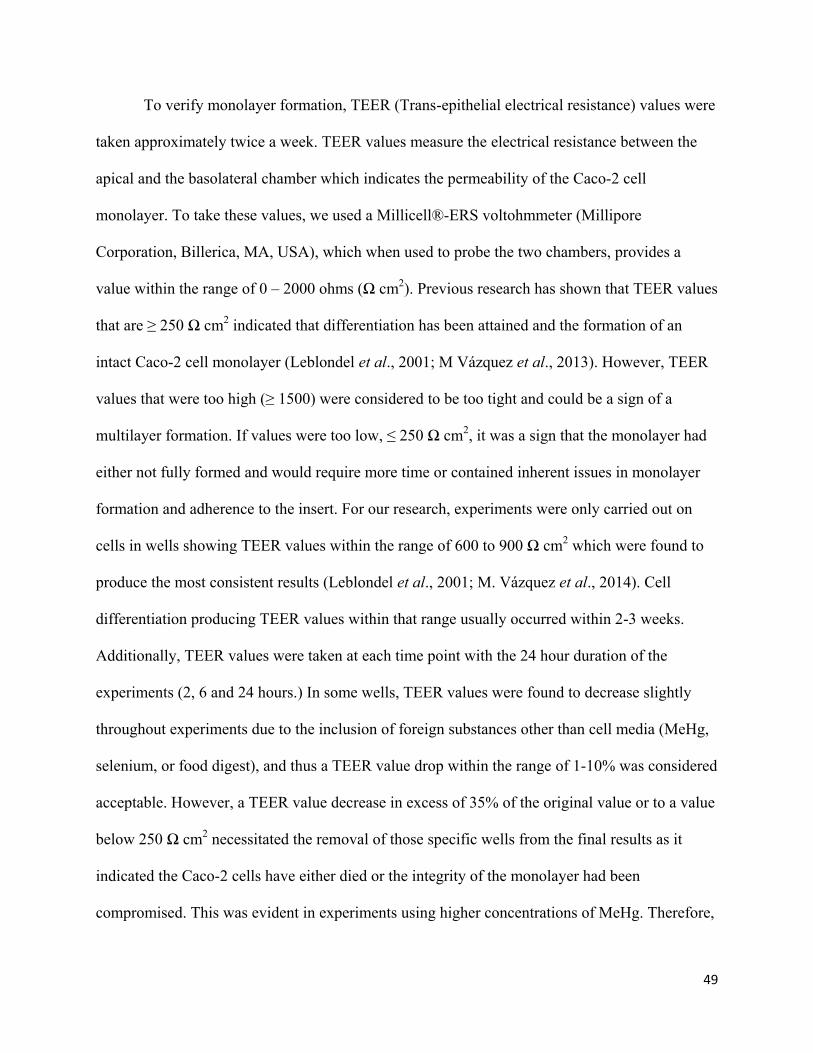
49
To verify monolayer formation, TEER (Trans-epithelial electrical resistance) values were
taken approximately twice a week. TEER values measure the electrical resistance between the
apical and the basolateral chamber which indicates the permeability of the Caco-2 cell
monolayer. To take these values, we used a Millicell®-ERS voltohmmeter (Millipore
Corporation, Billerica, MA, USA), which when used to probe the two chambers, provides a
value within the range of 0 – 2000 ohms (Ω cm2). Previous research has shown that TEER values
that are ≥ 250 Ω cm2 indicated that differentiation has been attained and the formation of an
intact Caco-2 cell monolayer (Leblondel et al., 2001; M Vázquez et al., 2013). However, TEER
values that were too high (≥ 1500) were considered to be too tight and could be a sign of a
multilayer formation. If values were too low, ≤ 250 Ω cm2, it was a sign that the monolayer had
either not fully formed and would require more time or contained inherent issues in monolayer
formation and adherence to the insert. For our research, experiments were only carried out on
cells in wells showing TEER values within the range of 600 to 900 Ω cm2 which were found to
produce the most consistent results (Leblondel et al., 2001; M. Vázquez et al., 2014). Cell
differentiation producing TEER values within that range usually occurred within 2-3 weeks.
Additionally, TEER values were taken at each time point with the 24 hour duration of the
experiments (2, 6 and 24 hours.) In some wells, TEER values were found to decrease slightly
throughout experiments due to the inclusion of foreign substances other than cell media (MeHg,
selenium, or food digest), and thus a TEER value drop within the range of 1-10% was considered
acceptable. However, a TEER value decrease in excess of 35% of the original value or to a value
below 250 Ω cm2 necessitated the removal of those specific wells from the final results as it
indicated the Caco-2 cells have either died or the integrity of the monolayer had been
compromised. This was evident in experiments using higher concentrations of MeHg. Therefore,

50
experiments required a balance between the use of high enough concentrations of MeHg to be
measurable by the mercury analyzer but low enough to ensure that cell death did not occur over
time throughout the experiment.
Determination of Bioavailability in Traditional Food Once cells reached differentiation, as indicated by TEER values within the optimal range
of 600 to 900 Ω cm2, they were ready to be used for experiments. However prior to using the
cells to test the bioavailability of Hg within the digested food samples, we determined the highest
concentration of Hg that would be tolerable for experiments without resulting in cell death.
Various dilutions of MeHg spikes were administered in each well and observed for cell death
over a 24h period, the experimental duration. Using this method, it was determined that a MeHg
concentration within the range of 0.005µM to 0.02µM was optimal for viability. TEER values of
each well were taken to ensure that the Caco-2 cell monolayer was within the optimal range of
experimentation. Our food digest experiments tested the mercury concentrations in each chamber
at 3 different time points (2, 6 and 24 hours.) Therefore, at each time point, aliquots were taken
from 2 different wells. Averages of THg concentrations were calculated using the two wells.
Two wells were used as blanks containing only cell culture media and one well contained the cell
medium with no Caco-2 cells to correct for basal TEER values. Each experimental plate counted
as 1 replicate, and each food experiment was carried out in triplicates.
The treatment solutions consisted of 18 mL of 0.005µM to 0.02µM Hg food digest (based
on food type) diluted in Caco-2 cell media. Each well required 1.5mL of treatment solution
(1.5mL x 12 wells = 18mL.)
All cell culture was conducted in the fume hood to ensure sterile conditions. To start the
experiment, we took the experimental plate out of incubation conditions and measured the TEER

51
values of each well prior to starting the experiment. We then aspirated all Caco-2 cell medium
out of the plate and placed 2mL of fresh medium into the basolateral chamber. Noting down the
starting time point, we dosed 1.5mL of the food treatment into the apical compartment. In the
two designated blank wells, we added 1.5mL of cell culture medium. The plates were then
placed back into incubation conditions (37ºC, within a 95% relative humidity atmosphere and a
CO2 flow of 5%.)
At each time point, 1mL aliquots were taken from the apical and basolateral
compartments of each well designated for that time point. Then, the Caco-2 cell monolayers
were detached from the insert of each well using 100µl of trypsin, followed by 9 minutes of
incubation and then collected using 100ul of PBS solution. The plate was then placed back into
incubation and this process was repeated at each time point. All mercury aliquots were frozen at
-20ºC until ready to be analyzed.
MeHg Uptake. To determine a baseline for the rate of absorption of Hg across the gastrointestinal tract, a
MeHg spike experiment was required. 1000ppm CH3HgCl was diluted to a concentration of
0.02µM MeHg in Caco-2 cell media, similar to the mercury concentration used in the food digest
experiment. The spike treatment was used to determine the rate of absorption of MeHg in the
absence of any food digest as well as other nutrients such as selenium.
The treatment solution of 0.02µM CH3HgCl spike was added to the Caco-2 cells and
incubated for two time points (6, and 24 hours.) This experiment was performed in replicates of
2 and the same parameters for blanks and quality assurance as in the food bioavailability
experiment were used.
52
Selenium-MeHg Co-incubation The last experiment was designed to determine if the presence of equimolar selenium had
an effect on the rate of absorption of MeHg. A 50g stock of Seleno-L-methionine was diluted to
1µM Selenomethionine in milli-Q water. The 1µM stock was then used to achieve a further
dilution of 0.02µM Selenomethionine in Caco-2 cell media, which is the dominant species of
selenium in our chosen foods.
This experiment used a modified design from the previous two protocols. One day prior
to conducting the experiment, all the Caco-2 cell media was pipetted from the apical and
basolateral chambers of the wells. Then, 2mL of fresh EMEM were added into the basolateral
chamber. However, in the apical chamber, 0.75mL of 0.02µM Seleno-L-methionine diluted in
Caco-2 cell media was added. This was followed by 24 hours of incubation in the appropriate
growth conditions. On the day of the experiment, TEER values were taken to ensure that cells
are still viable 24h post incubation with selenium. At experimental time point 0, 0.75mL of
0.02µM CH3HgCl spike treatment was added in addition to the already present selenium
treatment for a total volume of 1.5mL. This experiment was conducted in replicates of 2 and
similar to the previous MeHg spike experiment, aliquots were taken of both chambers at the 6h
and 24h time points.
Mercury Determination Total Hg for all experiments was determined using the MA-3000 Total Mercury Analyzer
(Nippon Instrument North America, College Station, USA.) The machine had a mercury
detection limit of 0.002 ng. Quality control was ensured using sample standards, replicates of
three for each time point, and blanks. The standard reference materials (SRMs) used were
DORM-4 (fish protein; National Research Council, ON, Canada), and DOLT-4 (dogfish liver;
National Research Council, ON, Canada), which yielded 90% and 95% recovery, respectively.

53
The SRMs were analyzed in unison with our samples and were found to be in range of the
certified standard (4.10 ± 0.55 µg Hg /g dw.) The blanks consisted of empty ceramic boats as
well as boats containing only the additive. All values for blanks were below the detection limit of
the machine (1 ng/g.)
Each experimental plate was analyzed separately using the MA-3000 mercury analyzer.
Aliquots taken from both compartments (apical and basolateral) as well as the cells at all three
time points, vortexed, and 200µl samples were added to ceramic wells containing additive B
(Aluminum Oxide). Results were obtained as nanograms of mercury found within the 200µl
sample as well as total concentration of Hg in the sample (µg/kg). To calculate the total amounts
of mercury transferred between compartments, the amount of mercury (ng) found in the 200µl
sample was multiplied by the total volume within the original compartment. Mass balance was
done to ensure that total Hg in all chambers reflected the amount of Hg within the treatment
solution.
Once all values were adjusted for their respective compartments, the sum of the amounts
of Hg in the apical and basolateral chambers as well as within the cells were calculated to find
the total amount of Hg in each well. The amount of Hg in each individual chamber was then
divided by the total Hg in the well and converted to percentages.
Statistical Analysis Results from all experiments were analyzed using one-tailed student’s t-test on “R”
software to determine the statistical significance of the increase in THg in the basolateral
compartment at each time point. In all cases, the results were considered significant at p≤0.05,
however, cases where p≤0.01 are outlined in the results. The experimental means were calculated

54
using triplicates in all food bioavailability experiments and duplicates in MeHg and selenium
spike experiments. The averages for all time points are expressed as ±standard deviation.
Results
Determination of Bioavailability in Traditional Food Of all our food samples, ringed seal liver showed the least uptake of mercury across the
Caco-2 cells and into the basolateral compartment over time (Table 4.1). There were no
significant differences between the amount of mercury found in the basolateral compartment at
the 2h time point and the 24 hour time point. The THg (%) in the apical compartment ranged
from 96.78 to 98.32 over the span of 24 hours with a standard deviation ranging from 0.63 to
0.94%. The basolateral compartment also remained consistent with 0.42 to 1.07 (%THg).
Standard deviation ranged between 0.32 to 0.44%. The percent recovery of Hg ranged from
102% to 105% for all time points.
The ringed seal muscle sample showed the second least uptake of mercury in the
basolateral chamber after a 24 hour period (Table 4.2) Between the 2 hour and 6 hour time point,
the amount of mercury in the basal chamber remained similar and no significant increase was
found. At the 2 hour time point, only 1.94% of total mercury was found in the basal chamber,
and at the 6 hour time point, 0.99%. However, between the 2 hour and 24 hour time points, the
amount of total Hg found in the basolateral compartment significantly increased (p < 0.05) to
5.24%. Standard deviation for all results was very low ranging from 0.48 to 2.22%. The percent
recovery for Hg ranged from 90% to 98% for all time points.
Using air-dried beluga muscle (Table 4.3), we found that similar to the ringed Seal
muscle sample, there was no significant increase in the amount of mercury in the basal
compartment between the 2 hour and 6 hour time points. At 2 hours, only 0.18% of the THg was
55
found in the basal compartment, while 98.9% was in the apical compartment and 0.89% was in
the cells. At the 6 hour time point, that mercury fraction in the basal compartment was 1.24%,
while the apical compartment contained 98.4% and the remaining 0.5% was in the cells.
However, after 24 hours of incubation, the amount of THg in the basolateral compartment
significantly increased in comparison to both the 2 hour and 6 hour time points to 7.3% (p <
0.01). The amount of mercury found within the Caco-2 cells remained consistent throughout the
24 hour incubation at levels ranging from 0.5% to 0.89%. The standard deviation for all results
was at the lowest 0.1% to the highest value of 1.05%. The percent recovery for Hg ranged from
86% to 94% for all time points.
The food sample that was found to have the highest rate of mercury uptake by the Caco-2
cells was lake trout muscle (Table 4.4.) In contrast to the remaining food samples, we saw a
significant increase (p < 0.05) in the amount of THg in the basolateral compartment at the 6 hour
time point in relation to the 2 hour time point. At the 2 hour time point, 1.49% of THg was found
in the basolateral compartment, while 95.91% was in the apical compartment and 2.97% in the
cells. At the 6 hour time point, the amount of THg in the basal compartment significantly
increased to 4.07%. At the 24 hour time point, we saw the highest rate of mercury uptake of all
the food samples. There was a significant increase (p < 0.01) in mercury uptake with 12.7% of
the THg found in the basolateral compartment, accompanied by a significant decrease (p < 0.01)
to 84.9% in the apical compartment. The amount of mercury in the cells remained consistent
ranging between 2.38% and 2.97. The standard deviation for all results ranged from 0.11% to
2.23%. The percent recovery for Hg ranged from 88 to 95% for all time points.

56
Table 4.1 Mercury values in the three compartment system of Caco-2 cells for ringed seal
liver food digest diluted to 0.015µM at three time points following a 24 hour incubation.
Mean Hg amount and percent of total Hg ± standard deviation is shown (N=3.)
2 Hours 6 Hours 24 Hours
Amount of Hg (ng)
[% of recovered Hg±StdDev]
Amount of Hg (ng)
[% of recovered Hg±StdDev]
Amount of Hg (ng)
[% of recovered Hg±StdDev]
Apical 4.56 [97.23 ± 0.81] 4.51 [96.78 ± 0.63] 4.67 [98.32 ± 0.94]
Basal 0.03 [0.64 ± 0.32] 0.05 [1.07 ± 0.44] 0.02 [0.42 ± 0.42]
Cells 0.1 [2.13 ± 0.65] 0.094 [1.93 ± 0.46] 0.06 [1.26 ± 1.25]
Mercury Recovered
[% of Added
Mercury]
4.69 [103.30 ± 8.09]
4.66 [102.64 ± 7.05]
4.75 [104.63 ± 5.68]
Mercury Added 4.54±0.30 4.54±0.30 4.54±0.30

57
Table 4.2. Mercury values in the three compartment system of Caco-2 cells for ringed seal
muscle food digest diluted to 0.007µM at three time points following a 24 hour incubation.
Mean Hg amount and percent of total Hg ± standard deviation is shown (N=3.) Statistical
significance was calculated using one-tailed student’s t-test on “R” software, * represents
p≤0.05, and ** represents p≤0.01.
2 Hours 6 Hours 24 Hours
Amount of Hg (ng)
[% of recovered Hg±StdDev]
Amount of Hg (ng)
[% of recovered Hg±StdDev]
Amount of Hg (ng)
[% of recovered Hg±StdDev]
Apical 2.01 [97.57 ± 2.22] 1.99 [98.51 ± 1.20] 1.8 [94.24 ± 1.24]
Basal 0.04 [1.94 ± 1.27] 0.02 [0.99 ± 0.68] 0.1 [5.24 ± 1.15] *
Cells 0.02 [0.97 ± 0.95] 0.01 [0.50 ± 0.49] 0.01 [0.52 ± 0.48]
Mercury Recovered
[% of Added
Mercury]
2.06 [97.6 ± 1.90]
2.02 [95.73 ± 3.03]
1.91 [90.52 ± 1.03]
Mercury Added 2.11±0.93 2.11±0.93 2.11±0.93

58
Table 4.3 Mercury values in the three compartment system of Caco-2 cells for air-dried
beluga muscle food digest diluted to 0.02µM at three time points following a 24 hour
incubation. Mean Hg amount and percent of total Hg ± standard deviation is shown (N=3.)
Statistical significance was calculated using one-tailed student’s t-test on “R” software, *
represents p≤0.05, and ** represents p≤0.01.
2 Hours 6 Hours 24 Hours
Amount of Hg (ng)
[% of recovered Hg±StdDev]
Amount of Hg (ng)
[% of recovered Hg±StdDev]
Amount of Hg (ng)
[% of recovered Hg±StdDev]
Apical 5.53 [98.9 ± 0.87] 5.55 [98.4 ± 0.98] 4.78 [91.9 ± 0.68]
Basal 0.01 [0.18 ± 0.10] 0.07 [1.24 ± 1.05] 0.38 [7.30 ± 0.90]**
Cells 0.05 [0.89 ± 0.79] 0.03 [0.5 ± 0.35] 0.03 [0.58 ± 0.34]
Mercury Recovered
[% of Added
Mercury]
5.59 [92.7 ± 2.24]
5.64 [93.4 ± 2.59]
5.2 [86.1 ± 3.36]
Mercury Added 6.04±0.99 6.04±0.99 6.04±0.99

59
Table 4.4 Mercury values in the three compartment system of Caco-2 cells for lake trout
muscle food digest diluted to 0.01µM at three time points following a 24 hour incubation.
Mean Hg amount and percent of total Hg ± standard deviation is shown (N=3.) Statistical
significance was calculated using one-tailed student’s t-test on “R” software, * represents
p≤0.05, and ** represents p≤0.01.
2 Hours 6 Hours 24 Hours
Amount of Hg (ng)
[% of recovered Hg±StdDev]
Amount of Hg (ng)
[% of recovered Hg±StdDev]
Amount of Hg (ng)
[% of recovered Hg±StdDev]
Apical 2.58 [95.91 ± 1.58] 2.52 [93.33 ± 1.04] 2.14 [84.92 ± 1.38]
Basal 0.04 [1.49 ± 1.02] 0.11 [4.07 ± 1.14]* 0.32 [12.70 ± 0.85]**
Cells 0.08 [2.97 ± 2.23] 0.08 [2.96 ± 0.11] 0.06 [2.38 ± 0.58]
Mercury
Recovered [% of
Added Mercury]
2.69 [94.06 ± 3.60]
2.7 [94.40 ± 1.53]
2.52 [88.25 ± 6.06]
Mercury Added 2.86±0.27 2.86±0.27 2.86±0.27

60
Figure 2. Percentage of Hg found in the basolateral compartment of Caco-2 cells at the 24
hour time point for three experiments as a function of increasing MeHg fraction in the
bioaccessible sample of the four chosen food types. R squared value is presented with
regression line (n=3.)
R² = 0.5087
0
4
8
12
16
Ringed Seal liver Ringed Seal muscle Lake Trout muscle Air-dried Beluga muscle
Tota
l H
g i
n B
asa
l C
ham
ber
(%
)
Food types
Experiment 1
Experiment 2
Experiment 3

61
Figure 3. Percentage of Hg found in the basolateral compartment of Caco-2 cells at the 24
hour time point for three experiments as a function of increasing selenium concentrations
in the bioaccessible sample of the four chosen food types. R squared value is presented with
regression line (n=3.)
R² = 0.7794
0
4
8
12
16
Lake Trout muscle Ringed Seal muscle Air-dried Beluga muscle Ringed Seal liver
Tota
l H
g i
n B
asa
l C
ham
ber
(%
)
Food types
Experiment 1
Experiment 2
Experiment 3

62
Table 4.5 Mean concentrations of mercury in raw food samples, bioaccessible fractions
after in-vitro digestion and transported across Caco-2 cells into the basolateral chamber as
well as bioaccessible MeHg concentrations.
1 Bioaccessible Hg concentration after undergoing in-vitro digestion (In-vitro bioaccessible)
2 Bioavailable Hg concentration found in the basolateral compartment in Caco-2 cells
3 Bioaccessible
Food
Part
Total raw Hg
(µg/g ww)1
IVBA1 Total Hg (µg/g)
[% of total raw Hg]
IVBA MeHg (µg/g)
[% of total BA3 Hg]
Hg IVTbasolateral (µg/g)2
[% of total IVBA Hg]
Ringed Seal (n=3)
Liver
24.3
7.89 [32.3]
0.270 [3.4]
0.033 [0.42]
Skeletal
Muscle
0.49
0.33 [69.0]
0.190 [55.9]
0.017 [5.24]
Lake Trout (n=3)
Skeletal
Muscle
1.1
0.32 [28.8]
0.275 [86.0]
0.040 [12.70]
Beluga Whale (n=3)
Skeletal
Muscle
2.87
0.97 [34.0]
0.870 [88.0]
0.070 [7.30]

63
Mercury and Selenium Uptake After a 6 hour incubation of 0.02µM MeHg, an uptake of 21.32% of the added mercury
was observed with a recovery rate of 73.66% of the total added Hg. After 24 hours, the amount
of mercury in the basolateral compartment significantly increased (p≤0.01) to 61.61% of the total
added Hg and recovered at 118.27% of total added Hg. Also observed was a corresponding
significant decrease in the concentration of mercury in the apical compartment from 72.44% to
35.46% at the 6 hour and 24 hour time point.
The second experiment involved pre-incubation with 0.02µM selenium for 24 hours,
followed by addition of an equimolar concentration of MeHg. At the 6 hour time point, the
concentration of MeHg in the basolateral compartment was 27.03% of the total added Hg. This
result is significantly higher (p≥0.05) than the sole MeHg treatment at the 6 hour time point. At
the 24 hour time point, the concentration of MeHg in the basolateral compartment remained
consistent at 28.32% of the total added Hg, which is significantly less (p≤0.01) than the sole
MeHg treatment at the 24 hour time point. A mean recovery rate of 95.69% and 94.0% was
observed for the 6 hour and 24 hour time points, respectively.

64
Table 4.6 Mercury values in the three compartment system of Caco-2 cells after a
0.02 µM MeHg spike treatment at two time points following 24 hour incubation. Mean Hg
amount and percent of total Hg ± standard deviation is shown (N=2.) Statistical significance was
calculated using one-tailed student’s t-test on “R” software, * represents p≤0.05, and **
represents p≤0.01.
6 Hours 24 Hours
Amount of Hg (ng)
[% of recovered Hg±StdDev]
Amount of Hg (ng)
[% of recovered Hg±StdDev]
Apical 5.23 [72.44 ± 0.58] 3.75 [35.46 ± 9.16]
Basal 1.53 [21.32 ± 1.15] 7.50 [61.61 ± 9.34]**
Cells 0.46 [6 .31± 1.73] 0.21 [2.93 ± 1.23]
Mercury
Recovered
[% of Added
Mercury]
7.22 [73.66 ± 8.76]
11.59 [118.27 ± 54.28]
Mercury Added 9.80±0.30 9.80±0.30

65
Table 4.7 Mercury values in the three compartment system of Caco-2 cells after a
0.02µM MeHg and Selenium co-incubation treatment. Mean Hg amount and percent of total
Hg ± standard deviation is shown (N=2.) Statistical significance was calculated using one-tailed
student’s t-test on “R” software, * represents p≤0.05, and ** represents p≤0.01.
6 Hours 24 Hours
Amount of Hg (ng)
[% of recovered Hg±StdDev]
Amount of Hg (ng)
[% of recovered Hg±StdDev]
Apical 3.98 [62.91 ± 8.36] 4.28 [68.92 ± 12.07]
Basal 1.71 [27.03 ± 8.82] 1.76 [28.32 ± 14.21]
Cells 0.64 [10.05± 6.70] 0.17 [2.74 ± 2.19]
Mercury
Recovered
[% of Added
Mercury]
6.33 [95.69 ± 14.52]
6.21 [94.0% ± 29.67]
Mercury Added 6.61±0.42 6.61±0.42

66
Discussion In our previous chapter, the oral bioaccessibility of Hg in ringed seal liver, ringed seal
muscle, lake trout muscle and air-dried beluga muscle was determined. Oral bioaccessibility is
defined as the fraction of the consumed substance that is released from the food matrix during
the process of digestion. The results showed that when incorporating the effects of the digestive
process, the concentration of ingested mercury in our body is significantly reduced for all food
types. Despite initial Hg concentrations in the raw food being higher than the Health Canada
recommended guideline of 0.5 ppm, the bioaccessible Hg concentrations fell below the guideline
for ringed seal muscle and lake trout muscle. However, ringed seal liver and air-dried beluga
muscle maintained Hg concentrations higher than the recommended guideline at 7.89µg/g and
0.97µg/g. Therefore, the objective of this chapter was to determine the fraction of remaining Hg
that would be absorbed across the gastrointestinal tract and into the bloodstream where it may
cross the blood-brain barrier and exert its neurodegenerative effects (Van de Wiele et al., 2007).
Our food bioavailability experiment results found that bioavailable Hg concentrations in all
four of our chosen foods were significantly less than bioaccessible Hg concentrations. In all
experimental replicates for all food types, the percentage of total mercury absorbed through the
Caco-2 cells and into the basolateral compartment did not exceed 13% of bioaccessible Hg
concentrations and ranged from 0.42% to 12.70%. These results are very similar to those
previously reported by (Calatayud et al., 2012) of 3% to 14% Hg uptake in samples of frozen
swordfish. This finding suggests that while all our chosen foods contained Hg concentrations that
met or exceeded the recommended guideline of 0.5µg/g, only 0.14% of the initial 24.3µg/g of
ringed seal liver is absorbed into the bloodstream after ingestion. Similarly, 3.7% of the initial
0.49µg/g of Hg in ringed seal muscle is absorbed and 3.63% of the initial 1.1µg/g for lake trout
muscle. Lastly 2.44% of the initial 2.87µg/g of Hg in air-dried beluga muscle was found to be

67
absorbed (Table 4.5), demonstrating that when incorporating the effects of digestion as well as
permeability of the gastrointestinal tract, the concentration of ingested Hg from food that is
absorbed into the bloodstream is significantly reduced. This finding advocates for the importance
of integrating the aforementioned parameters in the process of mercury risk assessment.
Furthermore, the current study relies on using digested food from its raw state that had not gone
through any process of cooking, steaming or grilling, which all have been found to also
significantly reduce the bioaccessibility of Hg (He & Wang, 2011).
The second objective of this study was to determine the effects of bioaccessible MeHg
concentration in the food on uptake by Caco-2 cells. We hypothesized that foods containing
higher percentages of MeHg in their bioaccessible total mercury fraction (THg) would be
absorbed through the Caco-2 cells and into the basal chamber at higher concentrations.
Specifically, the bioaccessible fractions of air-dried beluga muscle and lake trout muscle
containing a MeHg percentage of 88.0% and 86.0% should show higher Hg uptake than ringed
seal muscle and even more so ringed seal liver fractions containing 55.9% and 3.4% MeHg,
respectively. Our experiments showed that 12.7% and 7.30% of the total added Hg using
samples of lake trout muscle and air-dried beluga muscle was transported across the Caco-2
cells. These percentages were significantly higher than those found using ringed seal muscle and
especially ringed seal liver, which were 5.24% and 0.42%, respectively. Our results indicate that
the percentage of MeHg in the biaoccessible THg fraction of a food may play a role in
determining its bioavailability. Furthermore, (Figure 4.1) shows the correlation between
increasing fractions of MeHg in the bioaccessible digest and the percentage of Hg found in the
basolateral compartment (R2 = 0.51.) These findings are similar to those previously reported in
zebra fish exposed to two diets of MeHg (CH3HgCl) or inorganic mercury (HgCl2) (Korbas et
68
al., 2012). Analysis of Hg accumulation found concentrations as high as (0.4 – 0.96 µg/cm2) in
zebra fish exposed to a MeHg diet, while in zebra fish fed a diet of HgCl2, Hg concentrations
were significantly lower at (0.05 to 0.16 µg/cm2), indicating preferential accumulation for MeHg
(Korbas et al, 2012.)
As mercury speciation analysis was not conducted on the mercury found in the
basolateral compartment, the percentage of MeHg absorbed cannot be concluded. However, even
if it is assumed that all Hg found in the basolateral compartment is in the form of MeHg, then
only 8.0% to 14.5% of bioaccessible MeHg in the food was transported through the Caco-2 cells.
These results are contradictory to the previously reported finding that 95% of ingested MeHg is
bioavailable. This percentage was reported in human studies (Aberg et al., 1969) as well as in
monkeys (Berlin et al., 1975). However, a common aspect of these studies was that the MeHg
used was bound to a nitrate rather than the commonly found form of sulfur-bound MeHg and
more importantly, it was used in the absence of a food matrix as it would normally be ingested.
Traditional food has been found to be a remarkable source of nutrients such as selenium, omega-
3 fatty acids, vitamins A, D, and E as well as proteins (Laird, Goncharov, et al., 2013). These
nutrients have been found to affect the bioavailability, metabolism, and toxicity of Hg (Beyrouty
& Chan, 2006; Kaur et al., 2007; Zeng et al., 2011).
The final objective for this research was to determine the effects of selenium on the
uptake of mercury using Caco-2 cells. A baseline for MeHg uptake was established using a spike
treatment of 0.02µM MeHg chloride. After an incubation period of 24 hours, 61.61% of total
added MeHg was transported through the Caco-2 cells and into the basolateral compartment.
This was a significant increase (p≤0.01) from 21.32% uptake of added Hg at the 6H time point.
The significantly higher percentage of mercury transported through the Caco-2 cells when using

69
only MeHg-chloride in the absence of food digest suggests that components of the food may play
a role in the reduction of mercury uptake. To test this possibility, the cells were pre-incubated in
an equimolar (0.02µM) concentration of selenium 24 hours prior to the start of the experiment as
well as throughout the duration of the experiment. We hypothesized that the inclusion of
selenium would result in a significant reduction in the uptake of Hg after a 24 hour period. At the
end of the experiment, only 28.32% of the total added Hg was transported through the Caco-2
cells and into the basal chamber which is a significant decrease (p≤0.01) compared to 61.61% of
transported Hg in the absence of selenium. No increase in Hg uptake was found between the 6H
and the 24H time point. Additionally, the concentration of selenium was determined in the
biaoccessible fractions of all four foods. Ringed seal liver, which was found to contain the
highest concentration of selenium (0.230 µg/g) in its bioaccessible fraction showed a negligible
percentage of Hg transport into the basolateral compartment after 24 hours at 0.42%. In
comparison, lake trout muscle which was found to contain the least amount of bioaccessible
selenium (0.091µg/g), showed the highest rates of Hg transport across the Caco-2 cells and into
the basolateral compartment. When comparing the concentration of bioaccessible selenium in the
food digest with the decreasing percentage of Hg transport in Caco-2 cells after 24 hours of
incubation, a coefficient of determination (R2) value of 0.78 is found (Figure 4.2.) The molecular
mechanisms by which selenium impacts the uptake rate of Hg are still relatively unknown.
However, the phenomenon of selenium’s impact on the uptake of mercury has been reported in
several studies. A recent study by (Huang et al., 2013) used sturgeon fed a diet of MeHg
chloride, Selenomethionine, as well as a combination diet of Se/Hg. An increase in blood Hg
concentrations was found in both the MeHg group as well as the Se/Hg group. However, Hg
concentrations in the MeHg group peaked at 12 hours and maintained its elevation for 48 hours,

70
while the Se/Hg group peaked at 1.5 hours then promptly decreased to approximately half the
value of the peak and remained low for the remaining time period (Huang et al., 2013).
Similarly, in rats fed a diet of Hg or a Hg/Se diet with increasing concentrations of selenium, it
was found that the concentration of Hg in the brain and kidneys decreased with increased
selenium dosage. Brain Hg concentrations were 3 times higher and kidney concentrations were
12 times higher in the Hg-fed group than in the group fed the highest concentration of selenium
(Orct et al., 2009). These studies show that selenium not only impacts the uptake of mercury, but
the retention and distribution as well.
Conclusion The purpose of this chapter was to determine the in-vitro uptake of mercury in four food
types and to determine the potential impact of factors such as MeHg and selenium concentration
on the absorption of Hg. We determined that lake trout muscle was found to have the highest
concentration of transported Hg in the basolateral chamber at 12.70%, while ringed seal liver
was found to have the lowest amount of Hg in the basal chamber at 0.42%. These results agree
with our initial hypothesis that the fraction of bioaccessible MeHg in the food digest will show a
positive correlation with Hg uptake. The percentage of MeHg in the bioaccessible fraction of
ringed seal liver was the lowest at only 3.40%, while MeHg percentage in lake trout muscle was
86.0%, second only to air-dried beluga muscle at 88.8%.
The impact of selenium on the uptake of mercury was studied in two ways. Initially, the
concentrations of bioaccessible selenium in the food digest were determined in the previous
chapter. In this chapter, results showed that Hg transport was highest in foods such as lake trout
muscle which contained the lowest concentration of bioaccessible selenium (0.091µg/g) and
lowest in ringed seal liver which contained the highest amount of bioaccessible selenium

71
(0.230µg/g). The coefficient of determination (R2) value found by this relationship was 0.78.
Secondly, after a 24 hour incubation, the percentage of Hg transported to the basolateral
compartment decreased significantly from 61.61% of added Hg to only 28.32% when co-
incubating with selenium. These results are in line with our hypothesis that selenium co-
incubation will significantly reduce the uptake of Hg after 24 hours.
72
Chapter 5: Conclusions and Future Direction
Overall Conclusions The purpose of this study was to determine the bioaccessibility and bioavailability of four
key foods of the traditional Inuit diet to provide better estimation of Hg exposure and improve
the characterization of risk of Hg exposure to human health. The impact, if any, that selenium
may have on the uptake of Hg was also investigated using an in-vitro Caco-2 cell model. It was
hypothesized that bioaccessible Hg fractions would be dependent on original Hg concentrations
in the food, which was determined through an in-vitro digestion process. Our results showed that
while ringed seal liver was found to contain the highest concentration of Hg in the raw food
(24.43µg/g), after digestion, the bioaccessible Hg fraction was second lowest (32.3%.) Similarly,
ringed seal muscle contained the lowest concentration of Hg in the raw food (0.49µg/g), but post
in-vitro digestion, was found to contain the highest bioaccessible Hg fraction (69.0%.) These
findings are contrary to our hypothesis and highlight the need for risk assessment to include
factors such as bioaccessibility when estimating MeHg exposure from traditional food
consumption.
Secondly, the objective of chapter 4 of this study was to use Caco-2 cell model was used to
determine the uptake of mercury into the bloodstream from digested samples of traditional food.
A key finding of this experiment was that ringed seal liver having an initial Hg concentration
(24.43µg/g) approximately 30x higher than the mean of the three remaining foods, was absorbed
at the lowest percentage in Caco-2 cells (0.42%.) This result is in line with our hypothesis that
the fraction of bioaccessible MeHg in the food digest will show a positive correlation with the
percentage of Hg uptake in Caco-2 cells. Ringed seal liver was found to have the lowest fraction
of bioaccessible MeHg in its food digest at 3.4%. Hg from lake trout muscle was transported at

73
the highest percentage of (12.70%) which corresponded to its high bioaccessible MeHg fraction
of 86.0%.
Summarizing our analysis of Hg uptake, the Hg bioaccessibility percentage of ringed seal
liver was 32.3%, ringed seal muscle was 69.0%, lake trout muscle was 28.8%, and lastly air-
dried beluga muscle was 34.0% after in-vitro digestion. Furthermore, Hg bioavailability
percentages were determined to be 0.42% for ringed seal liver, 5.24% for ringed seal muscle,
7.30% for air-dried beluga muscle, and finally, 12.70% for lake trout muscle.
Lastly, the impact of the nutrient, selenium, was determined as Hg transport percentages in
Caco-2 cells were found to decrease with increasing selenium concentrations in the bioaccessible
fraction of the food. The concentration of bioaccessible selenium in lake trout muscle was the
lowest at (0.091µg/g) while the percentage of transported Hg was the highest. Additionally,
ringed seal liver bioaccessible selenium concentration was the highest at (0.230µg/g), and was
found exhibit the lowest fractions of transported Hg in Caco-2 cells. This relationship was
highlighted in (Figure 4.2) resulting in an R2 value of 0.78.
A baseline of MeHg uptake after a 24 hour incubation was found to be 61.61% of total added
MeHg. The effects of selenium co-incubation on Hg uptake were determined using an equimolar
concentration of selenium and MeHg treatment. A significant decrease in mercury uptake from
61.61% to 28.32% was observed after 24 hours. These results agree with our initial hypothesis
that selenium co-incubation will result in a significant reduction in Hg uptake after an incubation
period of 24 hours. The significance of this experiment is to emphasize the importance of
looking at a traditional diet as a whole. Selenium is one of many nutrients in traditional food
which includes omega-3 fatty acids, a multitude of vitamins, as well as other beneficial nutrients.
Aside from the ability of these nutrients to mitigate the neurotoxicity, uptake and overall

74
metabolism of mercury, a traditional diet also encourages a healthy active lifestyle and reduces
the risk of diseases (ex. diabetes and obesity) (Laird et al, 2013). Therefore, when considering
the risks of traditional food consumption due to potential Hg exposure, it is important to also
incorporate factors such as bioaccessibility, bioavailability as well as the host of inherent benefits
of a traditional Inuit diet.
Future Directions
The results presented in this study add to the recently growing research against the long-
held assumption that the fraction of ingested dietary Hg reaching systemic circulation is 95-
100%. Using an in-vitro digestion model as well as the caco-2 cell in-vitro gastrointestinal
uptake model, the bioaccessibility and bioavailability of Hg from the four chosen food types
were determined. When incorporating these factors, it was found that only 0.001% to 0.04% of
Hg from food was absorbed into the bloodstream. These results can be used to inform future Hg
risk assessments through the use of adjustment factors. The following adjustments factors can be
applied to future risk assessments; for RSL, Original THg (32.3 x 0.42) %, for RSM, Orig. THg
(69.0 x 5.24) %, for ADB, Orig. THg (34.0 x 7.30) %, and lastly, for LT, Orig. THg (28.8 x
12.70) %. Further research into the bioaccessibility and bioavailability of more traditional food
can increase the accuracy of risk assessment studies and reduce the overestimation of Hg risks.
For future studies, a more in-depth analysis of bioavailability experiments could be carried
out. A limitation of this study was that original MeHg concentrations in the food were not
determined and therefore the change in concentration of MeHg prior to and post-digestion is
unknown. This would provide further insight into the influence of the digestion process on
specifically MeHg rather than THg, which would, in turn, depict a more accurate representation
of the process of MeHg retention and elimination after digestion.

75
Inherent limitations still exist when using the in-vitro gastrointestinal model of Caco-2 cells.
Cell monolayer growth on the insert will differ based on many factors and therefore maximizing
the use of one 12-well plate is difficult. Due to cost and availability of material, this could
potentially lead to less experimental replicates. Further research into Caco-2 cell culture may in
the future result in more consistent growth and viability.
Lastly, in this research, reduced Hg uptake was observed when using the same concentration
of Hg from food digest instead of MeHg chloride spike treatment. A negative correlation
between increased selenium bioaccessible concentrations and Hg uptake was also seen.
However, traditional food is host to a variety of beneficial nutrients. Future studies can expand
on our current research by determining the impact of omega-3 fatty acids, and Vitamins A, D and
E on Hg uptake.

76
References
Aberg, B., Ekman, L., Falk, R., Greitz, U., Persson, G., & Snihs, J. (1969). Metabolism of
Methyl Mercury (203Hg) Compounds in Man. Archives of Environmental Health: An
International Journal, 19(4), 478–484. https://doi.org/10.1080/00039896.1969.10666872
Acra, S., & Ghishan, F. (1991). Methods of investigating intestinal transport. Journal of
Parenteral and Enteral Nutrition, 15(3), 93S–98S.
https://doi.org/10.1177/014860719101500393S
Afonso, C., Costa, S., Cardoso, C., Bandarra, N. M., Batista, I., Coelho, I., … Nunes, M. L.
(2015). Evaluation of the risk/benefit associated to the consumption of raw and cooked
farmed meagre based on the bioaccessibility of selenium, eicosapentaenoic acid and
docosahexaenoic acid, total mercury, and methylmercury determined by an in vitro
digestion model. Food Chemistry, 170, 249–256.
https://doi.org/10.1016/j.foodchem.2014.08.044
AMAP. (2009). AMAP Assessment 2009: Human Health in the Arctic. Arctic Monitoring and
Assessment Programme (AMAP).
AMAP. (2011). AMAP Assessment 2011: Mercury in the Arctic. Arctic Monitoring and
Assessment Programme (AMAP), Oslo, Norway. xiv + 193 ppAmlund, H., Lundebye, A.-
K., Boyle, D., & Ellingsen, S. (2015). Dietary selenomethionine influences the
accumulation and depuration of dietary methylmercury in zebrafish (Danio rerio). Aquatic
Toxicology, 158, 211–217. https://doi.org/10.1016/j.aquatox.2014.11.010
Arnold, A. P., Tan, K. S., & Rabenstein, D. L. (1986). Nuclear magnetic resonance studies of the
solution chemistry of metal complexes. 23. Complexation of methylmercury by
selenohydryl-containing amino acids and related molecules. Inorganic Chemistry, 25(14),
2433–2437. https://doi.org/10.1021/ic00234a030
Aschner, M., & Aschner, J. L. (1990). Mercury neurotoxicity: Mechanisms of blood-brain
barrier transport. Neuroscience and Biobehavioral Reviews, 14(2), 169–176.
https://doi.org/10.1016/S0149-7634(05)80217-9
Barthe, L., Woodley, J., & Houin, G. (1999). Gastrointestinal absorption of drugs: methods and
studies. Fundamental & Clinical Pharmacology, 13(2), 154–68.
Berlin, M. M., Grant, C. A., Hellberg, J., Hellstrom, J., & Shcutz, A. (1975). Neurotoxicity of
Methylmercury in squirrel monkeys. Archives of Environmental and Occupational Health,
30(1), 340–348.
Berry, M. J., & Ralston, N. V. C. (2008). Mercury toxicity and the mitigating role of selenium.
EcoHealth, 5(4), 456–9. https://doi.org/10.1007/s10393-008-0204-y
Beyrouty, P., & Chan, H. M. (2006). Co-consumption of selenium and vitamin E altered the
reproductive and developmental toxicity of methylmercury in rats. Neurotoxicology and
Teratology, 28, 49–58. https://doi.org/10.1016/j.ntt.2005.11.002
Bjerregaard, P., & Christensen, A. (2012). Selenium reduces the retention of methyl mercury in
77
the brown shrimp Crangon crangon. Environmental Science and Technology, 46(11), 6324–
6329. https://doi.org/10.1021/es300549y
Bjørklund, G. (2015). Selenium as an antidote in the treatment of mercury intoxication.
BioMetals, 28(4), 605–614. https://doi.org/10.1007/s10534-015-9857-5
Blanchet, C., Dewailly, E., Ayotte, P., Bruneau, S., Receveur, O., & Holub, B. J. (2000).
Contribution of Selected Traditional and Market Foods to the Diet of Nunavik Inuit
Women. Canadian Journal of Dietetic Practice and Research, 61(2), 50–59.
Boucher, O., Jacobson, S. W., Plusquellec, P., Dewailly, É., Ayotte, P., Forget-Dubois, N., …
Muckle, G. (2012). Prenatal methylmercury, postnatal lead exposure, and evidence of
attention deficit/hyperactivity disorder among Inuit children in Arctic Québec.
Environmental Health Perspectives, 120(10), 1456–1461.
https://doi.org/10.1289/ehp.1204976
Boucher, O., Muckle, G., Ayotte, P., Dewailly, E., Jacobson, S. W., & Jacobson, J. L. (2016).
Altered fine motor function at school age in Inuit children exposed to PCBs ,
methylmercury , and lead. Environment International.
https://doi.org/10.1016/j.envint.2016.08.010
Bradley, M., Barst, B., & Basu, N. (2017). A Review of Mercury Bioavailability in Humans and
Fish. International Journal of Environmental Research and Public Health, 14(2), 169.
doi:10.3390/ijerph14020169
Bridges, C. C., & Zalups, R. K. (2010). Transport of inorganic mercury and methylmercury in
target tissues and organs. Journal of Toxicology and Environmental Health. Part B, Critical
Reviews, 13(5), 385–410. https://doi.org/10.1080/10937401003673750
Bullock Jr, O. R. (2000). Modeling assessment of transport and deposition patterns of
anthropogenic mercury air emissions in the United States and Canada. Science of the Total
Environment, 259(1), 145–157.
Burger, J., & Gochfeld, M. (2004). Mercury in canned tuna: White versus light and temporal
variation. Environmental Research, 96(3), 239–249.
https://doi.org/10.1016/j.envres.2003.12.001
Cabañero, A. I., Madrid, Y., & Cámara, C. (2007). Mercury-selenium species ratio in
representative fish samples and their bioaccessibility by an in vitro digestion method.
Biological Trace Element Research, 119(3), 195–211. https://doi.org/10.1007/s12011-007-
8007-5
Calatayud, M., Devesa, V., Virseda, J. R., Barberá, R., Montoro, R., & Vélez, D. (2012).
Mercury and selenium in fish and shellfish: occurrence, bioaccessibility and uptake by
Caco-2 cells. Food and Chemical Toxicology : An International Journal Published for the
British Industrial Biological Research Association, 50(8), 2696–702.
https://doi.org/10.1016/j.fct.2012.05.028
Carr, K. E., & Toner, P. G. (1984). Morphology of the intestinal Mucosa. In Pharmacology of
Intestinal Permeation (pp. 1–3).
Ceccatelli, S., Bose, R., Edoff, K., Onishchenko, N., & Spulber, S. (2013). Long-lasting
78
neurotoxic effects of exposure to methylmercury during development. Journal of Internal
Medicine, 273(5), 490–7. https://doi.org/10.1111/joim.12045
Chapman, L., & Chan, H. M. (2000). The influence of nutrition on methyl mercury intoxication.
Environmental Health Perspectives, 108 Suppl(August 1999), 29–56.
Chen, C., Qu, L., Zhao, J., Liu, S., Deng, G., Li, B., … Chai, Z. (2006). Accumulation of
mercury, selenium and their binding proteins in porcine kidney and liver from mercury-
exposed areas with the investigation of their redox responses. Science of the Total
Environment, 366(2–3), 627–637. https://doi.org/10.1016/j.scitotenv.2005.12.021
Chételat, J., Amyot, M., Arp, P., Blais, J. M., Depew, D., Emmerton, C. a, … van der Velden, S.
(2014). Mercury in freshwater ecosystems of the Canadian Arctic: Recent advances on its
cycling and fate. The Science of the Total Environment.
https://doi.org/10.1016/j.scitotenv.2014.05.151
Clarkson, T. (1972). The Pharmacology of Mercury Compounds. Annual Review of
Pharmacology and Toxicology, 375–406. https://doi.org/10.1620/tjem.196.71
Clarkson, T. W. (2002). The three modern faces of mercury. Environmental Health Perspectives,
110(SUPPL. 1), 11–23. https://doi.org/10.1289/ehp.02110s111
Costa, S., Afonso, C., Cardoso, C., Batista, I., Chaveiro, N., Leonor, M., & Maria, N. (2015).
Fatty acids , mercury , and methylmercury bioaccessibility in salmon ( Salmo salar ) using
an in vitro model : Effect of culinary treatment. FOOD CHEMISTRY, 185, 268–276.
https://doi.org/10.1016/j.foodchem.2015.03.141
Cui, H. F., Ye, J. S., Chen, Y., Chong, S. C., & Sheu, F. S. (2006). Microelectrode array biochip:
Tool for in vitro drug screening based on the detection of a drug effect on dopamine release
from PC12 cells. Analytical Chemistry, 78(18), 6347–6355.
https://doi.org/10.1021/ac060018d
D.L. McPhee, D.M. Janz (2014).Dietary selenomethionine exposure alters swimming
performance, metabolic capacity and energy homeostasis in juvenile fathead minnow
Aquatic Toxicology 155, 91–100
Dringen, R. (2000). Metabolism and functions of glutathion in brain. Progress in Neurobiology,
62, 33–57. https://doi.org/10.1016/j.pneurobio.2014.01.002
Drover, V. a, Nguyen, D. V, Bastie, C. C., Darlington, Y. F., Abumrad, N. a, Pessin, J. E., …
Phillips, M. C. (2008). CD36 mediates both cellular uptake of very long chain fatty acids
and their intestinal absorption in mice. The Journal of Biological Chemistry, 283(19),
13108–15. https://doi.org/10.1074/jbc.M708086200
El-hayek, Y. H. (2007). Mercury Contamination in Arctic Canada : Possible Implications for
Aboriginal Health Mercury and the Environment of Arctic Canada. Journal on
Developmental Disabilities, 13(1), 67–90.
Environmental Investigation Agency. (2003). Mercury Rising: The sale of polluted whale ,
dolphin and porpoise meat in Japan.
Falnoga, I., Tušek-Žnidarič, M., & Stegnar, P. (2006). The influence of long-term mercury

79
exposure on selenium availability in tissues: An evaluation of data. BioMetals, 19(3), 283–
294. https://doi.org/10.1007/s10534-005-8642-2
Farina, M., Rocha, J. B. T., & Aschner, M. (2011). Mechanisms of methylmercury-induced
neurotoxicity: evidence from experimental studies. Life Sciences, 89(15–16), 555–563.
https://doi.org/10.1016/j.lfs.2011.05.019
Fitsanakis, V. A., & Aschner, M. (2005). The importance of glutamate, glycine, and ??-
aminobutyric acid transport and regulation in manganese, mercury and lead neurotoxicity.
Toxicology and Applied Pharmacology, 204(3), 343–354.
https://doi.org/10.1016/j.taap.2004.11.013
Fleming, E. J., Mack, E. E., Green, P. G., Nelson, D. C., & Al, F. E. T. (2006). Mercury
Methylation from Unexpected Sources : Molybdate-Inhibited Freshwater Sediments and an
Iron-Reducing Bacterium. Applied and Environmental Microbiology, 72(1), 457–464.
https://doi.org/10.1128/AEM.72.1.457
Fogh, J., Fogh, J. M., & Orfeo, T. (1977). One hundred and twenty-seven cultured human tumor
cell lines producing tumors in nude mice. Journal of the National Cancer Institute, 59(1),
221–6.
Franco, J. L., Posser, T., Dunkley, P. R., Dickson, P. W., Mattos, J. J., Martins, R., … Farina, M.
(2009). Methylmercury neurotoxicity is associated with inhibition of the antioxidant
enzyme glutathione peroxidase. Free Radical Biology & Medicine, 47(4), 449–57.
https://doi.org/10.1016/j.freeradbiomed.2009.05.013
Friedman, M. a, Eaton, L. R., & Carter, W. H. (1978). Protective effects of freeze dried
swordfish on methylmercury chloride toxicity in rats. Bulletin of Environmental
Contamination and Toxicology, 19(4), 436–443. https://doi.org/10.1007/BF01685823
Gagné, D., Blanchet, R., Lauzière, J., Vaissière, É., Vézina, C., Ayotte, P., … Turgeon O’Brien,
H. (2012). Traditional food consumption is associated with higher nutrient intakes in Inuit
children attending childcare centres in Nunavik. International Journal of Circumpolar
Health, 71(18401). https://doi.org/10.1002/jcp.24831
Guzzi, G., & La Porta, C. A. M. (2008). Molecular mechanisms triggered by mercury.
Toxicology, 244(1), 1–12. https://doi.org/10.1016/j.tox.2007.11.002
Ha, E., Basu, N., Bose-O ’reilly, S., Dórea, J. G., Mcsorley, E., Sakamoto, M., & Chan, H. M.
(2016). Current progress on understanding the impact of mercury on human health.
Environmental Research, 1–15. https://doi.org/10.1016/j.envres.2016.06.042
Häggqvist, B., Havarinasab, S., Björn, E., & Hultman, P. (2005). The immunosuppressive effect
of methylmercury does not preclude development of autoimmunity in genetically
susceptible mice. Toxicology, 208(1), 149–164. https://doi.org/10.1016/j.tox.2004.11.020
Harada, M. (1978). Congenital Minamata disease: intrauterine methylmercury poisoning.
Journal of Teratology, 285–288.
He, M., & Wang, W.-X. (2011). Factors affecting the bioaccessibility of methylmercury in
several marine fish species. Journal of Agricultural and Food Chemistry, 59(13), 7155–62.
https://doi.org/10.1021/jf201424g

80
Health Canada. (2004, October 1). Mercury, Your Health and the Environment. Retrieved from
Health Canada: http://www.hc-sc.gc.ca/ewh-semt/pubs/contaminants/mercur/index-
eng.php
Health Canada. (2007, March). Human Health Risk Assessment of Mercury in Fish and Health
Benefits of Fish Consumption. Retrieved from Health Canada: http://www.hc-sc.gc.ca/fn-
an/pubs/mercur/merc_fish_poisson-eng.php
Hill, K. E., Zhou, J., McMahan, W. J., Motley, A. K., Atkins, J. F., Gesteland, R. F., & Burk, R.
F. (2003). Deletion of selenoprotein P alters distribution of selenium in the mouse. Journal
of Biological Chemistry, 278(16), 13640–13646. https://doi.org/10.1074/jbc.M300755200
Hillgren, K. M., Kato, a, & Borchardt, R. T. (1995). In vitro systems for studying intestinal drug
absorption. Medicinal Research Reviews, 15(2), 83–109.
Huang, S. S. Y., Strathe, A. B., Fadel, J. G., Johnson, M. L., Lin, P., Liu, T. Y., & Hung, S. S. O.
(2013). The interactive effects of selenomethionine and methylmercury on their absorption,
disposition, and elimination in juvenile white sturgeon. Aquatic Toxicology, 126, 274–282.
https://doi.org/10.1016/j.aquatox.2012.09.018
Inuit Tapiriit Kanatami. (2014). Social Determinants of Inuit Health in Canada.
https://doi.org/10.1097/01.AOG.0000453605.35883.a0
IPY. (2008). Inuit Health Survey. International Polar Year.
Jacobson, J. L., Muckle, G., Ayotte, P., Dewailly, E., & Jacobson, S. W. (2015). Relation of
prenatal methylmercury exposure from environmental sources to childhood IQ.
Environmental Health Perspectives, 123(8), 827–833. https://doi.org/10.1289/ehp.1408554
Jadán-Piedra, C., Clemente, M. J., Devesa, V., & Vélez, D. (2016). Influence of Physiological
Gastrointestinal Parameters on the Bioaccessibility of Mercury and Selenium from
Swordfish. Journal of Agricultural and Food Chemistry, 64(3), 690–698.
https://doi.org/10.1021/acs.jafc.5b05046
Kannan, K., Smith, R. G., Lee, R. F., Windom, H. L., Heitmuller, P. T., Macauley, J. M., &
Summers, J. K. (1998). Environmental contamination and toxicology distribution of total
mercury and methyl mercury in water, sediment, and fish from South Florida estuaries.
Archives of Environmental Contamination and Toxicology, 34, 109–118.
Kaur, P., Schulz, K., Aschner, M., & Syversen, T. (2007). Role of docosahexaenoic acid in
modulating methylmercury-induced neurotoxicity. Toxicological Sciences : An Official
Journal of the Society of Toxicology, 100(2), 423–32. https://doi.org/10.1093/toxsci/kfm224
Keita, a V, & Söderholm, J. D. (2010). The intestinal barrier and its regulation by neuroimmune
factors. Neurogastroenterology and Motility : The Official Journal of the European
Gastrointestinal Motility Society, 22(7), 718–33. https://doi.org/10.1111/j.1365-
2982.2010.01498.x
Kellogg, J., Wang, J., Flint, C., Ribnicky, D., Kuhn, P., De Mejia, E. G., … Lila, M. A. (2010).
Alaskan wild berry resources and human health under the cloud of climate change. Journal
of Agricultural and Food Chemistry, 58(7), 3884–3900. https://doi.org/10.1021/jf902693r

81
Kerper, L. E., Ballatori, N., & Clarkson, T. W. (1992). Methylmercury transport across the
blood-brain barrier by an amino acid carrier. The American Journal of Physiology, 262(38),
R761–R765.
Kim, M. K., & Zoh, K. D. (2012). Fate and transport of mercury in environmental media and
human exposure. Journal of Preventive Medicine and Public Health, 45(6), 335–343.
https://doi.org/10.3961/jpmph.2012.45.6.335
Korbas, M., MacDonald, T. C., Pickering, I. J., George, G. N., & Krone, P. H. (2012). Chemical
form matters: Differential accumulation of mercury following inorganic and organic
mercury exposures in zebrafish larvae. ACS Chemical Biology, 7, 411–420.
https://doi.org/10.1021/cb200287c
Kuhnlein, H. V, & Chan, H. M. (2000). Environment And Contaminants in Traditional Food
Systems of Northern Indigenous Peoples. Annual Review of Nutrition, 20, 595–626.
Laird, B. D., & Chan, H. M. (2013). Bioaccessibility of metals in fish, shellfish, wild game, and
seaweed harvested in British Columbia, Canada. Food and Chemical Toxicology : An
International Journal Published for the British Industrial Biological Research Association,
58, 381–387. https://doi.org/10.1016/j.fct.2013.04.033
Laird, B. D., Goncharov, A. B., Egeland, G. M., & Chan, H. M. (2013). Dietary advice on Inuit
traditional food use needs to balance benefits and risks of mercury, selenium, and n3 fatty
acids. The Journal of Nutrition, 143(6), 923–930. https://doi.org/10.3945/jn.112.173351
Laird, B. D., James, K. J., Van de Wiele, T. R., Dodd, M., Casteel, S. W., Wickstrom, M., &
Siciliano, S. D. (2013). An investigation of the effect of gastrointestinal microbial activity
on oral arsenic bioavailability. Journal of Environmental Science and Health. Part A,
Toxic/hazardous Substances & Environmental Engineering, 48(6), 612–619.
https://doi.org/10.1080/10934529.2013.731357
Laird, B. D., Shade, C., Gantner, N., Chan, H. M., & Siciliano, S. D. (2009). Bioaccessibility of
mercury from traditional northern country foods measured using an in vitro gastrointestinal
model is independent of mercury concentration. The Science of the Total Environment,
407(23), 6003–6008. https://doi.org/10.1016/j.scitotenv.2009.08.014
Leblondel, G., Mauras, Y., Cailleux, a, & Allain, P. (2001). Transport measurements across
Caco-2 monolayers of different organic and inorganic selenium: influence of sulfur
compounds. Biological Trace Element Research, 83(3), 191–206.
https://doi.org/10.1385/BTER:83:3:191
Lemes, M., Wang, F., Stern, G. a., Ostertag, S. K., & Chan, H. M. (2011). Methylmercury and
selenium speciation in different tissues of beluga whales (Delphinapterus leucas) from the
western Canadian Arctic. Environmental Toxicology and Chemistry, 30(12), 2732–2738.
https://doi.org/10.1002/etc.684
Lemire, M., Fillion, M., Barbosa, F., Guimarães, J. R. D., & Mergler, D. (2010). Elevated levels
of selenium in the typical diet of Amazonian riverside populations. Science of the Total
Environment, 408(19), 4076–4084. https://doi.org/10.1016/j.scitotenv.2010.05.022
Lemire, M., Kwan, M., Laouan-Sidi, a E., Muckle, G., Pirkle, C., Ayotte, P., & Dewailly, E.

82
(2014). Local country food sources of methylmercury, selenium and omega-3 fatty acids in
Nunavik, Northern Quebec. The Science of the Total Environment, 1–12.
https://doi.org/10.1016/j.scitotenv.2014.07.102
Lennernäs, H., Palm, K., Fagerholm, U., & Artursson, P. (1996). Comparison between active and
passive drug transport in human intestinal epithelial (Caco-2) cells in vitro and human
jejunum in vivo. International Journal of Pharmaceutics, 127(1), 103–107.
https://doi.org/10.1016/0378-5173(95)04204-0
Li, W. C., & Tse, H. F. (2014). Health risk and significance of mercury in the environment.
Environmental Science and Pollution Research, 22(1), 192–201.
https://doi.org/10.1007/s11356-014-3544-x
Li, X., Yin, D., Yin, J., Chen, Q., & Wang, R. (2014). Dietary selenium protect against redox-
mediated immune suppression induced by methylmercury exposure. Food and Chemical
Toxicology : An International Journal Published for the British Industrial Biological
Research Association, 72C, 169–177. https://doi.org/10.1016/j.fct.2014.07.023
Li, Y. F., Dong, Z., Chen, C., Li, B., Gao, Y., Qu, L., … Chai, Z. (2012). Organic selenium
supplementation increases mercury excretion and decreases oxidative damage in long-term
mercury-exposed residents from Wanshan, China. Environmental Science and Technology,
46(20), 11313–11318. https://doi.org/10.1021/es302241v
Matos, J., Lourenço, H. M., Brito, P., Maulvault, A. L., Martins, L. L., & Afonso, C. (2015).
Influence of bioaccessibility of total mercury, methyl-mercury and selenium on the
risk/benefit associated to the consumption of raw and cooked blue shark (Prionace glauca).
Environmental Research, 143, 123–129. https://doi.org/10.1016/j.envres.2015.09.015
McAlpine, D., & Araki, S. (1958). Minamata disease. An unusual neurological disorder cause by
contaminated fish. Lancet, 629–631.
Mergler, D., Anderson, H. a, Chan, L. H. M., Mahaffey, K. R., Murray, M., Sakamoto, M., &
Stern, A. H. (2007). Methylmercury exposure and health effects in humans: a worldwide
concern. Ambio, 36(1), 3–11. https://doi.org/10.1579/0044-
7447(2007)36[3:MEAHEI]2.0.CO;2
Meunier, V., Bourrié, M., Berger, Y., & Fabre, G. (1995). The human intestinal epithelial cell
line Caco-2; pharmacological and pharmacokinetic applications. Cell Biology and
Toxicology, 11(3–4), 187–94.
Miettinen, J., Rahola, T., Hattula, T., Rissanen, K., Tillander, M., (1971). Elimination of 203Hg-
methylmercury in man. Annals of Clinical Research. 3 (2), 116–122.
Morgan, E. H., & Oates, P. S. (2002). Mechanisms and Regulation of Intestinal Iron Absorption.
Blood Cells, Molecules, and Diseases, 29(3), 384–399.
https://doi.org/10.1006/bcmd.2002.0578
Myers, G. J., Davidson, P. W., Cox, C., Conrad, F. S., Palumbo, D., Cernichiari, E., … Clarkson,
T. W. (2003). Prenatal Mehtylmercury Exposure From Ocean Fish Consumption in the
Seychelles Child Development Study. The Lancet, 361, 1686–1692.
https://doi.org/10.1016/S0140-6736(03)13371-5

83
Nielsen, J. B. (1992). Toxicokinetics of mercuric chloride and methylmercuric chloride in mice.
Journal of Toxicology and Environmental Health, 37(1), 85–122.
https://doi.org/10.1080/15287399209531659
Orct, T., Lazarus, M., Jurasović, J., Blanuša, M., Piasek, M., & Kostial, K. (2009). Influence of
selenium dose on mercury distribution and retention in suckling rats, (October 2008), 585–
589. https://doi.org/10.1002/jat.1444
Park, J., & Zheng, W. (2012). Human Exposure and Health Effects of Inorganic and Elemental
Mercury, 344–352.
Parsa, Y., Nabavi, S. S. M. B., Nabavi, S. N., & Hosseini, M. (2014). Mercury Accumulation in
Food Chain of Fish, Crab and Sea Bird from Arvand River. Journal of Marine Science:
Research & Development, 4(2), 1–6. https://doi.org/10.4172/2155-9910.1000148
Penglase, S., Hamre, K., & Ellingsen, S. (2014). Selenium and mercury have a synergistic
negative effect on fish reproduction. Aquatic Toxicology, 149, 16–24.
https://doi.org/10.1016/j.aquatox.2014.01.020
Piraino, M. N., & Taylor, D. L. (2009). Bioaccumulation and trophic transfer of mercury in
striped bass (Morone saxatilis) and tautog (Tautoga onitis) from the Narragansett Bay
(Rhode Island, USA). Marine Environmental Research, 67(3), 117–128.
https://doi.org/10.1016/j.marenvres.2008.12.006
Ralston, N. V. C., Blackwell, J. L., & Raymond, L. J. (2007). Importance of molar ratios in
selenium-dependent protection against methylmercury toxicity. Biological Trace Element
Research, 119(3), 255–268. https://doi.org/10.1007/s12011-007-8005-7
Ralston, N. V. C., & Raymond, L. J. (2010). Dietary selenium’s protective effects against
methylmercury toxicity. Toxicology, 278(1), 112–123.
https://doi.org/10.1016/j.tox.2010.06.004
Ranaldi, G., Islam, K., & Sambuy, Y. (1994). D-cycloserine uses an active transport mechanism
in the human intestinal cell line Caco 2. Antimicrobial Agents and Chemotherapy, 38(6),
1239–45.
Raymond, L. J., & Ralston, N. V. C. (2004). Mercury:selenium interactions and health
implications. Seychelles Medical and Dental Journal, 7(1), 72–77.
Sakamoto, M., Yasutake, A., Kakita, A., Ryufuku, M., Chan, H. M., Yamamoto, M., …
Watanabe, C. (2013). Selenomethionine protects against neuronal degeneration by
methylmercury in the developing rat cerebrum. Environmental Science and Technology, 47,
2862–2868. https://doi.org/10.1021/es304226h
Seixas, T. G., Moreira, I., Siciliano, S., Malm, O., & Kehrig, H. a. (2014). Differences in
methylmercury and inorganic mercury biomagnification in a tropical marine food web.
Bulletin of Environmental Contamination and Toxicology, 92(3), 274–8.
https://doi.org/10.1007/s00128-014-1208-7
Shanker, K., Mishra, S., Srivastava, S., Srivastava, R., Dass, S., Prakash, S., & Srivastava, M. M.
(1996). Study of mercury-selenium (Hg-Se) interactions and their impact on Hg uptake by
the radish (Raphanus sativus) plant. Food and Chemical Toxicology, 34(9), 883–886.
84
https://doi.org/10.1016/S0278-6915(96)00047-6
Shao, Y., & Chan, H. M. (2015). Effects of methylmercury on dopamine release in MN9D
neuronal cells. Toxicology Mechanisms and Methods, 25(8), 637–644.
https://doi.org/10.3109/15376516.2015.1053654
Shao, Y., Figeys, D., Ning, Z., Mailloux, R., & Chan, H. M. (2015). Methylmercury can induce
Parkinson’s-like neurotoxicity similar to 1-methyl-4- phenylpyridinium: A genomic and
proteomic analysis on MN9D dopaminergic neuron cells. Journal of Toxicological
Sciences, 40(6), 817–828. https://doi.org/10.2131/jts.40.817
Shao, Y., Yamamoto, M., Figeys, D., Ning, Z., & Chan, H. M. (2015). Proteomic analysis of
cerebellum in common marmoset exposed to methylmercury. Toxicological Sciences,
146(1), 43–51. https://doi.org/10.1093/toxsci/kfv069
Shen, L. (2009). Functional Morphology of the Gastrointestinal Tract. (C. Sasakawa, Ed.) (Vol.
337). Berlin, Heidelberg: Springer Berlin Heidelberg. https://doi.org/10.1007/978-3-642-
01846-6
Shim, S.-M., Ferruzzi, M. G., Kim, Y.-C., Janle, E. M., & Santerre, C. R. (2009). Impact of
phytochemical-rich foods on bioaccessibility of mercury from fish. Food Chemistry, 112(1),
46–50. https://doi.org/10.1016/j.foodchem.2008.05.030
Siedlikowski, M., Bradley, M., Kubow, S., Goodrich, J. M., Franzblau, A., & Basu, N. (2016).
Bioaccessibility and bioavailability of methylmercury from seafood commonly consumed in
North America: In vitro and epidemiological studies. Environmental Research, 149, 266–
273. https://doi.org/10.1016/j.envres.2016.02.013
Simeone, T. (2008, October 24). The Arctic: Northern Aboriginal Peoples. Retrieved from
PARLIAMENT of CANADA:
http://www.lop.parl.gc.ca/content/lop/researchpublications/prb0810-e.htm
Simmons-Willis, T. a, Koh, A. S., Clarkson, T. W., & Ballatori, N. (2002). Transport of a
neurotoxicant by molecular mimicry: the methylmercury-L-cysteine complex is a substrate
for human L-type large neutral amino acid transporter (LAT) 1 and LAT2. The Biochemical
Journal, 367(Pt 1), 239–246. https://doi.org/10.1042/BJ20020841
Steffen, A., Lehnherr, I., Cole, A., Ariya, P., Dastoor, A., Durnford, D., … Pilote, M. (2015).
Atmospheric mercury in the Canadian Arctic. Part I: A review of recent field
measurements. Science of The Total Environment, 509–510, 3–15.
https://doi.org/10.1016/j.scitotenv.2014.10.109
Stern, A. H. (2005). A revised probabilistic estimate of the maternal methyl mercury intake dose
corresponding to a measured cord blood mercury concentration. Environmental Health
Perspectives, 113(2), 155–163. https://doi.org/10.1289/ehp.7417
Storelli, M. M., Barone, G., Piscitelli, G., & Marcotrigiano, G. O. (2007). Mercury in fish:
concentration vs. fish size and estimates of mercury intake. Food Additives and
Contaminants, 24(12), 1353–7. https://doi.org/10.1080/02652030701387197
Stranges, S., Marshall, J. R., Natarajan, R., & Donahue, R. P. (2007). Effects of Long-Term
85
Selenium Supplementation on the Incidence of Type 2 Diabetes. Annals of Internal
Medicine, 147(4), 217. doi:10.7326/0003-4819-147-4-200708210-00175
Stringari, J., Nunes, A. K. C., Franco, J. L., Bohrer, D., Garcia, S. C., Dafre, A. L., … Farina, M.
(2008). Prenatal methylmercury exposure hampers glutathione antioxidant system
ontogenesis and causes long-lasting oxidative stress in the mouse brain. Toxicology and
Applied Pharmacology, 227(1), 147–154. https://doi.org/10.1016/j.taap.2007.10.010
Torres-Escribano, S., Vélez, D., & Montoro, R. (2010). Mercury and methylmercury
bioaccessibility in swordfish. Food Additives & Contaminants. Part A, Chemistry, Analysis,
Control, Exposure & Risk Assessment, 27(3), 327–37.
https://doi.org/10.1080/19440040903365272
UNEP Chemicals Branch, 2008. The Global Atmospheric Mercury Assessment: Sources,
Emissions and Transport. UNEP-Chemicals, Geneva.Valera, B., Dewailly, E., & Poirier, P.
(2009a). Environmental mercury exposure and blood pressure among Nunavik inuit adults.
Hypertension, 54(5), 981–986. https://doi.org/10.1161/HYPERTENSIONAHA.109.135046
Valera, B., Dewailly, E., & Poirier, P. (2009b). Environmental mercury exposure and blood
pressure among Nunavik Inuit adults. Hypertension, 54(5), 981–6.
https://doi.org/10.1161/HYPERTENSIONAHA.109.135046
Valera, B., Dewailly, E., & Poirier, P. (2013). Association between methylmercury and
cardiovascular risk factors in a native population of Quebec (Canada): a retrospective
evaluation. Environmental Research, 120, 102–8.
https://doi.org/10.1016/j.envres.2012.08.002
Van de Wiele, T. R., Oomen, A. G., Wragg, J., Cave, M., Minekus, M., Hack, A., … Sips, A. J.
A. M. (2007). Comparison of five in vitro digestion models to in vivo experimental results:
Lead bioaccessibility in the human gastrointestinal tract. Journal of Environmental Science
and Health, Part A, 42(9), 1203–1211. https://doi.org/10.1080/10934520701434919
Vázquez, M., Calatayud, M., Vélez, D., & Devesa, V. (2013). Intestinal transport of
methylmercury and inorganic mercury in various models of Caco-2 and HT29-MTX cells.
Toxicology, 311(3), 147–53. https://doi.org/10.1016/j.tox.2013.06.002
Vázquez, M., Vélez, D., & Devesa, V. (2014). In vitro characterization of the intestinal
absorption of methylmercury using a caco-2 cell model. Chemical Research in Toxicology,
27(2), 254–264. https://doi.org/10.1021/tx4003758
Vázquez, M., Vélez, D., & Devesa, V. (2014). In vitro evaluation of inorganic mercury and
methylmercury effects on the intestinal epithelium permeability. Food and Chemical
Toxicology, 74, 349–359. https://doi.org/10.1016/j.fct.2014.10.022
Vázquez-Carretero, M. D., Palomo, M., García-Miranda, P., Sánchez-Aguayo, I., Peral, M. J.,
Calonge, M. L., & Ilundain, A. a. (2014). Dab2, megalin, cubilin and amnionless receptor
complex might mediate intestinal endocytosis in the suckling rat. Journal of Cellular
Biochemistry, 115(3), 510–22. https://doi.org/10.1002/jcb.24685
Wagemann, R., Trebacz, E., Boila, G., & Lockhart, W. L. (1998). Methylmercury and total
mercury in tissues of arctic marine mammals. Science of the Total Environment, 218(1), 19–
86
31. https://doi.org/10.1016/S0048-9697(98)00192-2
Wagemann, R., Trebacz, E., Boila, G., & Lockhart, W. L. (2000). Mercury species in the liver of
ringed seals. Science of the Total Environment, 261(1–3), 21–32.
https://doi.org/10.1016/S0048-9697(00)00592-1
Wang, X., Tam, N. F. Y., Fu, S., Ametkhan, A., Ouyang, Y., & Ye, Z. (2014). Selenium addition
alters mercury uptake, bioavailability in the rhizosphere and root anatomy of rice (Oryza
sativa). Annals of Botany, 114(2), 271–278. https://doi.org/10.1093/aob/mcu117
Weber, D. N., Connaughton, V. P., Dellinger, J. A., Klemer, D., Udvadia, A., & Carvan, M. J.
(2008). Selenomethionine reduces visual deficits due to developmental methylmercury
exposures. Physiology and Behavior, 93(1–2), 250–260.
https://doi.org/10.1016/j.physbeh.2007.08.023
Wilson, T. H., & Wiseman, G. (1954). The use of sacs of everted small intestine for the study of
the transference of substances from the mucosal to the serosal surface. The Journal of
Physiology, 123, 116–125. https://doi.org/10.1113/jphysiol.1954.sp005036
Wiseman, B. Y. G. (1956). Active Transport of Amino Acids by Sacs of Everted Small Intestine
of the Golden Hamster (Mesocricetus Auratus). Journal of Physiology, 133(1955), 626–
630.
World Health Organization (WHO). (2003). Elemental mercury and inorganic mercury
compounds: human health aspects. World Health Organization Library.
Yang, H., Li, Y., Fang, J., Jia, X., & Li, N. (2013). Effect of vitamin E and selenium deficiency
on esophageal tumorigenesis and its oxidative stress mechanism. Wei Sheng Yan Jiu, 42(1),
23–30.
Yin, Z., Jiang, H., Syversen, T., Rocha, J. B. T., Farina, M., & Aschner, M. (2008). The
methylmercury-L-cysteine conjugate is a substrate for the L-type large neutral amino acid
transporter. Journal of Neurochemistry, 107(4), 1083–1090. https://doi.org/10.1111/j.1471-
4159.2008.05683.x
Zalups, R. K. (1995). Organic Anion Transport and Action of γGlutamyl Transpeptidase in
kidney linked mechanistically to renal tubular uptake of inorganic mercury. Journal of
Toxicology and Applied Pharmacology, 132, 289–298.
Zalups, R. K., & Ahmad, S. (2005). Transport of N -Acetylcysteine S -Conjugates of
Methylmercury in Madin-Darby Canine Kidney Cells Stably Transfected with Human
Isoform of Organic Anion Transporter 1. Journal of Pharmacology and Experimental
Therapeutics, 314(3), 1158–1168. https://doi.org/10.1124/jpet.105.086645.sites
Zeng, H., Jackson, M. I., Cheng, W.-H., & Combs, G. F. (2011). Chemical form of selenium
affects its uptake, transport, and glutathione peroxidase activity in the human intestinal
Caco-2 cell model. Biological Trace Element Research, 143(2), 1209–18.
https://doi.org/10.1007/s12011-010-8935-3
Zhang, L., & Wong, M. (2007). Environmental mercury contamination in China: Sources and
impacts. Environment International, 108–121.

87
Zhao, X., Yao, H., Fan, R., Zhang, Z., & Xu, S. (2014). Selenium Deficiency Influences Nitric
Oxide and Selenoproteins in Pancreas of Chickens. Biological Trace Element Research,
161(3), 341–349. https://doi.org/10.1007/s12011-014-0139-9