memory and cerebral blood flow in cases of transient global
TRANSCRIPT

Behavioural Neurology (1995),8,93-101
Memory and cerebral blood flow in cases of transient global amnesia during and after the attack
H. Kazui1, H. Tanabe2 , M. Ikeda2 , Y. Nakagawa1 , J. Shiraishi 1 and K. Hashikawa3
'Faculty of Health and Sport Sciences, Osaka University, 2Department of Neuropsychiatry, Osaka University Medical School, and 3Department of Nuclear Medicine, Biomedical Research Center, Osaka University Medical School, Osaka, Japan
Correspondence to: H. Kazui, Department of Neuropsychiatry, Osaka University Medical School, 2-2 Yamadaoka, Suita-shi, Osaka 565, Japan
We administered various memory tests and neuroimaging examinations to four pure cases who met Hodges' clinical criteria for transient global amnesia (TGA), during and after the attack. The purpose of the present study was to determine whether procedural learning is acquired during TGA and whether priming effects are preserved during TGA, and to investigate the anatomical basis of various memory subcomponents through these cases. Episodic memory was severely disturbed only during TGA, consistent with previous studies. Procedural learning during TGA examined by a drawing skill test and a reading skill test developed by us, and the Tower of Toronto, was preserved during TGA, consistent with one earlier report dealing with procedural memory during TGA. Priming effects during TGA have never been assessed. A word completion priming task with Kanji letters developed by us demonstrated that priming effects were preserved during TGA. Neuroradiologically, single photon emission computed tomograph hippocampal images clearly revealed a hypoperfusion confined to the medial portion of the bilateral temporal lobe only during the attack. These findings indicate that the medial portion of the temporal lobe is important for episodic memory as described in previous reports, but did not play an important role in procedural memory and priming effects.
Keywords: Hippocampal area - Priming effects - Procedural memory - Single photon emission computed tomography -Transient global amnesia
INTRODUCTION
Transient global amnesia (TGA) is a transient neurological condition in which memory impairment is the prominent deficit. The most evident clinical features of TGA are the inability to memorize new information and to recall the past (Kritchevsky et at., 1988). Previously, reports dealing with neuropsychological functions in patients during TGA demonstrated episodic memory disturbance but no apparent loss of semantic memory learned early in life (Hodges and Ward, 1989; Hodges, 1994). However whether nondeclarative memory such as classical conditioning, procedural learning and repetition priming can be acquired in the patient during TGA has seldom been examined. Goldenberg et at. (1991) reported that procedurallearning using recognition of fragmented digits was acquired during TGA. However priming effects on memory have never been investigated during TGA.
© 1995 Rapid Science Publishers
In spite of difficulties examining cerebral blood flow during TGA, there are a few reports of single photon emission computed tomography (SPECT) studies during TGA (Stillhard et at., 1990; Goldenberg et at., 1991; Tanabe et at., 1991; Evans et at., 1993; Lin et at., 1993). These studies have usually shown bilateral temporal lobe hypoperfusion, although hypoperfusion in other areas such as thalamus and occipital lobe was described in a few studies (Goldenberg et at., 1991; Lin et at., 1993).
The purposes of this study were: (1) to determine whether procedural learning was acquired and whether priming effects were preserved in the patient during TGA; (2) to examine the blood flow in the temporal lobes during TGA with specific SPECT images newly devised by us; and (3) to consider the anatomical basis of various memory subcomponents.
Behavioural Neurology. Vol 8 • 1995 93

TABLE I. Characteristics of patients
Time of testing after TGA onset
Age/Sex Duration During TGA AfterTGA
Case 1 63/F 8h 3h 7 days Case2 50/M 5h 0.5 h 19 h Case3 57/M 12 h 6h 23 h Case4 63/M 11 h 6h 25 h
METHODS
Subjects This study included four patients, three male and one female (Table I). Informed consent was obtained from patients and their families. The mean age was 58 years. All the cases met Hodges' clinical criteria for TGA (Hodges and Warlow, 1990).
The clinical features of our patients were as follows. (1) Attacks were witnessed. (2) First investigations administered during the
attack disclosed anterograde amnesia. (3) Patients were disoriented in time but not space
and person. They exhibited normal social interaction and their speech was normal. No confabulation was noted. They could readily name common objects, for instance, handkerchief, watch, ballpoint pen etc. They could also execute serial subtractions of the digit "7" rapidly and flawlessly, and explain abstract proverbs satisfactorily. Except for the sudden onset of amnesia, all other higher cortical functions were normal.
(4) Neurological examination was normal. (5) Epileptic features were absent. (6) They began to improve within 12 h (mean dura
tion 9 h). (7) No cases had recent head injury, epilepsy or
previous memory problems, including TGA. Electroencephalography (EEG) administered after TGA was normal. Only Case 1 had labile hypertension as a risk factor for cerebrovascular accidents. Case histories are as follows.
Case 1. Case 1 was a 63 year old, right-handed housewife. A full case description has already been published (Tanabe et al., 1991). One morning she became suddenly perplexed and repeatedly asked her son the same questions over and over despite repeated explanations. For instance, she asked him where her husband was, evidently not remembering that he had been hospitalized with a fracture of his foot I month
94 Behavioural Neurology. Vol 8 . 1995
H. KAZur ET AL.
before. She was immediately admitted to hospital. General examination was normal except for her blood pressure which was 170/100 mmHg. Her retrograde amnesia based on informal enquiry was considered to cover at least 10 months with fragments of memory during the acute phase.
Case 2. Case 2 was a 50 year old, right-handed former office clerk. For a few days before the attack of TGA he had been unwell, and during preparatory gymnastics before swimming at the pool side, he became suddenly perplexed and repeatedly asked the same questions despite repeated explanations. He was admitted to hospital urgently. Clinical examination was started half an hour later. His blood pressure was 130/86 mmHg. His consciousness was clear. His digit span was seven forward and six backward. He repeatedly asked for his whereabouts. He could not remember the doctor's name or recognize his face despite repeated introductions. His retrograde amnesia based on informal enquiry was considered to cover at least 2 years and 8 months.
Case 3. Case 3 was a 57 year old, right-handed neuropsychiatrist with experience of giving lectures on TGA. He had occasionally complained of migraine for 3 years. One morning he complained of a headache at his office. Before noon, his secretary wondered why he had not left his office because he was due to meet his colleagues at noon. When she asked him about the appointment, he had forgotten it and repeatedly asked her the date despite being given the same answer. On the way to our hospital by car, he repeatedly asked where he was going and why he had to go to hospital, despite being always given the same reply. When he arrived at our hospital, he was still in the acute phase. His blood pressure was 105/70 mmHg. In a while, he became able to retain fragments of some events, i.e. he moved out of the acute phase and entered the recovery stage. His retrograde amnesia, assessed using informal questioning, was considered to be 6 weeks at that time, and 1 week some 30 minutes later.
Case 4. Case 4 was a 63 year old, right-handed white-collar worker in a pharmaceutical company. For a few days before the attack of TGA, he had been fatigued due to overwork. One day during lunch, he asked the same questions over and over despite repeated explanations. When he was taken to our hospital about 5 hours later, he still repeated the same questions such as "Did I do anything wrong?" and "Why am I here?" after receiving an adequate reply. His blood pressure was 152/82 mmHg and
MEMORY AND CEREBRAL BLOOD FLOW IN TGA
pulse was regular, at 88/min. About 7 hours after the onset, his retrograde amnesia concerning autobiographical and public events was investigated (Kazui et ai., 1994). Autobiographical memory was assessed using informal questioning about recent and past events that had been recorded by family and colleagues. Knowledge for public events was evaluated using part of the questionnaire for famous public events, that had been developed by us. There were 85 questions, five for each of the 17 years from 1975 to 1991. The patient was mainly assessed for recognition of past public events. The results of these investigations revealed a retrograde amnesia of at least 3 years in both domains of memory. About 1 hour later his retrograde amnesia had diminished to 10 months. On the next day, the retrograde amnesia had reduced to only 2 hours.
Procedure In order to assess episodic memory, we administered the three object test and Wechsler Memory ScaleRevised (WMS-R; Wechsler, 1987). As non-declarative memory tests, procedural memory tests such as the Drawing Skill Test (DST), the Reading Skill Test (RST), both developed by us (Komori et ai., 1992), and the Tower of Toronto (Saint-Cyr et ai., 1988) and our own priming task were administered. Neuroimaging examinations including computed tomography (CT), magnetic resonance imaging (MRI) and SPECT scans were performed.
Memory tests. Three object test. Patients were shown three objects and asked to name them and immediately afterwards were asked to recall them. If patients could recall them, they were given serial seven subtractions as a distracter and then asked to recall them again. The delay between the two recall tests was a few minutes. Normal subjects should perform perfectly at this test.
Tower of Toronto. Tower of Toronto is a simplified variant of the Tower of Hanoi puzzle (Cohen et ai., 1985). This serves as a measure of cognitive procedural learning. In the present study we generally followed Saint-Cyr's procedure and rules (Saint-Cyr et ai., 1988). In short, Tower of Toronto consists of three pegs standing on the stand in a single file and four discs with a hole to permit easy sliding onto the pegs. The four discs were colour coded: white, yellow, red and black. At the beginning, all four discs were arranged according to colour (with the darkest on the bottom and the lightest on top) on the leftmost peg. The goal of this puzzle is to reconstruct the tower on the right peg, while respecting the rules of only moving one disc at a time and never placing a
darker disc on top of a lighter one. To solve the puzzle, the subjects had to shuffle the discs back and forth among three pegs. Three trials of a three disc puzzle are first given to familiarize subjects with the rule structure and the task environment (TO). After TO, five trials are given (TTl). Then there is a 1.5 h hiatus and then five more trials (TT2) are given. A simple tally of the number of moves per trial is tabulated. For the 10 trials of TTl and TT2, the number of trials requiring only 17 moves or better (N1) and that of trials at 30 or more moves (N2) are counted. The index score is calculated by the formula N1 - N2 + 10. The performance can be considered impaired if the index score is below 9.
DST and RST. The subjects were asked as quickly and as accurately as possible to copy a series of four geometric figures in the DST, and to read a brief legend written in Hiragana letters in the RST. We used two different series of four geometric figures in the DST and two different brief legends in the RST. In both the DST and the RST, one was given as a learning trial and the other as a test trial. Figure 1 outlines the study procedure. Five blocks of trials were given, each consisting of a learning trial of the DST and the RST. In addition, the first and fifth blocks included test trials of the DST and the RST. The time to copy a series of four geometric figures in the DST and to read a brief legend in the RST were measured with a digital stopwatch and recorded for each trial. To minimize declarative memory effects, procedural learning was operationally defined as the skill index. The skill index in the DST could be calculated by dividing the difference in the drawing time of the test trials of Block 5 and Block 1, by the drawing time of the test trial in Block 1 multiplied by 100. The skill index in the RST was calculated in the same manner.
Acquisition of drawing and reading skills should be reflected in a proportional decrease in response time over trials, showing transfer of learning.
Ten age-matched normal control males (mean age 62.7 years, S.D. 1.7, range 60--66 years) underwent these tests for comparison with Case 4.
Priming task. The priming task in this study was a word completion task written in Kanji letters. Cases 3 and 4 were shown 15 Kanji letter idioms, which consisted of two Kanji letters, and were requested to read them slowly and audibly. Three filler Kanji letter idioms were placed at the beginning of the list and two at the end in order to reduce primacy and recency effects, respectively. These represented prime stimulations. About 25 minutes later, 20 consecutive cards were presented, in each of which one Kanji letter and one square were placed in a row. The
Behavioural Neurology • Vol 8 • 1995 95

H. KAZUI ET AL.
Block I Block 2 Block 3 Block 4 Block 5
(learning trials)
(test trials)
Skill index = time of test trials
~r-- Time
D Drawing Skill Test (DST)
III Reading Skill Test (RST)
( Block 1 - Block 5 ) Block 1 X 100
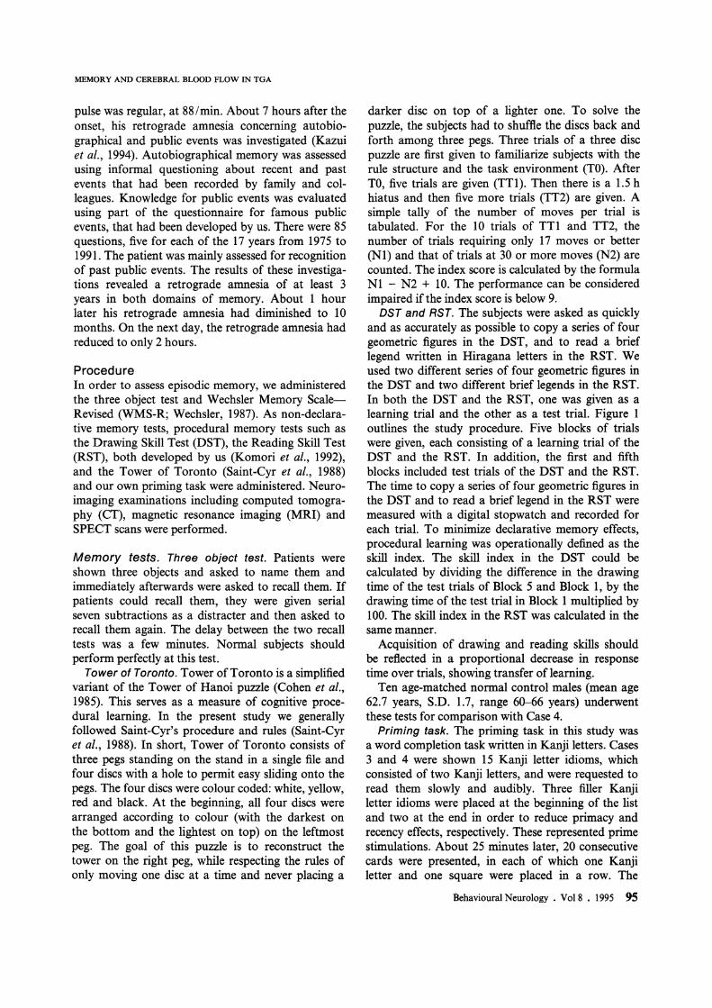
FIG. 1. Timetable for the DST and RST. Five blocks of trials were given, each consisting of learning trials for DST and RST, and test trials were added twice in the first and the fifth blocks; i.e. five learning trials and two test trials of both DST and RST were performed. We administered test trials with different materials from those used in the learning trials. Procedural learning in DST and RST was evaluated by the skill index.
patients were asked to put the first Kanji letter that came to mind in the square to make Kanji letter idioms. Half of the 20 Kanji letters in these cards could be completed with the Kanji letters presented in prime stimulations. There were at least five possible Kanji letters that could be used to make each target Kanji letter idiom among Kanji letters taught in elementary school. We calculated the percentage of completions.
Fifty normal age-matched control males were also administered this task. The control group was divided into two groups, each of which consisted of25 men. In one control group the procedure was completely the same as in the TGA patients [this group (mean age 56.7 years) was called "normal controls with prime stimulations (NCWS)]". In the other control group we presented a different series of prime stimulations, consisting of no Kanji letters placed in the priming cards [this group (mean age 56.4 years) was called "normal controls without prime stimulations (NCWOS)"]. In the NCWOS group, the other procedure was the same as in the TGA and NCWS groups, so the percentage of completIOns in NCWOS are baseline completion rates.
In Case 2 during and after TGA we initially presented him with seven Kanji idiom stimulations, and then 25 minutes later, 12 Kanji letters were presented consecutively in order to construct Kanji letter idioms from the presented Kanji letters.
Neuroimaging examinations. CT and MRI. CT scans were performed during the episodes of TGA
96 Behavioural Neurology. Vol 8 . 1995
and MRI scans after recovery from TGA in all cases. SPECT. SPECT scans using N-isopropyl-(I-123)-p
iodoamphetamine (IMP) were carried out both during and after TGA in Case 1. Data acquisitions were started 10 min after the intravenous injection of 222 MBq of IMP and continued for 35 min. In Cases 2, 3 and 4, SPECT scans were performed using Tc-99m hexamethylpropyleneamine oxime (HMPAO). Data acquisitions were started 5 min after the intravenous injection of 740 MBq of HMPAO and continued for 35 min. The SPECT unit was a four headed rotating gamma camera with a low energy high resolution collimator (Kimura et al., 1990). The spatial resolution was 9.8 mm in full width at half maximum at the centre. The raw data were reconstructed using a standard filtered back projection algorithm with pretreatment by Butterworth filter. The attenuation correction was performed by the post-correction method (jl = 0.08). Using the set of two light projectors for position setting, almost the same anatomical slices could be obtained for studies during and after TGA.
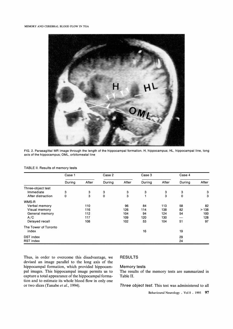
The hippocampal formation is rather a small structure. Its total length is between 4 and 4.5 cm, the width of its body on average 1 cm and that of its head 1.5-2 cm. In addition, its long axis is inclined at an angle 20° or 30° negative to the orbitomeatalline (Fig. 2). Accordingly, in images parallel to the orbitomeatal line commonly used, the hippocampal formation is divided into several parts and shown on several separate slices. As a result, it became difficult to evaluate blood flow of the small part in each slice.
MEMORY AND CEREBRAL BLOOD FLOW IN TGA
FIG. 2. Parasagittal MR image through the length of the hippocampal formation. H. hippocampus; HL. hippocampal line. long axis of the hippocampus; OML. orbitomeatal line
TABLE Ii. Results of memory tests
Case 1 Case 2
During After During
Three-object test Immediate 3 3 3 After distraction 0 3 0
WMS-R Verbal memory 110 Visual memory 116 General memory 112 AI C 117 Delayed recal l 108
The Tower of Toronto index
DST index RST index
Thus, in order to overcome this disadvantage, we devised an image parallel to the long axis of the hippocampal formation, which provided hippocampal images. This hippocampal image permits us to capture a total appearance of the hippocampal formation and to estimate its whole blood flow in only one or two slices (Tanabe et aI., 1994).
Case 3 Case 4
After During After During After
3 3 3 3 3 3 1 3 0 3
96 84 113 58 82 126 114 138 82 > 138 104 94 124 54 100 109 120 130 128 102 53 104 51 97
16 19
29 24
RESULTS
Memory tests The results of the memory tests are summarized in Table II.
Three object test. This test was administered to all
Behavioural Neurology. Vol 8 . 1995 97

70
----- • DTGA ?f2. 60
0 ATGA 0 50
~ 40 .....l
~ 0 30 U CIl
~ 20
0 ~ 10
o Case 2 Case 3 Case 4 NCWS NCWOS
FIG. 3. Word stem completion performance of patients during (DTGA) and after TGA (ATGA) and age-matched normal control males. NCWS, normal controls with prime stimulations; NCWOS, normal controls without prime stimulations (baseline completion rates). Bars show standard errors of the mean.
patients both during and after the attack. During the episode, all cases could readily name three objects, and immediately recall all of them, but after distraction Cases 1, 2 and 4 could recall none of them. Moreover, they could not remember at all that they had seen them. Case 3 could recall only one of the three objects after distraction and recognize only one of the remainder during TGA. However, after resolution of the TGA all patients performed normally in these tasks.
WMS-R.WMS-R was carried out both during and after the episode in Cases 3 and 4. The performance on the WMS-R of Case 3 during the attack was within normal range except for the delayed recall index. However, he showed a 30 point discrepancy between verbal and visual memory. The indices after the attack returned to normal and were superior to those during TGA. In addition, the discrepancy between verbal and visual memory disappeared. In Case 4 the space span subtest was not performed during the attack, due to tiring of the patient. His digit span was eight forward and four backward during TGA. In Case 4 all the indices during the episode were abnormal. Nevertheless after the attack all the performances returned to normal. In Cases 1 and 2, this test was carried out only after the attack when the scores were within normal limits.
Tower of Toronto. This task was carried out during the attack of TGA by Cases 3 and 4. Case 3 could register some events that happened during TGA so that he was in the recovery phase, while Case 4 who was still in the acute phase could recall none of three
98 Behavioural Neurology. Vol 8 . 1995
H. KAZUI ET AL.
objects in the three object test. The indices of Tower of Toronto were 16 in Case 3 and 19 in Case 4 during the episode. These scores are within the normal range (Saint-Cyr et aI., 1988).
DST and RSr. These tests were carried out by Case 4 during the episode and by 10 age-matched normal control males (mean age 62.7 years, range 60-66 years). At the time of these tests, Case 4 could recall none of the three objects in the three object test. The indices in Case 4 were, however, 29 in the DST and 24 in the RST. The mean indices of 10 normal controls were 26.7 (S.D. 8.3) in the DST and 24.1 (S.D. 10.1) in the RST. Hence the performance of Case 4 was considered to be normal in both the DST and RST.
Priming task. This task was performed incompletely by Case 2, and completely by Cases 3 and 4 both during and after TGA, and by age-matched controls. When this task was carried out during the attack, Cases 2, 3 and 4 were so amnesic that they could not recall or recognize the prime stimulations. The majority of normal controls and all three cases after TGA could recognize the prime stimulations but not recall them. The word completion performances of the three patients and NCWS and NCWOS are shown in Fig. 3. During the attack the percentage of completions (DTGA) were 42.9% in Case 2, 30% in Case 3 and 50% in Case 4. After TGA the results were 42.9%, 40% and 40%, respectively. Score in NCWS was 41.6% (S.D. 12.8) and NCWOS 16% (S.D. 11.2).
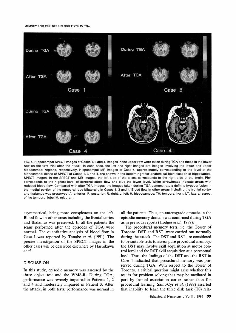
Neuroimaging examinations CT and MRI. CT scans carried out during the episodes of TGA in all cases revealed no abnormalities. MRI in Case 1, performed 1 month after the episode, revealed a circumscribed abnormal intensity spot in the left hippocampus (Tanabe et al., 1991). In the other cases, MRI scans revealed no abnormalities.
SPECr. SPECT scans were performed once during and twice after TGA in Cases 1,3 and 4. Scans were carried out in Case 2 only after TGA. During TGA SPECT scans were performed 6 or 7 h after the onset. After TGA the timing of the first scans was 1 week after the episodes of TGA in Cases 1 and 3, and the next day after the attack in Case 4. When compared with after-TGA images, the images taken during the TGA episode demonstrated a remarkable hypoperfusion in the medial portion of both temporal lobes in Cases 1, 3 and 4 (Fig. 4). The reductions of cerebral blood flow in the mesial temporal lobes were symmetrical in Cases 1 and 4, while in Case 3 it was
MEMORY AND CEREBRAL BLOOD FLOW IN TGA
FIG. 4. Hippocampal SPECT images ·of Cases 1, 3 and 4. Images in the upper row were taken during TGA and those in the lower row on the first trial after the attack. In each case, the left and right images are images involving the lower and upper hippocampal regions, respectively. Hippocampal MR images of Case 4, approximately corresponding to the level of the hippocampal slices of SPECT of Cases 1, 3 and 4, are shown in the bottom right for anatomical identification of hippocampal SPECT images. In the SPECT and MR images, the left side of the slices corresponds to the right side of the brain . Pink corresponds to the highest level of cerebral blood flow and blue the lower level. White arrowheads indicate areas with reduced blood flow. Compared with after-TGA images, the images taken during TGA demonstrate a definite hypoperfusion in the medial portion of the temporal lobe bilaterally in Cases 1, 3 and 4. Blood flow in other areas including the frontal cortex and thalamus was preserved. A, anterior; P, posterior; R, right; L, left; H, hippocampus; TH, temporal horn; L T, lateral aspect of the temporal lobe; M, midbrain.
asymmetrical, being more conspicuous on the left. Blood flow in other areas including the frontal cortex and thalamus was preserved. In all the patients the scans performed after the episodes of TGA were normal. The quantitative analysis of blood flow in Case 1 was reported by Tanabe et at. (1991). The precise investigation of the SPECT images in the other cases will be described elsewhere by Hashikawa et al.
DISCUSSION
In this study, episodic memory was assessed by the three object test and the WMS-R. During TGA, performance was severely impaired in Patients 1, 2 and 4 and moderately impaired in Patient 3. After the attack, in both tests, performance was normal in
all the patients. Thus, an anterograde amnesia in the episodic memory domain was confirmed during TGA as in previous reports (Hodges et at., 1989).
The procedural memory tests, i.e. the Tower of Toronto, DST and RST, were carried out normally during the attack. The DST and RST are considered to be suitable tests to assess pure procedural memory: the DST may involve skill acquisition at motor controllevel and the RST skill acquisition at a perceptual level. Thus, the findings of the DST and the RST in Case 4 indicated that procedural memory was preserved during TGA. With respect to the Tower of Toronto, a critical question might arise whether this test is for problem solving that may be mediated in part by frontal association cortex rather than for procedural learning. Saint-Cyr et al. (1988) asserted that inability to learn the three disk task (TO) rela-
Behavioural Neurology. Vol 8 . 1995 99

tively efficiently is taken as evidence of impaired problem-solving ability and usually signifies frontal dysfunction. Recently, a PET study during TGA revealed reduction in cerebral blood flow and oxygen consumption over the entire lateral frontal cortex on the right side (Baron et al., 1994). There have been reports of cognitive changes, in addition to amnesia proper, during episodes of TGA, such as behavioural passivity (Kritchevsky et al., 1988), reduced verbal fluency (Lin et al., 1993) and impaired "frontal lobe tests" (Stillhard et al., 1990), all of which might suggest frontal lobe dysfunction. We did not administer the formal frontal lobe tests to our TGA patients, but did not detect frontal lobe dysfunction in any of our cases during TGA through the neuropsychological examinations. In addition, Cases 3 and 4 could solve the Tower of Toronto and their indices of the task were normal; this indicates that both problem-solving ability and procedural memory were preserved during TGA. Thus, our tests to assess procedural memory indicated that procedural memory was preserved in spite of a severe amnesia for events during TGA. This finding is consistent with one earlier report (Goldenberg et al., 1991), in which the patient markedly improved from the first to the second trial of a procedural learning test which assessed recognition of fragmented digits.
Priming effects have been studied using various methods in amnesic patients and normal sUbjects. In the present study, a word completion priming task with Kanji letters was used. During the attack the percentages of completions were 42.9% in Case 2 and 50% in Case 4. These scores were 2 S.D. beyond the baseline completion rate, i.e. the mean score in NCWOS. Thus, in these cases, priming effects were definitely retained during TGA. In Case 3 during TGA, the score was 30%, i.e. within 2 S.D. of the baseline completion rate and within 1 S.D. of the mean score in NCWS. Accordingly, it is probable that in Case 3 priming effects were also retained during TGA. However, it seems likely that in Case 3 priming effects were also retained.
In the current study, our new method of construction of SPECT images, namely hippocampal images, allowed us to evaluate total blood flow of the hippocampal areas. These images demonstrated, in our three cases, a hypoperfusion confined to the medial aspects of the bilateral temporal lobe during TGA. The reductions of cerebral blood flow were symmetrical in Cases 1 and 4, and asymmetrical, being more conspicuous on the left, in Case 3.
Thus the results of the memory tests and SPECT findings in this study indicate that medial aspects of the temporal lobe play an important role in episodic memory, but not in procedural memory and priming
100 Behavioural Neurology. Vol 8 . 1995
H. KAZUI ET AL.
effects. In addition, medial aspect of the left temporal lobe could be related to episodic verbal memory.
All of our TGA patients exhibited a retrograde amnesia of up to 3 years. The retrograde amnesia was for both personal and public events. In Case 4, especially, the lengths of retrograde amnesia for personal and public events were shown to be similar. Therefore, this finding confirms the results of previous studies (Stillhard et al.,1990; Goldenberg et al., 1991; Tanabe et al., 1991; Evans et al., 1993; Lin et al., 1993) that the medial temporal lobes are important for retrieval of events in both autobiographical and social domains within several years previous to the onset of TGA. On the other hand, these areas do not seem to play an important role in retrieving memory of events occurring more than several years before the episode.
In contrast to studies of memory function in cases of permanent amnesia, those in TGA have an advantage: it is possible to compare the memory disturbance during TGA with that after TGA in the same person. The present study demonstrates the potential value of SPECT studies combined with neuropsychological examinations during TGA in understanding the neural basis of various memory subcomponents.
Acknowledgements We thank Professor David Neary for his assistance in the preparation of the manuscript.
REFERENCES Baron JC, Petit-Taboue MC, Le Doze F et al. (1994) Right
frontal cortex hypometabolism in transient global amnesia. A PET study. Brain, 117, 545-552.
Cohen NJ, Eichenbaum H, DeAcedo BS et al. (1985) Different memory systems underlying acquisition of procedural and declarative knowledge. Annals of the New York Academy of Sciences, 444, 54-71.
Evans J, Wilson B, Wraight EP et al. (1993) Neuropsychological and SPECT scan findings during and after transient global amnesia: evidence for the differential impairment of remote episodic memory. Journal of Neurology, Neurosurgery and Psychiatry, 56,1227-1230.
Goldenberg G, Podreka I, Pfaffelmeyer N et al. (1991) Thalamic ischemia in transient global amnesia: ASPECT study. Neurology, 41,1748-1752.
Hodges JR (1994) Semantic memory and frontal executive function during transient global amnesia. Journal of Neurology, Neurosurgery and Psychiatry, 57, 605-608.
Hodges JR and Ward CD (1989) Observations during transient global amnesia. A behavioural and neuropsychological study of five cases. Brain, 112, 595-620.
Hodges JR and Warlow CP (1990) Syndromes of transient amnesia: towards a classification. A study of 153 cases. Journal of Neurology, Neurosurgery, and Psychiatry, 53, 834-843.
Kazui H, Tanabe H, Ikeda M et al. (1994) Retrograde
MEMORY AND CEREBRAL BLOOD FLOW IN TGA
amnesia during transient global amnesia. Japanese Journal of Neuropsychology, 10, 122-130.
Kimura K, Hashikawa K, Etani H et al. (1990) A new apparatus for brain imaging: four head rotating gamma camera single photon emission computed tomograph. Journal of Nuclear Medicine, 31, 603-609.
Komori K, Ikeda M, Kazui H et al. (1992) Procedural memory in the normal aged: simple tasks of procedural memory available to patients with dementia. Japanese Journal of Neuropsychology, 8,182-190.
Kritchevsky M, Squire LR and Zouzounis JA (1988) Transient global amnesia: Characterization of anterograde and retrograde amnesia. Neurology, 38, 213-219.
Lin KN, Liu RS, Yeh TP et at. (1993) Posterior ischemia during an attack of transient global amnesia. Stroke, 24, 1093-1095.
Saint-Cyr JA, Taylor AE and Lang AE (1988) Procedural learning and neostriatal dysfunction in man. Brain, 111, 941-959.
Stillhard G, Landis T, Schiess R et al. (1990) Bitemporal hypoperfusion in transient global amnesia: 99m-Tc-HMPAO SPECT and neuropsychological findings during and after an attack. Journal of Neurology, Neurosurgery and Psychiatry, 53, 339-342.
Tanabe H, Hashikawa K, Nakagawa Y et al. (1991) Memory loss due to transient hypoperfusion in the medial temporal lobes including hippocampus. Acta Neurologica Scandinavica, 84, 22-27, 463.
Tanabe H, Ikeda M and Hashikawa K (1994) Neuroimaging of the human hippocampus in the context of disturbance of the higher brain functions. Shinkei Kenkyu no Shinpo,38,161-172.
Wechsler D (1987) Wechsler Memory Scale-Revised. Psychological Corporation, New York.
(Received 26 October 1994; accepted as revised 11 May 1995)
Behavioural Neurology. Vol 8 . 1995 101

Submit your manuscripts athttp://www.hindawi.com
Stem CellsInternational
Hindawi Publishing Corporationhttp://www.hindawi.com Volume 2014
Hindawi Publishing Corporationhttp://www.hindawi.com Volume 2014
MEDIATORSINFLAMMATION
of
Hindawi Publishing Corporationhttp://www.hindawi.com Volume 2014
Behavioural Neurology
EndocrinologyInternational Journal of
Hindawi Publishing Corporationhttp://www.hindawi.com Volume 2014
Hindawi Publishing Corporationhttp://www.hindawi.com Volume 2014
Disease Markers
Hindawi Publishing Corporationhttp://www.hindawi.com Volume 2014
BioMed Research International
OncologyJournal of
Hindawi Publishing Corporationhttp://www.hindawi.com Volume 2014
Hindawi Publishing Corporationhttp://www.hindawi.com Volume 2014
Oxidative Medicine and Cellular Longevity
Hindawi Publishing Corporationhttp://www.hindawi.com Volume 2014
PPAR Research
The Scientific World JournalHindawi Publishing Corporation http://www.hindawi.com Volume 2014
Immunology ResearchHindawi Publishing Corporationhttp://www.hindawi.com Volume 2014
Journal of
ObesityJournal of
Hindawi Publishing Corporationhttp://www.hindawi.com Volume 2014
Hindawi Publishing Corporationhttp://www.hindawi.com Volume 2014
Computational and Mathematical Methods in Medicine
OphthalmologyJournal of
Hindawi Publishing Corporationhttp://www.hindawi.com Volume 2014
Diabetes ResearchJournal of
Hindawi Publishing Corporationhttp://www.hindawi.com Volume 2014
Hindawi Publishing Corporationhttp://www.hindawi.com Volume 2014
Research and TreatmentAIDS
Hindawi Publishing Corporationhttp://www.hindawi.com Volume 2014
Gastroenterology Research and Practice
Hindawi Publishing Corporationhttp://www.hindawi.com Volume 2014
Parkinson’s Disease
Evidence-Based Complementary and Alternative Medicine
Volume 2014Hindawi Publishing Corporationhttp://www.hindawi.com