melt blown polymeric nanofibers for medical applications ... · citation: hiremath n, bhat g (2015)...
TRANSCRIPT

*Corresponding author email: [email protected] Group
Symbiosis www.symbiosisonline.org www.symbiosisonlinepublishing.com
Melt blown Polymeric Nanofibers for Medical Applications- An Overview
Nitilaksha Hiremath, and Gajanan Bhat*Department of Materials Science and Engineering, University of Tennessee, Knoxville, TN 37996, USA
Nanoscience & Technology: Open Access Open AccessReview Article
polymers of biological origin, polymer blends or block copolymers, or polymers with additives. Possible methods for the production of nanofibers are electrospinning, melt spinning, centrifugal spinning, bicomponent spinning followed by dissolution of the matrix, and melt blowing.
Nanofibers or microfibers produced by either of the techniques are used in tissue restoration, compact organs, construction of biocompatible prostheses, cosmetics, face masks, bone substitutes, artificial blood vessels, and valves; and drug delivery applications [3]. Scaffold materials produced from nanofibers offer a very large surface area that supports cell growth [2]. Polymers such as Polyglycolide (PGA), Polylactic Acid (PLA), and their random copolymer Poly (Glycolide-co-lactide) are often used as the base materials for implant devices, such as suture fibers and scaffolds, for tissue engineering [4]. These materials meet several controlled-release criteria: they are biocompatible, biodegradable, and they can provide high efficiency in drug loading. Polycaprolactone (PCL) has been investigated mainly for long-term implants for drug release and support of mineralized tissue formation and may be a suitable substrate for the treatment of bone defects. An improvement in the mechanical properties of PCL has been achieved by copolymerization with PLA, enabling its use for orthopedic applications, such as the repair of bone defects. The ultimate goal of the novel modified nanofibrous scaffold design is the production of an ideal structure that can replace the natural Extra Cellular Matrix (ECM) until host cells can repopulate and resynthesize a new natural matrix. Collagen in its native state is a natural substrate for cell attachment, growth, and differentiation.
Nanofibers provide a connection between the nanoscale world and the macroscale world, because the diameters are in the nanometer range and the lengths are in kilometers [5]. Therefore, the current emphasis of research is on exploiting such properties and focusing on determining appropriate conditions for making various polymers into products for eventual applications including multifunctional membranes, biomedical structural elements [6] (scaffolds used in tissue engineering, wound dressing, drug delivery, artificial organs, vascular grafts), protective shields in specialty fabrics, filter media for submicron particles in the separation industry, composite reinforcement,
AbstractNanotechnology has enabled the production of novel products,
which have superior control over a wide range of applications. One of the most successful areas is that of nanofibers. Because of their very high specific surface area, nanofibers may be used in a range of applications including but not limited to, high efficiency filter media, scaffolds for tissue engineering, sorbents, artificial blood vessels, and wound dressing material. Melt blowing is a technology that has been commercially used for the past three decades to produce microfibers. Recently, University of Tennessee Nonwovens Research Laboratory (UTNRL) in collaboration with many companies has been leading the applied research on meltblown nanofiber nonwovens from various thermoplastic polymers. Not only that the nanofiber nonwovens are produced without the use of any solvent, but also the production rates are very high with a good commercialization potential. Such nanofibers have properties that make them suitable for many medical applications. Recent approaches to achieve large-scale production of nanofibers by meltblowing as well as some of the medical applications where meltblown nanofibers have an advantage and the market potential are discussed in the following sections.
Keywords: Melt blowing; Nanofibers; Scaffolds; Tissue engineering; Skin; Cartilage; Bone; Nerve
Received: May 24, 2015; Accepted: June 26, 2015; Published: June 29, 2015
*Corresponding author: Gajanan Bhat, Department of Materials Science and Engineering, University of Tennessee, Knoxville, TN 37996, USA, Tel: +1-865-974-0976; E-mail: [email protected]
IntroductionPolymeric nanofibers find use in a broad range of applications
in various areas such as nano reinforcement and nanofluidics, medicine and pharmacy, tissue engineering etc. Although true definition of nanofibers is that the diameters have to be less than 100nm, fibers less than one micron in diameter are considered as nanofibers, also referred to as submicron fibers in the textile industry [1]. Fibers of less than one denier per filament are considered microfibers, and their diameters are typically in the range of 1-10 microns. Depending on the area of application, the properties of the polymer materials are optimized to achieve the required performance. For example the polymer needs to be hydrophobic or hydrophilic, biocompatible and/ or biodegradable, possess a high stiffness and strength, or show piezo-electric properties, etc. Often a combination of properties is desired and cannot be achieved by a single synthetic material [2]. Nanofibers are made from various materials including from

Page 2 of 9Citation: Hiremath N, Bhat G (2015) Melt blown Polymeric Nanofibers for Medical Applications- An Overview. Nanosci Technol 2(1): 1-9.
Melt blown Polymeric Nanofibers for Medical Applications- An Overview Copyright: © 2015 Bhat et al.
and structures for nanoelectronic machines. This mini review discusses the meltblowing process for producing nanofibers and medical related applications of polymeric nanofibers.
Nanofiber production from Polymers
One of the major success stories of nanotechnology has been that of submicron fibers, also referred to as nanofibers in the textile industry. Polymeric submicron fibers (< 1 µm) have enormous specific surface area and high flexibility. As a result, webs containing predominantly submicron fibers have large surface-to-volume ratio, micropores, and high porosity, and hence find numerous applications in the areas of decontamination, catalysis, and filtration, super absorbents, as scaffolds for tissue engineering and wound dressings, for energy storage and many electronic applications.
Polymer submicron fibers can be produced by a number of different techniques utilizing physical, chemical, thermal, and electrostatic fabrication techniques such as super drawing, phase separation, self-assembly, kinetically controlled solution synthesis and electrospinning. One of the successful methods has been by spinning immiscible polymers using the sea-island technique and then dissolving the matrix to leave with a bundle of nanofibers. Electrospinning is one of the most researched topics and has been very successful in the laboratory to produce nanofibers from a wide range of polymers with unique properties. However, this process is struggling to be one of commercial success, except finding applications in some niche areas. The major disadvantages of electrospinning are that the solvents used can be toxic, inability to obtain 3D structures efficiently with sufficient pore size distribution for tissue engineering, process depends on a lot of variables, the polymer feed rate is very small, which is not commercially viable. Another new technology that is in the early stage of development is based on centrifugal spinning. Melt blowing has shown the potential to produce submicron fibers. In addition to that, the meltblown process is commercially practiced at a higher throughput and can be extended for submicron fibers production [1]. Recent developments in this area are discussed in the following sections.
Melt blowing
Melt blowing is a one-step process (Figure 1) in which high-velocity hot air is used to draw the molten thermoplastic polymer fiber. A typical melt blowing process is a single integrated process, consisting of polymer-feeding systems, extruder, metering pumps, die assembly, web formation, and a collector [7]. Processing through extruder, metering pumps, filter, and up to the die assembly is quite similar to melt spinning. However, die configuration and collection systems are designed in a completely different manner. As the polymer is extruded through a linear die (Figure 2A) into converging streams of hot air, the high velocity air attenuates the fibers. The same air streams convey the fibers on to a collector. As the fibers move to the collector, they are quenched, form entanglements and bonding takes place at the fiber-to-fiber contact points, there by forming a cohesive nonwoven web. Generally, vacuum is applied at the collector to facilitate disengagement of air from the fabric.
Typical melt blown webs will have fibers in the range of 2-5 µm on the average with a wide distribution in diameter. Because of the type of the polymer used and the process, the fibers have relatively poor mechanical properties, but the fabrics have good barrier properties. Meltblown webs are extensively used, either alone or as part of composite structures, in fine filtration to remove finer particles and bacteria and as absorbent products for many applications.
Melt blown nanofibers
As the melt blowing process has allowed the production of microfibers as small as one micron average diameter in a commercial process, there has been continuing effort to push the technology to further reduce fiber diameter. Recent efforts to design and develop modular dies with smaller holes and larger
Figure 1: Schematic of a melt blowing line.
A)
B)
Figure 2: Images of a typical Melt Blown Die (A) and continuous pro-duction of nanofiber webs (B).

Page 3 of 9Citation: Hiremath N, Bhat G (2015) Melt blown Polymeric Nanofibers for Medical Applications- An Overview. Nanosci Technol 2(1): 1-9.
Melt blown Polymeric Nanofibers for Medical Applications- An Overview Copyright: © 2015 Bhat et al.
number of holes allow the production of submicron fibers in a cost effective way. One of the modular dies consists of stacked plates, with one of the plates having an inlet for molten polymer and heated air inlets unlike the typical meltblown die with linear holes and air knives to allow the process air from both sides. The plates are configured to define channels to create a path through which material flows, one of the plates having outlet acting as a spinneret orifice through which polymer can be extruded. Modular die operates at low throughput per hole and hence, low melt pressures for fiber extrusion. The molten polymer is extruded at relatively low flow rates through the orifice of the modular dies to produce submicron fibers.
Several thermoplastic polymers such as Polypropylene (PP), Polyethylene terephthalate (PET), Polybutylene terephthalate (PBT) and PLA have been processed successfully using commercially available resins to produce uniform webs using our pilot lines [8]. Submicron fibers produced using modular meltblowing dies had diameters in the range of 50 nm to 1000 nm, with average diameters in the range of 400-600 nm (Figure 3A and 3B). Hence, this modified melt blowing process would be a unique and novel approach to produce submicron fibers, in the industrial scale from thermoplastic polymers. Fiber diameter distribution (Figure 4) is comparable to that of, and many times narrower than that observed in typical microfiber meltblowing. Also under controlled process conditions, submicron fiber webs free of roping, shots or fiber breakage can be successfully produced continuously at production rates several times faster than that possible in other techniques such as electro spinning [9].
The submicron fibers provide the advantage as seen from many properties, especially the filter quality factor values compared to microfiber webs of similar materials. Further investigations have been done with another modular die from Hills, Inc., that consisted of 100 holes per inch with very fine holes. Both the modular dies helped in the production of submicron fibers from typical commercial melt blown grade polypropylenes. Unlike in the typical melt blown die, the modular dies have higher number of holes per cm, and the overall productivity does not decrease much. In fact, our trials on the pilot lines demonstrated
that production speeds could be more than 100 meters per minute if only a small amount of nanofibers are to be deposited on a nonwoven or any other substrate.
Medical applications of nanofibers
Fibers in general have been used in a wide range of medical applications. Over the years development and availability of micro and nanofibers has increased their use in such products. Some of the examples are shown in Figure 5. Electrospun products are employed in medical applications for tissue engineering and prosthetics. A skin mask is fabricated by electrospinning fibers on the skin surface directly to protect or heal wounds. Electrospun fiber mats were also explored as drug delivery vehicles, with promising results. Mats were made from either PLA, Poly (ethylene-co-vinyl alcohol (PEVA), or their 50:50 blend using tetracycline hydrochloride as a model drug [4,11,12]. PLA has been widely employed for various biomedical applications because of its biodegradability, biocompatibility, good mechanical properties, and its ability to be dissolved in common solvents for processing. Polymers such as silicone rubber, nylons, polyesters, polyurethanes, acrylics, PEG, PLA, hydrogels etc. have been used in biomedical applications. Biomaterials are used in dental implants, bone replacements, cartilages, heart valves, cosmetic surgeries, and vascular grafts [5,13-15]. Electrospinning could be used to fabricate biocompatible thin film coating design and texture that can be deposited on implantable devices to facilitate the unification of these devices with the body. Silk-like polymer with fibronectin functionality (ECM proteins) has been electrospun for making biocompatible films used on prosthetic devices aimed for implantation in the central nervous system. Of particular interest are electrospun membranes composed of elastomeric fibers, for the development of several protective clothing applications. Much work is being done with the aim of developing garments for soldiers that reduce risks of chemical exposure. The idea is to lace several types of polymers and fibers to make protective ultrathin layers that would enhance chemical reactivity and environmental resistance.
Skin and wound dressing
Tissue engineering offers potential opportunity for living
A) B)
Figure 3: SEM images of Melt blown Submicron Fiber Webs from PP (A) and PLA (B), magnification 2µm [1].

Page 4 of 9Citation: Hiremath N, Bhat G (2015) Melt blown Polymeric Nanofibers for Medical Applications- An Overview. Nanosci Technol 2(1): 1-9.
Melt blown Polymeric Nanofibers for Medical Applications- An Overview Copyright: © 2015 Bhat et al.
-5
0
5
10
15
20
25
0.25
0.30
0.35
0.40
0.45
0.50
0.55
0.60
0.65
0.70
0.75
0.85
0.95
Fibe
r Co
unt
Fiber Diameter, µm
Figure 4: Fiber diameter distribution of melt blown submicron fibers [10].
Figure 5: A schematic applications of tissue engineering in various fields such as Skin and Wound dressing [16], Cartilage [17], Bone [18], Neural tissue [19], Heart valves [20], Muscle fibers [21], SEM picture of nano fibers [22], the nano fibers are also used in cosmetics (facial masks, cleansing agents etc.) and pharmaceutical industries for localized drug delivery applications.
Page 5 of 9Citation: Hiremath N, Bhat G (2015) Melt blown Polymeric Nanofibers for Medical Applications- An Overview. Nanosci Technol 2(1): 1-9.
Melt blown Polymeric Nanofibers for Medical Applications- An Overview Copyright: © 2015 Bhat et al.
tissue replacements, thereby decreasing the dependence on donor tissue and organs. Natural and synthetic fibers are employed widely for tissue repair, and these nanofibrous scaffolds are stable mechanically and biologically at the implant site [5,23-26]. Mechanical stability of the biomaterials is dependent on the design and interactions of the scaffold with the cells. Wound dressings for humans have been aimed at protection, absorption of exudates, inhibition to external microbial invasion, and appearance. Polyurethane is mostly used in wound dressings because of its oxygen permeability and good barrier properties. Electro spun nano fibrous membranes permit higher gas flow and protect wound from infection and dehydration. For efficient barrier, wound-dressing materials must be chosen carefully, and should be ensured that the material adheres to the surface of the wound providing barrier against atmospheric conditions and permit the oxygen flow. The fibroblast cell culture on PCL nanofibers and membranes support human dermal fibroblast and keratinocytes in tissue-engineered skin in regenerative medicine [27]. The fibroblast cells migrate through pores in amoeboid fashion and push the fibers if the hole size is small and shows that the migration is independent of the hole size of the nanofiber scaffold. PCL nanofibers degrade enzymatically [11,12]. These nanofibers show adherence to the wet wound surface and help to heal the wound, without accumulating fluid exudates.
It is now widely accepted that chitosan derivatives are highly efficient against bacteria and fungi as the target site of these cationic polymers is the cytoplasmic membrane of bacterial cells [6]. The photo-cross-linked electrospun mats of quaternized chitosan were efficient in hindering growth of gram-positive and gram-negative bacteria and suggest their potential use for wound dressing [28]. The composite nanofibrous membranes of chitosan/ collagen promote wound healing and induce cell migration and proliferation [4]. From animal studies, the nanofibrous membrane was found to be better than gauze and commercial collagen sponge in wound healing [6]. Cell infiltration and proliferation are crucial for a scaffold to support and guide tissue regeneration. Figure 6 shows the confocal laser scanning microscopy (CLSM) images of the human fibroblasts cultured for three days in the collagen/chitosan scaffold treated by 0.25% GA. From the sequential scanning mode, the fibroblasts ‘A’ could be distinguished from the scaffold ‘B’. ‘C’ reveals the fibroblasts adhered on the walls of the scaffold [29].
Cosmetics
The current skin care products for facial creams, lotions or ointments may include dusts or liquid sprays of fibrous materials to migrate into sensitive areas of the face skin. Polymer nanofibers are used as cosmetic skin care masks for skin healing, cleansing, or other therapeutic or medical properties with or without various additives. The polymer nanofibers-based cosmetic skin mask could be applied to the skin effortlessly conforming to the three-dimensional topography of the skin to provide healing or treatment to the skin [4,12,23]. Taepaiboon et al. [30] and coworkers electro spun cellulose acetate nanofibers which act as carriers for delivery of the model vitamins, all-trans retinoic acid (Retin –A) or vitamin A (Vit-A) or vitamin E (Vit-E).
Round and smooth fiber mats of average diameters of 247 and 265nm loaded with vitamins were immersed in the acetate buffer solutions with 0.5 vol % Tween 80 and 10 vol % methanol. The fiber mats exhibited a gradual release of the vitamins over a 24 hrs time period compared to a sudden release with vitamin loaded cellulose acetate films. The gradual release of vitamins helps to protect the skin and to retain the surface nutrition from the atmospheric shocks.
Cartilage
Various Elastin-like Polypeptides (ELPs) are studied for applications associated with cartilage tissue engineering and regeneration. Betre and coworkers captured chondrocytes in an ELP solution for transition and coacervation at 35°C, furthering entrapment of cells. The use of ELP for encouraging chondrogenesis, led to the use of coacervated ELP as a scaffold for later cartilage tissue engineering; the synthesis and accumulation of articular cartilage extracellular matrix ECM for both primary chondrocytes and adult stem cells (hADAS cells) [31]. Cartilage being a load bearing tissue, McHale and coworkers [32] developed enzymatically cross linked glutamine- and lysine-containing ELPs to increase load-bearing capabilities twice as high as that of the uncross linked ELP. However, this method of crosslinking took more time than clinically required. Lim and co-workers crosslinked lysine-containing ELPs with (β- [Tris (hydroxymethyl) phosphino] propionic acid (betaine) (THPP) and possessed shear modulus three orders of magnitude greater than that of uncrosslinked ELP after only a five minute reaction time [33]. Such ELPs were later used for in vitro tissue engineering in cartilage regeneration in a goat model of an osteochondral defect. Both in vitro and in vivo studies showed the ability of ELPs to be used as injectable biomaterials for cartilage regeneration.
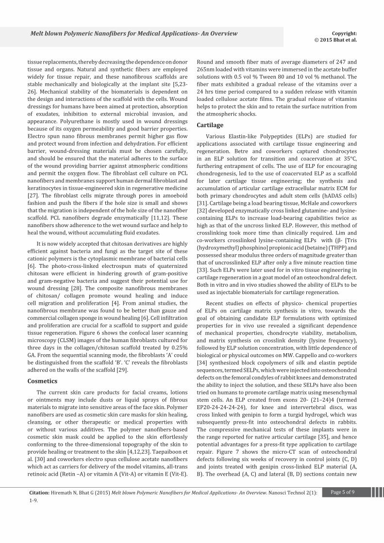
Recent studies on effects of physico- chemical properties of ELPs on cartilage matrix synthesis in vitro, towards the goal of obtaining candidate ELP formulations with optimized properties for in vivo use revealed a significant dependence of mechanical properties, chondrocyte viability, metabolism, and matrix synthesis on crosslink density (lysine frequency), followed by ELP solution concentration, with little dependence of biological or physical outcomes on MW. Cappello and co-workers [34] synthesized block copolymers of silk and elastin peptide sequences, termed SELPs, which were injected into osteochondral defects on the femoral condyles of rabbit knees and demonstrated the ability to inject the solution, and these SELPs have also been tried on humans to promote cartilage matrix using mesenchymal stem cells. An ELP created from exons 20- (21–24)4 (termed EP20-24-24-24-24), for knee and intervertebral discs, was cross linked with genipin to form a turgid hydrogel, which was subsequently press-fit into osteochondral defects in rabbits. The compressive mechanical tests of these implants were in the range reported for native articular cartilage [35], and hence potential advantages for a press-fit type application to cartilage repair. Figure 7 shows the micro-CT scan of osteochondral defects following six weeks of recovery in control joints (C, D) and joints treated with genipin cross-linked ELP material (A, B). The overhead (A, C) and lateral (B, D) sections contain new

Page 6 of 9Citation: Hiremath N, Bhat G (2015) Melt blown Polymeric Nanofibers for Medical Applications- An Overview. Nanosci Technol 2(1): 1-9.
Melt blown Polymeric Nanofibers for Medical Applications- An Overview Copyright: © 2015 Bhat et al.
Figure 6: CLSM images of human dermal fibroblasts (A) cultured over the collagen/chitosan scaffold (B, rhodamine-labeled) for 3 days; (C) is the merged image of (A) and (B).
subchondral infill as a fine white trabecular network distinct from the larger network in neighboring undamaged tissue.The experimentally treated wounds (A, B), cross-linked ELP material appears dark, with repair tissue visible along the periphery of the pad surfaces confirming the formation of large volume of new tissue.
The prime function of cartilage is weight cushioning during movement. Hydrated pads fitted into a confinement testing chamber under PBS at 37 °C and compressed to 10%, 20%, 30% and 40%, with each compressive cycle being followed by complete relaxation, as shown in Figure7F. Furthermore, a new composite scaffold with EP4 combined (four repeats of exons 20–24) with a thiol-modified hyaluronan and a Polyethylene Glycol Diacrylate (PEGDA) cross linker for nucleus pulposus repair and/ or treatment of early degenerative disc degeneration (DDD) was evaluated for its viability and gene expression of NP-associated genes for pathologic human disc cells. The aggregate modulus of 27.6 kPa and cell viability was observed [36]. However, when injected acellularly in rabbits, no evidence of an inflammatory response was observed and there was no difference between treated and untreated discs in disc volume suggesting limited benefit of injection of the ELP. Further modifications to the current scaffold model, could improve efficiency.
Bones
Nanofibers used for three-dimensional scaffolds, and starch/PCL (30:70%)-based scaffolds for bone were studied [37]. as the cell carrier has a higher ability to enhance cell attachment and organization, when compared to carriers having no nanofibers. Yoshimoto et al. [38] seeded PCL nano-scaffolds in neonatal rat bone marrow-derived MSCs. Osteogenic supplements containing cell-polymer dynamic culture maintained the original size and shape of the used scaffolds penetrated by cells and abundant ECM was observed after one week in culture. The surfaces of the scaffolds covered with cell multilayers, mineralization, and type I collagen after four weeks of culture, suggest that e-spun PCL could be a potential scaffold for engineering of bone. Bioactive silk fibroin scaffolds combined with either BMP-2, nanoparticles of hydroxyapatite (nHAp), or both were also investigated [39]. Scaffolds seeded with human bone marrow-derived MSCs, for 31 days in osteogenic media, support MSC growth and osteogenic
differentiation. Apatite that formed on silk fibroin BMP-2 scaffolds contained higher crystallinity than the one on silk fibroin control scaffolds. Furthermore nHAp particles were associated with improved bone formation. Two types of PCL nanofiber-CaCO3 nanoparticle composites with a ratio of PCL to CaCO3 of either 75:25 or 25:75, human osteoblasts cultured on these scaffolds had good cell attachment and proliferation. Badami et al. [40] studied MC3T3-E1 (mouse calvaria-derived osteoprogenitor cell line) for cell adhesion, orientation, proliferation, and osteoblastic differentiation on various scaffolds of copolymers PDLLA and PLLA, and block copolymers PEG-PDLLA and PEGPLLA on films and electro spun nanofibers. Post 14 days of incubation, cell proliferation was increased, cell density was found to be lower on fibers than on the smooth surfaces in the absence of osteogenic factors.
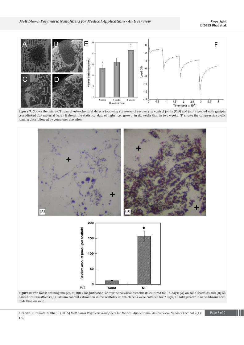
The cell density on fibers was equal to or greater, as the fiber diameter increased, than that on smooth surfaces in the presence of osteogenic factors. The authors suggested additional work to be done on the effect of fiber diameter, synthesis of osteocalcin and collagen (phenotypic markers), and the deposition of calcium–phosphate. When MSCs seeded PCL nano scaffolds were implanted in rats for four weeks, the constructs were rigid and bonelike with cells formed throughout the constructs [41]. As PCL properties could be further improved, developed nanoscaffolds of blends of PCL and hyaluronan were exposed to osteoblasts (cell line) and it was observed that the cells had attached more to PCL-hyaluronan blends than to plain hyaluronannanoscaffolds. In a report by Woo et al. [42] von Kossa staining after two weeks of culture showed the mineral deposition (brownish yellow precipitates) on the nano-fibrous scaffolds, while there was little observable mineralization in the solid-walled scaffolds. Calcium content assay further revealed that there was 13-fold greater amount of calcium produced in the nanofibrous scaffolds, than on the solid-walled scaffolds, as shown in Figure 8.
Nerves
Yang et al. [43] studied aligned PLLA nano/micro fibrous scaffolds for neonatal mouse cerebellum C17.2 stem cells and found that the cells elongated with neurite outgrowth parallel to the direction of aligned fibers. Though fiber diameter was not affecting the growth of the cells, nanofibers were proven to be
Page 7 of 9Citation: Hiremath N, Bhat G (2015) Melt blown Polymeric Nanofibers for Medical Applications- An Overview. Nanosci Technol 2(1): 1-9.
Melt blown Polymeric Nanofibers for Medical Applications- An Overview Copyright: © 2015 Bhat et al.
F
Figure 7: Shows the micro-CT scan of osteochondral defects following six weeks of recovery in control joints (C,D) and joints treated with genipin cross-linked ELP material (A, B). E shows the statistical data of higher cell growth in six weeks than in two weeks. ‘F’ shows the compressive cyclic loading data followed by complete relaxation.
Figure 8: von Kossa staining images, at 100 x magnification, of murine calvarial osteoblasts cultured for 14 days: (A) on solid scaffolds and (B) on nano-fibrous scaffolds. (C) Calcium content estimation in the scaffolds on which cells were cultured for 7 days, 13 fold greater in nano-fibrous scaf-folds than on solid.
Page 8 of 9Citation: Hiremath N, Bhat G (2015) Melt blown Polymeric Nanofibers for Medical Applications- An Overview. Nanosci Technol 2(1): 1-9.
Melt blown Polymeric Nanofibers for Medical Applications- An Overview Copyright: © 2015 Bhat et al.
better than microfibers, irrespective of fiber alignment. Scaffolds with fiber diameter of 272 nm, median pore diameter of 21 μm, and surface roughness of 172 nm were subjected to the growth of neural cells and 61.4% of the cells had adhered to the scaffolds by two hours, and 70% differentiated by one day as indicated by exhibiting spindle like shape with extended processes. PLLA is hydrophobic in nature and does not favor cell adherence, and effective surface modification for improved adhesion and the properties of the scaffolds is necessary.
SummaryModified meltblown process is a unique approach to produce
submicron fibers at rates many orders of magnitude higher than that possible from electrospinning, at the industrial scale. One of the advantages of meltblown technology is the ability to handle many different polymers as well as blends of polymers. There are no issues of solvent handling in the process, residual solvent in the webs, and environmental pollution. So far, we have successfully and consistently formed melt blown submicron fibers from high melt flow rate PP, PET, PBT and PLA, to name a few. One has to remember that the meltblown process is suitable for only thermoplastic polymers. The results suggest that it may be possible to fit the commercial lines with modular dies and continually produce submicron fiber webs. As the process seems to be technically and commercially feasible, further optimization of the process is needed and additional ongoing research will help find out possible challenges and appropriate solutions for successful implementation of this technology for large-scale production of submicron fiber nonwovens. The submicron fiber webs are suitable for many medical applications. Currently many such webs produced by electrospinning have been investigated, and the meltblown webs may have distinct advantage over electrospun webs both in processing, and structure and performance. Also, many additional applications are possible with the advancement in technology and further research.
References1. Bhat G. Polymeric Nanofibers: Recent Technology Advancements
Stimulating their Growth. Journal of textile science and engineering. 2015;(1):1.
2. Huang ZM, Zhang YZ, Kotaki M, and Ramakrishna S. A review on polymer nanofibers by electrospinning and their applications in nanocomposites. Composites science and technology. 2003;63(15):2223-2253.
3. Bhat G, Uppal R. Melt Blown Nonwovens for Medical Applications, Polymer Society. 2009;(2):26-29.
4. Schiffman JD, Schauer CL. A review: electrospinning of biopolymer nanofibers and their applications. Polymer reviews. 2008;(2):317-352.
5. Ashammakhi N, Ndreu A, Yang Y, Ylikauppila H, Nikkola L. Nanofiber-based scaffolds for tissue engineering. European journal of plastic surgery. 2012;35(2):135-149.
6. Jayakumar R, Prabaharan M, Nair SV, Tamura H. Novel chitin and chitosan nanofibers in biomedical applications. Biotechnology advances. 2010;(1):142-150.
7. Bhat GS, Malkan SR. Extruded continuous filament nonwovens:
Advances in scientific aspects. Journal of applied polymer science. 2002;(3):572-585.
8. Uppal R, Bhat G, Eash C, Akato K. Meltblown Nanofiber Media for Enhanced Quality Factor. Polymers and Fibers. 2013;14(4):660-668.
9. Han W, Wang X, Bhat G. Structure and Air permeability of Melt Blown Nanofiber Webs. Journal of Nanomaterials and Molecular Nanotechnology. 2013;2:3. doi:http://dx.doi.org/10.4172/2324-8777.1000115.
10. Bhat G. Meltblown submicron fibers for filter media and other applications. International fiber journal. 2015; 20-23.
11. Venugopal J, Ramakrishna S. Applications of polymer nanofibers in biomedicine and biotechnology. Appl Biochem Biotechnol. 2005;125(3):147-58.
12. Zhang Y, Lim CT, Ramakrishna S, Huang ZM. Recent development of polymer nanofibers for biomedical and biotechnological applications. J Mater Sci Mater Med. 2005;16(10):933-946.
13. Lee KY, Jeong L, Kang YO, Lee SJ, Park WH. Electrospinning of polysaccharides for regenerative medicine. Advanced drug delivery reviews. 2009;61(12):1020-1032. doi: 10.1016/j.addr.2009.07.006.
14. Bouten C, Dankers P, Driessen-Mol A, Pedron S, Brizard A, Baaijens F. Substrates for cardiovascular tissue engineering. Adv Drug Deliv Rev. 2011;63(4):221-241. doi: 10.1016/j.addr.2011.01.007.
15. Holzwarth JM, Ma PX. Biomimetic nanofibrous scaffolds for bone tissue engineering. Biomaterials. 2011;32(36): 9622-9629. doi: 10.1016/j.biomaterials.2011.09.009.
16. Dimond PF. Regenerative Medicine Stays Its Course. Genetic engineering and biotechnology news. 2011;(5).
17. Reiffel AJ, Zhou S, Chan S, Kafka C, Popa S, Spector JA, Lawrence JB. CAD-CAM Tissue Engineering of Auricular Cartilage Scaffolds for Reconstruction of Pediatric Microtia. Retrieved 2015 May 18.
18. Vallet-Regí M, Colilla M, González B. Medical applications of organic–inorganic hybrid materials within the field of silica-based bioceramics. Chemical society reviews. 2011;40(2):596-607. doi: 10.1039/c0cs00025f.
19. Hadjizadeh A. Neural tissue engineering-NSC differentiation-confocal microscopy. Retrieved May 18, 2015.
20. Schmidt D, Dijkman PE, Driessen-MolA, Stenger R, Mariani C, Puolakka A, et al. Minimally-invasive implantation of living tissue engineered heart valves: a comprehensive approach from autologous vascular cells to stem cells. Journal of the american college of cardiology. 2010;56(6): 510-520. doi: 10.1016/j.jacc.2010.04.024.
21. Silvernagel J. Fast and furious: how muscle fiber type influences basketball performance. SportsnScience Writer. 2014 April 27.
22. Nanosicence instruments. Nanosicence instruments. Retrieved 2015 May 18.
23. Burger C, Hsiao BS, Chu, B. Nanofibrous materials and their applications. Annu. Rev. Mater. Res. 2006;36:333-368.
24. Nettles DL, Chilkoti A, Setton LA. Applications of elastin-like polypeptides in tissue engineering. Advanced drug delivery reviews. 2010;62(15):1479-1485. doi: 10.1016/j.addr.2010.04.002.
25. Zhu J. Bioactive modification of poly (ethylene glycol) hydrogels for tissue engineering. Biomaterials. 2010 ;31(17):4639-4656. doi: 10.1016/j.biomaterials.2010.02.044.
26. Chen Q, Liang S, Thouas GA. Elastomeric biomaterials for tissue

Page 9 of 9Citation: Hiremath N, Bhat G (2015) Melt blown Polymeric Nanofibers for Medical Applications- An Overview. Nanosci Technol 2(1): 1-9.
Melt blown Polymeric Nanofibers for Medical Applications- An Overview Copyright: © 2015 Bhat et al.
engineering. Progress in polymer science. 2013;38(3):584-671.
27. Engel E, Michiardi A, Navarro M, Lacroix D, Planell JA. Nanotechnology in regenerative medicine: the materials side. Trends biotechnol. 2008;26(1):39-47.
28. Ignatova M, Manolova N, Rashkov I. Novel antibacterial fibers of quaternized chitosan and poly (vinyl pyrrolidone) prepared by electrospinning. European polymer journal. 2007;43(4):1112-1122.
29. Ma L, Gao C, Mao Z, Zhou J, Shen J, Hu X, et al. Collagen/chitosan porous scaffolds with improved biostability for skin tissue engineering. Biomaterials. 2003;24(26):4833-4841.
30. Taepaiboon P. Rungsardthong U, Supaphol P. Vitamin-loaded electrospun cellulose acetate nanofiber mats as transdermal and dermal therapeutic agents of vitamin A acid and vitamin E. Eur J Pharm Biopharm. 2007;67(2):387-397.
31. Betre H, Ong SR, Guilak F, Chilkoti A, Fermor B, Setton LA. Chondrocytic differentiation of human adipose-derived adult stem cells in elastin-like polypeptide. Biomaterials. 2006;27(1):91-99.
32. McHale MK, Setton LA, Chilkoti A. Synthesis and in vitro evaluation of enzymatically cross-linked elastin-like polypeptide gels for cartilaginous tissue repair. Tissue engineering. 2005;11(11-12):1768-1779.
33. Lim DW, Nettles DL, Setton LA, Chilkoti A. Rapid cross-linking of elastin-like polypeptides with (hydroxymethyl) phosphines in aqueous solution. Biomacromolecules. 2007;8(5):1463-1470.
34. Cappello J, Crissman J, Dorman M, Mikolajczak M, Textor G, Marquet M, Ferrari F. Genetic engineering of structural protein polymers. Biotechnol prog. 1990;6(3):198-202.
35. Hrabchak C, Rouleau J, Moss I, Woodhouse K, Akens M, Bellingham C, et al. Assessment of biocompatibility and initial evaluation of
genipin cross-linked elastin-like polypeptides in the treatment of an osteochondral knee defect in rabbits. Acta biomaterialia. 2010;6(6):2108-2115. doi: 10.1016/j.actbio.2009.12.034.
36. Ashammakhi N, Ndreu A, Piras A, Nikkola L, Sindelar T, Ylikauppila H, et al. Biodegradable nanomats produced by electrospinning:expanding multifunctionality and potential for tissue engineering. J nanosci nanotechnol. 2007;6(9-10):2693-711.
37. Tuzlakoglu K, Bolgen N, Salgado A, Gomes ME, Piskin E, Reis R. Nano-and micro-fiber combined scaffolds:a new architecture for bone tissue engineering. J Mater Sci Mater Med. 2005;16(12):1099-1104.
38. Yoshimoto H, Shin Y, Terai H, Vacanti J. A biodegradable nanofiber scaffold by electrospinning and its potential for bone tissue engineering. Biomaterials. 2003;24(12):2077-2082.
39. Li C, Vepari C, Jin HJ, Kim HJ, Kaplan DL. Electrospun silk-BMP-2 scaffolds for bone tissue engineering. Biomaterials. 2006;27(16):3115-3124.
40. Badami AS, Kreke MR, Thompson MS, Riffle JS, Goldstein AS. Effect of fiber diameter on spreading proliferation and differentiation of osteoblastic cells on electrospun poly (lactic acid) substrates. Biomaterials. 2006;27(4):596-606.
41. Shin M, Yoshimoto H, Vacanti JP. In vivo bone tissue engineering using mesenchymal stem cells on a novel electrospun nanofibrous scaffold. Tissue engineering. 2004;10(1-2):33-41.
42. Woo KM, Jun JH, Chen VJ, Seo J, Baek JH, Ryoo HM, et al. Nano-fibrous scaffolding promotes osteoblast differentiation and biomineralization. Biomaterials. 2007;28(2):335-343.
43. Yang F, Murugan R, Wang S, Ramakrishna S. Electrospinning of nano/micro scale poly (L-lactic acid) aligned fibers and their potential in neural tissue engineering. Biomaterials. 2005;26(15):2603-2610.