medical & vision research foundations files/in072001.pdf · medical & vision research...
TRANSCRIPT
Vol. XIX No. 2 JULY 2001
Scientific Journal of
MEDICAL & VISION RESEARCH FOUNDATIONS
18, COLLEGE ROAD, CHENNAI - 600 006, INDIA
Editorial
Perspective — Strabismus Evaluation — Sumitha Agarkar
Eales' Disease With Neurological Involvement - case report — Jyotirmoy Biswas, Rupesh V. Agrawal and Veena Noronha
Influence of Spatial frequency adaptation on human face perception — Irene Sophia Joseph and Shrikant R Bharadwaj
Photodynamic Therapy — Anand Subramanyam and Mahesh P Shanmugam
Keratoconus associated with Cone-Rod Dystrophy - A case report — Geetha K Iyer and Rajesh Fogla
Last Page — Telemedicine - What is it? — Rajesh Fogla

Editorial Evaluating patients with strabismus especially children, requires a great deal of patience along with good clinical method of examination. One needs to follow a systematic approach to complete examination within a short period of time, as children often tend to lose interest and become uncooperative towards the later part of the examination. Our perspective article on strabismus evaluation provides a detailed clinical approach to the patient with strabismus. Eales' disease is an idiopathic inflammatory venous occlusive disease of the retina affecting young adult males. Dr Biswas reports a rare case with neurological involvement, wherein cerebral ischemia resulted in areas of infarction. Photodynamic therapy (PDT) utilizes the intravascular dyes (like Verteporfin) to produce vascular occlusion of new vessels by a photochemical reaction. No collateral thermal injury occurs to adjacent non vascular tissues, unlike laser photocoagulation. The article on PDT provides information on this new mode of treatment of new vessels in age related macular degeneration (ARMD). Last page takes a look at "Telemedicine", which may prove to be the cost effective means for improving access to quality health care to people in rural areas.
Dr Rajesh Fogla
Editor SIR RATAN TATA FELLOWSHIP
TRAINING IN CATARACT SURGERY
(Basic & Advanced) Offered at
Sankara Nethralaya
With Stipend and Accommodation
Holders of Postgraduate Diploma or Degree in Ophthalmology may apply in own hand enclosing a detailed Bio-data with no objection certificate from the employer (if employed) to undergo training at Sankara Nethralaya. Fellowship programme of 3 months duration is offered four times a year commencing every 1st January, 1st April, 1st July and 1st October. For the 1st October programme, apply before 7th September 2001.
For further details contact:
The Academic Officer
Medical Research Foundation,
18, College Road, Chennai 600 006
Fax: 91-44-8254180 e-mail: [email protected] (Use Fax/E-mail/Speed Post for Correspondence)
Perspective:
Strabismus Evaluation Dr Sumitha Agarkar
The goals of a strabismus evaluation are (1) To establish a cause for strabismus, i.e., whether infantile esotropia, restrictive or paralytic causes. (2) To asses binocular sensory status (3) To measure the deviation (4) To diagnose amblyopia. Thus an evaluation which is done with these goals in mind will prevent lengthy examination which often results in an uncooperative patient and a confused clinician. It is equally true that despite a focussed evaluation, patient’s strabismus may refuse to fall in a specific category. The order for examination of a patient with strabismus is - History - Visual acuity - Sensory tests - Measurement of deviation - Ductions and version - Special tests - Cycloplegic refraction - Fundus examination HISTORY : A detailed history is a must before you start examining the patient. History taking should be based on patient’s chief complaint like squinting, diplopia or asthenopia. Duration of squinting, age of onset of squint, any apparent precipitating factors like trauma or febrile episodes or cerebrovascular accident should be asked for. Past history should include history of patching, spectacle wear, any history of trauma, any history of previous surgery for strabismus, cataract, retinal

detachment, blow out fracture, glaucoma implant or any periocular surgery like sinus surgery or neurosurgery. Birth history should include ; age of gestation, birth weight and any significant events in antenatal and postnatal period. There should be a mention of developmental milestones if it is normal or delayed. Family history is extremely important in cases of certain hereditary forms of strabismus and response of other family members to surgery will give a clue to patients response to surgery. Leading questions should be asked about associated neurological signs and symptoms like seizures, ataxia, muscle weakness, fatigue, ptosis. VISUAL ACUITY (VA) This is one of the most important part of the examination. It can often be a test of patience for the examiner if the patient happens to be a child. Visually a new born has a V.A of 6/240 which increases to 6/90 by 1st month. By age of 4-6 months VA varies between 6/18 – 6/6 and by 3 years it should be 6/6. Behaviour of child can often give a clue to visual acuity specially in extremely young children. By the age of one month most infants turn eyes and head at light source and can track light source horizontally. Pre verbal children -In preverbal children visual acuity can be assessed in following ways. Fixation pattern : Fixation should be central, steady and maintained. Unsteady or wandering or eccentric fixation usually indicates poor visual acuity. In a patient with strabismus, fixation preference for a particular eye also indicates poor visual acuity in the other eye. Fixation preference can also be tested by using a vertical prism. In this test, we place a vertical prism of 15∆ base up or base down in front of one eye, which induces a vertical strabismus and look for refixational movement. For example, if we place the prism base down in front of the right eye. And there is a refixational movement in upward then it indicates right eye is fixing but if there is no movement in either eye it indicates that left eye is fixing. This test is useful to detect amblyopia in straight eyes, or those in whom angle of strabismus is small. Optokinetic nystagmus (OKN) – It can be used to asses visual acuity in very young children. Highest spatial frequency which produces a response can be quantified to give visual acuity. It has limitations in the sense, that target must be presented in a rigidly standardized conditions which is difficult in clinical settings. More over OKN response has a sensory and a motor component so an infant with a normal sensory system may still have abnormal OKN, If a motor problem exists. Another falacy of the test is that OKN response may be normal in cases of cortical blindness. Preferential looking tests (Teller cards)

It is a behavioral test in which subject is offered a choice between black and white grating and plain area of same size and equal luminance. It is based on principal that child will prefer to look at grating than plain area. Visual acuity can be quantified by spatial frequency of grating presented. Response of child is observed by observer through a peep hole. Spatial frequency of grating presented gives an estimate of visual acuity. This test usually over estimates visual acuity. Visually evoked potential – It can also be used to get an idea of visual acuity. Though it tends to be a little unreliable in child below one year. Preschool children - There are a number of matching tests in which child is asked to match the letter or symbol by a replica or a matching card. These include Sheridian Gardinar, HOTV Tumbling E test, Allen cards, STYCAR Lea symbols etc. All these tests should be first demonstrated at near distance so that child is able to comprehend the test. Older children – VA can be tested using. Snellen’s chart or near vision test. Visual acuity in nystagmus – It is difficult to asses monocular vision in a patient with nystagmus. Because occluder placed over one eye often worsens the nystagmus and causes decline of VA so while recording VA we have to provide some peripheral binocular cues to prevent worsening of nystagmus at the same time permit monocular assessment of vision. It can be done by remote occlusion i.e., occluder is placed at some distance in front of one eye or we can use high plus lenses to fog the other eye. Neutral density filters can also be used. Binocular VA should also be recorded because that is often better than monocular VA. SENSORY TESTING : It is an integral part of strabismus evaluation. It is done with patient wearing full refractive correction. It includes, (1) Tests for stereopsis (2) Tests for retinal correspondence (3) Tests for suppression Tests for stereopsis usually incorporate 2 essential features they dissociate eyes i.e., each eye is presented with a separate field of view and each of the two views must contain elements imaged on corresponding retinal areas. Stereopsis must be noted for near as well as for distance. Distance stereoacuity often gives a better indication of control of intermittant squints. Near Stereoacuity : It is assessed by following methods; Titmus Stereo test Randot stereograms Langs test

Frisby test Titmus Stereo test - There are vectograph cards which dissociate eyes optically. Patient needs to wear polaroid glasses to appreciate stereo images. It is simple and can be used in clinical setting. Only disadvantage is that this test has monocular clues, it tends to over estimate stereopsis. Randot stereograms – These stereograms are devoid of monocular clues. Patient needs to use polaroid glass. TNO test is also based on random dot but patient needs to wear red green glasses to get stereoscopic effect. Langs test - It is useful in children who refuse to wear red green or polaroid spectacles. It is based on pantographic presentation of random dot pattern. Eyes are dissociated through the cylindrical elements imprinted on surface lamination of card. Frisby test - can be used for assess stereopsis for near. It also does not require special spectacle. Distance Stereoacuity : It can be measured by American Optical vectograph which uses polarized glasses. Mentor B Vat system is new addition to asses stereopsis for distance. It is computerized systems in which liquid crystal binocular glasses are provided which are connected to a microprocessor. Each eye is presented with disparate images at a high frequency. So that stereopsis is achieved. Test for retinal correspondence Retinal correspondence is ability of sensory system to appreciate the perceived direction of fovea in each eye, relative to the other eye. The two eyes have corresponding retinal elements that have a common visual direction. For example, both foveas share the straight-ahead direction. Normal retinal correspondence is seen in straight eyes or when subjective and objective angles of deviation are same. In anamolous retinal correspondence, fovea of deviating eye loses common visual direction with the fovea of fixing eye, and fovea of fixing eye shares a common visual direction with a peripheral retinal element of the deviating eye. Anomolous retinal correspondence can be harmonious or unharmonious. Retinal correspondence can be assessed by using :- Bagolini’s striated glasses which have, narrow striations oriented at 45° and 135°. These glasses allow evaluation of retinal correspondence in physiological conditions. After image test can also be used to evaluate ARC. In this test each fovea is stimulated separately and labels each eye with a linear after image. Vertical after image is presented to deviating eye because suppression scotomas are along horizontal meridian while horizontal after image is presented to the

fixing eye. In NRC a cross is seen with a central gap. In esotropia with ARC the after images are crossed, in exotropia with ARC, after images are uncrossed. Major amblyoscope can also be used to assess retinal correspondence. If subjective and objective angle of deviation are same. If indicates NRC. Worth 4 dot test can also be used to assess anomolous retinal correspondence. If patient reports 4 lights in presence of manifest deviation. It indicates anomolous retinal correspondence. Test for suppression Suppression is alteration of visual sensation resulting from inhibition of one eye’s images from reaching consciousness. It is a sensory adaptation to avoid diplopia. It is a purely binocular phenomenon. Suppression can be central or peripheral, monocular or alternating, facultative or obligatory. Worth 4 dot test - It should be done for near and distance. Patient wears red – green goggles and views 4 lights, 1 red, 1 white and 2 green. Red is traditionally worn over right eye, if patient reports only 2 red or 3 green lights suppression is there. 5 lights indicates diplopia. 4 lights indicates fusion or ARC. It is a useful test though it tends to dissociate the eyes and poor quality of red green glasses permits monocular clues. A new polarized version of 4 dot test is now available. 4 ∆ Base out test – This test demonstrates small foveal suppression scotoma associated with microtropias. In a person with bifoveal fixation if a 4 prism dioptre base out prism is placed on right eye. Right eye will move nasally and left eye temporally followed by a refixational movement in left eye (i.e., eye without prism). If a suppression scotoma exists in right eye the placing of prism will not elicit any movement in either eye. While if we place the prism on left eye, right eye will show initial conjugate saccade towards apex of prism but there will be no refixational movement. Bagolini’s glasses can also detect suppression. Patient will perceive only 1 line. MOTOR TESTING Head posture Comitant heterotropias usually have a normal head posture. Abnormal head posture is seen in incomitant squints, A/V patterns, nystagmus or in some strabismic entities like Duanes or Brown’s. Anomolous head posture is adopted either to avoid diplopia or achieve binocularity. Abnormal head posture can take form of face turns or head tilts. There can be a chin elevation or depression. However, non ocular causes of anomolous head posture like hearing loss, psychogenic torticollis, should also be kept in mind.

Position of lids Position of lids and palpebral fissure should also be examined. Any ptosis or pseudoptosis should be differentiated and recorded. Binocular motor functions There are 4 types of ocular alignment tests. Corneal light reflex tests Cover tests Dissimilar image tests Dissimilar target tests. Corneal light reflex tests These tests are useful to assess ocular alignment in patients with poor cooperation and those patients who have poor fixation. Hirschbergs test : This test is based on premise that 1 mm shift in light reflex from the center is equal to 7° of deviation of visual axis. Therefore a light reflex at pupillary margin which is 2 mm away from centre corresponds to 15° deviation. In mid iris region it is equal to 30° and at limbus it corresponds to 45° deviation. Modified Krimsky’s test : This test utilizes light reflex from both eyes. The patient fixates a pen light with his better eye and prisms are added till light reflex is centered in the deviated eye. This roughly gives the amount of deviation. The observer should be seated directly in front to avoid parallax. Bruckner test : In this test a direct ophthalmoscope is used to elicit a red reflex from both eyes simultaneously Reflex from deviating eye is usually brighter and lighter. Cover tests Cover test remains gold standard for assessing and measuring deviation. Basic requisites for cover tests are eye movement, capability for image formation and perception, foveal fixation and co-operation of the patient. Cover test has 3 components – cover test, cover uncover test and alternate cover test. In cover test we cover the apparently fixing eye and note the movement of other eye. If other eye moves to take up fixation it indicates presence of manifest squint. If there is no movement in fellow eye then that eye is covered and other eye is observed. Cover test is performed for both near and distance fixation. Cover test not only establishes presence of a manifest strabismus but it also helps to diagnose latent nystagmus which becomes obvious when one eye is covered. It also gives an idea of degree of alternation of strabismus. In

a true alternator when fixing eye is covered other eye takes up fixation and maintains fixation atleast through a blink, when cover is removed. Cover test also indicates presence of eccentric fixation if any. The eye with eccentric fixation continues to fix in from a deviated position if the fellow eye is covered. In small children cover test can be facilitated by putting occluder at a distance or using a translucent speilman occluder. A pen light should never be used as fixation target as it has a poor control of accommodation. Cover/uncover test is a test to detect latent strabismus. In this test as one and then other eye is covered while patient fixates at a target, movement of the eye under the cover is noted as the cover is removed. If eye moves in to take fixation it indicates exophoria and if it moves out it indicates esophoria. In alternate cover test total deviation is measured. It doesn’t differentiate between latent and manifest strabismus. It can be combined with prisms to measure the angle of deviation. To perform this test we alternately cover each eye while patient maintains fixation. Prisms of increasing strength are placed in one eye with apex oriented towards direction of squint (i.e., base in for exotropia) till no redressal movement is elicited. This is also called as simultaneous prism and cover test. Horizontal and vertical deviation can be measured by prism and alternate cover test. If they exist together then first horizontal and then vertical angle is measured. The amount of prism required to offset all redressal movements is the angle of deviation. This test is also done for both near and distance. Small accommodative fixation targets should be used for near and 6/9 vision acuity line for distance fixation. Pen light should never be used. It is necessary to dissociate eyes before measuring the angle so cover should be placed alternately few times and patient should not be allowed to regain fusion and compensatory mechanisms to check the deviation. If patient has a refractive error the angle should be measured both with and without glasses for near and distance. It is important to remember that high plus lenses decrease and minus lenses increase the measured deviation. In incomitant squint, test should be performed with either eye fixing to give a measurement of primary and secondary deviation. Prisms used for this test can be loose prisms or prism bar. Glass prism should be held in Prentice position and plastic prisms in frontal plane position. Wrong positioning can give rise to wrong measurements. While measuring large angles it is preferable to divide prism in both eyes. Stacking of prism often produces errors, specially if a low power prism is added to a high powered one. Dissimilar image tests These are tests based on diplopia principal. The diplopia is produced by presenting two dissimilar images. Diplopia can be spontaneous as in case of paralytic squint or has to be elicited, if there is suppression or ARC as in case of comitant squint. In these tests we determine subjective localization of a single object imaged on fovea of one eye and extrafoveal point in other eye.

Distance of double images is the measure of deviation. Double image can be crossed as in exotropia or uncrossed as in esotropia. The different tests are; Red filter test Maddox rod Double maddox rod test In Red glass test a red filter is placed over fixating eye to differentiate 2 visual fields. Patient fixates over a small light and reports whether red image is crossed or uncrossed, up or down. Red glass should be dark enough to differentiate as well as dissociate the eyes. Sometimes a vertical prism may be added to the red filter to appreciate diplopia better. Maddox rod is a lens which consists of a series of parallel cylinders which convert a point source of light to a streak which is oriented perpendicular to that in maddox rod. Maddox rod also dissociates the eyes. One eye perceives s point of light while the other sees a red vertical line, if Maddox rod is placed horizontally. If the line bisects the light there is orthophoria, if it is on right side of white light there is an esodeviation is present while if it is on left side an exodeviation is present. This test can’t differentiate between heterophoria and tropia. Maddox rod is also traditionally worn over right eye. It can be used to detect horizontal as well as vertical deviations. To measure vertical deviation, cylinders are placed vertically, to measure the angle, prisms are placed with apex in direction of deviation till line crosses the light. Maddox rod can also be used to measure torsion. It is placed in vertical direction. Patient is allowed to rotate the lens till one becomes horizontally straight. Degree of torsion can be read off the trial frame. Double Maddox Rod test – This is used for diagnosing cyclodeviations. A red maddox rod is placed in front of right eye while a white one is placed on left eye with cylinders aligned vertically so patient sees 2 horizontal lines. Patient is allowed to rotate axis of rod till 2 lines are aligned and parallel. Deviation can be read in degrees directly from the trial frame. Dissimilar target tests They are based on haploscopic principal i.e., Patient’s response to dissimilar images created by each eye viewing a different target. These tests are invaluable for incomitant squints. The only prerequisite for this test is the presence of NRC. The deviation is measured with first one eye fixing and then other. The common test done are; Hess’s screening - This test utilizes red green goggles and a screen that has a red light in 8 inner and 16 outer positions and a green slit projection. Patient sits at 50 cm and is asked to put green slit over each of red dot. Goggles are then reversed to record secondary deviation. A polarized version of Hess screen is also available. Lancaster red/green test – This test also has a screen with squares. Patient wears red-green goggles and sits at 2 meters. Examiner projects red slit on

screen while patient tries to coincide with green slit in his hand. Goggles are then reversed to record sec. deviation. Major Amblyoscope – It projects dissimilar target which patient is asked to superimpose. The deviation can be read directly off the scale. DUCTIONS/VERSIONS: There are 3 types of ocular movements. Ductions are monocular pursuit movements. Versions are binocular pursuit movements while vergences are binocular movements where eyes move in opposite directions. To check for ocular motility patient fixates at a small fixation target and it is moved in the diagnostic positions of gaze i.e., up right, right, down right, up, down, up left, left, down left and primary position. Ductions are first tested and then versions. Any overaction or underaction of muscle are noted any A or V pattern should also be noted. To measure motility Kastenbaum’s limbus test of Motility can be used. This test is done by holding a transparent ruler in front of eye and if you want to measure abduction, position of nasal limbus is marked on ruler in primary position and on maximum abduction the difference is noted in m.m. Similar way adduction elevation and depression can be noted. Normal values for abduction adduction and depression are 10 mm and for elevation it is 5 – 7 mm. Clinically speaking in normal adduction an imaginary vertical line through lower lacrimal punctum should coincide with inner 1/3 and outer 2/3 of cornea. If more cornea is hidden, there is excessive adduction and if sclera remains visible it is defective. In normal abduction corneal limbus should touch outer canthus, if limbus passes that point abduction is excessive and vice-versa. Fusional vergence amplitudes should be noted using synaptophore or prisms. It is useful to measure near point of convergence and AC/A ratio in selected cases of strabismus. SPECIAL TESTS: Special test which are done are: Forced duction test - This is done to differentiate between restrictive and paralytic strabismus. Indications for forced duction test are incomitant squints, thyroid ophthalmopathy, blow out fracture, Duane’s and Brown’s syndrome. To perform this test topical anesthetic drops are applied. Eye is passively moved with forceps in direction of limitation of movement. Passive movement is possible in neurogenic pathology while it is not possible in restrictive pathology. Force generation testing – Done in paralytic squint. In this patient is asked to move the eye in a given direction while examiner fixes the eye. Saccadic velocity – This is also a useful test to differentiate between neurogenic and restrictive pathology, it provides a graphic record of speed and direction of eye movements. It needs special apparatus.
Field of binocular vision – This is done on Goldman perimeter or on tangent screen. This tests the limit of version movement. Normal field of binocular fixation measures around 45 – 50° from fixation point. CYCLOPLEGIC REFRACTION: It is absolutely essential to get a refraction under cycloplegia for a case of strabismus. Atropine which is the strongest cycloplegic is preferred in very young children. It is contraindicated in infants and patients with Down’s syndrome. Adverse reactions to atropine like allergic reaction, flushing, dryness and fever should be explained to parents. In older children we can use shorter acting cycloplegics like Cyclopentolate and Homide. It is advisable not to use tropicamide or which has very weak cycloplegic action. FUNDUS EXAMINATION : Fundus examination is last but not the least part of evaluation of strabismus. Many cases of sensory esotropia and exotropia are associated with disorders of optic nerve and retina. Similarly fundus examination can also give information about inferior oblique overactions which produces extortion of macula which is visible on indirect ophthalmoscopy. Bibilography 1. Binocular vision and ocular motility by Bunter Von Noorden. 2. Pediatric Ophthalmology and Strabismus. (AAO Section 6) 1999-2000 Pgs. 56-72 3. Clinical Strabismus Management – Rosenbaum, Santiago W-B Saunders and Co. – 1999 Pgs 3-52. 4. Text Book of Pediatric Ophthalmology Kenneth Wright.

Eales' Disease with Neurological Involvement - Case Report Jyotirmoy Biswas, Rupesh V. Agrawal and Veena Noronha* INTRODUCTION Eales' disease is an idiopathic inflammatory venous occlusive disease primarily affecting the peripheral retina of young adult males. The essential features of Eales' disease are retinal periphlebitis, peripheral capillary non-perfusion and retinal neovascularisation. Aetiopathogenesis of Eales' disease is still unknown. No systemic disease has been found to be consistently associated.1 However, association of neurologic diseases like multiple sclerosis2,3, acute or subacute myelopathy4, multifocal white matter abnormality,5 cerebral stroke,6 and chronic progressive noncompressive myelopathy7 have been reported. We now report on a case with Eales' disease with neurologic lesions which on magnetic resonance imaging showed infarcts in the periventricular region and who developed neurological symptoms after the onset of ocular symptoms. He was diagnosed to be having Eales' and was on treatment for the same. In view of onset of neurological symptoms, magnetic resonance imaging of the brain was done, which showed periventicular infarcts and cerebellar atrophy. The lesion was thought to be due to an ischaemic insult. CASE REPORT A 23 year old Asian Indian presented to our institute with complaints of decrease in vision & floaters in left eye since three days; of spontaneous onset. There was no history of pain, redness, preceding history of trauma. He was blind in the right eye since seven years, following sudden onset dimunition of vision in right eye associated with flashes & floaters. He had undergone cryotherapy for the same & subsequently retinal detachment surgery was also done. However he did not recover any vision following surgery and was blind in the right eye at the time of presentation. On examination his visual acuity was no light perception in right eye and 20/60 in left eye. Slit lamp examination showed pre-phthisical changes in right eye whereas left eye anterior segment was normal. Applanation tension was 6 mm of Hg in right eye and 18 mmHg in left eye. No fundus details were appreciated in right eye whereas in left eye there were presence of scattered superficial retinal haemorrhages, active periphlebitis, area of retinal edema at the posterior pole, dull foveal reflex, normal optic disc (Fig.1). The clinical diagnosis of Eales' disease was made and patient was started on oral prednisolone 60mg per day for active vasculitis & tablet Azathioprine 50mg three times daily as the patient was one eyed and presence of severe vision threatening retinopathy involving macula in the seeing eye. Posterior subtenon's injection of Triamcinolone acetonide 40mg was also given for macular edema. Ultasound B-scan was done for the right eye, which revealed decreased axial length with choroidal thickening and calcific changes in the

coats of eyeball suggestive of prephthisical changes. A thorough laboratory evaluation was done to rule out any other disease condition leading to peripheral vasculitis. Haemoglobin was 13.1gm%, PCV 41%, total leukocyte count 6400 cells/cmm, platelets were 2,06,000/cmm, Erythrocyte sedimentation rate was 13mm at end of one hour, differential leukocyte count showed 57%neutrophils, 26%lymphocytes, 10% eosinophils, 7% monocytes. Bleeding time was 2.30 min & clotting time was 11min. Normal clot retraction. Prothrombin time was 14sec, partial thromboplastin time was 26 sec, thrombin time was 9 sec, factor XIII was stable, no clot lysis, random blood sugar was 78mg%, blood urea was 22mg%. The patient was regularly followed up. A week later; patient presented with further decrease in vision and increased haemorrhages with macular edema. Three doses of 1 gram intravenous methylprednisolone was given on three consecutive days in view of florid retinopathy. His vision improved to 6/9,N8 with stable fundus picture & healed vasculitis. He was asked to continue oral prednisolone and Azathioprine. Patient was reviewed three months later; that time he had visual acuity of 6/5, N6 and regressed vasculitis with decreased number of retinal haemorrhages and normal optic disc with no areas of neovascularization. Patient was advised to taper oral steroids and stop immuno-suppressive.
Fig-1: Color fundus photograph of the left eye showing active vasculitis, preretinal haemorrhages involving the macula * Vita Diagnostics & Research Centre, Egmore, Chennai - 600 006, India However, two months later patient presented with sudden onset decrease of vision in the left eye. Visual acuity was 6/12, N8. On fundus examination he had subhyaloid haemorrhage, retinal haemorrhages, neovascularization elsewhere and healed periphlebitis. In view of active leak seen on fluorescein angiography he was given panretinal laser photocoagulation and was advised to continue oral steroids and was restarted on azathioprine. Patient was reviewed a month later; his visual acuity was 6/6, N6 and fundus showed presence of fibrovascular proliferation at the disc and scattered retinal haemorrhages; but there were no areas of leakage on fluorescein angiography.

Patient presented again a month later with visual acuity of counting fingers at one metre in left eye and vitreous haemorrhage precluding the view of fundus. Ultrasound examination revealed presence of posterior vitreous detachment and low to moderate reflective intragel echoes. Patient was advised vitrectomy with endophotocoagulation with belt buckle under general anaesthesia. Patient recovered after the surgery (Fig.2). After eight weeks, he started developing neurological symptoms in the form of paresthesia at the extremities and an unstable waddling gait. A detailed neurological examination was carried out by the neurologist, which revealed ataxic gait characteristic of cerebellar lesions. Other cerebellar signs such as finger - nose test, dysdiadokokinesia, Romberg's sign were suggestive of cerebellar lesions. There was presence of facial asymmetry with uvula directed to the left suggestive of some form of abnormality present at the brain stem level. Besides that sensory system, motor system & higher neurological functions were within normal limits. There were no signs & symptoms suggestive of spinal cord lesions. Based on this evaluation an MRI scan was ordered. MRI scan revealed bilateral periventricular white matter and centrum semiovale hyperintense lesion along with right perisylvian hyperintense signal suggestive of focal edema or infarct on coronal FLAIR
Fig. 2 Color fundus photograph showing resolution of lesions, resolving vasculitis, decreased retinal haemorrhages with macular oedema focal edema or infarct on coronal FLAIR sequence (Fig.3), mild cerebellar & cerebral atrophy and small old infarct in the left caudate nucleus on axial T1 scan (Fig.4). The changes were suggestive of vasculitis. One month later, at last follow up patient has best corrected visual acuity was 6/6, N6 in the left eye, anterior segment examination was within normal limits and posterior segment revelaed a normal disc, normal retina, no neovascularisation, vitreous skirt and buckle effect. However the neurological symptoms were still persisting. Repeat MRI scan after one month revealed resolution of the right perisylvian white matter signal on FLAIR sequence and rest of the findings were same (Fig.5).

Such infarction in the brain we believe occurred as a result of episode of inflammation in the cerebral vessels. These neurological lesions were known to be self-limiting. DISCUSSION Association of Eales' disease with multiple sclerosis has been described by Fielo and Foster2 and Opala et al.3 Singhal and Dastur4 have described acute or subacute myelopathy to be associated with Eales'. Garg and co-workers have reported on a 27-year-old male with Eales' disease and neurological involvement in the form of chronic progressive noncompressive motor myelopathy.7 Masson et al5 have reported two cases of Eales' disease with neurological involvement. In their first case neurological symptoms of cerebellar ataxia was seen ten years after Eales' disease was diagnosed. In the second case neurological symptoms and signs of myelopathy followed ocular symptoms by eight years. There was no time relationship observed between the onset of ocular manifestations and neurological symptoms. Magnetic resonance imaging showed multifocal white matter abnormality.5 Kutsal and others9 have reported a patient with right hemiparesis and a left hemiplegia in a patient of Eales' disease. The CT scan of the patient showed multiple hypodense lesions in the right and left hemispheres. Digital subtraction angiography showed bilateral occlusion of anterior cerebral
Fig. 3 MRI of the brain showing hyperintense lesion in the perisylvian region on coronal FLAIR sequence.

Fig. 4 MRI scan of the brain showing old left caudate nucleus infarct with changes of diffuse cerebral atrophy on axial T1 scan.
Fig. 5 Axial FLAIR sequence showing periventricular and corona radiata white matter hyperintense lesions on repeat MRI scan after one month. arteries. Gordon et al6 have reported cerebral stroke in a case of Eales' disease. Antiguedad and Zarranz10 have described a 34-year-old male with Eales' disease and central nervous system involvement. Magnetic resonance imaging in this patient was suggestive of demyelination. CSF showed moderate pleocytosis with intrathecal production of immunoglobulins with oligoclonal bands while other values were normal. The basic pathology involved in Eales' disease is inflammation, ischemia, neovascularisation and its sequelae. The site of involvement is predominantly in the peripheral retinal vessels. Histopathological studies have uniformly demonstrated infiltration of chronic inflammatory cells, especially lymphocytes. Stock and Gilbert11,12 have demonstrated acid fast bacilli in the peripheral retinal lesion and perivascular sheath respectively. The association of Eales' with tuberculosis has been debatable. Although tuberculosis is an infective disease with propensity for multi-system involvement, there were no signs and symptoms of systemic tuberculosis in any of these patients to explain the neurological symptoms. Primary involvement of the central nervous system and the eye due to tuberculosis without systemic disease is extremely rare. Moreover imaging findings in these three cases were not

suggestive of a space occupying tuberculoma which could present as focal fits or thickening of the meninges, which is characteristic of basal meningitis, which could lead to isolated nerve palsies as in the case our first patient. Renie and coworkers reported sensorineural hearing loss in a subset of patients of Eales' disease in the USA.13 However studies in patients of Eales' disease in India did not show any such sensorineural hearing abnormalities.14 We have reported three cases with presumed Eales' disease with neurological involvement15. Neurological symptoms have been described to occur in association with a few uveitic entities such as Vogt-Koyanagi-Harada's syndrome, sympathetic ophthalmia, Behcet's disease and acute posterior multifocal placoid pigment epitheliopathy. The features of meningismus, headache and CSF pleocytosis have been described as central nervous system manifestations of Vogt-Koyanagi-Harada's syndrome.16 Neurological involvement in Behcet's disease occurs as headache, cerebrovascular accidents with hemiplegia (CT and MRI), syncopial attacks and epileptiform fits. These are more common than the reported recurrent facial palsy, diabetes insipidus, paranoid schizophrenia and behavioural changes.17
This case of Eales' disease with neurological involvement indicate a possible variant of this disease where cerebral ischemia results in areas of infarction. We also feel that neurological history should be obtained in all cases of Eales' disease. Out of 2,569 cases of Eales' disease seen in last 10 years, this is one of the four case with well documented neurological lesions. This indicates the rarity of such an association. REFERENCES 1. Das T, Biswas J, Kumar A, Nagpal PN, Namperumalsamy P, Patnaik B, Tewari HK: Eales' Disease. Ind J Ophthalmol 1994; 42:3-18. 2. Fielo EJ, Foster JB: Periphlebitis retina and multiple sclerosis. J Neurol Neurosurg Psychiat 1962; 25:269. 3. Opala G, Wajgt A, Ochud OS, et al: Eales' disease and multiple sclerosis. Case Report. (Pol). Neurologia I Neurochirurgia Polska 1988; 22:340-342. 4. Singhal BS, Dastur DK: Eales' disease with neurologic involvement. Part I. Clinical features in 9 patients. J Neurol Sci. 1976; 27:313-321. 5. Masson C, Denis P, Prier S, et al: Eales' disease with neurologic disorders. Fre Revue Neulogique 1988;144:817-819. 6. Gordon MF, Coyle PK, Golub B. Eales' disease presenting as stroke in the young adult. Annals Neurol. `963; 24:264-266. 7. Garg RK, Kar AM, Varma M:Eales' disease with progressive spastic paraparesis. J Assoc Physicians India 1993; 41:179.

8. Das T, Biswas J, Kumar A, Nagpal PN, Namperumalswamy P, Patnaik B and Tiwari HK: Eales' disease. Indian Journal of Ophthalmology 1994;42 (1): 3-18. 9. Kutsal-YG, Altioklar.K, Atasu S, Kutluk K, Atmaca L: Eales' disease with hemiplegia, Clin-Neurol-Neurosurg. 1987; 89:283-86. 10. Antiguedad-A, Zarranz-JJ: Eales' disease involving central nervous system white matter; Neurologia 1994; 9:307-10. 11. Mourin S, Ronnian C, Rigaignon G, Lyon G: Epilepsy disclosing neuroborreliosis. Rev Neurol Paris 1993;149:481-491. 12. Gilbert TW, Periphlebitis and endovasculitis of retinal vessels. . Klin Monatsol augengh. 1935; 94:335-49. 13. Renie WA, Murphy RP, Anderson KC, et al. The evaluation of patients with Eales' disease. Retina 1983; 3: 243-248. 14. Gieser SC, Thomas R, Sen S, et al: Risk factors of Eales' disease in South India. Invest Ophthalmol Vis. Sci. 34 (suppl) 1993;126. 15. Biswas J., Ramesh R., Pinakin G., Arjundas D.: Presumed Eales' disease with neurological involvement Report of three cases. Retina 2001; 21; 2 : 141-145. 16. Murthy RS, Inomata H, Rao NA: Vogt-Koyanagi-Harada's syndrome. Surv. Ophthalmol 1995; 39;4:265-292. 17. Madanat WY, Zureikat HY, Fayyad FT: Behcet's disease in Jordan. Uveitis Today; 161-164.

Influence of Spatial Frequency Adaptation on Human Face Perception Irene Sophia Joseph and Shrikant R Bharadwaj Introduction Studies on the spatial and the temporal aspects of human vision began as early as the 1950's when, techniques for predicting the response of human visual system to any intensity distribution were developed. By late 1950's this came to be recognized as a powerful tool for the analysis of the spatial properties of the human visual system. These techniques were called linear systems analysis or Fourier analysis and the data it yields is called the modulation transfer function. Sinusoidal gratings were first used in the evaluation of the optical systems because they are always imaged as sine waves of the same spatial frequencies even when degraded by defocus, aberration, diffraction and light scatter. In addition, it is known from the works of Fourier that they constitute the building blocks of complex periodic waveforms. In a sine wave grating, closer the gratings are packed, more are the Fourier components and hence higher the spatial frequency and vice versa. Cut off frequencies of 50-60cycles/degree with the peak sensitivity at 16cycles/degree is often found in subjects with healthy eyes1. In a highly influential study by Campbell and Robson (1968)2, it was suggested that the neurons in the visual cortex might process spatial frequencies instead of particular features of the visual world. Putting it in simple terms, the entire visual world is broken down into its individual spatial frequency components by Fourier analysis, and then is reconstructed in the visual cortex. This prompted vision scientists to extrapolate the results of sine wave gratings to human face perception. Perception of normal face texture is by a definite ratio of high and low spatial frequencies, although the definite ratio is not known. Analyzing which specific frequency components in the stimulus contribute to the perception of blur versus sharpness was the major aim of our study. Basic termiology Blur: Optically, blur is defined as defocus of the image. In terms of spatial frequencies, blur can be a change in the ratio of the high and the low spatial frequencies. Sine Wave Grating: A sine wave grating is a series of alternating light and dark bands whose luminance varies sinusoidally as a function of position.

Cycle: This describes an adjacent pair of a dark and light bar in a Grating. This is also known as the spatial period and can be measured between the troughs and peaks of the luminance profile of the grating. Spatial Frequency: This describes the thickness of the wave gratings in terms of cycles/degree of Visual Angle. Closer the sine wave gratings are, higher is their spatial frequency and vice versa. Spatial frequencies described for images other than sine-wave gratings are usually represented in terms of cycles/image width. Fourier Theorem: This theorem states that any waveform or distribution can by generated by summing up appropriate sine waves. The procedure for finding the particular set of sine waves that must be added in order to obtain a given waveform if called Fourier Analysis. This is also known as Linear Systems Analysis. The procedure of finding the particular waveform that is produced when a given set of sine waves are added together is called Fourier Synthesis. The Sine Wave components obtained during Fourier analysis are called Fourier Components. A plot of the amplitudes of each Fourier component against its' spatial frequency is called the Amplitude Spectrum. It is also called the Power Spectrum. Band Pass Function: A band pass function is one whose sensitivity has a definite peak, with a decreased sensitivity on either side of this peak. Any sine wave grating is composed of both high and low spatial frequencies. The procedure of cutting off or filtering one particular spatial frequency is termed Band-Pass filtering. Two types of band pass filtering have been identified. A High-pass filtering is one, which permits only high spatial frequencies, cutting off frequencies at the lower end of the spectrum. A Low-pass filtering cuts of all high spatial frequencies allowing only low frequencies to pass. Octave Bands: This is a measurement of the bandwidth of a band pass function. A band pass function with a bandwidth x octaves, is one where the highest spatial frequency is 2x times greater than the lowest spatial frequency. Spatial Modulation Transfer Function (SMTF): It is the measure of the ability of an optical lens to transfer spatial modulation of intensity from the object to image. Methods Adaptation effects were measured for different images of face that were filtered into a narrow range of frequencies by Band-Pass filtering. Figure I shows the set of adapting stimuli obtained by filtering the original image into successive 1-Octave bands3. Seven such images were generated with their cut-off frequencies in cycles/face equivalent to 4 times the frequency in cycles/degree.
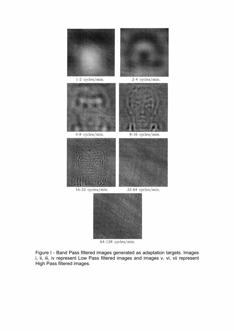
Figure I - Band Pass filtered images generated as adaptation targets. Images i, ii, iii, iv represent Low Pass filtered images and images v, vi, vii represent High Pass filtered images.

Figure II - Samples of Test Images. Set I represents an array of sharpened images and Set II represents an array of blurred images. An array of test images was generated by blurring or sharpening the original image over a range of magnitudes. The filtering was achieved by multiplying the original amplitude spectrum by ð-, where ð was the spatial frequency in cycles/face and the exponent - controlled the magnitude of change. Negative values of - progressively reduced the amplitude of higher frequencies and thus blurred the image, while positive values increased the amplitude with increasing frequencies and thus sharpened the images. The values of - varied from -0.50 to +0.50 in steps of 0.001 forming a series of 101 images. This allowed us to vary the blur magnitude in very fine steps. Once filtered, these images were again rescaled to have the same luminance and RMS contrast as the original image so that the degree of blur did not correlate with the image contrast. Figure II illustrates the array of test images. To examine the effects of adaptation, the test and the adapt images were displayed on a standard computer screen with a 12x16 uniform gray background. The images had a resolution of 256x256 pixels and 256 luminance levels. All were adjusted to have mean gray level of 100, equivalent to a mean luminance of approximately 10cd/m2. Observers viewed the screen at a distance of 100cms. At which the targets subtended a 4-degree visual angle. Subjects first viewed a Band-Pass filtered adapting image for a period of 120secs. After which, the test images were displayed for a period of 0.5secs. The test images displayed were interleveled with 6 seconds periods of readaptation duration of the staircase, with the test and adapt images separated by 0.25 seconds. The perceived focus was measured using an Alternative Forced Choice procedure (AFC staircase procedure). The observer used a pair of buttons to indicate whether the image appeared "too blurred" or "too sharp". If the subject responded, "too blurred", then the next presented image was sharpened or vice versa, so that over trials, the staircase converged at the - value at which the two responses were equally likely. This procedure continued for 9 reversals each in the responses of 2 randomly interleveled staircases, with the focus point estimated from the mean of the final 6 reversals points of each staircase. These procedures were similar to those used by Webster et al.,4 Tadmor and Tolhurst5 and Field and Brady.6

Similar procedure was followed for all the set of adapt images twice in order to ensure repeatability and reliability of the results. The influences of adaptation to each of the Band-Pass filtered images were qualitatively analyzed in 23 emmetropic observers (10 males and 13 females). Adaptation to any of the Band-Pass filtered images caused the original image to appear blurred and thus shifted the perceived focus to sharper images. The mean adaptation effects to each of these Band-Pass filtered images are tabulated in Figure III. Note that the values plotted show the change in the physical image blur required to null the change in perceived image blur, and thus are opposite in sign to the actual visual effect. Figure IV illustrates the adaptation after-effects for each adapt images. Adaptation effects were minimal for the Band-Pass filtered image of 32-64cycles/face followed by the 16-32-cycles/face image. However, the result should be interpreted with caution. Figure V illustrates the variations in the adaptation effects obtained for the 32-64 cycles/face image and the 16-32 cycles/face image. It is evident that the adaptation effects for the 32-64 cycles/face image was inconsistent when compared to the latter, corroborating the fact that the adaptation effects were minimal and more stable for the 16-32 cycles/face image. Figure IV - Graph illustrating mean adaptation effects
Band-pass filtered images Adaptation effects 64 - 128 cycles/face -0.07857 32 - 64 cycles/face -0.02811 16 - 31 cycles/face -0.07586 08 - 16 cycles/face -0.12086 04 - 08 cycles/face -0.12222 02 - 04 cycles/face -0.16332 01 - 02 cycles/face -0.15724
A strong negative correlation of -0.87 was obtained between the spatial frequencies and the adaptation effects allowing us come to a conclusion that adaptation was maximum for the least spatial

frequency image (1-2 cycles/face). However, the fact that the adaptation effects for the higher spatial frequencies (32-64 cycles/face and 64-128 cycles/face) were inconsistent and deviant should not be neglected.
A p-value of <0.001 proved the fact that adaptation effects for the different Band-Pass filtered images were statistically significant.
Figure V - Adaptation effects for 32-64 cycles/face and 16-32 cycles/face.
Discussion The role of high and low spatial frequencies on face perception is quite intrinsic. Our results, present only an overview of this complex physiology. Adaptation to either low or high spatial frequencies caused the original image to appear blurred with the adaptation effects being maximum for the low spatial frequencies. The results are probably due to the decrease in sensitivity for the adapted spatial frequency causing a shift in the perceived focus. In other words, adapting to a specific band-pass filtered image causes a reduction in sensitivity for that particular spatial frequency. This results in the equilibrium shifting towards the other set of spatial frequencies thereby altering the perceived focus. Also, a minimal adaptation to the mid-range spatial frequencies (16-32 cycles/face) was noted which can be rightly attributed to the equivalent sensitivity changes at the higher and lower spatial frequencies. These results are similar to those obtained by Webster et al.4
The mid range spatial frequencies (16-32cycles/face) are found to be the most sensitive for the recognition of face images and hand-written numerals8. Our study showed minimal adaptation to this range of spatial frequencies, thereby allowing us to conclude that mid-range spatial frequencies dominate judgment of faces. It is interesting to note that, Webster et al reported a shift in the perceived focus to opposite directions between the high and low spatial frequency

images.4 Our results contradict their findings. Our study reports that the adaptations to high spatial frequency images were inconsistent and the perceived focus was in the same direction as that of the low spatial frequency images. This result should be interpreted with caution. Campbell and Green7 found high frequency attenuation owing to optical aberrations and the finite size of the individual receptors. The pupil size, which is a major contributing factor for optical aberration was not standardized in our study. Further, our study population consisted of corrected emmetropes. The spectacles worn by the patient can alter the modulation transfer function. Thus, these two factors to an extent, can explain the inconsistency in the adaptation effects at the higher levels. Understanding the adaptation patterns has lead us to another debatable question. Where is the specific site of adaptation? This question is still left unanswered. Rolls et al9, found specific neurons in the superio-temporal sulcus in the cortex of monkeys that respond to high and low spatial frequencies. Can we thereby conclude that adaptation is a cortical phenomenon? High spatial frequencies are being conducted by the slow acting P-cells in the retina1. Does the inconsistency in the adaptation effects point out the poor adaptable properties of the P-cells, thus making adaptation a retinal phenomenon? Our results only hint at these probable selectivities. Further experiments should thus be performed to arrive at any conclusion. Acknowledgments We are indebted to Prof., Michael. A Webster, Director, Experimental Psychology, University of Nevada, Reno and late Dr. E Vaithiligam for their invaluable advise during the course of the study. References 1. F.W Campbell and J.G Robson, "Application of Fourier Analysis to the visibility of gratings." J. Physiol, 197:551-566. 2. Borish's Clinical Refraction, Edition I 3. R Nasanen, "Spatial frequency band width used in the recognition of facial images," Vision research, Nov. 39(23): pp. 3824-33, 1999. 4. M.A Webster, S.A Webster, J Macdonald and S.R Bharadwaj, "Adaptation to blur." In B.E Regowitz and T.N Pappas (Eds.) Human vision and Electronic imaging, SPIE, in press. 5. Y Tadmor and D.J Tolhurst, "Discrimination of changes in the second-order statistics of natural and synthetic images," Vision Research. 34: pp. 5541-554, 1994. 6. D Field and N Brady, "Visual sensitivity, blur and the sources of variability in the amplitude spectra of natural images," Vision Research. 23: pp. 3367-3383, 1997.

7. F.W Campbell and D.G Green, "Optical and retinal factors affecting visual resolution," J. Physiol. (London); 181: pp. 576-593, 1965. 8. R Nasanen and C O'Leary, "Recognition of Band-Pass filtered hand-written numerals in facial and peripheral vision," Vision Research. Dec (38) 23: pp. 3691-701, 1998. 9. E.T Rolls, G.C Baylis and C.M Leonard, "Role of low and high spatial frequencies in the face selective responses of neurons in the cortex in the superio-temporal sulcus in the monkey," Vision Research. 25(8): pp. 1021-35, 1985
AN APPEAL A lot of things in this world depend on money - security, shelter, education and even health. But at Nethralaya, money has ceased to be a pre-requisite for sight.Day after day, year after year, Nethralaya treats hundreds of patients absolutely free of cost and gives them back their sight. Treatment is provided free of cost to all patients with a monthly income below Rs.1,750/-. Yet there is no discrimination between the free patient and the one who pays. Apart from the treatment, food, medicines and travel expenses are absolutely free. Those free patients depend on Nethralaya, and Nethralaya depends on you. So, come and join the Ophthalmic Mission Trust. For questions about tax exempt status and contributions, please contact:
Mr S V Acharya,
Secretary and Treasurer Ophthalmic Mission Trust Inc. (OM Trust)
14613, Pommel Drive, Rockville, MD 20850, U.S.A.
Phone: (301)251 0378 INTERNET e-mail : [email protected], [email protected]
For those of you in India and elsewhere, please contact:
Dr S S Badrinath,
Chairman Sankara Nethralaya
(Unit of Medical Research Foundation) 18 College Road, Chennai 600 006
Phone: 826 1265, 827 1616 Fax: (044) 825 4180, 821 0117
INTERNET e-mail : [email protected] LOOK US UP ON THE WEB at http://www.sankaranethralaya.org
COME, GIVE THE GIFT OF SIGHT

Photodynamic Therapy (PDT) Anand Subramanyam and Mahesh P Shanmugam Intravascular dyes cause vascular occlusion by a photochemical reaction. In this way, isolated vessel occlusion may be achieved without severe collateral thermal injury to the nonvascular tissues (as in laser photocoagulation). Mechanism of action: Photodynamic compounds, which usually react with water to create oxygen and hydroxyl free radicals, are stimulated with a specific light wavelength; the intensity of this wavelength is low enough to spare the irradiated tissues from thermal damage. The free radicals, in turn, react with cell membranes of the endothelium and blood cells to induce massive platelet activation and thrombosis. Important variables in this reaction include the intravascular concentration of dye; the photochemical behavior of the dye; the interval from the injection to the onset of irradiation; and the intensity, specificity, and duration of the irradiation. Photodynamic therapy involves the following mechanism: Verteporfin competes with low-density lipoproteins (LDLs). Like tumor cells , vascular endothelial cells are rich in LDL receptors. When irradiated by non-thermal laser, the dye progresses from a stable ground state to an excited triplet state. This reaction releases oxygen free radicals & induces vascular occlusion by causing intraluminal damage. Agents used & Results: Researchers are developing several photosensitizers in animal models, such as phthalocyanines, rose bengal, chlorine, lutetium texaphyrin, tin ethyl etiopurpurin, and benzoporphyrin derivative.Only BPD is currently being used commercially. Liposome-encapsulated benzoporphyrin derivative (BPD)has undergone a phase III multicenter trial. BPD (verteporfin) is a modified porphyrin with an absorption maximum near 689 nm. BPD is cleared rapidly from the body, resulting in minimal skin sensitivity after 1-3 days.It is liposome-encapsulated to enhance solubility for IV administration. Very low laser energies are employed to release the dye from the liposomes and to stimulate the photodynamic action. Phase III data by the Treatment of Age-Related Macular Degeneration with Photodynamic Therapy (TAP) Study Group after 2 years showed that 59% of the verteporfin-treated eyes with predominantly classic CNV lost fewer than 15 letters of visual acuity at 24 months compared to 31% of eyes that received placebo. The percentage of verteporphin-treated eyes that experienced an improvement in vision remained at 13% over 2 years. It also

confirmed that fewer treatments were necessary with verteporphin (5.6) than with placebo (6.5). The mean no. of treatments decreased from 3.4 at 12 months to 2.2 at 24 months.In the sub-group who had minimally classic CNV-Visudyne In Minimally classic CNV (VIM) Trial, a clinically relevant difference in vision outcomes using verteporphin was identified if the lesion was no bigger than 4 Macular Photocoagulation Study (MPS) disc areas with a visual acuity of ³20/50. Phase II & III VIM trials will be initiated. Two-year results from the phase IIIb Verteporphin In Photodynamic therapy(VIP) Trial show that verteporphin therapy reduced the risk of moderate vision loss in patients with occult CNV with no classic CNV: 55% of treated patients lost ³3 lines as compared to 68% of who received placebo. The risk of severe vision loss was also less for the treated patients: 29% of treated patients lost ³6 lines as compared to 47% of who received placebo. The benefits were greater with smaller lesions or with lower levels of visual acuity. It shows that CNV from pathologic myopia also benefit: 86% of treated patients lost ³3 lines as compared to 67% of who received placebo. CONCLUSION: PDT is able to stabilize vision.Positive resu l t s have been ob ta ined in p redominan t l y c lass ic as we l l as m in ima l l y c lass ic & occu l t t ypes o f CNVs due to AMD.CNV due to d iseases inc lud ing pa tho log ic myop ia , ocu la r h is top lasmos is syndrome, ang io id s t reaks can a lso bene f i t ted .

Keratoconus associated with Cone-Rod Dystrophy - A case report Geetha K Iyer and Rajesh Fogla Introduction Keratoconus is known to be associated with a variety of ocular and systemic disorders. The common posterior segment disorders known to be associated with keratoconus are retinitis pigmentosa, macular coloboma, Leber's congenital amaurosis, retinal aplasia and retrolental fibroplasia.1-3 Occurrence of keratoconus in association with tapetoretinal degeneration is rare and has been reported infrequently.4,5 Visualization of the fundus is often difficult in cases of keratoconus due to the associated refractive error and corneal opacities. This may make it difficult for the ophthalmologist to clinically diagnose associated macular degenerative changes preoperatively. We report a case of keratoconus who was diagnosed to have cone-rod dystrophy following successful corneal transplantation. A 31 year old man presented to the outpatient clinic with complaints of sudden painless decrease of vision, associated with watering in the left eye for the past one month.
Fig 1. Slit lamp photograph showing a clear corneal graft in the left eye
There was no history of any recent trauma. The patient was a known high myope since 20 years and a contact lens wearer for the last 10 years. On examination, his best corrected visual acuity in the right eye was 6/60 and 1/60 in the left eye. Slit lamp biomicroscopy of both eyes revealed features suggestive of advanced keratoconus, with acute hydrops in the left eye. Fundus examination through the hazy cornea revealed pigmentary changes at the macula in both eyes. Following medical management of the hydrops in the left eye, best corrected visual acuity with contact lens did not improve beyond 6/60. He underwent corneal transplantion in the left eye one year later. Postoperatively despite a clear graft (Fig-1) his best corrected visual acuity in the left eye did not improve beyond 6/36. Keratomertry readings in the left eye were 48.25 x 128° / 44.12 x 38°. Fundus examination of the left eye revealed

minimal temporal pallor of the optic disc associated with pigmentary changes at the macula giving the appearance of bull's eye maculopathy. (Fig-2) Electroretinogram of both the eyes was done. Grossly delayed implicit time with reduced amplitudes of the
Fig 2. Fundus photograph of the left eye with bull's eye maculopathy
Fig 3. ERG ( scotopic / blue flash ) showing abnormal waveforms (reduced amplitude and delayed implicit time) in both eyes
rod response and extinguished cone waveforms were noted indicating cone rod dystrophy.(Fig-3&4) Colour blindness was also noted with the Ishihara's pseudoisochromatic color plates. Genetic counseling revealed it to be an isolated defect with negative family history. Comments: A correlation between keratoconus and tapetoretinal degeneration has been reported by Moschos and coworkers.4 Progressive cone dystrophy in association with keratoconus has also been reported in a 33 year old woman.5 Coexistence of these two conditions, especially photoreceptor dysfunction has prognostic implications on the outcome of corneal transplantation in these cases. High refractive error and corneal opacities in keratoconus, may often prevent visualization of the fundus. In our case a clear view of the fundus was not possible due to the corneal scarring which occurred following resolution of corneal hydrops. However following corneal transplantation, it was possible to make a clinical diagnosis of bull's eye maculopathy, which was confirmed to be cone rod dystrophy on electrophysiologic study.
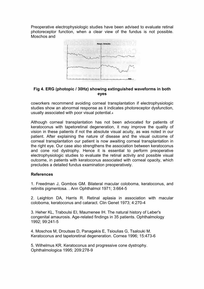
Preoperative electrophysiologic studies have been advised to evaluate retinal photoreceptor function, when a clear view of the fundus is not possible. Moschos and
Fig 4. ERG (photopic / 30Hz) showing extinguished waveforms in both eyes
coworkers recommend avoiding corneal transplantation if electrophysiologic studies show an abnormal response as it indicates photoreceptor dysfunction, usually associated with poor visual potential.4
Although corneal transplantation has not been advocated for patients of keratoconus with tapetoretinal degeneration, it may improve the quality of vision in these patients if not the absolute visual acuity, as was noted in our patient. After explaining the nature of disease and the visual outcome of corneal transplantation our patient is now awaiting corneal transplantation in the right eye. Our case also strengthens the association between keratoconus and cone rod dystrophy. Hence it is essential to perform preoperative electrophysiologic studies to evaluate the retinal activity and possible visual outcome, in patients with keratoconus associated with corneal opacity, which precludes a detailed fundus examination preoperatively. References 1. Freedman J, Gombos GM. Bilateral macular coloboma, keratoconus, and retinitis pigmentosa. . Ann Ophthalmol 1971; 3:664-5 2. Leighton DA, Harris R. Retinal aplasia in association with macular coloboma, keratoconus and cataract. Clin Genet 1973; 4:270-4 3. Heher KL, Traboulsi EI, Maumenee IH. The natural history of Leber's congenital amaurosis. Age-related findings in 35 patients. Ophthalmology 1992; 99:241-5 4. Moschos M, Droutsas D, Panagakis E, Tsioulias G, Tsalouki M. Keratoconus and tapetoretinal degeneration. Cornea 1996; 15:473-6 5. Wilhelmus KR. Keratoconus and progressive cone dystrophy. Ophthalmologica 1995; 209:278-9


Last Page
Telemedicine - What is it ? Rajesh Fogl Broadly defined, "Telemedicine" is the transfer of electronic medical data including high resolution images, sounds, video and patient records from one location to another. This transfer of medical data may utilize a variety of telecommunications technology like telephone lines, ISDN, internet and satellites. Current interactive telemedicine workstations provide the medical specialist with the ability to interact with the patient at the remote location through live audio and / or video. A multimedia patient record permits the reviewing physician to examine information from current or previous consultations, while continuing to interact with the patient. It also permits the reviewing physician to store information from the consultation for later review or for progress tracking. Worldwide, especially in the developing countries, people living in the rural and remote areas often have substandard access to specialty health care, primarily because specialists physician are more likely to be located in areas of concentrated population. For more than 30 years clinicians, health service providers, researchers and others have been investigating the use of advanced telecommunications and information technologies to improve health care. Innovations in computing and telecommunication technology has now made healthcare delivery possible when the patient and health care provider are geographically separated across town, across a state, or even across the world. Telemedicine attempts to provide access to the basic needs of timely and quality specialty medical care, to the people living in remote rural areas. Telemedicine is now utilized by health providers in a growing number of medical specialties, including but not limited to - dermatology, oncology, radiology, surgery, cardiology, psychiatry, and home health care. Early applications of telemedicine often focused on remote populations scattered across mountainous areas, islands, open plains, and arctic regions where medical specialists and some times primary care practitioners were not easily reached. Most of the telemedicine projects from the 1960s through the early 1980s failed, as telecommunications costs tended to be high, and the technologies were awkward to use. The National Aeronautics and Space Administration (NASA) provided most of the technology and funding for most of the early telemedicine projects. STARPAHC (Space Technology Applied to Rural Papago Advanced Health Care) was one of the earliest endeavors in telemedicine. Its goals were to provide general medical care to the Papago Reservation. A van carrying two paramedics and a variety of medical instruments was linked to the Public Health Service hospital and another hospital with specialists by a two-way
microwave telemedicine and audio transmission. In 1989 NASA conducted the first international telemedicine program. United States offered medical consultation from several medical centers in the U.S. to the earthquake hit Soviet Republic of Armenia. Telemedicine consultations were conducted using one-way video, voice, and facsimile between a medical center in Yerevan, Armenia and four medical centers in the U.S. The program was extended to Ufa, Russia to facilitate burn victims after a terrible railway accident. This project demonstrated that medical consultation could be conducted over satellite network crossing political, cultural, social and economic borders. The last decade has seen a steady increase in the number of telemedicine projects internationally, mainly as a result more government funding and improved technology. Telemedicine offers a mechanism for centralizing specialists and supporting primary care clinicians. Some academic medical centers and other organizations, faced with reduced revenues and even exclusion from local managed care networks, are exploring telemedicine as they seek to develop new regional, national, and international markets for their highly specialized clinicians. Teleradiology appears to be the most common application, in part because Medicare and other payers reimburse for radiology consultations without demanding the face-to-face relationship required for most other consultations. Telemedicine has a variety of applications in patient care, education, research, administration and public health. Use of telemedicine can significantly reduce the time and costs of patient transportation. Telemedicine permits physicians doing clinical research to be linked together despite geographical separation. It also improves medical education for rural health care professionals. Telemedicine has captured the interest of the medical community, the government, and the public as a cost-effective means of improving access to quality healthcare. In rural areas, where specialized medical care may be unavailable, telemedicine can have an especially large impact on the quality and speed of patient care. However despite recent growth, obstacles to widespread use of clinical telemedicine persist. While many people acknowledge the practical advantages of telemedicine, not everyone is at ease with the transmission of confidential patient data over electronic networks. Many groups are working to develop hardware and software standards, to put together systems in which the components operate predictably and smoothly in different settings without extensive adaptation. Telemedicine has numerous and ever expanding applications to reduce the burdens of inferior health care access through utilization of technology.

PLEASE LOOK US UP ON THE WEB at http://www.sankaranethralaya.org
Please forward your correct address with your E-mail address if any, to enable us to update our address data base.
This issue of Insight is sponsored by : Rampion Eyetech Pvt. Ltd., Kalash, New Sharda Mandir Road, Paldi, Ahmedabad - 380 007 Apex Laboratories Pvt. Ltd., 44, Gandhi Mandapam Road, Kotturpuram, Chennai - 600 085 Pharmacia & Upjohn India Pvt. Ltd., SCO 27, Sector 14, Gurgaon 122 001, Haryana Medical & Vision Research Foundations thank the above sponsors for their generosity.
Editor : Dr. Mahesh P Shanmugam
For Private Circulation Only