medical review - dlv.org.rsdlv.org.rs/wp-content/uploads/2017/06/medicinskipregled5-6-2017... ·...
TRANSCRIPT
Publishing Sector of the Society of Physicians of Vojvodina of the Medical Society of Serbia, Novi Sad, Vase Stajica 9,
MEDICAL REVIEW
JOURNAL OF THE SOCIETY OF PHYSICIANS OF VOJVODINA OF THE MEDICAL SOCIETY OF SERBIA
THE FIRST ISSUE WAS PUBLISHED IN 1948
Editor-in-ChiefLJILJA MIJATOV UKROPINA
Assistant to the Editor-in-Chief for Clinical Branches: PETAR SLANKAMENACAssistant to the Editor-in-Chief for Imaging Methods: VIKTOR TILL
Assistants to the Editor-in-ChiefBOJANA KRSTONOŠIĆ
ŽELJKO ŽIVANOVIĆ
EDITORIAL BOARD
Proof-reading for Serbian Language: Dragica PantićProof-reading for English Language: Marija Vučenović
Technical Secretary: Vesna ŠaranovićTechnical Support: ”Grafit” Novi Sad
UDC and descriptors prepared by: the Library of the Faculty of Medicine, Novi Sad
MEDICAL REVIEW is published bimonthly (six issues per year) with a circulation of 1.000 co¬pies. The annual payment fee in 2017, for individuals from the territory of Serbia, is 3,000.00 dinars (the value-added tax included), 4,000.00 dinars for individuals from Serbia who are not members of the Society of Physicians of Vojvodina of the Medical Society of Serbia, 60 Euros for members outside the territory of Serbia, and 8,000.00 dinars (+ VAT) for institutions. The payment account is: 340-1861-70 or 115-13858-06, ”Annual membership fee for Medical Review”.
Copyright ® Društvo lekara Vojvodine Srpskog lekarskog društva Novi Sad 1998
The manuscripts can be submited on the web-page: aseestant.ceon.rs/index.php/medpreg/.Address Editorial: Društvo lekara Vojvodine Srpskog lekarskog društva, 21000 Novi Sad, Vase Stajića 9,
Tel. 021/521-096; 063/81 33 875, E-mail: [email protected]; Web: www.dlv.org.rs
OKAN AKHAN, AnkaraANDREJ ALEKSANDROV, BirminghamSTOJANKA ALEKSIĆ, HamburgVLADO ANTONIĆ, BaltimorITZHAK AVITAL, BethesdaKAREN BELKIĆ, StockholmJEAN-PAUL BEREGI, Lille CedexHELENA BERGER, LjubljanaMILAN BREBERINA, Novi SadRADOVAN CVIJANOVIĆ, Novi SadVLADIMIR ČANADANOVIĆ, Novi SadIVAN DAMJANOV, Kansas CityDRAGAN DANKUC, Novi SadOMER DEVAJA, MeidstonePETAR DRVIŠ, SplitTATJANA ĐURĐEVIĆ-MIRKOVIĆ, Novi SadVERA GRUJIĆ, Novi SadIRENA HOČEVAR BOLTEŽAR, LjubljanaMARINA JOVANOVIĆ, Novi SadDRAGAN KATANIĆ, Novi SadALEKSANDAR KIRALJ, Novi SadDRAGAN KOVAČEVIĆ, Novi SadDUŠKO KOZIĆ, Novi SadDUŠAN LALOŠEVIĆ, Novi SadJORGE MANUEL COSTA LAINS, CoimbraVELJKO MARIĆ, FočaSMILJANA MARINKOVIĆ, Novi Sad
VLADIMIR MARTINEK, Bad AiblingSINIŠA MASLOVARA, OsijekJASNA MIHAILOVIĆ, Novi SadLJILJA MIJATOV UKROPINA, Novi SadMIROSLAV MILANKOV, Novi SadIGOR MITIĆ, Novi SadNADA NAUMOVIĆ, Novi SadALEKSANDRA NOVAKOV MIKIĆ, Novi SadAVIRAM NISSAN, Ein KaremJANKO PASTERNAK, Novi SadLJUBOMIR PETROVIĆ, Novi SadMIHAEL PODVINEC, BaselJOVAN RAJS, DanderydPETAR E. SCHWARTZ, New HavenMILAN SIMATOVIĆ, Banja LukaTOMAŠ SKRIČKA, BrnoPETAR SLANKAMENAC, Novi SadEDITA STOKIĆ, Novi SadALEXANDER STOJADINOVIĆ, Glen AlenGORAN STOJILJKOVIĆ, Novi SadVIKTOR TILL, Novi SadTIBOR TOT, FalunTAKASHI TOYONAGA, KobeKONSTANTIN VIKTOROVIĆ SUDAKOV, MoskvaNADA VUČKOVIĆ, Novi SadZORAN VUJKOVIĆ, Banja LukaPETAR VULEKOVIĆ, Novi Sad
Printed by: Department for Joint Affairs of Provincial Authorities - Sector for Printing

Izdavačka delatnost Društva lekara Vojvodine Srpskog lekarskog društva, Novi Sad, Vase Stajića 9
MEDICINSKI PREGLED
ČASOPIS DRUŠTVA LEKARA VOJVODINE SRPSKOG LEKARSKOG DRUŠTVAPRVI BROJ JE ŠTAMPAN 1948. GODINE.
Glavni i odgovorni urednikLJILJA MIJATOV UKROPINA
Pomoćnik urednika za kliničke grane: PETAR SLANKAMENACPomoćnik urednika za imidžing metode: VIKTOR TILL
Pomoćnici urednika: BOJANA KRSTONOŠIĆ
ŽELJKO ŽIVANOVIĆ
REDAKCIJSKI ODBOR
Lektor za srpski jezik: Dragica PantićLektor za engleski jezik: Marija Vučenović
Tehnički sekretar: Vesna ŠaranovićTehnička podrška: „Grafit”, Novi Sad
Izrada UDK i deskriptora: Biblioteka Medicinskog fakulteta, Novi Sad
MEDICINSKI PREGLED izlazi dvomesečno (šest dvobroja godišnje), u tiražu od 1000 primeraka. Pretplata za pojedince sa teritorije Srbije za 2017. godinu iznosi 3.000,00 dinara (sa uračunatim PDV-om), a 4.000,00 dinara za pojedince iz Srbije koji nisu članovi DLV-SLD, 60 eura za članove van Srbije, a za ustanove 8.000,00 dinara (uz dodavanje PDV-a). Uplate se vrše na račun broj 340-1861-70 ili 115-13858-06, s naznakom „Dodatna članarina za Medicinski pregled”.
Copyright ® Društvo lekara Vojvodine Srpskog lekarskog društva Novi Sad 1998.
Prijem rukopisa vrši se u elektronskoj formi na stranici: aseestant.ceon.rs/index.php/medpreg/. Adresa Redakcije: Društvo lekara Vojvodine Srpskog lekarskog društva,
21000 Novi Sad, Vase Stajića 9, Tel. 021/521-096; 063/81 33 875E-mail: [email protected]; Web: www.dlv.org.rs
OKAN AKHAN, AnkaraANDREJ ALEKSANDROV, BirminghamSTOJANKA ALEKSIĆ, HamburgVLADO ANTONIĆ, BaltimorITZHAK AVITAL, BethesdaKAREN BELKIĆ, StockholmJEAN-PAUL BEREGI, Lille CedexHELENA BERGER, LjubljanaMILAN BREBERINA, Novi SadRADOVAN CVIJANOVIĆ, Novi SadVLADIMIR ČANADANOVIĆ, Novi SadIVAN DAMJANOV, Kansas CityDRAGAN DANKUC, Novi SadOMER DEVAJA, MeidstonePETAR DRVIŠ, SplitTATJANA ĐURĐEVIĆ-MIRKOVIĆ, Novi SadVERA GRUJIĆ, Novi SadIRENA HOČEVAR BOLTEŽAR, LjubljanaMARINA JOVANOVIĆ, Novi SadDRAGAN KATANIĆ, Novi SadALEKSANDAR KIRALJ, Novi SadDRAGAN KOVAČEVIĆ, Novi SadDUŠKO KOZIĆ, Novi SadDUŠAN LALOŠEVIĆ, Novi SadJORGE MANUEL COSTA LAINS, CoimbraVELJKO MARIĆ, FočaSMILJANA MARINKOVIĆ, Novi Sad
VLADIMIR MARTINEK, Bad AiblingSINIŠA MASLOVARA, OsijekJASNA MIHAILOVIĆ, Novi SadLJILJA MIJATOV UKROPINA, Novi SadMIROSLAV MILANKOV, Novi SadIGOR MITIĆ, Novi SadNADA NAUMOVIĆ, Novi SadALEKSANDRA NOVAKOV MIKIĆ, Novi SadAVIRAM NISSAN, Ein KaremJANKO PASTERNAK, Novi SadLJUBOMIR PETROVIĆ, Novi SadMIHAEL PODVINEC, BaselJOVAN RAJS, DanderydPETAR E. SCHWARTZ, New HavenMILAN SIMATOVIĆ, Banja LukaTOMAŠ SKRIČKA, BrnoPETAR SLANKAMENAC, Novi SadEDITA STOKIĆ, Novi SadALEXANDER STOJADINOVIĆ, Glen AlenGORAN STOJILJKOVIĆ, Novi SadVIKTOR TILL, Novi SadTIBOR TOT, FalunTAKASHI TOYONAGA, KobeKONSTANTIN VIKTOROVIĆ SUDAKOV, MoskvaNADA VUČKOVIĆ, Novi SadZORAN VUJKOVIĆ, Banja LukaPETAR VULEKOVIĆ, Novi Sad
Štamparija: Uprava za zajedničke poslove pokrajinskih organa - Odsek za poslove štamparije

M E D I C A L R E V I E WJOURNAL OF THE SOCIETY OF PHYSICIANS OF VOJVODINA OF THE MEDICAL SOCIETY OF SERBIA
Novi Sad Vase Stajića 9 Serbia
Med Pregl 2017; LXX (5-6): 133-192. Novi Sad: May-June.
CONTENTS
EDITORIAL
Ksenija Bošković and Snežana Tomašević TodorovićTHERAPEUTIC EFFECTS OF PHYSICAL AGENTS IN THE TREATMENT OF CHRONIC PAIN .................................
ORIGINAL STUDIES
Sonja Apostolska, Elizabeta Gjorgievska, Vasilka Rendžova, Marina Eftimoska, Rade Živković and Ivica StančićADAPTABILITY OF DIFFERENT CANAL SEALERS TO THE ROOT CANAL DENTIN – SCANNING ELECTRON MICROSCOPY ANALYSIS ..............................................................................................................................................................
Maja Bogdan, Rajko Jović and Tanja ArbutinaTHE QUALITY OF VOICE AND SPEECH BEFORE AND AFTER SURGICAL TREATMENT OF BILATERAL RE-CURRENT LARYNGEAL NERVE PARALYSIS ...........................................................................................................................
Vedrana Karan, Aleksandra Rakovac, Mladen Karan, Milan Popović, Jelena Klašnja and Damir LukačEVALUATION OF BODY COMPOSITION AND MUSCULAR STRENGTH IN DIFFERENT SPORTS..........................
PROFESSIONAL ARTICLES
Stanislava Nikolić, Nikola Ćurić, Romana Mijović, Branislava Ilinčić and Damir BencSIGNIFICANCE AND ROLE OF HOMEOSTATIC MODEL ASSESSMENT IN THE EVALUATION OF GLUCOSE REGULATION MECHANISMS ................................................................................................................................................................
Slađana J. Vasiljević and Aleksandra T. CvetkovićMONITORING THE QUALITY OF ORAL HEALTH AMONG THE POPULATION OF SCHOOLCHILDREN ..........
CASE REPORTS
Larisa Dizdarević Hudić, Zumreta Kušljugić, Irma Bijedić and Igor HudićSYNCOPE DUE TO SINUS NODE DYSFUNCTION AFTER SURGICAL PATCH CLOSURE OF ATRIAL SEPTAL DEFECT – A CASE REPORT ..........................................................................................................................................................
Samir Delibegović, Edvin Mulalić and Sejo ButurovićFOREIGN BODY INGESTION – GLASS IN COLON AND RECTUM – A CASE REPORT AND LITERATURE REVIEW
Anja Stojšin, Vedrana Petrić, Grozdana Čanak, Vesna Turkulov, Siniša Sević and Maja RužićRHABDOMYOLYSIS AND INFLUENZA A (H3N2) INFECTION – A CASE REPORT ......................................................
SEMINAR FOR PHISICIANS
Vojislav Stanojević and Marija JevtićDIETARY REGIMENS FOR PERSONS WITH TYPE 2 DIABETES: RECOMMENDATIONS, ISSUES AND POTEN-TIAL SOLUTIONS .............................................................................................................................................................................
HISTORY OF MEDICINE
Rade R. Babić, Ankica Jelenković, Gordana Stanković Babić, Strahinja R. Babić and Nevena R. BabićBRANISLAV NUŠIĆ AND X–RAYS IN THE STORY “ROENTGEN’S PHOTOGRAPHY” .................................................
137-140
141-145
146-149
150-154
155-161
162-165
167-169
170-172
173-175
177-182
183-188

M E D I C I N S K I P R E G L E DČASOPIS DRUŠTVA LEKARA VOJVODINE SRPSKOG LEKARSKOG DRUŠTVA
Novi Sad Vase Stajića 9 Srbija
Med Pregl 2017; LXX (5-6): 133-192. Novi Sad: maj-juni.
SADRŽAJ
UVODNIK
Ksenija Bošković i Snežana Tomašević TodorovićMEHANIZAM DEJSTVA FIZIKALNIH AGENASA U TERAPIJI HRONIČNOG BOLA .........................................................................
ORIGINALNI NAUČNI RADOVI
Sonja Apostolska, Elizabeta Gjorgievska, Vasilka Rendžova, Marina Eftimoska, Rade Živković i Ivica StančićADAPTABILNOST RAZLIČITIH KANALNIH PUNЈENЈA ZA DENTIN KORENSKOG KANALA – ANALIZA ELEKTRONSKI SKENIRANIH MIKROFOTOGRAFIJA ...........................................................................................................................................................
Maja Bogdan, Rajko Jović i Tanja ArbutinaKVALITET GLASA I GOVORA PRE I NAKON HIRURŠKOG LEČENJA OBOSTRANE TRAJNE ODUZETOSTI POVRATNOG GRKLJANSKOG ŽIVCA .....................................................................................................................................................................................
Vedrana Karan, Aleksandra Rakovac, Mladen Karan, Milan Popović, Jelena Klašnja i Damir LukačPROCENA TELESNOG SASTAVA I MIŠIĆNE SNAGE KOD RAZLIČITIH SPORTOVA ...........................................................................
STRUČNI ČLANCI
Stanislava Nikolić, Nikola Ćurić, Romana Mijović, Branislava Ilinčić i Damir BencZNAČAJ I UPOTREBA PARAMETARA MODELA HOMEOSTAZE U PROCENI GLIKOREGULATORNIH MEHANIZAMA .........
Slađana J. Vasiljević i Aleksandra T. CvetkovićPRAĆENJE KVALITETA ORALNOG ZDRAVLJA KOD POPULACIJE ŠKOLSKE DECE ..........................................................................
PRIKAZI SLUČAJEVA
Larisa Dizdarević Hudić, Zumreta Kušljugić, Irma Bijedić i Igor HudićSINKOPA ZBOG DISFUNKCIJE SINUSNOG ČVORA NAKON HIRURŠKOG ZATVARANJA ATRIJALNOG SEPTALNOG DE-FEKTA – PRIKAZ SLUČAJA .............................................................................................................................................................................
Samir Delibegović, Edvin Mulalić i Sejo ButurovićPROGUTANA STRANA TELA – STAKLO U KOLONU I REKTUMU – PRIKAZ SLUČAJA I PREGLED LITERATURE...............................
Anja Stojšin, Vedrana Petrić, Grozdana Čanak, Vesna Turkulov, Siniša Sević i Maja RužićRABDOMIOLIZA I INFLUENZA A (H3N2) INFEKCIJA – PRIKAZ SLUČAJA ..........................................................................................
SEMINAR ZA LEKARE U PRAKSI
Vojislav Stanojević i Marija JevtićISHRANA OSOBA SA DIJABETESOM TIPA 2: PREPORUKE, PROBLEMI I MOGUĆA REŠENJA ........................................................
ISTORIJA MEDICINE
Rade R. Babić, Ankica Jelenković, Gordana Stanković Babić, Strahinja R. Babić i Nevena R. BabićBRANISLAV NUŠIĆ I X–ZRACI U PRIČI “RENTGENOVA FOTOGRAFIJA“ ............................................................................................
137-140
141-145
146-149
150-154
155-161
162-165
167-169
170-172
173-175
177-182
183-188

Errata
At the request of Prof. Dr. Sandra Stefan Mikić, Ph. D., author of the paper “ANTIMICROBIAL SUSCEPTI-BILITY PATTERN OF ACINETOBACTER SPP IN THE PERIOD 2012 - 2015”, published in the journal Medi-cal Review, 3 - 4/2017, pages 99 – 106, we hereby provide the correct list of coauthors of this paper: Sandra Stefan-Mikić, Siniša Sević, Ivana Hrnjaković Cvjetković, Vesna Milošević, Vedrana Petrić and Milica Rupar.
Acknowledgment - This work was supported and funded by the Ministry of Education, Science and Techno-logical Development of the Republic of Serbia (grant TR31084).
Na molbu prof. dr Sandre Stefan Mikić, autorke rada „Ispitivanje osetljivosti Acinetobacter spp na antimikrobne lekove u periodu 2012−2013. godine“, objavlјenog u dvobroju 3-4/2017, na stranama 99−106, objavlјujemo ispravke koje se odnose na koautore rada, a koji glasi: Sandra Stefan Mikić, Siniša Sević, Ivana Hrnjaković Cvjetković, Vesna Milošević, Vedrana Petrić i Milica Rupar, kao i na Zahvalnicu koja je izostavlјena, a treba da glasi: Ac-knowledgments - This work was supported by project grants TR31084 funded by the Ministry of Education, Sci-ence and Technological Development of the Republic of Serbia
*****
At the request of Dr. Uroš Milošević, author of the article “CENTRAL PANCREATECTOMY IN SURGICAL TREATMENT OF PANCREATIC INSULINOMA”, published in the journal Medical Review, 3 - 4/2017, pages 111 – 114, we hereby provide the correct email address of the author: [email protected].
Na molbu dr Uroša Miloševića, autora rada „Centralna pankreatektomija u hirurškom tretmanu insulinoma pankreasa – prikaz slučaja“, objavlјenog u dvobroju 3-4/2017, na stranama 111−114. objavlјujemo ispravku koja se odnosi na imejl adresu autora, a koja bi trebalo da glasi [email protected].

Med Pregl 2017; LXX (5-6): 137-140. Novi Sad: maj-juni. 137
Chronic pain is a physiological response to tissue damage, often associated with emotional reaction, somatic and autonomic disorders, and changes in behavior [1]. Therefore, it requires a multidiscipli-nary approach among which physical agents have a very important place [2, 3]. In order to apply ade-quate physical agents in the treatment of chronic
pain, it is necessary to accurately determine pain characteristics, as well as to know the physiological mechanisms of action of the applied physical agent (Table 1) [4].
The explanation of therapeutic effects of physical agents is based on the theory of control and modulation of painful impulses at: 1) peripheral level (mechanosen-
EDITORIALUVODNIKClinical Center of Vojvodina, Novi Sad EditorialDepartment of Medical Rehabilitation UvodnikUniversity of Novi Sad, Faculty of Medicine, Novi Sad UDK 616.8-009.7-036.1:615.8 https://doi.org/10.2298/MPNS1706137B
THERAPEUTIC EFFECTS OF PHYSICAL AGENTS IN THE TREATMENT OF CHRONIC PAIN
MEHANIZAM DEJSTVA FIZIKALNIH AGENASA U TERAPIJI HRONIČNOG BOLA
Ksenija BOŠKOVIĆ and Snežana TOMAŠEVIĆ TODOROVIĆ
Corresponding Author: Prof. dr Ksenija Bošković, Klinički centar Vojvodine, Klinika za medicinsku rehabilitaciju,21000 Novi Sad, Hajduk Veljkova 1-7, E-mail: [email protected]
Table 1. Potentials of physical therapy effects on chronic painTabela 1. Mogućnosti delovanja fizikalne terapije na hronični bol
ProcessProces
Therapeutic mechanismTerapijski mehanizam
Therapeutic interventionsTerapijske intervencije
TransductionTransdukcija
Damage reductionRedukcija oštećenja
Nociorector excitability decreaseSmanjenje ekscitabilnosti nociorectora
Protective positions/Zaštitni položajiExercises reducing joint stress and re-
newing the function/Vežbe koje redukuju zglobni stres i obnavljaju funkciju
Cryotherapy/Krioterapija Small power laser/Laser male snage
Ultrasound/UltrazvukElectrotherapy/Elektroterapija
TransmissionTransmisija
Competition (sensor receptors stimuli with faster conduc-tion of „gate control“). Transmission decrease (decrease of
hyperexcitability at the level of spinal ganglia and back horns of spinal cord)/Kompeticija (nadražaj senzornih receptora sa bržim provođenjem kroz „kontrolne kapije signala“). Sman-jenje transmisije (smanjenje hiperekscitabilnosti na nivou
spinalnih ganglija i zadnjih rogova kičmene moždine)
TENS/TENSMassage/Masaža
Heat/ToplotaLaser/Laser
Acupuncture/Akupunktura
PerceptionPercepcija
Cognitive insight/Kognitivni uvidMotivation-emotional processes
Motivaciono-emocionalni procesi
Information/InformacijaEducation/Edukacija
Support/PodrškaModulationModulacija
Strengthening of pain control descedent mechanismsJačanje descendentnih mehanizama kontrole bola
Information/InformacijaEducation/Edukacija
Support/PodrškaExercises/Vežbe
Electrotherapy/ElektroterapijaAcupunture/Akupunktura
TENS - transcutaneous electrical nerve situmulation/transkutana električna nervna stimulacija

Bošković K, et al. Physical Agents in the Treatment of Chronic Pain 138
sitive, thermosensitive and chemosensitive pain recep-tors); 2) spinal level (segmental – presynaptic system); 3) supra-spinal (central blocking system) [5, 6]. The analgesic effects of physical therapy may be expressed on afferent or efferent segments of the consignment spinal system. In order to explain afferent analgesia, the theory most often used is “the gate control“ i.e. the signal input gate control theory. The stimulation of pe-ripheral thick fast-conducting nerve fibers activates dorsal horn neurons before signals arrive via Aδ and C fibers that cannot additionally activate them (the gate has been closed). It is considered that most physical modalities achieve their effects in this way [7, 8].
The efferent analgesic system is complex. The voluntary stimuli from the cerebral cortex (sugges-tion, biofeedback and other cognitive demands) in-crease the process of release of the matter decreasing the pain. Those are endogenous opiates (enkephalins, endorphins) of the middle brain which decrease neu-rotransmitter release in afferent path of pain through the downstream paths, directly or indirectly, over se-rotonin to distal part of the first neurone of afferent path of pain, by presynaptic inhibition over µ and partly kappa receptors. This inhibited state of the sec-ond neuron in the afferent path decreases the pain [9].
Physical therapy recognizes the term “fatigue process of the Aδ afferent fibers“ through axonal blockage (repetitive, high-frequency stimuli) as well as neuropeptide transmitters release (direct and modulated current) followed by pain transfer block-age on the peripheral and spinal level. Hyperstimu-lation analgesia and analgesia via application of transcutaneous electrical nerve stimulation (TENS), in addition to the already mentioned mechanisams of action, also act at supraspinal level, inhibiting the pain transfer up to conscious level at the somato-sensory cerebral cortex [10, 11].
Therapeutic effects of certain physical agents
Kinesitherapy is a therapy based on movement and exercise used in the management of chronic pain. Effects of this physical agent are explained by seg-mental blockage activation, suprasegmental blockage activation, as well as by the psycho-tonus increase with the characteristics of joy, independence, com-munication and feeling of security [7, 11, 12].
Galvanic current, named after Luigi Galvani, is a direct current where iones move constantly, with the same intensity in one direction. The analgesic effects of direct current are explained by effects of iones on C afferent nerve fibers, as well as by ap-plication of different ionic content, while neuro-modulatory effect is explained by release of neu-ropeptide transmitters [3, 9].
Electrophoresis (iontophoresis) in the chronic pain therapy is the use of galvanic current to intro-duce analgesic ions into the organism through non-
injured skin or mucosa. Aqueous solution medicine is inserted from electrode that has the same charge as the active component of the medicine. The anal-gesic effect is explained by the combination effects of the galvanic current and the given medicine.
Diadynamic currents, or Bernard’s currents, which belong to the group of low frequency direct currents, may be semi-wave and full-wave sinusoi-dal currents, 50 - 100 Hz frequency. These currents are the combination of 5 differently modulated im-pulses by their intensity, shape and amplitude (im-pulse one) and galvanic component: DF (diphase fixe), MF (monophase fixe), CP (courte “short” pe-riod), LP (long periods), RS (rhythmic syncope). By combining diadynamic currents of different modu-lation, we can enhance the desired effect even more, firstly in the form of analgesia, but also in vasodil-atation, sympaticolytic effect or the possibility of muscle contraction [9, 10, 13].
Transcutaneous electrical nerve stimulation (TENS) is a basic physical therapeutic procedure for pain modulation and its effects are based on the pain entrance gate control theory and pain neuro-humoral modulation, via endogenous opiate system. TENS is the use of low-frequency currents with rectangular impulses of certain duration; analgesia is achieved with electric stimulation of neuron in-hibitory system of the spinal cord last horn, via de-scending paths mediated by opiates (endorphins and enkephalins) [3, 9].
Interference currents or Nemec’s currents are produced by 2 alternating currents of middle-fre-quencies, but more painlessly and deeply than each would individually; deep in the tissue, low-frequen-cy alternating current is generated. In this way, the skin resistance is avoided, as well as discomfort. Interference currents act in an analgesic way and longer than other types of currents, on local in-crease of tissue blood supply and edema reduction, accelerate bone healing after fracture, stimulate recovery of the injured nerves, etc. [9, 13].
High-frequency current is applied as short-wave and micro-wave diathermy, with the purpose to heat deep muscular tissues. It belongs to alternating cur-rents where dipoles change their orientation, tend to be positioned in direction of circuit flow and friction between dipole molecules and viscous environment heats the tissue. The analgesic effect of diathermy is explained by thermal conditioning of mesencephalic blockage and in humoral way - via thermal/stress reaction and excretion of cortisol [9, 10].
Cryotherapy is the use of low temperatures for therapeutic purposes. The analgesic effect is achieved through metabolic activities, slowing down nerves conduction in the treated region, decreasing the activ-ity of the inflammatory mediator, as well as of en-zymes responsible for destructive changes in some inflammatory rheumatic diseases [3, 9].
Heat therapy is mostly applied in the chronic pain therapy by application of surface and deep methods of heating. Heating causes vasodilation
AbbreviationsTENS – transcutaneous electrical nerve stimulation

Med Pregl 2017; LXX (5-6): 137-140. Novi Sad: maj-juni. 139
and relaxation of muscles in the treated area. In this way oxygenation is increased, as well as accelera-tion of cellular metabolism. The initial feeling of a mild heat may have analgesic effects and results in decrease of local soreness and muscle spasm [9].
Hydrotherapy is one of the oldest methods of physical therapy; it is the use of water, especially thermal mineral water in order to treat different dis-eases. The therapeutic effect is reached by the com-bination of physical characteristics with heat, chem-ical and mechanical action, and also by the combina-tion with exercises in the water. The analgesic effect of hydrotherapy is explained by the theory of the pain entrance gate control and segmental competitive blockage through the thermal-receptor stimulation, while humoral effect can be seen through heat and cold stress, i.e. through cortisol secretion [3].
Laser therapy - low level laser therapy is the use of red and infrared laser rays, i.e. stimulated light fotons on the region treated. The analgesic effect of laser therapy is explained by the mechanism of clos-ing the entrance gate on ascendant path, with seg-mental competitive blockage through activation of peripheral axonal blockage i.e. blockage of A-d af-ferent neural fibers; the neuromodulatory effect of laser therapy is explained by inhibition of inflam-matory mediators formation [9, 14].
Sonotherapy is a method of using ultrasound at frequency higher than 20 kHz, directed by applica-tor to the certain region of the body where sound oscilations are transmitted into mechanical vibra-tions. The analgesic effect is explained by the influ-ence on the painful pulses, via activation of mesen-cephalic system of blockade, while humoral effect
is explained by heat reaction and reduction of algo-genic substances at the level of receptors [3, 9].
Ultrasonophoresis is a treatment where active ingredients are pushed through non-injured skin by ultrasound. The analgesic effect is explained by the combination of the galvanic current and given med-ication effect.
Magnotherapy is the application of constant or impulse magnetic field of low or high frequency. It affects chemical and physiological processes in the organism, acting at a molecular level; it changes the activity of neural and endocrine systems that rep-resent the main regulatory systems in the organism, influencing metabolism, microcirculation and the blood content. This way, both the analgesic and anti-edematous effect are explained [9, 10].
Acupuncture, as part of the traditional Chinese medicine and a special philosophical approach to the human body and spirit, has been succesfully applied as an additional method in the chronic pain therapy. The neural thalamic network is the gate towards the cortex and, in that way, cortical projec-tions influence the analysis of all signals going through the thalamus. Also, the existance of interac-tion between somatosensory regions, close to the thalamus, has been found [15].
Conclusion
Adequate pain asessment, associated with good knowledge of therapeutic effects of physical agents, and a multimodal approach to the treatment of pain, provide expected results in the management of chronic pain.
References
1. Jensen M, Ehde D, Day MA. The behavioral activation and inhibition systems: implications for understanding and treating chronic pain. J Pain. 2016;17(5):529.e1-18.
2. Scascighini L, Toma V, Dober-Spielmann S, Sprott H. Multidisciplinary treatment for chronic pain: a systematic review of interventions and outcomes. Rheumatology. 2008;47(5):670-8.
3. Rakel B, Barr JO. Physical modalities in chronic pain man-agement. Nurs Clin North Am. 2003;38(3):477-94.
4. Kumar SP, Saha S. Mechanism-based classification of pain for physical therapy management in palliative care: a clinical com-mentary. Indian J Palliat Care. 2011;17(1):80-6.
5. Harden RN, Bruehl SP. Diagnostic criteria: the statistical derivation of the four criterion factors. In: Wilson PR, Stanton-Hicks M, Harden RN, editors. CRPS: current diagnosis and ther-apy progress in pain research and management. Seattle: IASP Press; 2005. p. 45-58.
6. Minor MA, Sanford MK. The role of physical therapy and physical modalities in pain management. Rheum Dis Clin North Am. 1999;25(1):233-48.
7. van Middelkoop M, Rubinstein SM, Kuijpers T, Verhagen AP, Ostelo R, Koes BW, et al. A systematic review on the effec-tiveness of physical and rehabilitation interventions for chronic non-specific low back pain. Eur Spine J. 2011;20(1):19-39.
8. Slater H, Sluka K, Söderlund A, Watson PJ. IASP Curriculum outline on pain for physical therapy. International Association for the Study of Pain; c2015 [cited 2016 Dec 25]. Available from: http://www.orthopt.org/uploads/content_files/CSM_2013/Handouts/IASP_Curriculum_Outline_on_Pain_for_Physical_Therapy.pdf.
9. Akyuz G, Kenis O. Physical therapy modalities and reha-bilitation techniques in the treatment of neuropathic pain. Int J Phys Med Rehabil. 2013;1(4):124.
10. Lubkowska A. Cryotherapy: physiological considerations and applications to physical therapy. In: Bettany-Saltikov J, Pay-Lourido B, editors. Physical therapy perspectives in the 21st cen-tury – challenges and possibilities. Rijeka: InTech; 2012. p. 155-76.
11. Smart KM, Wand BM, O’Connell NE. Physiotherapy for pain and disability in adults with complex regional pain syndrome (CRPS) types I and II. Cochrane Datebase Syst Rev. 2016;2: CD010853.
12. Cuesta-Vargas A, Garsia-Romero JC, Arroyo-Morales M, Diego-Acosta AM, Daly DJ. Exercise, manual therapy, and educa-tion with or without high-intensity deep-water running for non-specific chronic low back pain: a pragmatic randomized controlled trial. Am J Phys Med Rehabil. 2011;90(7):526-34.
13. Bošković K. Mogućnosti fizikalnog lečenja subjektivnih simptoma cervikalnog sindroma. Med Pregl. 1999;52(11-12):495-500.

140
14. Fuentes JP, Olivo S, Magee DJ, Gross DP. Effectiveness of interferential current therapy in the management of muscu-loskeletal pain: a systematic review and meta-analysis. Phys Ter. 2010;90(9):1219-38.
15. Fulop AM, Dhimmer S, Deluca JR, Johanson DD, Lenz RV, Patel KB, et al. A meta-analysis of the efficacy of laser pho-totherapy on pain relief. Clin J Pain. 2010;26(8):729-36.
16. Vickers AJ, Cronin AM, Maschino AC, Lewith G, MacPherson H, Foster NE, et al. Acupuncture for chronic pain: individual patient data meta-analysis. Arch Intern Med. 2012;172(19):1444-53.
Rad je primljen 17. III 2017.Prihvaćen za štampu 17. III 2017.BIBLID.0025-8105:(2017):LXX:5-6:137-140.
Bošković K, et al. Physical Agents in the Treatment of Chronic Pain

Med Pregl 2017; LXX (5-6): 141-145. Novi Sad: maj-juni. 141
Introduction
The endodontic treatment is a complex procedure which includes preparation, instrumentation, and root canal obturation.
A successful outcome of endodontic therapy de-pends on numerous factors, including appropriate canal instrumentation, successful irrigation and de-contamination of the whole root canal system, as well
ORIGINAL STUDIESORIGINALNI NAUČNI RADOVIUniversity “Ss. Cyril and Methodius” Skopje, Original studyFaculty of Dental Medicine, Republic of Macedonia Originalni naučni radDepartment of Restorative Dentistry and Endodontics1 UDK 616.314.16-77:615.461Department of Pediatric and Preventive Dentistry2, https://doi.org/10.2298/MPNS1706141A University of Belgrade, School of Dental Medicine, Republic of Serbia, Department of Prosthodontics3
ADAPTABILITY OF DIFFERENT CANAL SEALERS TO THE ROOT CANAL DENTIN – SCANNING ELECTRON MICROSCOPY ANALYSIS
ADAPTABILNOST RAZLIČITIH KANALNIH PUNЈENЈA ZA DENTIN KORENSKOG KANALA – ANALIZA ELEKTRONSKI SKENIRANIH MIKROFOTOGRAFIJA
Sonja APOSTOLSKA1, Elizabeta GJORGIEVSKA2, Vasilka RENDŽOVA1, Marina EFTIMOSKA1, Rade ŽIVKOVIĆ3 and Ivica STANČIĆ3
SummaryIntroduction. The aim of this in vitro study was to test and analyze the sealing ability of three endodontic materials used for permanent obturation, in between the dentin walls and the gutta-percha points, using a scanning electron microscope. Material and Methods. Forty-five recently extracted single-root teeth, treated by a step-back technique, were divided into three groups (15 teeth in each); the canals were filled with three different permanent obturation materi-als: N2 - zinc oxide root canal cement, Gutta Flow (Coltene), and Endomethasone N (Septodont). Their sealing ability and adhesive properties were analyzed using field emission gun scanning electron microscope, at the time when they were applied between the dentin walls of the canal and the gutta-percha. Results. The results of the scanning electron microscope analysis have shown that all the three sealers have good adhesion properties when used over the root canal walls in the apical third. Good adaptation of the filling used for the root canal walls in the middle and the cervical third was found only in teeth obturated using Gutta Flow, while samples obturated by N2 - zinc oxide root canal cement and Endomethasone showed the weak-est adhesion, and greatest number of cracks between the sealer and the canal wall. Conclusion. When using a single-cone obturation technique, compared to other obturation materials, Gutta Flow shows considerably better adaptation to the root canal wall and the gutta-percha points in the apical, middle, and the cervical third of the root.Key words: Root Canal Filling Materials; Root Canal Prepara-tion; Root Canal Obturation; Dentin; Microscopy, Electron, Scanning; Gutta-Percha
SažetakUvod. Cilj ove in vitro studije je testiranje i analiziranje sposob-nosti zaptivanja tri različita endodontska materijala koji se upotre-bljavaju za definitivno punjenje između dentinskih zidova i guta-perke poena, primenom elektronskog mikroskopa. Materijal i metode. Četrdeset pet ekstrahovanih jednokorenih zuba, tretiranih step-back tehnikom bili su podelјeni u tri grupe (po 15 zuba u sva-koj od njih), kanali su bili ispunjeni trima različitim materijalima za definitivnu opturaciju kanala: cink-oksid eugenolom, Gutta Flow (Coltene) i Endomethasone N (Septodont). Elektronskim mikroisko-pom sa emisijom polja analizirani su adhezivna sposobnost i zapti-vanje materijala za punjenje između dentinskih zidova korencskog kanala i gutaperke. Rezultati. Analiza elektronski skeniranih mikro-fotografija pokazala je da sva tri materijala imaju dobru adheziju karakterističnu za njihovu primenu na zidovima kanala korena u apikalnoj trećini. Dobra adaptacija punjenja za zidove kanala ko-rena u srednje i cervikalne trećine pronađena je samo na uzorke opturisanih metodom Gutta Flow, a uzorci koji su bili opturisani sa cink-oksid eugenolom, i Endomethason-om N imali su najslabiju adheziju i najveće prisustvo pukotina između silera i zida kanala. Kada se primenjuje single-cone tehnika za opturaciju kanala, Gutta Flow pokazuje znatno bolјu adaptaciju za zid korenskog kanala i gutaperke u apikalni deo kao i u srednjoj i cervikalnoj trećini ko-rena u poređenju sa drugim materijalima za opturaciju kanala.Klјučne reči: materijali za punjenje kanala korena zuba; pre-paracija kanala korena zuba; opturacija kanala korena zuba; dentin; skenirajuća elektronska mikroskopija; gutaperka
Corresponding Author: Prof. dr Sonja Apostolska, Univerzitet “Sv. Ćiril i Metodije”, Stomatološki fakultet, 1000 Skopje, Majke Tereze 43, Republic of Macedonia, E-mail: [email protected]

Apostolska S, et al. Adaptibility of Different Canal Sealers142
as complete obturation, using biocompatible mate-rials [1, 2].
One of the main purposes of the root canal filling materials is to prevent penetration of microorganisms and their products, in order to prevent reinfection of the root canal system. That is why the biocompatibil-ity of the obturation material, the antimicrobial ef-fect, as well as the sealing and adaptation ability of the material used over dentin walls, are important preconditions for achieving that goal [3].
For years, gutta-percha has been the most com-monly used material for permanent obturation of root canals. As it does not have the ability to bond to dentin, it is mostly used in combination with seal-ers or cements that fill the space gap between the gutta-percha and the root canal wall, and between the gutta-percha and the dentin walls [4].
In order to achieve quality permanent sealing, as well as complete obturation of the whole canal space, it is necessary to prevent micro crack formation, which in turn prevents micro leakage that occurs between the canal walls, the filing material, and the gutta-percha points after certain period of time [5].
One of the most important properties that per-manent obturation cements must possess is viscos-ity (the fluidity) of the material, in order to fill in the space between the gutta-percha and the dentin walls better, and be able to reach and fill in all the irregularities of the canal space and to obturate the holes of the lateral and the accessory canals [6].
Gutta Flow (Coltene/Whaledent, Altstatten, Swit-zerland) is a a cold liquid self-bonding cement com-posed of powdered gutta-percha, polydimethylsi-loxane and nanosilver particles. The structure and the size of the particles enable good adaptability be-cause of its leakage ability and expansion of 0.2% when bonding [7].
The solubility of Gutta Flow is virtually equal to zero, which should ensure good sealing for a longer period of time.
The sealing ability is the main characteristic that needs to be tested for every material or technique that is used for permanent obturation of the root canal system. Nowadays, many techniques are used for evaluation of the adhesion level of the root canal fill-ing materials. These methods include penetration of ink, the fluid filtration method, the radioactive iso-topes method, penetration of bacteria and scanning electron microscope (SEM) analysis [8, 9].
The SEM analysis enables evaluation of the seal-ing and the adhesion properties of the sealer to the
root canal wall, as well as to the gutta-percha point, at various levels of sectioning [10].
The purpose of this in vitro study was to test and analyze the sealing ability of three endodontic ma-terials used for permanent obturation, applied be-tween the dental walls and the gutta-percha points, using SEM.
Material and Methods
This in vitro study examined forty-five single-root upper anterior intact teeth (incisors) recently extracted due to periodontal reasons. The teeth were cut transversely, down to the level of the enamel-cement junction, using a high speed diamond burr, under water coolant and the crowns were removed from the root part of the teeth. The working length of the canals was established 0.5 mm short of the apex, using K-file # 10. The instrumentation was performed using nickel titanium (NiTi) hand files size #40, by a step-back technique. During the canal preparation, 5 ml 3% sodium hypochlorite was used as an irrigant. Towards the end of canal preparation, root canals were irrigated with 1 ml 15% ethylenedi-aminetetraacetic acid (EDTA) followed by 2 ml 3% sodium hypochlorite to remove the smear layer. Fi-nally, the root canals were rinsed with 2 ml deion-ized water and dried with paper points. The roots were divided into three groups (15 in each) and the canals were filled with three different permanent obturation materials: the first group was obturated with Endomethasone N (Septodont) and gutta-percha point, the second with Gutta Flow system (Coltene), and the third group with zinc oxide root canal ce-ment (N2) (Hager&Werken) and gutta-percha point. The root canal sealer was applied using a #25 len-tulo spiral. Then, a single Master gutta-percha point #35 was inserted in each of the canals down to the previously determined length. The excessive gutta-percha cones were removed and the cervical part of the root samples were obturated using glass-ionomer cement (Fuji IX GP, GC Japan).
The root samples were placed and kept in a phys-iological saline for 7 days, at a temperature of 37°C, in order to enable bonding of the material intended to be used for permanent obturation. After this pe-riod of time, the teeth were cut transversely on three levels: in the cervical third, in the middle third, and in the apical third, i.e. 10, 5, and 2 mm from the apex.
These surfaces that were previously cut were polished and appropriately prepared for field emis-sion gun scanning electron microscope (FEG-SEM) analysis. The adhesion and the sealing properties of the sealer used between the dental walls of the canal and the gutta-percha were analyzed under dif-ferent magnifications, ranging from x 100 to x 200. These representative micrographs were taken in order to classify the results, using modified Ray and Seltzer criteria [11] as follows:
• Grade 1. Exceptionally good adhesion – flat line of contact at the border between the sealer
AbbreviationsSEM – scanning electron microscopeNiTi – nickel titaniumEDTA – ethylenediaminetetraacetic acidFEG-SEM – field emission gun - scanning electron microscope N2 – zinc oxide root canal cementNaOCl – sodium hypochloriteAHplus – epoxy-amine resin

Med Pregl 2017; LXX (5-6): 141-145. Novi Sad: maj-juni. 143
and the dentin, without presence of flaws and space gaps, with considerable penetration of the material in the openings of the dental tubules;
• Grade 2. Good adhesion – slightly uneven contact surface on the border between the sealer and the dentin wall, with a few noticeable cracks and space gaps;
• Grade 3. Relatively good adhesion – space gaps on the contact surface between the sealer and the dentin of the canal walls was noticed, with un-clear and uneven contact surface on the area of bondage.
Results
The micrographs of root samples obturated with N2, Gutta Flow, and Endomethasone cut transverse-ly in the apical third are shown in Figure 1. Good adaptation and bondage of sealers with the dentin walls was noticed.
At the contact surface of the gutta-percha and the three types of sealers, presence of a few cracks was evidenced. The adhesion at the apical third of all of tested materials was ranked with grade 2.
At cross sections of samples made in the middle third (Figure 2), a slightly uneven contact surface on the bondage area of the sealer and the dentin walls was found, as well as presence of a small number of cracks and space gaps in root canals filled with En-domethasone and N2 (relatively good adhesion – grade 3). A bit better edge closure and less number of cracks were evidenced in samples filled with Gutta Flow (good adhesion – grade 2). At the contact area between the Gutta Flow and the gutta-percha points, a continuous contact was evidenced, without pres-ence of cracks or space gaps (grade 1).
In the cervical third (Figure 3), the adhesion of sealers Endomethasone and Gutta Flow to the dentin was good, with slightly uneven contact surface and small number of cracks (grade 2), while the adhesion in the root canals filled with N2 could be ranked as relatively good, because of the presence of space gaps between the dentin and the sealer (grade 3).
Discussion
Obturation of the root canal is the last phase of the endodontic therapy, which is particularly sig-nificant for the long-term success of the endodontic therapy. For many years, gutta-percha is the most commonly used material for permanent obturation of the root canals. Due to its inability to bond to the dentin, it has been used in combination with sealers or cements that fill in the space gap between the gutta-percha and the root canal wall [12].
Based on the previous tests, modern endodontics prefers lesser thickness of the sealer, due to its con-traction and dissolution, and occurrence of micro leakage. Of all filling techniques, the largest volume of the sealer compared to the gutta-percha point appears in the single cone technique, which enables occurrence of space gaps in the filling, thus result-ing in lower quality. By calibrating the gutta-percha points in the preparatory period, one enables to lower the volume of the sealer even in the single cone technique [13].
Today, there are more techniques that are used for evaluation of the root canal walls filling such as penetration of ink, fluid filtrating method, radioac-tive isotope method, penetration of bacteria. The SEM analysis allows evaluation of the sealing and adhesion ability of the sealer to the wall of the root
EndomethasoneEndometazon
Gutta FlowTečna gutaperka
N2Cink-oksid eugenol
Figure 1. SE micrographs of permanent obturation of the root canals by Endomethasone, Gutta Flow and N2 (Transverse cross-section in the apical third)Slika 1. Elektronski skenirane mikrofotografije perma-nentno opturiranih zubnih kanala korišćenjem: endo-metazona, tečne gutaperke i cink-oksid eugenola (tran-sverzalni poprečni presek apikalne trećine korena)
EndomethasoneEndometazon
Gutta FlowTečna gutaperka
N2Cink-oksid eugenol
Figure 2. SE micrographs of permanent obturation of the root canal using Endomethasone, Gutta Flow and N2 (Transverse cross-section in the middle third)Slika 2. Elektronski skenirane mikrofotografije perma-nentno opturiranih zubnih kanala korišćenjem: endo-metazona, tečne gutaperke i cink-oksid eugenola (tran-sverzalni poprečni presek srednjeg dela korena)

Apostolska S, et al. Adaptibility of Different Canal Sealers144
canal and the gutta-percha point on various levels of sectioning.
The adaptation of the sealer to the root canal walls depends of the presence or the absence of a smear layer. Previous tests have shown that the use of so-dium hypochlorite (NaOCl) in combination with EDTA as a solution for irrigation of the root canals is exceptionally effective for removal of the smear layer [14]. In the present study, 3% NaOCl in combi-nation with EDTA for root canal irrigation was used.
The results of SEM analysis have shown that all the three sealers have good adhesion to the root canal walls in the apical third. In the middle and the cervical third, there was good adaptation of the sealer only in the samples obturated with Gutta Flow, while the samples obturated with N2 and En-domethasone show weak adhesion of the sealer and noticeable presence of cracks in-between the sealer and the canal wall. Exceptionally good adhesion of the sealer was not found in any of the samples.
In their study, Vujasković et al. came to a conclu-sion that Gutta Flow shows great adhesion to the root canal wall and the gutta-percha point, without presence of gaps and cracks [15].
El Ayouti found that even with the presence of a small number of gaps in the same material, Gutta Flow shows good adaptability to the wall of the canal. As a sealer based on a resin, Gutta Flow has a ho-mogenous structure with particles of gutta-percha that allow leakage of the sealer in the open dentin tubules and lateral canals, and complete obturation of the space between the canal wall and the gutta-percha point [16].
According to Varun Kapoor, Gutta Flow is a good alternative for thermoplastic gutta-percha, in all of the cases where application of lateral compac-
tion was contraindicated, providing good apical obturation [17].
Better sealing of apical depressions and lateral canals while applying Gutta Flow was evidenced by various authors [18–20].
According to Dhanya Kumar, Gutta Flow enables significantly better sealing in the apical 3 mm of the root canal as a result of a better leakage in the lateral canals and present depressions in the apical third of the tooth. Unlike in the apical third of the tooth, the technique of vertical condensation of gutta-percha has shown better adaptation to the walls of the root canal in the middle third of the tooth compared to Gutta Flow [21, 22]. The research of Upadhay and associates pointed to significantly more superior sealing of the root canal over the whole length when using Gutta Flow, compared to zinc oxide eugenol sealer and lat-eral condensation of the gutta-percha [10].
Single-cone technique includes application of one gutta-percha point, at a room temperature, and a sealer (of various thickness), depending on the adaptation of the point to the walls of the root canal. Studies show lower effectiveness of the single-cone technique in the sealing of the canal system because of a greater thickness of the sealer, which may be expected in the absence of the gutta-percha’s con-densation and presence of variability in the shape of the root canals, that aren’t always corresponding to the shape of the instruments that are used for this treatment and the master points. Porosities, contraction, cement dissolution and a lower adaptation of the master point in the middle and the cervical third, especially in root canals with irregu-lar shape, are main flaws of this technique [13].
In the present study, Gutta Flow has shown bet-ter adaptation to the walls of the root canal in the middle and the cervical third of the tooth, compared to the other two sealers. Along with liquid consist-ency, the size of the particles (< 0.9 µm), and the ability of Gutta Flow to expand while bonding by 0.2% additionally improving the adaptation, this may be the reason for better adhesion in the middle and the cervical third of the samples obturated with Gutta Flow. To the opposite, another in vitro re-search shows that Gutta Flow has maximum apical leakage compared to epoxy-amine resin (AH plus) as a sealer, which may be due to the weak chemical bondage between the gutta-percha particles and the master gutta-percha [23].
Savariz et al. have tested the sealing ability of Gutta Flow and AH plus, in combination with var-ious sealing techniques, for a period of 3, 30, and 120 days. Their test results showed that after a cer-tain period of time Gutta Flow shows greater capac-ity for sealing apically and coronary compared to AH plus, regardless if the single cone technique or lateral condensation was applied. The application of Gutta Flow as the only solution for filling the root canal without application of a master point has re-sulted in greater coronal and apical leakage [24].
EndomethasoneEndometazon
Gutta FlowTečna gutaperka
N2Cink-oksid eugenol
Figure 3. SE micrographs of permanent obturation of the root canals using Endomethasone, Gutta Flow and N2. (Transverse cross-section in the cervical third)Slika 3. Elektronski skenirane mikrofotografije perma-nentno opturiranih zubnih kanala korišćenjem: endo-metazona, tečne gutaperke i cink-oksid eugenola (tran-sverzalni poprečni presek cervikalnog dela korena)

Med Pregl 2017; LXX (5-6): 141-145. Novi Sad: maj-juni. 145
Conclusion
The results of scanning electron microscope analysis have shown that in single cone technique of obturation, Gutta Flow shows better adaptation
to the wall of the root canal and the gutta-percha points than Endomethasone and zinc oxide root ca-nal cement, and that it is better in the apical com-pared to the middle and the cervical third of the root.
References1. Vujašković M, Bacetić D. Reakcija tkiva na materijale za
trajno punenje kanala korena zuba. Stomatološki glasnik Srbije. 2004;51(3):136-41.
2. Karadzic B, Vujaskovic M. Biocompatibility of root canal obturation materials implanted in rats muscular tissue. Acta Vet (Beogr). 2009;59(2-3):267-76.
3. Gilbert SD, Witherspoon DE, Berry CW. Coronal leakage fol-lowing three obturation techniques. Int Endod J. 2001;34(4):293-9.
4. Cohen S, Burns RC, editors. Pathways of the pulp. 8th ed. St. Louis: Mosby; 2002. p. 293-356.
5. Teodorovic N, Matovic I. Scanning electron microskopic analysis of the sealing ability of guttaflow and acroseal endodontic sealers. Stomatološki glasnik Srbije. 2008;55(10):15-22.
6. Lee KW, Williams MC, Camps JJ, Pashley DH. Adhesion of endodontic sealers to dentin and gutta-percha. J Endod. 2002;28(10):684-8.
7. Elayouti A, Achleithner C, Löst C, Weiger R. Homogeneity and adaptation of a new gutta-percha paste to root canal walls. J Endod. 2005;31(9):687-90.
8. Gopikrishna V, Parameswaren A. Coronal sealing ability of three sectional obturation techniques--SimpliFill, Thermafil and warm vertical compaction--compared with cold lateral condensa-tion and post space preparation. Aust Endod J. 2006;32(3):95–100.
9. Chailertvanitkul P, Saunders WP, Saunders EM, MacKenzie D. An evaluation of microbial coronal leakage in the restored pulp chamber of root-canal treated multirooted teeth. Int Endod J. 1997;30(5):318-22.
10. Upadhyay V, Upadhyay M, Panday RK, Chturvedi TP, Ba-jpai U. A SEM evaluation of dentinal adaptation of root canal ob-turation with Gutta Flow and conventional obturation material. Indian J Dent Res. 201;22(6):881.
11. Ray H, Seltzer S. A new glass ionomer root canal sealer. J Endod. 1991;17(12):598-603.
12. Goodman A, Schilder H, Aldrich V. Thermomechanical properrtiesof gutta percha. The history and molecular chemistry of gutta percha. Oral Surg Oral Med Oral Pathol. 1974;37(6):954-61.
13. Pereira AC, Nishiyama CK, de Castro Pinto L. Single-cone obturation technique: a literature review. RSBO. 2012;9(4):442-7.
14. Bystrom A, Sundqvist G. The antibacterial action of so-dium hypochlorite and EDTA in 60 cases of endodontic therapy. Int Endod J. 1985;18(1):35-40.
15. Vujaskovic M, Teodorovic N. Analysis of sealing ability of root canal sealers using scanning electronic microscopy tech-nique. Srp Arh Celok Lek. 2010;138(11-12):694-8.
16. Elayouti A, Achleithner C, Lost C, Weiger R. Homogene-ity and adaptation of a new gutta-percha paste to root canal wals. J Endod. 2005;31(9):687-90.
17. Varun K, Harpreet S, Rajinder B, Samrity P. Qualitative and quantitative comparative evaluation of sealing ability of gut-taflow, thermoplasticized gutta percha and lateral compaction for root canal obturation: a cohort, controlled, ex-vivo study. Oral Health Dent Manag. 2013;12(3):155-61.
18. Aminsobhani M, Ghorbanzadeh A, Bolhari B, Shok-ouhinejad N, Ghabraei S, Assadian H,et al. Coronal microleakage in root canals obturated with lateral compaction, warm vertical compaction and guttaflow system. Iran Endod J. 2010;5(2):83–7.
19. Saraf-Dadpe A, Kamra A. A scanning electron micro-scopic evaluation of the penetration of root canal dentinal tubules by four different endodontic sealers: a zinc-oxide eugenol-based sealer, two resin-based sealers and a polydimethylsiloxane-based sealer: an in vitro study. Endodontology. 2012;24(2):50-3.
20. Elias I, Guimarães GO, Caldeira CL, Gavini G, Cai S, Akisue E. Apical sealing ability comparison between GuttaFlow and AH Plus: in vitro bacterial and dye leakage. J Health Sci Inst. 2010;28(1):77-9.
21. Kumarnm D, Shivanna V, Joshi V. Evaluation of Gutta-flow and guttapercha in filling of lateral grooves and depressions in a single rooted tooth - an in vitro study. Endodontology. 2012;24(1):33-9.
22. Alicia Karr N, Baumgartner JC, Marshall JG. A com-parison of gutta-percha and Resilon in the obturation of lateral grooves and depressions. J Endod. 2007;33(6):749-52.
23. Punia SK, Nadig P, Punia V. An in vitro assessment of apical microleakage in root canals obturated with gutta-flow, resilon, thermafil and lateral condensation: a stereomicroscopic study. J Conserv Dent. 2011;14(2):173–7.
24. Savariz A, González-Rodríguez MP, Ferrer-Luque CM. Long-term sealing ability of GuttaFlow versus Ah Plus using different obturation techniques. Med Oral Patol Oral Cir Bucal. 2010;15(6):e936-41.
Rad je primljen 19. XII 2016.Recenziran 2. IV 2017.Prihvaćen za štampu 3. IV 2017.BIBLID.0025-8105:(2017):LXX:5-6:141-145.

146
Introduction
The voice production refers to the optimal coordina-tion among the respiratory, phonatory, resonation, and articulation subsystems, and changes at any level of this complex mechanism lead to certain voice and speech quality disorders [1–5]. Bilateral vocal cord paralysis usually occurs as an effect of operations and reopera-tions of malignant thyroid gland tumors, because of the close anatomical connection [5, 6]. The number of causes leading to recurrent laryngeal nerve paralysis
requires a systematic and multidisciplinary approach to diagnosis and therapy, whereas functional recovery of the nerve depends on the cause and severity of the injury [6]. Bilateral paralysis of the recurrent laryngeal nerve manifests as dysphagia, slight changes in voice quality, dyspnea, aspiration and stridor, which increase during physical activity and sleep [7]. According to the literature data, surgical techniques can eliminate the symptoms, but cannot restore the vocal fold physiolog-ical mobility [8]. Early surgical treatment is indicated in patients with aspiration pneumonia, dyspnea, non-
University of Novi Sad, Faculty of Medicine, Novi Sad1 Original studyDepartment of Physiology2 Originalni naučni radClinical Center of Vojvodina, Novi Sad UDK 616.225-089.87:612.78Ear, Nose and Throat Clinic3 https://doi.org/10.2298/MPNS1706146B
THE QUALITY OF VOICE AND SPEECH BEFORE AND AFTER SURGICAL TREATMENT OF BILATERAL RECURRENT LARYNGEAL NERVE PARALYSIS
KVALITET GLASA I GOVORA PRE I NAKON HIRURŠKOG LEČENJA OBOSTRANE TRAJNE ODUZETOSTI POVRATNOG GRKLJANSKOG ŽIVCA
Maja BOGDAN1,2, Rajko JOVIĆ1,3 and Tanja ARBUTINA3
SummaryIntroduction. Bilateral recurrent laryngeal nerve paralysis usually occurs after thyroid surgery. In bilateral vocal cord paralysis, the voice is clear or slightly hoarse. The aim of this study was to deter-mine whether the quality of voice and speech significantly deterio-rates after the surgical treatment of bilateral recurrent laryngeal nerve paralysis. Material and Methods. The study included 16 patients with bilateral vocal cord paralysis and 16 age- and sex-matched healthy controls. The patients underwent partial arytenoid-ectomy with posterior cordectomy at the Clinical Center of Vojvo-dina in the period from January to April 2014. The quality of voice and speech was determined before and after surgical treatment by subjective, objective, aerodynamic voice analysis, and analysis of spontaneous speech, and then compared to the control group. Re-sults. The results of this study showed that in patients with bilateral vocal cord paralysis the voice and speech quality was significantly worse compared to the healthy subjects. The results of subjective and aerodynamic analysis showed that there was a statistically sig-nificant deterioration in voice quality after the surgical treatment (p<0.05; p=0.001). The values of objective analysis and analysis of spontaneous speech parameters did not significantly change after the surgery (p>0.05; p=0.401). Conclusion. The patients with bilat-eral vocal cord paralysis have a poorer voice and speech quality compared to the healthy subjects. After the surgical treatment, pa-tients presented with a lower voice quality, but there were no sig-nificant changes regarding the ability of spontaneous speech.Key words: Vocal Cord Paralysis; Voice Quality; Surgical Pro-cedures, Operative; Treatment Outcome; Postoperative Com-plications; Speech; Recurrent Laryngeal Nerve; Acoustics
SažetakUvod. Obostrana trajna oduzetost povratnog grkljanskog živca najčešće nastaje kao posledica operacija tumora štitaste žlezde. Kod obostrane oduzetosti povratnog grkljanskog živca, glas je čist ili neznatno promukao. Cilj istraživanja bio je da se utvrdi da li se kvalitet glasa i govora značajno pogoršava posle hirurškog lečenja trajne obostrane oduzetosti povratnog grkljanskog živca. Ma-terijal i metode. Analiza je obuhvatala 16 pacijenata sa obos-tranom oduzetošću glasnica i 16 zdravih osoba slične uzrasne i polne distribucije. Pacijenti su operisani metodom parcijalne ari-tenoidektomije sa zadnjom hordektomijom u Kliničkom centru Vojvodine, u periodu od januara do aprila 2014. godine. Kvalitet glasa i govora kod pacijenata, pre i nakon hirurškog tretmana, utvrđen je subjektivnom, objektivnom, aerodinamičkom analizom glasa i analizom spontanog govora i upoređen sa kontrolnom grupom. Rezultati. Rezultati ove studije pokazali su da je kvalitet glasa i govora kod pacijenata sa obostranom oduzetošću glasnica značajno lošiji u poređenju sa zdravom grupom. Razultati subjek-tivne i aerodinamičke analize pokazali su da postoji statistički značajno pogoršanje kvaliteta glasa posle hirurškog tretmana (p = 0,001; 0,026). Vrednosti parametara objektivne analize i analize spontanog govora nisu se značajno promenile posle operacije (p = 0,401; 0,876). Zaključak. Pacijenti sa obostranom oduzetošću glasnica imaju lošiji kvalitet glasa i govora u poređenju sa zdravi-ma. Posle hirurškog tretmana glas ima lošiji kvalitet, ali u pogledu sposobnosti spontanog govora nema značajnih promena.Ključne reči: paraliza glasnih žica; kvalitet glasa; operativne hirurške procedure; ishod lečenja; postoperativne komplikaci-je; govor; povratni laringealni nerv; akustika
Corresponding Author: Dr Maja Bogdan, Klinički centar Vojvodine, Klinika za bolesti uva, grla i nosa, 21000 Novi Sad, Hajduk Veljkova 1-7, E-mail: [email protected]
Bogdan M. Quality of Voice and Speech after Surgery of BRLNP

Med Pregl 2017; LXX (5-6): 146-149. Novi Sad: maj-juni. 147
productive cough, and with poor response to vocal therapy [6]. Voice and speech disability after emergency tracheotomy has a strong impact on patients’ psychoso-cial life, which is why the surgeons have now developed various alternative surgical techniques, such as total arytenoidectomy, partial arytenoidectomy with poste-rior cordectomy, laser-assisted arytenoidectomy, CO2 laser cordectomy, laterofixation, etc. Laterofixation is the first alternative to tracheotomy in fresh bilateral le-sions up to 8 weeks old. If the nerve does not recover after 10 to 12 months post laterofixation, or more than six-month after the injury, the submucosal arytenoidec-tomy is indicated. Arytenoidectomy is an irreversible method that permanently changes the relationships in the glottic part of larynx, making breathing easier, but the voice becomes worse because of the insufficient glottic occlusion. Therefore, some patients refuse or give up the intervention, despite severe breathing difficulties. The optimal treatment of bilateral vocal cord lesions has not yet been found.
The aim of this study was to determine whether the quality of voice and speech significantly deterio-rates after the surgical treatment of bilateral recurrent laryngeal nerve paralysis (BRLNP). We also hypoth-esized that in patients with bilateral vocal cord pa-ralysis the quality of voice and speech is significant-ly lower compared to the healthy subjects.
Material and Methods
The analysis included 16 patients, 5 male (31.25%) and 11 female patients (68.75%), aged 40 to 80 years, with bilateral vocal cord paralysis following thyroid gland tumor surgery using partial arytenoidectomy with posterior cordectomy in the period from January to April 2016. The other group included 16 age- and
sex-matched controls of healthy subjects. The study was conducted at the Ear, Nose and Throat Clinic of the Clinical Center of Vojvodina. Each patient’s quality of voice and speech was examined pre- and postopera-tively, and the results were compared to the healthy subjects. Subjective, objective, acoustic, and aerody-namic analysis were used to test the voice quality. The GIRBAS scale (G - grade, I - instability, R - roughness, B - breathiness, A -asthenia, S - strain) was used for subjective acoustic analysis. The evaluation of the voice using GIRBAS scale implies that three voice therapists independently examined the voice. The scale has four levels: 0 - normal voice, 1- somewhat worse, 2 - mod-erately worse, 3 - significantly worse. Using the Praat software package, parameters of objective acoustic analysis, the fundamental laryngeal tone (F0), jitter (the variable vocal fundamental laryngeal tone in short in-tervals) and shimmer (the intensity fluctuations of each vocal cord vibration) were examined. Reference values of these parameters were determined by the above men-tioned software program. Within the aerodynamic analysis, the maximum phonation time (MPT) (the longest time of phoning the sound /a/) was measured as well as the friction of voiceless sound /s/ and voiced sound/z/ (MPTs, MPTz). Aerodynamic tests estimated the ratio of respiration and phonation. This method de-termines the vital capacity and MPT. The MPT is a parameter that indicates the possibility of a vocal ap-paratus to maintain phonation in a certain period. The patients were asked to take a deep breath and try to articulate one vowel sound with a comfortable height and intensity as long as possible. This was measured by a stopwatch. The test was performed in a standing position. Each patient tried this for three times and the longest value was used. The function of spontaneous speech was also examined in a way that each patient was asked to give some information about himself/herself in one breath. Statistical analysis was done using the software package Microsoft Excel 2010. Regarding statistical functions offered by this program, the arith-metic mean, standard deviation, and T-test were used.
Results
The results of subjective, aerodynamic analysis, and analysis of spontaneous speech showed that there
AbbreviationsMPT – maximum phonation timeMPTa – MPT of sound aMPTs – MPT of voiceless sound sMPTz – MPT of voiced sound zGIRBAS scale – G - grade, I - instability, R - roughness,
B - breathiness, A -asthenia, S – strainFO – fundamental laryngeal toneBRLNP – bilateral recurrent laryngeal nerve paralysis
Table 1. The parameters of objective acoustic analysis (Jitter, Shimmer, F0) in patients before and after the surgery (**p>0.05)Tabela 1. Parametri objektivne akustičke analize (podrhtavanje, treperenje, fundamentalna frekvencija) kod pacijenata pre i posle operacije (**p>0,05)
MedianMedijana
Standard deviation (SD)Standardna devijacija (SD)
T-test/T-test p/p
F0 - before surgery/pre operacije F0 - after surgery/posle operacije
182.0000195.2500
77.7980392.83713 0.864 0.401**
Jitter - before surgery/Podrhtavanje pre operacijeJitter - after surgery/Podrhtavanje posle operacije
189.5000400.0569
481.58876673.64207 -0.939 0.363**
Shimmer - before surgery/Treperenje pre operacijeShimmer - after surgery/Treperenje posle operracije
1587.06252524.2500
3950.936324975.42820 -0.595 0.561**
FO - fundamentalna frekvencija

Bogdan M. Quality of Voice and Speech after Surgery of BRLNP148
was a statistically significant difference in the voice and speech quality between the healthy control group and patients with bilateral vocal cord paralysis. The parameters of objective acoustic analysis, F0 and jitter, did not show lower voice quality in patients with bilat-eral laryngeal paralysis compared to healthy controls, unlike the parameter shimmer. The results of subjective acoustic analysis showed that there was a statistically significant deterioration in the voice quality after the surgical treatment (Graph 1). The parameters of the objective acoustic analysis did not statistically change after the surgery compared to the preoperative values (p=0.401; 0.363; 0.561) (Table 1). After the surgical treatment of BRLNP, the parameters of aerodynamic analysis statistically changed compared to the preop-erative ones (p=0.001; 0.026; 0.002; 0.026) (Table 2). The analysis of the number of words spoken per breath showed that there was no statistically significant change in speech after the surgery (p=0.876). Graph 2 shows parameters of aerodynamic analysis and analysis of the spontaneous speech.
Discussion
Based on previous studies, we investigated the quality of voice and speech in patients with bilateral vocal cord paralysis after partial arytenoidectomy with posterior cordectomy. In our study, there was a higher
incidence of female patients (68.75%) compared to male patients (31.25%), which is in accordance with the literature data [9]. In BRLNP, vocal cords are placed in the medial or paramedial (phonation) posi-tion, which further reduces the airway, disables inspi-ration, with a high risk for aspiration and suffocation. The results of subjective, aerodynamic analysis, and analysis of spontaneous speech showed that the qual-ity of voice and speech in patients with bilateral vocal cord paralysis is significantly poorer compared to the healthy subjects, regardless the paramedian position of the vocal cords, as we assumed. The parameters of objective acoustic analysis, F0 and jitter, did not show a decrease in the voice quality in patients with bilat-eral vocal cord paralysis compared to healthy controls, unlike the parameter shimmer. This means that the voice retains the frequency range after the lesion, but loses the strength and endurance, because of passive vocal cords vibrations.
The results of our subjective and aerodynamic voice analysis confirmed that the quality of voice signifi-cantly deteriorated after partial arytenoidectomy with posterior cordectomy. Arytenoidectomy provides a larger airway for respiration, which reduces the voice quality, because of the insufficient occlusion on the glot-tic level. The same results were obtained by many other authors [10–14]. Comparing the parameters of objective acoustic analysis, we found that the mean val-
Table 2. The parameters of aerodynamic analysis (MPTa, MPTs, MPTz) and analysis of the spontaneous speech in patients before and after the surgery (*p<0.05;** p>0.05)Tabela 2. Parametri aerodinamičke analize (maksimalno vreme fonacije – MVF) i analize spontanog govora kod pacijenata pre i posle operacije (*p<0,05;** p>0,05)
MedianMedijana
Standard deviation (SD)Standardna devijacija (SD)
T-testT-test p/p
MPTa – before surgery/MVPa pre operacijeMPTa – after surgery/MVFa posle operacije
9.37505.5625
4.660111.99896 4.071 0.001*
MPTs – before surgery/MVFs pre operacijeMPTs – after surgery/MVFs posle operacije
11.187510.5000
4.534592.87518 2.505 0.026*
MPTz – before surgery/MVFz pre operacijeMPTz – after surgery/MVFz posle operacije
9.56255.4375
5.137692.39357 3.716 0.002*
Number of words – before surgery/Broj reči pre operacijeNumber of words – after surgery/Broj reči posle operacije
3.68753.6250
0.704151.08781 0.159 0.876**
MVFa - MVF samoglasnika a; MVFs - MVF suglasnika s; MVFz - MVF suglasnika z
Graph 2. A graphical representation of preoperative and postoperative mean values of the parameters of aerody-namic acoustic analysis and analysis of spontaneous speechGrafikon 2. Grafički prikaz preoperativnih i postop-erativnih srednjih vrednosti parametara aerodinamičke analize i analize spontanog govora
Graph 1. The parameters of subjective acoustic analysis before and after the surgery (GIRBAS scale)Grafikon 1. Parametri subjektivne akustičke analize pre i posle operacije (stepen disfunkcije - G; promenljivost kval-iteta fonacije - I; hrapavost glasa - R; pneumofoničnost - B; slabost glasa - A; napetost glasa - S)

Med Pregl 2017; LXX (5-6): 146-149. Novi Sad: maj-juni. 149
ues of F0 ranged from 182.00 Hz preoperatively to 165.25 Hz postoperatively, while the mean values of jitter ranged from 189.500 preoperatively to 400.0569 postoperatively. Despite the obvious differences, the standard deviation was high; therefore, this difference was not significant. Other authors have reported similar results when examining the impact of total, partial, and laser-assisted arytenoidectomy and CO2-laser cordec-tomy on the voice quality [11, 12, 15–18]. On the other hand, Gorph et al. [19] showed that the parameters of objective acoustic analysis (shimmer, jitter) were worse after the endoscopic laser medial arytenoidectomy. Considering the results of objective acoustic analysis in our and other studies, it may be assumed that the pa-rameters of objective acoustic analysis are closely re-lated to the type of surgical technique. However, we did not examine the correlation between the applied surgi-cal technique and subjective, objective and aerody-namic analysis of voice quality. Maybe in the future we will investigate which of these analyses has the highest correlation with the applied surgical technique.
The number of spontaneous words spoken per breath, which represents the integration of vital lung capacity, vocal cords and resonatory function, showed no statistical deterioration after partial arytenoidectomy
with posterior cordectomy, which was unexpected. No deterioration in spontaneous speech can be explained by the fact that patients with bilateral vocal cord paraly-sis have difficulties with respiration, but resonatory and phonatory functions are acceptable. On the other hand, after surgery, the patients’ respiratory function is estab-lished, the resonatory function remains the same, but their phonatory function becames abnormal. In both cases, one of the three components of this complex mechanism is damaged and two others work properly, which might be the reason why the ability of spontane-ous speech did not significantly change in patients after the surgical treatment.
Conclusion
We can conclude that patients with bilateral vocal cord paralysis have a poorer voice and speech qual-ity compared to the healthy subjects. After partial arytenoidectomy with posterior cordectomy, the quality of voice is slightly worse, but there are no significant changes regarding the ability of spontane-ous speech. Improvements in surgical techniques and introduction of new ones in the future will probably help preservation of the voice and speech quality.
References1. Кotby MN. The accent method of voice therapy. San Diego:
Singular Publishing Group Inc; 1995. 2. Mumović G. Konzervativni tretman disfonija. Novi Sad:
Medicinski fakultet; 2004. 3. Heđever M. Osnove fiziološke i govorne akustike. Zagreb:
Sveučilište u Zagrebu, Edukacijsko-rehabilitacijski fakultet; 2010. 4. Jović MP, Mumović G, Mitrović S, Golubović S. Medicin-
ske osnove poremećaja glasa i govora. Novi Sad: Medicinski fakultet; 2014.
5. Radulović R, Stanković P. Otorinolaringologija i maksilo-facijalna hirurgija. Beograd: Medicinski fakultet; 2004.
6. Mitrović S, Mumović G, Jović R, Kljajić V. Unilateral la-ryngeal paralysis. Med. Pregl. 2003;56(1-2):59-62.
7. Finck C. Laryngeal dysfunction after thyroid surgery: diag-nosis, evaluation and treatment. Acta Chir Belg. 2006;106(4):378-87.
8. Heavner SB, Rubin AD, Fung K, Old M, Hogikyan ND, Feldman EL. Dysfunction of the recurrent laryngeal nerve and the potential of gene therapy. Ann Otol Rhinol Laryngol. 2007;116 (6):441-8.
9. Đilas-Todorović Lj. Oboljenja štitaste žlezde. In: Pejin D, Anđelić B, Antonić M, Baltić V, Belić A, Benc D, et al. Interna medicina 2. Novi Sad: Medicinski fakultet; 2009. p. 797-823.
10. Yilmaz T. Endoscopic total arytenoidectomy for bilateral abductor vocal fold paralysis: a new flap technique and personal experience with 50 cases. Laryngoscope. 2012;122(10):2219-26.
11. Huang YD, Zhou SM, Zheng HL, Li ZJ, Wen W, Zhang SQ, et al. Glottic measurement and vocal evaluation before and after adult arytenoidectomy. Zhonghua Er Bi Yan Hou Ke Za Zhi. 2004;39(9):554-7.
12. Zhang QF, Zhang JJ, Zhang Y, She CP, Ma L. Endoscopic coblation assisted arytenoidectomy in the treatment of bilateral
vocal cord paralysis. Zhonghua Er Bi Yan Hou Tou Jing Wai Ke Za Zhi. 2013;48(7):589-91.
13. Misiolek M, Klębukowski L, Lisowska G, Czecior E, Ścierski W, Orecka B, et al. Usefulness of laser arytenoidectomy and laterofixation in treatment of bilateral vocal cord paralysis. Otolaryngol Pol. 2012;66(2):109-16.
14. Huang YD, Zheng HL, Zhou SM, Chen JF, Li ZJ, Xia SW, et al. Glottic measurement and vocal evaluation after three surgical techniques in the treatment of bilateral vocal cord paralysis. Zhon-ghua Er Bi Yan Hou Touing Wai Ke Za Zhi. 2006;41(9):648-52.
15. Hans S, Vassiere J, Crevier-Buchman L, Laccourreye O, Brasnu D. Aerodynamic and acoustic parameters in CO2 laser posterior transverse cordotomy for bilateral vocal fold paralysis. Acta Otolaryngol. 2000;120(2):330-5.
16. Yılmaz T, Süslü N, Atay G, Özer S, Günaydın RÖ, Bajin MD. Comparison of voice and swallowing parameters after endo-scopic total and partial arytenoidectomy for bilateral abductor vocal fold paralysis: a randomized trial. JAMA Otolaryngol Head Neck Surg. 2013;139(7):712-8.
17. Asik MB, Karasimav O, Birkent H, Merati AL, Gerek M, Yildiz Y. Impact of unilateral carbon dioxide laser posterior trans-verse cordotomy on vocal and aerodynamic parameters in bilat-eral vocal fold paralysis. J Laryngol Otol. 2016;130(4):373-9.
18. Testa D, Guerra G, Landolfo PG, Nunziata M, Conzo G, Mesolella M, et al. Current therapeutic prospectives in the func-tional rehabilitation of vocal fold paralysis after thyroidectomy: CO2 laser aritenoidectomy. Int J Surg. 2014;12 Suppl 1:S48-51.
19. Gorphe P, Hartl D, Primov-Fever A, Hans S, Crevier-Buchman L, Brasnu D. Endoscopic laser medial arytenoidec-tomy for treatment of bilateral vocal fold paralysis. Eur Arch Otorhinolaryngol. 2013;270(5):1701-5.
Rad je primljen 13. X 2016.Recenziran 19. X 2016.Prihvaćen za štampu 14. XI 2016.BIBLID.0025-8105:(2017):LXX:5-6:146-149.

150
Introduction
Today, professional sports function as strictly controlled systems and nothing is random. The en-tire training process is conducted by a multidisci-
plinary team of experts that cover all aspects neces-sary for optimal development of each athlete. An integral part of this process is monitoring the progress of athletes and training results. Methods used to achieve the best results are various and nu-
SummaryIntroduction. Body composition measurements and dynamomet-ric parameters are very reliable indicators of performance and progress in the training process. The aim of this study was to assess the body composition and muscle strength and to establish the cor-relation between these parameters in various sports. Material and Methods. The study was conducted at the Department of Physiol-ogy, Laboratory of Functional Diagnostics, Faculty of Medicine in Novi Sad. It included 45 male examiners: 15 handball players, 15 football players, and 15 sprinters. The following parameters were measured: body weight, body height, body mass index, skinfold thickness, and dynamometric parameters of calf extensors and forearm flexors. Results. In comparison to football players and sprinters, handball players presented with statistically significantly higher body mass index (p < 0.05). In terms of skinfold thickness, there were statistically significant differences (p < 0.05) between the groups. The values of dynamometric parameters of calf exten-sors (average value of load of the calf extensors, maximum value of load of the calf extensors, strength of muscle contraction of calf extensors) showed no statistically significant differences between the groups of athletes (p > 0.05). Compared to football and handball players, sprinters had significantly higher values of dynamometric parameters of forearm flexors (average value of load of forearm flexors, maximum value of load of forearm flexors, strength of muscle contraction of forearm flexors). Conclusion. In the group of handball players there was a positive correlation between the percentage of the body fat and muscle power in both examined muscle groups; however, among the football players and sprinters, a correlation was found between the body composition and strength of calf extensors.Key words: Body Composition; Muscle Strength; Sports; Athletic Performance; Body Mass Index; Skinfold Thickness; Exercise Test
SažetakUvod. Određivanje telesnog sastava i merenje dinamometrijskih parametara su veoma pouzdani pokazatelji uspešnosti i napre-dovanja trenažnog procesa. Cilj rada bio je procena telesnog sas-tava i mišićne snage kao i utvrđivanje postojanja njihove povezanos-ti kod različitih sportova. Materijal i metode. Ispitivanje je sprovedeno na Zavodu za fiziologiju u Laboratoriji za funkcionalnu dijagnostiku Medicinskog fakulteta u Novom Sadu. Studija je obuh-Studija je obuh-vatila 45 osoba muškog pola: 15 rukometaša, 15 fudbalera i 15 sprintera. Izmereni su: telesna težina, telesna visina, indeks telesne mase, debljina kožnih nabora i dinamometrijski parametri ekstenzo-ra potkolenice i fleksora podlaktice. Rezultati. Uočene su statistički značajno više vrednosti indeksa telesne mase kod rukometaša u odnosu na fudbalere i sprintere (p < 0,05). Statistički značajna ra-zlika (p < 0,05) postoji u pogledu vrednosti debljine kožnih nabora između posmatranih grupa. U vrednostima dinamometrijskih parametara ekstenzora potkolenice: prosečna (Aen) i maksimalna (MAXen) vrednost savladanog opterećenja ekstenzora potkolenice, snaga mišićne kontrakcije ekstenzora potkolenice (Pen) između posmatranih grupa sportista nisu nađene statistički značajne razlike (p > 0,05). U poređenju sa fudbalerima i sprinterima, rukometaši imaju značajno više vrednosti dinamometrijskih parametara fle-ksora podlaktice: prosečna (Afr) i maksimalna (MAXfr) vrednost savladanog opterećenja fleksora podlaktice, snaga mišićne kontrak-cije fleksora podlaktice (Pfr). Zaključak. U grupi rukometaša postoji pozitivna korelacija između procenta telesne masti i snage obe ispitivane mišićne grupe, dok kod fudbalera i sprintera korel-acija postoji između telesnog sastava i snage ekstenzora potkolenice. Ključne reči: telesna kompozicija; mišićna snaga; sportovi; sportski učinak; indeks telesne mase; debljina kožnog nabora; vežbe, testovi
University of Novi Sad, Faculty of Medicine, Novi Sad, Department of Physiology1 Original studyDepartment of Surgery2 Originalni naučni radClinical Center of Vojvodina, Novi Sad, Clinic of Neurosurgery3 UDK 612.745:796.012.11 i 796.071.2University of Novi Sad, Faculty of Medicine, Novi Sad, https://doi.org/10.2298/MPNS1706150KDepartment of Histology and Embryology4 Clinical Center of Vojvodina, Novi Sad, Laboratory of Functional Diagnostics5
EVALUATION OF BODY COMPOSITION AND MUSCULAR STRENGTH IN DIFFERENT SPORTS
PROCENA TELESNOG SASTAVA I MIŠIĆNE SNAGE KOD RAZLIČITIH SPORTOVA
Vedrana KARAN1, Aleksandra RAKOVAC1, Mladen KARAN2,3, Milan POPOVIĆ4, Jelena KLAŠNJA5 and Damir LUKAČ1
Corresponding Author: Dr Aleksandra Rakovac, Medicinski fakultet, Katedra za fiziologiju, 21000 Novi Sad, Hajduk Veljkova 3, E-mail: [email protected]
Karan V, et al. Body Composition and Muscular Strength in Different Sports

Med Pregl 2017; LXX (5-6): 150-154. Novi Sad: maj-juni. 151
merous. Among other methods, determination of body composition and measurements of dynamo-metric parameters, are highly reliable indicators of performance and progress in the training process.
Body height (BH) and body mass (BM) are the fundamental indicators of body growth and devel-opment. They are the result of the genetic potential and their interaction with environmental factors [1]. Determination of body composition provides infor-mation about the relationship between the fat and muscle mass, which is essential in monitoring the training process and sports achievements. The amount and distribution of fat and muscle mass are affected by many factors such as age, sex, race, diet, physical activity, etc. [2]. Due to a balanced diet and intense physical activity, athletes have different an-thropometric parameters and energy capacity com-pared with non-athletes [3].
There are many methods which are used to analyze the body composition. The most reliable methods are computerized tomography (CT) and magnetic reso-nance imaging (MRI). These methods are too expen-sive and complicated for routine use. However, there are simpler, but less precise methods, which are eas-ily accessible, cheaper and can be applied in large samples. In everyday practice, the most frequently used methods are anthropometric measurements and bioelectrical impedance method.
Measurements of muscle strength are among the most specific and the most accurate assessments of the functional state of the locomotor system. In addi-tion, the muscular strength in athletes is also an indi-rect indicator of sports achievement [4]. Muscles have different strength, which depends on the needs, and muscle strength is directly proportional to the muscle profile. Methods for assessing muscle strength include subjective assessment of muscle strength, manual muscle test, dynamometry and dynamography.
The aim of this study was to evaluate body com-position and muscle strength in different sports, as well as to establish the correlation between anthropometric and dynamometric parameters of athletes.
Material and Methods
The study included 45 male participants who were divided into three groups. The first group included 15 handball players, the second included 15 football players, and the third group included 15 sprinters. The study was conducted at the Laboratory of Func-tional Diagnostics of the Department of Physiology, Faculty of Medicine in Novi Sad. The subjects were healthy at the time of testing, and after a detailed explanation of the test protocol, they signed a volun-tary informed consent to participate in the research.
The athletes underwent basic anthropometric measurements, analysis of body composition, and anthropometric determination of the percentage of body fat mass based on the skinfold thickness, as well as dynamometric measurement of muscle strength.
The following anthropometric parameters were measured - BM, BH, 8 body circumferences (forearm, flexed and relaxed upper arm, chest, waist, hip, mid-thigh and calf), 7 skinfold thickness sites (chest, sub-scapular, middle axillary, biceps, triceps, abdominal, suprailiac, supraspinal, front thigh and medial calf). Stadiometer with an accuracy of 0.1 cm was applied for the measuring the BH. The BM was measured by medical decimal scales with sliding weights with an accuracy of 0.1 kg. Tape measure with precision of 0.1 cm was used to measure body circumferences. Holtain Koln caliper with an accuracy of 0.1 mm was used to
AbbreviationsBH – body heightBM – body massBMI – body mass indexBF % – body fat percentageAen – average value of load of the calf extensors MAXen – maximum value of load of the calf extensors Pen – strength of muscle contraction of calf extensors Afr – average value of load of forearm flexors MAXfr – maximum value of load of forearm flexors Pfr – strength of muscle contraction of forearm flexorsWHO – World Health OrganizationCT – computerized tomographyMRI – magnetic resonance imaging
Table 1. Average values of BM, BH, age (years) and sports experience (years) of examineesTabela 1. Prosečne vrednosti telesne mase, telesne visine, godina života i sportskog staža ispitanika
Body weight (kg)Telesna masa (kg)
Height (cm)Visina (cm)
Age (years)Starost (god)
Sport experience (years)Sportsko iskustvo (god)
Handball players/RukometašiX 92.8 189 21 8SD 8.9 8.9 1 2Football players/FudbaleriX 80.8 186 20 9SD 5.7 5 1 2Sprinters/SprinteriX 74 182.1 19 7SD 7.6 3.6 2 2

152 Karan V, et al. Body Composition and Muscular Strength in Different Sports
measure skinfold thickness. The measurements were performed on the right side of the body at 7 sites, ac-cording to the standards of the International Society for the Advancement of Kinanthropometry [5]. These values were used to calculate the total body fat by us-ing a regression equation.
According to the World Health Organization (WHO) recommendations, normal body mass index (BMI) is 18.5–24.9 kg/m2, overweigh - 25–29.9 kg/m2, and all values over 30 kg/m2 are considered as obesity. Reference values for body fat in athletes depend on the sports discipline. Fleck and Wilmore [6] recommend-ed the following: for handball players – 10–12%, foot-ball players - 10% and for sprinters 8–16%.
Dynamometric testing of the strength of the calf extensors and forearm flexors was performed using the machine Concept 2 DYNO. Maximum (MAXen and MAXfr) and average values (Aen and Afr) of load in kilograms (kg) and strength of muscle con-tractions of the calf extensors and forearm flexors (Pen and Pfr) expressed in watts (W) were analyzed.
The results were statistically analyzed, especially the mean value (X), standard deviation (SD), student’s t-test, and Pearson’s coefficient of correlation (r).
Results
The average values of BM, BH, age (years), and sports experience (years) in all three groups of ath-letes, are presented in Table 1. Between the tested groups, in terms of BM, there was a statistically sig-nificant difference (p < 0.05), while in terms of BH, there were no statistically significant differences (p > 0.05) between handball and football players and sprint-ers, but between handball players and sprinters there was a statistically significant difference (p < 0.05).
Values of BMI and body fat percentage (BF %) are presented in Table 2. Between handball and foot-ball players, as well as handball players and sprinters, there was a statistically significant difference in the BMI (p < 0.05), but between football players and
sprinters, no significant difference (p > 0.05) was found. However, there was a statistically significant difference in values of BF % (p <0.05) between the groups of athletes.
The Aen, Maxen and Pen of calf extensors are shown in Table 3. In all observed groups of athletes there was not a statistically significant difference (p > 0.05) between measured values.
The Afr and MAXfr in the examined groups of athletes are presented in Graph 1. There were sta-tistically significant differences in the values of Afr and MAXfr (p < 0.05) between handball and foot-
Table 2. The values of BMI and BF %Tabela 2. Vrednosti BMI i BF %
Handball players/Rukometaši Football players/Fudbaleri Sprinters/SprinteriBMI (kg/m2) 25.9 23.4 22.3BF (%) 12 10 8BMI - indeks telesne mase; BF % - % telesne masti
Table 3. Values of Aen, Maxen, Pen, Afr, MAXfr and Pfr Tabela 3. Vrednosti Aen, MAXen, Pen, Afr, MAXfr i Pfr
Parameters/Parametri Handball players/Rukometaši Football players/Fudbaleri Sprinters/SprinteriAen (kg) - X ± SD 182 ± 29.3 177 ± 32.7 174 ± 35.4MAXen (kg) - X ± SD 193 ± 29.1 189 ± 30.1 185 ± 33.4Pen (W) - X ± SD 987 ± 219 974 ± 217 893 ± 180.5Legenda: Aen - prosečna vrednost savladanog opterećenja ekstenzora potkolenice, MAXen - maksimalna vrednost savladanog opterećenja ekstenzora potkolenice, Pen - snaga mišićne kontrakcije ekstenzora potkolenice, Afr - prosečna vrednost savladanog opterećenja fleksora podlaktice, MAXfr - maksimalna vrednost savladanog opterećenja fleksora podalktice, Pfr - snaga mišićne kontrakcije fleksora podlaktice
Graph 1. The average and maximum values of load of forearm flexorsGrafikon 1. Prosečna i maksimalna vrednost savlada-nog opterećenja fleksora podlaktice
Graph 2. The strength of muscle contraction of forearm flexorsGrafikon 2. Snaga mišićne kontrakcije fleksora podlaktice

Med Pregl 2017; LXX (5-6): 150-154. Novi Sad: maj-juni. 153
ball players, and handball players and sprinters. However, between football players and sprinters there were no statistically significant differences in values of the observed parameters (p > 0.05).
The values of Pfr are shown in Graph 2. There was a statistically significant difference (p < 0.05), in the values of Pfr, among handball and football players, as well as handball players and sprinters, but between football players and sprinters, a differ-ence was not found (p > 0.05).
There was a positive correlation between the BF % and all observed dynamometric parameters of calf ex-tensors: Aen, Maxen, Pen (r = 0,42) in groups of hand-ball and football players, while in the group of sprinters, the correlation was found between body composition and BF % and Pen (r = 0,41).
Discussion
The training process in elite athletes is highly specialized and requires constant upgrading. Its main task is to maximize the sport results fueled by high individual and team potential.
This study included three groups of profession-al athletes: handball players, football players and sprinters. By analyzing the basic anthropometric parameters, it was established that handball players were significantly heavier compared to football players and athletes. In terms of BH, handball play-ers were the highest, but not statistically signifi-cantly higher from football players; however, the difference was statistically significant compared to the BH of sprinters. These basic anthropometric differences can be attributed to the specific require-ments of individual sports, which influence the initial selection of athletes at an early age.
There was a statistically significant difference (p < 0.05) in the BMI between handball and football players, and handball players and sprinters, while no significant difference (p > 0.05) was found between football play-ers and sprinters. In handball players the average BMI was 25.9 ± 2.6 kg/m2, football players 23.4 ± 1.2 kg/m2 while in sprinters it was 22.3 ± 2.1 kg/m2. These results were expected, and explained by difference in BM of examined groups. Our results are in accordance with literature findings where handball players also pre-sented with higher BMI [7]. According to the reference values of the WHO, the BMI in handball players may be regarded as increased. The BMI takes into account only the height and weight of the individual; the body composition is not taken into account [8], and therefore it is not considered a good indicator in terms of the amount of body fat mass in athletes, because high val-ues indicate a developed muscle mass, rather than an increased amount of adipose tissue [9].
There was a statistically significant difference among the observed groups of athletes in assessment of BF % (p < 0.05). The average BF % among handball
players was 12 ± 3%, among football players it was 10 ± 2%, and among sprinters 8 ± 3%. These values are in the range of reference values for given sports disci-plines. Skinfold thickness measurement provides a good estimate of body fat mass in people with normal weight and normal distribution of fat mass, because it reflects the subcutaneous adipose tissue.
The differences in motor skills, sources of energy for muscle contraction and psychological profile in certain sports, imposed a division of sports into groups. In one group of sports, training of endurance is dominant, while in the other, the emphasis is on developing power [10, 11]. However, all sports apply strength training. The volume and intensity of train-ing in different sports is different [12].
In respect to Aen, MAXen, and Pen of calf exten-sors, measured in handball players, football players and sprinters, no statistically significant differences were found (p > 0.05). The values were high in all three groups, because of the training process where, among other things, strength exercises of the lower extremities were dominant. In terms of Afr, MAXfr, and Pfr of forearm flexors, there were statistically significant dif-ference in values (p < 0.05) between handball and foot-ball players, and handball players and sprinters, while no statistically significant difference was found (p > 0.05) between football players and sprinters. Signifi-cantly higher values of all parameters were found in handball players, due to a specific training, which in-cludes a wide variety of exercises developing strength of the upper extremities and the development of explo-sive throwing strength, important in handball. Unlike handball players, football players and sprinters do not have strength training of the upper limbs, so it is not so emphasized.
In handball and football players, there was a cor-relation between the BF % and Aen, MAXen, and Pen of calf extensors. On the other hand, in the group of sprinters, a correlation exists only between the BF % and Pen of calf extensors. The registered correlation may be explained by specific require-ments of individual sports and development of mus-cular strength of certain muscle groups, which is reflected on the body composition of sprinters.
Conclusion
There are significant differences between the ex-amined groups related to the values of body fat meas-ured by anthropometric methods. In athletes, the body mass index is not a good indicator of the amount of body fat. The dynamometric parameters of the calf extensors were high in all three groups, whereas the dynamometric parameters of the forearm flexors were significantly higher in the group of handball players. The parameters of body composition were in positive correlation with the dynamometric parameters of low-er extremities in all three groups of athletes.

154
Rad je primljen 17. I 2017.Recenziran 13. IV 2017.Prihvaćen za štampu 14. IV 2017.BIBLID.0025-8105:(2017):LXX:5-6:150-154.
1. Simić S, Vasić G, Jakonić D. Telesna visina, telesna masa i uhranjenost studenata Univerziteta u Novom Sadu. Medicina danas. 2010;9|(4-6):141-6.
2. Srdić B, Stokić E, Polzović A. Odnos parametara koji definišu veličinu i raspored masnog tkiva. Med Pregl. 2003;56(5-6):232-6.
3. Radu LE, Popovici IM, Puni AR. Comparison of anthro-pometric characteristics between athletes and non-athletes. Pro-cedia Soc Behav Sci. 2015;191:495-9.
4. Obradović D, Milutinović B, Ulić D, Božić-Krstić V. Kin-eziologija. Beograd: Zavod za udžbenike i nastavna sredstva; 2002.
5. International Society for the Advancement of Kinanthro-pometry (ISAK). International Standards for anthropometric assessment. Underdale: The International Society for the Ad-vancement of Kinanthropometry; 2001.
6. DeVries HA, Čolak-Antić B, Zarić M. Fiziologija fizičkih napora u sportu i fizičkom vaspitanju. Beograd: Zajednica fizičke kulture SR Srbije; 1976.
7. Karaba-Jakovljević D, Jovanović G, Erić M, Klašnja A, Slavić D, Lukač D. Anthropometric characteristics and func-tional capacity of elite rowers and handball players. Med Pregl. 2016;69(9-10):267-73.
8. Rajkumar RV. Endomorphy dominance among non-athlete population in all the ranges of body mass index. Int J Physiother Res. 2015;3(3):1068-74.
9. Snijder MB, Kuyf BE, Deurenberg P. Effect of body build on the validity of predicted body fat from body mass index and bioelectrical impedance. Ann Nutr Metab. 1999;43(5):277-85.
10. Popadić-Gaćeša J. Komparativna analiza razvoja snage kod osoba različitih sportskih specijalnosti [magistarska teza]. Novi Sad: Univerzitet u Novom Sadu, Medicinski fakultet; 2003.
11. Jones DA, Rutherford OM. Human muscle strength train-ing the effects of three different regimens and the nature of the resultant changes. J Physiol. 1987;391:1-11.
12. Drapšin M. Fiziološki odgovor organizma sportista i ne-sportista na programiranu fizičku aktivnost [magistarska teza]. Novi Sad: Univerzitet u Novom Sadu, Medicinski fakultet; 2003.
Karan V, et al. Body Composition and Muscular Strength in Different Sports
References

Med Pregl 2017; LXX (5-6): 155-161. Novi Sad: maj-juni. 155
SummaryIntroduction. Mathematical formulas, such as homeostatic model assessment indexes, proved to be useful for the estimation of insulin resistance. Nevertheless, numerous published results point to a con-siderable variability of their reference values. The aim of this study was to use homeostatic model assessment indexes and evaluate lev-els of insulin resistance in nondiabetic patients. Material and Meth-ods. The study included 486 individuals (mean age 36.84 ± 12.86; 17% of males and 83% of females). Blood sampling was performed in order to determine glucose and insulin plasma levels, at the 0th and 120th minute of the oral glucose tolerance test. The indexes were calculated by the use of homeostatic model assessment 2 calculator, homeostatic model assessment of insulin resistance, homeostatic model assessment of insulin sensitivity, and homeostatic model as-sessment of β-cells function. The results were statistically analyzed using a Data Analysis programme. Results. In the examined popu-lation, the average glycemic values of the oral glucose tolerance test were within the euglycemic scope (Gluc 0 = 4.76 ± 0.45 mmol/L; Gluc 120 = 5.24 ± 1.17 mmol/L), while the average values of calcu-lated homeostatic model assessment indexes were: insulin resistance - 1.41 ± 0.82; β-cells function - 131.54 ± 49.41%, and insulin sensitiv-β-cells function - 131.54 ± 49.41%, and insulin sensitiv--cells function - 131.54 ± 49.41%, and insulin sensitiv-ity - 91.94 ± 47.32%. According to study cut-off values, homeo-static model assessment of insulin resistance was less than 2. We found 84 (17.28%) individuals with increased insulin resistance. Also, we set the lowest reference value for homeostatic model as-sessment of insulin sensitivity at less than 50%. With the probabil-ity of 66.67% (x̄ ± 1SD), basal insulin level under 11.9 mIU/L can be considered to correspond to physiologic level of insulin resistance. Conclusion. The follow-up of increased insulin resistance and al-tered secretion of pancreatic β-cells, at early stages of glucose regu-β-cells, at early stages of glucose regu--cells, at early stages of glucose regu-lation disturbances, may be useful in assessing dynamics and level of glucose regulation disturbances and their appropriate treatment.Key words: Insulin Resistance; Prediabetic State; Insulin; Blood Glu-cose; Glucose Tolerance Test; Early Diagnosis; Models, Theoretical; Risk Factors; Diabetes Mellitus, Type 2; Insulin-Secreting Cells
SažetakUvod. Za relativno brzu procenu insulinske rezistencije primenjuju se matematički izrazi, poput modela za procenu homeostaze. Međutim, podaci iz literature ukazuju na veliku varijabilnost njihovih graničnih vrednosti, namećući potrebu za njihovim određivanjem u našoj popu-laciji. Cilj rada bio je da se ispita nivo insulinske rezistencije pomoću indeksa homeostaze u grupi pacijenata bez dijabetesa. Materijal i me-tode. U studiju je uključeno 486 ispitanika, od kojih su 17% osobe muškog pola. Prosek godina života ispitanika je 36,84. Svim ispitanici-ma laboratorijski je određivana koncentracija glukoze i insulina, iz uzoraka plazme, u toku nultog i 120. minuta oralnog testa tolerancije glukoze, a zatim primenom kalkulatora 2 homeostaze, izračunati sledeći indeksi: indeks insulinske rezistencije, indeks insulinske senzitivnosti i sekretorni indeks oslobađanja insulina. Rezultati su statistički obrađivani programom Data Analysis i potom prikazani grafički i tabelarno. Re-zultati. Na ispitivanoj populaciji, srednje vrednosti glikemije u nultom i 120. minutu oralnog testa tolerancije glukoze su unutar euglikemijskog opsega (4,76 ± 0,45 mmol/L i 5,24 ± 1,17 mmol/L), dok su srednje vred-nosti izračunatih indeksa homeostaze: indeks insulinske rezistencije (1,41 ± 0,82); sekretorni indeks oslobađanja insulina (131,54 ± 49,41%) i indeks insulinske senzitivnosti (91,94 ± 47,32%). Prema preporuci iz literature i rezultatima studije, definisana je gornja cut-off vrednost indeks insu-linske rezistencije < 2. U ispitivanoj populaciji je bilo 84 ispitanika (17,28%) sa povišenom insulinskom rezistencijom. Takođe, postavili smo donju graničnu vrednost za indeks insulinske senzitivnosti na < 50%. Sa verovatnoćom od 66,67% ( x̄ ± 1SD), može se smatrati da vrednosti bazalnog insulina ispod 11,9 mIU/L odgovaraju fiziološkom nivou in-sulinske sekrecije. Zaključak. Praćenje promene insulinske rezistenci-je i sekrecije beta ćelija pankreasa, u početnim fazama poremećaja glikoregulatornog sistema, mogao bi biti od koristi u proceni dinamike i težine narušavanja glikoregulatornog sistema i eventualnom preduzi-manju pravovremenih, odgovarajućih terapijskih mera. Ključne reči: insulinska rezistencija; predijabetes; insulin; šećer u krvi; oralni glukoza tolerans test; rana dijagnoza; teoretski modeli; faktori rizika; dijabetes melitus, tip 2; insulin-sekretujuće ćelije
PROFESSIONAL ARTICLESSTRUČNI ČLANCIUniversity of Novi Sad, Faculty of Medicine Novi Sad1 Professional articleDepartment of Pathophysiology and Laboratory Medicine2 Stručni članakClinical Center of Vojvodina, Novi Sad3 UDK 577.175.72:616-008-07 i 612.349.8Center of Laboratory Medicine4 https://doi.org/10.2298/MPNS1706155N Clinic of Endocrinology, Diabetes and Metabolic Diseases5
SIGNIFICANCE AND ROLE OF HOMEOSTATIC MODEL ASSESSMENT IN THE EVALUATION OF GLUCOSE REGULATION MECHANISMS
ZNAČAJ I UPOTREBA PARAMETARA MODELA HOMEOSTAZE U PROCENI GLIKOREGULA-TORNIH MEHANIZAMA
Stanislava NIKOLIĆ1-4, Nikola ĆURIĆ1-4, Romana MIJOVIĆ1-4, Branislava ILINČIĆ1-4 and Damir BENC1,3,5
Corresponding Author: Dr Stanislava Nikolić, Univerzitet u Novom Sadu, Medicinski fakultet, 21000 Novi Sad, Hajduk Veljkova 3, E-mail: [email protected]

Nikolić S, et al. HOMA Models and Glucose Regulation Mechanisms 156
Introduction
The incidence of type 2 diabetes mellitus (T2DM), associated with obesity, physical inactiv-ity and unhealthy diet, is increasing worldwide, especially in developing countries [1]. It is charac-terized by abnormalities in insulin secretion and elevated insulin resistance leading to increased glu-cose levels [2]. Years before the T2DM is diagnosed, patients usually have a few or even no signs or symptoms typical for the disease itself. Therefore, it is of great importance to have a suitable diagnos-tic tool which may point to the initial pathophysi-ological processes in apparently healthy population.
The gold standard for the assessment of insulin sensitivity and/or resistance is the glucose clamp technique. However, these tests are expensive and difficult to perform, and as such they are not suit-able for routine use [3].
According to the results primarily obtained by clamp techniques (euglycemic, hyperinsulinemic, and hyperglycemic clamp), certain mathematical models were developed. These equations are based on simple, single determination of glucose, insulin
or C-peptide levels, and as such, relatively accu-rately estimate the level of insulin resistance, as well as the secretory capacity of pancreatic β-cells.
These mathematical equations, described in 1985, called homeostatic model assessment (HOMA) in-dexes, comprise two different formulas: homeostatic model assessment of insulin resistance (HOMA-IR), and homeostatic model assessment of β-cell capacity (HOMA-B) [4]. Since that time, other mathematical expressions have been developed, such as quantitative insulin sensitivity check index (QUICKI), and McAu-ley index which assesses the degree of insulin sensitiv-ity [5]. At the same time, the existing formulas have been upgraded by glucose and insulin levels obtained from oral glucose tolerance test (OGTT), as well as the demographic and anthropometric data (body mass index, age, and gender). These equations are known as Stumvoll indexes [6].
The fact that these relatively simple mathemati-cal models highly correlate with the clamp tech-nique, has enabled their use in a large number of clinical and epidemiological studies. However, in clinical practice, the most widely used are HOMA indexes: HOMA-IR and HOMA-B, using both glu-cose and insulin under basal conditions.
Insulin resistance is associated with an increased cardio-metabolic risk in obese people as well as in individuals with altered glucose control. Considering that insulin resistance is the pathophysiological basis of T2DM, there is a need for its measurement, as well as for setting the reference (cut off) intervals, espe-cially in people at increased risk of developing T2DM [7]. In most of the published papers, HOMA-IR val-ue taken for the upper limit is less than 2.5 [8].
The aim of this study was to analyze the level of insulin resistance using HOMA indexes in subjects with clear euglycemic values in basal conditions, as well as at the 120th minute of the OGT-test.
AbbreviationsT2DM – type 2 diabetes mellitusHOMA – homeostatic model assessmentHOMA-IR – homeostatic model assessment of insulin resistanceHOMA-S – homeostatic model assessment of insulin sensitivityHOMA-B – homeostatic model assessment of secretory β-cells capacityQUICKI – quantitative insulin sensitivity check indexOGTT – oral glucose tolerance testCLIA – direct chemiluminescence immunoassay
Table 1. Mean values and standard deviations of all of examined parameters (n = 486)Tabela 1. Srednje vrednosti i standardne devijacije analiziranih parametara u celom ispitivanom uzorku
n=486 x̄ SDMean age (years)/Godine života (god) 36.84 12.86Gluc 0 (mmol/L)/Gluc 0 (mmol/l) 4.76 0.45Gluc 120 (mmol/L)/Gluc 120 (mmol/l) 5.24 1.17Ins 0 (mIU/L)/Ins 0 (mIU/l) 11.14 6.59Ins 120 (mIU/L)/Ins 120 (mIU/l) 52.40 46.46HOMA-IR/HOMA-IR 1.41 0.82HOMA-B (%)/HOMA-B (%) 131.54 49.41HOMA-S (%)/HOMA-S (%) 91.94 47.32Legend: Gluc 0 and Gluc 120 – plasma glucose level during the 0th and 120th OGT test; Ins 0 and Ins 120 – plasma insulin level during the 0th and 120th OGT test; HOMA-IR - calculated index of insulin resistance (homeostasis model assessment, calculator HOMA 2); HOMA-B - calculated index of insulin secretion (homeostasis model assessment, calculator HOMA 2); HOMA-S - calculated index of insulin sensitivity (homeostasis model assessment, calculator HOMA 2)Legenda: Gluc 0 i Gluc 120 – koncentracija glukoze u toku oralnog testa tolerancije glukoze; Ins 0 i Ins 120 – koncentracija insulina u toku oralnog testa tolerancije glukoze; HOMA-IR – izračunati indeks insulinske rezistencije; HOMA-B% - izračunati indeks insulin-ske sekrecije; HOMA-S% - izračunati indeks insulinske senzitivnosti
Acknowledgments and appreciation- - - - - - - - - - - - - - - - - - - - - - - - - - - - - This study was carried out and supported as a national project of the Ministry of Education, Science and Technology of the Repub-lic of Serbia (No: III46005).

Med Pregl 2017; LXX (5-6): 155-161. Novi Sad: maj-juni. 157
Material and Methods
The study included 486 subjects who were ex-amined at the Center for Laboratory Medicine, Clinical Center of Vojvodina, from January 2014 to December 2015.
All the participants have undergone adequate prep-aration, including eight-hour, overnight fasting, as well as half an hour standstill before blood sampling. Blood was collected in EDTA (venous blood collection tubes) vacutainer tubes, centrifuged at a speed of 4000 rpm for 10 minutes. Glucose and insulin levels were deter-mined from obtained plasma samples.
Glucose determination was done using a spe-cific enzyme, GOD-pap method (reference range from 4.0 to 6.1 mmol/L), while insulin was mea-sured using an automated immunometric system (ADVIA Centaur system XP), based on direct chemiluminescence immunoassay (CLIA). The sen-(CLIA). The sen-sitivity and the range of insulin was 0.5 to 300 mIU/L. The recommended reference value for the basal insulin values was 3.0 – 25.0 mIU/L.
Based on the measured values, exclusion criteria were as folows: fasting glucose during 0th of OGT testing > 6.1 mmol/L; glucose during 120th minute of OGT testing > 7.8 mmol/L. According to the above criteria, the study included only nondiabetic, euglycemic subjects [9].
HOMA indexes were calculated in all partici-pants. According to HOMA2 calculator, the follow-ing indexes were determined: HOMA-IR (insulin resistance index), HOMA-S (index of insulin sensi-tivity %) and HOMA-B (index of β-cells secretory capacity %). HOMA2 calculator was downloaded from the official website of the University of Oxford, Oxford Centre for Diabetes, Endocrinology and Me-tabolism [10]. The content of the formulas used by the mentioned calculator is not known, but it most
certainly represents a perfected model in comparison to previously defined HOMA equations.
The results were statistically analyzed using a Data Analysis program, and then presented in graphical and tabular forms.
Results
The study included 401 (83%) women, and 85 (17%) men. The average age of respondents was 36.84 ± 12.86, the youngest participant was 19 years old, and the oldest 70 years old (Table 1).
In the examined subjects, both average glycemic values (at 0th and 120th minute) of OGTT were with-in the euglycemic range (Gluc 0 = 4.76 ± 0.45 mmol/L; Gluc 120 = 5.24 ± 1.17 mmol/L). The mean values of calculated HOMA indexes are presented in Table 1 (HOMA-IR = 1,41±0,82; HOMA-B = 131.54 ± 49.41% and HOMA-S = 91.94 ± 47.32%) (Table 1).
The upper cut-off value for HOMA-IR has been defined to be less than 2 (Table 2).
Based on study results, 84 (17.28%) individuals manifested increased insulin resistance, while 402 patricipants had HOMA-IR values less than 2.0 and accounting for 82.7% (healthy subjects without in-sulin resistance) of the total of examined subjects (Table 2). According to the results presented in Graph 1, most subjects (83) had the interval of HOMA–IR values between 0,8 and 1,0 (Graph 1).
The T-test showed a statistically significant dif-ference of all analyzed parameters between the group with initially elevated insulin resistance (HOMA-IR>2) and healthy subjects (HOMA-IR<2) (Table 2).
By definition, HOMA-S index represents the per-centage of deviation from the expected, ideal 100% insulin sensitivity. Therefore, only the lowest reference value for HOMA-S less than 50% was set (Table 2).
Table 2. The significance of differences of examined parametersTabela 2. Značajnost razlika ispitivanih parametara
HOMA-IR>2 x̄±SDn=84
HOMA-IR<2x̄±SDn=402
P
Gluc 0 (mmol/L) 4.91 ± 0.48 4.72 ± 0.42 p < 0,001Gluc 120 (mmol/L) 5.88 ± 1.31 5.11 ± 1.10 p < 0.001Ins 0 (mIU/L) 22.27 ± 7.45 8.81 ± 3.09 p < 0.001Ins 120 (mIU/L) 106.28 ± 67.90 41.14 ± 30.36 p < 0.001HOMA-IR 2.79 ± 0.89 1.12 ± 0.39 p < 0.001HOMA-B (%) 200.90 ± 58.68 117.05 ± 31.97 p < 0.001HOMA-S (%) 38.44 ± 8.89 103.12 ± 44.34 p < 0.001Legend: Gluc 0 and Gluc 120 –plasma glucose level during the 0th and 120th OGT test; Ins 0 and Ins 120 – plasma insulin level during the 0th and 120th OGT test; HOMA-IR - calculated index of insulin resistance (homeostasis model assessment, calculator HOMA 2); HOMA-B - calculated index of insulin secretion (homeostasis model assessment, calculator HOMA 2); HOMA-S - calculated index of insulin sensitivity (homeostasis model assessment, calculator HOMA 2)Legenda: Gluc 0 i Gluc 120 – koncentracija glukoze u toku oralnog testa tolerancije glukoze; Ins 0 i Ins 120 – koncentracija insulina u toku oralnog testa tolerancije glukoze; HOMA-IR – izračunati indeks insulinske rezistencije; HOMA-B% - izračunati indeks insulinske sekrecije; HOMA-S% - izračunati indeks insulinske senzitivnosti

158
No overlap in the mean basal insulin values (-1SD < x̄ <+1SD) has been established between the two groups with (5.72 < 8.81 < 11.9 mIU/L) and without (14.82 < 22.27 < 29.72 mIU/L) elevated insulin resist-ance. With the probability of 66.67% (x̄±1SD), basal insulin level under 11.91 mIU/L can be considered to correspond the physiologic level of insulin resist-ance (Table 3).
Linear correlation analysis revealed a statisti-cally significant connection of a moderate degree between calculated HOMA-IR values and Ins 120 (r = 0.632, p <0.01). A high degree of positive cor-relation was observed between HOMA-IR and HOMA-B (r = 0.831, p <0.01), while there was a high degree of negative correlation between HO-MA-IR and HOMA-S (Table 4).
Discussion
This study included a total of 486 subjects with normal blood glucose levels during the 0th and 120th OGT-test (baseline glucose < 6.1 mmol/L and 120th glucose <7.8 mmol/L, respectively). Since the glu-cose reference range during OGT testing is inde-
pendent of gender and age, the study included 83% of women and 17% of men, with an average age of 36.8 years (range - 19–70 years) [11].
Based on the values and results obtained in the study, the recommended cut-off value for HOMA-IR less than 2 was accepted [12]. The adequacy of the defined upper limit has been confirmed by analyzing the calculated value of HOMA-IR in the entire examined population. In the group of par-ticipants without insulin resistance, the level of HOMA-B and HOMA-S had the slightest deviation from the optimal values defined according to the euglycemic population (HOMA-S = 103.12%; HO-MA-B = 117.05%) [4].
At the same time, the lower reference limit for HOMA-S to less than 50% has been established. HOMA-S represents a very important parameter because it defines condition of insulin resistance in peripheral tissues more closely. This index enables assessment of peripheral tissues insulin sensitivity. Due to lower insulin sensitivity, the insulin resist-ance is increased. Disturbances of insulin resistance represent the first link in the chain of numerous pathophysiological events leading to disruption of the complex metabolic processes in the body. Ini-tially, higher insulin secretion is compensated with elevated insulin resistance, introducing the system in a vicious circle, until the collapse of glucose regulation and other metabolic homeostasis mecha-nisms resulting in a permanent expression of hy-perglycemia and clinical manifestations of T2DM.
Based on the upper limit for HOMA-IR, 17.28% (84 of 486) of participants presented with elevated insulin resistance. This result is in accordance with numerous results obtained from different studies conducted in various populations. Japanese authors determined the upper limit value for HOMA-IR at 2.4 [13]. Another study established reference value for HOMA-IR at 2.05, and it included adult popula-tion in Spain [14]. Similar to our values, Swedish group of authors defined the limit for HOMA-IR at 2.0 [15], while HOMA-IR indexes were higher among Italians (HOMA-IR = 2.77) [16] and the French [17]. The study conducted among the adult population in the USA set a cut off value at 4.39 [18].
Table 3. Basal insulin levels and HOMA-S % in the groups of subjects with and without elevated insulin resistanceTabela 3. Vrednosti bazalnog insulina i HOMA-S% u grupi zdravih i grupi ispitanika sa povišenom insulinskom rezistencijom
HOMA-IR < 2N = 402
HOMA-IR > 2N = 84
1 SD x̄ + 1 SD 1 SD x̄ + 1 SDIns 0 (mIU/L) 5.72 8.81 11.91 14.82 22.27 29.72HOMA-S (%) 58.78 103.10 147.47 47.33 38.44 29.55Legend: Ins 0 - plasma insulin level during the 0th of OGT test; HOMA-B% - calculated index of insulin secretion (homeostasis model assessment. calculator HOMA 2); x̄ – mean value; SD – standard deviationLegenda: Ins 0 – koncentracija insulina u toku oralnog testa tolerancije glukoze; HOMA-S% – izračunati indeks insulinske senzitiv-nosti; x̄ – srednja vrednost; SD – standardna devijacija
Graph 1. Histogram of HOMA-IR distribution in the group of subjects without insulin resistance (HOMA-IR<2)Grafikon 1. Histogram distribucije učestalosti insulinske rezistencije u grupi zdravih ispitanika (HOMA-IR<2)Legend: x–axis: intervals of HOMA-IR; y-axis: frequency of HOMA–IR intervals Legenda: na apcisi (x – osa) intervali vrednosti insulinske rezisten-cije; na ordinati (y – osa) učestalost vrednosti insulinske rezistencije u datom intervalu
Nikolić S, et al. HOMA Models and Glucose Regulation Mechanisms

Med Pregl 2017; LXX (5-6): 155-161. Novi Sad: maj-juni. 159
The differences between the defined reference values of HOMA indexes can be explained by nu-merous facts. It is noteworthy that in many studies HOMA indexes have been calculated according to the original equation, using plasma values of glu-cose and insulin measured in basal conditions (orig-inal HOMA model equations). It is known that this original equation is defined based on the earlier generation of insulin tests, and as such underesti-mates insulin sensitivity and insulin resistance and overestimates the secretory capacity of pancreatic β-cells [4]. In contrast, in this study, HOMA2 com- com-com-puter model was used, which is more reliable and widespread providing the ability of calculating three different HOMA values at the same time (HOMA-IR, HOMA-B and HOMA-S). This model offers a non-linear solution. It is calibrated according to the lat-est generation of insulin or even c-peptide values as a direct indicator of insulin secretion. HOMA2 calculator is widely used in hyperglycemic states, taking into account the renal glucose loss [4].
Obesity and socioeconomic status are also im-portant factors for the development of insulin resis-tance, which can explain the significantly higher value of the index among Americans compared to other populations, including our examinees [15]. A Mexican study dealt with the influence of heritage on the level of insulin resistance. The results of that study proved that Mexicans of Indian origin had higher HOMA indexes than those whose origins were European [19]. This is an interesting fact con-sidering that the population of Vojvodina is multi-ethnic and future studies should take into account the ethnicity of the population being tested.
The participants were divided into two groups, based on the set cut-off HOMA-IR and were statis-tically different in all parameters (plasma glucose and insulin during the 0th and 120th minute of OGTT, HOMA-B and HOMA-S). Both groups had blood glucose levels in the reference range (from
4.0 to 6.1), with significantly higher values in the group with elevated insulin resistance (4.91 mmol/L vs. 4.72 mmol/L) (Table 2).
Also, the baseline plasma insulin was signifi-cantly lower in the group without elevated insulin resistance (8.81 mIU/L vs. 22.27 mIU/L). These insulin levels may be used in a rough estimation of insulin resistance and increased insulin secretion. Although the reference value of baseline insulin levels is between 3.0 and 25.0 mIU/L (manufac-turer’s recommendation), according to this study, with the probability of 66.67% (x̄ ± 1SD), basal in-sulin level under 11.91 mIU/L can be considered to correspond to physiologic level of insulin resistance.
Numerous publications were aimed to define the upper limit for insulin levels under basal conditions. McAuley and associates have set the limit at 12 mIU/L [20–22]. Other studies have defined the up-per reference range at 16 mIU/L [23]. In this study, we noticed that the limit at 15.0 mIU/L resulted in the absence of false-negative patients (none of the 84 subjects with insulin resistance had the basal insulin level less than 15.0 mIU/L), and only 2 par-ticipants were false-positive (only 2 of 403 subjects without insulin resistance had fasting insulin levels above 15.0 mIU/L).
HOMA-B defines a compensatory mechanism which maintains the level of glucose in the optimal range. Since HOMA-B is about the degree of de-viation of beta cells activity from the optimal glu-cose homeostasis, it provides very useful informa-tion on the current level of the compensatory mech-anism in advanced stages of the disorder.
The study focuses on subjects whose glucose levels did not reflect a realistic insight into the physiological mechanism of glucose regulation, dur-ing the period when the pancreatic beta cells were sufficient to maintain glucose in the reference range. Therefore, it is very important to detect el- Therefore, it is very important to detect el-herefore, it is very important to detect el-evation of insulin resistance using indirect methods
Table 4. Linear correlationTabela 4. Linearna korelacija
HOMA-IRn=486
r pGluc 0 (mmol/L) 0.311 < 0.001Gluc 120 (mmol/L) 0.300 < 0.001Ins 0 (mIU/L) 0.998 < 0.001Ins 120 (mIU/L) 0.632 < 0.001HOMA-B (%) 0.831 < 0.001HOMA-S (%) -0.775 < 0.001Legend: r-Pearson’s coefficient of correlation; p - statistical significance; Gluc 0 and Gluc 120 – plasma glucose level during the 0th and 120th OGT test; Ins 0 and Ins 120 – plasma insulin level during the 0th and 120th OGT test; HOMA-IR - calculated index of insulin resistance (homeostasis model assessment, calculator HOMA 2); HOMA-B % - calculated index of insulin secretion (homeostasis model assessment, calculator HOMA 2); HOMA-S % - calculated index of insulin sensitivity (homeostasis model assessment, calculator HOMA 2)Legenda: r-Pirsonov korelacioni koeficijent; p – statistička značajnost; Gluc 0 i Gluc 120 – koncentracija glukoze u toku oralnog testa tolerancije glukoze; Ins 0 i Ins 120 - koncentracija insulina u toku oralnog testa tolerancije glukoze; HOMA-IR – izračunati indeks insulin-ske rezistencije; HOMA-B% - izračunati indeks nsulinske sekrecije; HOMA-S% -izračunati indeks insulinske senzitivnosti

160
that can measure the degree of insulin resistance, particularly in people with an increased risk of dis-ruption of glucose regulatory mechanisms, obesity and poor physical activity. For individuals with de-tected elevated HOMA-IR it is necessary to repeat the testing in order to exclude certain conditions that may compromise the credibility of the obtained re-sults (tests performed in non-standard conditions). After confirming the elevation of HOMA-IR and/or reduction of HOMA-S, it is recommended to monitor these individuals in appropriate time intervals, since it is thought they are at increased risk for developing T2DM. In addition to regular laboratory monitoring of this population and calculation of HOMA indexes, it is very useful to implement education for these persons. However, today there is very little data avail-able on monitoring and taking appropriate measures in nondiabetic population with early established in-crease in insulin resistance [24]. Therefore, it would be of particular importance to continuously monitor individuals with established insulin resistance, ac-
cording to set criteria (HOMA-IR >2). Eventually, slightly elevated insulin resistance will cause chang-es in blood glucose levels. The follow-up of changes in insulin resistance and secretion of pancreatic beta cells, may be useful in assessing the dynamics and severity of disruptions of glucose regulatory mech-anisms, as well as in taking adequate therapeutic measures in due time.
Conclusion
Continuous monitoring of individuals with ele-vated insulin resistance will give more precise an-swers about the efficacy of homeostatic model as-homeostatic model as-sessment indexes in early detection of the glucose regulation disturbances. The calculated homeo-. The calculated homeo-homeo-static model assessments of insulin sensitivity (%) and of secretory β-cells capacity (%) are useful in-dicators that provide additional information neces-sary for proper interpretation of homeostatic model assessment of insulin resistance.
References
1. Sobngwi E, Kengne AP, Echouffo-Tcheugui JB, Choukem S, Sobngwi-Tambekou J, Balti EV, et al. Fasting insulin sensitiv-ity indices are not better than routine clinical variables at predict-ing insulin sensitivity amoung black Africans: a clamp study in a sub-Saharan Africans. BMC Endocr Disord. 2014;14:65.
2. World Health Organization. Diabetes programme: about diabetes. [cited 2016 Apr 5]. Available from: http://www.who.int/diabetes/action_online/basics/en/index1.html
3. Bonora E, Targher G, Alberichie M, Bonadonna RC, Sag-gianni F, Zenere MB, et al. Homeostasis model assessment close-ly mirrors the glucose clamp technique in the assessment of insu-lin sensitivity. Diabetes Care. 2000;23(1):57-63.
4. Wallace TM, Levy JC, Matthews DR. Use and abuse of HOMA modeling. Diabetes Care. 2004;27(6):1487-95.
5. Ascaso JF, Pardo S, Real JT, Lorente RI, Priego A, Car-mena R. Diagnosing insulin resistance by simple quantitative methods in subjects with normal glucose metabolism. Diabetes Care. 2003;26(12):3320-5.
6. Stumvoll M, Van Haeften T, Fritsche A, Gerich J. Oral glucose tolerance test indexes for insulin sensitivity and secretion based on various availabilities of sampling time. Diabetes Care. 2001;24(4):796-7.
7. Singh B, Saxena A. Surrogate markers of insulin resistance: a review. World J Diabetes. 2010;1(2):36-47.
8. Matthews DR, Hosker JP, Rudenski AS, Naylor BA, Treach-er DF, Turner RC. Homeostasis model assessment: insulin resist-ance and beta-cell function from fasting plasma glucose and insu-lin concentrations in man. Diabetologia. 1985;28(7):412–9.
9. Kojić Damjanov S, Đerić M, Eremić Kojić N. Glycated he-moglobin A1c as modern biochemical marker of glucose regula-tion. Med Pregl. 2014;67(9-10):339-44.
10. Diabetes Trials Unit, The Oxford Centre for Diabetes, En-docrinology and Metabolism. HOMA Calculator. [cited 2016 Apr 5]. Available from: https://www.dtu.ox.ac.uk/homacalculator/
11. Qu HQ, Li Q, Rentfro AR, Fisher-Hoch SP, McCormick JB. The definition of insulin resistance using HOMA-IR for Amer-icans of Mexican descent using machine learning. PLoS ONE. 2011;6(6):e21041.
12. Esteghamati A, Ashraf H, Khalilzadeh O, Zandieh A, Na-khjavani M, Rashidi A, et al. Optimal cut-off homeostasis model assessment of insulin resistance (HOMA–IR) for the diagnosis of metabolic syndrome: third national surveillanceof risk factors of non-communicable diseases in Iran (SuRFNCD-2007). Nutr Me-tab (Lond). 2010;7:26.
13. Yamada C, Mitsuhashi T, Hiratsuka N, Inabe F, Araida N, Takahashi E. Optimal reference interval for homeostasis model assessment of insulin resistance in a Japanese population. Diabe-tes Investig. 2011;2(5):373-6.
14. Gayoso-Diz P, Otero-González A, Rodriguez-Alvarez MX, Gude F, García F, De Francisco A, et al. Insulin resistance (HOMA-IR) cut-off values and the metabolic syndrome in a general adult population: effect of gender and age: EPIRCE cross-sectional study. BMC Endocr Disord. 2013;13:47.
15. Hedblad B, Nilsson P, Janzon L, Berglund G. Relation between insulin resistance and carotid intima-media thickness and stenosis in non-diabetic subjects. Results from a cross-sectional study in Malmo, Sweden. Diabet Med. 2000;17(4):299-307.
16. Miccoli R, Bianchi C, Odoguardi L, Penno G, Caricato F, Giovannitti MG, et al. Prevalence of the metabolic syndrome among Italian adults according to ATP III definition. Nutr Metab Cardiovasc Dis. 2005;15(4):250-4.
17. Marques-Vidal P, Mazoyer E, Bongard V, Gourdy P, Rui-davets JB, Drouet L, et al. Prevalence of insulin resistance syn-drome in Southwestern France and its relationship with inflam-matory and hemostatic markers. Diabetes Care. 2002;25(8):1371-7.
18. Lee JM, Okumura MJ, Davis MM, Herman WH, Gurney JG. Prevalence and determinants of insulin resistance among U.S. adolescents: a population-based study. Diabetes Care. 2006;29(1): 2427-32.
19. Qu HQ, Li G, Lu Y, Hanis CL, Fisher-Hoch SP, Mccormick JB. Ancestral effect on HOMA-IR levels quantitated in an American population of Mexican origin. Diabetes Care. 2012;35(12):2591-3.
20. McAuley KA, Williams SM, Mann JI, Walker RJ, Ledwis-Barned NJ, Temple LA, et al. Diagnosing insulin resistance in the general population. Diabetes Care. 2001;24(3):460-4.
Nikolić S, et al. HOMA Models and Glucose Regulation Mechanisms

Med Pregl 2017; LXX (5-6): 155-161. Novi Sad: maj-juni. 161
Rad je primljen 24. XI 2016.Recenziran 17. XII 2016.Prihvaćen za štampu 23. I 2017.BIBLID.0025-8105:(2017):LXX:5-6:155-161.
21. Haffner SM, Mykkanen L, Festa A, Burke JP, Stern MP. Insulin-resistant prediabetic subjects have more atherogenic risk factors than insulin-sensitive prediabetic subjects implications for preventing coronary heart disease during the prediabetic state. Circulation. 2000;101(9):975-80.
22. Fontbonne A, Eschwege E, Cambien F, Richard JL, Duci-metiere P, Thibult N, et al. Hypertriglyceridemia as a risk factor
of coronary heart disease mortality in subjects with impaired glu-cose tolerance or diabetes: results from the 11 year follow-up of the Paris Prospective Study. Diabetologia. 1989;32(5):300-4.
23. Garnett SP, Gow M, Ho M, Baur LA, Noakes M, Wood-head HJ, et al. Optimal macronutrient content of the diet for ado-lescents with prediabetes, RESIST a rendomised control trial. J Clin Endocrinol Metab. 2013;98(5):2116-25.

162
SummaryIntroduction. Improvement of the quality of dental care is necessary for efficient prevention of oral diseases. The aim of this study was to analyze: the efficiency of the recommended work quality param-eters in the Dental Health Care Service of the Health Center Zemun in 2014 and 2015; compare 7- and 12-year-olds in terms of having all healthy teeth and topical application of fluoride; presence of ortho-dontic anomalies in 12- and 14-year-olds, and assess the caries index (decayed-missing-filled teeth) in 12-year-old children. Material and Methods. The retrospective study included 7-, 12-, 14-, and 18-year-olds and assessed their oral health in 2014 (n = 4.317) and 2015 (n = 6.575). Results. A higher percentage of examinees kept their dental appointments in 2015 than in 2014 (82% and 60%, respectively). Out of 3.723 seven-year-olds, 36.6% had all healthy teeth, as well as 43.69% out of 3.170 of 12-year-olds. Out of 3.723 seven-year-olds, 65.26% had topical application of fluoride, as well as 78.73% out of 3.170 of twelve-year-olds. High percentages of orthodontic anomalies were found in both fourteen and eighteen-year-olds in 2015 (p<0.05). The average decayed-missing-filled teeth index in twelve-year-olds was 1.30 in 2014 and 1.68 in 2015. Conclusion. A higher percentage of all healthy teeth, and of topical application of fluoride in twelve-year-olds compared to the seven-year-olds, indicates that seven-year-olds keep their dental appointments more regularly, and consequent-ly the prevention of oral diseases is more successful. Since the pres-ence of orthodontic anomalies is high in both fourteen and eighteen-year-olds, and fewer children of both age groups respond to regular dental checkups, an intensified prevention of oral diseases is neces-sary in children.Key words: Oral Health; Quality Control; Child; Adolescent; Pre-ventive Dentistry; Dental Caries; Fluorides, Topical; Malocclusion
SažetakUvod. Unapređenje kvaliteta stomatološke zdravstvene zaštite pred-stavlja proces čiji je cilj efikasna preventiva bolesti usta. Cilj rada je analiza efikasnosti primene preporučenih parametara kvaliteta rada u Službi za stomatološku zdravstvenu zaštitu u 2014. i 2015. godini u Domu zdravlja Zemun; uporediti decu u sedmoj i dvanaestoj godini u pogledu postojanja svih zdravih zuba i lokalne aplikacije fluorida; odrediti prisustvo ortodontskih anomalija kod četrnaestogodišnjaka i osamnaestogodišnjaka, i ustanoviti indeks karijesa kod dvanaesto- godišnjaka. Materijal i metode. Procena kvaliteta oralnog zdravlja dece u 7, 12, 14. i 18. godini života obavljana je retrospektivnom analizom podataka iz 2014. (n = 4 317). i 2015. (n = 6 575) godine. Rezultati. Znatno veći procenat dece je pregledan 2015. godine (82%) nego 2014. godine (60%). Od 3 723 sedmogodišnjaka, 36,6% imalo je sve zdrave zube, a takođe i 43,69% od 3 170 dvanaestogodisnjaka. Od 3 723 sedmogodišnjaka, 65,26% imalo je lokalnu aplikaciju fluorida, kao i 78,73% od 3 170 dvanaestogodišnjaka. Ortodontske anomalije su zabeležene u većem procentu kod četrnaestogodišnjaka i osamnaestogodišnjaka tokom 2015. god. (p < 0,05). Prosečan indeks karijesa kod dvanaestogodišnjaka godine 2014. je iznosio 1,30, a go-dine 2015 1,68. Zaključak. Povećan procenat svih zdravih zuba, kao i povećan procenat lokalne aplikacije fluorida kod dvanaestogodišnjaka u odnosu na sedmogodišnjake ukazuje na to da je posećenost stoma-tologu redovnija kod sedmogodišnjaka, pa je i prevencija uspešnija. Visok procenat ortodontskih anomalija kod četrnaestogodišnjaka i osamnaestogodišnjaka, a znatno manji broj dece obe uzrasne grupe koja koji dolaze na sistematski stomatološki pregled razlozi su za in-tenzivniji rad na preventivi bolesti usta kod dece. Ključne reči: oralno zdravlje; kontrola kvaliteta; dete; adolescent; pre-ventivna stomatologija; zubni karijes; topikalni fluoridi; malokluzija
Introduction
The generally accepted definition of healthcare quality, which proved to be useful in the development and formulation of strategies, regardless of the avail-able resources, points out that the quality healthcare provides the most effective and safe prevention and treatment for healthcare users, efficiently organizing its resources, without unnecessary losses, and meeting high level demands [1]. Constant quality improvement is a continuous process aimed at achieving greater ef-
ficiency and success at work, as well as at increasing the satisfaction of customers and health care providers.
Organized efforts to assess and improve the quality of healthcare are of more recent date. In Europe, the activities to ensure quality have esca-lated after the adoption of the World Health Or-ganization program, “Health for All” [2] and for-mulation of specific objectives pertaining to the improvement of the quality [3]. Since then, the ac-tivities and mechanisms for quality improvement have become more numerous and more diverse.
Health Center Zemun, Zemun Professional articleDepartment of Social Medicine and Informatics1 Stručni članakDental Health Care Service2 UDK 616.31-084-053.5 https://doi.org/10.2298/MPNS1706162V
MONITORING THE QUALITY OF ORAL HEALTH AMONG THE POPULATION OF SCHOOLCHILDREN
PRAĆENJE KVALITETA ORALNOG ZDRAVLJA KOD POPULACIJE ŠKOLSKE DECE
Slađana J. VASILJEVIĆ1 and Aleksandra T. CVETKOVIĆ2
Corresponding Author: Slađana J. Vasiljević, zdravstveni saradnik, Dom zdravlja Zemun, Služba socijalne medicine, 11080 Zemun, Rade Končara 46, E-mail: [email protected]
Vasiljević JS, et al. Quality of Oral Health among Schoolchildren

Med Pregl 2017; LXX (5-6): 162-165. Novi Sad: maj-juni. 163
In accordance with the Strategy for healthcare re-form [1], the Institute of Public Health of Serbia “Dr. Milan Jovanović Batut”, prepared detailed guidelines for health care institutions in terms of reporting data on work quality indicators [4, 5]. The recommended quality of work indicators for the Dental Health Care Service included the number of children aged 7 and 12 years covered by dental examination and topical application of fluoride, and the number of children with orthodontic anomalies aged 14 and 18 years.
The aim of this study was to analyze the recom-mended quality of work parameters for the Dental Health Care Service of the Health Center Zemun in the period 2014 and 2015; determine whether there were differences between children aged seven and twelve years with all healthy teeth and topical ap-plication of fluoride; differences between fourteen- and eighteen-year-olds in terms of untreated ortho-dontic anomalies, as well as to determine the caries index, and distribution of decayed-missing-filled teeth (DMFT) in twelve-year-olds in this period.
Material and Methods
This retrospective study included 7-, 12-, 14-, and 18-year-old schoolchildren having preventive check-ups at the Health Center Zemun during 2014 and 2015. The total number of examined children during 2014 and 2015 was 7.202 and 7.961, respectively. The total number of children who responded to preventive dental check-ups in 2014 and 2015, was 4.317 and 6.575, re-spectively.
We determined the percentage of children who re-sponded to systematic dental examinations, the number of children with all healthy teeth, and percentage of topical application of fluoride in seven- and twelve-year-olds, as well as the caries index in 12-year-olds. The pres-ence of orthodontic anomalies was determined in 14- and 18-year-olds. The number of participants under ortho-dontic treatment in 2014 and 2015 was 631 and 1.311, respectively.
Statistical data processing: frequencies and percent-ages were used as descriptive statistics. Differences be-tween groups were determined by X2 test. Statistical significance was defined at the level of probability of the null hypothesis of p ≤ 0.05 to p < 0.01. Statistical process-ing and analysis was done using SPSS ver. 20 (Statistical Package for Social Sciences).
Results
In comparison with 2014, a significantly higher percentage of children were covered by systematic dental examination (60% and 82%, respectively) during 2015 (Table 1). However, out of the total number of children who were scheduled for system-atic dental check-ups during both years, 72% of children were actually examined (Table 1).
Out of 3.723 examined seven-year-olds, 1.364 (36.6%) had all healthy teeth, and out of 3.170 exam-ined 12-year-olds, 1.385 children (43.69%) had all healthy teeth. Out of 3.723 seven-year-olds, 2.430 (65.26%) had topical application of fluoride, as well as 2.496 (78.73%) of 3.170 twelve-year-olds. There were no statistically significant differences between the data obtained in 2014 and 2015 (p> 0.05) (Graph 1).
The response of children to preventive dental examination was not satisfactory neither in 2014 nor in 2015. More orthodontic anomalies were found in fourteen- and eighteen-year-olds in 2015 than in 2014 (p<0.05). In relation to the examined popula-tion of eighteen-year-olds, the presence of ortho-dontic anomalies was significantly higher (p<0.05) in 2015 than in 2014 (Graph 2).
In 2014, there were 1.134 cases of caries, 68 ex-tractions and 1.056 fillings in twelve-year-olds, while the total DMFT-12 was 2.258 (average DMFT-12 = 1.30). In 2015, among children of the same age, there were 1.877 cases of caries, 285 extractions, 1.430 fillings, the total DMFT-12 = 3.592, and the average DMFT-12 = 1.68. The total DMFT index was sig-nificantly higher (p <0.05) in 2015 (Graph 3).
Discussion
Our study points to the increasing trend of dental preventive examinations in the total population of schoolchildren, indicating the introduction of a qual-ity culture and continuous monitoring of the quality of work. However, it is evident that the number of children who respond to systematic preventive dental examinations declines with age. Dental checkups are more regular in younger children (seven-year-olds), which means that the timely prevention of caries is successful, so that the percentage of twelve-year-olds with all healthy teeth is larger. Prevention programs in most developed countries have resulted in perma-nent decline of tooth decay in children [6, 7], so it is necessary to revitalize the preventive health care in our country, according to recommendations, strategy and protocol already adopted [1, 8].
Table 1. The ratio of planned and completed dental examinations of schoolchildren in 2014 and 2015Tabela 1. Odnos planiranih i izvršenih stomatoloških pregleda školske dece u 2014. i 2015. godini
Period/Period Planned number of childrenPlanirani broj dece
Number of children covered by dental examinationBroj dece kod kojih je izvršen stomatološki pregled
%%
1. 2014 7.202 4.317 60%2. 2015 7.961 6.575 82%3. Total/Ukupno 15.163 10.892 72%
AbbreviationsDMFT – decayed-missing-filled teeth
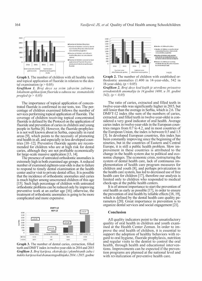
Vasiljević JS, et al. Quality of Oral Health among Schoolchildren164
The importance of topical application of concen-trated fluoride is confirmed in our tests, too. The per-centage of children examined follows the number of services performing topical application of fluoride. The coverage of children receiving topical concentrated fluoride is defined by the Protocol on the application of fluoride and prevention of caries in children and young people in Serbia [8]. However, the fluoride-prophylax-is is not well known about in Serbia, especially in rural areas [9], which points to the necessity of promoting oral health in all, and especially in less developed coun-tries [10–12]. Preventive fluoride agents are recom-mended for children who are at high risk for dental caries, although they are not profitable economically for large-scale massive application [13, 14].
The presence of untreated orthodontic anomalies is extremely high in both examined age groups. A reduced number of examined eighteen-year-olds is due to failure to respond to timely dental examination at the health center and/or visit to private dental office. It is possible that the incidence of orthodontic anomalies and caries is much higher among unscreened children of this age [15]. Such high percentage of children with untreated orthodontic problems can be reduced only by improving preventive work at an earlier age [16]; otherwise, the treatment of orthodontic anomalies is going to be more complicated and more expensive.
The ratio of caries, extracted and filled teeth in twelve-year-olds was significantly higher in 2015, but still lower than the average in Serbia, which is 2.6. The DMFT-12 index (the sum of the numbers of caries, extracted, and filled teeth in twelve-year-olds) is con-sidered a very good indicator of oral health. Average caries index in twelve-year-olds in the European coun-tries ranges from 0.7 to 4.2, and in most countries of the European Union, the index is between 0.5 and 1.5 [3]. In developed European countries, this index has been constantly improving since the beginning of the nineties, but in the countries of Eastern and Central Europe, it is still a public health problem. Slow im-provement in these countries is attributed to the change in the health system due to political and eco-nomic changes. The economic crisis, restructuring the system of dental health care, lack of continuous im-plementation of health care program measures for children and youth [8], and perhaps privatization in the health care system, has led to decreased use of free health care for children [17], therefore our analysis is limited only to children who responded to medical check-ups at the public health centers.
It is of utmost importance to start the prevention of oral health as early as possible [17], in order to ensure the prevention of oral health by reliable effects [18, 19], which is defined by the dental health care quality pa-rameters [20]. Great importance in prevention is to organize dental services and social engagement [21].
Conclusion
All quality indicators point to the unsatisfactory quality of oral health in children and youth exam-ined at the Health Center Zemun. In order to im-prove the oral health of children, it is essential to support the adoption of healthy behaviors with re-gard to oral hygiene, fluoride prophylaxis, nutrition and regular visits to the dentist to control the oral health, through health and educational interven-tions. Improvements can be expected if the preven-tion programs are planned at the national level and with revitalization of preventive health care.
Graph 1. The number of children with all healthy teeth and topical application of fluoride in relation to the den-tal examination (p > 0.05)Grafikon 1. Broj dece sa svim zdravim zubima i lokalnom aplikacijom fluorida u odnosu na stomatološki pregled (p > 0,05)
Graph 2. The number of children with established or-thodontic anomalies (1.400 in 14-year-olds, 542 in 18-year-olds), (p < 0.05)Grafikon 2. Broj dece kod kojih je utvrđeno prisustvo ortodontskih anomalija (u 14.godini 1400, u 18. godini 542), (p < 0,05)
Graph 3. The number of dental caries, extraction, filled teeth and DMFT index in twelve-year-olds in 2014 and 2015Grafikon 3. Broj karijesa, ekstrakcija i plombiranih zuba i indeks karijesa kod dvanaestogodišnjaka 2014. i 2015. godine

Med Pregl 2017; LXX (5-6): 162-165. Novi Sad: maj-juni. 165
References1. Strategy for continuous improvement of healthcare qual-
ity and patient safety, art.45, paragraph 1, Law on Government. Official Gazette of the Republic of Serbia. 2005;(75); correction; 2007;(101); 2008;(65).
2. World Health Organization, The objectives of the WHO Global Oral Health Programme (ORH). [cited 2017 Jan 5]. Available from: http://www.who.int/oral_health/objectives/en/.
3. World Health Organization Regional Office for Europe. European Health for All family of databases. [cited 2017 Jan 5]. Available from: http://www.euro.who.int/en/data-and-evidence/databases/european-health-for-all-family-of-databases-hfa-db. World Health Organization Regional Office for Europe. Euro-pean Health for All database: Available at: http://www.euro.who.int/en/data-and-evidence/databases/european-health-for-all-database-hfa-dbl
4. The Institute of Public Health of Serbia “Dr Milan Jo-vanovic Batut”. Health Statistical Yearbook of the Republic of Serbia 2011 [serial on the Internet]. 2012 [cited 2017 Jan 5]. Available from: http://www.batut.org.rs/download/publikacije/pub2011.pdf).
5. The Ministry of Health of the Republic of Serbia, Public Health Survey of the Republic of Serbia, 2013 - Final Report, May 20 [cited 2017 Jan 5]. Available from: http://www.batut.org.rs/download/publikacije/Finalni%20izvestaj%202014.pdf).
6. Petersen PE. Global policy for improvement of oral health in the 21st century: implications to oral health research of World Health Assembly 2007, World Health Organization. Commu-nity Dent Oral Epidemiol. 2009;37(1):1-8.
7. Ramos-Gomez F, Crystal YO, Ng MW, Tinanoff N, Featherstone JD. Caries risk assessment, prevention, and man-agement in pediatric dental care. Gen Dent. 2010;58(6):505-17.
8. Protocol of promotion of oral health in the prevention of oral diseases in children and youth in the Republic of Serbia. Belgrade: Clinic for Pediatric and Preventive Dentistry, Fac-ulty of Dental Medicine; 2009.
9. Igic M, Apostolovic M, Kostadinovic I, Trickovic – Jan-jic O, Surdilovic D. [The quantity of information which parents and their seven-year-old children have on the affects of nutri-tion, oral hygiene and fluoride prophylaxis on dental health]. Med Pregl. 2009;62(9-10):421-6.
10. Gao XL, Hsu CY, Xu YC, Loh T, Koh D, Hwarng HB. Promoting positive health behaviours - ’tooth worm’ phenomenon and its implications. Community Dent Health. 2012;29(1):55-61.
11. Ferro R, Besostri A, Olivieri A, Stellini E, Denotti G, Campus G. Caries experience in 14-year-olds from Northeast Italy. Is socioeconomic-status (SES) still a risk factor? Eur J Paediatr Dent. 2012;13(1):46-52.
12. Gilinsky A, Swanson V, Merrett M, Power K, Marley L. Development and testing of a theory-based behavioural change intervention: a pilot investigation in a nursery school in a deprived area of Scotland. Community Dent Health. 2012;29(1):62-7.
13. Vermaire JH, Hoogstraten J, van Loveren C, Poorterman JH, van Exel NJ. Attitudes towards oral health among parents of 6-year-old children at risk of developing caries. Community Dent Oral Epidemiol. 2010;38(6):507-20.
14. World Health Organization (WHO) [home page on the Internet]. ©2017[cited 2017 Jan 5]. Health promotion and oral health. Available from: http://www.who.int/oral_health/strate-gies/hp/en/.
15. Davies GM, Duxbury JT, Boothman NJ, Davies RM. Challenges associated with the evaluation of a dental health promotion programme in a deprived urban area. Community Dent Health. 2007;24(2):117-21.
16. Jürgensen N, Petersen PE. Promoting oral health of chil-dren through schools: results from a WHO global survey 2012. Community Dental Health. 2013;30(4):204-18.
17. Radic M, Benjak T, Deckovic Vukres V, Rotim Z, Filipovic Zore I. Presentation of DMFT/dmft Index in Croatia and Europe. Acta Stomatol Croat. 2015;49(4):275-84.
18. Rules on health care quality indicators. Official Gazette of the Republic of Serbia. 2010;(49).
19. World Health Organization [home page on the Internet]. ©2017 [cited 2017 Jan 5]. The objectives of the WHO Global Oral Health Programme (ORH). Available from: http://www.who.int/oral_health/objectives/en/.
20. Petersen PE. World Health Organization global policy for improvement of oral health - World Health Assembly. 2007. Int Dent J. 2008;58(3):115-21.
21. Jevtić M, Pantelinac J. Jovanović Ilić T, Petrović V, Grgić O, Blažić L. Te role of nutrition in caries prevention and maintenance of orl health during pregnancy. Med Pregl. 2015;68(11-12):387-93.
Rad je primljen 10. X 2016.Recenziran 22. XII 2016.Prihvaćen za štampu 28. XII 2016.BIBLID.0025-8105:(2017):LXX:5-6:162-165.


Med Pregl 2017; LXX (5-6): 167-169. Novi Sad: maj-juni. 167
CASE REPORTSPRIKAZI SLUČAJEVAUniversity Clinical Center Tuzla, Clinic of Internal Diseases, Case reportDepartment of Cardiology, Tuzla, Bosnia and Herzegovina1 Prikaz slučajaMedical School Tuzla, Tuzla, Bosnia and Herzegovina2 UDK 616.125-089.84-06:616.8-009.832-07 https://doi.org/10.2298/MPNS1706167D
SYNCOPE DUE TO SINUS NODE DYSFUNCTION AFTER SURGICAL PATCH CLOSURE OF ATRIAL SEPTAL DEFECT – A CASE REPORT
SINKOPA ZBOG DISFUNKCIJE SINUSNOG ČVORA NAKON HIRURŠKOG ZATVARANJA ATRIJAL-NOG SEPTALNOG DEFEKTA – PRIKAZ SLUČAJA
Larisa DIZDAREVIĆ HUDIĆ1, Zumreta KUŠLJUGIĆ1, Irma BIJEDIĆ1 and Igor HUDIĆ2
Introduction
Syncope is a sudden and brief loss of conscious-ness followed by spontaneous recovery. Syncope develops because of temporary reduction in blood flow to the brain with consecutive cerebral oxygen deprivation. Therefore, syncope is defined as a tran-sient, self-limited loss of consciousness with an in-ability to maintain postural tone that is followed by spontaneous recovery. Various causes are well de-scribed in the literature and include cardiac, vascular, neurological, metabolic and miscellaneous origins [1]. Sometimes it is difficult to determine the real cause of syncope, so numerous analyses need to be performed. Sick sinus syndrome (SSS) refers to a combination of symptoms such as dizziness, confu-
sion etc. (symptoms and signs of end-organ hypop-erfusion) caused by sinus node dysfunction (SND). SND is a frequent cause of syncope, and in this case it was a result of cerebral hypoperfusion.
We report here the case of a patient with history of syncope and dizziness. Three years before admission the patient underwent surgical repair of atrial septal defect (ASD) by pericardial patch closure of ASD. Re-current syncope was a result of SSS and this syndrome rarely occurs after surgical patch closure of ASD.
Case report
A 38-year-old woman was referred to our depart-ment due to repeated episodes of syncope during the past month. Three years before, she underwent
SummaryIntroduction. Sick sinus syndrome, a frequent cause of syn-cope, refers to a combination of symptoms caused by sinus node dysfunction. Case report. We report the case of a 38-year-old female patient presenting with recurrent syncope, who under-went surgical patch closure of atrial septal defect three years before admission. Ambulatory twenty-four-hour Holter moni-toring was done capturing only sinus tachycardia. A series of examinations were warranted after admission, and recurrent syncope was found to be the result of sinus node dysfunction. This syndrome rarely occurs after surgical closure of atrial sep-tal defect. The patient underwent permanent pacemaker implan-tation. Conclusion. A rigorous search for every possible cause of syncope is mandatory. A structural, multidisciplinary ap-proach is required in order to achieve an optimal outcome.Key words: Sick Sinus Syndrome; Syncope; Tachycardia; Post-operative Complications; Pacemaker, Artificial; Heart Septal De-fects, Atrial
SažetakUvod. Sinkopa je iznenadni i kratki gubitak svesti sa gubitkom pos-turalnog tonusa i najčešće spontanim oporavkom. Postoje brojni uzroci sinkope. Sindrom bolesnog sinusnog čvora, tzv. sik sinus sin-drom, odnosi se na kombinaciju simptoma kao što su konfuzija ili sinkopa uzrokovane bolešću sinusnog čvora. Prikaz slučaja. Prikazan je slučaj 38-godišnje bolesnice sa istorijom sinkope. Pre tri godine podvrgnuta je hirurškom zahvatu zatvaranja pretkomorskog septalnog defekta. Ambulantni 24-satni monitoring holterom kod naše pacijent-kinje pokazao je samo sinusnu tahikardiju. Ostale ambulantne analize takođe su bile u granicama normale. Nakon niza pretraga, uz multi-disciplinarni pristup i ponavljanje monitoringa holterom utvrđeno je da je sinkopa rezultat disfunkcije sinusnog čvora kao retke komp-likacije hiruškog zatvaranja pretkomorskog septalnog defekta. Potom je podvrgnuta implantaciji veštačkog vodiča srčanog ritma − pejsme-jkera. Zaključak. Treba pomno istražiti sve moguće uѕroke sinkope uz poseban akcenat na multidisciplinarni pristup pacijentu.Ključne reči: disfunkcija sinusnog čvora; sinkopa; postopera-tivne komplikacije; pejsmejker; pretkomorski septalni defekt
Corresponding Author: Dr Larisa Dizdarević-Hudić, Univerzitetsko klinički centar Tuzla, Klinika za interne bolesti, 75000 Tuzla, Trnovac bb, Bosna i Hercegovina, E-mail: [email protected]; [email protected]

Dizdarević Hudić L, et al. Syncope due to Sinus Node Dysfunction168
a surgical patch closure of secundum ASD. She denied having other medical issues in the past and she was a non-smoker. She did not use any medica-tions during the previous year.
Several months prior to admission, she started having symptoms of dizziness and weakness, and three months ago, she had the first episode of syn-cope. She did not have a chest pain, typical for an-gina. The patient was evaluated by a neurologist in ambulatory settings. A transcranial Doppler ex-amination showed bilateral patency of carotid arter-ies, without significant stenosis. Computed tomog-raphy of the brain and electroencephalogram were requested. Ambulatory 24-hour Holter monitoring was done and it showed only sinus tachycardia with maximum heart rate 135/min during physical effort.
On admission, the patient was fully alert and ori-ented. Her vital signs (blood pressure, pulse and res-piration) unremarkable. Physical examination of the heart, chest, abdomen and extremities were normal. No neurological deficit was present. Laboratory test results were within the normal range.
The chest X-ray showed no abnormalities. Both awake and asleep electroencephalography showed normal results. Computed tomography (CT) of the brain did not show any signs of intracranial hemor-rhage or ischemia. Echocardiography showed slight-ly dilated right ventricle (3,5 cm) and patch closure of ASD, essentially preserved left-ventricular systo-lic function and no other abnormalities. Several electrocardiograms (ECG) were within normal lim-its (Figure 1).
During the 24-hour Holter recording, the patient was in normal sinus rhythm with an average heart rate of 68/min, and maximum heart rate 137/min. PQ (time it takes for the sinus impulse to travel from the atria to the ventricles) and QT (measure be-tween Q wave and T wave in the heart’s electri-cal cycle) interval were within normal ranges, with-out signs of pre-excitation. Only rare atrial prema-ture contractions were recorded. In the morning hours (08h 03 min to 08h 25min) a few significant sinus pauses were registered that correlated with the occurrence of syncope. The longest episode was 12820 ms (Figure 2).
The patient was diagnosed with symptomatic SND and underwent permanent pacemaker implan-tation (dual chamber rate adaptive pacemaker - DDDR). She did not suffer from syncope, weakness or dizziness after the treatment.
Discussion
Sinus node dysfunction is a term used for nu-merous rhythm abnormalities such as: persistent sinus bradycardia, persistent sinus arrest with es-cape rhythms, and chronotropic incompetence [2, 3]. This disorder is often associated with conduction system diseases and supraventricular tachyarrhyth-mias (in this case, SND is termed tachy-brady syn-drome) [3]. Most people with SSS are asymptomat-ic or oligosymptomatic. Well-described symptoms of SND are bradycardia, fainting (syncope), fatigue, weakness, dyspnea, angina, disturbed sleep, confu-sion, and palpitations.
The most common cause of SND is idiopathic sinoatrial node fibrosis, sometimes accompanied by degeneration of lower elements of the conducting system. Risk factors for development of SND in-
AbbreviationsSSS – sick sinus syndromeSND – sinus node dysfunctionASD – atrial septal defectECG – electrocardiogramDDDR – dual chamber rate adaptive pacemakerCHD – congenital heart diseaseSVASD – sinus venosus atrial septal defect
Figure 1. Electrocardiogram of our patient on admission (no significant abnormalities)Slika 1. Elektrokardiogram pacijentkinje na prijemu (bez značajnih abnormalnosti)
Figure 2. Holter monitoring - a few significant sinus pauses were registered in the morning hours (08 h 03 min to 08 h 25 min); the longest episode was 12820 ms, as shown above. This figure shows only the beginning of this longest episode, the whole pause is not shown due to technical circumstancesSlika 2. Monitoring holterom – nekoliko značajnih si-nusnih pauza u jutarnjim satima (8 h i 3 min. do 8 h i 25 min.); najduža epizoda trajale je 12 820 ms (prikazano gore). Na slici je prikazan samo početak najduže epiz-ode; cela pauza nije prikazana iz tehničkih razloga

Med Pregl 2017; LXX (5-6): 167-169. Novi Sad: maj-juni. 169
clude: age, medications, hyperkalemia, myocardial infarction, heart surgery, sleep apnea, diphtheria, hemochromatosis, muscular dystrophy, amyloidosis (many ischemic, inflammatory and infiltrative dis-orders). In younger patients, SND is often second-ary to other cardiac processes/disease [4] and it may be present in patients who have undergone surgery for congenital heart disease, including ASD. In ad-dition, the later in life ASD is repaired, the more likely arrhythmia will develop. The cause of SND in these patients is probably related to the underly-ing structural heart disease and surgical trauma (of the sinus node and/or sinus node artery). When re-pairing ASDs, sinus venosus atrial septal defect (SVASD), SND often occurs because ASD is close-ly related to the sinus node tissue [4, 5]. Postopera-tive SND is more common in patients after SVASD repair than after secundum ASD repair [6].
The only effective treatment for patients with chronic symptomatic SND is pacemaker implanta-tion. It is important to mention that asymptomatic patients do not require device therapy. Pacemakers are indicated in patients with certain symptomatic bradyarrhythmias caused by SND and in patients with frequent, prolonged sinus pauses [3]. Sympto-matic bradyarrhythmias are the most common indi-cations for pacemaker placement. Approximately one-half of devices are implanted for SND. Our pa-tient was symptomatic, with very significant periods of asystoles (12 820 ms), so she underwent pacemak-er implantation (DDDR) as mentioned above.
We believe that a certain number of syncope pa-tients remain misdiagnosed. For example, in our case, a psychiatrist made a diagnosis of anxiety-depressive
disorder with panic attacks and dissociative reactions. On the other hand, it was difficult to establish the real diagnosis because the ambulatory Holter monitoring and serial ECG-s showed no abnormalities, as well as laboratory reports and neurological examinations. It is often difficult to capture the moment when syncope occurs on Holter or ECG. Differentiating true syn-cope from other similar non-syncopal conditions (e.g. epilepsy, severe metabolic disorders, intoxications, psychogenic pseudo-syncope etc.) is the first diagnos-tic step with huge influence on the subsequent diag-nostic strategy. Among the neutrally mediated inves-tigations, the tilt-test and carotid sinus massage are the most useful. The most useful cardiac examinations here are echocardiography, prolonged ECG monitor-ing, stress test, electrophysiological study and implant-able loop recorder [7]. Patients with unexplained syn-cope are more likely to have an underlying arrhythmia mechanism than unselected patients with syncope. However, arrhythmias causing syncope may occur at varying and long intervals, giving standard ECG-a and short–lasting ECG monitoring little chance of providing symptoms vs. ECG correlation when com-pared with continuous long term ECG-monitoring (sometime a few days of ECG monitoring may be valuable). A structured, multidisciplinary approach to evaluation of syncope is the best model to achieve an optimal outcome [8].
Conclusion
In conclusion, a structured multidisciplinary ap-proach to syncope of unexplained origin is the best model to achieve an optimal outcome.
References1. Kuo FY, Hsiao HC, Chiou CW, Liu CP. Recurrent syn-
cope due to carotid sinus hypersensitivity and sick sinus syn-drome. J Chin Med Assoc. 2008;71(10):532–5.
2. Ferrer MI. The sick sinus syndrome in atrial disease. JAMA. 1968;206(3):645-6.
3. Gillis AM, Russo AM, Ellenbogen KA, Swerdlow CD, Olshansky B, Al-Khatib SM, et al. HRS/ACCF expert consen-sus statement on pacemaker device and mode selection: devel-oped in partnership between the Heart Rhythm Society (HRS) and the American College of Cardiology Foundation (ACCF) and in collaboration with the Society of Thoracic Surgeons. Heart Rhythm. 2012;9(8):1344–65.
4. Dobrzynski H, Boyett MR, Anderson RH. New insights into pacemaker activity: promoting understanding of sick sinus syndrome. Circulation. 2007;115(14):1921-32.
5. Gatzoulis MA, Freeman MA, Siu SC, Webb GD, Harris L. Atrial arrhythmia after surgical closure of atrial septal de-fects in adults. N Engl J Med. 1999;340(11):839–46.
6. Attenhofer Jost CH, Connolly HM, Danielson GK, Bai-ley KR, Schaff HV, Shen WK, et al. Sinus venosus atrial septal defect: long-term postoperative outcome for 115 patients. Cir-culation. 2005;112(13):1953-8.
7. Brignole M, Alboni P, Benditt DG, Bergfeldt L, Blanc JJ, Bloch Thomsen PE, et al. Guidelines on management (diagno-sis and treatment) of syncope-update 2004. Europace. 2004; 6(6):467-537.
8. Edvardsson N, Wolff C, Tsintzos S, Rieger G, Linker NJ. Costs of unstructured investigation of unexplained syncope: insights from a micro-costing analysis of the observational PIC-TURE registry. Europace. 2015;17(7):1141-8.
Rad je primljen 18. VI 2016.Recenziran 10. I 2017.Prihvaćen za štampu 2. II 2017.BIBLID.0025-8105:(2017):LXX:5-6:167-169.

Delibegović S, et al. Foreign Body Ingestion170
University Clinical Center Tuzla, Surgery Clinic, Case reportDepartment of Colorectal Surgery, Tuzla, Bosnia and Herzegovina1 Prikaz slučajaGeneral Hospital, Konjic, Bosnia and Herzegovina2 UDK 616.348/.35-003.6-07 https://doi.org/10.2298/MPNS1706170D
FOREIGN BODY INGESTION – GLASS IN COLON AND RECTUM – A CASE REPORT AND LITERATURE REVIEW
PROGUTANA STRANA TELA – STAKLO U KOLONU I REKTUMU – PRIKAZ SLUČAJA I PREGLED LITERATURE
Samir DELIBEGOVIĆ1, Edvin MULALIĆ1 and Sejo BUTUROVIĆ2
Introduction
Ingestion of a foreign body is not a common emer-gency condition in small hospitals. Accidental ingestion is more frequent in children. Intentional ingestion is usually found in cases of mental disorders, prisoners, attempted suicides, and in persons with intellectual dis-abilities. The most frequently ingested foreign bodies are pieces of food, most often bones, toothpicks, but real foreign bodies are orthodontic implants, needles, nails etc. [1].
Glass foreign bodies are among the most unusual ingested foreign bodies, and it is difficult to predict the consequences of their passing through the gastrointes-tinal tract (GIT). We report a case of accidental swal-lowing of a large quantity of glass pieces, diagnosed by X-ray in the ascending colon and the rectum. The pa-tient did not have signs of perforation, so an expectant attitude was taken, and elimination occurred naturally.
Case report
A 36-year-old woman with intellectual disabilities was admitted to the Clinic due to abdominal pain. Het-eroanamnesis showed that she had allegedly had stom-
ach pains for a month. Physical findings showed pain-ful sensitivity in the upper right quadrant, with no signs of peritonitis. The abdominal X-ray showed a great number of intensive shadows in the area of the ascend-ing colon and the rectum (Figure 1). Digital rectal ex-amination produced a lump of stool with pieces of glass.
Upon admission, a computed tomography (CT) of the abdomen was performed (Figure 2). In the area of the ascending colon and the rectum there were numer-ous hyperdense areas of various shapes and sizes, mostly with sharp contours, and the character of a for-eign body, with a density of 500 to 1500 HJ, 5 to 20 mm in diameter. The colonic loop was distended, es-pecially in the area of the transverse colon, with a lu-men up to 5 cm in width.
Due to the persistent pain, surgical exploration was considered, but in the absence of signs of perforation, an expectant attitude was taken, and laxatives were prescribed. The patient presented with a gradual regres-sion of pain, and a control X-ray confirmed that the foreign bodies shifted through the intestines (Figure 3 and 4) but they were filled with air, at multiple air fluid levels.
A rectoscopy was performed to establish the condi-tion of the mucous membranes of the rectum, showing
SummaryIntroduction. Ingestion of foreign body is one of the most com-plex and serious emergency conditions to diagnose. Accidental ingestion is more frequent in children than in adults, whereas intentional ingestion is usually found in cases of mental disor-ders, prisoners, attempted suicides, and in persons with intel-lectual disabilities. Case report. Glass ingestion is very rare and it is very difficult to predict the consequences of its passing through the gastrointestinal tract. We report a case of acciden-tal swallowing of a large quantity of glass pieces in the ascend-ing colon and rectum diagnosed by abdominal X-ray. The pa-tient did not have any signs of perforation. An expectant attitude was taken, and the elimination occurred naturally. Key words: Foreign Bodies; Deglutition; Accidents; Glass; Diagnosis; Treatment Outcome; Gastrointestinal Tract
SažetakUvod. Progutano strano telo predstavlja veoma kompleksno i hitno stanje, teško za postavljanje dijagnoze. Slučajno gutanje je češće kod dece nego kod odraslih, a namerno gutanje nalazi se obično kod osoba sa psihološkim poremećajima, kod zat-vorenika, osoba koje pokušaju suicid, te kod osoba sa poremećajima u mentalnom razvoju. Staklo je vrlo retko strano telo koje se guta i kod kojeg je teško predvideti posledice pro-laska kroz gastrointestinalni trakt. Prikaz slučaja. Mi izveštavamo o nenamernom gutanju veće količine komada stakla koji su radiološki dijagnostikovani u ushodnom kolonu i rektumu. Pacijent nije imao znakove perforacije. Zauzet je ekspektativan stav, te je prirodnim putem došlo do eliminacije.Ključne reči: strana tela; gutanje; nesreće; staklo; dijagnoza; ishod lečenja; gastrointestinalni trakt
Corresponding Author: Prof. dr Samir Delibegović, Univerzitetski klinički centar, Klinika za hirurgiju,75000 Tuzla, Trnovac bb, E-mail: [email protected]

Med Pregl 2017; LXX (5-6): 170-172. Novi Sad: maj-juni. 171
intact mucous membranes, with several balls of stool. In each ball there was a piece of glass (Figure 5 a and b).
On the fifth day of hospitalization, X-ray of the native abdomen indicated the absence of glass (Fig-ure 6), but showed several air fluid levels.
On the twelfth day of stay, the patient was dis-charged in a satisfactory general condition.
Discussion
Most intentionally or accidentally ingested foreign bodies pass through the GIT without complications, and only a minority require surgical intervention [2–4]. The occurrence of abdominal pain alerts the physician of possible complications, primarily perforation with the resulting peritonitis. Fatal cases after ingestion have also been described, due to intestinal obstruction [5].
The intestines have a significant ability to protect themselves from perforation in cases of ingestion of sharp objects, such as glass. When a sharp object is stuck in the intestinal mucous, an area of ischemia with a large central concavity develops. The walls of the intestines increase the lumen at the place of contact, enabling easier passage of the sharp object [6]. Moreo-ver, when a sharp object is swallowed, the flow of the intestinal content and the relaxation of the intestinal walls tend to direct the head of the object to the front, and the sharp end to the back [7]. When it arrives in the colon, the foreign body is covered with faecal material, protecting the intestinal wall.
AbbreviationsCT – computed tomographyGIT – gastrointestinal tract
Figure 1. Abdominal X-ray showing a great number of in-tensive shadows in the area of the ascending colon and the rectum Slika 1. Rendgenogram abdomena pokazuje veći broj inten-zivnih senki u području ascedentnog kolona i rektuma
Figure 2. Abdominal CT in the area of the ascending colon and the rectum showing numerous hyperdense areasSlika 2. Kompjuterizovana tomografija abdomena, u području ascedentnog kolona i rektuma veći broj hiperden-znih area
Figures 3 and 4. The X-ray confirmed that the foreign bodies shifted through the intestines, but they were filled with air at several air fluid levelsSlike 3 i 4. Rendgenogram abdomena pokazuje pomer-anje stranih tela kroz creva, ali crevima ispunjenim vazduhom i sa nekoliko aerolikvidnih nivoa

Delibegović S, et al. Foreign Body Ingestion172
Isolated groups of glass in the rectum (Figures 1 and 2) cause concern, because it is forgotten that this collection has already passed through the digestive tract, wrapped in stool, which protects the intestinal wall. In this case, the inability of the patient with intel-lectual disabilities to describe in detail how the inges-tion occurred, that is, how the glass entered the GIT, led to difficulties in diagnosis and therapy.
The aim of clinical evaluation was to identify the type, quantity, size and location of the foreign bodies in the rectum. As a rule, removal of a foreign body from the rectum requires experience, with particular care when using various means of extraction, to minimize damage to the mucous membrane. Most foreign bodies can be successfully removed trans-anally, under ap-propriate anesthesia and using appropriate instruments. Alternative methods are the use of a Foley catheter [8], trans-anal vacuum extraction of glass foreign bodies [9], and trans-anal minimally invasive surgery [10].
Only a small number of foreign body cases, if they go deeper into the sigmoid colon, require extraction by
colonoscopy [11]. Rare cases with signs of perforation, peritonitis, or pelvic sepsis, Fouriner gangrene [12], or unsuccessful manual extraction, require open surgery or laparoscopy, by massaging (milking) towards the anus or a colostomy. After removal of the rectal foreign body, a proctosigmoidoscopy is mandatory, to exclude injury to the mucous membrane [13].
Conclusion
In the case presented, it was clearly a matter of glass that had passed through the gastrointestinal tract. The attitude of waiting and monitoring the clin-ical condition of the patient seemed most appropriate; otherwise unnecessary resection of the large intestine would have been performed. This approach requires a great deal of patience, constant monitoring, and a prompt reaction in the case of intestinal perforation.
References1. Ricci S, Massoni F, Schiffino L, Pelosi M, Salesi M. Foreign
bodies ingestion: what responsibility? J Forensic Leg Med. 2014;23:5-8.
2. Schwartz GF, Polsky HS. Ingested foreign bodies of the gas-trointestinal tract. Am Surg. 1976;42(4):236-8.
3. Mcpherson RC, Karlan M, Williams RD. Foreign body per-foration of the intestinal tract. Am J Surg. 1957;94(4):564-6.
4. Maleki M, Evans WE. Foreign-body perforation of the in-testinal tract. Report of 12 cases and review of the literature. Arch Surg. 1970;101(4):475-7.
5. Betz P, van Meyer L, Eisenmenger W. Fatalities due to intes-tinal obstruction following the ingestion of foreign bodies. Forensic Sci Int. 1994;69(2):105-10.
6. Exner A. Wie schuetzt sich der verdanungstract ver verlet-zungen durch spitze fremdkoerper. Arch F D Ges Physiol. 1902; 89:253.
7. Goh BK, Chow PK, Quah HM, Ong HS, Eu KW, Ooi LL, et al. Perforation of the gastrointestinal tract secondary to ingestion of foreign bodies. World J Surg. 2006;30(3):372–7.
8. Cologne KG, Ault GT. Rectal foreign bodies: what is the current standard? Clin Colon Rectal Surg. 2012;25(4):214-8.
9. Johnson SO, Hartranft TH. Nonsurgical removal of a rectal foreign body using a vacuum extractor. Report of a case. Dis Colon Rectum. 1996;39(8):935-7.
10. Cawich SO, Mohammed F, Spence R, Albert M, Narayns-ingh V. Colonic foreign body retrieval using a modified TAMIS technique with standard instruments and trocars. Case Rep Emerg Med. 2015;2015:815616.
11. Lin XD, Wu GY, Li SH, Wen ZQ, Zhang F, Yu SP. Re-moval of a large foreign body in the rectosigmoid colon by colonos-copy using gastrolith forceps. World J Clin Cases. 2016;4(5):135-7.
12. Abate G, Shirin M, Kandanati V. Fournier gangrene from a thirty-two-centimeter rectosigmoid foreign body. J Emerg Med. 2013;44(2):e247-9
13. Ayantunde AA. Approach to the diagnosis and management of retained rectal foreign bodies: clinical update. Tech Coloproctol. 2013;17(1):13-20.
Rad je primljen 23. II 2017.Recenziran 28. III 2017.Prihvaćen za štampu 18. IV 2017.BIBLID.0025-8105:(2017):LXX:5-6:170-172.
Figure 5 a and b. One of the balls of stool containing a piece of glassSlika 5 a i b. Jedna od “kuglica” stolice u kojoj se nala-zi komadić stakla
Figure 6. X-ray of the native abdomen indicated the absence of glassSlika 6. Rendgenogram nativnog abdomena ukazuje na odsustvo stakla, ali sa nekoliko aerolikvidnih nivoa
Med Pregl 2017; LXX (5-6): 173-175. Novi Sad: maj-juni. 173
University of Novi Sad, Faculty of Medicine Case reportClinical Center of Vojvodina Prikaz slučajaClinic of Infectious Diseases UDK 616.921.5-06:616.74 https://doi.org/10.2298/MPNS1706173S
RHABDOMYOLYSIS AND INFLUENZA A (H3N2) INFECTION – A CASE REPORT
RABDOMIOLIZA I INFLUENZA A (H3N2) INFEKCIJA – PRIKAZ SLUČAJA
Anja STOJŠIN, Vedrana PETRIĆ, Grozdana ČANAK, Vesna TURKULOV, Siniša SEVIĆ and Maja RUŽIĆ
Introduction
Influenza causes respiratory tract diseases with acute respiratory distress syndrome (ARDS) as the leading and most severe life threatening complication. In terms of early diagnosis, extrapulmonary complica-tions are less common, but clinically a significant prob-lem [1]. In the territory of Vojvodina, influenza occurs in epidemic forms, usually in the winter months. The most common cause is the virus Influenza A. In the season 2014/2015, the dominant type was A/H3N2. Pulmonary complications were the most common com-plications, and also the most common cause of death [2, 3]. However, we are witnessing the appearance of
other rare and often unrecognized, life threatening complications.
Rhabdomyolysis (RM) is the destruction of skeletal muscle followed by the release of intracellular myofi-brils into the bloodstream and extracellular fluid. Clinical manifestations vary, from asymptomatic to life threatening conditions, such as acute renal failure and disseminated intravascular coagulation [4]. Al-though myositis is the most common symptom of the flu syndrome, the development of RM is rare, and it is more likely to occur in individuals with a genetic pre-disposition.
The aim of this paper is to highlight RM as a po-tentially life threatening complication of influenza.
SummaryIntroduction. Extrapulmonary complications of influenza infec-tions are often unrecognized. The aim of this paper is to point to rhabdomyolysis as a potentially life threatening complication of influenza. Case Report. A month after the onset of influenza com-plicated by bronchopneumonia, the general condition of a nineteen year old female deteriorated with development of progressive mus-cle weakness and dark-colored urine. Despite intensive hydration and antibiotic therapy, her condition got worse, laboratory findings showed pancytopenia, hypoalbuminemia and creatine phosphoki-nase about 1000 times higher than normal. Influenza A H3N2 was confirmed by polymerase chain reaction analysis of the throat swab sample. Electromyoneurography showed severe acute polyneu-ropathy of muscles innervated by perineal nerve and signs of poly-myositis; pathohistological examination of gastrocnemius muscle biopsy sample confirmed chronic myositis with necrotic neuro-genic atrophy. In spite of intense hydration, the patient’s status continued deteriorating, so methylprednisolone was administered. Six weeks later, the patient was discharged in a good general condi-tion, with blood test results within reference ranges, with weakness of foot dorsiflexors and tilting of the pelvis to the left during verti-calization. Conclusion. Rhabdomyolysis caused by influenza-A is on the increase, and given the degree of morbidity and mortality, thorough assessment of patients is necessary.Key words: Rhabdomyolysis; Influenza A Virus, H3N2 Subtype; Polyneuropathies; Risk Factors; Signs and Symptoms; Disease Progression; Treatment Outcome
SažetakUvod. Ekstrapulmonalne komplikacije influenca virusne infekcije su često neprepoznate. Cilj ovog rada je da se ukaže na rabdomi-olizu kao potencijalnu vitalno ugrožavajuću komplikaciju gripa. Prikaz slučaja. Mesec dana nakon početka gripa komlikovane bronhopneumonijom, kod devetnaestogodišnje devojke došlo je do pogoršanja opšteg stanja razvojem progresivne mišićne slabosti, uz pojavu tamne prebojenosti urina. Na sprovedenu intenzivnu rehid-rataciju i antibiotsku terapiju, opšte stanje se i dalje pogoršava, a u laboratorijskim nalazima se zapaža pancitopenija, hipoalbumine-mija i oko 1 000 puta povišena aktivnost kreatin fosfokinaze. Analizom polimerazne lančane reakcije uzorka brisa grla doka-zana je Influenza A H3N2. Elektromioneurografijom se verifikuje akutna teška polineuropatija muskulature koju inerviše n. perone-us i znaci polimiozitisa, a patohistološki pregled bioptata levog m. gastrocnemius potvrđuje myositis chronica cum atrophia neuro-genes cum necrosis. Kako i pored obilne hidratacije nije došlo do očekivanog poboljašnja stanja bolesnice, ordiniran je metilpredni-zolon. Nakon šest nedelja lečenja, pacijentkinja je otpuštena dobrog opšteg stanja, urednih laboratorijskih nalaza, sa slabosću dorzifle-ksora stopala i naginjanjem karlice na levu stranu tokom vertika-lizacije. Zaključak. Rabdomioliza uzrokovani influencom-A se sve češće beleži u praksi, a s obzirom na stepen morbiditeta i mor-taliteta iziskuje pažljiv pristup proceni stanja pacijenta.Ključne reči: rabdomioliza; influenca A virus, podtip H3N2; po-lineuropatije; faktori rizika; znaci i simptomi; progresija bolesti; ishod lečenja
Corresponding Author: Dr Anja Stojšin, Klinički centar Vojvodine, Klinika za infektivne bolesti, 21000 Novi Sad, Hajduk Veljkova 1-7, E-mail: [email protected]
Stojšin A, et al. Rhabdomyolysis and Influenza A (H3N2) Infection174
Case Report
At the beginning of January 2015, a seventeen year old girl presented with Influenza syndrome (dry cough, elevated body temperature up to 38°C). She was under regular medical supervision due to asthma therapy in the past two years (budesonide and formoterol as need-ed). Five days after the onset, bronchopneumonia was detected by auscultation, and cefpodoxime (200 mg per 12 hours) was initiated. The respiratory symptoms have resolved, but at the same time muscle weakness with intense pain and inability to move started. The patient presented with dark discoloration of the urine, and was admitted to a regional hospital in the second week of February with the diagnosis of suspected in-fectious myositis.
On admission, the patient was afebrile, hypotensive, tachycardic, cyanotic with generalized skeletal muscle pain, and with paresis of all extremities and hypore-flexia. The laboratory findings showed the following: C-reactive protein (CRP) 21.7 (below 5 mg/l), aspartate aminotransferase (AST) 572 (below 37 U/l), alanine transaminase (ALT) 130 (below 40 U/l), lactate dehy-drogenase (LDH) 1684 (460 U/L and creatine phos-phokinase (CPK) 19344 (below 171 U/l), and after basic diagnostic procedures were performed, intrave-nous hydration and antibiotic therapy (ceftriaxone, amikacin) were initiated. Despite the administered therapy, the laboratory findings got worse: an increase of leukocytes, anemia, thrombocytopenia, hypoalbu-minemia, CPK 176 421 (below 168 U/l), AST 3080 (below 37 U/l), ALT 467 (below 40 U/l), LDH 3209 (below 460 U/l) and after 24 hours the patient was transferred to the Clinic of Infectious Diseases for sus-pected sepsis and infectious myositis.
Intensive intravenous hydration with mannitol and correction of metabolic acidosis were performed. Im-munological tests (anti nuclear antibodies - ANA, anti neurotrophic cytoplasmatic antibody virus - ANCA, anti-double-stranded deoxyribonucleid acid At) were negative. Assessments were done for Adeno, Coxsack-ie A and B, Influenza A and B, Parainfluenza, Vari-cella Zoster, Herpes Simplex, Respiratory Syncytial Virus, Epstein Barr Virus, Parvo B19, Cytomegalovirus infections, Mycoplasma pneumonia, Hepatitis B Virus surface antigen (HBsAg), anti Hepatitis C Virus anti-body (AntiHCV), which proved negative. However, polymerase chain reaction (PCR) microbiological anal-ysis of the throat swab was Influenza A H3N2 positive. As there was no satisfactory response, corticosteroids (methylprednisolone 120 mg) was initiated, that resulted in moderate clinical and laboratory improvements.
During further treatment at the Clinic of Infectious Diseases the patient was gradually recovering. Elec-tromyoneurography (EMNG) indicated a severe acute polyneuropathy of muscles innervated by perineal nerve and signs of polymyositis. Histopathological ex-amination of the left gastrocnemius confirmed chron-ic myositis with necrotic neurogenic atrophy. After six weeks of treatment, the patient was discharged recov-ered, with blood results within reference ranges, with weakness of foot dorsiflexor, tilting of the pelvis to the left during verticalization. Eighteen months later, there were mild sequelae - occasional pain in the muscles of the lower extremities and difficulty doing squats.
Discussion
Myositis is a well-known manifestation of numer-ous infectious diseases, viral infections being the most common (Influenza A and B, Coxsackie Virus, Epstein Barr Virus, Herpes Simplex Virus, Parainfluenza, Ad-enovirus, Echovirus, Human Immunodeficiency Virus (HIV) and Cytomegalovirus (CMV). In general, my-ositis is not the main symptom and rarely progresses to life threatening RM.
Presented Influenza A virus infection was con-firmed by detection with PCR isolation from the phar-ynx swab and serology test - detection of IgM Influ-enza A antibodies by the ELISA method. Prolonged duration of viral shedding, unusual for Influenza A virus, can be explained by the application of corticos-teroid therapy in the treatment of asthma [5, 6].
Although there are indications of direct viral myo-toxic effect (EBV), RM is most often the result of T-cell hyperactivity [7, 8].
A number of exogenous and endogenous factors may accelerate destructive immune response to the muscle tissue [4]. The most common concomitant factors, which may contribute to the development of RM in viral infec-tions, are alcohol abuse and use of certain drugs, which was excluded by toxicological analyses. Cytochrome P450 inhibitors (statins, acetaminophen) are described as the most provoking factors of RM; although the patho-genesis is still not proven, it is believed that they reduce production of adenosine triphosphate (ATP) within the muscle cells which destabilizes the membrane [8]. How-ever, our patient denied taking drugs from this group. Genetic predisposition for the development of RM should also be taken into consideration.
According to recent studies, people with virus in-duced RM are often carriers of mutations of RYR1 gene that creates defective ryanodine receptor 1 on myocytes, thus creating susceptibility to malignant hyperthermia, which leads to RM. We could not perform genetic analysis, but we considered genetic predisposition un-likely to be the cause of RM due to the long latent pe-riod (one month), and absence of high body tempera-tures (up to 38°C). A number of metabolic, genetically determined RM can also occur, but they are usually recurrent and occur after intense physical activity, re-gardless of the infection. The patient regularly practiced folklore and never complained of myalgia, nor reported darker urine color.
AbbreviationsARDS – acute respiratory distress syndromeRM – rhabdomyolysisCRP – C-reactive proteinAST – aspartate aminotransferaseLDH – lactate dehydrogenaseALT – alanine transaminasePCR – polymerase chain reactionCPK – creatine phosphokinaseNaHCO3 – sodium bicarbonate

Med Pregl 2017; LXX (5-6): 173-175. Novi Sad: maj-juni. 175
Progression of symptoms in terms of difficulty of movement, inability of verticalization and dark urine, immediately pointed to RM, especially when blood re-sults showed five times elevated CPK levels and acidosis, with pathological urine [9]. Myoglobinuria is the last of three classical RM symptoms (nausea, myalgia, dark urine) that appeared. It is present in less than 10% of patients with RM, and most patients do not have weak-ness or myalgia, but do have systemic manifestations such as tachycardia, fever, nausea and vomiting [3]. My-oglobinuria does not occur without RM, but RM is not a mandatory result in myoglobinuria. It occurs when the threshold of renal filtration from 0.5 to 1.5 mg/dl is ex-ceeded, and it is macroscopically visible only when the quantities reach 100 mg/dl [10]. Myoglobin in the urine of our patients was not determined, since this analysis is not included in the standard diagnostic procedure.
The main complications of RM are acute kidney failure, electrolyte imbalance, especially hyperkalemia which leads to arrhythmia, hypophosphatemia and hy-pocalcaemia. In order to prevent this, quick and ade-quate therapy is needed. Intense i.v. hydration with sa-line 300 – 500 ml/h should be given. Since our patient was hemodynamically stable, these quantities of fluid were not exceeded. Although there is no protocol in the literature to define duration of intense hydration, it is considered that it should be applied until the level of serum CPK decreases to 1000 U/l or less [4]. Alkaliza-tion of urine with sodium bicarbonate (NaHCO3) is beneficial in three ways: it reduces precipitation of Tamm-Horsfall protein-myoglobin complex, it inhibits oxidation-reductive process of myoglobin and lipid per-oxidase reducing the damage to the tubules, and it re-duces vasoconstriction caused by metmyoglobin in acidic environment [11, 12]. It is recommended to apply infusion of one ampoule of NaHCO3 (20 ml, 1 ml con-tains 84 NaHCO3) in one saline bottle or 2 – 3 ampoules
in one 5% glucose bottle at 100 ml/h rate, in order to maintain urine pH above 6.5 preventing acute kidney failure. Hemodialysis is considered in cases with exces-sive hyperkalemia and metabolic acidosis [13, 14]. Given the small amount of data on the efficacy of cor-ticosteroids in the treatment of RM, and bearing in mind they are used in recurrent RM with chronic my-ositis of autoimmune etiology, they were not adminis-tered during hospitalization in our clinic [15].
Delayed development of RM (one month after the first symptoms) and good therapeutic response to cor-ticosteroids, despite negative immunological test re-sults, may point to autoimmune diseases in the back-ground (dermatomyositis, polymyositis). Biopsy of m. gastrocnemius has been done according to the recom-mendations - 8 weeks after the first symptoms and four weeks after completion of corticosteroid therapy. The result was chronic myositis with necrotic neurogenic atrophy, reactive inflammation with no signs of im-mune or dystrophy myositis. Considering good recov-ery and minimal sequelae, with no signs of recur-rence, we concluded that RM was caused by Influ-enza A virus (H3N2).
Conclusion
Complications of Influenza A are a global prob-lem, so it is of crucial importance to take into con-sideration the range of clinical manifestations, be-yond the typical symptoms of respiratory infections. Reports on rhabdomyolysis caused by influenza-A are increasing and given the degree of morbidity and mortality, assessment of patients suspected for the development of rhabdomyolysis should be done carefully, especially those with general weakness and muscle pain.
References1. World Health Organization. Influenza. 2014 March [cited
2015 Jul 12]. Available from: http://www.who.int/mediacentre/factsheets/fs211/en/.
2. Zarazne bolesti u AP Vojvodini 2015. godina [Communicable diseases in Vojvodina, 2015]. Novi Sad: Institut za javno zdravlje Vojvodine; 2016. Serbian.
3. Khan FY. Rhabdomyolysis: a review of the literature. Neth J Med. 2009;67(9):272-83.
4. Huerta-Alardin AL, Varon J, Marik PE. Bench-to-bedside review: Rhabdomyolysis – an overview for clinicians. Crit Care. 2005;9(2):158-69.
5. Pesik NT, Otten EJ. Severe rhabdomyolysis following a viral illness: a case report and review of the literature. J Emerg Med. 1996;14(4):425-8.
6. Fadila MF, Wool KJ. Rhabdomyolysis secondary to influ-enza a infection: a case report and review of the literature. N Am J Med Sci. 2015;7(3):122-4.
7. Hodel C. Myopathy and rhabdomyolysis with lipid-lowering drugs. Toxicol Lett. 2002;128(1-3):159-68.
8. Ruzic M, Fabri M, Pobor M, Jovelic A, Lukac D. Exercise induced rhabdomyolysis. Vojnosanit Pregl. 2009;66(9):754-7.
9. Bosch X, Poch E, Grau JM. Rhabdomyolysis and acute kidney injury. N Engl J Med. 2009;361(1):62-72.
10. Lane R, Philps M. Rhabdomyolysis has many causes, includ-ing statins, and may be fatal. BMJ. 2003;327(7407):115-6.
11. Heyman SN, Greenbaum R, Shina A, Rosen S, Brezis M. Myoglobinuric acute renal failure in the rat: a role for acidosis? Exp Nephrol. 1997;5(3):210-6.
12. Malik GH. Rhabdomyolysis and myoglobin-induced acute renal failure. Saudi J Kidney Dis Transpl. 1998;9(3):273-84.
13. Chatzizisis YS, Misirli G, Hatzitolios AI, Giannoglou GD. The syndrome of rhabdomyolysis: complications and treatment. Eur J Intern Med. 2008;19(8):568-74.
14. Antoon JW, Chakraborti C. Corticosteroids in the treatment of alcohol-induced rhabdomyolysis. Mayo Clin Proc. 2011;86(10): 1005-7.
Rad je primljen 27. II 2017.Recenziran 20. III 2017.Prihvaćen za štampu 29. III 2017.BIBLID.0025-8105:(2017):LXX:5-6:173-175.


Med Pregl 2017; LXX (5-6): 177-182. Novi Sad: maj-juni. 177
SEMINAR FOR PHISICIANSSEMINAR ZA LEKARE U PRAKSICommunity Health Center, General Medical Practice, Zaječar1 Seminar for phisiciansUniversity of Novi Sad, Faculty of Medicine, Novi Sad2 Seminar za lekare u praksiInstitute of Public Health of Vojvodina, Novi Sad UDK 616.379-008.64:613.2.03Department of Hygiene3 https://doi.org/10.2298/MPNS1706177S
DIETARY REGIMENS FOR PERSONS WITH TYPE 2 DIABETES: RECOMMENDATIONS, ISSUES AND POTENTIAL SOLUTIONS
ISHRANA OSOBA SA DIJABETESOM TIPA 2: PREPORUKE, PROBLEMI I MOGUĆA REŠENJA
Vojislav STANOJEVIĆ1 and Marija JEVTIĆ2,3
Introduction
Public health aspectsType 2 diabetes mellitus (T2DM) is a global public
health problem, and the number of patients in the
world is reaching epidemic proportions [1]. The World Health Organization (WHO) estimated that, globally, 422 million adults aged over 18 years were living with diabetes in 2014, out of which over 95% with T2DM [2]. The number of patients worldwide is constantly
SummaryIntroduction. Type 2 diabetes mellitus is a global public health problem. Altered dietary habits and modern lifestyle lead to obes-ity and insulin resistance, the main factors in the pathogenesis of this disease. Dietary Regimens for Patients with Type 2 Diabetes. There is no consensus on the most appropriate dietary therapy for glycemic control and long-term weight loss. Individualized ap-proach, based on metabolic needs and goals of each patient, is rec-ommended. Because of the relationship between the body mass and insulin resistance, permanent weight loss is the strategy recom-mended to obese patients with diabetes. Permanent weight loss is achieved by reducing caloric intake and increased physical activity. Issues. Although careful nutrition is an essential control element of this disease, most patients with type 2 diabetes mellitus consider dietary recommendations too restrictive and reject them, leading to poor glycemic control in over 60% of patients. The feeling of frustration and hopelessness, fear that they will be deprived of their favourite foods, fear that treatment of diabetes will negatively affect their social life, lead to escapism into forbidden foods. Potential solutions. Understanding, knowledge, attitudes and beliefs of pa-tients about the importance of dietary regimens in the treatment of type 2 diabetes mellitus are crucial in the new approach of education and public health policies that will support wider acceptance of dietary habits and lead to a better control of the disease. Providing more quality time between doctors and patients for better com-munication is part of this comprehensive approach, which is the only way to stop the global epidemic of type 2 diabetes.Key words: Diabetes Mellitus, Type 2; Diet, Diabetic; Weight Loss; Life Style; Nutrition Therapy; Health Knowledge, Atti-tudes, Practice; Motivation; Energy Intake; Recommended Dietary Allowances
SažetakUvod. Dijabetes melitus tipa 2 je globalna epidemija savremenog sveta i veliki javnozdravstveni problem. Izmenjene dijetetske navike i stil života savremenog čoveka doveli su do ogromnog porasta gojaznosti u populaciji koja dovodi do insulinorezistencije, domi-nantnog faktora u patogenezi ove bolesti. Ishrana osoba sa dija-betesom tipa 2. Ne postoji konsenzus o dijetetskom tretmanu koji najviše odgovara kontroli glikemije i dugoročnom gubitku težine. Preporučuje se individualizovani pristup koji odgovara metaboličkim ciljevima svakog pojedinca. Zbog odnosa telesne mase i insulinorezistencije, trajni gubitak težine jeste strategija koja se preporučuje gojaznim osobama sa dijabetesom kroz smanjen ukupni kalorijski unos i povećanu fizičku aktivnost. Problemi. Iako je pažljiva dijetetska praksa esencijalni element kontrole ove boles-ti, većina pacijenata smatra dijetetske preporuke preterano restrik-tivnim i odbija da ih se pridržava, dovodeći do loše kontrole boles-ti u preko 60% slučajeva. Osećaj frustracije i beznađa; strah da će im omiljena hrana biti oduzeta; da će biti hendikepirani neučestvovanjem u društvenom životu i da su promene potrebne za uspešan tretman dijabetesa nedostižne vode čestim „bekstvima u zabranjenu hranu“. Moguća rešenja. Upućenost pacijenta, njego-va znanja, shvatanja i verovanja o ulozi ishrane u tretmanu ove bolesti od ključnog su značaja u novom pristupu edukaciji i javnozdravstvenim politikama koje bi išle u prilog prihvatanju di-jetetskih navika i postizanju bolje kontrole bolesti. Dovoljno kval-itetnog vremena koje lekar može da posveti svakom pacijentu deo je ovog sveobuhvatnog pristupa kojim se jedino može zaustaviti globalna epidemija dijabetesa tipa 2.Ključne reči: dijabetes melitus, tip 2; dijabetesna dijeta; sman-jenje težine; stil života; nutritivna terapija; znanje o zdravlju stavovi, praksa; motivacija; energetski unos; dijetetske preporuke
Corresponding Author: Prof. dr Marija Jevtić, Institut za javno zdravlje Vojvodine, 21000 Novi Sad, Futoška 121, E-mail: [email protected], [email protected]

Stanojević V, et al. Dietary Regimens for Persons with Type 2 Diabetes178
rising, and 80% live in low- and middle-income coun-tries [3], Serbia being among them. According to data from 2012, 1.5 million deaths were caused directly by diabetes, and 2.2 million were the result of cardiovas-cular and renal complications caused by diabetes; thus, diabetes is on the high eighth place among the leading causes of death in both sexes, and 43% of all deaths caused by elevated values of serum glucose that occur in the age group of 20 – 69 years [2]. It has been esti-mated that the direct annual cost of diabetes to the world is more than 827 billion dollars [4]. According to data from the Institute of Public Health of Serbia, in 2014 there were 710.000 registered persons with diabetes or 12.4% of the adult population, which cor-responds to the prevalence rate of 9.9% [5] and repre-sents the highest rate in the region [6].
Pathogenesis of T2DM and lifestyle changes
Type 2 diabetes mellitus represents a potential end of a long and extremely complicated path that starts with insulin resistance, caused by chemical substances from adipose tissue, reducing the sensi-tivity of liver cells and skeletal muscles to insulin effects [7]. Most patients with T2DM are over-weight, obese or have predominantly abdominal distribution of visceral fat, and the body composi-tion in T2DM patients is the main cause of insulin resistance [8]. Due to insulin resistance, the level of glycemia is kept within normal ranges, but at the cost of overloading the pancreas and secretion of large quantities of insulin [7]. This temporary equi-librium is called prediabetes, and the American Diabetes Association (ADA) defines it as fasting glucose of 5.6 – 6.9 mmol/l and values of glyco-sylated hemoglobin (HbA1c) of 5.7 – 6.4% [8, 9]. Prediabetes is a potentially reversible condition. The reduction in body weight will unload the pancreas of excessive insulin secretion and thus the develop-ment of T2DM will be avoided. Otherwise, over time, pancreas loses the ability to maintain fasting glycemia within normal limits causing excessive secretion of insulin that leads to diabetes [7].
Global epidemic of T2DM can be attributed to changes in dietary habits, lifestyle changes, and ag-ing population [7, 10]. The traditional dietary patterns have disappeared, while foods with high energy den-sity, large portions with highly refined carbohydrates, unhealthy fats and sugary drinks are becoming the dominant model [10, 11]. These eating habits lead to obesity, that is the driving force of this global disease [11]. According to statistics from 2014, an astonishing 43% of population older than 18 years is overweight or obese. Changing patterns of everyday life and re-duced energy consumption contribute to the patho-
genesis of obesity [10, 11]. The “thrifty gene” hypoth-esis is an excellent interpretation of the impact of lifestyle transition on the T2DM epidemics. “Thrifty genes” are genes, which enable individuals to effi-ciently collect and process food to deposit fat during periods of food abundance in order to provide for periods of food shortage. The same genes, in condi-tions of food abundance, make the population vulner-able and prone to diabetes. The adaptive genetic re-sponse has become maladaptive, contributing to obesity that is a base for insulin resistance and de-velopment of T2DM [7]. On the other side, if a person manages eating habits and physical activity properly, in spite of genetic risk, it is unlikely that the disease will develop [2, 7].
Dietary regimens for persons with T2DM
Diet and management of diabetes For many people with diabetes, the most challeng-
ing aspect of the treatment is determining what to eat [12]. Although careful dietary regimen is one of the essential elements of controlling diabetes, WHO data indicate that approximately 80% of patients with T2DM refuse or avoid dietary recommendations [13]. Researches indicate that eating habits contribute with 51% to overall variations of HbA1c in the population of diabetic patients [14], whereas non-adherence of patients with T2DM to dietary recommendations remains high and ranges from 48% to 74% [15]. Negative attitudes of patients with T2DM towards dietary recommendations inevitably lead to poor management of the disease. The results of the inter-national prospective study conducted in 28 countries show that almost 60% of patients with T2DM have poor management of the disease [16], that is HbA1c ≥ 7%, which is the recommended limit for avoiding micro-vascular complications of this type of diabetes [17]. In the neighbouring Croatia, only 21.6% of peo-ple with T2DM have good control of glycemia, while 87% of patients are obese or overweight [18].
Dietary recommendations for persons with T2DMAlthough medical nutrition therapy and lifestyle
modifications are considered cornerstones in the treatment of T2DM, there is still no consensus on the most appropriate dietary treatment for glycemic control and long-term weight loss [19]. The prima-ry goals of dietary intervention in T2DM is adop-tion of eating habits to achieve long-term energy balance, optimize body weight, reduce cardiovas-cular risk factors and prevent complications [1, 12]. Therefore, 2017 recommendations of ADA empha-size individualized approach based on metabolic needs and goals of each patient [12]. However, in-dividualized approach means there are also some general principles to follow.
The key dietary approach in the management of T2DM is limited intake of calories in general, not of specific foods [1]. Since almost three out of four
AbbreviationsT2DM – type 2 diabetes mellitus WHO – World Health OrganizationADA – American Diabetes AssociationHbA1c – glycosylated hemoglobin

Med Pregl 2017; LXX (5-6): 177-182. Novi Sad: maj-juni. 179
persons with T2DM are overweight, and over 50% are obese, and due to the relationship between the body mass and insulin resistance, the ADA recom-mends a weight loss strategy to obese patients with diabetes [12]. Reduced intake of calories and in-creased physical activities decrease blood sugar even before the body weight loss, thus relieving the pancreas and reducing insulin resistance [7]. Sig-nificant weight loss is associated to T2DM remis-sion. The risk of developing T2DM after a signifi-cant loss of body weight remains reduced even if lost weight is partially regained under the influence of psychosocial factors and physiological changes (compensatory hormonal changes that influence weight gain) [20]. Current nutrition recommenda-tions do not support any particular dietary approach for reducing excess weight, but different dietary patterns with overall quality diet, resulting in re-duced energy intake [10, 12].
The total amount of carbohydrates is a key strat-egy in achieving glycemic control in T2DM [1]. Al-though carbohydrates with lower glycemic index further improve glycemic control [1, 19], the choice of carbohydrates should be seen primarily in terms of direct impact on energy balance and body weight [12]. Consequently, sucrose and other sugars should not be strictly avoided, but seen from the perspective of overall energy balance and replaced with other carbohydrate sources [1]. Although fibers of plant origin slow down the absorption of carbohydrates, consumption of whole grains is not associated with improved glycemic control in T2DM patients, and in its latest guidelines ADA does not recommend in-creased fiber intake i. e. more than in general popu-lation [12]. Fructose (“free fructose”), potentially is not harmful, unless exceeds 10% of the total caloric intake due to the relatively slow digestion and absorp-tion [10]. The intake of “sweets for diabetics” must also be limited, because they almost always contain a large amount of fructose which turns into lipids in the liver, leading to dyslipidemia, visceral obesity and insulin resistance [11]. Low calorie sweeteners reduce the total calorie intake, do not have glycemic effect whatsoever, but there is not enough evidence of their effects on the reduction of body weight and metabolic risk factors [10, 12].
Data on the ideal amount of fat in the diet of persons with diabetes are still controversial. Type of fat that is consumed, from the viewpoint of the objectives of the metabolic control and cardiovas-cular risk, is much more important than the total amount of fat. A large number of randomized con-trolled studies of patients with T2DM suggest that a diet rich in monounsaturated fats can improve the glycemic control and lipids in serum [12]. In the cohort study of women with diabetes, higher intake of fish and omega-3 fatty acids with a long chain was associated with a lower incidence rate of coro-nary artery disease [21]. A large study including 12.536 people with diabetes or at potential risk of diabetes has shown that omega-3 polyunsaturated
fatty acids used as a supplement did not lead to a reduction of mortality and rate of cardiovascular accidents [22]. Therefore, the ADA does not recom-mend omega-3 polyunsaturated fatty acids as a sup-plement neither in patients with diabetes, nor in general population; the ADA recommends their higher intake through foods [10, 12].
Dietary barriers perceived by patients with T2DM
Studies show that patients with T2DM prefer pharmacological treatment rather than strict dietary modifications that lead to weight loss [19, 23]. Rea-sons for this are complex and include:
Lack of knowledge and understanding Patients feel they cannot keep in mind all the
information they receive and feel adrift [19]. Em-bracing the information may be reduced, especially at the time of diagnosis [24]. Repeating the informa-tion given during the first visit may be necessary, so patients would not make wrong choices of food [19]. Some of the most common problems that pa-tients are facing include [1, 19]:
• lack of understand the concept of carbohy-drates (carbohydrates are just sugar and starchy foods, but biscuits, snacks and corn rolls are free to consume; brown bread (not necessarily whole grain) is better for glycemic control and its intake is al-lowed in larger quantities;
• issue of portion size (especially the quan-tity of bread);
• should fruits be consumed, which ones and how many per day;
• should patients be more concerned about sugars than fats (diet should have low level of car-bohydrates, while the quantity and ingredients of fats are irrelevant).
All aforementioned indicates that effective and useful dietary guidelines are necessary for patients with T2DM, with developed cultural directions and involvement of patients [25]. The health profession-als recommend the term “blood sugar” to be re-placed by the term “blood glucose” in order to avoid visualization of sugar in food that is directly trans-ferred into the bloodstream, which contributes to poor conceptual understanding of carbohydrates [1].
Negative perception towards the new dietary regimens The feeling that some foods are limited, but
tempting sometimes, in patients with T2DM cause negative emotions like frustration and hopelessness. There is a fear that they will be deprived of their favourite foods [14], and that successful treatment of diabetes requires unattainable lifestyle changes (7). The sense of deprivation, often accompanied by a sense of loss of control (21) leads to an increased tendency towards uncontrolled consumption of “for-bidden foods”, emotional overeating, and self decep-
180
tion, later followed by increased stress [1]. Therefore, a new dietary regimen should be presented as an opportunity to improve health, without associating it with deprivation and dietary restraint [1, 25].
Obstacles associated with motivation factors Results of dietary changes are not immediately
visible; therefore, compliance with new dietary reg-imen can be difficult. Motivation is influenced by the objectives and priorities: evaluation of good gly-cemic control versus freedom from everyday burdens of T2DM is a question of personal choice [24]. Al-though knowledge of the risk that the disease brings can act as a motivating factor, individuals perceive these risks differently (believing that they person-ally will not be affected) [24]. Lack of short-term consequences, that is characteristic of T2DM, acts as a demotivating factor. Individuals have no symp-toms, so they are not motivated to change [19]. Los-ing the illusions, due to the lack of change and the abandonment of the dietary regimen is evident espe-cially in patients who see blood glucose control as the primary therapeutic target. A high level of blood glucose, despite dietary changes is the reason for giv-ing up [1, 19]. Some patients do not consider T2DM a serious disease, so they do not see the need to change their diet, believing that taking prescription medications is sufficient and that “they do not have to starve themselves” [7, 19]. Responsibility towards family is considered a motivational factor [25].
Psychosocial obstacles Food plays an important role in maintaining so-
cial relations and there is a considerable pressure to eat with too many temptations and often with inad-equate offer for patients with T2DM which is a ma-jor challenge for adherence to certain dietary re-quirements [19]. In these situations, family mem-bers and friends have different, positive and/or negative, impact on behavior related to the dietary regimen [1, 19]. Although family support is gener-ally considered a significant factor that influences changes in lifestyle, only 13% of diabetic patients stated that their family has made any change ben-eficial for them [24]. Very often the needs of people with T2DM are secondary in relation to the prefer-ences of other family members, so their food choic-es at home can be challenging [19, 25]. On the other hand, overprotective families show that they have little confidence in the abilities [24] of patient and increase their sense of stigma [26].
A significant problem exists among the elderly population, who make half of the patients with T2DM. Memory loss and cognitive problems reduce the compliance to dietary advices [3]. Older people with long-established dietary habits frequently dis-card dietary recommendations with limited food choices. Food can be one of the greatest pleasures of their daily routine [27]. With the increasing dura-tion of disease, adherence towards dietary recom-mendations declines which can be explained by the
fact that these patients simply grow tired complying to dietary regimens [3].
Solution must be comprehensive
Diet significantly contributes to the control of glycemia and body weight in diabetic patients. A reduced intake of carbohydrates, especially simple sugars, as well as replacing saturated fat with un-saturated, represents great advantage in improving these parameters, thus reducing the risk of cardio-vascular complications and mortality.
Subjective knowledge, attitudes and beliefs which patients adhere, in regard to importance of dietary regimen in treatment of T2DM, are influ-enced by many factors, including dietary advice patients receive from health professionals, other patients, literature and media. Unfortunately, modus operandi in everyday life is often different. The study conducted in 2011, by the American Academy of Nutrition and Dietetics, reveals that the most popular sources of information on nutrition are tel-evision, newspapers and the internet; however, in-formation obtained from the dieticians, doctors and nurses are considered more credible [28].
Necessities for organizational changes in the healthcare systemMoving away from fragmented healthcare and
using an integrated management of diabetes in pri-mary health care, assures health promotion, preven-tion and treatment of this disease [2]. Population-based screening for T2DM in Serbia, as an impor-tant strategic decision, was not accompanied by analysis of existing resources able to carry the in-creased workload caused by screening itself or new cases. Thus, due to the workload of doctors and short time they really spend “face to face” with their patients, the messages about healthy eating and physical activity are often left unsaid. Recommen-dations of health professionals are highly valued, considered credible, and even the smallest advices that patients receive are of great importance [28].
A new approach to patient education It has been proven that patients appreciate more
the visual experience of food model and portion sizes, which are much easier to adopt, opposed to approaches that include discussions about healthy food [1]. Training in groups also turned out to be a cost-effective strategy [2]. Successful education, be-side changes related to knowledge, needs to introduce a change in beliefs, understanding and way of think-ing about diabetes [1]. In order to achieve that, it is necessary to introduce research about the experi-ences, problems and obstacles that people with T2DM encounter while managing the disease. Many attitudes about dietary regimens are geographically, culturally, religiously, traditionally, and otherwise specific. Most of the attitudes, knowledge and recom-mendations that we use originate from research done
Stanojević V, et al. Dietary Regimens for Persons with Type 2 Diabetes

Med Pregl 2017; LXX (5-6): 177-182. Novi Sad: maj-juni. 181
within Western countries and are specific for that given area. Unfortunately, researches on knowledge, attitudes and problems that patients suffering from T2DM in Serbia encounter, almost do not exist. Di-etary choices must be individual, but patients must be familiar with all moderators, specific to this giv-en area, which affect these choices.
Changes in public health policy Public health policy should create a healthy food
environment, through favoring the production and distribution of healthy foods, promoting socially responsible business as well as additional taxation of highly refined food products.
Conclusion
Type 2 diabetes mellitus represents a global epidemic of the modern world that can be attrib-
uted to altered dietary habits and lifestyle of the modern man leading to obesity as the main cause of this disease. It is known that a careful diet prac-tice represents an essential control element of this disease. Despite the knowledge, most patients with type 2 diabetes mellitus continue to be non-adherent towards dietary recommendations, which leads to poor control of the disease in over 60% of patients. Understanding, knowledge, attitudes and beliefs of patients about the importance of dietary regimen in the treatment of type 2 diabetes mellitus are crucial in the new approach of education and public health policies that should support wider acceptance of dietary habits and lead to a better control of the disease. Providing doctors enough quality time with their patients is part of this comprehensive ap-proach, which is the only way to stop the global epidemic of type 2 diabetes.
References1. Breen C, McKenzie K, Yoder R, Ryan M, Gibney MJ,
O’Shea D. A qualitative investigation of patients’ understanding of carbohydrate in the clinical management of type 2 diabetes. J Hum Nutr Diet. 2016;29(2):146–55.
2. World Health Organisation. Global report on diabetes. France: MEO Design & Communication; 2016 [cited 2016 Oct 6]. Available from: http://apps.who.int/iris/bitstream/10665/204871/ 1/9789241565257_eng.pdf.
3. Parajuli J, Saleh F, Thapa N, Ali L. Factors associated with nonadherence to diet and physical activity among Nepalese type 2 diabetes patients; a cross sectional study. BMC Res Notes. 2014;7: 758.
4. Seuring T, Archangelidi O, Suhrcke M. The economic costs of type 2 diabetes: a global systematic review. Pharmacoeconomics. 2015;33(8):811-31.
5. Rakočević I, Miljuš D, editors. Incidencija i mortalitet od dijabetesa u Srbiji 2014 - Incidence and mortality of diabetes in Serbia 2014 [monograph on the Internet]. Beograd: Institut za javno zdravlje Srbije ,,Dr Milan Jovanović Batut”; 2014 [cited 2016 Oct 6]. Available from: http://www.batut.org.rs/download/publikacije/2014IzvestajDijabetes.pdf.
6. Šipetić-Grujičić S. Nacionalna patologija. 2011 [cited 2016 Oct 6]. Available from: http://www.mfub.bg.ac.rs/dotAsset/73932.pdf.
7. Nathan DM, Delahanty LM. Beating diabetes. The first com-plete program clinically proven to dramatically improve your glu-cose tolerance. 1th ed. New York: McGraw Hill; 2005.
8. American Diabetes Association. Classification and diagno-sis of diabetes. Diabetes Care. 2017;40(Suppl 1):S11-24.
9. Kodali VRR, Ali I. Prediabetes: the cat is out! Int J Diabetes Dev Ctries. 2016;36(2):143-5.
10. Ley SH, Hamdy O, Mohan V, Hu FB. Prevention and man-agement of type 2 diabetes: dietary components and nutritional strategies. Lancet. 2014;383(9933):1999-2007.
11. Martins MR, Ambrosio AC, Nery M, Aquino Rde C, Que-iroz MS. Assessment guidance of carbohydrate counting method in patients with type 2 diabetes mellitus. Prim Care Diabetes. 2014;8(1):39-42.
12. American Diabetes Association. Lifestyle management. Diabetes Care. 2017;40(Suppl 1):S33-43.
13. Ozcariz SG, Bernando Cde O, Cembranel, Peres MA, Gonzalez-Chica DA. Dietary practices among individuals with diabetes and hypertension are similar to those of healthy people: a population-based study. BMC Public Health. 2015;15:479.
14. Marcy TR, Britton ML, Harrison D. Identification of bar-riers to appropriate dietary behavior in low-income patients with type 2 diabetes mellitus. Diabetes Ther. 2011;2(1):9-19.
15. Mumu SJ, Saleh F, Ara F, Afnan F, Ali L. Non-adherence to life-style modification and its factors among type 2 diabetic pa-tients. Indian J Public Health. 2014;58(1):40-4.
16. Litwak L, Gog SY, Hussein Z. Malek R, Prusty V, Khamseh ME. Prevalence of diabetes complications in people with type 2 diabetes mellitus and its association with baseline characteristics in the multi¬national A1chieve study. Diabetol Metab Syndr. 2013;5(1):57.
17. Kojić Damjanov S, Đerić M, Eremić Kojić N. Glycated hemoglobin A1C as a modern biochemical marker of glucose regu-lation. Med Pregl. 2014;67(9-10):339-44.
18. Magaš S, Poljičanin T, Šekerija M, Metelko Ž, Car N, Kern J. Lifestyle habits of Croatian diabetic population: observations from the Croatian adult health survey. Coll Antropol. 2009;33 Suppl 1:S115–9.
19. Booth AO, Lowis C, Dean M, Hunter SJ, McKinley MC. Diet and physical activity in the self-management of type 2 diabetes: barriers and facilitators identified by patients and health profession-als. Prim Health Care Res Dev. 2013;14(3):293–306.
20. Rehackova L, Arnott B, Araujo-Soares V, Adamson AA, Taylor R, Sniehotta FF. Efficacy and acceptability of very low en-ergy diets in overweight and obese people with type 2 diabetes mellitus: a systematic review with meta-analyses. Diabet Med. 2016;33(5):580-91.
21. Cheng L, Leung DY, Sit JW, Li XM, Wu YN, Yang MY, et al. Factors associated with diet barriers in patients with poorly con-trolled type 2 diabetes. Patient Prefer Adherence. 2016;10:37-44.
22. Bosch J, Gerstein HC, Dagenais GR, Diaz R, Dyal L, Jung H, et al. N-3 fatty acids and cardiovascular outcomes in patients with dysglycemia. N Engl J Med. 2012;367(4):309–18.
23. Vijan S, Stuart NS, Fitzgerald JT, Ronis DL, Hayward RA, Slater S, et al. Barriers to following dietary recommenda-tions in type 2 diabetes. Diabet Med. 2005;22(1):32-8.

182
24. Ahola AJ, Groop PH. Barriers to self-management of diabetes. Diabet Med. 2013;30(4):413-20.
25. Ranasinghe P, Pigera AS, Ishara MH, Jayasekara LM, Jayawardena R, Katulanda P. Knowledge and perceptions about diet and physical activity among Sri Lankan adults with diabetes mellitus: a qualitative study. BMC Public Health. 2015;15:1160.
26. Browne JL, Ventura A, Mosely K, Speight J. ‘I call it the blame and shame disease’: a qualitative study about perceptions
of social stigma surrounding type 2 diabetes. BMJ Open. 2013;3(11):e003384.
27. Stanley K. Nutrition considerations for the growing population of older adults with diabetes. Diabetes Spectr. 2014;27(1):29-36.
28. Swift CS. Promoting healthful eating patterns among patients with diabetes. Diabetes Spectr. 2012;25(2):67-8.
Stanojević V, et al. Dietary Regimens for Persons with Type 2 Diabetes
Rad je primljen 3. X 2016.Recenziran 9. III 2017.Prihvaćen za štampu 10. III 2017.BIBLID.0025-8105:(2017):LXX:5-6:177-182.

Med Pregl 2017; LXX (5-6): 183-188. Novi Sad: maj-juni. 183
Introduction
Branislav Nušić (October 20, 1864, Belgrade, the Principality of Serbia – January 19, 1938, Belgrade, Yugoslavia) (Figure 1) was a Serbian writer, play-wright, comedian, journalist, photographer, politi-cian, diplomat, member of the Serbian Royal Acad-emy, President of the Association of Yugoslav Play-wrights, manager of the theaters in Belgrade, Novi Sad, Skopje and Sarajevo, member of the Radical party, a military volunteer in the Serbian - Bulgar-
ian war (1885) (Figure 2) and a convict sentenced to two years of prison in Požarevac, because of us-ing “filthy language” (1888) (Figure 3) [1–6].
In 2010, the Post of Serbia issued an edition of postage stamps “Giants of Serbian Literature” and in honor of Branislav Nušić a post stamp with his image. The Post of Yugoslavia has also issued a post stamp with the image of Branislav Nušić (Figure 4).
Also, in honor of the 150th birthday anniversary of the great Serbian writer Branislav Nušić, the National Library of Serbia digitized the greatest achievements
HISTORY OF MEDICINEISTORIJA MEDICINEClinical Center of Niš, Center for Radiology1 History of medicineUniversity of Beograde, Institute for Biological Research “Siniša Stanković”2 Istorija medicineUniversity of Niš, Faculty of Medicine3 UDK 535-34(091) i 615.849:929 Nušić B.Clinical Center of Niš, Clinic for Eye Diseases4 https://doi.org/10.2298/MPNS1706183B
BRANISLAV NUŠIĆ AND X–RAYS IN THE STORY “ROENTGEN’S PHOTOGRAPHY”
BRANISLAV NUŠIĆ I X–ZRACI U PRIČI “RENTGENOVA FOTOGRAFIJA“
Rade R. BABIĆ1, Ankica JELENKOVIĆ2, Gordana STANKOVIĆ BABIĆ3,4, Strahinja R. BABIĆ3 and Nevena R. BABIĆ3
Corresponding Author: Dr Rade R. Babić, Klinički centar Niš, Centar za radiologiju, 18000 Niš, Zorana Đinđića 48, E-mail: [email protected]
SummaryIntroduction. Shortly after the discovery of X-rays, their practical application in the clinical practice became the object of interest of many non-medical individuals. One of them was the famous Serbian writer, Branislav Nušić. This paper presents the life and work of Branislav Nušić, as well as his article: “Roentgen’s Photography” which was published in the journal “Politics” (July 8, 1906; Nº 892, p. 3), under the alias Ben Akiba, in the Cyrillic script. The life and work of Branislav Nušić. Alchiviadi Nuşa, later Branislav Nušić (1864 - 1938) was a great Serbian literate, playwright, journalist, photographer, politician, diplomat, member of the Serbian Royal Academy, President of the Association of Yugoslav Playwrights, manager of the theaters in Belgrade, Novi Sad, Skopje and Sarajevo, and a military volunteer in the Serbian - Bulgarian war (1885). “Roentgen’s Photography”. The author wrote this text in his own way, the only way he could and knew, vividly and wittily. He knew about the great power and strength of X-ray radiation, and he wrote of his knowledge in this short story. Without Branislav Nušić, the history of Serbian radiology would be poorer for not seeing the X-rays by the eyes of an educated, intelligent and, above all, humorous writer. Conclusion. Branislav Nušić, alongside Nikola Tesla, Mi-hajlo Pupin Idvorski, Dr. Abraham Vinaver, and all past and present Serbian radiologists, has become an essential and memorable link in the chain of the history of the Serbian radiology. Key words: Famous Persons; History of Medicine; Radiology; Radiography; X-Rays; Medicine in Literature
SažetakUvod. Rendgenski zraci, vrlo brzo posle otkrivanja i praktične primene u kliničkoj praksi, postaju predmet interesovanja i nemedicinskih poslenika. Jedan od njih je i čuveni srpski književnik Branislav Nušić. Radom se predstavlja život i delo Branislava Nušića kao i njegov tekst pod naslovom “Rentgenova fotografija”, objavljen u listu “Politika” (8. jul 1906; No 892, str. 3) pod pseudonimom Ben Akiba, na ćiriličnom pismu. Život i rad Branislava Nušića. Alkibijad Nuša (Alchiviadi Nuşa), kas-nije Branislav Nušić (1864–1938) bio je veliki srpski književnik, dramski pisac, novinar, fotograf, političar, diplomata, član Srpske kraljevske akademije, predsednik Udruženja jugoslovenskih dramskih pisaca, upravnik pozorišta u Beogradu, Novom Sadu, Skoplju i Sarajevu, ratnik dobrovoljac u Srpsko-bugarskom ratu (1885). “Rentgenova fotografija”. Autor je napisao tekst na svoj način, kako je samo on mogao i znao, projektovano i duhovito. Znao je za veliku moć i snagu rendgenskog zračenja, te znanje pretočio u ovu kratku priču. Bez Branislava Nušića istorija srpske radiologije bila bi siromašnija za viđenje X-zračenja očima obra-zovanog, pronicljivog i nadasve, duhovitog kniževnika. Zaključak. Rame uz rame sa Nikolom Teslom, Mihajlom Idvor-skim Pupinom, dr Avramom Vinaverom i svim srpskim radi-olozima prošlosti i sadašnjosti, Nušić je postao bitna i neizbrisiva alka u lancu istorije srpske radiologije. Ključne reči: poznate osobe; istorija medicine; radiologija; radiografija; X-zraci; medicina u literaturi

Babić R.R, et al. Branislav Nušić and his story “Roentgen’s Photography” 184
of this distinguished playwright. Digital editions of twenty-five Nušić’s books published by Geca Kon, are part of the Digital Library of the National Library of Serbia. Four titles have a searchable text, and audio recordings are being prepared, so that Nušić’s books will be available to blind and visually impaired persons for the first time. Digital copies of Nušić are available at: www.digitalna.nb.rs/sf/NBS/Knjige/II_4042 [6].
The life and work of Branislav Nušić
Branislav Nušić was born in a Cincar family. His father Đorđe, who was a cereal merchant, and his mother Ljubica, named him Alkibijad Nuša (Al-chiviadi Nuşa). When he was eighteen, he changed his name into Branislav Nušić. He spent his child-hood in Smederevo, where his family moved after the bankruptcy of his father. He finished his ele-mentary school and two grades of high school in Smederevo, and graduated in Belgrade. Nušić start-ed his law studies in Graz, and continued in Bel-grade, where he graduated from the Law School of the University of Belgrade in 1886 [1–3].
He wrote his first comedy “A Member of Parlia-ment” at the age of nineteen.
Apart from writing under his real name, he also wrote under an alias - Ben Akiba.
After publishing his controversial poem “Two Slaves” (1887) in the Daily Gazette, King Milan ordered his arrest in 1888. Branislav Nušić was sen-tenced to two years of prison, but after spending one year in Požarevac, at the request of his father, he was pardoned. After imprisonment, King Milan and the Minister of Foreign Affairs awarded him with a position in civil service, where he spent ten years as a clerk of Serbian consulates in Bitola, Sko-pje, Thessaloniki, Serres and Priština, then part of the Ottoman Empire [1–3].
In 1900, Nušić was appointed as a Secretary of the Ministry of Education, Head of the Art Depart-ment of the Ministry of Education (1918), Head Dramaturge of the National Theatre in Belgrade (1902), Head of the Serbian National Theatre in Novi Sad (1904), Head of the Sarajevo National Theatre (1927). He was the founder of the Theatre in Skopje (1913), Children’s Theatre in Novi Sad (1904) and “Stork Theatre” in Belgrade, today the theatre “Boško Buha”. He became a full member of the Ser-bian Royal Academy on February 10, 1933 [1–6].
Branislav Nušić made a great contribution to the beginnings and development of Serbian photogra-phy. His knowledge and work in photography was truly a work of a pioneer, cultural, theoretical, in-novative and creative.
His major works are:
Figure 1. Branislav Nušić, alias Alchiviadi Nuşa, alias Ben AkibaSlika 1. Branislav Nušić, alias Alkibijad Nuša (Alchivia-di Nuşa), alias Ben Akiba
Figure 2. Corporal Branilsav Nušić in the Serbian-Bul-garian WarSlika 2. Desetar Branilsav Nušić u Srpsko-bugarskom ratu

Med Pregl 2017; LXX (5-6): 183-188. Novi Sad: maj-juni. 185
Comedies: A Member of the Parliament (1883), A Suspicious Person (1887), Favoritism (1889), An Ordi-nary Man (1899), The Publicity (1906), Travel Around the World (1910), The Cabinet Minister’s Wife (1929), Mister Dollar (1932), SYEW - Society of Yugoslav Emancipated Women (1933), Bereaved Family (1935), PhD (1936), The Deceased (1938), Never Despair, Au-thority (unfinished), Đole Kermit (unfinished);
– Novels: County’s Child (1902), The Outlaws (1933), The 1915th (1920), Autobiography (1924);
– Dramas: Offing (1901), It Had to Be This Way, Autumn Rain (1907), Behind God’s Back (1910), Rental Fee, Ignoramus;
– Short stories: Political Rival, Eulogy, Class, The Corporal’s Stories; Photography (1906), Roent-; Photography (1906), Roent-Roent-gen’s Photography (1906);
– Tragedies: Prince Ivo of Semberia , Hodža-Loja, Foundling;
– Textbook: A Discourse on Rhetorics; – Travelogue: Kosovo, Description of the
Country and People [1–6]; – Poems: Two Slaves.
Branislav Nušić was a holder of the Order of St. Sava - I grade (Kingdom of Serbs, Croats and Slovenes), Order of Prince Danilo I - IV degree (Kingdom of Montenegro), Order of St. Sava - IV grade (Kingdom of Serbia), and the Order of the White Eagle - V class (Kingdom of Serbs, Croats and Slovenes) (Figure 5) [5].
“Roentgen’s Photography” by Branislav Nušić
Shortly after the discovery of X-rays, their prac-tical application in the clinical practice became the object of interest of many non-medical individu-als. One of them was the famous Serbian writer Branislav Nušić. His article: “Roentgen’s Photogra-phy”, was published in the journal “Politics” (July 8, 1906, No 892, p. 3) under the alias Ben Akiba, in the Cyrillic script, in a column From Belgrade Life (Figure 6) [8].
Not much is known how Branislav Nušić wrote and published the story about X-ray radiation. His godfather, a photographer Milan Jovanović, made a photo of Branislav Nušić. Branislav Nušić signed his name at the beginning and at the end of the story “Roentgen’s Photography” and wrote his say-ing: “My humor provokes laughter and softens the hardships of life”.
Branislav Nušić wrote this text in his own way, the only way he could and knew, vividly and wit-tily. He begins the story “Roentgen’s Photography”:
Figure 3. Branislav Nušić serving a prison sentence in PožarevacSlika 3. Branislav Nušić na odsluženju zatvorske kazne u Požarevcu
Figure 4. Branislav Nušić on a postage stamp of the Post Office of YugoslaviaSlika 4. Branislav Nušić na poštanskoj marki koju je izdala Pošta Jugoslavije

186
“So now even we have installed a roentgen photog-raphy in Belgrade. This is the novelty that newspa-pers reported about a few days ago. It was about time we got an X-ray machine, because we took enough pictures of our looks, and it is time we be-gan taking pictures of our inner selves” [8].
It was an excellent beginning for a story. A few sentences in the first paragraph reveal everything and he says:
– Belgrade got the first X-ray machine in July 1906;
– The news was published in all newspapers in the city, including “Politics”;
– Roentgen’s Photography is the name for a ra-diograph. Branislav Nušić was not far from the true name, because he was the one of the first Serbian photographers and a press photographer of that time;
– Capture - Branislav Nušić’s word for radiog-raphy, the word “radio-paint” that we radiologists often use in jargon;
– Of course, Branislav Nušić would not be true to himself, if he did not use the power of this scientific discovery to ridicule the society - “it is time we began taking pictures of our inner selves”.
We can see that he knew the power and strength of X-ray radiation well, and that this scientific dis-covery was a great novelty for Serbs from the fol-lowing: “It will have endless practical consequenc-es. Imagine this: ....” [8].
As the story develops, Nušić is describing sev-eral persons from the urban environment, trying to ridicule human weaknesses, greed, love, and to
make things even funnier, he tries to visualize and show them on “roentgen photography”.
Nušić begins with a description of love between Mr. Živko and Ms. Stanka, arranged by the match-maker Savka, which is confirmed, magnified, and visualized by a “roentgen photo”. Thus, Ms. Stanka says to Ms. Savka - I would like to have the gentleman’s roentgen images...just to see if his heart is empty” [8].
The story continues: “Imagine now if X-rays were used during proposals and falling in love...and lovers could take X-rays…for example, Ms. Dana takes a heart X-ray and the roentgen photography shows the entire 25th Class of Military Academy, in her heart…” And concludes: “If we now start “tak-ing pictures”, who knows what we will find in eve-ryone of us under the roentgen rays” [8].
The text continues: “... if Mr. Jova Zdravković takes an X-ray, just his stomach will be visible. Eve-rything else, the heart and the soul, will be transpar-ent and will not appear on the image, and only the stomach will be seen, beautifully painted”. Or, for
Figure 5. Branislav Nušić is a holder of the Order of St. Sava - I grade (Kingdom of Serbs, Croats and Slovenes), Order of Prince Danilo I - IV degree (Kingdom of Mon-tenegro), Order of St. Sava - IV grade (Kingdom of Ser-bia) and the Order of the White Eagle - V class (King-White Eagle - V class (King-dom of Serbs, Croats and Slovenes)Slika 5. Branislav Nušić je nosilac Ordena Svetog Save - I reda (Kraljevine Srba, Hrvata i Slovenaca), Ordena Kneza Danila I - IV stepena (Kraljevine Crne Gore), Ordena Svetog Save - IV reda (Kraljevina Srbija) i Or-dena belog orla – V klase (Kraljevina Srba, Hrvata i Slovenaca)
Figure 6. “Roentgen’s Photography” by Ben Akiba. The beginning and the end of the story published in the Jour-nal Politics, No. 892, page 3Slika 6. „Rentgenova fotografija”, pisac: Ben Akiba. Početak i kraj priče objavljene u dnevnom listu „Poli-tika“, broj 892, str. 3.
Babić R.R, et al. Branislav Nušić and his story “Roentgen’s Photography”

Med Pregl 2017; LXX (5-6): 183-188. Novi Sad: maj-juni. 187
example, Ms. Olga takes an X-ray, and the image does not show a single drop of blood, and then you can see why she never blushes when someone de-clares love to her” [8].
So, for transparency Nušić uses the word “trans-parent”, of course, to describe a translucent and emp-ty soul of Mr. Jovan Zdravković, who is probably obese and who thinks only of food and insensitivity of Ms Olga. At the same time, in this part of the story Nušić shows the importance of centering the object in radiography: “only the stomach will be seen, beauti-fully painted”; in this way Nušić enters the sphere of the “Fundamentals of roentgenography”.
Nušić further wrote: “Or, for example, master Pera, who complains of some stomach cramps since his youth, takes an X-ray. The image of master Pera is transparent, but in one corner of his stomach a coin can be seen... since 1875, everything becomes clear. While he was an apprentice of master Sava…he took it and put into his mouth as in a cashbox...saving like that three of four dinars per month” [8].
Nušić continues the story: “... Let’s say, Ms. Nata, a young girl who has just turned seventeen, has stomach ache. She is cured in different ways…. and someone counsels her to take an X-ray. An X-ray is taken ... .she is all transparent like every little girl at sixteen should be... There is nothing to be seen in the image, just one little love letter:
My dearest, I received your letter Receive my heart I love you immensely I love you strongly, faithfully Yours, Mita.
The thing becomes immediately clear... her father came in ... and she swallowed it. As an experienced doctor for the soul, Nušić concludes: “She always knew the cause for her stomach cramps, and now it is clear to her father, clear that the child is not only suffering from stomach ache, but also from heart ache” [8].
The last paragraph of the article particularly stands out, and is the final word of Branislav Nušić: “It is a good thing, the X-ray photography, but I would rather not take a photo. What I am, and what I had to swallow during my life, and what I carried in my heart all my life; well, thank you very much, but I’d rather not show it all in one picture” [8].
We will add to this Nušić’s article the great ac-complishments of our doctors and scientists:
– Nikola Tesla published ten papers on X-radia-tion from March 11, 1896 to August 11, 1897, and laid the basic principles of radiology and first proved the harmfulness of X-radiation on human body [7, 9];
– Mihajlo Pupin began his research on X-rays right after their discovery; he studied X-fluoroscopy and shortening the exposure to X-rays in February 1896 [7, 10].
– In 1899, at the initiative of the engineer Đorđe Stojanović, the first X-rays were applied in the High School in Belgrade.
– In 1900, Dr. Avram Vinaver installed the first X-ray machine in Šabac, at the time when many developed countries of the world did not have X-ray machines, and wrote the first papers on the applica-tion of X-rays [7, 11];
– Dr. Nikola Krstić (1905) made the first roent-genogram; it was the roentgenogram of the king Petar I Karađorđević’s hand [12].
These are the works, assembled and blended like puzzles into the first Serbian X-ray image, which is the first and real foundation of radiology in Serbia.
Conclusion
In this paper, the authors showed the life and work of Branislav Nušić, as well as his short story entailed “Roentgen’s Photography”, published in the journal “Politics” (July 8, 1906, No 892, p. 3), under the alias Ben Akiba. The story is written in the Cyrillic script. At that time, such a story could not have been written by any other playwright and writer of the world than Branislav Nušić himself.
The story “Roentgen’s Photography” reveals Branislav Nušić to our and world public as a great promoter of world scientific discovery of roentgen radiation. Branislav Nušić, alongside Nikola Tesla, Mihajlo Pupin Idvorski, Dr. Abraham Vinaver, and all past and present Serbian radiologists, has be-come an essential and memorable link in the chain of the history of the Serbian of radiology.
We brought the story “Roentgen’s Photography” from oblivion to the light; we were also the first to include Branislav Nušić into the Serbian history of ra-diology.
References1. Vikipedija slobodna enciklopedija [homepage on the In-
ternet]. [cited 2016 Oct 3]. Branislav Nušić. Available from: https://sr.wikipedia.org/sr/Бранислав_Нушић.
2. www.sanu.ac.rs [home page on the Internet]. Beograd: SANU; ©2015 [cited 2016 Oct 3]; [about 1 screen]. Branislav Nušić. Available from: http://www.sanu.ac.rs/Clanstvo/IstClan.aspx?arg=387.
3. Branislav Nušić In: Mišić M, editor. Enciklopedija Britanika M-P. Knjiga 6. Beograd: Narodna knjiga; 2005. p. 87.
4. Nušić B. Kosovo. Opis zemlje i naroda. Novi Sad: Matica srpska; 1902.
5. Arhiv Jugoslavije [homepage on the Internet]. Beograd: Arhiv Jugoslavije; ©2008 [cited 2016 Dec 3]. Diplome i od-likovanja. Available from: http://www.arhivyu.gov.rs/active/srlatin/home/glavna_navigacija/izlozbe/internet_izlozbe/i_ovo_je_arhivska_gradja/diplome_i_odlikovanja.html.
6. Digitalizovana Nušićeva dela [Internet]. [cited 2016 Oct 3]. Available from: www.digitalna.nb.rs/sf/NBS/Knjige/II_4042.
7. Babić R, Stanković-Babić G, Babić S, Babić N. 120 years after the discovery of X-rays. Med Pregl. 2016;69(9-10):323-33.
8. Nušić B. Rendgenova fotografija. Politika. 1906 8 jul.

188
9. Babić RR. Tesla o X-zracima. Vojnosanit Pregl. 2006; 63(11):979-82.
10. Babić RR. Povodom 155. godina od rođenja Mihajla Idvorskog Pupina. Vojnosanit Pregl. 2009;66(5):407–10.
11. Babić R, Stanković-Babić G. Dr Avram Josif Vinaver (1862-1915) – pionir srpske radiologije. Med Pregl. 2015;68(5-6):204-10.
12. Čikarić S. Radioterapija u Srbiji. Naučnoistraživačka studija. Beograd: Društvo Srbije za borbu protiv raka; 2005.
Rad je primljen 27. II 2017.Recenziran 15. III 2017.Prihvaćen za štampu 20. III 2017.BIBLID.0025-8105:(2017):LXX:5-6:183-188.
Babić R.R, et al. Branislav Nušić and x–rays in the story “Roentgen’s photography”


UPUTSTVO ZA AUTOREČasopis Medicinski pregled objavljuje radove koji prethod-
no nisu objavljeni niti poslati u drugi časopis. U Časopisu mogu biti objavljeni radovi iz različitih oblasti biomedicine, koji su namenjeni lekarima različitih specijalnosti.
Od 1. januara 2013. godine Medicinski pregled je počeo da koristi usluge e-Ur − Elektronskog uređivanja časopisa. Svi korisnici sistema − autori, recenzenti i urednici, moraju biti registrovani korisnici sa jednom elektronskom adresom.
Korisnici časopisa treba da se registruju na adresi:http://aseestant.ceon.rs/index.php/medpreg/user/registerPrijava rada treba da se učini na adresi:http://aseestant.ceon.rs/index.php/medpreg/U postupku prijave neophodno je da se pošalje saglasnost i
izjava autora i svih koautora da rad nije delimično ili u celini objavljen ili prihvaćen za štampu u drugom časopisu.
Elektronsko uređivanje časopisa obezbeđuje korišćenje sis-tema CrossCheck, koji prijavljene radove automatski proverava na plagijarizam i autoplagijarizam. Autori ne bi smeli da pošalju isti rad u više časopisa istovremeno. Ukoliko se to desi, glavni urednik časopisa Medicinski pregled ima pravo da rad vrati autorima bez prethodnog slanja rada na recenziju; da odbije štampanje rada; da se obrati urednicima drugih časopisa u koje je rad poslat ili da se obrati direktoru ustanove u kojoj su autori rada zaposleni.
Primaju se samo radovi koji su napisani na engleskom jez-iku, uz sažetak rada i naslov rada koji treba da budu napisani na engleskom i srpskom jeziku.
Radove koji su pristigli u časopis Medicinski pregled preg-leda jedan ili više članova Uređivačkog odbora Časopisa. Oni radovi koji su napisani prema pravilima Časopisa šalju se na anonimnu recenziju kod najmanje dva recenzenta, stručnjaka iz odgovarajuće oblasti biomedicine. Načinjene recenzije ra-dova pregleda glavni urednik ili članovi Uređivačkog odbora i one nisu garancija da će rad biti prihvaćen za štampu. Ma-terijal koji je pristigao u časopis ostaje poverljiv dok se rad nala-zi na recenziji, a identitet autora i recenzenata su zaštićeni, osim u slučaju ako oni odluče drugačije.
U časopisu Medicinski pregled objavljuju se: uvodnici, orig-inalni članci, prethodna ili kratka saopštenja, pregledni članci, stručni članci, prikazi slučajeva, članci iz istorije medicine i drugi članci.
1. Uvodnici – do 5 strana. Sadrže mišljenja ili diskusiju o posebno značajnoj temi za Časopis, kao i o podacima koji su štampani u ovom ili nekom drugom časopisu. Obično ih piše jedan autor po pozivu.
2. Originalni članci – do 12 strana. Predstavljaju rezultate istraživanja autora rada i njihovo tumačenje. Istraživanje treba da bude obrađeno i izloženo na način da se može ponoviti, a analiza rezultata i zaključci jasni da bi se mogli proveriti.
3. Pregledni članci – do 10 strana. Predstavljaju sistematsko, sveobuhvatno i kritičko izlaganje problema na osnovu anal-iziranih i diskutovanih podataka iz literature, a koji oslikavaju postojeću situaciju u određenom području istraživanja. Liter-atura koja se koristi u radu mora da sadrži najmanje 5 radova autora članka iz uže naučne oblasti koja je opisana u radu.
4. Prethodna ili kratka saopštenja – do 4 strane. Sadrže izuzetno važne naučne rezultate koje bi trebalo objaviti u što kraćem vremenu. Ne moraju da sadrže detaljan opis metod-ologije rada i rezultata, ali moraju da imaju sva poglavlja kao originalni članci u sažetoj formi.
5. Stručni članci – do 10 strana. Odnose se na proveru ili prikaz prethodnog istraživanja i predstavljaju koristan izvor za širenje znanja i prilagođavanja originalnog istraživanja potrebama postojeće nauke i prakse.
6. Prikazi slučajeva – do 6 strana. Opisuju retke slučajeve iz prakse. Slični su stručnim člancima. U ovim radovima pri-
kazuju se neuobičajeni oblici i tokovi oboljenja, neočekivane reakcije na primenjenu terapiju, primene novih dijagnostičkih procedura ili retke i nove bolesti.
7. Članci iz istorije medicine – do 10 strana. Ovi članci opis-uju događaje iz prošlosti sa ciljem da omoguće očuvanje medicinske i zdravstvene kulture. Imaju karakter stručnih članaka.
8. Ostali članci – U časopisu Medicinski pregled objav-ljuju se feljtoni, prikazi knjiga, izvodi iz strane literature, izveštaji sa kongresa i stručnih sastanaka, saopštenja o radu pojedinih zdravstvenih organizacija, podružnica i sekcija, saopštenja Uredništva, pisma Uredništvu, novosti u medicini, pitanja i odgovori, stručne i staleške vesti i članci napisani u znak sećanja (In memoriam).
Priprema rukopisaKompletan rukopis, uključujući tekst rada, sve priloge i
propratno pismo, treba poslati na elektronsku adresu koja je prethodno navedena.
Propratno pismo:– mora da sadrži izjavu svih autora da se radi o originalnom
radu koji prethodno nije objavljen niti prihvaćen za štampu u drugim časopisima;
– autori svojim potpisom preuzimaju odgovornost da rad ispunjava sve postavljene uslove i da ne postoji sukob interesa i
– autor mora navesti kategoriju članka (originalni rad, pre-gleni rad, prethodno saopštenje, stručni rad, prikaz slučaja, rad iz istorije medicine, itd.).
RukopisOpšta uputstvaTekst rada treba da bude napisan u programu Microsoft
Word za Windows, na A4 formatu stranice (sve četiri margine 2,5 cm), proreda 1,5 (isto važi i za tabele), fontom Times New Roman, veličinom slova 12 pt. Neophodno je koristiti međunarodni sistem mernih jedinica (SI), uz izuzetak tem-perature (° C) i krvnog pritiska (mmHg).
Rukopis treba da sadrži sledeće elemente: 1. Naslovna stranaNaslovna strana treba da sadrži: kratak i sažet naslov rada,
bez skraćenica, skraćeni naslov rada (do 40 karaktera), imena i prezimena autora (ne više od 6) i afilijacije svih autora. Na dnu strane treba da piše ime, prezime i titula autora zaduženog za korespondenciju, njena/njegova adresa, elektronska adresa, broj telefona i faksa.
2. SažetakSažetak ne može da sadrži više od 250 reči niti skraćenice.
Treba da bude strukturisan, kratak i sažet, sa jasnim pregledom problema istraživanja, ciljevima, metodama, značajnim rezul-tatima i zaključcima.
Sažetak originalnih i stručnih članaka treba da sadrži uvod (sa ciljevima istraživanja), materijale i metode, rezultate i zaključak.
Sažetak prikaza slučaja treba da sadrži uvod, prikaz slučaja i zaključak.
Sažetak preglednih članaka treba da sadrži Uvod, podnas-love koji odgovaraju istima u tekstu i Zaključak.
Navesti do 10 ključnih reči ispod sažetka. One su pomoć prilikom indeksiranja, ali autorove ključne reči mogu biti iz-menjene u skladu sa odgovarajućim deskriptorima, odnosno terminima iz Medical Subject Headings, MeSH.
Sažetak treba da bude napisan na srpskom i engleskom jez-iku. Sažetak na srpskom jeziku trebalo bi da predstavlja prevod sažetka na engleskom, što podrazumeva da sadrži jednake delove.
3. Tekst člankaOriginalni rad treba da sadrži sledeća poglavlja: Uvod (sa
jasno definisanim ciljevima istraživanja), Materijal i metode, Rezultati, Diskusija, Zaključak, spisak skraćenica (ukoliko su

korišćene u tekstu). Nije neophodno da se u posebnom poglav-lju rada napiše zahvalnica onima koji su pomogli da se istraživanje uradi, kao i da se rad napiše.
Prikaz slučaja treba da sadrži sledeća poglavlja: Uvod (sa jasno definisanim ciljevima), Prikaz slučaja, Diskusija i Zaključak.
Uvod U poglavlju Uvod potrebno je jasno definisati predmet
istraživanja (prirodu i značaj istraživanja), navesti značajne na-vode literature i jasno definisati ciljeve istraživanja i hipoteze.
Materijal i metodeMaterijal i metode rada treba da sadrže podatke o vrsti
studije (prospektivna/retrospektivna, uslove za uključivanje i ograničenja studije, trajanje istraživanja, demografske podatke, period praćenja). Detaljno treba opisati statističke metode da bi čitaoci rada mogli da provere iznesene rezultate.
RezultatiRezultati predstavljaju detaljan prikaz podataka koji su do-
bijeni istraživanjem. Sve tabele, grafikoni, sheme i slike moraju biti citirani u tekstu rada i označeni brojevima po redosledu njihovog navođenja.
DiskusijaDiskusija treba da bude koncizna, jasna i da predstavlja
tumačenje i poređenje rezultata studije sa relevantnim studi-jama koje su objavljene u domaćoj i međunarodnoj literaturi. U poglavlju Diskusija potrebno je naglasiti da li su postavljene hipoteze potvrđene ili nisu, kao i istaknuti značaj i nedostatke istraživanja.
ZaključakZaključci moraju proisteći isključivo iz rezultata istraživanja
rada; treba izbegavati uopštene i nepotrebne zaključke. Zaključci koji su navedeni u tekstu rada moraju biti u saglas-nosti sa zaključcima iz Sažetka.
4. LiteraturaPotrebno je da se literatura numeriše arapskim brojevima
redosledom kojim je u tekstu navedena u parentezama; izbega-vati nepotrebno velik broj navoda literature. Časopise bi treba-lo navoditi u skraćenom obliku koji se koristi u Index Medi cus (http://www.nlm.nih.gov/tsd/serials/lji.html). Pri citiranju lit-erature koristiti Vankuverski sistem. Potrebno je da se navedu svi autori rada, osim ukoliko je broj autora veći od šest. U tom slučaju napisati imena prvih šest autora praćeno sa et al.
Primeri pravilnog navođenja literature nalaze se u nastavku.Radovi u časopisima* Standardni radGinsberg JS, Bates SM. Management of venous thromboembo-
lism during pregnancy. J Thromb Haemost 2003;1:1435-42.* Organizacija kao autorDiabetes Prevention Program Research Group. Hypertensi on,
insulin, and proinsulin in participants with impaired gluco se toler-ance. Hypertension 2002;40(5):679-86.
* Bez autora21st century heart solution may have a sting in the tail. BMJ.
2002;325(7357):184.* Volumen sa suplementomMagni F, Rossoni G, Berti F. BN-52021 protects guinea pig
from heart anaphylaxix. Pharmacol Res Commun 1988;20 Suppl 5:75-8.
* Sveska sa suplementomGardos G, Cole JO, Haskell D, Marby D, Pame SS, Moore P.
The natural history of tardive dyskinesia. J Clin Psychophar macol 1988;8(4 Suppl):31S-37S.
* Sažetak u časopisuFuhrman SA, Joiner KA. Binding of the third component of
complement C3 by Toxoplasma gondi [abstract]. Clin Res 1987;35:475A.
Knjige i druge monografije* Jedan ili više autoraMurray PR, Rosenthal KS, Kobayashi GS, Pfaller MA. Me-
dical microbiology. 4th ed. St. Louis: Mosby; 2002.* Urednik (urednici) kao autor (autori)Danset J, Colombani J, eds. Histocompatibility testing 1972.
Copenhagen: Munksgaard, 1973:12-8.* Poglavlje u knjiziWeinstein L, Shwartz MN. Pathologic properties of invading
microorganisms. In: Soderman WA Jr, Soderman WA, eds. Patho-logic physiology: mechanisms of disease. Philadelphia: Saunders; 1974. p. 457-72.
* Zbornik radova sa kongresaChristensen S, Oppacher F. An analysis of Koza’s computa-
tional effort statistic for genetic programming. In: Foster JA, Lut-ton E, Miller J, Ryan C, Tettamanzi AG, editors. Genetic program-ming. EuroGP 2002: Proceedings of the 5th European Conference on Genetic Programming; 2002 Apr 3-5; Kinsdale, Ireland. Berlin: Springer; 2002. p. 182-91.
* DisertacijaBorkowski MM. Infant sleep and feeding: a telephone sur vey
of Hispanic Americans [dissertation]. Mount Pleasant (MI): Cen-tral Michigan University; 2002.
Elektronski materijal* Članak iz časopisa u elektronskom formatuAbood S. Quality improvement initiative in nursing homes:
the ANA acts in an advisory role. Am J Nurs [Internet]. 2002 Jun [cited 2002 Aug 12];102(6):[about 1 p.]. Available from: http://www.nursingworld.org/AJN/2002/june/Wawatch.htmArticle
* Monografija u elektronskom formatuCDI, clinical dermatology illustrated [monograph on CD-
ROM]. Reevs JRT, Maibach H. CMEA Multimedia Group, pro-ducers. 2nd ed. Version 2.0. San Diego:CMEA;1995.
* Kompjuterska datotekaHemodynamics III: the ups and downs of hemodynamics [com-
puter program]. Version 2.2. Orlando (FL): Computerized Educa-tional Systems; 1993.
5. Prilozi (tabele, grafikoni, sheme i slike)BROJ PRILOGA NE SME BITI VEĆI OD ŠEST!Tabele, grafikoni, sheme i slike se postavljaju kao posebni
dokumenti. – Tabele i grafikone bi trebalo pripremiti u formatu koji je
kompatibilan programu u kojem je napisan tekst rada. Slike bi trebalo poslati u jednom od sledećih oblika: JPG, GIF, TIFF, EPS.
– Svaki prilog mora biti obeležen arapskim brojem prema redosledu po kojem se navodi u tekstu rada.
– Naslovi, tekst u tabelama, grafikonima, shemama i leg-ende slika bi trebalo da budu napisani na srpskom i engles kom jeziku.
– Nestandardne priloge označiti u fusnoti uz korišćenje sledećih simbola: *, †, ‡, §, | |, ¶, **, † †, ‡ ‡ .
– U legendi slika trebalo bi napisati korišćeno uveličanje okulara i objektiva mikroskopa. Svaka fotografija treba da ima vidljivu skalu.
– Ako su tabele, grafikoni, sheme ili slike već objavljene, navesti originalni izvor i priložiti pisano odobrenje autora za njihovo korišćenje.
– Svi prilozi će biti štampani kao crno-bele slike. Ukoliko autori žele da se prilozi štampaju u boji, obavezno treba da plate dodatne troškove.
6. Dodatne obavezeAUTORI I SVI KOAUTORI RADA OBAVEZNO TREBA
DA PLATE GODIŠNJU PRETPLATU ZA ČASOPIS MEDICINSKI PREGLED. U PROTIVNOM, RAD NEĆE BITI ŠTAMPAN U ČASOPISU.

INFORMATION FOR AUTHORSMedical Review publishes papers (previously neither pub-
lished in nor submitted to any other journals) from various fiel ds of biomedicine intended for broad circles of doctors.
Since January 1th, 2013 the Medical Review has been using the service e-Ur: Electronic Journal Editing. All users of the Registration system, i.e. authors, reviewers, and editors have to be registered users with only one e-mail address. Registration should be made on the web address:
http://aseestant.ceon.rs/index.php/medpreg/user/register.Manuscript submission should be made on the web address:http://aseestant.ceon.rs/index.php/medpreg/A SUPPLEMENTARY FILE, WITH THE STATEMENT
THAT THE PAPER HAS NOT BEEN SUBMITTED OR AC-CEPTED FOR PUBLICATION ELSEWHERE AND A CON-SENT SIGNED BY ALL AUTHORS, HAVE TO BE EN-CLOSED WITH THE MANUSCRIPT.
Authors may not send the same manuscript to more than one journal concurrently. If this occurs, the Editor may return the paper without reviewing it, reject the paper, contact the Editor of the other journal(s) in question and/or contact the author’s employers.
Papers should be written in English language, with an abstract and title page in English, as well as in Serbian language.
All papers submitted to Medical Review are seen by one or more members of the Editorial Board. Suitable articles are sent to at least two experts to be reviewed, thier reports are returned to the assigned member of the Editorial Board and the Editor. Revision of an article gives no guarantee of acceptance and in some cases revised articles are rejected if the improvements are not sufficient or new issues have arisen. Material submitted to the Journal re-mains confidential while being reviewed and peer-reviewers’ iden-tities are protected unless they elect to lose anonymity.
Medical Review publishes the following types of articles: editorials, original studies, preliminary reports, review articles, professional articles, case reports, articles from history of medicine and other types of publications.
1. Editorials – up to 5 pages – convey opinions or discus-sions on a subject relevant for the Journal. Editori als are com-monly written by one author by invitation.
2. Original studies – up to 12 pages – present the authors’ own investigations and their interpreta tions. They should contain data which could be the basis to check the obtained results and reproduce the investigative procedure.
3. Review articles – up to 10 pages – provide a con densed, comprehensive and critical review of a problem on the basis of the published material being analyzed and discussed, reflecting the current situation in one area of research. Papers of this type will be accepted for publica tion provided that the authors confirm their expertise in the relevant area by citing at least 5 self-cita-tions.
4. Preliminary reports – up to 4 pages – contain scientific results of significant importance requiring ur gent publishing; however, it need not provide detailed description for repeating the obtained results. It presents new scientific data without a detailed explanation of methods and results. It contains all parts of an original study in an abridged form.
5. Professional articles – up to 10 pages – examine or repro-duce previous investigation and represent a valu able source of knowledge and adaption of original inve stigations for the needs of current science and practice.
6. Case reports – up to 6 pages – deal with rare casu istry from practice important for doctors in direct charge of patients and are similar to professional articles. They emphasize unu-sual characteristics and course of a disea se, unexpected reac-tions to a therapy, application of new diagnostic procedures and describe a rare or new disease.
7. History of medicine – up to 10 pages – deals with history with the aim of providing continuity of medi cal and health care culture. They have the character of professional articles.
8. Other types of publications – The journal also pu blishes feuilletons, book reviews, extracts from foreign li terature, re-ports from congresses and professional mee tings, communica-tions on activities of certain medical in stitutions, branches and sections, announcements of the Editorial Board, letters to the Editorial Board, novelti es in medicine, questions and answers, professional and vocational news and In memoriam.
Preparation of the manuscriptThe complete manuscript, including the text, all su pple men-
tary material and covering letter, is to be sent to the web address above.
The covering letter:– It must contain the proof given by the author that the
paper represents an original work that it has neither been previ-ously published in other journals nor is un der consideration to be published in other journals.
– It must confirm that all the authors meet criteria set for the authorship of the paper, that they agree comple tely with the text and that there is no conflict of interest.
– It must state the type of the paper submitted (an original study, a review article, a preliminary report, a professional ar-ticle, a case report, history of medicine).
The manuscript:General instructions.Use Microsoft Word for Windows to type the text. The text
must be typed in font Times New Roman, page format A4, space 1.5 (for tables as well), margins set to 2.5 cm and font size 12pt. All measurements should be reported in the metric system of the International System of Units – SI. Tem perature should be ex-pressed in Celsius degrees (°C) and pressure in mmHg.
The manuscript should contain the following elements:1. The title page. The title page should contain a con cise and clear title of the
paper, without abbreviations, then a short title (up to 40 characters), full names and sur names of the authors (not more than 6) indexed by num bers corresponding to those given in the heading along with the full name and place of the institutions they work for. Contact information including the academic degree(s), full ad-dress, e-mail and number of phone or fax of the corresponding author (the author responsible for corres pondence) are to be given at the bottom of this page.
2. Summary. The summary should contain up to 250 words, without ab-
breviations, with the precise revi ew of problems, objectives, methods, important results and conclusions. It should be struc-tured into the para graphs as follows:
– Original and professional papers should have the introduc-tion (with the objective of the paper), materials and methods, results and conclusion
– Case reports should have the introduction, case re port and conclusion
– Review papers should have the introduction, subtitles cor-responding to those in the paper and conclusi on.
The authors should provide up to 10 keywords below the summary. These keywords will assist indexers in cross-indexing the article and will be published with the summary, but the authors’ keywords could be changed in accordance with the list of Medical Subject Headings, MeSH of the American National Medical Library.
The summary should be written in both languages, English as well as Serbian. The summary in Serbian language should be the translation of the summary in English; therefore, it has to contain the same paragraphs.

3. The text of the paper.The text of original stu dies must contain the following: in-
troduction (with the clearly defined objective of the study), materials and methods, results, discussion, conclusion, list of abbreviations (if used in the text) and not necessarily, the ac-knowledgment mentioning those who have hel ped in the inves-tigation and preparation of the paper.
The text of a case report should contain the fo llowing: in-troduction (with clearly defined objective of the study), case report, discussion and conclusion.
Introduction contains clearly defined problem dealt with in the study (its nature and importance), with the relevant refer-ences and clearly defined objective of the investigation and hypothesis.
Materials and methods should contain data on design of the study (prospective/retrospective, eligibility and exclusion criteria, duration, demographic data, follow-up period). Statis-tical methods applied should be clear and described in details.
Results give a detailed review of data obtained du ring the study. All tables, graphs, schemes and figures must be cited in the text and numbered consecutively in the order of their first citation in the text.
Discussion should be concise and clear, interpreting the basic findings of the study in comparison with the results of relevant studies published in international and national litera-ture. It should be stated whether the hypot hesis has been con-firmed or denied. Merits and demerits of the study should be mentioned.
Conclusion must deny or confirm the attitude towar ds the 0based solely on the author’s own results, corroborating them. Avoid generalized and unnecessary conclusions. Conclusions in the text must be in accor dance with those given in the sum-mary.
4. References are to be given in the text under Arabic nu-merals in parentheses consecutively in the order of their first citation. Avoid a large number of citations in the text. The title of journals should be abbreviated according to the style used in Index Medi cus (http://www.nlm.nih.gov/tsd/serials/lji.html). Apply Vancouver Group’s Criteria, which define the order of data and punctuation marks separating them. Examples of cor-rect forms of references are given below. List all authors, but if the number exceeds six, give the names of six authors followed by ‘et al’.
Articles in journals* A standard articleGinsberg JS, Bates SM. Management of venous thromboembo-
lism during pregnancy. J Thromb Haemost 2003;1:1435-42.* An organization as the author Diabetes Prevention Program Research Group. Hypertensi on,
insulin, and proinsulin in participants with impaired gluco se toler-ance. Hypertension 2002;40(5):679-86.
* No author given 21st century heart solution may have a sting in the tail. BMJ.
2002;325(7357):184.* A volume with supplement Magni F, Rossoni G, Berti F. BN-52021 protects guinea pig
from heart anaphylaxix. Pharmacol Res Commun 1988;20 Suppl 5:75-8.
* An issue with supplement Gardos G, Cole JO, Haskell D, Marby D, Pame SS, Moore P.
The natural history of tardive dyskinesia. J Clin Psychophar macol 1988;8(4 Suppl):31S-37S.
* A summary in a journal Fuhrman SA, Joiner KA. Binding of the third component of com-
plement C3 by Toxoplasma gondi [abstract]. Clin Res 1987;35:475A.
Books and other monographs * One or more authors Murray PR, Rosenthal KS, Kobayashi GS, Pfaller MA. Me-
dical microbiology. 4th ed. St. Louis: Mosby; 2002.* Editor(s) as author(s) Danset J, Colombani J, eds. Histocompatibility testing 1972.
Copenhagen: Munksgaard, 1973:12-8.* A chapter in a book Weinstein L, Shwartz MN. Pathologic properties of invading
microorganisms. In: Soderman WA Jr, Soderman WA, eds. Patho-logic physiology: mechanisms of disease. Philadelphia: Saunders; 1974. p. 457-72.
* A conference paper Christensen S, Oppacher F. An analysis of Koza’s computa-
tional effort statistic for genetic programming. In: Foster JA, Lut-ton E, Miller J, Ryan C, Tettamanzi AG, editors. Genetic program-ming. EuroGP 2002: Proceedings of the 5th European Conference on Genetic Programming; 2002 Apr 3-5; Kinsdale, Ireland. Berlin: Springer; 2002. p. 182-91.
* A dissertation and theses Borkowski MM. Infant sleep and feeding: a telephone sur vey
of Hispanic Americans [dissertation]. Mount Pleasant (MI): Cen-tral Michigan University; 2002.
Electronic material * A journal article in electronic formatAbood S. Quality improvement initiative in nursing homes:
the ANA acts in an advisory role. Am J Nurs [Internet]. 2002 Jun [cited 2002 Aug 12];102(6):[about 1 p.]. Available from: http://www.nursingworld.org/AJN/2002/june/Wawatch.htmArticle
* Monographs in electronic formatCDI, clinical dermatology illustrated [monograph on CD-
ROM]. Reevs JRT, Maibach H. CMEA Multimedia Group, pro-ducers. 2nd ed. Version 2.0. San Diego:CMEA;1995.
* A computer fileHemodynamics III: the ups and downs of hemodynamics [com-
puter program]. Version 2.2. Orlando (FL): Computerized Educa-tional Systems; 1993.
5. Attachments (tables, graphs, schemes and pho tographs). THE MAXIMUM NUMBER OF ATTACHMENTS AL-
LOWED IS SIX!– Tables, graphs, schemes and photographs are to be submit-
ted as separate documents, on separate pages.– Tables and graphs are to be prepared in the format com-
patible with Microsoft Word for Windows progra mme. Photo-graphs are to be prepared in JPG, GIF, TIFF, EPS or similar format.
– Each attachment must be numbered by Arabic numerals consecutively in the order of their appearance in the text
– The title, text in tables, graphs, schemes and legen ds must be given in both Serbian and English languages.
– Explain all non-standard abbreviations in footnotes using the following symbols *, †, ‡, §, | |, ¶, **, † †, ‡ ‡ .
– State the type of color used and microscope ma gnification in the legends of photomicrographs. Photo micrographs should have internal scale markers.
– If a table, graph, scheme or figure has been previ ously published, acknowledge the original source and submit written permission from the copyright holder to reproduce it.
– All attachments will be printed in black and whi te. If the authors wish to have the attachments in colo r, they will have to pay additional cost.
6. Additional requirementsSHOULD THE AUTHOR AND ALL CO-AUTHORS
FAIL TO PAY THE SUBSCRIPTION FOR MEDICAL RE-VIEW, THEIR PAPER WILL NOT BE PUBLISHED.