medical radioisotopes production: a comprehensive cross ...cdn.intechweb.org/pdfs/23687.pdf ·...
TRANSCRIPT

1
Medical Radioisotopes Production: A Comprehensive Cross-Section
Study for the Production of Mo and Tc Radioisotopes Via Proton Induced
Nuclear Reactions on natMo
A. A. Alharbi1,2 et al.* 1Faculty of Sciences, Physics Department,
Princess Nora University Riyadh, 2Cyclotron institute, Texas A&M University,
College Station, TX, 1Saudi Arabia,
2USA
1. Introduction
1.1 Radioisotopes in nuclear medicine
Nowadays, many different stable and radioactive isotopes, each with unique physical and chemical properties, play significant roles in technological applications of importance to our modern society and are substantial to scientific research. One of the most common applications is the use of the radioisotopes in medicine. Medical radioisotopes are used to label some special chemical compounds to form radiopharmaceuticals. Radiopharmaceuticals are used extensively in the field of nuclear medicine in three main branches. The largest and the most common type involve diagnostic procedures in which a radionuclide in a chemically suitable form is administered to the patient, and the distribution of the radioactivity in the body is determined by an external radiation detector (Qaim, 2008). The results are in the form of image of the involved organ, which provides information about the functioning of person’s specific organs via emission tomography. The second branch of nuclear medicine deals with radionuclide techniques that are used for the analysis of concentration of hormones, antibodies, drugs and other important substances in samples of blood or tissues. The third branch is radiation therapy, which is the ultimate aim of all diagnostic investigations. Here the tissues or organs are treated with radiation and restored to the normal functions in the human body (Loveland, et al., 2006). * A. Azzam1,3, M. McCleskey2, B. Roeder2, A. Spiridon2, E. Simmons2, V.Z. Goldberg2, A. Banu2, L. Trache2 and R. E. Tribble2 1Faculty of Sciences, Physics Department, Princess Nora University Riyadh, Saudi Arabia, 2Cyclotron institute, Texas A&M University, College Station, TX, USA 3Nuclear Physics Department., Nuclear Research Center, AEA, Cairo, Egypt
www.intechopen.com

Radioisotopes – Applications in Bio-Medical Science 4
The two fundamental considerations in the administration of radioactivity to the human body are (Krane, 1987): Efficient detection of the radiation from outside the body,
Radiation dose caused to the patient. Diagnostic techniques in nuclear medicine use radioactive tracers which are easily detectable and which help to investigate various physiological and metabolic functions of the human body. Diagnosis is usually conducted by short-lived radionuclides, generally attached to a suitable chemical compound. Depending on the nature of the radiopharmaceutical, it may be inhaled, ingested, or injected intravenously (Stőcklin, et al., 1995). The radiation emitted by the radionuclide provides different kinds of information, as required for diagnosis. Radionuclides are powerful tools for diagnosis due to three reasons: 1. The mass of the sample is infinitesimally small, as low as 10-10 g of radioactive material,
so it does not disturb the biological equilibrium. 2. The radioactive form of an element behaves exactly the same way as the non-
radioactive element. 3. Each radioactive material spontaneously decays into some other form with emission of
radiation. This radiation can be detected from outside the body. Depending upon the nature of radionuclide, today two different tomographic procedures are available for imaging:
Single photon emission computed tomography (SPECT) Positron emission tomography (PET) In SPECT, a single or a dominant photon is detected by a gamma camera, which can view organs from many different angles (Khan, 2003). The camera makes an image from the points where the radiation is emitted; this image achieved by the camera is enhanced on a computer and can be viewed by a physician. Positron Emission Tomography (PET) is a more modern technique in which a positron-emitting radionuclide, attached to a proper chemical compound, is introduced in the body, usually by injection, where it accumulates in the target tissue. As it decays it emits a positron, which at first loses its kinetic energy in the tissue and then promptly combines with a nearby electron resulting in the simultaneous emission of two identifiable photons in opposite directions (180o). These are detected by two detectors in coincidence. An array of such detectors is known as a PET camera, it gives very precise and sophisticated information on the place of annihilation. The most important clinical role of PET is in oncology, with a suitable fluorine-18 labelled compound as the tracer, since it has been found to be the best non-invasive method of detecting and evaluating most cancers. It is also well used in cardiac and brain imaging (Qaim, et al., 1993). The radiation therapy is often done by using external beams of protons, neutrons, electrons, or photons (Wolf & Jones, 1983). As far as radionuclides are concerned, there are many possibilities to utilize them in therapy. One such possibility is to use the radiation emitted by the radionuclides, e.g. electrons and high-energy ┛-rays as in the case of 60Co. However, in recent years internal radiotherapy has also been gaining enhanced attention. Internal radiotherapy involves the use of radionuclides of suitable decay characteristics (Qaim, 2003). When a therapeutic radionuclide is delivered to a specific organ by using a biochemical pathway, it is known as open source therapy or endoradiotherapy (Qaim, 2003; Krane, 1987; Wolf & Barclay Jones, 1983). This type of
www.intechopen.com

Medical Radioisotopes Production: A Comprehensive Cross-Section Study for the Production of Mo and Tc Radioisotopes Via Proton Induced Nuclear Reactions on
natMo 5
radiotherapy is a unique cancer treatment modality. It is systemic and non-invasive. The uptake and retention in the tumour can be assessed with a tracer study before administering a therapeutic dose to the patient. The major criteria for the choice of a radionuclide for endotherapeutic use are suitable decay
characteristics and suitable biochemical reactivity. Concerning the decay properties, the
desired half-life is between 6 hours and 7 days and the emitted corpuscular radiation should
have a suitable linear energy transfer (LET) value and range in the tissue (Qaim, 2003;
Sharp, et al., 2005). The ratio of non-penetrating to penetrating radiation should be high. The
daughter should be short-lived or stable. The stability of the therapeutically pharmaceutical
is demanded over a much longer period than that in the case of a diagnostic pharmaceutical.
Thus, the choice falls on about 30 radionuclides. Most of them are ┚- emitters but several of
them are emitters and Auger electron emitters.
1.2 Medical radioisotopes production
The main processes to produce the medical radioisotopes are neutron activation, nuclear
fission, charged particles induced reactions and radionuclide generators. Mostly, chemical
separation is needed to separate the required isotope from targets and any produced
impurities before using in the labeling process.
The medical radioisotopes can be produced using nuclear reactors either by neutron
activation or by nuclear fission. The first procedure depends mostly on the thermal neutron
capture process (n,┛). These isotopes will decay by means of ┚- emission accompanied with
some gamma rays and could be used in treatment or Single Photon Emission Computed
Tomography (SPECT). The second procedure based on the fission of a heavy nucleus, from
the fuel after thermal neutron absorption. Some of the produced fission fragments have
found medical applications such as 99Mo (used as 99Mo/99mTc generator), 131I, and 133Xe
(Qaim, 2004).
Charged particle accelerators are another tool for producing medical radioisotopes using
charged particle induced reactions on some stable isotopes. The accelerators used for this
purpose should deliver ion beam with enough energy suitable for the used nuclear reaction
and high beam intensity for production of reasonable radioactive yield in a reasonable
irradiation time. Usually cyclotron accelerators with energies in the range 10 to 50 MeV are
suitable for this purpose.
Cyclotron radionuclide production involves various constraints. First, a target has to be prepared, quite often from isotopically enriched material and energy should be carefully chosen to reduce, as much as possible, the impurities level. Second, the target should be stable in respect to ionizing radiation and heat generated by slowing down of the charged particles. Therefore, targets should be as thin as possible, just enough to degrade the incident energy to the required threshold energy, and they should display good heat conductivity to allow efficient cooling. After irradiation, the target is dissolved and various radiochemical operations are performed to isolate and purify the radionuclide. The produced isotopes will usually be neutron deficient. This type of isotopes decay with ┚+ and/or EC accompanied with specific gamma rays and can be used for Positron Emission Tomography (PET) such as 11C, 15O, 13N, and 18F or SPECT such as 111I, 67Ga and 201Tl (Lamberecht, 1979; Qaim, 2001). A number of isotopes as shown in Table 1 are technically available for use in medical applications (Troyer & Schenter, 2009).
www.intechopen.com

Radioisotopes – Applications in Bio-Medical Science 6
Purpose Accelerator-produced Reactor-produced
Therapeutic Isotopes
64Cu, 67Cu, 77Br, 88mBr, 88Y, 89Zr, 103Pd, 111In, 124I, 186Re, 211At
32P, 47Sc, 60Co, 64Cu, 67Cu, 89Sr, 90Sr, 90Y, 103Pd, 103Ru, 106Ru, 109Cd, 109Pd, 117mSn, 115Cd, 125I, 131I, 137Cs, 145Sm, 153Sm, 165Dy, 166Dy, 166Ho, 169Er, 169Yb, 180Tm, 175Yb, 177Lu, 186Re, 188Re, 192Ir, 195mPt, 198Au, 199Au, 211At, 213Bi, 225Ac, 241Am
Diagnostic Isotopes
11C, 13N, 15O, 18F, 55Fe, 57Co, 61Cu, 64Cu, 67Ga, 74As, 76Br, 81mKr, 82mRb, 94mTc, 97Ru, 111In, 123I, 124I, 179Ta, 201Tl
3H, 14C, 51Cr, 64Cu, 97Ru, 99mTc, 123I, 131I, 133Xe, 153Gd, 195mPt
Table 1. Common medical isotopes sorted by use category and production method (Troyer & Schenter, 2009)
1.3 Molybdenum and technetium in nuclear medicine
Molybdenum is used as a target material for the production of medically important
radioisotopes, such as 99mTc/99Mo, 96(m+g)Tc and 94mTc. 94mTc (52min), has shown its applicability as a PET isotope (Rösch and Qaim, 1993; Nickles, et al., 1993; Sajjad and Lambrecht, 1993; Rösch, et al., 1994; Fabbender, et al., 1994; Qaim, 2000; Hohn, et al., 2008). 96Tc (4.28d) has been proposed for the use in prevention of coronary restenosis by Fox (2001). Despite of favorable moderate half-life, other isotopes of technetium, like, 93Tc (2.75h), 94Tc (4.883h) and 95Tc (20.0h) are seldom discussed. Specially, radiological half-life of 94Tc is ideal for diagnostic purposes. 95Tc (20.0h), due to its comparatively longer half-life is also promising for tracking long processes, like, metabolic pathways for brain and heart, studies with proteins, anti bodies, etc. Among short-lived radionuclides, 93Tc (2.75 h) is another promising isotope for imaging as suggested by (Lambrecht and Montner, 1982). One of the most important medical radioisotopes is 99mTc (T½= 6.01 h), which has a gamma ray energy of about 140 keV. The fact that both its physical half-life and its biological half-life are very short, as seen in Table 2, leads to a very fast clearing from the body after an imaging process. A further advantage is that the gamma is a single energy, not accompanied by beta emission, and that permits a more precise alignment of imaging detectors.
Isotope Half-lives in days
TPhysical TBiological TEffective
99mTc 0.25 1 0.20
Table 2. The physical, biological and effective half lives for 99mTc
99mTc is a vital part of diagnostic tests for heart diseases and cancers; It accounts for over
80% of all diagnostic nuclear medicine procedures worldwide. According to the latest
survey, the world demand for production of 99Mo/99mTc is estimated to be around 7
kCi/week and further growth is predicted (Takács, et al., 2003). Currently, only five nuclear
reactors produce 99Mo/99mTc leading to a predicted shortage in covering the world demand.
Consequently, many studies nowadays concentrate on producing 99Mo generators with an
alternative method using cyclotron accelerators (Van der Marck, 2010; Gull, 2001).
www.intechopen.com

Medical Radioisotopes Production: A Comprehensive Cross-Section Study for the Production of Mo and Tc Radioisotopes Via Proton Induced Nuclear Reactions on
natMo 7
99mTc is obtained from the decay of its parent isotope 99Mo. It was discovered in 1937, and
the first 99Mo/99mTc generator was invented at the Brookhaven National Laboratory in the
U.S. in 1957. General usage of 99mTc began in the early seventies when the Chalk River
Laboratory established routine production of 99Mo, its parent isotope (Tammemagi and
Jackson, 2009; Ullyett, 1997). 99mTc is versatile and can be used to produce some 20 different
compounds of radiopharmaceuticals. There are various technological options for the
production of 99mTc/99Mo listed in Table 3.
Rea
cto
rs
Fission of 235U n+235U→99Mo + xn + other fission products
Neutron activation of 98Mo n + 98Mo→99Mo
Acc
eler
ato
rs Photo-fission of 238U Photon+238U→99Mo + xn + other fission products
100Mo transmutation Photon + 100Mo→ 99Mo + n
Direct 99mTc production P + 100Mo→ 99mTc + 2n
Table 3. The various technological options for the production of 99mTc/99Mo
The usual production of 99Mo for nuclear medicine depends on:
1. The neutron induced fission of 235U, which results in expensive but high specific activity 99Mo (IAEA-TECDOC-1065, 1999), or
2. The (n,┛) nuclear reaction with 98Mo, 24% using natural Molybdenum, resulting in inexpensive but low-specific activity 99Mo.
Thus, for either method, at least one neutron is required for the reaction.
Neutrons can be produced from accelerator reactions where the charged particles strike
heavy atoms, also from alpha or gamma reactions with light atoms, such as beryllium or
lithium. However, to produce the large quantities of neutrons needed for production of
useful quantities of 99Mo, the most effective source is a critical nuclear reactor operating at
powers in the range of megawatts. Each fission process of an atom of 235U produces an
average of about 2.5 neutrons. In an operating reactor, these neutrons either are absorbed by
materials in the reactor or escape from the boundaries of the reactor. One neutron must
cause fission in another 235U atom. Of the remaining 1.5 neutrons from each fission process
in a critical reactor, some small fractions are available for production. The most appropriate
target material for low specific activity 99Mo production is molybdenum trioxide (MoO3);
neutron activation occurs via the reaction 98Mo(n,┛)99Mo.
The potential use of accelerators for these purposes is another issue of current scientific and
technological interest. Recently, a matter of concern has been the availability and supply of 99Mo for the manufacturing of generators. These concerns arose from several factors
including, amongst others, the shutdown of some nuclear reactors, uncertainty of reliable
operating condition for radioisotope production and easy availability of enriched 235U target
materials.
More recently, the utilization of charged particle accelerators, either LINAC's or cyclotrons,
has been discussed as a potential alternative technology to the fission route. These
discussions have been prompted by basic research concerns as well as the need to explore
www.intechopen.com

Radioisotopes – Applications in Bio-Medical Science 8
new production routes to offset the perceived situation of future problems with the
availability of 99Mo if no new dedicated reactors are licensed.
The production of 99Mo via the 100Mo(p,pn) reaction was evaluated. A good agreement was found among the different excitation functions available. However, because of the rather low cross-section values found in these measurements, the production of 99Mo via this potential process was found to be largely impractical. A significant limiting factor of this approach appears to be the need for a large inventory (tens of kg quantities) of enriched 100Mo, the logistical considerations of its distribution and recovery, and the cost (2 US $/mg). Furthermore, proton accelerators delivering mA beam on target would be required including the development of high power targets. The production of 99mTc via the 100Mo(p,2n) reaction was also evaluated, and the cross
section data available were found to be consistent and in good agreement. Extrapolating 99mTc yields obtained from this data, using the operational conditions of the existing 30 MeV
accelerator technologies, suggest that large-scale (kCi) production of 99mTc is possible
(Glenn, et al., 1997).
1.4 Nuclear data needs
The excitation function measurements of charged particle induced reactions are needed to
improve and study the ideal way for medical radioisotope production. The optimization of
nuclear reaction for the production of radioisotope at a cyclotron involves a selection of the
projectile energy range that will maximize the yield of the product and minimize that of
radionuclide impurities. The IAEA Coordinated Research program (CRP) which deals with
all aspects of the production of medical radioisotopes that can be used for diagnostic and
therapeutic purposes, requires a reliable database for production cross sections, not only for
the main and the monitor reactions but also for the associated producing impurity reactions
(IAEA-TECDOC-468, 2009). The program includes targetry (preparation, cooling and
chemistry), yields, radionuclidic impurities, radiation dose from targets and target backings.
By revising the database situation for 99Mo & 94,95g,95m,96(m+g),96g,99mTc production, it could be
seen that the status of the present information is still not satisfactory for a detailed
optimization of the production processes. Several authors (Kormali, et al., 1976; Takács, et
al., 2002; Bonardi, et al., 2002; Uddin, et al., 2004; Khandaker, et al., 2006; Khandaker, et al.,
2007; Uddin, et al., 2008) have reported a variety data for proton-induced reaction cross-
sections on molybdenum in the medium-energy range, but large discrepancies can be found
among them. These discrepancies limit the reliability of data evaluations.
2. Experimental techniques
The reaction cross-section of the proton-induced reactions on molybdenum were measured,
in this work, as a function of proton energy in the range from the respective threshold for
each contributing reaction (Ethr) to about 40 MeV using the activation method and the well-
established stacked foil technique combined with high resolution gamma-ray spectroscopy.
2.1 Stacked foil technique
By this method a series of thin target foils are put together to form the target as in Figure 1.
Each target foil (Mo in this study) is followed by another material (mainly Al in our case) to
www.intechopen.com

Medical Radioisotopes Production: A Comprehensive Cross-Section Study for the Production of Mo and Tc Radioisotopes Via Proton Induced Nuclear Reactions on
natMo 9
catch the ejected product nuclides (recoils) from the preceding Mo foil. This catcher foil is
selected so that it does not produce any radioactive product by the given bombarding
particle at the energy range used. The catchers should be also as low Z- material as possible
to decrease the gamma attenuation during the activity measurements. Therefore, a pair of
foils (Mo+Al catcher) will contain the total produced radioactive isotopes from the given Mo
foil after the irradiation. The catcher Al foil contains only the ejected atoms (radionuclides)
from the Mo implanted into it. The advantage of the stacked foil method is that one can get a
whole excitation function curve using a lower number of irradiations. Another advantage of
this method is that each target of the stack is irradiated with the same integrated beam
charge. The main conceptual disadvantage of the staked foil technique is concerned with the
energy straggling that is induced in the beam by passing through the stack of thin foils,
recoil catchers and energy degraders (Zeigler, J.F., 1995). The inaccuracy of the foil thickness
and surface roughness, which cause the accumulation of the error in energy calculations
from the first to the last foil of the stack, which can be corrected by inserting some beam
current monitor foils in different regions over the stack.
Fig. 1. Schematic diagram of the stacked foil arrangements
2.2 Target holder and experimental setup
An aluminum target holder (12 mm aperture) was designed as shown in Figure 2. It also
acts as a Faraday cup equipped with secondary electron suppressor by applying -300 Volts
to an electrically isolated cylinder attached to the target holder. An earthed collimator ring
(10 mm diameter) was placed in front of the holder facing the beam. This target holder was
attached to a reaction chamber shown in Figure 3, which adapted for the activation purpose.
The total charges collected by the Faraday cup have been integrated using current integrator
circuit with good linearity at low current values. The target foils of 10 mm diameter were
sufficiently larger than the proton beam diameter. Care was taken to ensure that equal areas
of the monitor and the target foils intercepted the beam. The irradiation geometry used
guaranteed that practically the whole beam passed through every foil. The secondary effect
www.intechopen.com

Radioisotopes – Applications in Bio-Medical Science 10
of the interactions of the secondary produce neutrons with the molybdenum targets was
checked by placing some foils in the end of the stack far behind the range of the fully
stopped proton beam followed by the measurement of its activities.
Fig. 2. Schematic diagram of the target holder and the Faraday cup
Fig. 3. A photograph of the experimental setup
2.3 Targets and irradiations
Thin foils of molybdenum with natural isotopic composition were used as our main targets.
There are 35 known isotopes of molybdenum ranging in atomic mass from 83 to 117, as well
www.intechopen.com

Medical Radioisotopes Production: A Comprehensive Cross-Section Study for the Production of Mo and Tc Radioisotopes Via Proton Induced Nuclear Reactions on
natMo 11
as four metastable nuclear isomers. The seven stable isotopes are listed in Table 2 (Audi, et
al., 2003). All unstable isotopes of molybdenum decay into isotopes of niobium, technetium,
and ruthenium.
Isotope Natural abundance (%)
92Mo 14.84
94Mo 9.25
95Mo 15.92
96Mo 16.68
97Mo 9.55
98Mo 24.13
100Mo 9.63
Table 4. Most stable radioisotopes of molybdenum
The irradiations were performed using an external beam of accelerated protons with energy
of about 40 MeV provided by K500 superconducting cyclotron at Texas A&M University,
Cyclotron institute, USA. Two different sets of stacks were irradiated to cover the energy
range from the respective threshold for each reaction up to 40 MeV. Each stack was made of
several groups of targets; natMo (99.999% and 50 µm thickness) as the main target foils, natCu
(99.98% and 125 µm thickness) were used as monitor foils that acted also as beam degraders
and natAl (99.999% and 50,100 µm thickness) as catcher foils, all foils were supplied by
Goodfellow, Cambridge, UK. The set of foils was pressed together to avoid air gaps between
them, which could have influence on the vacuum and particles stopping. The proton energy
degradation along the stack was determined using the computer program SRIM-2003
assuming the incident energy was 40 MeV (Ziegler, et al., 1985). The irradiation conditions
for each stack are shown in Table 5.
Stack number
Incident energy (MeV)
Energy range (MeV)
Irradiation time (hour)
Beam current (nA)
Stack 1 39.4 ± 0.4 39.4 - 19 30 min 27
Stack 2 20.3 ± 0.8 20.3 - 0 50 min 24
Table 5. Irradiation conditions in the experiments relevant to cross-section measurements
2.4 Monitor reactions
To confirm the cyclotron beam intensity and energy, a thin copper monitor foil (50 ┤m) was
placed in the front of the stack (Al-Saleh, et al., 2006). Copper is an ideal target material with respect to its availability, physical, mechanical and chemical properties to be used in monitoring process. This Cu foil was irradiated simultaneously with the main target foils
www.intechopen.com

Radioisotopes – Applications in Bio-Medical Science 12
and then analyzed with the same gamma ray spectrometer in a comparable geometry. Thus, the ratio (αexp) between the measured cross section values for the 63Cu(p,n)63Zn and 63Cu(p,2n)62Zn nuclear reactions can be calculated using equation (1) (Piel, et al., 1992):
63
62
6262
63 63
1
1
b
Zn
bZn
t
exp t
A e
A e
(1)
where, tb is the irradiation time, A62 and A63 are the measured decay activities for both 62Zn and 63Zn, respectively. By comparing the determined ratio which found to be (0.0118) with the ratios obtained from the recommended cross-section values by the IAEA (Tárkányi, et al., 2001) and plotted, in dotted line, as a function of the proton energy in Figure 4. The energy value of the accelerated protons was estimated to be Ep=39.4 ± 0.4 MeV.
Fig. 4. The energy calibration for the Proton beam using the ┙exp ratio for σ62Zn and σ63Zn
The measured Cu monitoring reactions were also used for beam intensity calculations, using the reverse relation to the well-known reaction cross section values. The charge collected in the Faraday cup was registered, from which the average beam current was deduced. The two results generally agreed within 10%. The uncertainty of the proton energy along the stack was checked by inserting Al and Cu monitor foils into different points of the stack then by comparing the measured excitation functions for natAl(p,x)22,24Na and natCu(p,x)62,63,65Zn monitor reactions with their recommended values (Tárkányi, et al., 2001), as shown in Figure 5. The individual uncertainties of the contributing reactions were taken into account considering the cumulative effects. The total uncertainty for each energy point depends on the irradiation circumstances and the position of each foil in the stack. These are the uncertainties of the target homogeneity and thickness, the incident beam energy and the beam straggling. Typical uncertainty in the energy was (±0.3 MeV) at the beginning of the stack and (±1.2 MeV) at the end. Furthermore, the very good agreement with the recommended values for the measured cross-sections of the studied monitoring reactions confirms the reliability of our experimental setup.
www.intechopen.com

Medical Radioisotopes Production: A Comprehensive Cross-Section Study for the Production of Mo and Tc Radioisotopes Via Proton Induced Nuclear Reactions on
natMo 13
Fig. 5. Excitation functions of the monitor reactions compared with the recommended cross-sections by the IAEA.
2.5 Radioactivity measurements
The radioactivity of the residual nuclei in the activated foils was measured nondestructively
using a HPGe ┛-ray detector with 70% efficiency relative to a (3"x3") NaI detector, and
energy resolution of 2.2 keV for the 1.332 MeV ┛-line of the 60Co standard source, a peak to
Compton ratio of 58: l. The detector absolute efficiencies for various source-detector
distances and photon energies were determined experimentally by using a selected set of ┛-
ray standard sources (60Co, 137Cs, 133Ba and 152Eu), of known activities, to cover the whole
energy range of the studied ┛-rays. The detector-sample distance was kept large enough to
ensure the point source geometry and to keep the dead time within 8% or less. In addition to
the main characteristic ┛-lines for each studied radioisotope, some other weaker ┛-lines were
also considered to minimize the relative errors due to counting statistics, wherever possible.
In the cases of the longer-lived radionuclides, activity measurements were carried out after
sufficient cooling time, which is enough for the complete decay of most of the undesired
short-lived isotopes, to avoid any possible interference of nearly equal energies ┛-lines. The
stack was dismantled and each foil was counted 2-3 times after different cooling times
following the end of bombardment EOB to avoid disturbance by overlapping ┛-lines from
undesired sources and to evaluate accurately the cross-sections for cumulative formation of
the corresponding longer-lived daughter radionuclide.
Figure 6 presents an example of the calibrated measured ┛-ray spectrum with identified ┛-
lines covering the energy range up to 1350 keV. Table 6 shows the contributing reactions
and the decay data of all the investigated radionuclides, which were taken from the Table of
Isotopes (Firestone, 1998 and T-16, Nuclear Physics Group, LANL 1997).
www.intechopen.com

Radioisotopes – Applications in Bio-Medical Science 14
Fig. 6. A calibrated Gamma ray spectrum with identified ┛-lines
Nuclide Half life
Principal contributing
reactions
Q-value MeV
Decay mode Eγ
keV Iγ %
99Mo 2.75 d
100Mo(p,pn) 99Nb→decay
-8.30 -11.14
┚- (100) 140.51 181.07 739.5
89.43 5.99 12.13
94gTc 4.88 h
94Mo(p,n)95Mo(p,2n) 96Mo(p,3n)
-5.03-12.41 -21.56
EC (87.94%) ┚+ (11.71%)
702.63
849.92
871.08
99.6 95.7 100
95gTc 20 h
95Mo(p,n)96Mo(p,2n) 97Mo(p,3n) 96mTc→ decay
-02.47 -11.63 -18.45
EC (100%) 765.79 947.67 1073.71
93.82 01.95 03.74
96gTc 4.28 d
96Mo(p,n)97Mo(p,2n) 98Mo(p,3n)
-03.76-10.58 -19.22
EC (100%)
778.22 812.58 849.92
99.76 82.0 98.0
96mTc 51.50
min
96Mo(p,n) 97Mo(p,2n) 98Mo(p,3n)
-03.76 -10.58 -19.22
IT (98%)34.28 100.0
EC (2%) 778.22 1200
01.90 01.08
99mTc 6.01 h
100Mo(p,2n)99Mo→decay
-7.60
IT +┚- (100) 140.51 89.06
Table 6. The contributing reactions and the decay data of the investigated radioisotopes
www.intechopen.com

Medical Radioisotopes Production: A Comprehensive Cross-Section Study for the Production of Mo and Tc Radioisotopes Via Proton Induced Nuclear Reactions on
natMo 15
2.5.1 Separation of interfered γ-lines
Some investigated radionuclides emit ┛-rays that have very close energies, which were
difficult to be separated using the HPGe spectrometer.
The individual activities of those overlapped ┛-rays were analyzed using the difference in half-lives of the contributing nuclides by plotting the ┛-ray emission rate as a function of time. Figure 7 shows the radioactive decay curve for the 140.5 keV ┛-peak which resulted from the decay of the directly produced 99Mo (65.94 h, 140.51 keV), the directly and indirectly produced 99mTc (6.01 h, 140.51 keV), and 90Nb (14.6 h, 141.2 keV). The radionuclides decay completely in the order of their half-lives, 99Mo the longest-lived nuclide is the last to decay. After more than 14 days, the remaining activity was due to decay of the daughter nuclide 99mTc in transient equilibrium with the parent 99Mo radionuclide. The activities of the radionuclide; 99Mo(A2) →99mTc(A1) at the end of bombardment (EOB) were estimated by using equation (2) (Uddin, et al., 2004):
1 2 2 11( )
1 2 1 2
( )
exp [exp exp ]EOB
c c c
AA
t t t (2)
where tc is the respective cooling time, ┣1 and ┣2 are the decay constants of 99Mo and 99mTc,
respectively, and A1(EOB) is the activity of 99Mo at the EOB. To separate the activities after the
EOB of 90Nb(A3(EOB)) and 99mTc(A4(EOB)), we used the following equation (3):
3 4 3( ) 3 4( ) 4exp expEOB c EOB cA A t A t (3)
Fig. 7. Resolving the 140 keV ┛-line which produced from three different radioisotopes 99Mo, 99mTc and 90Nb
The daughter 99mTc activity decreases from the maximum at a constant rate, which
depends on the decay rate of 99M. Then the directly produced 99mTc completely decayed
out before the measurement. The measured activity for the 140.5 keV ┛-line was the sum
of the ┛-line from the daughter 99mTc and from 90Nb. We deduced the activities of 140.5
www.intechopen.com

Radioisotopes – Applications in Bio-Medical Science 16
and 141.2keV ┛-lines from the independent ┛-lines of 99Mo and 90Nb, respectively; an
excellent agreement was obtained when compared with the results of radioactive decay
curve.
2.6 Cross section calculations and uncertainty
The reaction cross sections for the nuclear reactions natMo(p,x) were calculated using the
activation formula as in equation 4 considering the decay data and rates of the radioactive
isotopes produced, the detector absolute efficiency, and the measured beam intensity (Helus
& Colombetti, 1980).
b c m
γ-λ t -λ t λ t
A abs
M Z e λ Tσ
I x N f I ε (1 - e ) e (1 e ) (4)
Whereas; M is the target molecular weight, Ze is the projectile charge, ┣ is the decay
constant, Tγ is the net area under each ┛-peak, Iγ is the gamma line intensity, Δx is the
thickness of each target foil, NA is the Avogadro’s number, f is the abundance of the isotope,
ρ is the target density, I is the beam intensity, εabs is the detector efficiency corresponding to
each ┛-line energy, tc is the cooling time and tm is the measuring time.
The total experimental error was calculated by combining the individual errors as a square
root of the sum of squares of the contributing relative errors, which are the lack of precision
in: measuring the absolute detector efficiency of 3-6%, the calculation of the area under the
photoelectric peak 1-4%, measuring the current intensity 4-7%, the calculation of irradiation
time 2 %, determining the foil thicknesses and composition 1-4% and the nuclear decay data
of 3%. The total experimental errors were obtained to be (8-12%). The total uncertainty in
each energy point depends on the irradiation circumstances and the position of the foil in
the stack.
3. Nuclear model calculations
All the measured cross sections over the whole energy range were simulated using TALYS
(Koning, et al., 2008) code. A short description for the codes is given in the following:
3.1 TALYS code
We calculated the independent formation cross sections for both the ground and/or the
isomeric states by using the TALYS code, which is a computer program that integrates all
types of nuclear reactions in the energy range of 1 keV-200 MeV. TALYS incorporates
modern nuclear models for the optical model, level densities, direct reactions, compound
reactions, pre-equilibrium reactions, fission reactions, and a large nuclear structure database
(Koning, et al., 2008). The database of this code is derived from the (Reference Input
Parameter Library, http://www-nds.iaea.org/ripl2/). The pre-equilibrium particle
emission is described using the two-component exciton model. The model implements new
expressions for internal transition rates and new parameterization of the average squared
matrix element for the residual interaction obtained using the optical model potential. The
phenomenological model is used for the description of the pre-equilibrium complex particle
emission. The contribution of direct processes in inelastic scattering is calculated using the
www.intechopen.com
Medical Radioisotopes Production: A Comprehensive Cross-Section Study for the Production of Mo and Tc Radioisotopes Via Proton Induced Nuclear Reactions on
natMo 17
ECIS-94 code (Raynal, 1994) incorporated in TALYS (Raynal, 1994). The equilibrium particle
emission is described using the Hauser-Feshbach model. The default optical model
potentials (OMP) which used in TALYS are the local and the global parameterizations for
neutrons and protons. These parameters can be adjusted in some cases by the user. The
present results of all the calculated excitation functions were evaluated using the default
values of the code.
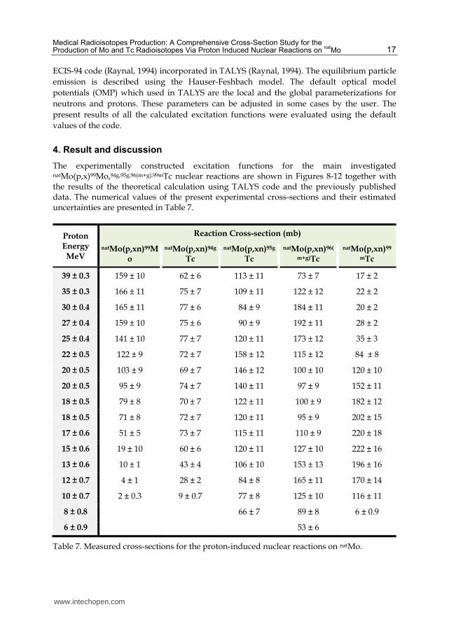
4. Result and discussion
The experimentally constructed excitation functions for the main investigated
natMo(p,x)99Mo,94g,95g,96(m+g),99mTc nuclear reactions are shown in Figures 8-12 together with the results of the theoretical calculation using TALYS code and the previously published data. The numerical values of the present experimental cross-sections and their estimated uncertainties are presented in Table 7.
Proton Energy MeV
Reaction Cross-section (mb)
natMo(p,xn)99Mo
natMo(p,xn)94g
Tc
natMo(p,xn)95g
Tc
natMo(p,xn)96(
m+g)Tc
natMo(p,xn)99
mTc
39 ± 0.3 159 ± 10 62 ± 6 113 ± 11 73 ± 7 17 ± 2
35 ± 0.3 166 ± 11 75 ± 7 109 ± 11 122 ± 12 22 ± 2
30 ± 0.4 165 ± 11 77 ± 6 84 ± 9 184 ± 11 20 ± 2
27 ± 0.4 159 ± 10 75 ± 6 90 ± 9 192 ± 11 28 ± 2
25 ± 0.4 141 ± 10 77 ± 7 120 ± 11 173 ± 12 35 ± 3
22 ± 0.5 122 ± 9 72 ± 7 158 ± 12 115 ± 12 84 ± 8
20 ± 0.5 103 ± 9 69 ± 7 146 ± 12 100 ± 10 120 ± 10
20 ± 0.5 95 ± 9 74 ± 7 140 ± 11 97 ± 9 152 ± 11
18 ± 0.5 79 ± 8 70 ± 7 122 ± 11 100 ± 9 182 ± 12
18 ± 0.5 71 ± 8 72 ± 7 120 ± 11 95 ± 9 202 ± 15
17 ± 0.6 51 ± 5 73 ± 7 115 ± 11 110 ± 9 220 ± 18
15 ± 0.6 19 ± 10 60 ± 6 120 ± 11 127 ± 10 222 ± 16
13 ± 0.6 10 ± 1 43 ± 4 106 ± 10 153 ± 13 196 ± 16
12 ± 0.7 4 ± 1 28 ± 2 84 ± 8 165 ± 11 170 ± 14
10 ± 0.7 2 ± 0.3 9 ± 0.7 77 ± 8 125 ± 10 116 ± 11
8 ± 0.8 66 ± 7 89 ± 8 6 ± 0.9
6 ± 0.9 53 ± 6
Table 7. Measured cross-sections for the proton-induced nuclear reactions on natMo.
www.intechopen.com

Radioisotopes – Applications in Bio-Medical Science 18
4.1 Excitation functions 4.1.1
natMo(p,xn)
99Mo
99Mo is produced by proton activation on natMo target via the contribution of two reaction channels 100Mo(p,pn)99Mo (Q= 8.3 MeV) and 100Mo(p,2p)99Nb (Q= 11.14 MeV) through the ┚- decay of the parent isotope 99Nb(15 s). The highest cross-section value of about 160 mb corresponds to Ep= 30 MeV. A comparison between our measured cross-sections and the previously reported data together with the theoretical calculations using TALYS code is presented in Figure 8. (Takács, et al., 2003) reported cross-section data up to 37 MeV and (Levkovskij, 1991) reported up to 29 MeV for 99Mo production on the enriched 100Mo isotope. Our measured values are consistent with the data presented by (Uddin, et al., 2004). The data reported by (Scholten, et al.,1999) are consist with our data in energy range lower than 22 MeV, although his results at the higher energies are scattered. Our results showed agreement with (Takács, et al., 2003) in low energy region. The data presented by (Levkovskij, 1991) are about 25% higher than our data. (Lagunas-solar, et al., 1991) reported numerical cross-section data that are much lower than our measured data and the other published data as well in the energy region above 20 MeV. A good agreement exists between the measured cross-sections and the TALYS code calculations within the experimental error and that fact confirms the reliability of our measured data.
Fig. 8. Excitation function of the natMo(p,x) reaction (full red dots with vertical and horizontal error bars) compared to some previously published results and the TALYS code calculations (curve).
4.1.2 nat
Mo(p,xn)94g
Tc 94Tc has two isomeric states, metastable state 94mTc (T½ = 52 min, 2+) and ground state (T½ = 4.86 h, 7+). We studied the excitation function for the ground state only due to the relatively short half-life of the metastable state. The contribution of the isomeric transition (IT< 0.1) for 94mTc is small enough to be neglected. Therefore, we can study each state separately by eliminating the interfering gamma rays from the measurements, such as 849.92 keV and
www.intechopen.com

Medical Radioisotopes Production: A Comprehensive Cross-Section Study for the Production of Mo and Tc Radioisotopes Via Proton Induced Nuclear Reactions on
natMo 19
871.08 keV as listed in Table 6. Mainly we used the 702.63 keV ┛-line, which has no interference with any other ┛-lines from any other produced isotopes in a cooling time of about 5 hours, to determine the cross section for 94gTc production. The present experimental excitation function for the reaction natMo(p,xn)94gTc is presented in
Figure 9 together with the previously published results and the calculated cross sections by
the used nuclear model code TALYS.
A good agreement is found between our measured cross sections and the ones reported by
(Bonardi, et al., 2002 and Uddin, et al., 2004) over the entire energy range. There is a
remarkable difference between the present results and the reported data by (Khandaker, et
al., 2007) especially for the energies lower than 20 MeV and above 30 MeV. The measured
cross sections by (Kormali, et al., 1976) show about 40% lower values than our data in the
energy range from 11-20 MeV. The TALYS code calculation is about 50% higher than our
measured data and higher than all the previously reported data sets.
Fig. 9. Excitation function of the natMo(p,x) reaction (full red dots) compared to some previously published results and the TALYS code calculations (curve).
4.1.3 nat
Mo(p,xn)95g
Tc 95Tc is formed in two different states: the longer lived isomeric state 95mTc (T½ = 61 d, 1/2-) and the shorter lived ground state 95gTc (T½ = 20 h, 9/2+). In this study, we report only the measured cross sections for 95gTc due to the difficulty in measuring the interfering characteristic ┛-rays for 95mTc as shown in Table 6. The 95gTc activity measurement was based on detecting the main ┛-line at 765.79 keV. A comparison of the present measured data with some previously reported data and the TALYS code calculations is shown in Figure 10. The cross section is only measurable at 8 MeV, then increases gradually due to the 95Mo (p,n) reaction. The contribution of the 96Mo (p,2n) reaction appears as a little plateau starting at about 12 MeV, while the 97Mo (p,3n) reaction contribution starts at about 20 MeV, creating another small peak. There is a good agreement between our experimental excitation function
www.intechopen.com

Radioisotopes – Applications in Bio-Medical Science 20
and the previously published data by (Bonardi, et al., 2002 and Khandaker, et al., 2007) within the experimental error, while the earlier presented study by (Khandaker, et al., 2006) shows 35% higher value than our experimental data at energies above 26 MeV. However, the data reported by (Birattari, et al., 2002) shows higher cross-section values in the proton energy range > 10 MeV. The presented data by (Uddin, et al., 2004) shows inconsistency with most of the other experimental data, , especially for the point at about 22 MeV. The TALYS code calculation results are in good consistency with our experimental data
within the experimental error, but there exists a small drop in the measured cross section
values in the higher values of the energy range.
Fig. 10. Excitation function of the natMo(p,x)95gTc reaction compared to some previously published results and the TALYS code calculations.
4.1.4 nat
Mo(p,xn)96(m+g)
Tc 96Tc is formed in two energy states: 96mTc (T½ = 51.5 min, 4+) that decays by 98% isomeric
transition to the ground state 96gTc (T½ = 4.28 d, 7+). In this study we measured the cross-
section of 96gTc using the main characteristic ┛-line 778.2 keV, while it was not possible to
measure the characteristic isomeric transition 34.28 keV of the metastable state due to the
intensive interfering of the X-rays. According to the short half-life and the high IT decay rate
of the metastable state, we can consider the measured cross section as the total cross section
of 96(m+g)Tc without measuring the metastable state independently. Figure 11 illustrates a
comparison between our measured cross sections and the available published data together
with the TALYS code calculations. Some findings can be summarized from this figure as
follows:
The first part of the curve is due to 96Mo(p,n) reaction. It starts to increase rapidly to form a peak at 12 MeV. Then it decreases slowly and forms a plateau in the range 16-21 MeV due to the contribution of the 97Mo(p,2n) and 98Mo(p,3n) reactions which start at
www.intechopen.com

Medical Radioisotopes Production: A Comprehensive Cross-Section Study for the Production of Mo and Tc Radioisotopes Via Proton Induced Nuclear Reactions on
natMo 21
11 and 19 MeV, respectively. The rapid increase in the cross-section values at energies higher than 22 MeV indicates the increasing contribution of the (p,3n) reaction.
Very good agreement is found in the energy range above 9MeV between the present
data and those reported by (Takács, et al., 2002; Uddin, et al., 2004 & Khndaker, et al
2006,2007).
The results by (Bonardi, et al., 2002) overestimate the cross-section value in the energy
range < 10 and >26 MeV.
The data by (Khandaker, et al., 2007) are somewhat low in the proton energy range
below 10 MeV.
An overall good agreement is found between the present experimental excitation function for 96(m+g)Tc formation and the calculated theoretical results by TALYS code and the recommended data (Takács, et al., 2002), within the experimental error.
Fig. 11. Excitation function of the natMo(p,x)96(m+g)Tc reaction compared to some previously published results and the TALYS code calculations.
4.1.5 nat
Mo(p,xn)99m
Tc
Three reactions contribute to the production of 99mTc by direct way are 98Mo(p,┛), 100Mo(p,2n), and indirect way by 100Mo(p,pn). Possibly, the highest contribution is from the 100Mo(p,2n)99mTc reaction (on the 9.63% 100Mo present in the highly chemically pure Mo sample). Activity of 99mTc was measured in this work by detecting the gamma peak at energy 140.5 MeV after the resolution of this peak as described before. The measured excitation function is compared with some earlier published data and the TALYS code calculations in Figurer 12. The data of (Takács, et al. 2003) and ( Kandaker, et al. 2007) fit nicely our measured data specially in the low energy part up to 20 MeV. At higher energies (Kandakar, et al. 2007) data clearly over estimate our results. The results of (Challan, et al. 2007) agree with our results except the last two points. The cross section data for (Scholtan, et al. 1999) are clearly lower than our values over the hall energy range. The TALYS
www.intechopen.com

Radioisotopes – Applications in Bio-Medical Science 22
calculations over estimate the present results, especially in the energy range lower than 18 MeV , while they fit, within the experimental errors in the higher range.
Fig. 12. Excitation function of the natMo(p,x)99mTc reaction compared to some previously published results and the TALYS code calculations.
4.2 Integral yield calculations
The integral yields, at the end of bombardment, for the production of the different isotopes
were derived using the measured excitation functions for the production of these
radioisotopes. The method was done by assuming the thick target as dividend to several
thin targets each of an equivalent thickness of about 0.5 MeV. The cross section at each thin
target is assumed constant, because of the small energy interval through the target. The
number of target atoms/cm2 was calculated using the target thickness, which reduce the
proton energy by 0.5 MeV. The differential yield produced in each thin target was calculated
using the following equation (5):
30( ) . . ( ).10 . 1.
btMBq
Y E N P E eA h
(5)
Whereas, 購博岫継岻 (mb) is the average cross section at a specific energy; N is the number of target atoms/cm2; λ is the decay constant for the produced isotopes; P is the number of incident protons/sec for (1 ┤A) and the irradiation time (tb= 1 h). We then calculated the integral target yield by summing up the differential yields. Figure 13 represents the values of the integral target yield for the studied reactions as a function of the proton energies. Obviously, the yields of the investigated radioisotopes increase with the proton energy and start to saturate at energy of about 30 MeV. The nearly saturation values for 99Mo, 94gTc, 95gTc, 96(m+g)Tc, and 99mTc are equal to 110, 600, 310, 90 and 910 MBq/┤A.h, respectively.
www.intechopen.com

Medical Radioisotopes Production: A Comprehensive Cross-Section Study for the Production of Mo and Tc Radioisotopes Via Proton Induced Nuclear Reactions on
natMo 23
For the production of 99mTc via cyclotron, it is highly recommended to use an enriched target of 100Mo to exclude all the other impurities by using the indirect 100Mo(p,pn)99Mo and the direct 100Mo(p,2n)99mTc nuclear reactions. From the present data we conclude that the optimum energy range for the production of 99mTc directly and indirectly using protons is Ep= 35-18 MeV, the integral target yield amounting to to 412 MBq/┤A.h to 1000 MBq/┤A.h at saturation with respect to the half lives of both 99Mo and 99mTc.
Fig. 13. Integral Yields for the natMo(p,x)99Mo,94g,95g,96(m+g),99mTc nuclear reactions calculated from the excitation functions measured in this work.
5. Conclusion
99mTc radioisotope is a very important medical radioisotope for diagnostic tests. In this work an alternative root of producing this isotope, either directly or through the generator 99Mo (99mTc ) , namely using cyclotrons, is introduced and discussed. The excitation functions for the different proton-induced nuclear reactions on natMo target are measured and compared with some previously measured data. This study aims to resolve some contradictions between the existing data, and to give a reliable data set for the production of 99mTc and some other isotopes of importance in nuclear medicine beside some impurities. Monitoring reactions on Al and Cu targets are also measured and compared with the recommended IAEA data sets, in order to give high degree of consistency to our results. The present excitation functions confirm some previously measured sets, while contradict with others. Theoretical code calculations using TALYS code are performed and show a good consistency with the measured cross section values. The code calculations can be used for cross section estimations, when not enough experimental data exist. Furthermore, the integral or thick target yields are estimated based on the measured excitation functions for all the investigated reactions. Finally, it is well known that for medical uses, enriched targets have to be used in the production to avoid the secondary produced unwanted impurities. While the studies on natural targets, gives an idea about the suitable energy range for maximum production of the wanted isotope and minimum of the impurities.
www.intechopen.com

Radioisotopes – Applications in Bio-Medical Science 24
6. Acknowledgment
We thank G. J. Kim, D. P. May, and the staff of the CI for delivering the stable beam of protons. One of the authors Dr. A. Alharbi wish to express her appreciation to the international Fulbright U.S. exchange Scholar Program. This work was supported in part by the United States Department of Energy Office of Nuclear Physics under award number DE-FG02-93ER40773, and the Texas A&M University Cyclotron Institute.
7. References
Al-Saleh, F.S.; Al-Harbi, A.A. & Azzam, A. (2006). Excitation functions of proton induced nuclear reactions on natural copper using a medium-sized cyclotron, Radiochim. Acta, 94, 391.
Audi, G.; Bersillon, O.; Blachot, J. & Wapstra, A. H. (2003). The NUBASE evaluation of nuclear and decay properties, Nuclear Physics A, 729, 3–128.
Birattari, C.; Bonardi, M.; Gini, L.; Groppi, F. & Menapace, E. (2002) J. Nucl. Scien. Tech., Suppl.2, 1302.
Bonardi, M.; Birattari, C.; Groppi, F. & Sabbioni, E. (2002). Thin-target excitation functions, cross-sections and optimised thick-target yields for natMo(p,xn)94g,95m,95g,96(m+g)Tc nuclear reactions induced by protons from threshold up to 44MeV. No Carrier Added radiochemical separation and quality control, Appl. Radiat. & Isot., 57, 617.
Challan, M.B.; Comsan, M.N.H. & Abou-Zeid, M.A. (2007) Thin Target Yields and Empire-II Predictions on the accelerator production of Technetium-99m, Journal of Nuclear and Radiation Physics, 2, 1, 1-12
Fabbender, M.; Novgorodov, A.F.; Rösch, F. & Qaim, S.M. (1994). Excitation functions of 93Nb(3He,xn)93m,g,94m,g,95m,gTc-processes from threshold up to 35 MeV :possibility of production of 94mTc in high radiochemical purity using a thermo chromatographic separation technique, Radiochim. Acta, 65, 215–221.
Firestone, R.B. (1998). Table of Isotopes, 8th edition, John Wiley & Sons, New York , USA. Glenn, D.; Heger, S. & Hladik, W. (1997), Comparison of Characteristics of Solution and
Conventional Reactors for Mo-99 Production, Nuclear Technology, Vol 118. Gull, K.; Hermanne, A.; Mustafa, M.G.; Nortier, F.M.; Oblozinsky, P.; Qaim, S.M.; Scholten,
B.; Shubin, Yu.; Takács, S.; Tárkányi, T.F. & Zhuang, Y. (2001). Charged particle cross-section database for medical radioisotope production: diagnostic radioisotopes and monitor reactions, Final Report of a Co-ordinated Research Project, IAEA-TECDOC-1211, IAEA, Vienna, Austria.
Helus, F. and Colombetti, L.G. (1980). Radionuclides Production. CRC Press Inc., Boca Raton, Florida.
Hohn, A.; Zimmermann, K.; Schaub, E.; Hirzel, W.; Schubiger, P.A. & Schibli, R. (2008). Production and separation of ‘‘non-standard’’ PET nuclides at a large cyclotron facility: the experiences at the Paul Scherrer Institute in Switzerland, Q. J., Nucl. Med. Mol. Imaging, 52, 145–150.
IAEA-TECDOC-1065, (1999). Production technologies for molybdenum-99 and technetium-99m, IAEA, Vienna, Austria, ISSN 1011-289.
IAEA-TECDOC-468, (2009). Cyclotron produced radionuclides: physical characteristics and production methods, IAEA, Vienna, ISSN 0074–1914, ISBN 978–92–0–106908–5.
Khan, F.M. (2003).The Physics of Radiation Therapy, Third Edition, Lippincott Williams & Wilkins, USA.
www.intechopen.com

Medical Radioisotopes Production: A Comprehensive Cross-Section Study for the Production of Mo and Tc Radioisotopes Via Proton Induced Nuclear Reactions on
natMo 25
Khandaker, M.U.; Meaze, A.K.M.M.H.; Kim, K.; Son, D.; Kim, G. & Lee, Y.S. (2006). Measurements of the proton-induced reaction cross-sections of natMo by using the MC50 cyclotron at the Korea Institute of Radiological and Medical Sciences, J. Korean Phys. Soc., 48, 821.
Khandaker, M.U.; Uddin, M.S.; Kim, K.S.; Lee, Y.S. & Kim, G.N. (2007). Measurement of cross-sections for the (p,xn) reactions in natural molybdenum, Nucl. Instrum. Methods Phys. Res., B 262, 171.
Koning, A. J.; Hilaire, S. & Duijvestijn, M.C. (2008). TALYS-1.0, Proceedings of the International Conference on Nuclear Data for Science and Technology - ND2007, April 22-27, 2007, Nice, France, eds. O. Bersillon, F. Gunsing, E. Bauge, R. Jacqmin and S. Leray, EDP Sciences, 211. Available online: http://www.talys.eu/home/
Kormali, S.M.; Swindle, D.L. & Schweikert, E.A. (1976). Charged particle activation of medium Z elements. II. Proton excitation functions, J. Radiat. Chem., 31, 437.
Krane; K.S. (1987). Introductory Nuclear Physics, John Wiley and Sons, Inc. New York, ISBN 978-812-6517-85-5, USA.
Lagunas-Solar, M.C.; Kiefer, P.M.; Carvacho, O.F.; Lagunas, C.A.; Ya Po Cha, (1991). J. Appl. Radiation Isotopes, 42, 463.
Lambrecht, R.M. & Montner, S.M. (1982). Production and radio chemical separation of 92Tc and 93Tc for PET, J. Labelled Compd. Radiopharm., 19, 1434–1435.
Levkovoskii, N. (1991). Middle mass nuclides (A=40-100) activation cross sections by medium energy (E+10-50 MeV) protons and -particles (Experiment and Systematics). Inter-Vesti, Moscow.
Loveland, W.; Morrissey, D.J. & Seaburg, G.T. (2006). Modern Nuclear Chemistry, John Wiley & Sons, ISBN: 978-047-1115-32-0, USA.
Lamberecht, R.M. (1979). Positron emitting radionuclides-present and future status, Proceedings second international symposium on radiopharmaceuticals, 19-22 March, Seattle, Washington. The Society of Nuclear Medicine Inc., New York.
Nickles, R.J.; Nunn, A.D.; Stone, C.K. & Christian, B.T., (1993). Technetium-94m -teboroxime-synthesis, dosimetry and initial PET imaging studies, J. Nucl. Med., 34, 1058–1066.
Piel, H.; Qaim, S.M. & Stöcklin, G. (1992). Excitation functions of (p,xn) reactions on natNi and highly enriched 62Ni – possibility of production of medically important radioisotope 62Cu at a small cyclotron, Radiochem. Acta, 57, 1.
Qaim, S.M.; Clark, J.C.; Crouzel, C.; Guillaume, M.; Helmeke, H.J.; Nebeling, B.; Pike, V.W. & Stöcklin, G. (1993). PET radionuclide production. In: Radiopharmaceuticals for Positron Emission Tomography. (Stöcklin, G., Pike, V. W., Eds.), Kluwer Academic Publishers, Dordrecht, The Netherlands, pp. 1–42.
Qaim, S.M. (2000). Production of high purity 94mTc for positron emission tomographic studies, Nucl. Med. Biol., 27, 323–328.
Qaim, S.M. (2001). Nuclear data relevant to the production and application of diagnostic radionuclides, Radiochim. Acta, 89, pp. 223–232.
Qaim, S.M. (2003). Cyclotron production of medical radionuclides, Handbook of nuclear chemistry 4, Kluwer Academic Publishers, Dordrecht, Netherlands.
Qaim, S.M. (2004). Use of cyclotron in medicine, Radiation Physics and Chemistry, 71, pp. 917-926. Raynal, J. (1994). Notes on ECIS-94, CEA Saclay Report No. CEA-N-2772. Reference Input Parameter Library, Available online: http://www-nds.iaea.org/ripl2/ Rösch, F. & Qaim, S.M. (1993). Nuclear data relevant to the production of the positron emitting
technetium isotope 94mTc via the 94Mo (p,n) reaction, Radiochim. Acta, 62, 115–121.
www.intechopen.com

Radioisotopes – Applications in Bio-Medical Science 26
Rösch, F.; Novgorodov, A.F. & Qaim, S.M. (1994). Thermo chromatographic separation of 94mTc from enriched molybdenum targets and its large scale production for nuclear medical application, Radiochim. Acta, 64, 113–120.
Sajjad, M. & Lambrecht, R.M. (1993). Cyclotron production of medical radionuclides, Nucl. Instrum. Meth., B79, 911–915.
Scholten, B.; Lambrecht, R.M.; Cogneau, M.; Ruiz, H.V. & Qaim, S.M. (1999). Excitation functions for the cyclotron production of 99mTc and 99Mo, J. of Applied Radiation and Isotopes, 51, 69-80
Sharp, P.F.; Germmell, H.G. & Murray, A.D. (3ed.) (2005), Practical Nuclear Medicine, ISBN-13: 978-1852338756, Springer-Verlag London Limited, USA.
Stőcklin, G.; Qaim, S.M. & Rösch, F. (1995). The impact of radioactivity on medicine, Radiochim. Acta, 70/71, 249-272.
T-16, Nuclear Physics Group, Theoretical Division of the Los Alamos National Laboratory (1997). Nuclear Information Service, Los Alamos, USA. Available online: http://t2.lanl.gov/data/data.html
Takács, S.; Tárkányi, F.; Sonck, M. & Hermanne, A. (2002). New cross sections and intercomparison of proton monitor reactions on Ti, Ni and Cu, Nucl. Instrum. Methods Phys. Res., B 188, 106.
Takács, S.; Szűcs, Z.; Tárkányi, F.; Hermanne, A. & Sonck, M. (2003). Evaluation of proton induced reactions on 100Mo:New cross sections for production of 99mTc and 99Mo, Journal of Radio analytical and Nuclear Chemistry, Vol. 257, No. 1, pp. 195.201
Tammemagi, H., Jackson, D. (2009). Half-Lives A Guide to Nuclear Technology in Canada, Oxford University Press, pp. 11-13, 156.
Tárkányi, F.; Takács, S.; Gul, K.; Hermanne, A.; Mustafa, M.G.; Nortier, M.; Obložinský, P.; Qaim, S.M.; Scholten, B.; Shubin, Yu.N. and Zhuang Y. (2001). Beam Monitor Reactions, IAEA-TECDOC-1211, IAEA, Vienna, p. 49, Updated version January, 2007. Available from: http://www-nds.iaea.org/medical
Troyer, G.L. & Schenter, R.E. (2009). Medical isotope development and supply opportunities in the 21st century, J. Radioanal. Nucl. Chem., 282:243–246, DOI 10.1007/s10967-009-0267-4.
Uddin, M.S.; Hagiwara, M.; Tárkányi, F.; Ditrói, F. & Baba, M. (2004). Experimental studies on the proton-induced activation reactions of molybdenum in the energy range 22-67 MeV, Appl. Radiat. & Isot., 60, 911.
Uddina, M.S. & Baba, M. (2008). Proton-induced activation cross-sections of the short-lived radionuclides formation on molybdenum, Appl. Radiat. & Isot., 66,208–214.
Ullyett, B. (1997), Chapter Five - Canada Enters the Nuclear Age, published for Atomic Energy of Canada Limited, McGill-Queen's University Press.
Van der Marck, S. C., Koning, A. J. & Charlton, K. E., (2010). The options for the future production of the medical isotope 99Mo, Eur J Nucl Med Mol Imaging, 37:1817–1820 DOI 10.1007/s00259-010-1500-7
Wolf, A.P.; Barclay Jones, W. (1983). Cyclotrons for biomedical radioisotope production, Radiochim. Acta, 34, 1.
Ziegler, J.F. (1995). TRIM 95.4 code: The Transport of Ions in Matter, IBM-Research, Yorktown, New York, USA.
Ziegler, J.F.; Biersack, J.P. & Littmark, U. (1985). The Stopping and Range of Ions in Solids. Vol. 1 of the Stopping and Ranges of Ions in Matter, Pergamon Press, New York.
www.intechopen.com
Radioisotopes - Applications in Bio-Medical ScienceEdited by Prof. Nirmal Singh
ISBN 978-953-307-748-2Hard cover, 320 pagesPublisher InTechPublished online 21, November, 2011Published in print edition November, 2011
InTech EuropeUniversity Campus STeP Ri Slavka Krautzeka 83/A 51000 Rijeka, Croatia Phone: +385 (51) 770 447 Fax: +385 (51) 686 166www.intechopen.com
InTech ChinaUnit 405, Office Block, Hotel Equatorial Shanghai No.65, Yan An Road (West), Shanghai, 200040, China
Phone: +86-21-62489820 Fax: +86-21-62489821
The book Radioisotopes - Applications in Bio-Medical Science contains two sections: Radioisotopes andRadiations in Bioscience and Radioisotopes and Radiology in Medical Science. Section I includes chapters onmedical radioisotope production, radio-labeled nano-particles, radioisotopes and nano-medicine, use ofradiations in insects, drug research, medical radioisotopes and use of radioisotopes in interdisciplinary fieldsetc. In Section II, chapters related to production of metal PET (positron emission tomography) radioisotopes,3-dimensional and CT (computed tomography) scan, SS nuclear medicine in imaging, cancer diagnose andtreatments have been included. The subject matter will by highly useful to the medical and paramedical staff inhospitals, as well as researchers and scholars in the field of nuclear medicine medical physics and nuclear bio-chemistry etc.
How to referenceIn order to correctly reference this scholarly work, feel free to copy and paste the following:
A. A. Alharbi, A. Azzam, M. McCleskey, B. Roeder, A. Spiridon,E. Simmons, V.Z. Goldberg, A. Banu, L. Tracheand R. E. Tribble (2011). Medical Radioisotopes Production: A Comprehensive Cross-Section Study for theProduction of Mo and Tc Radioisotopes Via Proton Induced Nuclear Reactions on natMo, Radioisotopes -Applications in Bio-Medical Science, Prof. Nirmal Singh (Ed.), ISBN: 978-953-307-748-2, InTech, Availablefrom: http://www.intechopen.com/books/radioisotopes-applications-in-bio-medical-science/medical-radioisotopes-production-a-comprehensive-cross-section-study-for-the-production-of-mo-and-tc

© 2011 The Author(s). Licensee IntechOpen. This is an open access articledistributed under the terms of the Creative Commons Attribution 3.0License, which permits unrestricted use, distribution, and reproduction inany medium, provided the original work is properly cited.