medical neurosciences 731 - global anatomy … neurosciences 731 spring 2002 ... patrick forcada,...
TRANSCRIPT
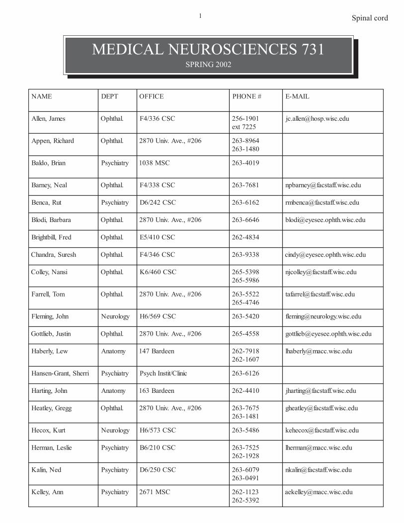
Spinal cord1
MEDICAL NEUROSCIENCES 731SPRING 2002
EMAN TPED ECIFFO #ENOHP LIAM-E
semaJ,nellA .lahthpO CSC633/4F 1091-6525227txe
drahciR,neppA .lahthpO 602#,.evA.vinU0782 4698-3620841-362
nairB,odlaB yrtaihcysP CSM8301 9104-362
laeN,yenraB .lahthpO CSC833/4F 1867-362 ude.csiw.ffatscaf@yenrabpn
tuR,acneB yrtaihcysP CSC242/6D 2616-362 ude.csiw.ffatscaf@acnebmr
arabraB,idolB .lahthpO 602#,.evA.vinU0782 6466-362 ude.csiw.hthpo.eeseye@idolb
derF,llibthgirB .lahthpO CSC014/5E 4384-262
hseruS,ardnahC .lahthpO CSC643/4F 8339-362 ude.csiw.hthpo.eeseye@ydnic
isnaN,yelloC .lahthpO CSC064/6K 8935-5626895-562
ude.csiw.ffatscaf@yellocjn
moT,llerraF .lahthpO 602#,.evA.vinU0782 2255-3626474-562
ude.csiw.ffatscaf@lerrafat
nhoJ,gnimelF ygolorueN CSC965/6H 0245-362 ude.csiw.ygoloruen@gnimelf
nitsuJ,beilttoG .lahthpO 602#,.evA.vinU0782 8554-562 ude.csiw.hthpo.eeseye@beilttog
weL,ylrebaH ymotanA needraB741 8197-2627061-262
ude.csiw.ccam@ylrebahl
irrehS,tnarG-nesnaH yrtaihcysP cinilC/titsnIhcysP 6216-362
nhoJ,gnitraH ymotanA needraB361 0144-262 ude.csiw.ffatscaf@gnitrahj
ggerG,yeltaeH .lahthpO 602#,.evA.vinU0782 5767-3621841-362
ude.csiw.ffatscaf@yeltaehg
truK,xoceH ygolorueN CSC375/6H 6845-362 ude.csiw.ffatscaf@xocehek
eilseL,namreH yrtaihcysP CSC012/6B 5257-3628291-262
ude.csiw.ccam@namrehl
deN,nilaK yrtaihcysP CSC052/6D 9706-3621940-362
ude.csiw.ffatscaf@nilakn
nnA,yelleK yrtaihcysP CSM1762 3211-2622935-262
ude.csiw.ccam@yellekea
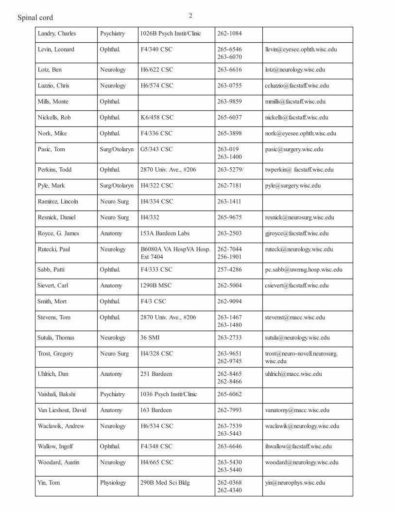
Spinal cord 2
selrahC,yrdnaL yrtaihcysP cinilC/titsnIhcysPB6201 4801-262
dranoeL,niveL .lahthpO CSC043/4F 6456-5620706-362
ude.csiw.hthpo.eeseye@nivell
neB,ztoL ygolorueN CSC226/6H 6166-362 ude.csiw.ygoloruen@ztol
sirhC,oizzuL ygolorueN CSC475/6H 5570-362 ude.csiw.ffatscaf@oizzulcc
etnoM,slliM .lahthpO 9589-362 ude.csiw.ffatscaf@sllimm
boR,sllekciN .lahthpO CSC854/6K 7306-562 ude.csiw.ffatscaf@sllekcin
ekiM,kroN .lahthpO CSC633/4F 8983-562 ude.csiw.hthpo.eeseye@kron
moT,cisaP nyralotO/gruS CSC343/5G 910-3620041-362
ude.csiw.yregrus@cisap
ddoT,snikreP .lahthpO 602#,.evA.vinU0782 /9725-362 ude.csiw.ffatscaf@nikrepwt
kraM,elyP nyralotO/gruS CSC223/4H 1817-262 ude.csiw.yregrus@elyp
nlocniL,zerimaR gruSorueN CSC433/4H 1141-362
leinaD,kcinseR gruSorueN 233/4H 5769-562 ude.csiw.grusoruen@kcinser
semaJ.G,ecyoR ymotanA sbaLneedraBA351 3052-362 ude.csiw.ffatscaf@ecyorjg
luaP,ikcetuR ygolorueN .psoHAVpsoHAVA0806B4047txE
4407-2621091-652
ude.csiw.ygoloruen@ikcetur
ittaP,bbaS .lahthpO CSC333/4F 6824-752 [email protected]
lraC,treveiS ymotanA CSMB0921 4005-262 ude.csiw.ffatscaf@treveisc
troM,htimS .lahthpO CSC3/4F 4909-262
moT,snevetS .lahthpO 602#,.evA.vinU0782 7641-3620841-362
ude.csiw.ccam@tsnevets
samohT,alutuS ygolorueN IMS63 3372-362 ude.csiw.ygoloruen@alutus
yrogerG,tsorT gruSorueN CSC823/4H 1569-3625479-262
naD,hcirlhU ymotanA needraB152 5648-2626648-262
ude.csiw.ccam@hcirlhu
ihskaB,ilahsiaV yrtaihcysP cinilC/titsnIhcysP6301 2606-562
divaD,tuohseiLnaV ymotanA needraB361 3997-262 ude.csiw.ccam@ymotanav
werdnA,kiwalcaW ygolorueN CSC435/6H 9357-3623445-362
ude.csiw.ygoloruen@kiwalcaw
flognI,wollaW .lahthpO CSC843/4F 6466-362 ude.csiw.ffatscaf@wollawhi
nitsuA,dradooW ygolorueN CSC566/4H 0345-3620445-362
ude.csiw.ygoloruen@dradoow
moT,niY ygoloisyhP gdlBicSdeMB092 8630-2620434-262
ude.csiw.syhporuen@niy

Spinal cord3
Medical Neurosciences 731
YRAURBEFNUS NOM EUT DEW RUHT IFGT TAS
NAJ72
NAJ 82 92NAJ8: :erutceL00
noitcudortnIgnitraH.rD
llamS00:01:spuorg
niarBssorGnoitatneirO
03NAJ:erutceL00:8
snmuloclasroDgnitraH.rD
1300:8 :erutceL
syslaretaloretnA .)SLA( lasroD
ebereconips all r)TCSD(tcart
erutceL00:01 :-ocitroclaretaL
lanips (tcart )TSCLnrohlartneVdna
gnitraH.rD
1:erutceL00:8nrohlaretaLgnitraH.rD
2
3 4:erutceL00:8llatignignirB
rehtegotgnitraH.rD
5:erutceL00:8,ssenkaeW
yhtapoymztoL.rD
:erutceL00:01esaesidCHAtcefedTMN
ztoL.rDseihtapolucidaR
ikcetuR.rD
6:erutceL00:8noisserpmoCseihtapoleymikcetuR.rD
7:erutceL00:8
stnioPmetsniarB1 dna 2
gnitraH.rD
00:01 erutceLstnioPmetsniarB
3 dna 4gnitraH.rD
8:erutceL00:8
stnioPmetsniarB5 dna 6
gnitraH.rD
9
01 11
ygolohtaPmaxE
21:erutceL00:8
stnioPmetsniarB7 dna 8
gnitraH.rD
llamS00:01:spuorg lacinilC
seidutsesac
31:erutceL00:8
stnioPmetsniarB9 dna 01
gnitraH.rD
41:erutceL00:8
stnioPmetsniarB11 dna 21
gnitraH.rD:erutceL00:01
tnioPmetsniarB31
51:erutceL00:8
tnioPmetsniarB31
gnitraH.rD00:5 mp lanoitpo
weiveredilsneedraB041
gnitraH.rD
61
71 81:erutceL00:8
tnioPmetsniarB31
gnitraH.rD
91:erutceL00:8
tnioPmetsniarB51dna41gnitraH.rD
:erutceL00:01tneitaP
noitatneserp
02:erutceL00:8
stnioPmetsniarB61 dna 71
gnitraH.rD
12:erutceL00:8
stnioPmetsniarB81 dna 91
gnitraH.rD:erutceL00:01
stnioPmetsniarB12,02 dna 22
gnitraH.rD
22:erutceL00:8
stnioPmetsniarB32 dna 42
gnitraH.rD00:5 mp lanoitpo
weiveredilsneedraB041
gnitraH.rD
32
42 52ygoloisyhP
maxE
62llamS00:01
:spuorgesaclacinilC
seiduts
7200:1 :erutceL
noitargetnI
82:erutceL00:8noitargetnI
00:01:spuorgllamS
metsniarBnoitcessid

Spinal cord 4
Medical Neurosciences 731
HCRAMNUS NOM EUT DEW RUHT IFGT TAS
1MA00:8
noitargetnIikcetuR.rD
00:5 mp lanoitpoweiveredils
needraB041gnitraH.rD
2WEIVERMA00:9
041needraB
eerFstunuod
3 4MA00:8
IMAXE
needraB041
5:erutceL00:01
6 7 8 9
01 11 21 31 41 51 61
71 81 91 02 12 22 32
4213
52 62 72 82 92 03
Spinal cord5
MEDICAL NEUROSCIENCES 731
Medical Neurosciences is aimed at capturing the excitement and relevance of the dynamicfield of basic and clinical neuroscience. Our goal is to present an overview of important and timelyconcepts regarding the structure and function of the nervous system. When you leave our course youshould be able to handle the problems (listed in the box below) that the American Academy ofNeurology is distributing to lay people to “talk to your physician” about. Every doctor should knowthe serious implications of these.
Many times in your career, no matter what type of doctor you become, both family membersand patients will tell you these things. It is important to have several important diseases in yourmind so your patients will avoid serious neurologic dysfunction and you will be known as a compe-tent doctor, instead of one who gives false reassurance. Know the questions to ask and the signs tolook for.
1. Dizziness 7. Unsteadiness2. Headache 8. Tremors/ Twitches3. Numbness/Tingling 9. Head injury4. Memory/Concentration loss 10. Sleep problems5. Blackouts/Seizure 11. Sudden vision change6. Muscle weakness/Pain 12. Slurred speech
7 credits
Lectures are usually scheduled at 8AM on M, T, W, Th and F in 140 Bardeen and at 10AM onTuesdays and Thursdays
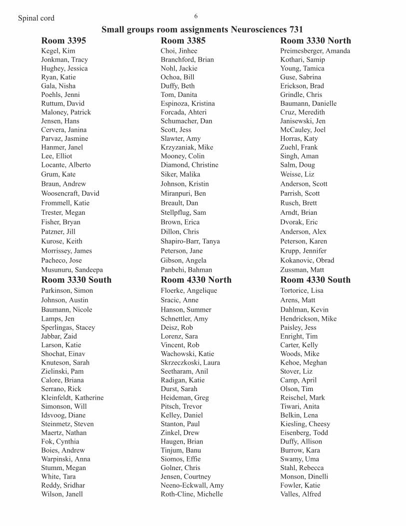
Small group/lab discussions are held at the normal 10AM lecture time on Tuesdays and Thursdaysin rooms 3330N, 3330S, 4330N, 4330S, 3385, and 3395. Your room assignment is listed on thefollowing page. Check your schedule each day for small group meetings.
During the semester we will strive to:• place you in a supportive environment• give you timely feedback on how well you understand the material• make the instruction problem-centered• help you to be active rather than passive learners
We cannot overemphasize how important it is to:• read the material before coming to lecture• come to the lecture, relax, and just listen• answer the practice questions• attend all small group discussions• enjoy learning about the nervous system and have a great time
Medical Neurosciences 731
Spinal cord 6
Small groups room assignments Neurosciences 731Room 3395 Room 3385 Room 3330 NorthKegel, Kim Choi, Jinhee Preimesberger, AmandaJonkman, Tracy Branchford, Brian Kothari, SamipHughey, Jessica Nohl, Jackie Young, TamicaRyan, Katie Ochoa, Bill Guse, SabrinaGala, Nisha Duffy, Beth Erickson, BradPoehls, Jenni Tom, Danita Grindle, ChrisRuttum, David Espinoza, Kristina Baumann, DanielleMaloney, Patrick Forcada, Ahteri Cruz, MeredithJensen, Hans Schumacher, Dan Janisewski, JenCervera, Janina Scott, Jess McCauley, JoelParvaz, Jasmine Slawter, Amy Horras, KatyHanmer, Janel Krzyzaniak, Mike Zuehl, FrankLee, Elliot Mooney, Colin Singh, AmanLocante, Alberto Diamond, Christine Salm, DougGrum, Kate Siker, Malika Weisse, LizBraun, Andrew Johnson, Kristin Anderson, ScottWoosencraft, David Miranpuri, Ben Parrish, ScottFrommell, Katie Breault, Dan Rusch, BrettTrester, Megan Stellpflug, Sam Arndt, BrianFisher, Bryan Brown, Erica Dvorak, EricPatzner, Jill Dillon, Chris Anderson, AlexKurose, Keith Shapiro-Barr, Tanya Peterson, KarenMorrissey, James Peterson, Jane Krupp, JenniferPacheco, Jose Gibson, Angela Kokanovic, ObradMusunuru, Sandeepa Panbehi, Bahman Zussman, MattRoom 3330 South Room 4330 North Room 4330 SouthParkinson, Simon Floerke, Angelique Tortorice, LisaJohnson, Austin Sracic, Anne Arens, MattBaumann, Nicole Hanson, Summer Dahlman, KevinLamps, Jen Schnettler, Amy Hendrickson, MikeSperlingas, Stacey Deisz, Rob Paisley, JessJabbar, Zaid Lorenz, Sara Enright, TimLarson, Katie Vincent, Rob Carter, KellyShochat, Einav Wachowski, Katie Woods, MikeKnuteson, Sarah Skrzeczkoski, Laura Kehoe, MeghanZielinski, Pam Seetharam, Anil Stover, LizCalore, Briana Radigan, Katie Camp, AprilSerrano, Rick Durst, Sarah Olson, TimKleinfeldt, Katherine Heideman, Greg Reischel, MarkSimonson, Will Pitsch, Trevor Tiwari, AnitaIdsvoog, Diane Kelley, Daniel Belkin, LenaSteinmetz, Steven Stanton, Paul Kiesling, CheesyMaertz, Nathan Zinkel, Drew Eisenberg, ToddFok, Cynthia Haugen, Brian Duffy, AllisonBoies, Andrew Tinjum, Banu Burrow, KaraWarpinski, Anna Siomos, Effie Swamy, UmaStumm, Megan Golner, Chris Stahl, RebeccaWhite, Tara Jensen, Courtney Monson, DinelliReddy, Sridhar Neeno-Eckwall, Amy Fowler, KatieWilson, Janell Roth-Cline, Michelle Valles, Alfred

Spinal cord7
EXAMS: There will be three exams and each is worth approximately 33 1/3% of your final grade.
GRADING: A=96% and above, A/B=92%-95%, B=87%-91%, B/C=80%-86%, C=below 80%.
IN ORDER TO RESCHEDULE ANY EXAM YOU MUST GET PERMISSION PRIORTO THE EXAM FROM ASSOCIATE DEAN OF STUDENTS MIKEL SNOW
Composition of examsEach exam will consist of several parts. The first part (about 60-70% of the questions) will be a
test of your knowledge of the material that has been presented in lectures and in the course book. Youwill receive considerable help and “coaching” with this material. The second part (10-20%) will berelated to web-based, self learning excercises. In particular, you will need to visit designated websitesfrom which you will learn on your own. The aim of this exercise is to simulate a situation on the wardwhere you need to gather information quickly from the nearest comptuer with web access. Since youdon’t have a lot of time, I will suggest the amount of time you should spend at each website to handlethe questions on the exam. I don’t want you spending days and days! Finally, the third part of eachexam (about 20-30%) will consist of clinical vignettes, similar to the case histories that you will do insmall groups. These vignettes are similar to those on the National Boards Part I and are designed totest your integrative problem solving skills!!
Medical Neurosciences 731

Spinal cord 8
Medical Neurosciences 731
SPINAL CORDSelf learn websites 10MRI orientation 11Weigert stained spinal cord sections 15Point 1. Dorsal Columns 23Point 2. Anterolateral System 41Point 3. Dorsal Spinocerebellar Tract 53Point 4. Lateral Corticospinal Tract 63
Problem Solving Answers-Orientation and Points 1-4 74
Point 5. Ventral Horn 75Point 6. Lateral (Intermediolateral) Horn 81Point 7. Bringing it all together 87Point 8. Practice Questions on Somatotopic Organization 105
Problem Solving Answers-Points 5-8 and Integration 111
Clinical vignette 115Weakness; myopathy, anterior horn cell diseases, neuropathies and neuromuscular transmission defects 119
Problem Solving Answers 142
Radiculopathy 143Problem Solving Answers 156
Compression Myelopathy 157Problem Solving Answers 171
Clinical Integration 172Problem Solving Answers 178
CASE HISTORIES SPINAL CORD
Spinal cord Case Histories I - VI 179
BRAIN STEMClinical vignette 191Introduction/Orientation 193Weigert sections 201Point 1. Pyramids (Approximate Reading Time=20min.) 211Point 2. Anterolateral System (Reading Time=12 min.) 219Point 3. Spinal nucleus and tract V (Reading Time=20 min.) 225Point 4. Nucleus Gracilis-Cuneatus-Medial Lemniscus (Reading Time=12 min.) 235Point 5. Accessory Cuneate Nucleus (Reading Time=20 min.) 244
Problem Solving Answers-Points 1-5 254
Point 6. Inferior Olivary Complex (Reading Time=10 min.) 255Point 7. Hypoglossal Nucleus (Reading Time=10 min.) 263Point 8. Dorsal Motor Nucleus of X (Reading Time=10 min.) 271Point 9. Nucleus Ambiguus (Reading Time=15 min) 277Point 10. Inferior Salivatory Nucleus (Reading Time=10 min.) 287
Problem Solving Answers-Points 6-10 292
MODULE 1

Spinal cord9
Point 11.Nucleus and Tractus Solitarius (Reading Time=20 min.) 295Point 12.Dorsal & Ventral Cochlear Nuclei (Reading Time=15 min) 305Point 13. Vestibular Nuc.-Abducens Nuc. (Reading Time=60 min.) 313Point 14. Motor Nucleus VII (Reading Time=15 min.) 343Point 15.Superior Salivatory-Lacrimal Nuc. (Reading Time=10 min.) 351
Problem Solving Answers-Points 11-15 358
Point 16. Pontine Grey-Middle Cerebellar. Ped. (Reading Time=10 min.) 359Point 17.Motor-Chief Sens.-Mesencep. V (Reading Time=15 min.) 369Point 18.Superior Cerebellar Peduncle (Reading Time=10 min.) 383Point 19.Trochlear Nucleus (Reading Time=10 min.) 391Point 20.Substantia Nigra (Reading Time=5-10 min.) 401
Problem Solving Answers-Points 16-20 408
Point 21. Oculomotor Nuclear Complex (Reading Time=10 min.) 411Point 22. Red Nucleus (Reading Time=10 min.) 419Point 23.Superior Colliculus (Reading Time=10 min.) 427Point 24.Periaquductal Grey (Reading Time=10 min.) 437
Problem Solving Answers-Points 21-24 443
Deficits related to specific brain stem regions (medulla, pons or midbrain) 444
Cranial Nerve Review 453
Cranial nerve review Answers 470
BRAIN STEM CASE HISTORIES
Case Histories VII - XI 471
Brain Dissection Lab I 478
Brain Dissection Lab II 483
Medical Neurosciences 731

Spinal cord 10
SELF LEARNING WEBSITES FOR MODULE I (All can be directly accessed from www.anatomy.wisc.edu)
These are relatively short readings that you are to do on your own. There are several rules to thisgame. First, I cannot help you!! We know that this is 180 degrees different from the way we arepresenting the course book material, but you need to do these exercises on your own so as to becomeindependent learners. In other words, we will help you with the basic important facts about the brainand spinal cord so that you are in a position to then learn a little bit more on your own. I suggest thatyou do not wait till the night before the exam to do this web based learning. In order to give you afeel for what I want you to get out of these readings, I have included some practice questions for selflearning reading #11. You will see from these practice questions that what you will be tested onfrom these readings are things that are related to what you have learned thus far in thecourse. The new information should broaden your horizons and pique your interest in clinicalneurology, and help to reinforce the topics and concepts that we have stressed.
One final note. As you become more web-based in your learning, you will realize that material inour course book might sometimes differ from what you read on the internet. So the rule is, go bythe course book if there are conflicting data. I know that this sounds like a cop-out, but asyou become doctors you will realize that textbooks (and research and teaching groups) differ in theirterminology and interpretations, so WHEN IN DOUBT, THE MODULE IS THE LASTWORD!!
1. Cervical Spondylotic Myelopathyhttp://www.spine-health.com/topics/cd/undermy/undermy01.html
2. Trigeminal Neuralgiahttp://www.geocities.com/HotSprings/Villa/7047/fran.htm
3. Spina Bifidahttp://spinabifida.org/Spina%20Bifida.htm
4. Cerebellopontine Angle Meningiomahttp://neurosurgery.mgh.harvard.edu/rounds/mening17.htmsee alsohttp://www.cid.ch/TEACH/AF/AF15.html for a smaller lesion
5. Autonomic Hyper-Reflexia (or Dysreflexia)http://rehabnurse.org/ce/010299/auto.htm
6. Blood supplyhttp://www.anatomy.wisc.edu/brainstem/bldsup.html
Self learn
Spinal cord11
LET’S GET ORIENTED
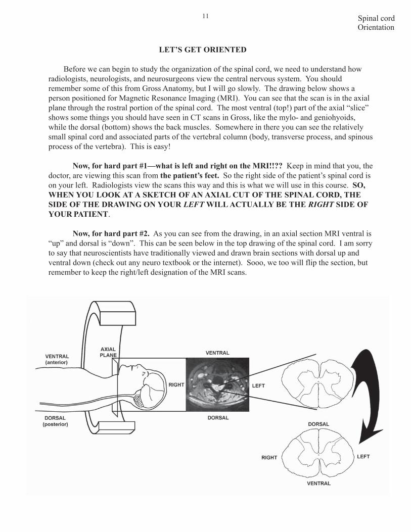
Before we can begin to study the organization of the spinal cord, we need to understand howradiologists, neurologists, and neurosurgeons view the central nervous system. You shouldremember some of this from Gross Anatomy, but I will go slowly. The drawing below shows aperson positioned for Magnetic Resonance Imaging (MRI). You can see that the scan is in the axialplane through the rostral portion of the spinal cord. The most ventral (top!) part of the axial “slice”shows some things you should have seen in CT scans in Gross, like the mylo- and geniohyoids,while the dorsal (bottom) shows the back muscles. Somewhere in there you can see the relativelysmall spinal cord and associated parts of the vertebral column (body, transverse process, and spinousprocess of the vertebra). This is easy!
Now, for hard part #1—what is left and right on the MRI!!?? Keep in mind that you, thedoctor, are viewing this scan from the patient’s feet. So the right side of the patient’s spinal cord ison your left. Radiologists view the scans this way and this is what we will use in this course. SO,WHEN YOU LOOK AT A SKETCH OF AN AXIAL CUT OF THE SPINAL CORD, THESIDE OF THE DRAWING ON YOUR LEFT WILL ACTUALLY BE THE RIGHT SIDE OFYOUR PATIENT.
Now, for hard part #2. As you can see from the drawing, in an axial section MRI ventral is“up” and dorsal is “down”. This can be seen below in the top drawing of the spinal cord. I am sorryto say that neuroscientists have traditionally viewed and drawn brain sections with dorsal up andventral down (check out any neuro textbook or the internet). Sooo, we too will flip the section, butremember to keep the right/left designation of the MRI scans.
Orientation

Spinal cord 12
Now, I know you are really P.O.’ed about all of this flipping around, etc. Well, take my wordfor it, you will get over it and start flying through the course. As you will find out, I will constantlytest you on important concepts and you will not have any trouble with these drawings. To show youhow much you already know, here are several practice questions to help alleviate your stress. Thatis, you will get the questions correct and see that you are in fine shape. Here we go, and don’t hangup without doing these.
The spinal cord is an extremely important component of the central nervous system. For thebasic science component of this module, I have tried to organize the material in a way that will makeyou comfortable with the fundamental organization of the spinal cord. This will hopefully prepareyou for solving the clinical case problems that the clinicians spring on you!!
I am aware of what you have learned about the spinal cord in Gross Anatomy, Histology andPhysiology, and therefore will not dwell on gross structure, meninges or muscle spindles. What Iwill dwell on ad nauseum is the organization of ascending sensory pathways and descending motorpathways in the spinal cord. Following a discussion of each major pathway or topic, I have inserteda group of practice questions. BE SURE TO COMPLETE THESE PRACTICE QUESTIONS!These questions will help you to evaluate your progress as we move through the various topics andbuild a more global view of spinal cord organization and function(s). If you get the answers correct,you are doing great! Trust me, there are no tricks!!! However, if you miss a few questions, go backand review the material that you have not understood (or that I have not clearly written ordiscussed??). Then go forward!!!! As you go through the spinal cord material the practice questionsbuild on earlier material (and will therefore be more inclusive and difficult). If you do the questionsfaithfully, you will have the material well in hand. You will then be ready to problem solve andimpress the clinicians.
Orientation

Spinal cord13
Refer to the Table of contents for the Problem solving ANSWER set page num-bers.
1. Which of the following statements is TRUE regarding the drawing below?
A. the area designated C the ventral surface of the bodyB. the area designated A is the dorsal horn of the spinal cordC. the area designated E is the left side of the patient’s cerebral hemisphereD. the area designated D is the dorsal surface of the body
E. the area designated B is the dorsal surface of the body2. Which of the following statements is TRUE regarding the drawing below?
A. the area designated A is the left side of the patients MRIB. the area designated C is the right side of the patient’s MRIC. the area designated E shows the mid-sagittal planeD. the area designated D is the ventral surface of the patient’s MRIE. the area designated B is the ventral surface of the patient’s MRI
Orientation
Spinal cord 14
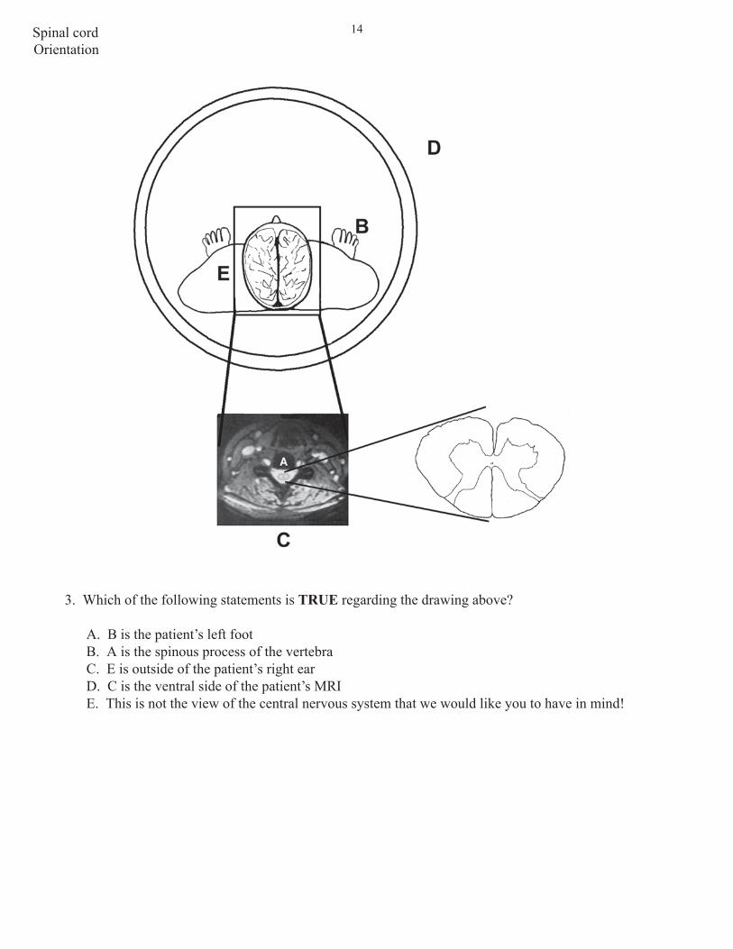
3. Which of the following statements is TRUE regarding the drawing above?
A. B is the patient’s left footB. A is the spinous process of the vertebraC. E is outside of the patient’s right earD. C is the ventral side of the patient’s MRIE. This is not the view of the central nervous system that we would like you to have in mind!
Orientation

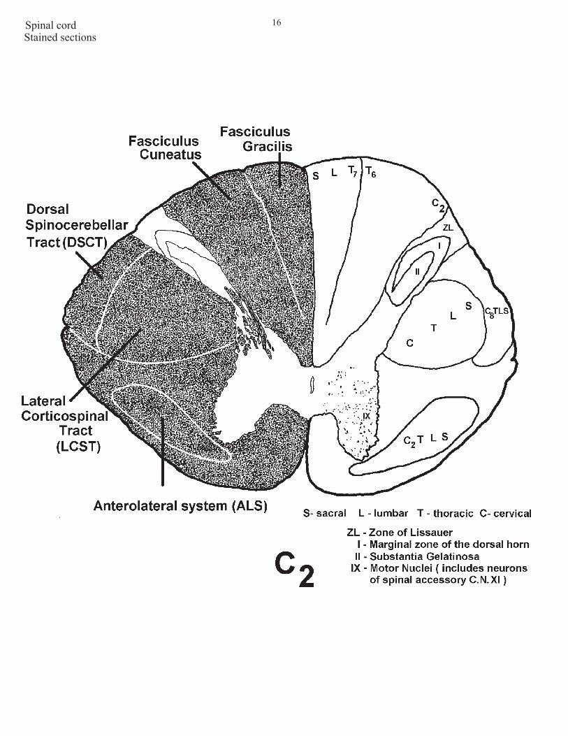
Spinal cord15
Stained sections
Spinal cord 16
Stained sections
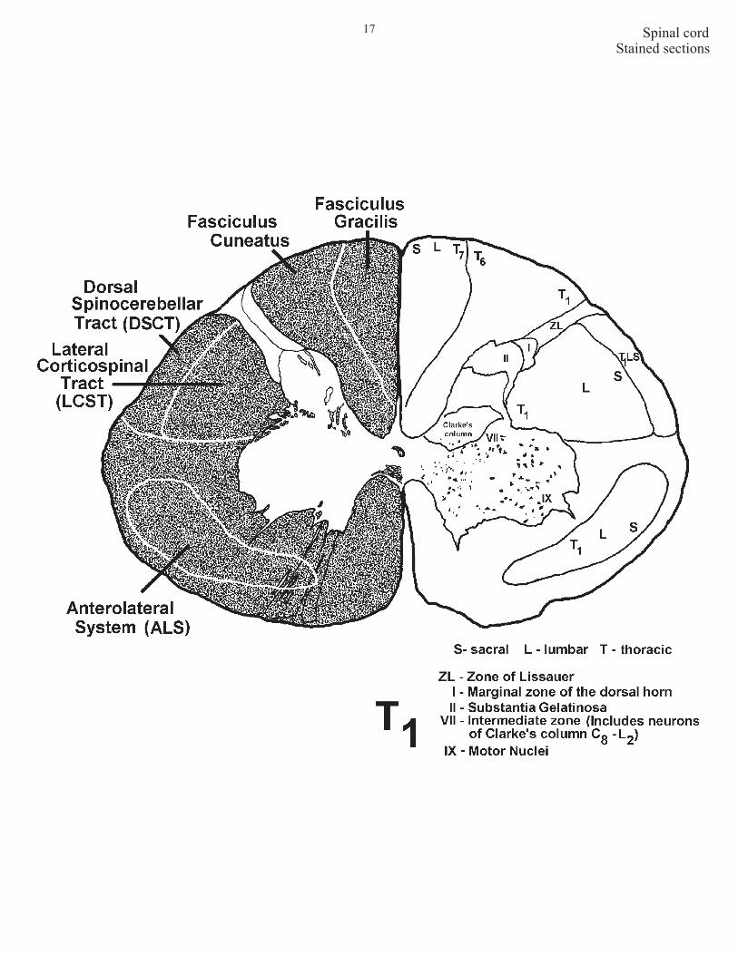
Spinal cord17
Stained sections
Spinal cord 18Stained sections

Spinal cord19
Stained sections

Spinal cord 20
CLINICAL VIGNETTE I
An important part of learning is to use the information (facts!) that you have learned in onesetting (i.e lecture), to solve more in-depth problems that involve findings that are beyond those that areneurological. This “case-based” approach is used on National Boards and in the third and fourth years.Below you will find a vignette that will completely bewilder you. Keep in mind that at the end of thespinal cord module you will understand all of the neurological symptoms presented here and that is ourprimary goal. However, you should pay attention to all of the findings as you need to learn to look at the“big picture.”
Case history
Ray Farrell is a 25-YOM who presents with pain in the left chest, anteriorly, around the left side,and in the adjacent left back. This pain began 4 months previously. It is severe, constant, and isworse when he moves his body but not when he moves his arm. The pain does not radiate into thearm. Taking a deep breath or walking do not make it worse. There is no numbness, peculiarfeeling or loss of feeling in the chest, arm or elsewhere on the body. He has wondered about somedecreased coordination in the left leg. He has not had any intellectual dysfunction, visual disturbance,dizziness, vertigo, or loss of bladder or bowel control. He has not had any temporary spells ofneurologic dysfunction. There has been no recent trauma to the area.
Past Medical History
There are no previous neurologic problems, serious illnesses, or abnormalities in the review ofsystems. He does not take any medications other than ibuprofen for pain, and has no allergies.
Social History
He does not smoke or drink alcohol. He has not been exposed to any occupational chemicals. Mr.Farrell is unmarried and lives alone. He is employed by a heating & cooling company as an air-conditioning repairman.
Family History
There is no family history of neoplasia or neurological diseases, early cerebrovascular or coronaryartery disease, diabetes, or hypertension.
PHYSICAL EXAMINATION:General: This is an alert person in no distress.
Vital signs:Temperature = 98.6°FBlood pressure = 130/60Pulse = 68 and regularRespiration = 12Height = 5’10"Weight = 165 lbs.

Spinal cord21
HEENT (head, eyes, ears, nose throat): Fundoscopy revealed normal optic nerves withoutpapilledema or optic atrophy, normal arteries without AV nicking, emboli,hemorrhages or exudates. Visual acuity normal. Visual fields normal.
Neck: Neck was supple and without lymphadenopathy. No carotid bruits are present.
Skin: No cutaneous lesions.
Chest: Clear to auscultation and percussion.
Heart: Normal heart sounds were present. No cardiac murmurs were present.
Extremities: Normal. There was no cyanosis, edema, or skin lesions. Arterial pulses were normal.
Neurologic: Normal function of cranial nerves II-XII. The motor strength and fine coordinationwere normal, except for slight weakness in the left lower extremity. Motor function isnormal in the left upper extremity (LUE), RUE, and RLE. Gait is normal except forminimal dysfunction of the LLE. Finger to finger and heel to shin are normal. The deeptendon reflexes are normal in all 4 extremities, except the left knee jerk and left anklejerk are slightly more active than the corresponding RLE reflexes. Direction of plantarreflex was downward bilaterally. Temperature sensation is not as well appreciated inthe RLE up to just below the right costal margin. Temperature sensation is normalabove the costal margin on the right and is normal on the entire left side of the body. Nosacral sparing of temperature sensation is present. Proprioceptive sensation is minimallydecreased in the LLE, but is normal in the RLE and both UE’s. No bruit is present overthe back.
Pinprick sensation is normal above the costal margin on the right and is normal on theleft.
LABORATORY TEST RESULTS:
CBC:Hgb = 16 GM/dl (14.0 - 18.0)Hct = 44% (40 - 54)WBC = 6 K/µL (5 - 10)Platelets = 220 K/cc (150 - 400)PT = 11.2 sec (9.5 - 12.5)PTT = 28 sec (23 - 35)
Routine Chemistry:BUN = 11 mg/dl (5 - 24)Creatinine = 1.0 mg/dl (0.7 - 1.3)Glucose = 80 mg/dl (65 - 115)Na+ = 139 mmol/l (136 - 146)K+ = 4.1 mmol/l (3.7 - 5.3)Cl- = 104 mmol/l (101 - 111)CO
2= 22 mmol/l (21 - 31)

Spinal cord 22
Chest x-ray: Normal heart and lungs without any masses.ECG: Normal.Thoracic vertebral x-rays: Normal.
MRI of the thoracic cord: Reveals an extra medullary intradural mass at T-5 on the left.
The mass was surgically removed without complication. The pathological diagnosis was meningioma. The patient made a full recovery.
This is a case of the gradual onset of localized spinal cord dysfunction. Make sure that you candifferentiate between segmental spinal cord and spinal tract signs and between upper motor neuronand lower motor neuron weakness of the lower extremity.
The tumor is on the left. The physical exam and MRI define the lesion and surgical removal is theonly reasonable option. Lumbar puncture or biopsy have no role in this case—they don’t affectmanagement.This patient could present to an office or clinic or an emergency department—the approach to thecase would be the same, since the symptoms had been present for 4 months.

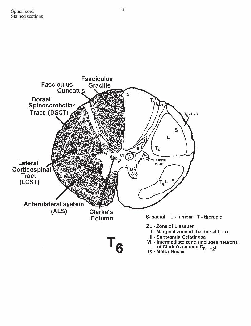
Spinal cord23
The spinal cord is comprised of an outer zone of white matter and a butterfly-shaped central component ofcells and fibers (grey [or gray] matter). The peripherally located white matter consists of three funiculi orcolumns (funiculus = L., little cord) dorsal, lateral and ventral. I want to focus now on the ascendingsensory pathways within the dorsal funiculus, or dorsal columns.
1 DORSAL COLUMNS(Fasciculus Gracilis and Cuneatus)
Dorsal columns

Spinal cord 24
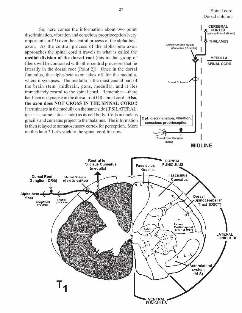
As shown below, the dorsal columns convey 2 point discrimination, vibration and consciousproprioception to nuclei in the medulla. These nuclei then send the information to the opposite side(contralateral) thalamus. Cells in the thalamus then project to the cerebral cortex where theperception of stimuli occurs.
Dorsal columns

Spinal cord25
Now that we have an overview of the pathway let’s get down to the nitty gritty (before we goto the Kohl Center). All incoming (afferent) information to the spinal cord is conveyed via thedorsal root fibers. Cells in dorsal root ganglia (DRG) possess two processes, one that passesperipherally to pick up information from a sensory receptor and one that passes centrally into thespinal cord. In the case of the dorsal columns, these axons are called alpha-beta fibers.
Dorsal columns
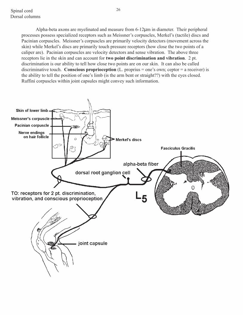
Spinal cord 26
Alpha-beta axons are myelinated and measure from 6-12µm in diameter. Their peripheralprocesses possess specialized receptors such as Meissner’s corpuscles, Merkel’s (tactile) discs andPacinian corpuscles. Meissner’s corpuscles are primarily velocity detectors (movement across theskin) while Merkel’s discs are primarily touch pressure receptors (how close the two points of acaliper are). Pacinian corpuscles are velocity detectors and sense vibration. The above threereceptors lie in the skin and can account for two point discrimination and vibration. 2 pt.discrimination is our ability to tell how close two points are on our skin. It can also be calleddiscriminative touch. Conscious proprioception (L. proprius = one’s own; ceptor = a receiver) isthe ability to tell the position of one’s limb (is the arm bent or straight??) with the eyes closed.Ruffini corpuscles within joint capsules might convey such information.
Dorsal columns
Spinal cord27
So, here comes the information about two pointdiscrimination, vibration and conscious proprioception (veryimportant stuff!!) over the central process of the alpha-betaaxon. As the central process of the alpha-beta axonapproaches the spinal cord it travels in what is called themedial division of the dorsal root (this medial group offibers will be contrasted with other central processes that lielaterally in the dorsal root [Point 2]). Once in the dorsalfuniculus, the alpha-beta axon takes off for the medulla,where it synapses. The medulla is the most caudal part ofthe brain stem (midbrain, pons, medulla), and it liesimmediately rostral to the spinal cord. Remember—therehas been no synapse in the dorsal root OR spinal cord. Also,the axon does NOT CROSS IN THE SPINAL CORD!!It terminates in the medulla on the same side (IPSILATERAL;ipsi = L., same; latus = side) as its cell body. Cells in nucleusgracilis and cuneatus project to the thalamus. The informationis then relayed to somatosensory cortex for perception. Moreon this later!! Let’s stick to the spinal cord for now.
Dorsal columns

Spinal cord 28
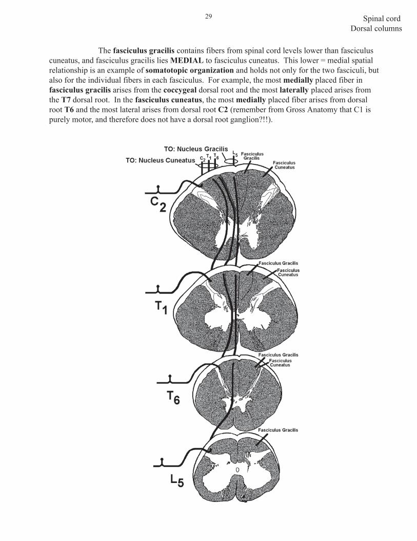
There are two components to the dorsal columns, called fasciculus gracilis and fasciculuscuneatus (fasciculus = L., little bundle; gracilis = slender; cuneatus = wedge). The central processof the alpha-beta fiber travels within the fasciculus gracilis if it arises from dorsal root ganglia T7and below. In contrast, if the central process of the alpha-beta fiber arises from cells in dorsal rootsT6 and above (toward your head), it is part of fasciculus cuneatus.
CUNEATUS = “ARM” = T6 and upGRACILIS = “LEG” = T7 and down
Fasciculus gracilis and fasciculus cuneatus are thus comprised of the alpha-beta axons whosecell bodies lie in IPSILATERAL DORSAL ROOT GANGLIA. That is, the cell bodies are on theSAME SIDE as the fasciculi. I have mentioned that fibers in the dorsal columns DO NOT CROSSin the spinal cord and eventually synapse in the medulla. While we will cover the medulla later inthe course, you might like to know that axons in fasciculus gracilis terminate in the ipsilateral (to thefasciculus) nucleus gracilis, while fibers in fasciculus cuneatus synapse in ipsilateral nucleuscuneatus (big surprise).
Dorsal columns
Spinal cord29
The fasciculus gracilis contains fibers from spinal cord levels lower than fasciculuscuneatus, and fasciculus gracilis lies MEDIAL to fasciculus cuneatus. This lower = medial spatialrelationship is an example of somatotopic organization and holds not only for the two fasciculi, butalso for the individual fibers in each fasciculus. For example, the most medially placed fiber infasciculus gracilis arises from the coccygeal dorsal root and the most laterally placed arises fromthe T7 dorsal root. In the fasciculus cuneatus, the most medially placed fiber arises from dorsalroot T6 and the most lateral arises from dorsal root C2 (remember from Gross Anatomy that C1 ispurely motor, and therefore does not have a dorsal root ganglion?!!).
Dorsal columns

Spinal cord 30
What happens when there is a lesion anywhere in the system involving the peripheralprocesses of the dorsal root neurons, the fasciculus gracilis, the fasciculus cuneatus, the nucleusgracilis and the nucleus cuneatus? Let’s take the peripheral processes first and include all alpha-betafibers which can carry TWO POINT DISCRIMINATION, VIBRATION, AND CONSCIOUSPROPRIOCEPTION. Such a lesion would result in interruption of the information from the regionof the body innervated by that dorsal root. This is called a dermatome. While there is overlap ofadjacent dermatomes, don’t worry about that now. Think about the distribution of the peripheralprocess of each dorsal root.
Following a lesion of the dorsal root the resulting deficits are manifest on the same side asthe lesion = IPSILATERAL.
Deficits that result from a lesion in thedorsal column system (i.e. in the spinal cord)would differ depending on the precise locationof the lesion. For instance, following a lesion atspinal cord level C2 which damages bothfasciculi, information from the entire ipsilateralside of the body (and the back of the head,which is innervated by C2) would not reach thenucleus gracilis and nucleus cuneatus, and thuswe would never feel the sensations (they don’treach consciousness via pathways from themedulla to the cerebral cortex). If the spinalcord lesion involves only fasciculus gracilis atspinal segment C2, only the information fromspinal segments T7 and below (all the waydown) is lost. Information from the arm is OKbecause fasciculus cuneatus is fine. If the lesionlies at S1, then only the ascending informationfrom spinal segments S1 and below are affected.Information coming in above S1 (toward thehead) gets in OK and ascends to the caudalmedulla.
Dorsal columns

Spinal cord31
In addition to the loss of 2 pt. discrimination, vibration and conscious proprioception,you should know that dorsal column lesions result in astereognosia (Gr. steros = solid, gnosis =recognition), which is the inability to recognize objects or forms by touch. Put a key in your handwith your eyes closed and you can identify it as a key. Another problem is called agraphesthesia(inability to recognize letters, numbers, etc., drawn on the skin). In the upper extremity, thesesensory losses result in clumsiness or ataxia. Finally, damage to the dorsal columns sometimespresents as paresthesia (Gr.- para = abnormal, aisthesis = sensation) which is tingling andnumbness. Think of this as resulting from irritation of the fibers as they die.
The ataxia (lack of order orincoordination) that results from lesions ofthe dorsal columns is due to the loss ofproprioceptive information regarding theposition of our limbs. If the fasciculi graciliare involved, the patient will exhibit aRomberg sign. To test for this sign thepatient is asked to stand with their feettogether and their eyes open. If closing theeyes causes the patient to sway then there isa Romberg sign. That is, the patient has lostthe sensory proprioceptive input and oncevisual inputs are eliminated the deficitbecomes apparent.
When a patient with a dorsal columnlesion steps forward the legs are flungabruptly forward, often being lifted higherthan necessary. This is seen in tabes(wasting) dorsalis or neurosyphilis. Thereis an audible sound as the foot stamps theground (they are not sure when it hits) andsince they usually have a cane it is referedto as a “stick and stamp.”
Finally, patients with dorsal columndisease in the cervical region exhibit aLhermitte’s sign. This is described as thesensation of an “electric shock” that runsdown the vertebral column and permeatesthe arms and legs. These sensations are setoff by flexion at the neck which stretchesthe dorsal columns in the cervical region.This stretching causes demyelinated axonsin the dorsal columns to send “funny”messages to the cortex.
Dorsal columns
Spinal cord 32
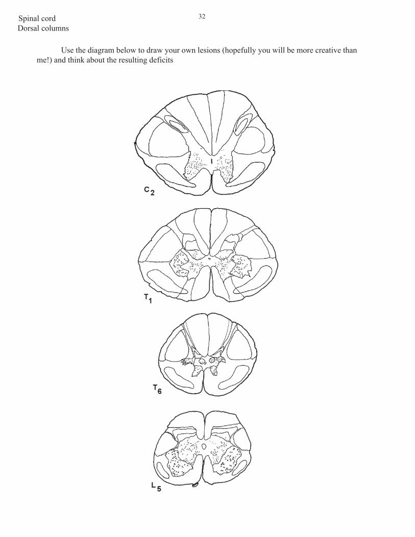
Use the diagram below to draw your own lesions (hopefully you will be more creative thanme!) and think about the resulting deficits
Dorsal columns
Spinal cord33
Dorsal columns
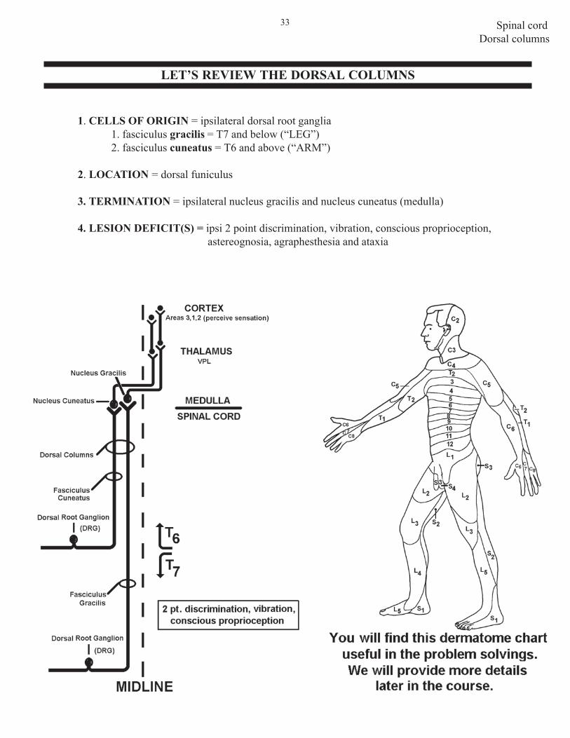
LET’S REVIEW THE DORSAL COLUMNS
1. CELLS OF ORIGIN = ipsilateral dorsal root ganglia1. fasciculus gracilis = T7 and below (“LEG”)2. fasciculus cuneatus = T6 and above (“ARM”)
2. LOCATION = dorsal funiculus
3. TERMINATION = ipsilateral nucleus gracilis and nucleus cuneatus (medulla)
4. LESION DEFICIT(S) = ipsi 2 point discrimination, vibration, conscious proprioception,astereognosia, agraphesthesia and ataxia
Spinal cord 34
Dorsal columns
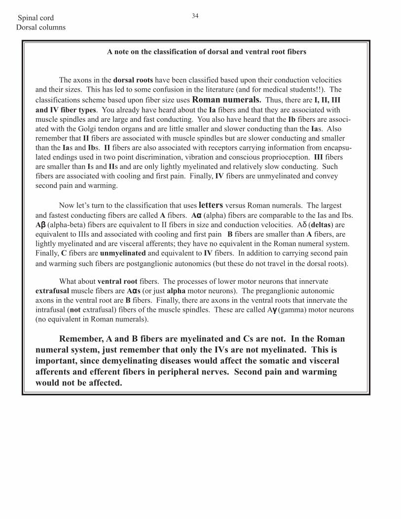
A note on the classification of dorsal and ventral root fibers
The axons in the dorsal roots have been classified based upon their conduction velocitiesand their sizes. This has led to some confusion in the literature (and for medical students!!). Theclassifications scheme based upon fiber size uses Roman numerals. Thus, there are I, II, IIIand IV fiber types. You already have heard about the Ia fibers and that they are associated withmuscle spindles and are large and fast conducting. You also have heard that the Ib fibers are associ-ated with the Golgi tendon organs and are little smaller and slower conducting than the Ias. Alsoremember that II fibers are associated with muscle spindles but are slower conducting and smallerthan the Ias and Ibs. II fibers are also associated with receptors carrying information from encapsu-lated endings used in two point discrimination, vibration and conscious proprioception. III fibersare smaller than Is and IIs and are only lightly myelinated and relatively slow conducting. Suchfibers are associated with cooling and first pain. Finally, IV fibers are unmyelinated and conveysecond pain and warming.
Now let’s turn to the classification that uses letters versus Roman numerals. The largestand fastest conducting fibers are called A fibers. Aααααα (alpha) fibers are comparable to the Ias and Ibs.Aβββββ (alpha-beta) fibers are equivalent to II fibers in size and conduction velocities. Aδ (deltas) areequivalent to IIIs and associated with cooling and first pain B fibers are smaller than A fibers, arelightly myelinated and are visceral afferents; they have no equivalent in the Roman numeral system.Finally, C fibers are unmyelinated and equivalent to IV fibers. In addition to carrying second painand warming such fibers are postganglionic autonomics (but these do not travel in the dorsal roots).
What about ventral root fibers. The processes of lower motor neurons that innervateextrafusal muscle fibers are Aαααααs (or just alpha motor neurons). The preganglionic autonomicaxons in the ventral root are B fibers. Finally, there are axons in the ventral roots that innervate theintrafusal (not extrafusal) fibers of the muscle spindles. These are called Aγγγγγ (gamma) motor neurons(no equivalent in Roman numerals).
Remember, A and B fibers are myelinated and Cs are not. In the Romannumeral system, just remember that only the IVs are not myelinated. This isimportant, since demyelinating diseases would affect the somatic and visceralafferents and efferent fibers in peripheral nerves. Second pain and warmingwould not be affected.

Spinal cord35
Dorsal columnsProblem solvingPROBLEM SOLVING #1
Which statement is true regarding the shaded area below? There is only one correct response.
A. pathway terminates in the ipsilateral (right) nucleus cuneatus
B. pathway arises from cells in the contralateral (left) dorsal horn
C. pathway arises from cells in the ipsilateral (right) dorsal root ganglia T6 and above (rostral)
D. pathway arises from cells in contralateral (left) dorsal root ganglia T7 and below
E. pathway consists of alpha-beta axons from the ipsilateral (right) dorsal root ganglia T7and below
RIGHT LEFTPROBLEM SOLVING #2
Which statement is true regarding the neurological deficit(s) that would be present following alesion involving the shaded area above? There might be deficits that are not included in theresponses. There is only one correct response.
A. deficit in two point discimination from the contralateral (left) index finger
B. deficit in two point discrimination from the ipsilateral (right) index finger
C. deficit in vibration sense from the contralateral (left) big toe
D. deficit in conscious proprioception from the contralateral (left) index finger
E. deficit in two point discrimination from the ipsilateral (right) big toe
Refer to Table of contents for Problem Solving ANSWER sets.

Spinal cord 36
Dorsal columnsProblem solving
PROBLEM SOLVING #3
Which statement is true regarding the shaded area below? There is only one correct response.
A. pathway arises from cells in the ipsilateral (right) dorsal root ganglia T7 and below
B. pathway arises from cells in the ipsilateral (right) dorsal horn
C. pathway consists of alpha-beta axons that terminate within the ipsilateral (right) nucleus cuneatus
D. pathway arises from cells in the contralateral (left) dorsal root ganglia T6 and above
E. pathway consists of alpha-beta axons from the ipsilateral (right) dorsal root ganglia T7 and below
RIGHT LEFTPROBLEM SOLVING #4
Which statement is true regarding the neurological deficit(s) that would be present following alesion involving the shaded area above? There might be deficits that are not included in theresponses. There is only one correct response. ( Remember to use the dermatome chart on the pointsummary page).
A. deficit in two point discrimination from the contralateral (left) index finger
B. deficit in two point discrimination from the ipsilateral (right) shoulder
C. deficit in vibration sense from the contralateral (left) big toe
D. deficit in conscious proprioception from the contralateral (left) index finger
E. deficit in two point discrimination from the ipsilateral (right) chest over the heart
Spinal cord37
Dorsal columnsProblem solving
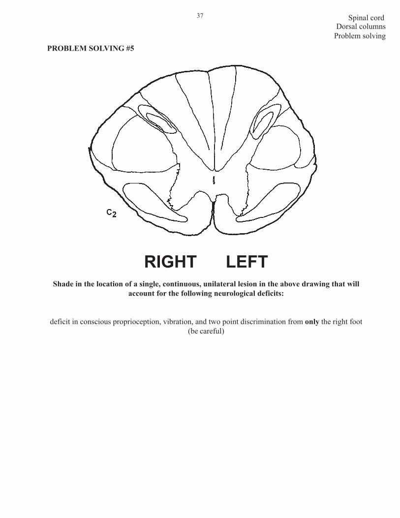
PROBLEM SOLVING #5
RIGHT LEFTShade in the location of a single, continuous, unilateral lesion in the above drawing that will
account for the following neurological deficits:
deficit in conscious proprioception, vibration, and two point discrimination from only the right foot(be careful)

Spinal cord 38
Dorsal columnsProblem solving
RIGHT LEFT
PROBLEM SOLVING #5 ANSWER
Spinal cord39
Dorsal columnsProblem solving
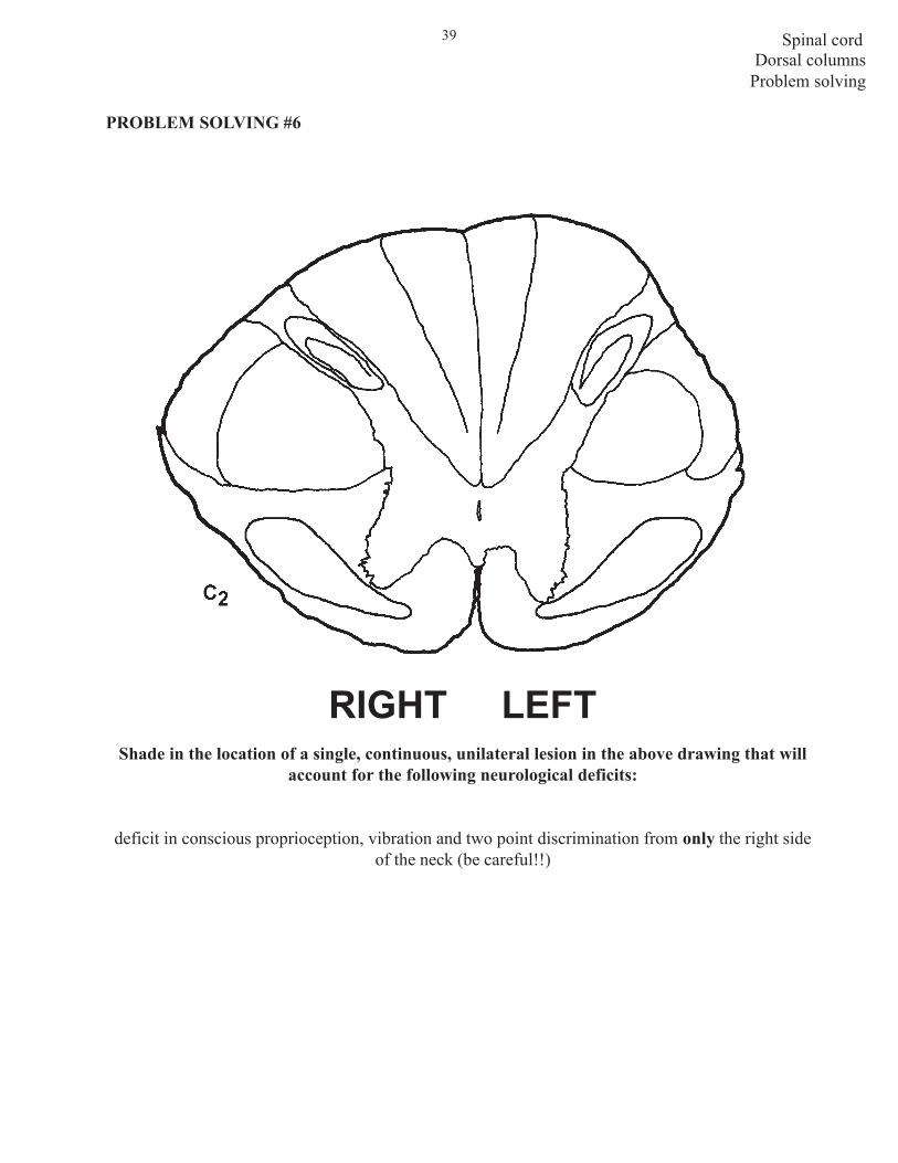
PROBLEM SOLVING #6
RIGHT LEFTShade in the location of a single, continuous, unilateral lesion in the above drawing that will
account for the following neurological deficits:
deficit in conscious proprioception, vibration and two point discrimination from only the right sideof the neck (be careful!!)
Spinal cord 40
Dorsal columnsProblem solving
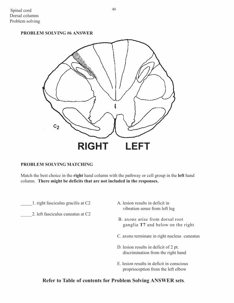
PROBLEM SOLVING #6 ANSWER
RIGHT LEFTPROBLEM SOLVING MATCHING
Match the best choice in the right hand column with the pathway or cell group in the left handcolumn. There might be deficits that are not included in the responses.
_____1. right fasciculus gracilis at C2 A. lesion results in deficit in vibration sense from left leg
_____2. left fasciculus cuneatus at C2 B. axons arise from dorsal root ganglia T7 and below on the right
C. axons terminate in right nucleus cuneatus
D. lesion results in deficit of 2 pt. discrimination from the right hand
E. lesion results in deficit in conscious proprioception from the left elbow
Refer to Table of contents for Problem Solving ANSWER sets.
Spinal cord41
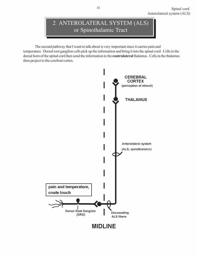
The second pathway that I want to talk about is very important since it carries pain andtemperature. Dorsal root ganglion cells pick up the information and bring it into the spinal cord. Cells in thedorsal horn of the spinal cord then send the information to the contralateral thalamus. Cells in the thalamusthen project to the cerebral cortex.
Anterolateral system (ALS)
2 ANTEROLATERAL SYSTEM (ALS)or Spinothalamic Tract
Spinal cord 42
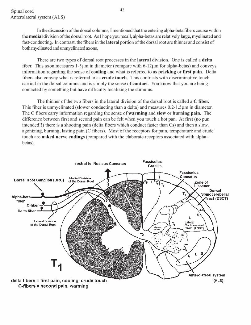
In the discussion of the dorsal columns, I mentioned that the entering alpha-beta fibers course withinthe medial division of the dorsal root. As I hope you recall, alpha-betas are relatively large, myelinated andfast-conducting. In contrast, the fibers in the lateral portion of the dorsal root are thinner and consist ofboth myelinated and unmyelinated axons.
There are two types of dorsal root processes in the lateral division. One is called a deltafiber. This axon measures 1-5µm in diameter (compare with 6-12µm for alpha-betas) and conveysinformation regarding the sense of cooling and what is referred to as pricking or first pain. Deltafibers also convey what is referred to as crude touch. This contrasts with discriminative touchcarried in the dorsal columns and is simply the sense of contact. You know that you are beingcontacted by something but have difficulty localizing the stimulus.
The thinner of the two fibers in the lateral division of the dorsal root is called a C fiber.This fiber is unmyelinated (slower conducting than a delta) and measures 0.2-1.5µm in diameter.The C fibers carry information regarding the sense of warming and slow or burning pain. Thedifference between first and second pain can be felt when you touch a hot pan. At first (no punintended!!) there is a shooting pain (delta fibers which conduct faster than Cs) and then a slow,agonizing, burning, lasting pain (C fibers). Most of the receptors for pain, temperature and crudetouch are naked nerve endings (compared with the elaborate receptors associated with alpha-betas).
Anterolateral system (ALS)

Spinal cord43
The central processes of delta and C fibers in the lateral division of the dorsal root do somethingquite different than the alpha-betas. These axons enter a zone at the top of the dorsal horn called the zoneof Lissauer (ZL) and then course ROSTRALLY for approximately 2 spinal segments within this zonebefore they dive into the dorsal horn, where they synapse. THERE HAS BEEN NOCROSSING YET!!!!! Cells in the DORSAL HORN that receive this pain and temperatureinformation then send axons which CROSS and enter the anterolateral portion of the lateral funiculus,where they ascend to the thalamus (the great gateway to the cortex). In particular, they terminate in theventral posterolateral nucleus (VPL). The VPL then relays the information to the somatosensory cortex(Areas 3, 1, and 2). Information carried over the pain, temperature and crude touch pathway begins in theprocesses of dorsal root ganglion cells, but the ANTEROLATERAL SYSTEM ( [ALS] axons in theanterolateral part of the white matter) TAKES ORIGIN FROM CELLS IN THECONTRALATERAL DORSAL HORN. THIS SYSTEM IS ALSO CALLED THESPINOTHALAMIC PATHWAY (ORIGIN IN SPINAL CORD, TERMINATION INTHALAMUS).
Anterolateral system (ALS)
Spinal cord 44
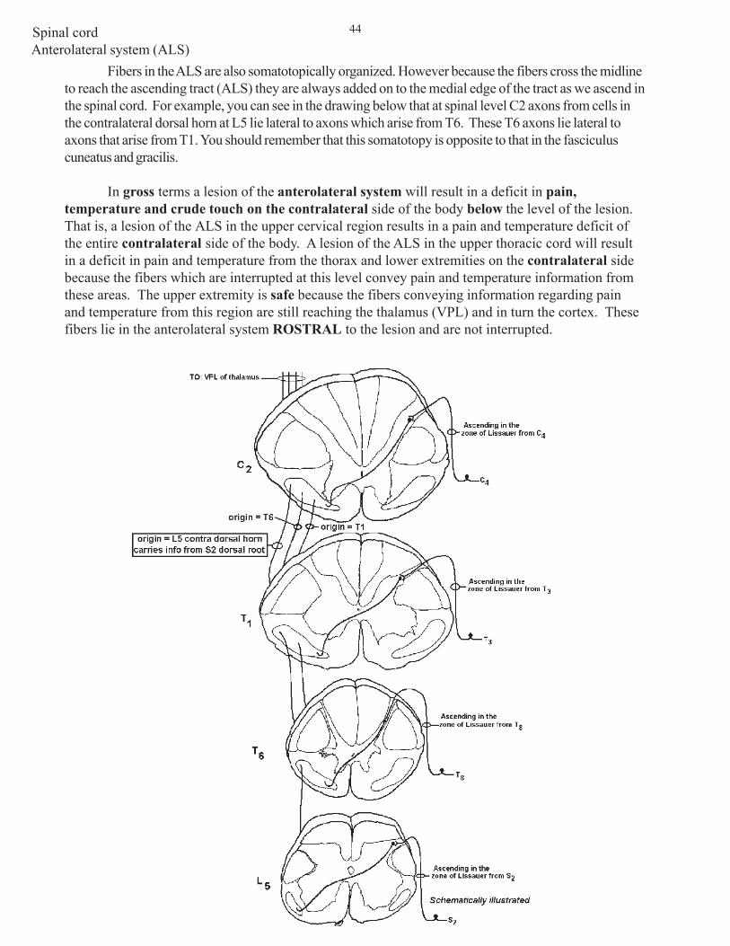
Fibers in the ALS are also somatotopically organized. However because the fibers cross the midlineto reach the ascending tract (ALS) they are always added on to the medial edge of the tract as we ascend inthe spinal cord. For example, you can see in the drawing below that at spinal level C2 axons from cells inthe contralateral dorsal horn at L5 lie lateral to axons which arise from T6. These T6 axons lie lateral toaxons that arise from T1. You should remember that this somatotopy is opposite to that in the fasciculuscuneatus and gracilis.
In gross terms a lesion of the anterolateral system will result in a deficit in pain,temperature and crude touch on the contralateral side of the body below the level of the lesion.That is, a lesion of the ALS in the upper cervical region results in a pain and temperature deficit ofthe entire contralateral side of the body. A lesion of the ALS in the upper thoracic cord will resultin a deficit in pain and temperature from the thorax and lower extremities on the contralateral sidebecause the fibers which are interrupted at this level convey pain and temperature information fromthese areas. The upper extremity is safe because the fibers conveying information regarding painand temperature from this region are still reaching the thalamus (VPL) and in turn the cortex. Thesefibers lie in the anterolateral system ROSTRAL to the lesion and are not interrupted.
Anterolateral system (ALS)
Spinal cord45
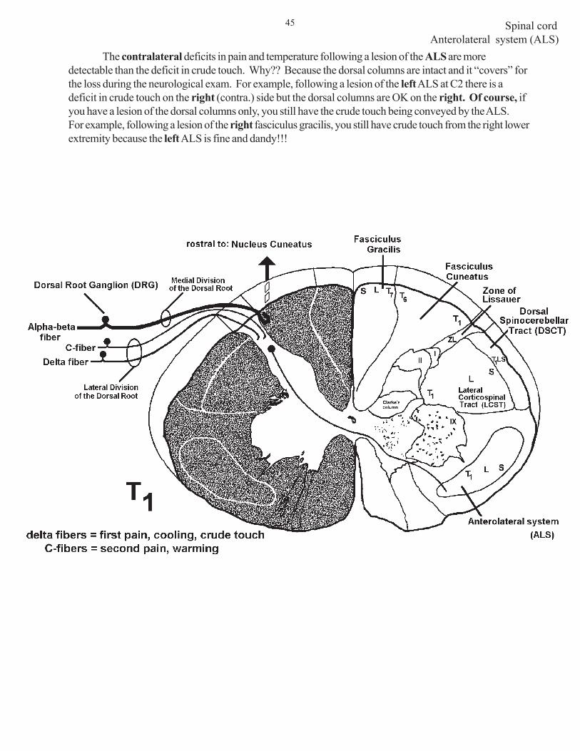
The contralateral deficits in pain and temperature following a lesion of the ALS are moredetectable than the deficit in crude touch. Why?? Because the dorsal columns are intact and it “covers” forthe loss during the neurological exam. For example, following a lesion of the left ALS at C2 there is adeficit in crude touch on the right (contra.) side but the dorsal columns are OK on the right. Of course, ifyou have a lesion of the dorsal columns only, you still have the crude touch being conveyed by the ALS.For example, following a lesion of the right fasciculus gracilis, you still have crude touch from the right lowerextremity because the left ALS is fine and dandy!!!
Anterolateral system (ALS)

Spinal cord 46
We know that the central processes of delta and C fibers ASCEND approximately 2 levels beforethey synapse in the dorsal horn. In other words, dorsal horn cells which send their axons across into theanterolateral system receive their pain and temperature information from TWO spinal segments below.Thus, instead of the gross approximation of deficits of pain and temperature below the level of the lesion onthe contralateral side, it is best to say the deficits start two levels below the lesion of the ALS and includeseverything below this level. Of course, the deficits are contralateral. For example, a lesion of theALS at T1 will result in deficits in pain, temperature, and crude touch from T3(2 segments below) and below on the contralateral side of the body.
“SPEED PLAY”
If there is reduced pain/temperature sensation in one limb and reduced position/vibrationsensation in the contralateral limb, the lesion must be somewhere in the spinal cord (on the
side of the position/vibration deficit.)
Anterolateral system (ALS)
Spinal cord47
LET’S REVIEW THE ANTEROLATERAL SYSTEM
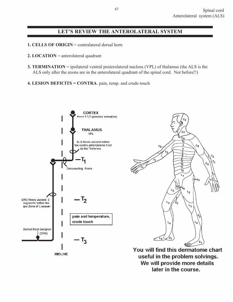
1. CELLS OF ORIGIN = contralateral dorsal horn
2. LOCATION = anterolateral quadrant
3. TERMINATION = ipsilateral ventral posterolateral nucleus (VPL) of thalamus (the ALS is theALS only after the axons are in the anterolateral quadrant of the spinal cord. Not before!!)
4. LESION DEFICITS = CONTRA. pain, temp. and crude touch
Anterolateral system (ALS)

Spinal cord 48
Anterolateral system (ALS)Problem solving
PROBLEM SOLVING #1
Which statement is true regarding the shaded areas below? There is only one correctresponse.
A. pathway consists of alpha-beta axons whose cell bodies lie in the contralateral (left)dorsal root ganglia
B. pathway arises from cells in the ipsilateral (right) dorsal horn
C. pathway terminates in the contralateral (left) VPL
D. pathway is comprised of the central processes of delta and C fibers
E. pathway consists of alpha-beta axons from the ipsilateral (right) dorsal root ganglia T7and below (caudal)
RIGHT LEFTPROBLEM SOLVING #2
Which statement is true regarding the shaded areas above? There is only one correct response.
A. pathway terminates in the ipsilateral (right) nucleus cuneatus in the medulla
B. pathway consists of alpha-beta axons from the contralateral (left) dorsal root ganglia T7and below
C. pathway arises from cells in the ipsilateral (right) dorsal root ganglia T6 and above
D. pathway arises from cells in the ipsilateral (right) dorsal horn
E. pathway arises from cells in the contralateral (left) dorsal horn

Spinal cord49
Anterolateral system (ALS)Problem solving
PROBLEM SOLVING #3
Which statement is true regarding the neurological deficit(s) that would be presentfollowing a lesion involving the shaded areas below? There might be deficits that are not includedin the responses. There is only one correct response.
A. deficit in two point discrimination from the contralateral (left) index finger
B. deficit in vibration sense from the ipsilateral (right) index finger
C. deficit in pricking pain from the contralateral (left) big toe
D. deficit in burning pain from the ipsilateral (right) index finger
E. deficit in the sense of cooling from the ipsilateral (right) big toe
RIGHT LEFTPROBLEM SOLVING #4
Which statement is true regarding the neurological deficit(s) that would be presentfollowing a lesion involving the shaded area above? There might be deficits that are not includedin the responses. There is only one correct response.
A. deficit in 2 pt. discrimination from the contralateral (left) hip
B. deficit in fast (first) pain from the ipsilateral (right) index finger
C. deficit in the sense of warming below the contralateral (left) knee
D. deficit in conscious proprioception from the contralateral (left) index finger
E. deficit in vibration sense from the ipsilateral (right) thumb

Spinal cord 50
Anterolateral system (ALS)Problem solving
PROBLEM SOLVING #5
Match the best choice in the right hand column with the pathway or cell group in the left handcolumn. There might be deficits that are not included in the responses.
_____1. right fasciculus gracilis at C2 A. lesion results in deficit in pain from the left arm
_____2. left anterolateral system at C1B. axons arise from dorsal roots T6 and above on the right
C. axons carry info. about vibration from the right thumb
D. lesion results in deficit in sense of cooling from the right foot
E. lesion results in deficit in conscious conscious proprioception from the right knee
Spinal cord51
Anterolateral system (ALS)Problem solving
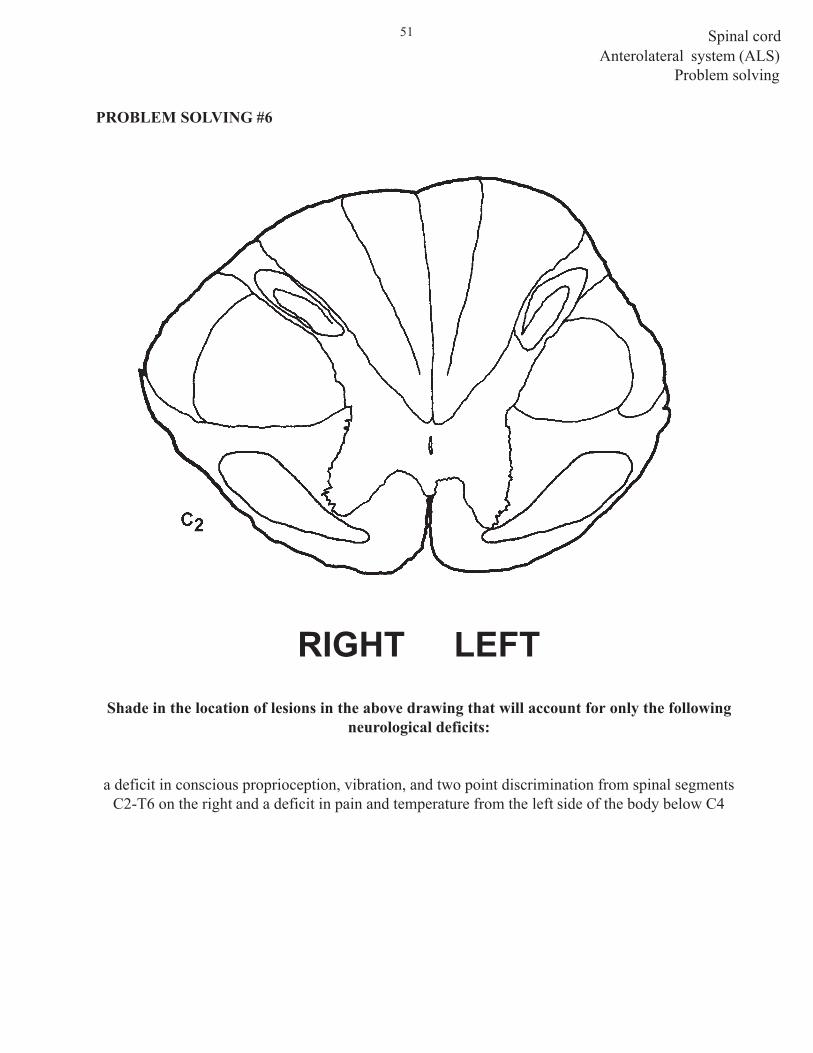
PROBLEM SOLVING #6
RIGHT LEFTShade in the location of lesions in the above drawing that will account for only the following
neurological deficits:
a deficit in conscious proprioception, vibration, and two point discrimination from spinal segmentsC2-T6 on the right and a deficit in pain and temperature from the left side of the body below C4

Spinal cord 52
Anterolateral system (ALS)Problem solving
PROBLEM SOLVING #6 ANSWER
RIGHT LEFT

Spinal cord53
So far we have discussed 3 types of fibers that comprise the dorsal root. The alpha-betas areassociated with the dorsal columns (fasc. gracilis and fasc. cuneatus) while the deltas and Cs are associatedwith the anterolateral system (ALS). The alpha-betas are bigger than the deltas and Cs, but there are fibersin the dorsal root that are even bigger (12-20µm) in diameter. These are called Ia, Ib and II fibers. Sincethese fibers (whose cell bodies lie in the dorsal root ganglia) are big, guess which division of the dorsal rootthey use when entering the spinal cord???? Of course, the MEDIAL, along with the alpha-betas.Remember that the skinny ones lie laterally (ahh, that hurts!!) and the more rotund ones medially.
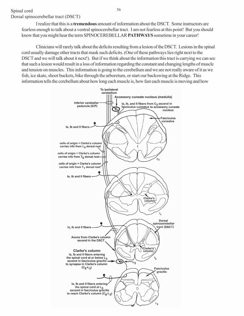
You have heard in Physiology that Ia and II fibers convey information from muscle spindles,while Ib fibers carry information from Golgi tendon organs. As Dr. Moss mentioned, this informationis utilized for reflexes. However, all of this information also ascends to the cerebellum (L., littlebrain) in order to participate in motor coordination. How does this information reach the cerebellum?
When the Ia, Ib, and II axons enter the spinal cord they dive into the grey matter of the dorsal hornuntil they reach its base. An investigator by the name of Rexed has divided the grey matter of the cord intolayers or laminae, and the base of the dorsal horn is called lamina VII. Within this lamina at spinal
Dorsal spinocerebellar tract (DSCT)
3 DORSAL SPINOCEREBELLAR TRACT(DSCT)

Spinal cord 54
segments C8-L3, AND ONLY AT THESE LEVELS, there isa very distinctive cell group called CLARKE’S NUCLEUS ORCOLUMN. The Ia, Ib and II fibers terminate on cells inClarke’s nucleus. From there, cells in Clarke’s nucleus sendaxons into the IPSILATERAL lateral funiculus where they arelocated dorsal and laterally. These axons comprise the DORSALSPINOCEREBELLAR TRACT (DSCT). The cells of originof this tract lie in the IPSILATERAL Clarke’s nucleus. Thepathway passes rostrally in the lateral funiculus and eventuallyterminates within the IPSILATERAL CEREBELLUM.
As fibers from cells in Clarke’s column enter the DSCTand ascend, they are organized such that the most caudal fibers lielaterally within the DSCT, while the most rostral (C8) liemedially in the DSCT. Compare this with the dorsal columns andALS.
To get into the cerebellum, the DSCT courses within (is acomponent of) the inferior cerebellar peduncle (L., a little foot)or restiform body. Think of a cerebellar peduncle as a bundle ofaxons connecting the spinal cord/brain stem and the overlyingcerebellum. There are three of these peduncles. More on thislater in the course!!
Dorsal spinocerebellar tract (DSCT)

Spinal cord55
It’s a crying shame that Clarke’s nucleus is not present at every spinal cord level. As I mentionedearlier, it is only present at spinal cord segments C8-L3. So, if a Ia, Ib, or II axon comes into the spinalcord between C8-L3, fine!!! There is a Clarke’s nucleus waiting for it and bingo, the fiber dives into thenucleus and the information that it is conveying is relayed to the cerebellum (via the DSCT). However,
think about a Ia, Ib or II fiber comingin at spinal level L5. It looks aroundand there is no Clarke’s nucleus tohitch a ride on. What would you do ifyou were a fiber who wanted to getyour information to the cerebellum??Personally, I would pass rostrally inthe fasciculus gracilis (remember, nofasciculus cuneatus is present here!!)until I got to L3, where there is aClarke’s column, and dive into thenucleus. This is exactly what happens!Ia, Ib, and II fibers that enter the cordat L4 or below travel in the fasciculusgracilis with the ascending alpha-betafibers to get to Clarke’s nucleus.
What about Ia, Ib and II fibersassociated with dorsal roots above C8?Well, they enter the cord, and find that likeL4 and below they don’t have a Clarke’snucleus, so they enter the fasciculus of theupper extremity (fasciculus cuneatus) untilthey reach the caudal medulla, where theysynapse in the ACCESSORYCUNEATE NUCLEUS. Cells in theaccessory cuneate nucleus project to theIPSI cerebellum via the inferior cerebellarpeduncle (just like cells in Clarke’s columndo).
Dorsal spinocerebellar tract (DSCT)
Spinal cord 56
I realize that this is a tremendous amount of information about the DSCT. Some instructors arefearless enough to talk about a ventral spinocerebellar tract. I am not fearless at this point! But you shouldknow that you might hear the term SPINOCEREBELLAR PATHWAYS sometime in your career!
Clinicians will rarely talk about the deficits resulting from a lesion of the DSCT. Lesions in the spinalcord usually damage other tracts that mask such deficits. (One of these pathways lies right next to theDSCT and we will talk about it next!). But if we think about the information this tract is carrying we can seethat such a lesion would result in a loss of information regarding the constant and changing lengths of muscleand tension on muscles. This information is going to the cerebellum and we are not really aware of it as wefish, ice skate, shoot buckets, bike through the arboretum, or start our backswing at the Ridge. Thisinformation tells the cerebellum about how long each muscle is, how fast each muscle is moving and how
Dorsal spinocerebellar tract (DSCT)

Spinal cord57
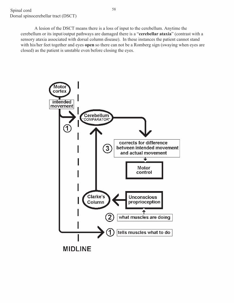
much tension is on each muscle (#2 above). The cerebellum then can compare this ascending informationregarding what the muscles are doing with other information (the sources of which we will learn later)regarding what higher motor centers want the muscles to do (#1 above). Then a correction can occur viapathways that leave the cerebellum to influence motor performance (#3 above). Whew!!
For our problem solving, let’s equate a lesion of the DSCT with loss of unconsciousproprioception and incoordination or ataxia. This incoordination deficit will be IPSILATERALto the lesion because there is no crossing of information in the spinal cord. The DSCT is IPSI tothe receptors. Also, the cerebellum influences the same or ipsilateral side of the body (viaseveral output pathways). Think about the dorsal columns. Is there crossing from the receptors tothe fasc. gracilis and fasc. cuneatus in the spinal cord?? How about the pain and temperaturepathways?
Dorsal spinocerebellar tract (DSCT)
Spinal cord 58
A lesion of the DSCT means there is a loss of input to the cerebellum. Anytime thecerebellum or its input/output pathways are damaged there is a “cerebellar ataxia” (contrast with asensory ataxia associated with dorsal column disease). In these instances the patient cannot standwith his/her feet together and eyes open so there can not be a Romberg sign (swaying when eyes areclosed) as the patient is unstable even before closing the eyes.
Dorsal spinocerebellar tract (DSCT)

Spinal cord59
LET’S REVIEW THE DORSAL SPINOCEREBELLAR TRACT
1. CELLS OF ORIGIN = ipsilateral Clarke’s column
2. LOCATION = dorsolateral part of lateral funiculus
3. TERMINATION = ipsilateral cerebellum
4. LESION DEFICIT(S) = ipsilateral muscle incoordination/ataxia
Dorsal spinocerebellar tract (DSCT)
Spinal cord 60
Dorsal spinocerebellar tract (DSCT)Problem solving
PROBLEM SOLVING #1
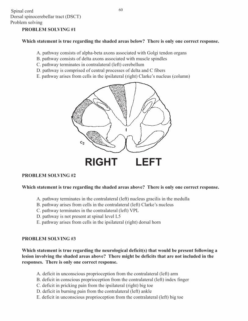
Which statement is true regarding the shaded areas below? There is only one correct response.
A. pathway consists of alpha-beta axons associated with Golgi tendon organsB. pathway consists of delta axons associated with muscle spindlesC. pathway terminates in contralateral (left) cerebellumD. pathway is comprised of central processes of delta and C fibersE. pathway arises from cells in the ipsilateral (right) Clarke’s nucleus (column)
RIGHT LEFTPROBLEM SOLVING #2
Which statement is true regarding the shaded areas above? There is only one correct response.
A. pathway terminates in the contralateral (left) nucleus gracilis in the medullaB. pathway arises from cells in the contralateral (left) Clarke’s nucleusC. pathway terminates in the contralateral (left) VPLD. pathway is not present at spinal level L5E. pathway arises from cells in the ipsilateral (right) dorsal horn
PROBLEM SOLVING #3
Which statement is true regarding the neurological deficit(s) that would be present following alesion involving the shaded areas above? There might be deficits that are not included in theresponses. There is only one correct response.
A. deficit in unconscious proprioception from the contralateral (left) armB. deficit in conscious proprioception from the contralateral (left) index fingerC. deficit in pricking pain from the ipsilateral (right) big toeD. deficit in burning pain from the contralateral (left) ankleE. deficit in unconscious proprioception from the contralateral (left) big toe

Spinal cord61
Dorsal spinocerebellar tract (DSCT)Problem solving
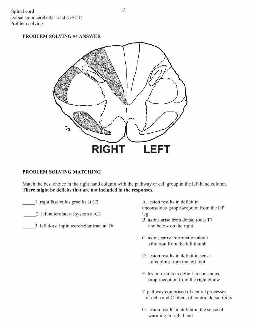
PROBLEM SOLVING #4
RIGHT LEFTShade in the location of unilateral lesions in the above drawing that will account for the
following neurological deficits:
deficit in conscious proprioception, vibration, and two point discrimination from spinal segments C2-T6 on the right, deficit in pain and temperature from the left side of the entire body (below the neck)
and deficit in unconscious proprioception from the entire right side of the body (Think about howfibers get to the accessory cuneate nucleus!!!)
Spinal cord 62
Dorsal spinocerebellar tract (DSCT)Problem solving
PROBLEM SOLVING #4 ANSWER
RIGHT LEFT
PROBLEM SOLVING MATCHING
Match the best choice in the right hand column with the pathway or cell group in the left hand column.There might be deficits that are not included in the responses.
_____1. right fasciculus gracilis at C2 A. lesion results in deficit inunconscious proprioception from the left
_____2. left anterolateral system at C2 legB. axons arise from dorsal roots T7
_____3. left dorsal spinocerebellar tract at T6 and below on the right
C. axons carry information about vibration from the left thumb
D. lesion results in deficit in sense of cooling from the left foot
E. lesion results in deficit in conscious proprioception from the right elbow
F. pathway comprised of central processes of delta and C fibers of contra. dorsal roots
G. lesion results in deficit in the sense of warming in right hand

Spinal cord63
Cells in the cerebral cortex, especially the motor cortex (area 4; precentral gyrus) possess very longaxons that descend through an extensive region of the brain to eventually reach the spinal cord. Right beforeentering the cord these corticospinal fibers cross or decussate (L., to make an X) and enter the LATERALFUNICULUS where they travel medial to the DSCT. These fibers, which are now called the lateral (theyare in the lateral funiculus) corticospinal tract (LCST), innervate neurons in the spinal cord along its entirelength. Once in the grey matter (where the cells are) LCST axons synapse upon cells in the ventral horn. Thisis the first synapse in a pathway over which the cerebral cortex informs cells in theCONTRALATERAL spinal cordabout a voluntary movementthat it wishes to perform. Oncethe cells in the ventral horn receivethis cortical information, theydirectly drive the muscles viaaxons that pass out the ventral root.The fastest way you can move abody muscle voluntarily is byutilizing 2 neurons. The first one isan upper motor neuron and lies inthe cerebral cortex. The secondone lies in the contralateral oropposite ventral horn and is alower motor neuron.
Descending fibers in theLCST are somatotopicallyorganized such that the mostmedially located fibers in the tractterminate before (rostral to) themore the laterally placed fibers.
The lateral corticospinal tract (LCST) is the most important pathway wehave for making voluntarily movements and is one of, if not THE, most
important pathways in clinical neurology.
Lateral corticospinal tract (LCST)
4 LATERAL CORTICOSPINAL TRACT (LCST)

Spinal cord 64
Lateral corticospinal tract (LCST)

Spinal cord65
Interruption of the LCST means that neurons in the spinal cord that innervate or drive muscles havelost a tremendously important input. These muscles are still innervated by the spinal cord neurons in theventral horn, but these cells have lost a large part of their drive. This results in weakness in those musclesthat are innervated by spinal neurons that have lost their LCST excitatory drive. Such a lesiondoes NOT result in paralysis because the muscles are still ALIVE. The problem is that theneurons that innervate the muscles have lost a large part of their drive. (There are still some otherinputs to these cells).
An early sign of damage to the corticospinal tract(for example, following a stroke) is called “pronator drift”and may be demonstrated as follows. You have the patienthold both supinated arms straight out with their eyes closed.Tapping gently down on each hand for weakness will resultin the “bad” arm/hand slowly drifting down and intopronation.
It is important to understand that lesions of theLCST at different levels of the spinal cord result indifferent muscles being affected. Let’s start with alesion at C1. Such a lesion will interrupt ALL of the LCSTfibers to the spinal cord on the SAME side as the lesion.The result is a loss of voluntary control of all of themuscles on the IPSILATERAL (to the lesion)side of the body. This is called HEMIPLEGIA(plegia = stroke). Notice that the muscles are notparalyzed, only weak.
Lateral corticospinal tract (LCST)
Spinal cord 66
Lateral corticospinal tract (LCST)
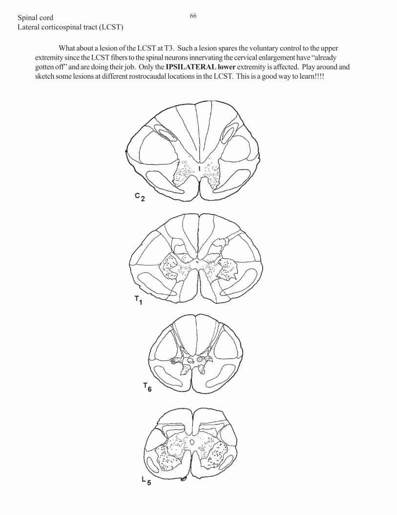
What about a lesion of the LCST at T3. Such a lesion spares the voluntary control to the upperextremity since the LCST fibers to the spinal neurons innervating the cervical enlargement have “alreadygotten off” and are doing their job. Only the IPSILATERAL lower extremity is affected. Play around andsketch some lesions at different rostrocaudal locations in the LCST. This is a good way to learn!!!!

Spinal cord67
The loss of descending input carried over the lateral corticospinal tract also results in clinical signsbesides weakness. Stroke patients in which the corticospinal or lateral corticospinal tract is damagedexhibit a flexed arm and an extended leg. That is, the resting length of these muscles is shortened.
When you passively lengthen the patient’s flexed arm or extended leg, you feel more resistance ortone than in a normal person. This is due to an increase in the muscle stretch reflexes that serve to maintainthe length of muscles. The hypertonia/hyperreflexia in a stroke patient is especially apparent in theflexor muscles of the arm and the extensor muscles of the leg (ipsilateral to the spinal cord lesion orcontralateral to the cortical lesion). These increased muscle stretch reflexes are velocity dependent. Thatis, the faster you try to extend the flexed arm and flex the extended leg the more resistance you will feel.The increase in muscle tone (hypertonia/hyperreflexia) seen when passively moving a limb iscalled SPASTICITY. This is a very important term in clinical neurology, one that you shouldNEVER FORGET!! PLEASE!!!!!!!
SPASTICITY!!!
The spastic gait can be recognized by the sound of the slow, rhythmic scuff of the foot along thefloor. This will result in the toe area of the shoe being unevenly worn down.
Physicians also tap on tendons and this stretches muscle fibers. Following a lesion of thecorticospinal system, the “tendon reflexes” are usually increased (hyperreflexia). However, since you arenot palpating the muscle when you hit someone’s patellar tendon, you are not testing for tone. Remember,spasticity refers only to the hypertonia/hyperreflexia that is observed during passive movement of alimb.
So, now we know that there is 1) spasticity and 2) increasedtendon reflexes following lesions of the corticospinal or lateralcorticospinal tract.
Now let’s look at two more problems associated with corticospinal/lateral corticospinal tract lesions. One of these is CLONUS (turmoil). Ifthe physician suddenly flexes the foot at the ankle and holds it flexed, therewill be spasmodic alternation of contraction and relaxation. Finally,there will be a BABINSKI sign. That is, stroking the ventral (plantar)surface of the lateral portion of the foot in a normal person results in the bigtoe going down (the plantar response is flexor). With lesions of thecorticospinal or lateral corticospinal tract the big toe goes up when strokingthe ventral surface of the foot “plantar response is extensor”. This iscalled a Babinski sign. Other less famous pathological reflexes that reflectcorticospinal damage include: the Chaddock sign (toe goes up uponstimulation of the lateral surface of the foot), the Bing sign (toe goes upfollowing jabs to the dorsal surface of the big toe), and the Hoffman sign(flicking the middle finger causes the index finger and thumb to reflexivelyflex).
Lateral corticospinal tract (LCST)

Spinal cord 68
I realize that you all are thinking “what causes spasticity, clonus and a Babinski?” Well, we don’tknow, but think about the fact that those gamma efferents (covered in Physiology) that innervate the bag andchain fibers of the muscle spindles have lost their cortical innervation and might be doing funny things to themuscle spindles. This might account for some of these problems, but we are not sure and therefore don’texpect you to understand the mechanism(s) either. But suggestions are certainly welcome!!
LET’S SUMMARIZE SOME DIFFICULT (AND CONFUSING??) MATERIAL INTO A
“SIMPLE RULE”:
A LESION OF THE LCST RESULTS IN IPSILATERAL WEAKNESS,SPASTICITY AND INCREASED TENDON REFLEXES OF MUSCLES THATARE INNERVATED BY VENTRAL HORN CELLS AT AND BELOW THE
SPINAL LEVEL OF THE LESION. THERE ALSO WILL BE IPSILATERALCLONUS AND A BABINSKI SIGN.
Lateral corticospinal tract (LCST)
Spinal cord69
LET’S REVIEW THE LATERAL CORTICOSPINAL TRACT
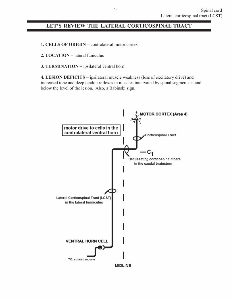
1. CELLS OF ORIGIN = contralateral motor cortex
2. LOCATION = lateral funiculus
3. TERMINATION = ipsilateral ventral horn
4. LESION DEFICITS = ipsilateral muscle weakness (loss of excitatory drive) andincreased tone and deep tendon reflexes in muscles innervated by spinal segments at andbelow the level of the lesion. Also, a Babinski sign.
Lateral corticospinal tract (LCST)

Spinal cord 70
Lateral corticospinal tract (LCST)PROBLEM SOLVING #1
Which statement is true regarding the shaded areas below? There is only one correct answer.
A. pathway arises from the ipsilateral (right) motor cortex
B. pathway terminates on cells in the ipsilateral (right) side of the spinal cord
C. pathway terminates in the contralateral (left) VPL
D. pathway arises from cells in the ipsilateral (right) Clarke’s column
E. two of the above
RIGHT LEFTPROBLEM SOLVING #2
Which statement is true regarding the neurological deficit(s) that would be presentfollowing a lesion involving the shaded areas above? There might be deficits that are notincluded in the responses. There is only one correct response.
A. left hemiplegia
B. left Babinski
C. deficit in first pain from the ipsilateral (right) big toe
D. spasticity in the contralateral (left) arm and leg
E. plantar response is extensor in the ipsilateral (right) foot (right Babinski)

Spinal cord71
Lateral corticospinal tract (LCST)Problem solving
PROBLEM SOLVING #3
RIGHT LEFTShade in the location of unilateral lesions in the above drawing that will account for the
following neurological deficits:
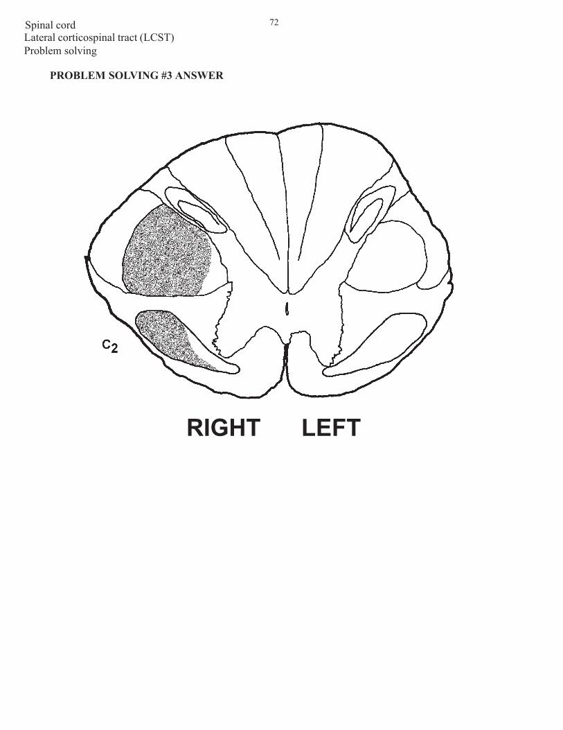
stroking the bottom of the right foot results in extension (up) of the right big toe, spastic right leg andarm, and deficit in fast pain from the left foot
Spinal cord 72
Lateral corticospinal tract (LCST)Problem solving
PROBLEM SOLVING #3 ANSWER
RIGHT LEFT

Spinal cord73
Lateral corticospinal tract (LCST)Problem solving
PROBLEM SOLVING MATCHING
Match the best choice in the right hand column with the pathway or cell group in the left hand column.There might be deficits that are not included in the responses.
_____1. right fasciculus gracilis at C1 A. lesion results in deficit in unconscious proprioception from the left leg
_____2. left anterolateral system at C1B. axons arise from dorsal roots T7
_____3. left dorsal spinocerebellar tract at T6 and below on the left
_____4. left lateral corticospinal tract C. axons carry information about vibration from the right big toe
D. lesion results in deficit in sense of cooling from the right foot
E. lesion results in deficit in conscious proprioception from the left elbow
F. lesion results in deficit in distinguishing position of the right arm in space with eyes closed
G. lesion results in deficit in sense of warming in left hand
H. lesion results in right Babinski
I. cells arise in right motor cortex

Spinal cord 74
ANSWERS TO PROBLEM SOLVING QUESTIONS RELATED TOORIENTATION AND POINTS 1-4
NOTE: The answers to ALL “shade-ins” are illustrated on the back side of the question.
Orientation problem solving
1. C2. D3. E
Point #1 Dorsal Columns
1. E2. E3. C4. EMatching B,E
Point #2 Anterolateral System
1. E D is false because delta and C fibers synapse upon dorsal horn cells. The axons ofthese dorsal horn cells (not the delta and Cs, whose cell bodies lie in the DRG’s) giverise to the contra ALS.
2. E3. C4. CMatching E,D
Point #3 Dorsal Spinocerebellar Tract
1. E2. D3. DMatching B,G,A
Point #4 Lateral Corticospinal Tract
1. B2. EMatching C,D,A,I
PROBLEM SOLVING ANSWERSPOINTS 1-4
FYI and enjoyment
The brain of the great physicist Albert Einstein weighed 1,230 grams. This is far below the averagebrain weight of 1,400 grams. (Reference: Neuroscience Letters, 210:161-164, 1996.)
The adult human spinal cord weighs about 35 grams (0.1 lb).The average length of the adult spinal cord is 45 cm for men and 43 cm for women.
The brain of an elephant weighs about 6 kg (13 lb)

Spinal cord75
The ventral horn contains some of the largest cells in the central nervous system. The large neuronsdirectly innervate muscles. We need to think about what happens when there is a lesion that damages theselarge ventral horn motor neurons. When such damage occurs the motor neurons die, their axons in the ventralroot die, and the muscles that they innervate eventually die. The muscle dies because it has lost its source ofnourishment or trophic source (trophic = nourishment). When the muscle dies, it ATROPHIES orshrinks up. (atrophy = wasting). Atrophy occurs only when the cells that DIRECTLY innervate the muscledie. Also, as the lower motor neurons die, the muscles they innervate sometimes twitch. These twitches, asseen by the naked eye, are called fasciculations.
CELLS THAT DIRECTLY INNERVATE MUSCLES ARE CALLED
LOWER MOTOR NEURONS (LMNs)
Let’s do some problem solving using this extremely important concept. What will happen ifthere is a lesion in the ventral horn at T1??? Well, it seems to me that the logical answer is that motorneurons in the ventral horn at this level, and only at this level, die. Their axons also die, and then themuscles innervated by these axons die, and atrophy. It’s as simple as that!!!! T1 motor neuronsinnervate the intrinsic hand muscles.
Since the muscles innervated by ventral motor neurons at spinal level T1 are dead, theyatrophy and are flaccid (flaccidus L = flabby). They will not respond to stretch and therefore there isatonia and areflexia.
Now, another important concept. As you already know, the lateral corticospinal tract (LCST)fibers synapse upon lower motor neurons in the spinal cord. This pathway is therefore called anUPPER MOTOR NEURON (UMN) pathway. Clinically, it is “THEEEEEE” UMN pathway. Star,underline in many colors, and remember for the rest of your life:
LCST = UMNs while VENTRAL HORN CELLS = LMNs.
UPPERS TELL LOWERS WHAT TO DO—BUT ONLY LOWERS TELLMUSCLES WHAT TO DO!!!!!!
LOWER MOTOR NEURON LESION = ATROPHY, FLACCIDITY, ATONICITYAND AREFLEXIA
5 VENTRAL HORN
Ventral horn

Spinal cord 76
Ventral horn
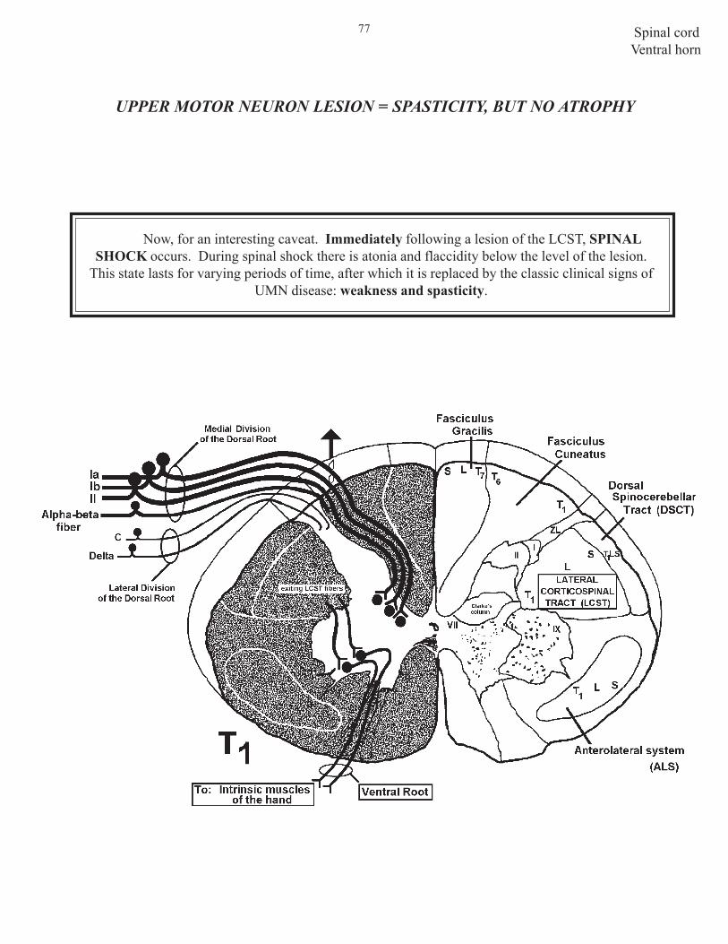
Spinal cord77
UPPER MOTOR NEURON LESION = SPASTICITY, BUT NO ATROPHY
Now, for an interesting caveat. Immediately following a lesion of the LCST, SPINALSHOCK occurs. During spinal shock there is atonia and flaccidity below the level of the lesion.
This state lasts for varying periods of time, after which it is replaced by the classic clinical signs ofUMN disease: weakness and spasticity.
Ventral horn

Spinal cord 78
Ventral hornProblem solving
PROBLEM SOLVING #1
Which statement is true regarding the shaded areas below? There is only one correct re-sponse.
A. cells send axons directly to muscles in the contralateral (left) handB. pathway terminates on cells in the ipsilateral (right) nucleus cuneatusC. pathway arises from cells in the ipsilateral (right) dorsal root ganglion from T6 and aboveD. pathway arises from cells in the ipsilateral (right) Clarke’s nucleusE. none of the above
RIGHT LEFT
PROBLEM SOLVING #2
Which statement is true regarding the neurological deficit(s) that would be presentfollowing a lesion involving the shaded areas below? There might be deficits that are notincluded in the responses. There is only one correct response.
A. atrophy of intrinsic muscles in the ipsilateral (right) handB. right BabinskiC. deficit in pricking pain from the ipsilateral (right) big toeD. spasticity in the contralateral (left) arm and legE. loss of unconscious proprioception from the right shoulder
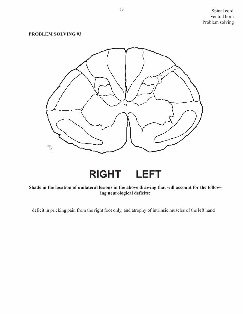
Spinal cord79
Ventral hornProblem solving
PROBLEM SOLVING #3
RIGHT LEFTShade in the location of unilateral lesions in the above drawing that will account for the follow-
ing neurological deficits:
deficit in pricking pain from the right foot only, and atrophy of intrinsic muscles of the left hand
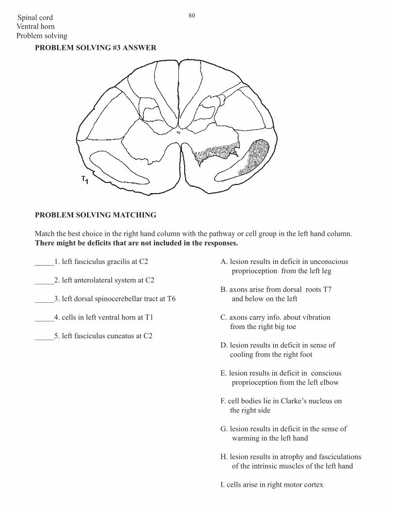
Spinal cord 80
Ventral hornProblem solving
PROBLEM SOLVING #3 ANSWER
PROBLEM SOLVING MATCHING
Match the best choice in the right hand column with the pathway or cell group in the left hand column.There might be deficits that are not included in the responses.
_____1. left fasciculus gracilis at C2 A. lesion results in deficit in unconscious proprioception from the left leg
_____2. left anterolateral system at C2B. axons arise from dorsal roots T7
_____3. left dorsal spinocerebellar tract at T6 and below on the left
_____4. cells in left ventral horn at T1 C. axons carry info. about vibration from the right big toe
_____5. left fasciculus cuneatus at C2D. lesion results in deficit in sense of cooling from the right foot
E. lesion results in deficit in conscious proprioception from the left elbow
F. cell bodies lie in Clarke’s nucleus on the right side
G. lesion results in deficit in the sense of warming in the left hand
H. lesion results in atrophy and fasciculations of the intrinsic muscles of the left hand
I. cells arise in right motor cortex

Spinal cord81
Hopefully, you will remember from Gross Anatomy some of the important details regarding theorganization of the autonomic nervous system within the spinal cord. What I want to emphasize at this time is thesympathetic outflow. There are PREGANGLIONIC SYMPATHETIC cells present in the spinalcord at spinal levels T1-L2. This clump of cells comprises the LATERAL CELL COLUMN (also called theintermediolateral cell column by some investigators). Axons of these cells pass ventrally and comprise part ofthe ventral root (from T1-L2). These preganglionic sympathetics can then do a number of things.
6 LATERAL (INTERMEDIOLATERAL) HORN(preganglionic autonomics)
Lateral horn

Spinal cord 82
The preganglionic sympathetics pass to the sympathetic trunk via the white communicating rami.They can synapse in the autonomic trunk (paravertebral ganglia), go up or down and synapse, or go throughto comprise the splanchnics. Preganglionic sympathetics from spinal levels T1 to T5 ascend to the superiorcervical ganglion, but most of these fibers arise from T1. Cells in the superior cervical ganglion (whichreceive their main drive from cells in the lateral horn at spinal level T1) then innervate, via postganglionicsympathetics, the smooth muscle of the dilator pupillae, the smooth muscle of the upper eyelid, the bloodvessels, sweat glands, and hair of the head and face. A LESION at spinal level T1, either in the spinalcord or the ventral root, interrupts the sympathetic drive to these structures. This results in what is calledHORNER’S SYNDROME. On the side IPSILATERAL to the spinal T1 lesion there is adrooping eyelid (PTOSIS), a constricted pupil (MIOSIS; remember, the boring parasympathetics are “incharge”), and a flushed (vasodilation, since sympathetics to the skin vasoconstrict) and dry face.
If lesions interrupt the lateral horn or the ventral roots as far caudal as L2, the loss of sympatheticdrive to the thorax, abdomen and pelvis is most noticeable in the lack of sweating in the area innervatedby the particular nerve (heart rate and visceral control are relatively normal at rest).
Lateral horn

Spinal cord83
NOW FOR A DIFFICULT CONCEPT. There are sympathetic-related fibers that arise fromcells in the hypothalamus and descend through the brain stem to reach the spinal cord. In the spinal cordthey travel in the lateral funiculus in the most medial part of the LCST. If these fibers are interruptedanywhere above T1, (in the brain stem or between spinal levels C1 and T1) cells in the lateral horn at spinallevel T1 have lost their major drive. The result is similar to a lesion in the lateral horn or ventral root at T1.That is, the sympathetic outflow to the head is interrupted, resulting in a Horner’s syndrome. ALSOSWEATING OVER THE REST OF THE BODY IS AFFECTED because cells in the lateral horn belowT1, have lost their drive. So, a lesion ofthe descending sympathetics above T1 =loss of sweating over the entire body ipsito the lesion. Lesion of descendingsympathetics below T1 spares the headand involves loss of sweating below thelevel of the lesion.
Lateral horn
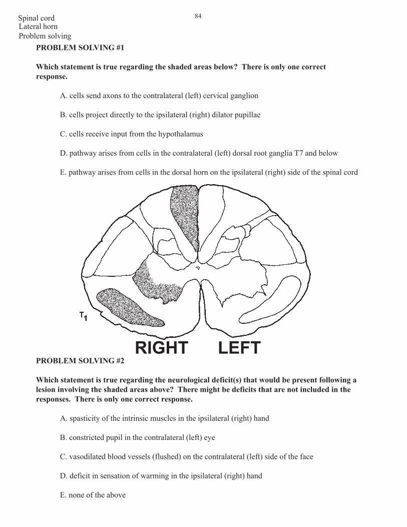
Spinal cord 84Lateral hornProblem solving
PROBLEM SOLVING #1
Which statement is true regarding the shaded areas below? There is only one correctresponse.
A. cells send axons to the contralateral (left) cervical ganglion
B. cells project directly to the ipsilateral (right) dilator pupillae
C. cells receive input from the hypothalamus
D. pathway arises from cells in the contralateral (left) dorsal root ganglia T7 and below
E. pathway arises from cells in the dorsal horn on the ipsilateral (right) side of the spinal cord
RIGHT LEFTPROBLEM SOLVING #2
Which statement is true regarding the neurological deficit(s) that would be present following alesion involving the shaded areas above? There might be deficits that are not included in theresponses. There is only one correct response.
A. spasticity of the intrinsic muscles in the ipsilateral (right) hand
B. constricted pupil in the contralateral (left) eye
C. vasodilated blood vessels (flushed) on the contralateral (left) side of the face
D. deficit in sensation of warming in the ipsilateral (right) hand
E. none of the above
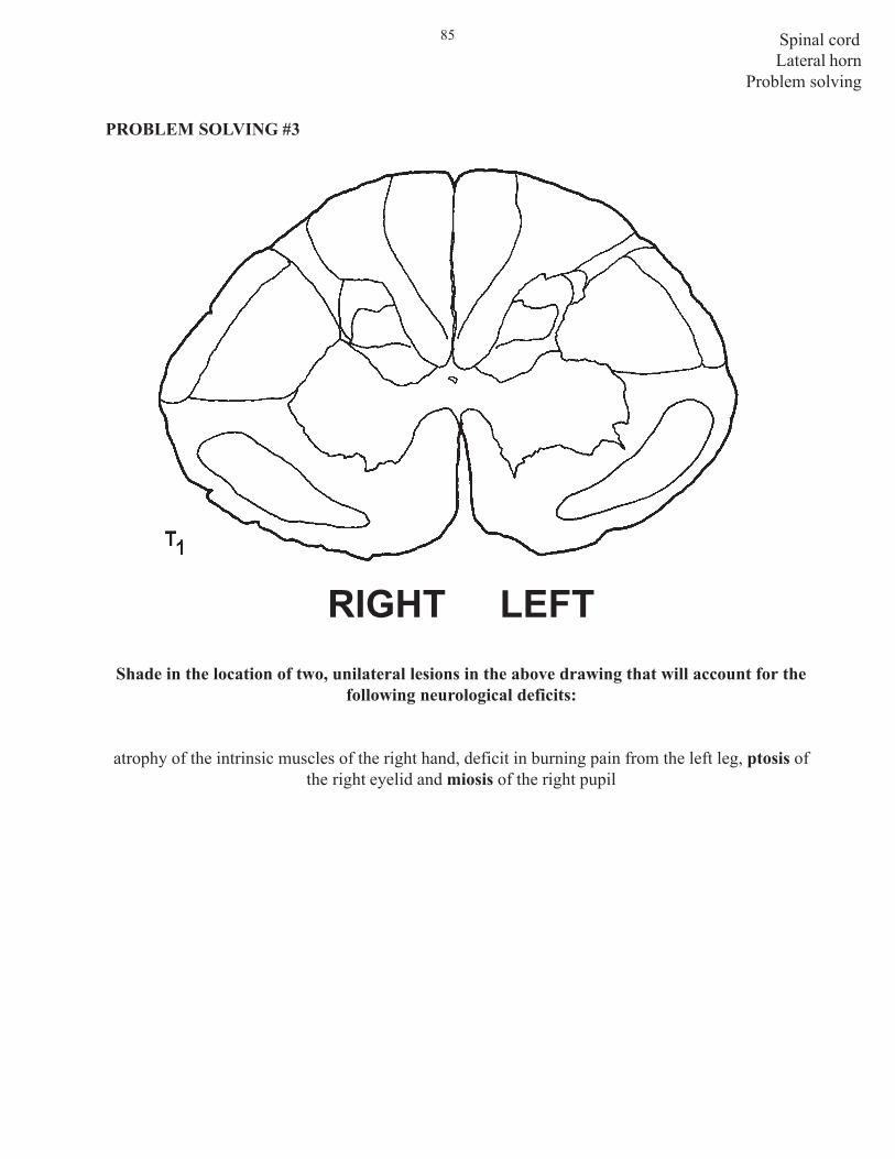
Spinal cord85
Lateral hornProblem solving
PROBLEM SOLVING #3
RIGHT LEFTShade in the location of two, unilateral lesions in the above drawing that will account for the
following neurological deficits:
atrophy of the intrinsic muscles of the right hand, deficit in burning pain from the left leg, ptosis ofthe right eyelid and miosis of the right pupil

Spinal cord 86
Lateral hornProblem solving
PROBLEM SOLVING #3 ANSWER
RIGHT LEFTPROBLEM SOLVING MATCHING
Match the best choice in the right hand column with the pathway or cell group in the left hand column.There might be deficits that are not included in the responses.
_____1. left fasciculus gracilis at C1 A. lesion results in deficit in unconscious proprioception from the right leg
_____2. left lateral corticospinal tract at C1B. axons arise from the dorsal roots T7
_____3. left dorsal spinocerebellar tract at C1 and below on the left
_____4. cells in left ventral horn at T1 C. axons carry info. about vibration from the right big toe
_____5. left fasciculus cuneatus at C1D. lesion results in deficit in sense of
_____6. left lateral horn at C6 cooling from the right foot
E. lesion results in deficit in conscious proprioception from the left elbow
F. cell bodies lie in the left Clarke’s nucleus
G. lesion results in deficit in sense of warming from the left hand
H. lesion results in atrophy of the intrinsic muscles of the left hand
I. axons arise from cells in right motor cortex
J. NO appropriate response
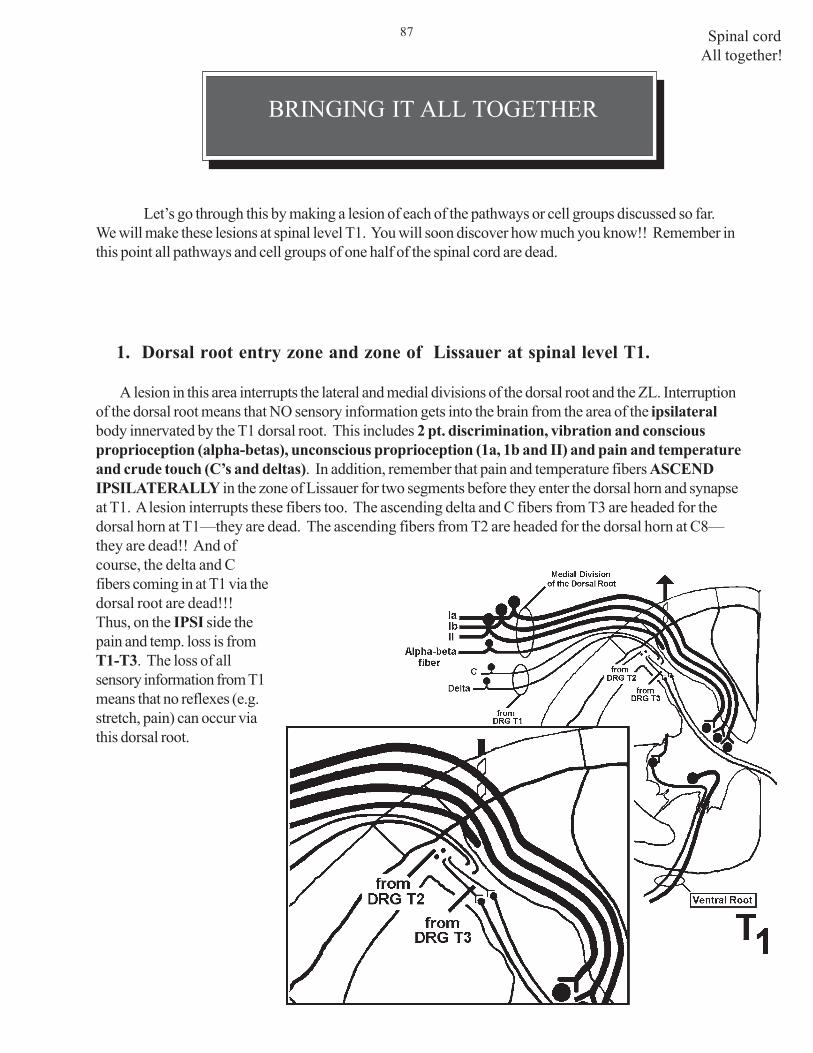
Spinal cord87
Let’s go through this by making a lesion of each of the pathways or cell groups discussed so far.We will make these lesions at spinal level T1. You will soon discover how much you know!! Remember inthis point all pathways and cell groups of one half of the spinal cord are dead.
1. Dorsal root entry zone and zone of Lissauer at spinal level T1.
A lesion in this area interrupts the lateral and medial divisions of the dorsal root and the ZL. Interruptionof the dorsal root means that NO sensory information gets into the brain from the area of the ipsilateralbody innervated by the T1 dorsal root. This includes 2 pt. discrimination, vibration and consciousproprioception (alpha-betas), unconscious proprioception (1a, 1b and II) and pain and temperatureand crude touch (C’s and deltas). In addition, remember that pain and temperature fibers ASCENDIPSILATERALLY in the zone of Lissauer for two segments before they enter the dorsal horn and synapseat T1. A lesion interrupts these fibers too. The ascending delta and C fibers from T3 are headed for thedorsal horn at T1—they are dead. The ascending fibers from T2 are headed for the dorsal horn at C8—they are dead!! And ofcourse, the delta and Cfibers coming in at T1 via thedorsal root are dead!!!Thus, on the IPSI side thepain and temp. loss is fromT1-T3. The loss of allsensory information from T1means that no reflexes (e.g.stretch, pain) can occur viathis dorsal root.
BRINGING IT ALL TOGETHER
All together!
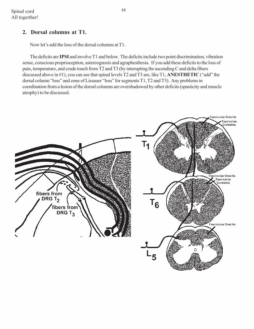
Spinal cord 88
2. Dorsal columns at T1.
Now let’s add the loss of the dorsal columns at T1.
The deficits are IPSI and involve T1 and below. The deficits include two point discrimination, vibrationsense, conscious proprioception, astereognosis and agraphesthesia. If you add these deficits to the loss ofpain, temperature, and crude touch from T2 and T3 (by interupting the ascending C and delta fibersdiscussed above in #1), you can see that spinal levels T2 and T3 are, like T1, ANESTHETIC (“add” thedorsal column “loss” and zone of Lissauer “loss” for segments T1, T2 and T3). Any problems incoordination from a lesion of the dorsal columns are overshadowed by other deficits (spasticity and muscleatrophy) to be discussed.
All togerther!
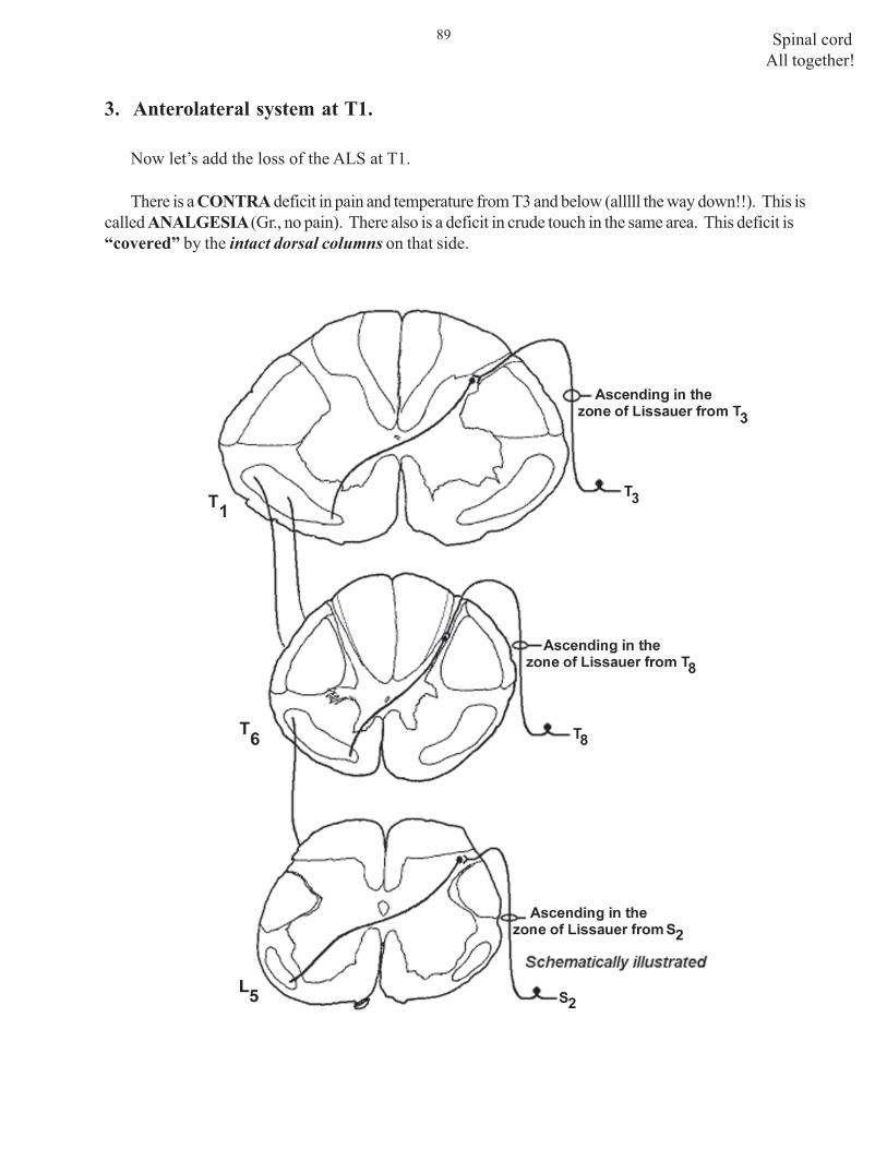
Spinal cord89
3. Anterolateral system at T1.
Now let’s add the loss of the ALS at T1.
There is a CONTRA deficit in pain and temperature from T3 and below (alllll the way down!!). This iscalled ANALGESIA (Gr., no pain). There also is a deficit in crude touch in the same area. This deficit is“covered” by the intact dorsal columns on that side.
All together!

Spinal cord 90
4. Dorsal spinocerebellar tract at T1.
Now let’s add the loss of the DSCT at T1.
There is a loss of unconscious proprioception IPSILATERAL to the lesion from T1 and below. Thedeficit involves incoordination of the lower extremity (and the forearm). This deficit will be camouflagedby problems related to interruption of the LCST.
All togerther!
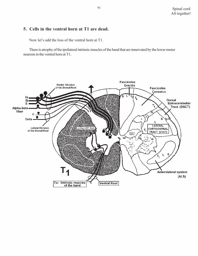
Spinal cord91
All together!
5. Cells in the ventral horn at T1 are dead.
Now let’s add the loss of the ventral horn at T1.
There is atrophy of the ipsilateral intrinsic muscles of the hand that are innervated by the lower motorneurons in the ventral horn at T1.

Spinal cord 92
All togerther!
6. Lateral horn at T1.
Now let’s add the loss of the lateral horn at T1.
There is an IPSILATERAL Horner’s Syndrome (ptosis, miosis, dry face, etc.). The interruptionof the descending sympathetics (that no longer pass to regions of the spinal cord below T1) meansthat there is no sweating on the entire IPSI side of the body.
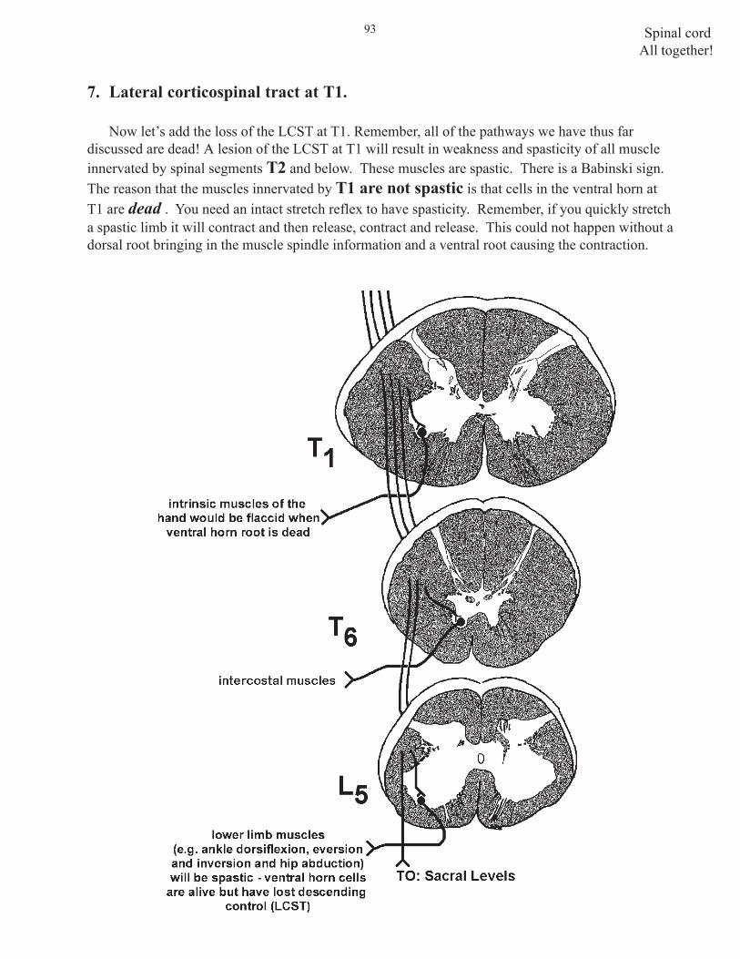
Spinal cord93
All together!
7. Lateral corticospinal tract at T1.
Now let’s add the loss of the LCST at T1. Remember, all of the pathways we have thus fardiscussed are dead! A lesion of the LCST at T1 will result in weakness and spasticity of all muscleinnervated by spinal segments T2 and below. These muscles are spastic. There is a Babinski sign.The reason that the muscles innervated by T1 are not spastic is that cells in the ventral horn atT1 are dead . You need an intact stretch reflex to have spasticity. Remember, if you quickly stretcha spastic limb it will contract and then release, contract and release. This could not happen without adorsal root bringing in the muscle spindle information and a ventral root causing the contraction.

Spinal cord 94
As you can see from the figure below, there are several spinal cord pathways that we have notdiscussed. These pathways will be covered in the next few weeks. Right now, realize that a lesionof the LCST, following a stroke, will not disrupt all descending motor control. You may have seensuch patients and it is clear that there is some motor control that is conveyed via several of thepathways labeled below.
All togerther!
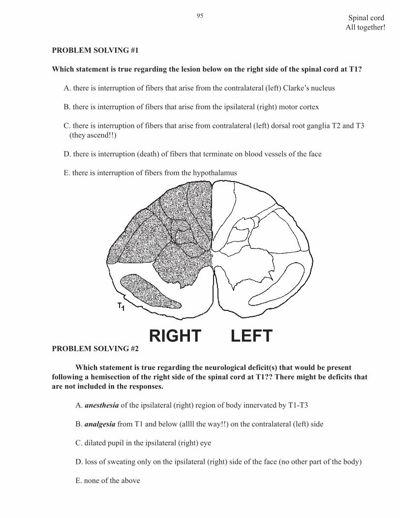
Spinal cord95
All together!
PROBLEM SOLVING #1
Which statement is true regarding the lesion below on the right side of the spinal cord at T1?
A. there is interruption of fibers that arise from the contralateral (left) Clarke’s nucleus
B. there is interruption of fibers that arise from the ipsilateral (right) motor cortex
C. there is interruption of fibers that arise from contralateral (left) dorsal root ganglia T2 and T3(they ascend!!)
D. there is interruption (death) of fibers that terminate on blood vessels of the face
E. there is interruption of fibers from the hypothalamus
RIGHT LEFTPROBLEM SOLVING #2
Which statement is true regarding the neurological deficit(s) that would be presentfollowing a hemisection of the right side of the spinal cord at T1?? There might be deficits thatare not included in the responses.
A. anesthesia of the ipsilateral (right) region of body innervated by T1-T3
B. analgesia from T1 and below (allll the way!!) on the contralateral (left) side
C. dilated pupil in the ipsilateral (right) eye
D. loss of sweating only on the ipsilateral (right) side of the face (no other part of the body)
E. none of the above
Spinal cord 96
All togerther!
PROBLEM SOLVING #3
Which of the following are true regarding a lesion that would involve the entire LEFT side of thespinal cord at T1??
1. spasticity of the left leg
2. flushed on the left side of the face
3. lack of sweating on left side of face
4. lack of sweating on left side of body
5. miosis of left pupil
6. ptosis of left eyelid
7. spasticity of left intrinsic muscles of the hand
8. spasticity of left biceps
9. atrophy of left toe muscles
10. loss of pain and temp from dermatome innervated by left dorsal root T3 (forget aboutdermatome overlap)
11. bilateral loss of crude touch at T3
12. loss of tone in left intrinsic muscles of the hand muscles
13. spasticity of left shoulder muscles
14. loss of vibration sense from the left toe
15. right Babinski
16. loss of unconscious proprioception from the region of the body innervated by theipsilateral dorsal root L4 (remember, Clarke’s column ends at L3)
17. increased muscle stretch reflex in the left quadriceps upon tapping the patellar tendon
18. some of Clarke’s column on the left is involved
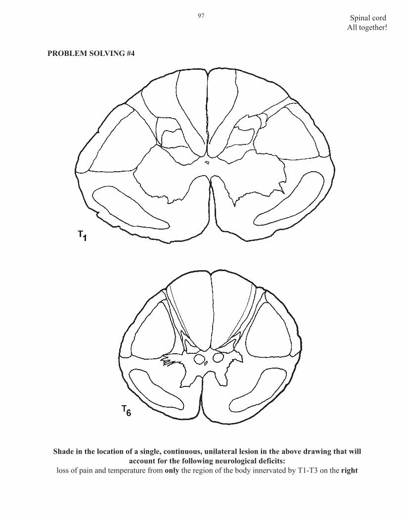
Spinal cord97
All together!
PROBLEM SOLVING #4
Shade in the location of a single, continuous, unilateral lesion in the above drawing that willaccount for the following neurological deficits:
loss of pain and temperature from only the region of the body innervated by T1-T3 on the right

Spinal cord 98
All togerther!

Spinal cord99
All together!
PROBLEM SOLVING #5
RIGHT LEFTShade in the location of a single, continuous, unilateral lesion in the above drawing that will
account for the following neurological deficits:
loss of sweating from the entire right side of the body and head
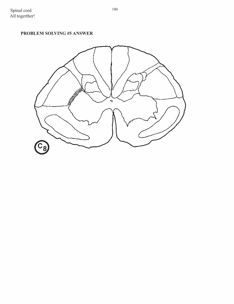
Spinal cord 100
All togerther!
PROBLEM SOLVING #5 ANSWER

Spinal cord101
All together!
PROBLEM SOLVING #6
RIGHT LEFTShade in the location of a single, continuous, unilateral lesion in the above drawing that will
account for the following neurological deficits:
loss of pain and temperature from the region of the body innervated by spinal segments T3 andbelow on the left
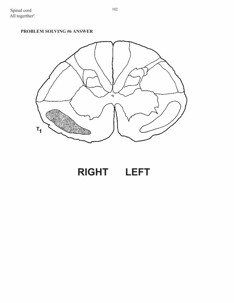
Spinal cord 102
All togerther!
PROBLEM SOLVING #6 ANSWER
RIGHT LEFT

Spinal cord103
All together!
PROBLEM SOLVING #7
RIGHT LEFTShade in the location of a single, continuous, unilateral lesion in the above drawing that will
account for the following neurological deficits:
loss of unconscious proprioception from regions of the body innervated by spinal segments T1 andbelow on the right
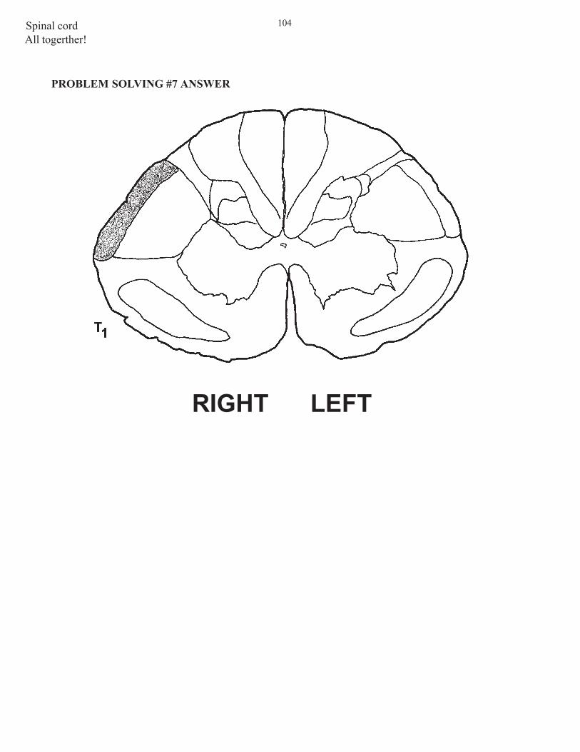
Spinal cord 104
All togerther!
PROBLEM SOLVING #7 ANSWER
RIGHT LEFT
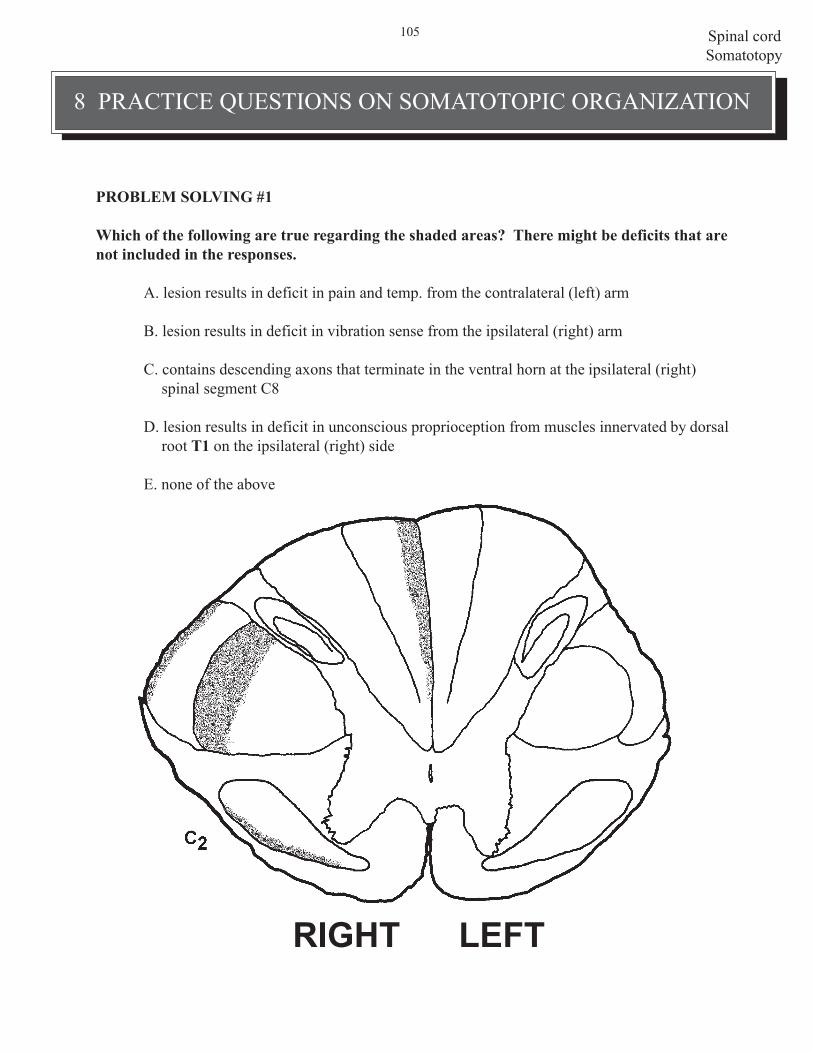
Spinal cord105
PROBLEM SOLVING #1
Which of the following are true regarding the shaded areas? There might be deficits that arenot included in the responses.
A. lesion results in deficit in pain and temp. from the contralateral (left) arm
B. lesion results in deficit in vibration sense from the ipsilateral (right) arm
C. contains descending axons that terminate in the ventral horn at the ipsilateral (right)spinal segment C8
D. lesion results in deficit in unconscious proprioception from muscles innervated by dorsalroot T1 on the ipsilateral (right) side
E. none of the above
RIGHT LEFT
8 PRACTICE QUESTIONS ON SOMATOTOPIC ORGANIZATION
Somatotopy
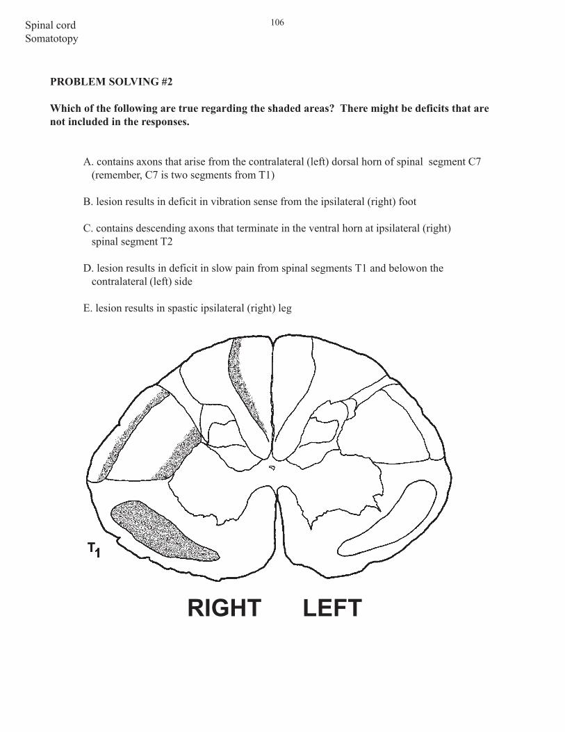
Spinal cord 106
PROBLEM SOLVING #2
Which of the following are true regarding the shaded areas? There might be deficits that arenot included in the responses.
A. contains axons that arise from the contralateral (left) dorsal horn of spinal segment C7(remember, C7 is two segments from T1)
B. lesion results in deficit in vibration sense from the ipsilateral (right) foot
C. contains descending axons that terminate in the ventral horn at ipsilateral (right)spinal segment T2
D. lesion results in deficit in slow pain from spinal segments T1 and belowon thecontralateral (left) side
E. lesion results in spastic ipsilateral (right) leg
RIGHT LEFT
Somatotopy

Spinal cord107
PROBLEM SOLVING #3
Which of the following are true regarding the shaded areas? There might be deficits that arenot included in the responses.
A. contains axons that arise from the contralateral (left) dorsal horn of the spinal cord atS1 (To help you answer this, use the S1 drawing below and connect a cell in the leftdorsal horn with the right ALS).
B. lesion results in spasticity of muscles innervated by ipsilateral (right) S1 lower motorneurons
C. contains descending axons that terminate in the ventral horn at ipsilateral (right)spinal segment L5
D. lesion results in deficit in unconscious proprioception from muscles innervated bydorsal root L5 on the ipsilateral (right) side (be careful!!—remember how this info.gets to Clarke’s nucleus)
E. none of the above
Somatotopy

Spinal cord 108
PROBLEM SOLVING #4
Match the best choice in the right hand column with the number of the structure in the drawing below.
1.____ A. lesion results in muscle atrophy
2.____ B. primary source of information about how fast a muscle is changing length
3.____C. pathway carries pain and temperature
4.____ and terminates within the ipsilateral thalamus
5.____ D. axons carry primarily information regarding the constant length of muscle
6.____E. pathway arises from Clarke’s column (nucleus)
7.____F. axons convey information about fast pain and
8.____ cooling
9.____ G. smallest and slowest conducting axon in the dorsal root (conveys info. about warming)
10.____H. axon conveys information about vibration
I. axon conveys info. about muscle tension
J. axon carries info. about pain and temperature from regions of the body innervated by contralateral dorsal root T3
Somatotopy
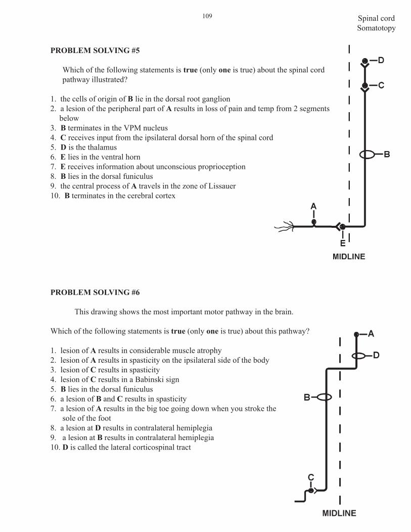
Spinal cord109
Somatotopy
PROBLEM SOLVING #5
Which of the following statements is true (only one is true) about the spinal cordpathway illustrated?
1. the cells of origin of B lie in the dorsal root ganglion2. a lesion of the peripheral part of A results in loss of pain and temp from 2 segments
below3. B terminates in the VPM nucleus4. C receives input from the ipsilateral dorsal horn of the spinal cord5. D is the thalamus6. E lies in the ventral horn7. E receives information about unconscious proprioception8. B lies in the dorsal funiculus9. the central process of A travels in the zone of Lissauer10. B terminates in the cerebral cortex
PROBLEM SOLVING #6
This drawing shows the most important motor pathway in the brain.
Which of the following statements is true (only one is true) about this pathway?
1. lesion of A results in considerable muscle atrophy2. lesion of A results in spasticity on the ipsilateral side of the body3. lesion of C results in spasticity4. lesion of C results in a Babinski sign5. B lies in the dorsal funiculus6. a lesion of B and C results in spasticity7. a lesion of A results in the big toe going down when you stroke the
sole of the foot8. a lesion at D results in contralateral hemiplegia9. a lesion at B results in contralateral hemiplegia10. D is called the lateral corticospinal tract
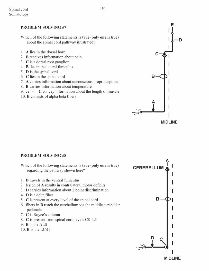
Spinal cord 110
Somatotopy
PROBLEM SOLVING #7
Which of the following statements is true (only one is true)about the spinal cord pathway illustrated?
1. A lies in the dorsal horn2. E receives information about pain3. C is a dorsal root ganglion4. B lies in the lateral funiculus5. D is the spinal cord6. C lies in the spinal cord7. A carries information about unconscious proprioception8. B carries information about temperature9. cells in C convey information about the length of muscle10. B consists of alpha beta fibers
PROBLEM SOLVING #8
Which of the following statements is true (only one is true)regarding the pathway shown here?
1. B travels in the ventral funiculus2. lesion of A results in contralateral motor deficits3. D carries information about 2 point discrimination4. D is a delta fiber5. C is present at every level of the spinal cord6. fibers in B reach the cerebellum via the middle cerebellar
peduncle7. C is Royce’s column8. C is present from spinal cord levels C8- L39. B is the ALS10. B is the LCST

Spinal cord111
ANSWERS TO PROBLEM SOLVING QUESTIONS RELATED TO POINTS 5-8
NOTE: The answers to ALL #3s (#4s for points 5 and 6) illustrated on the back side of the question.
Point #5 Ventral Horn
1. E2. AMatching B,D,A,H,E
Point #6 Lateral (Intermediolateral) Horn
Matching B,I,F,H,E,J2. C3. E
Point #7 Lesion of the spinal cord at T1
1. E2. ATrue/False 7, 8, 9, 13, and 15 are false
Point #8 Somatotopic Organization
1. E2. C3. A4. B,F,D,C,A,G,E,H,J,I5. 96. 87. 108. 8
PROBLEM SOLVING ANSWERSPOINTS 5-8
Spinal cord 112
THIS CONCLUDES THE FIRST PART OF THE SPINAL CORD MODULE. TOPICSTHAT WILL BE COVERED IN THE FOLLOWING CLINICAL PORTION OF THE
MODULE INCLUDE:
1. RELATIONSHIPS OF SPINAL NERVES TO VERTEBRAL LEVELS
2. CAUDA EQUINA LESIONS
3. CONUS MEDULLARIS LESIONS
4. EFFECTS OF CORD LESIONS ON BLADDER CONTROL
5. DISTINGUISHING BETWEEN LESIONS OF THE:
a) VENTRAL HORNb) PERIPHERAL NERVE (NEUROPATHY)c) NEUROMUSCULAR JUNCTIONd) MUSCLE (MYOPATHY)
6. PROBLEM-SOLVING—CLINICAL CASE STUDIES
Spinal cord113

Spinal cord 114