mechanisms of thrombus formation
TRANSCRIPT

review article
T h e n e w e ngl a nd j o u r na l o f m e dic i n e
n engl j med 359;9 www.nejm.org august 28, 2008938
Mechanisms of Disease
Mechanisms of Thrombus FormationBruce Furie, M.D., and Barbara C. Furie, Ph.D.
From the Division of Hemostasis and Thrombosis, Beth Israel Deaconess Medi-cal Center, and Harvard Medical School — both in Boston.
N Engl J Med 2008;359:938-49.Copyright © 2008 Massachusetts Medical Society.
Hemostasis is the process that maintains the integrity of a closed, high-pressure circulatory system after vascular damage. Vessel-wall injury and the extravasation of blood from the circulation rapidly initiate
events in the vessel wall and in blood that seal the breach. Circulating platelets are recruited to the site of injury, where they become a major component of the develop-ing thrombus; blood coagulation, initiated by tissue factor, culminates in the genera-tion of thrombin and fibrin. These events occur concomitantly (Fig. 1A; also see Video, available with the full text of this article at www.nejm.org), and under nor-mal conditions, regulatory mechanisms contain thrombus formation temporally and spatially.
When pathologic processes overwhelm the regulatory mechanisms of hemo-stasis, excessive quantities of thrombin form, initiating thrombosis (Fig. 1B; and Video, Chap. 2). Thrombosis is a critical event in the arterial diseases associated with myocardial infarction and stroke, and venous thromboembolic disorders ac-count for considerable morbidity and mortality. Moreover, venous thrombosis is the second leading cause of death in patients with cancer. Our understanding of the molecular and cellular basis of thrombus formation has advanced greatly through the use of novel techniques for studying mouse models of thrombosis. In this article, we review recent advances in knowledge about thrombus formation. We also offer new hypotheses and some speculations about thrombus formation and the preven-tion and treatment of thrombosis.
Figure 1 (facing page). Thrombus Formation In Vivo.
The developing thrombus in a living mouse after vessel-wall injury (Panel A) is characterized by the deposition of platelets (red), tissue factor (green), and fibrin (blue). Platelet thrombus for-mation and fibrin deposition occur concomitantly. Platelets and tissue factor appear yellow; tis-sue factor and fibrin, turquoise; platelets and fibrin, magenta; and platelets, fibrin, and tissue factor, white. A three-dimensional, confocal optical reconstruction of a thrombus in the lumen of an arteriole (Panel B) shows the platelet thrombus (red and yellow) being formed in the ves-sel wall, which is lined with the endothelium (labeled green with antibodies to platelet-endothe-lial cell-adhesion molecule [PECAM-1]). Platelets are labeled red using antibodies to CD41; platelets stained with both CD41 and PECAM-1 appear yellow. Calcium is mobilized during platelet activation. Panel C shows platelets loaded with a calcium-sensitive dye during thrombus formation; resting platelets appear green, and activated platelets appear yellow. Labeled mi-croparticles bearing tissue factor (Panel D, green) infused into a recipient mouse accumulate in the developing thrombus. In Panel E, expression of protein disulfide isomerase (PDI, green) is shown during thrombus formation. Panel F shows fibrin (green) and platelets (red), which ap-pear rapidly after vessel-wall injury and form a thrombus; yellow indicates colocalization of fi-brin and platelets. In Panel G, inhibition of PDI blocks platelet accumulation and the generation of fibrin, and neither is observed. A video showing the process of thrombus formation in live mice is available with the full text of this article at www.nejm.org.
The New England Journal of Medicine Downloaded from nejm.org on May 4, 2013. For personal use only. No other uses without permission.
Copyright © 2008 Massachusetts Medical Society. All rights reserved.

mechanisms of disease
n engl j med 359;9 www.nejm.org august 28, 2008 939
For m ation of a Pl atele t Thrombus
The vessel wall, with its inner lining of endotheli-um, is crucial to the maintenance of a patent vas-culature. The endothelium contains three throm-boregulators — nitric oxide,1,2 prostacyclin,3 and the ectonucleotidase CD394 — which together provide a defense against thrombus formation. Collagen in the subendothelial matrix and tissue
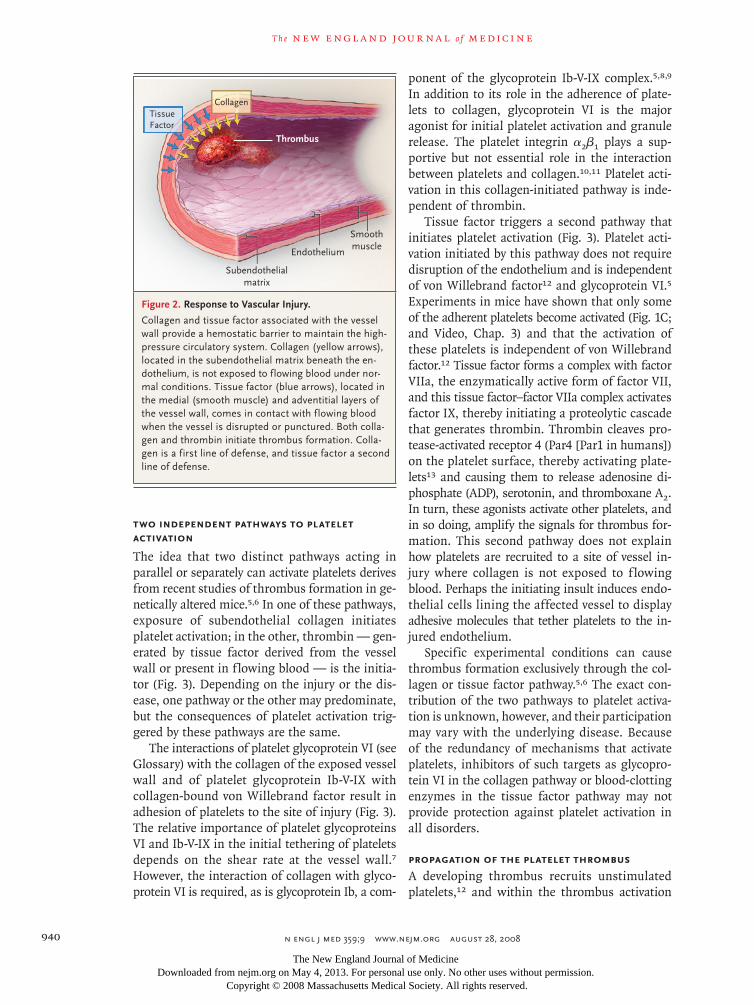
factor facilitate the maintenance of a closed cir-culatory system. When the vessel wall is breached or the endothelium is disrupted, collagen and tis-sue factor become exposed to the flowing blood, thereby initiating formation of a thrombus (Fig. 2). Exposed collagen triggers the accumulation and activation of platelets, whereas exposed tis-sue factor initiates the generation of thrombin, which not only converts fibrinogen to fibrin but also activates platelets.
33p9
AUTHOR
FIGURE
JOB: ISSUE:
4-CH/T
RETAKE 1st
2nd
SIZE
ICM
CASE
EMail LineH/TCombo
Revised
AUTHOR, PLEASE NOTE: Figure has been redrawn and type has been reset.
Please check carefully.
REG F
FILL
TITLE3rd
Enon ARTIST:
Furie
1a-f
8-28-08
mst
35909
A
Blood flow
B
DC
F GE
The New England Journal of Medicine Downloaded from nejm.org on May 4, 2013. For personal use only. No other uses without permission.
Copyright © 2008 Massachusetts Medical Society. All rights reserved.
T h e n e w e ngl a nd j o u r na l o f m e dic i n e
n engl j med 359;9 www.nejm.org august 28, 2008940
Two Independent Pathways to Platelet Activation
The idea that two distinct pathways acting in parallel or separately can activate platelets derives from recent studies of thrombus formation in ge-netically altered mice.5,6 In one of these pathways, exposure of subendothelial collagen initiates platelet activation; in the other, thrombin — gen-erated by tissue factor derived from the vessel wall or present in flowing blood — is the initia-tor (Fig. 3). Depending on the injury or the dis-ease, one pathway or the other may predominate, but the consequences of platelet activation trig-gered by these pathways are the same.
The interactions of platelet glycoprotein VI (see Glossary) with the collagen of the exposed vessel wall and of platelet glycoprotein Ib-V-IX with collagen-bound von Willebrand factor result in adhesion of platelets to the site of injury (Fig. 3). The relative importance of platelet glycoproteins VI and Ib-V-IX in the initial tethering of platelets depends on the shear rate at the vessel wall.7 However, the interaction of collagen with glyco-protein VI is required, as is glycoprotein Ib, a com-
ponent of the glycoprotein Ib-V-IX complex.5,8,9 In addition to its role in the adherence of plate-lets to collagen, glycoprotein VI is the major agonist for initial platelet activation and granule release. The platelet integrin α2β1 plays a sup-portive but not essential role in the interaction between platelets and collagen.10,11 Platelet acti-vation in this collagen-initiated pathway is inde-pendent of thrombin.
Tissue factor triggers a second pathway that initiates platelet activation (Fig. 3). Platelet acti-vation initiated by this pathway does not require disruption of the endothelium and is independent of von Willebrand factor12 and glycoprotein VI.5 Experiments in mice have shown that only some of the adherent platelets become activated (Fig. 1C; and Video, Chap. 3) and that the activation of these platelets is independent of von Willebrand factor.12 Tissue factor forms a complex with factor VIIa, the enzymatically active form of factor VII, and this tissue factor–factor VIIa complex activates factor IX, thereby initiating a proteolytic cascade that generates thrombin. Thrombin cleaves pro-tease-activated receptor 4 (Par4 [Par1 in humans]) on the platelet surface, thereby activating plate-lets13 and causing them to release adenosine di-phosphate (ADP), serotonin, and thromboxane A2. In turn, these agonists activate other platelets, and in so doing, amplify the signals for thrombus for-mation. This second pathway does not explain how platelets are recruited to a site of vessel in-jury where collagen is not exposed to f lowing blood. Perhaps the initiating insult induces endo-thelial cells lining the affected vessel to display adhesive molecules that tether platelets to the in-jured endothelium.
Specific experimental conditions can cause thrombus formation exclusively through the col-lagen or tissue factor pathway.5,6 The exact con-tribution of the two pathways to platelet activa-tion is unknown, however, and their participation may vary with the underlying disease. Because of the redundancy of mechanisms that activate platelets, inhibitors of such targets as glycopro-tein VI in the collagen pathway or blood-clotting enzymes in the tissue factor pathway may not provide protection against platelet activation in all disorders.
Propagation of the Platelet Thrombus
A developing thrombus recruits unstimulated platelets,12 and within the thrombus activation
08/07/08
AUTHOR PLEASE NOTE:Figure has been redrawn and type has been reset
Please check carefully
AuthorFig #TitleDEMEArtist
COLOR FIGURE
Draft 1
Mech. of thrombus formation2
CHKLS
Furie
Schwartz
Smoothmuscle
Endothelium
Subendothelialmatrix
Thrombus
TissueFactor
Collagen
Figure 2. Response to Vascular Injury.
Collagen and tissue factor associated with the vessel wall provide a hemostatic barrier to maintain the high-pressure circulatory system. Collagen (yellow arrows), located in the subendothelial matrix beneath the en-dothelium, is not exposed to flowing blood under nor-mal conditions. Tissue factor (blue arrows), located in the medial (smooth muscle) and adventitial layers of the vessel wall, comes in contact with flowing blood when the vessel is disrupted or punctured. Both colla-gen and thrombin initiate thrombus formation. Colla-gen is a first line of defense, and tissue factor a second line of defense.
The New England Journal of Medicine Downloaded from nejm.org on May 4, 2013. For personal use only. No other uses without permission.
Copyright © 2008 Massachusetts Medical Society. All rights reserved.

mechanisms of disease
n engl j med 359;9 www.nejm.org august 28, 2008 941
occurs only in a subgroup of the recruited plate-lets. Others remain loosely associated with the thrombus but do not undergo activation and may ultimately disengage from the thrombus (Fig. 1C).12 In short, thrombus formation is a dynamic
process in which some platelets adhere to and others separate from the developing thrombus, and in which shear, f low, turbulence, and the number of platelets in the circulation greatly in-fluence the architecture of the clot.
08/13/08
AUTHOR PLEASE NOTE:Figure has been redrawn and type has been reset
Please check carefully
AuthorFig #TitleDEMEArtist
COLOR FIGURE
Draft 2
Mech. of thrombus formation2
?Williams
Furie
Schwartz
Encrypted tissue factor expressed consti-tutively on the vessel wall or active tissuefactor expressed within the vessel wall
Tissue factor activation by PDI
Tissue factor forms a complex with factor VIIa, leading to thrombin generation
Thrombin cleaves Par4 on platelet surface,activating platelets
Exposure of subendothelial matrixon endothelial-cell disruption
Platelets captured on vessel wall
Collagen in contact with flowing blood
Platelet glycoprotein VI interacts withcollagen and glycoprotein Ib-V-IX with von Willebrand factor, leading to platelet capture
Platelet–platelet interaction mediatedby binding of αIIbβ3 to fibrinogen andvon Willebrand factor
Collagen-mediated activation of platelets
A Tissue Factor Pathway
C Platelet Activation
B Collagen Pathway
Figure 3. Independent Pathways of Platelet Activation in Mouse Models of In Vivo Thrombosis.
In the tissue factor pathway (Panel A), tissue factor (green) is expressed on the vessel wall and requires protein disul-fide isomerase (PDI) to generate fibrin. Tissue factor generates thrombin by means of the blood-coagulation path-ways. Platelets are captured on the vessel wall, and platelet–platelet interaction and platelet activation by thrombin cleavage of protease-activated receptor (Par4) follow. In the collagen pathway (Panel B), on disruption of the en-dothelium, collagen (green) is exposed, rapidly leading to platelet (red) deposition. The yellow represents the merg-ing of the collagen and the platelets. Platelets are captured on the vessel wall, and platelet–platelet interaction and platelet activation follow. Thrombin is not required for platelet activation in this pathway. In the common pathway, platelet activation is monitored by calcium mobilization (Panel C). Unactivated platelets (green) become associated with the developing thrombus. Those that are activated (yellow) are detected by increases in calcium mobilization.
The New England Journal of Medicine Downloaded from nejm.org on May 4, 2013. For personal use only. No other uses without permission.
Copyright © 2008 Massachusetts Medical Society. All rights reserved.
T h e n e w e ngl a nd j o u r na l o f m e dic i n e
n engl j med 359;9 www.nejm.org august 28, 2008942
The platelet integrin αIIb
β3, when activated, mediates recruitment of platelets to the throm-bus as well as platelet–platelet interactions. Acti-vation of α
IIbβ3 requires an enzyme (protein
disulfide isomerase) that catalyzes cleavage or formation of disulfide bonds between cysteine residues.14-16 Activation of platelets bound to the wall of the injured vessel causes a conformational transition in α
IIbβ3 that increases the affinity of
the integrin for its ligands, fibrinogen and von Willebrand factor.17 At low shear rates, fibrinogen is the predominant ligand, whereas von Wille-brand factor plays an important role at higher shear rates.7,18 However, neither von Willebrand factor nor fibrinogen is absolutely required for
platelet accumulation.19 In addition, von Wille-brand factor is a ligand for glycoprotein Ib, but results in a mouse thrombosis model involving denudation of the endothelium suggest an as yet unidentified alternative ligand for glycoprotein Ib.9 During platelet activation, late signaling events enhance platelet–platelet affinity. Growth-arrest–specific gene 6,20 CD40 ligand,21 ephrin-Eph,22 and signaling lymphocyte activation mol-ecule23 participate in the platelet–platelet synapse to create a protected environment in the inter-stices of the clot that stabilizes the thrombus.24
Platelet activation releases the contents of platelet alpha granules and dense granules, each of which carries a cargo of components that are critical for thrombus formation. Proteins are packaged in various subpopulations of alpha granules,25 whereas ADP and calcium ions are packaged in the dense granules. The release of ADP stimulates platelet activation through two ADP receptors, P2Y1 and P2Y12. The role of these receptors in platelet function and the pharma-cology of drugs directed against these receptors has recently been reviewed.26
Bl o od Coagul ation
Tissue Factor
A membrane protein, tissue factor is present on cells in numerous anatomical compartments and has multiple functions. In addition to initiating blood coagulation, tissue factor mediates intracel-lular signaling events that are important for angio-genesis,27 tumor progression,28 metastasis,29 and maintenance of the yolk-sac vasculature.30 Tissue factor is constitutively expressed on fibroblasts and pericytes in the adventitia and medial smooth-muscle cells of the vessel wall. It is also constitu-tively expressed on many nonvascular cells, and its expression on monocytes and endo thelial cells can be induced by chemical stimuli.31,32 The idea that there may be functionally significant amounts of tissue factor on granulocytes and platelets re-mains controversial.33 The endothelium was thought to act as a barrier separating factor VIIa in flowing blood from cellular sources of tissue factor in order to prevent the initiation of coagu-lation in the absence of injury.34 However, tissue factor is also present in circulating blood, and this bloodborne tissue factor may participate in physiologic and pathologic processes.35
Tissue factor is associated with some micro-
Glossary
Adenosine diphosphate (ADP): An agonist of platelet activation.
α2 β1: A platelet integrin that plays a role in collagen binding.
αIIbβ3: An integrin that serves as the fibrinogen receptor on platelets.
β3 integrin: A subunit of integrins.
CD36: A leukocyte surface protein.
CD40 ligand: A platelet receptor for CD40 that triggers an inflammatory response.
CD55: A leukocyte surface protein associated with the cell membrane by means of a glycerolinositol anchor. The anchor is defective in paroxysmal noctur-nal hemoglobinuria.
CD59: A leukocyte surface protein associated with the cell membrane by means of a glycerolinositol anchor. The anchor is defective in paroxysmal noctur-nal hemoglobinuria.
Ephrin-Eph: Receptor kinases and ligands on platelet surfaces.
Glycoprotein Ib-V-IX: A cluster of adhesive receptors on platelets. Von Wille-brand factor binds to this complex.
Glycoprotein VI: A collagen receptor on platelets.
Growth-arrest–specific gene 6: A vitamin K–dependent membrane protein in-volved in cell signaling.
Protease-activated receptor 1 (Par1): A thrombin receptor on platelets; equiva-lent to Par4 on mouse platelets; activation initiates cell-signaling pathways.
P-selectin: An adhesion molecule on activated platelets and endothelial cells that binds to PSGL-1, its receptor on leukocytes.
P-selectin glycoprotein ligand 1 (PSGL-1): An adhesion molecule on leukocytes that binds to P-selectin.
P2Y1: An ADP receptor on platelets.
P2Y12: An ADP receptor on platelets.
Signaling lymphocyte activation molecule: An adhesion molecule found on platelets.
Tissue factor: A cytokine receptor analogue on the surface of cells that initiates blood coagulation and is engaged in cell-signaling events.
Tissue factor pathway inhibitor: A protein that, when bound to factor Xa, blocks the activity of the tissue factor–factor VIIa complex.
Von Willebrand factor: A plasma protein that is the carrier for factor VIII and that is critical for the adhesion of platelets to the vessel wall.
The New England Journal of Medicine Downloaded from nejm.org on May 4, 2013. For personal use only. No other uses without permission.
Copyright © 2008 Massachusetts Medical Society. All rights reserved.
mechanisms of disease
n engl j med 359;9 www.nejm.org august 28, 2008 943
particles in the circulating blood.35,36 These ve-sicular structures, which are less than 1000 nm in diameter, display proteins of the blood cells from which they were derived (e.g., leukocytes, platelets, endothelial cells, smooth-muscle cells, and mono cytes).36,37 During thrombus formation, platelets accumulate at the vessel wall, become activated, and express P-selectin.38 This adhesion molecule binds to microparticles that display the P-selectin counterreceptor, termed P-selectin gly-coprotein ligand 1 (PSGL-1), allowing the throm-bus to capture microparticles that display tissue factor derived from monocytes (Fig. 1D; and Video, Chap. 4).36 Fibrin propagation within the thrombus is dominat ed by bloodborne tissue fac-tor when vessel-wall injury is limited to endothe-lial-cell activation.39
What prevents tissue factor on microparticles from initiating blood coagulation? Tissue factor can exist in a latent (or “encrypted”) form that lacks coagulant activity or in an active form that initiates blood coagulation.40,41 The molecular ba-sis of encryption is uncertain, but dimerization,42 lipid reorganization,43 and cellular secretion of tis-sue factor–rich granules44 are among the proposed mechanisms. One of the two di sulfide bonds in tissue factor may be a labile allosteric disulfide bond45 that can undergo cleavage or formation, with effects on the structure and function of the protein.46,47 Oxidation of free thiols in encrypted tissue factor to form a disulfide bond yields a con-formation that allows the tissue factor–factor VIIa complex to bind to and activate factor X.45,48 How can these altered disulfide bonds explain the transformation of bloodborne tissue factor from the encrypted to the active form in response to vessel-wall injury? Activated endothelial cells and platelets at the site of injury release protein dis-ulfide isomerase, which catalyzes the formation and breakage of disulfide bonds between cysteine residues within proteins (Fig. 1E; and Video, Chap. 5).49 This enzyme is required for fibrin gen-eration and platelet thrombus formation (Fig. 1F and 1G; and Video, Chap. 6). Perhaps it acts by promoting the formation of a functionally critical disulfide bond in tissue factor.
Thrombin and Fibrin
Tissue factor is the sole initiator of thrombin generation and fibrin formation. The contact pathway of blood coagulation,50,51 a powerful tool for in vitro studies of the coagulation cas-
cade, is not required for initiation of hemostasis in vivo.52 A complete deficiency of factor XII, high-molecular-weight kininogen, or prekallekrein is associated with major defects in the initiation of the contact pathway of coagulation, as mani-fested by a markedly prolonged partial-thrombo-plastin time. Nevertheless, patients with any one of these deficiencies do not have a hemorrhagic disorder. The importance of factor XII in throm-bosis remains controversial, but in mice, a defi-ciency of factor XII or factor XI attenuates the development of thrombi.53-55 Furthermore, inhi-bition or deficiency of factor XII protects mice from ischemic brain injury without causing hem-orrhage.56 In humans, factor XI deficiency may be associated with a hemorrhagic phenotype. Factor XI may also participate in thrombosis in humans, because a deficiency of this protein is associated with a reduced risk of ischemic stroke but not of myocardial infarction.57
A new iteration of the coagulation pathways is required to accommodate these findings and hypotheses (Fig. 4). We propose that the activa-tion of encrypted tissue factor by protein disul-fide isomerase initiates coagulation. On activa-tion, platelets and endothelial cells secrete the isomerase,49 which converts inactive tissue fac-tor on cells or microparticles to its active form. In the case of direct tissue damage, tissue factor in the vessel wall or on cell surfaces may already exist in its active form, and the isomerase may not be required. This tissue factor pathway can be considered the fuse that ignites coagulation with a small amount of thrombin.62 Other salient features of these coagulation pathways indicate that the sole initiator of thrombin generation is active tissue factor. Before thrombin is generat-ed, the tissue factor pathway, proceeding through factor IX or factor X, is inefficient because factors VIII and V, the circulating pro-cofactors required in the tenase and prothrombinase complexes, are not yet available in their most active cofactor form. This inefficient mechanism generates a small amount of thrombin. Once formed, throm-bin converts factors VIII and V to their cofactor forms, factor VIIIa and factor Va, respectively. The tenase and prothrombinase complexes now pro-ceed efficiently to generate a large burst of throm-bin. The tissue factor pathway is down-regulated, or inhibited, by the action of tissue factor path-way inhibitor, but thrombin generation proceeds without replenishing active tissue factor.63
The New England Journal of Medicine Downloaded from nejm.org on May 4, 2013. For personal use only. No other uses without permission.
Copyright © 2008 Massachusetts Medical Society. All rights reserved.
T h e n e w e ngl a nd j o u r na l o f m e dic i n e
n engl j med 359;9 www.nejm.org august 28, 2008944
The question of what promotes continued thrombin generation in the absence of continued production of the active tissue factor–factor VIIa complex is unresolved. In vitro studies that can mimic thrombus formation in flowing blood by resupplying coagulation proteins in the absence of additional tissue factor and in the presence of factor XIIa inhibitors suggest that the prothrom-binase formed when tissue factor–factor VIIa ignites coagulation can sustain continued throm-bin generation. This newly formed thrombin feeds back to activate factors VIII and V, which form factors VIIIa and Va, triggering the greatly am-plified formation of additional thrombin through the pathway mediated by the fully active tenase and prothrombinase complexes. Alternatively, factor XI, which is activated by thrombin,64 cre-ates a reservoir of initiator activity after the tis-sue factor pathway is terminated.62 However, the ability of thrombin to activate factor XI in plas-ma has been questioned.65
What are the membrane surfaces on which the tenase and prothrombinase complexes as-semble? It has been thought that the membrane surface that is critical for thrombin generation is presented by the activated platelet. However, fi-brin generation in the Par4-null mouse, whose platelets cannot be activated by thrombin, is nor-mal,66 suggesting the importance of other mem-brane surfaces in vivo. Factor XII and factor XI are less important for hemostasis than for throm-bosis52; nevertheless, this framework includes an important, albeit not critical, role of factor XI in hemostasis. Three questions remain unanswered: What activates factor XII during thrombosis, and why is activation of this zymogen not important during hemostasis? What enzyme is responsible for the constitutive circulation of factor VIIa? What are the cellular surfaces on which the te-nase and prothrombinase complexes assemble if activated platelets are not required?
Tissue Fac t or a nd Micropa rticles
in Thrombo tic Disor der s
Acute inflammation and infection, sepsis, and endotoxemia can induce a hypercoagulable state. When regulatory mechanisms are overwhelmed, acute disseminated intravascular coagulation en-sues, with consumption of blood coagulation proteins and platelets and, hence, bleeding. In the chronic form of disseminated intravascular
coagulation, thrombosis rather than hemorrhage is predominant. Thrombosis and inflammation are related and mutually reinforcing processes, involving inflammatory mediators (e.g., endo-toxin, tumor necrosis factor α, and CD40 ligand), tissue factor expression on monocytes and the activated endothelium,67 and circulating tissue factor–bearing microparticles.37 A primary cause of thrombosis in disseminated intravascular co-agulation is disruption of endogenous anticoag-ulant pathways.
Hemostatic Microparticles versus Pathologic Microparticles
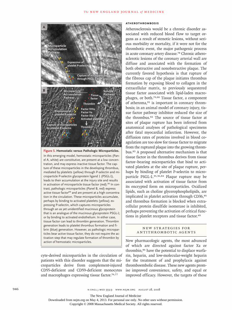
There is no detectable tissue factor activity in normal blood,68 yet tissue factor–bearing micro-particles circulate in healthy persons. Perhaps tis-sue factor–bearing microparticles contain inac-tive tissue factor, which may become activated only when the particles are recruited to the site of vascular injury (Fig. 5A). Pathologic microparticles may bear active tissue factor, which may confer a predisposition to thromboembolic events. Mi-
Figure 4 (facing page). Pathways of Blood Coagulation during Hemostasis and Thrombosis.
Coagulation can be divided into the initiation phase (Panel A) and the amplification phase (Panel B). Dur-ing initiation, the tissue factor–factor VIIa complex serves as a fuse to trigger blood coagulation by gener-ating small amounts of thrombin. Although the mecha-nism is not known, in this pathway the protein disul-fide isomerase (PDI) pathway is required for thrombin generation. Tissue factor forms a complex with circu-lating factor VIIa.34 This complex, which plays a major role in coagulation, has three substrates: factor VII, factor IX,58 and factor X. Factor IXa binds to factor VIII.59 This complex inefficiently activates factor X to form factor Xa. Factor Xa, generated by the tissue fac-tor–factor VIIa complex or the factor IXa–factor VIII complex, binds factor V on membrane surfaces. This complex converts prothrombin to thrombin. The rate of thrombin generation with factor V is less than 1% of the rate of thrombin generation in the presence of thrombin-activated factor Va.60,61 During amplifica-tion, the thrombin generated activates factors VIII and V, leading to a burst of thrombin-generating potential. Alternatively, or in addition, thrombin may activate fac-tor XI. During hemostasis, the tissue factor pathway that is the fuse for initiation of coagulation is inactivat-ed. The tenase complex and prothrombinase complex efficiently generate the large thrombin burst. In some mechanisms of thrombosis, tissue factor may require activation by protein disulfide isomerase, whereas in other mechanisms, active tissue factor may be avail-able as a consequence of a related disease process. TFE denotes encrypted tissue factor.
The New England Journal of Medicine Downloaded from nejm.org on May 4, 2013. For personal use only. No other uses without permission.
Copyright © 2008 Massachusetts Medical Society. All rights reserved.

mechanisms of disease
n engl j med 359;9 www.nejm.org august 28, 2008 945
croparticles bearing tissue factor derived from tu-mor cells or inflammatory cells can cause throm-botic events,37 and circulating microparticles bearing active tissue factor may be a biomarker for an increased thrombotic risk (Fig. 5B). The presence of high levels of such microparticles war-rant consideration as the predisposing cause of thrombosis in a variety of disorders.
Cancer-Associated Thrombosis
The molecular and cellular basis of the associa-tion of thrombosis with cancer is uncertain. Pro-posed causes for the increased risk of throm-bosis in cancer include activation of blood coagulation by tissue factor in tumors,70 a factor X–activating cysteine protease,71 mucinous glyco-
proteins,72 MET oncogene activation,73 and circu-lating tumor-derived, tissue factor–bearing mi-croparticles.69,74,75 A pilot study supports the hypothesis that elevated numbers of tumor- derived, tissue factor–bearing microparticles in plasma contribute to cancer-associated thrombo-sis (unpublished data). Although epithelial-derived tumors do not express PSGL-1, other mucinous glycoproteins are components of adenocarcino-mas72 and are probably surface components of tu-mor-derived microparticles that bind to P-selectin.
Paroxysmal Nocturnal Hemoglobinuria
Thrombosis of the hepatic and portal circulation is a feature of paroxysmal nocturnal hemoglobi-nuria. The abundance of procoagulant, leuko-
A Initiation of Thrombin Production
TFE
Activetissue factor VIIa
VIIaIXa
VII
XII
XI
IX
IXa
VIIIa
VIII
XIa
?
XIIa
VVIII
Prothrombin
Tenase complex
Prothrombinasecomplex
Thrombin
Prothrombin
Thrombin
Thrombin
Thrombin
Thrombin
IX
X
X
V
Xa
Va
IXa–VIII
IXa–VIIIa
Xa–V
Xa–Va
Xa
PDI
B Amplification: Burst of Thrombin Production
08/13/08
AUTHOR PLEASE NOTE:Figure has been redrawn and type has been reset
Please check carefully
Author
Fig #Title
ME
DEArtist
Issue date
COLOR FIGURE
Rev2Dr. Furie
08-28-2008
4
SchwartzDaniel Muller/KLS
The New England Journal of Medicine Downloaded from nejm.org on May 4, 2013. For personal use only. No other uses without permission.
Copyright © 2008 Massachusetts Medical Society. All rights reserved.
T h e n e w e ngl a nd j o u r na l o f m e dic i n e
n engl j med 359;9 www.nejm.org august 28, 2008946
cyte-derived microparticles in the circulation of patients with this disorder suggests that the mi-croparticles derive from complement-injured CD55-deficient and CD59-deficient monocytes and macrophages expressing tissue factor.76,77
Atherothrombosis
Atherosclerosis would be a chronic disorder as-sociated with reduced blood flow to target or-gans as a result of stenotic lesions, without seri-ous morbidity or mortality, if it were not for the thrombotic event, the major pathogenic process in acute coronary artery disease.78 Chronic athero-sclerotic lesions of the coronary arterial wall are diffuse and associated with the formation of both obstructive and nonobstructive plaque. The currently favored hypothesis is that rupture of the fibrous cap of the plaque initiates thrombus formation by exposing blood to collagen in the extracellular matrix, to previously sequestered tissue factor associated with lipid-laden macro-phages, or both.79,80 Tissue factor, a component of atheroma,81 is important in coronary throm-bosis; in an animal model of coronary injury, tis-sue factor pathway inhibitor reduced the size of the thrombus.82 The source of tissue factor at sites of plaque rupture has been inferred from anatomical analyses of pathological specimens after fatal myocardial infarction. However, the diffusion rates of proteins involved in blood co-agulation are too slow for tissue factor to migrate from the ruptured plaque into the growing throm-bus.83 A proposed alternative mechanism is that tissue factor in the thrombus derives from tissue factor–bearing microparticles that bind to acti-vated platelets at the site of plaque rupture, per-haps by binding of platelet P-selectin to micro-particle PSGL-1.35,36,84 Plaque rupture may be associated with activation of tissue factor from its encrypted form on microparticles. Oxidized lipids, such as choline glycerophospholipids, are implicated in platelet activation through CD36,85 and thrombus formation is blocked when extra-cellular protein disulfide isomerase is inhibited, perhaps preventing the activation of critical func-tions in platelet receptors and tissue factor.49
Ne w S tr ategies for A n ti thrombo tic Agen t s
New pharmacologic agents, the most advanced of which are directed against factor Xa or thrombin,86 have the potential to displace warfa-rin, heparin, and low-molecular-weight heparin for the treatment of and prophylaxis against thromboembolic disease. These new agents prom-ise improved convenience, safety, and equal or improved efficacy. However, the targets of these
AUTHOR
FIGURE
JOB: ISSUE:
4-CH/T
RETAKE 1st
2nd
SIZE
ICM
CASE
EMail LineH/TCombo
Revised
AUTHOR, PLEASE NOTE: Figure has been redrawn and type has been reset.
Please check carefully.
REG F
FILL
TITLE3rd
Enon ARTIST:
Furie
4
8-28-08
kls
35909
B
A
Microparticle Accumulation Pathway
Microparticle Accumulation Pathway
SolublePathway
Pa
croparticlecumulation thway
MicA
bleA
cropartticle Solubyyyway
atAccAcc
hway
pcumulaation Pathwcumulaation Tissue
factor
Expression of platelet P-selectin
Expression of platelet P-selectin
Capture of microparticlesthrough PSGL-1 by P-selectin
Capture of microparticles by platelet P-selectin
Thrombin
Thrombin
Factor VIIIaFactor VaFactor XIaActivated platelets
Figure 5. Hemostatic versus Pathologic Microparticles.
In this emerging model, hemostatic microparticles (Pan-el A, white) are constitutive, are present at a low concen-tration, and may express inactive tissue factor. The cap-ture of these microparticles in the developing thrombus, mediated by platelets (yellow) through P-selec tin and mi-croparticle P-selectin glycoprotein ligand 1 (PSGL-1), leads to their accumulation at the injury site and results in activation of microparticle tissue factor (red).36 In con-trast, pathologic microparticles (Panel B, red) express active tissue factor69 and are present at a high concentra-tion in the circulation. These microparticles accumulate, perhaps by binding to activated platelets (yellow); ex-pressing P-selectin, which captures microparticles through an as yet unidentified mucinous glycoprotein that is an analogue of the mucinous glyco protein PSGL-1; or by binding to activated endothelium. In either case, tissue factor can lead to thrombin generation. Thrombin generation leads to platelet thrombus formation and fi-brin (blue) generation. However, as pathologic micropar-ticles bear active tissue factor, they do not require the ac-tivation step that may regulate formation of thrombin by action of hemostatic micro particles.
The New England Journal of Medicine Downloaded from nejm.org on May 4, 2013. For personal use only. No other uses without permission.
Copyright © 2008 Massachusetts Medical Society. All rights reserved.
mechanisms of disease
n engl j med 359;9 www.nejm.org august 28, 2008 947
inhibitors are the same as those of heparin and warfarin, and they may compromise hemostasis, thereby causing hemorrhage while preventing thrombosis.
The ideal antithrombotic agent would inhibit thrombosis but spare hemostasis. Mechanisms of thrombosis differ among the various predis-posing conditions. The development of optimal pharmacologic agents for the prevention of throm-bosis associated with a particular disease should include consideration of specific mechanisms. Since injury to the vessel wall is the major hemo-static challenge, independent strategies for block-ing pathologic thrombosis should focus on path-ways that do not involve repair of breached vessels. Activated factor XII might be an example of a target for new inhibitors of thrombin genera-tion: occlusive thrombi do not form in mice lack-ing factor XII,53,56 and neither mice nor humans who are deficient in factor XII have a hemostatic defect. The evidence implicating microparticles that display tumor-derived or monocyte-derived tissue factor in the thrombotic complications of cancer or inflammation suggests that such par-ticles could be suitable targets for thrombopro-phylaxis. In a mouse model of laser-induced vas-cular injury, inhibition of the reaction between P-selectin and the PSGL-1 receptor has been shown to block the accumulation of micropar-ticles bearing monocyte-derived tissue factor in a developing thrombus.36 Although not yet tested
clinically, this observation suggests the possibil-ity that pharmacologic inhibitors that disrupt the P-selectin–PSGL-1 interaction may have the po-tential to act as antithrombotic agents, especially in disorders that are associated with activation of endothelial cells but in which the integrity of the endothelium is preserved.87-89 Inhibition of tissue factor or prevention of microparticle accumula-tion might provide prophylactic treatment against cancer-associated thrombosis.
Thrombosis remains a final pathway to dis-ease and death in some of our most common diseases: myocardial infarction, stroke, and can-cer. Although substantial progress has been made in understanding the biology of thrombus for-mation and the pathophysiology of thrombosis, all the pharmacologic agents available for preven-tion or treatment have been in use for decades or have been replaced with newer variants that offer a modest incremental improvement. The ideal drug for prophylaxis and treatment of thrombotic disease remains an agent that will inhibit thrombosis but not hemostasis. The trans-lation of new knowledge from in vitro and in vivo studies in animal models to pharmaceutical development presents opportunities for substan-tial advances in the prevention of thrombotic diseases.
Dr. Bruce Furie and Dr. Barbara Furie report receiving annual licensing fees for patents on P-selectin. No other potential con-flict of interest relevant to this article was reported.
References
Ignarro LJ, Buga GM, Wood KS, Byrns 1. RE, Chaudhuri G. Endothelium-derived relaxing factor produced and released from artery and vein is nitric oxide. Proc Natl Acad Sci U S A 1987;84:9265-9.
Palmer RM, Ferrige AG, Moncada S. 2. Nitric oxide release accounts for the bio-logical activity of endothelium-derived relaxing factor. Nature 1987;327:524-6.
Marcus AJ, Broekman MJ, Pinsky DJ. 3. COX inhibitors and thromboregulation. N Engl J Med 2002;347:1025-6.
Marcus AJ, Broekman MJ, Drosopou-4. los JH, et al. Role of CD39 (NTPDase-1) in thromboregulation, cerebroprotection, and cardioprotection. Semin Thromb Hemost 2005;31:234-46.
Dubois C, Panicot-Dubois L, Merrill-5. Skoloff G, Furie B, Furie BC. Glycoprotein VI-dependent and -independent pathways of thrombus formation in vivo. Blood 2006; 107:3902-6.
Mangin P, Yap CL, Nonne C, et al. 6. Thrombin overcomes the thrombosis de-fect associated with platelet GPVI/FcR-
gamma deficiency. Blood 2006;107:4346-53.
Ruggeri ZM. Old concepts and new 7. developments in the study of platelet ag-gregation. J Clin Invest 2000;105:699-701.
Massberg S, Gawaz M, Grüner S, et al. 8. A crucial role of glycoprotein VI for plate-let recruitment to the injured arterial wall in vivo. J Exp Med 2003;197:41-9.
Bergmeier W, Piffath CL, Goerge T, et 9. al. The role of platelet adhesion receptor GPIbalpha far exceeds that of its main li-gand, von Willebrand factor, in arterial thrombosis. Proc Natl Acad Sci U S A 2006; 103:16900-5.
Nieswandt B, Brakebusch C, Berg-10. meier W, et al. Glycoprotein VI but not alpha2beta1 integrin is essential for plate-let interaction with collagen. EMBO J 2001; 20:2120-30.
Holtkötter O, Nieswandt B, Smyth N, 11. et al. Integrin alpha 2-deficient mice de-velop normally, are fertile, but display par-tially defective platelet interaction with collagen. J Biol Chem 2002;277:10789-94.
Dubois C, Panicot-Dubois L, Gainor 12. JF, Furie BC, Furie B. Thrombin-initiated platelet activation in vivo is vWF indepen-dent during thrombus formation in a la-ser injury model. J Clin Invest 2007;117: 953-60.
Vu TK, Hung DT, Wheaton VI, Cough-13. lin SR. Molecular cloning of a functional thrombin receptor reveals a novel proteo-lytic mechanism of receptor activation. Cell 1991;64:1057-68.
Burgess JK, Hotchkiss KA, Suter C, et 14. al. Physical proximity and functional as-sociation of glycoprotein 1balpha and protein-disulfide isomerase on the plate-let plasma membrane. J Biol Chem 2000; 275:9758-66.
Essex DW, Li M, Miller A, Feinman 15. RD. Protein disulfide isomerase and sulf-hydryl-dependent pathways in platelet ac-tivation. Biochemistry 2001;40:6070-5.
Chen VM, Hogg PJ. Allosteric disul-16. fide bonds in thrombosis and thromboly-sis. J Thromb Haemost 2006;4:2533-41.
Du X, Gu M, Weisel JW, et al. Long 17.
The New England Journal of Medicine Downloaded from nejm.org on May 4, 2013. For personal use only. No other uses without permission.
Copyright © 2008 Massachusetts Medical Society. All rights reserved.

T h e n e w e ngl a nd j o u r na l o f m e dic i n e
n engl j med 359;9 www.nejm.org august 28, 2008948
range propagation of conformational changes in integrin alpha IIb beta 3. J Biol Chem 1993;268:23087-92. [Erratum, J Biol Chem 1994;269:11673.]
Goto S, Ikeda Y, Saldivar E, Ruggeri 18. ZM. Distinct mechanisms of platelet ag-gregation as a consequence of different shearing f low conditions. J Clin Invest 1998;101:479-86.
Ni H, Denis CV, Subbarao S, et al. Per-19. sistence of platelet thrombus formation in arterioles of mice lacking both von Willebrand factor and fibrinogen. J Clin Invest 2000;106:385-92.
Angelillo-Scherrer A, de Frutos P, Apa-20. ricio C, et al. Deficiency or inhibition of Gas6 causes platelet dysfunction and pro-tects mice against thrombosis. Nat Med 2001;7:215-21.
André P, Prasad KS, Denis CV, et al. 21. CD40L stabilizes arterial thrombi by a beta3 integrin–dependent mechanism. Nat Med 2002;8:247-52.
Prévost N, Woulfe DS, Jiang H, et al. 22. Eph kinases and ephrins support throm-bus growth and stability by regulating integrin outside-in signaling in platelets. Proc Natl Acad Sci U S A 2005;102: 9820-5.
Nanda N, Andre P, Bao M, et al. Plate-23. let aggregation induces platelet aggregate stability via SLAM family receptor signal-ing. Blood 2005;106:3028-34.
Brass LF, Zhu L, Stalker TJ. Minding 24. the gaps to promote thrombus growth and stability. J Clin Invest 2005;115:3385-92.
Italiano JE Jr, Richardson JL, Patel-25. Hett S, et al. Angiogenesis is regulated by a novel mechanism: pro- and antiangio-genic proteins are organized into sepa-rate platelet alpha granules and differen-tially released. Blood 2008;111:1227-33.
Davi G, Patrono C. Platelet activation 26. and atherothrombosis. N Engl J Med 2007; 357:2482-94.
Belting M, Dorrell MI, Sandgren S, et 27. al. Regulation of angiogenesis by tissue factor cytoplasmic domain signaling. Nat Med 2004;10:502-9.
Versteeg HH, Ruf W. Tissue factor co-28. agulant function is enhanced by protein disulfide isomerase independent of oxido-reductase activity. J Biol Chem 2007;282: 25416-24.
Dorfleutner A, Hintermann E, Tarui 29. T, Takada Y, Ruf W. Cross-talk of integrin alpha3beta1 and tissue factor in cell mi-gration. Mol Biol Cell 2004;15:4416-25.
Carmeliet P, Mackman N, Moons L, et 30. al. Role of tissue factor in embryonic blood vessel development. Nature 1996; 383:73-5.
Semeraro N, Biondi A, Lorenzet R, 31. Locati D, Mantovani A, Donati MB. Direct induction of tissue factor synthesis by en-dotoxin in human macrophages from di-
verse anatomical sites. Immunology 1983; 50:529-35.
Bevilacqua MP, Pober JS, Majeau GR, 32. Cotran RS, Gimbrone MA Jr. Interleukin 1 (IL-1) induces biosynthesis and cell sur-face expression of procoagulant activity in human vascular endothelial cells. J Exp Med 1984;160:618-23.
Panes O, Matus V, Sáez CG, Quiroga 33. T, Pereira J, Mezzano D. Human platelets synthesize and express functional tissue factor. Blood 2007;109:5242-50.
Morrissey JH, Macik BG, Neuen-34. schwander PF, Comp PC. Quantitation of activated factor VII levels in plasma using a tissue factor mutant selectively deficient in promoting factor VII activation. Blood 1993;81:734-44.
Giesen PL, Rauch U, Bohrmann B, et 35. al. Blood-borne tissue factor: another view of thrombosis. Proc Natl Acad Sci U S A 1999;96:2311-5.
Falati S, Liu Q, Gross P, et al. Accu-36. mulation of tissue factor into developing thrombi in vivo is dependent upon mi-croparticle P-selectin glycoprotein ligand 1 and platelet P-selectin. J Exp Med 2003; 197:1585-98.
Morel O, Toti F, Hugel B, et al. Proco-37. agulant microparticles: disrupting the vas-cular homeostasis equation? Arterioscler Thromb Vasc Biol 2006;26:2594-604.
Gross PL, Furie BC, Merrill-Skoloff G, 38. Chou J, Furie B. Leukocyte- versus micro-particle-mediated tissue factor transfer during arteriolar thrombus development. J Leukoc Biol 2005;78:1318-26.
Chou J, Mackman N, Merrill-Skoloff G, 39. Pedersen B, Furie BC, Furie B. Hemato-poietic cell-derived microparticle tissue factor contributes to fibrin formation dur-ing thrombus propagation. Blood 2004; 104:3190-7.
Maynard JR, Heckman CA, Pitlick FA, 40. Nemerson Y. Association of tissue factor activity with the surface of cultured cells. J Clin Invest 1975;55:814-24.
Bach R, Rifkin DB. Expression of tis-41. sue factor procoagulant activity: regula-tion by cytosolic calcium. Proc Natl Acad Sci U S A 1990;87:6995-9.
Stone MD, Harvey SB, Martinez MB, 42. Bach RR, Nelsestuen GL. Large enhance-ment of functional activity of active site-inhibited factor VIIa due to protein di-merization: insights into mechanism of assembly/disassembly from tissue factor. Biochemistry 2005;44:6321-30.
Dietzen DJ, Page KL, Tetzloff TA. 43. Lipid rafts are necessary for tonic inhibi-tion of cellular tissue factor procoagulant activity. Blood 2004;103:3038-44.
Osterud B. The role of platelets in de-44. crypting monocyte tissue factor. Semin Hematol 2001;38:Suppl:2-5.
Chen VM, Ahamed J, Versteeg HH, 45.
Berndt MC, Ruf W, Hogg PJ. Evidence for activation of tissue factor by an allosteric disulfide bond. Biochemistry 2006;45: 12020-8.
Wouters MA, Lau KK, Hogg PJ. Cross-46. strand disulphides in cell entry proteins: poised to act. Bioessays 2004;26:73-9.
Schmidt B, Ho L, Hogg PJ. Allosteric 47. disulfide bonds. Biochemistry 2006;45: 7429-33.
Reinhardt C, von Brühl ML, Manukyan 48. D, et al. Protein disulfide isomerase acts as an injury response signal that enhanc-es fibrin generation via tissue factor acti-vation. J Clin Invest 2008;118:1110-22.
Cho J, Furie BC, Coughlin SR, Furie B. 49. A critical role for extracellular protein disulfide isomerase during thrombus for-mation in mice. J Clin Invest 2008;118: 1123-31.
Ratnoff OD, Davie EW. Waterfall se-50. quence for intrinsic blood clotting. Science 1964;145:1310-2.
Macfarlane RG. An enzyme cascade 51. in the blood clotting mechanism, and its function as a biochemical amplifier. Na-ture 1964;202:498-9.
Furie B, Furie BC. Molecular and cel-52. lular biology of blood coagulation. N Engl J Med 1992;326:800-6.
Renné T, Pozgajová M, Grüner S, et al. 53. Defective thrombus formation in mice lacking coagulation factor XII. J Exp Med 2005;202:271-81.
Wang X, Cheng Q, Xu L, et al. Effects 54. of factor IX or factor XI deficiency on fer-ric chloride-induced carotid artery occlu-sion in mice. J Thromb Haemost 2005;3: 695-702.
Gailani D, Renné T. Intrinsic pathway 55. of coagulation and arterial thrombosis. Arterioscler Thromb Vasc Biol 2007;27: 2507-13.
Kleinschnitz C, Stoll G, Bendszus M, 56. et al. Targeting coagulation factor XII provides protection from pathological thrombosis in cerebral ischemia without interfering with hemostasis. J Exp Med 2006;203:513-8.
Salomon O, Steinberg DM, Koren-57. Morag N, Tanne D, Seligsohn U. Reduced incidence of ischemic stroke in patients with severe factor XI deficiency. Blood 2008;111:4113-7.
Osterud B, Rapaport SI. Activation of 58. factor IX by the reaction product of tissue factor and factor VII: additional pathway for initiating blood coagulation. Proc Natl Acad Sci U S A 1977;74:5260-4.
Ngo JC, Huang M, Roth DA, Furie BC, 59. Furie B. Crystal structure of human factor VIII: implications for the formation of the factor IXa-factor VIIIa complex. Structure 2008;16:597-606.
Nesheim ME, Taswell JB, Mann KG. 60. The contribution of bovine Factor V and
The New England Journal of Medicine Downloaded from nejm.org on May 4, 2013. For personal use only. No other uses without permission.
Copyright © 2008 Massachusetts Medical Society. All rights reserved.

mechanisms of disease
n engl j med 359;9 www.nejm.org august 28, 2008 949
Factor Va to the activity of prothrombi-nase. J Biol Chem 1979;254:10952-62.
Orfeo T, Brufatto N, Nesheim ME, Xu 61. H, Butenas S, Mann KG. The factor V ac-tivation paradox. J Biol Chem 2004;279: 19580-91.
Orfeo T, Butenas S, Brummel-Ziedins 62. KE, Mann KG. The tissue factor require-ment in blood coagulation. J Biol Chem 2005;280:42887-96.
Baugh RJ, Broze GJ Jr, Krishnaswamy 63. S. Regulation of extrinsic pathway factor Xa formation by tissue factor pathway in-hibitor. J Biol Chem 1998;273:4378-86.
Gailani D, Broze GJ Jr. Factor XI acti-64. vation in a revised model of blood coagu-lation. Science 1991;253:909-12.
Pedicord DL, Seiffert D, Blat Y. Feed-65. back activation of factor XI by thrombin does not occur in plasma. Proc Natl Acad Sci U S A 2007;104:12855-60.
Vandendries ER, Hamilton JR, Cough-66. lin SR, Furie B, Furie BC. Par4 is required for platelet thrombus propagation but not fibrin generation in a mouse model of thrombosis. Proc Natl Acad Sci U S A 2007; 104:288-92.
Lupu C, Westmuckett AD, Peer G, et 67. al. Tissue factor-dependent coagulation is preferentially up-regulated within arterial branching areas in a baboon model of Es-cherichia coli sepsis. Am J Pathol 2005; 167:1161-72.
Butenas S, Bouchard BA, Brummel-68. Ziedins KE, Parhami-Seren B, Mann KG. Tissue factor activity in whole blood. Blood 2005;105:2764-70.
Hron G, Kollars M, Weber H, et al. 69. Tissue factor-positive microparticles: cel-lular origin and association with coagula-tion activation in patients with colorectal cancer. Thromb Haemost 2007;97:119-23.
Callander NS, Varki N, Rao LV. Immu-70. nohistochemical identification of tissue
factor in solid tumors. Cancer 1992;70: 1194-201.
Gordon SG, Cross BA. A factor X-acti-71. vating cysteine protease from malignant tissue. J Clin Invest 1981;67:1665-71.
Varki A. Trousseau’s syndrome: multi-72. ple definitions and multiple mechanisms. Blood 2007;110:1723-9.
Boccaccio C, Sabatino G, Medico E, et 73. al. The MET oncogene drives a genetic programme linking cancer to haemosta-sis. Nature 2005;434:396-400.
Tesselaar ME, Romijn FP, Van Der 74. Linden IK, Prins FA, Bertina RM, Osanto S. Microparticle-associated tissue factor activity: a link between cancer and throm-bosis? J Thromb Haemost 2007;5:520-7.
Rauch U, Antoniak S. Tissue factor-75. positive microparticles in blood associat-ed with coagulopathy in cancer. Thromb Haemost 2007;97:9-10.
Hugel B, Socié G, Vu T, et al. Elevated 76. levels of circulating procoagulant micro-particles in patients with paroxysmal noc-turnal hemoglobinuria and aplastic ane-mia. Blood 1999;93:3451-6.
Liebman HA, Feinstein DI. Thrombo-77. sis in patients with paroxysmal noctural hemoglobinuria is associated with mark-edly elevated plasma levels of leukocyte-derived tissue factor. Thromb Res 2003; 111:235-8.
Gruppo Italiano per lo Studio della 78. Streptochinasi nell’Infarto Miocardico (GISSI). Effectiveness of intravenous thrombolytic treatment in acute myocar-dial infarction. Lancet 1986;1:397-402.
Falk E, Shah PK, Fuster V. Coronary 79. plaque disruption. Circulation 1995;92: 657-71.
Davies MJ. Stability and instability: 80. two faces of coronary atherosclerosis — The Paul Dudley White Lecture 1995. Cir-culation 1996;94:2013-20.
Marmur JD, Thiruvikraman SV, Fyfe 81.
BS, et al. Identification of active tissue factor in human coronary atheroma. Cir-culation 1996;94:1226-32.
Roqué M, Reis ED, Fuster V, et al. In-82. hibition of tissue factor reduces thrombus formation and intimal hyperplasia after porcine coronary angioplasty. J Am Coll Cardiol 2000;36:2303-10.
Hathcock JJ, Nemerson Y. Platelet de-83. position inhibits tissue factor activity: in vitro clots are impermeable to factor Xa. Blood 2004;104:123-7.
Mallat Z, Hugel B, Ohan J, Lesèche G, 84. Freyssinet JM, Tedgui A. Shed membrane microparticles with procoagulant poten-tial in human atherosclerotic plaques: a role for apoptosis in plaque thromboge-nicity. Circulation 1999;99:348-53.
Podrez EA, Byzova TV, Febbraio M, et 85. al. Platelet CD36 links hyperlipidemia, oxi-dant stress and a prothrombotic pheno-type. Nat Med 2007;13:1086-95.
Hirsh J, O’Donnell M, Eikelboom JW. 86. Beyond unfractionated heparin and war-farin: current and future advances. Circu-lation 2007;116:552-60.
Bedard PW, Clerin V, Sushkova N, et al. 87. Characterization of the novel P-selectin inhibitor PSI-697 [2-(4-chlorobenzyl)-3-hydroxy-7,8,9,10-tetrahydrobenzo[h] quino-line-4-carboxylic acid] in vitro and in ro-dent models of vascular inflammation and thrombosis. J Pharmacol Exp Ther 2008;324:497-506.
Downing LJ, Wakefield TW, Strieter 88. RM, et al. Anti-P-selectin antibody de-creases inflammation and thrombus for-mation in venous thrombosis. J Vasc Surg 1997;25:816-27.
Myers DD Jr, Schaub R, Wrobleski SK, 89. et al. P-selectin antagonism causes dose-dependent venous thrombosis inhibition. Thromb Haemost 2001;85:423-9.Copyright © 2008 Massachusetts Medical Society.
full text of all journal articles on the world wide web
Access to the complete text of the Journal on the Internet is free to all subscribers. To use this Web site, subscribers should go to the Journal’s home page (www.nejm.org) and register by entering their names and subscriber numbers as they appear on their mailing labels. After this one-time registration, subscribers can use their passwords to log on for electronic access to the entire Journal from any computer that is connected to the Internet. Features include a library of all issues since January 1993 and abstracts since January 1975, a full-text search capacity, and a personal archive for saving articles and search results of interest. All articles can be printed in a format that is virtually identical to that of the typeset pages. Beginning 6 months after publication, the full text of all Original Articles and Special Articles is available free to nonsubscribers.
The New England Journal of Medicine Downloaded from nejm.org on May 4, 2013. For personal use only. No other uses without permission.
Copyright © 2008 Massachusetts Medical Society. All rights reserved.