mechanisms of integral membrane protein insertion and folding · mechanisms of integral membrane...
TRANSCRIPT

Review
Florian Cymer
0022-2836/© 2014 Elsevi
Mechanisms of Integral Membrane ProteinInsertion and Folding
1, †, Gunnar von Heijne1, 2 ,
† and Stephen H. White3, †1 - Center for BiomembraneResearch,Department of Biochemistry and Biophysics, StockholmUniversity, SE-106 91 Stockholm, Sweden2 - Science for Life Laboratory, Stockholm University, Box 1031, SE-171 21 Solna, Sweden3 - Department of Physiology and Biophysics and the Center for Biomembrane Systems, University of California, Irvine, CA 92697, USA
Correspondence to Gunnar von Heijne: Center for Biomembrane Research, Department of Biochemistry andBiophysics, Stockholm University, SE-106 91 Stockholm, Sweden. [email protected]://dx.doi.org/10.1016/j.jmb.2014.09.014Edited by F. Jacob-Dubuisson
Abstract
The biogenesis, folding, and structure of α-helical membrane proteins (MPs) are important to understandbecause they underlie virtually all physiological processes in cells including key metabolic pathways, such asthe respiratory chain and the photosystems, as well as the transport of solutes and signals acrossmembranes. Nearly all MPs require translocons—often referred to as protein-conducting channels—forproper insertion into their target membrane. Remarkable progress toward understanding the structure andfunctioning of translocons has been made during the past decade. Here, we review and assess this progresscritically. All available evidence indicates that MPs are equilibrium structures that achieve their final structuralstates by folding along thermodynamically controlled pathways. The main challenge for cells is the targetingand membrane insertion of highly hydrophobic amino acid sequences. Targeting and insertion are managedin cells principally by interactions between ribosomes and membrane-embedded translocons. Our reviewexamines the biophysical and biological boundaries of MP insertion and the folding of polytopic MPs in vivo. Atheme of the review is the under-appreciated role of basic thermodynamic principles in MP folding andassembly. Thermodynamics not only dictates the final folded structure but also is the driving force for theevolution of the ribosome–translocon system of assembly. We conclude the review with a perspectivesuggesting a new view of translocon-guided MP insertion.
© 2014 Elsevier Ltd. All rights reserved.
Introduction
Membrane proteins (MPs) occur in all living cellsand form part of key metabolic pathways, such asthe respiratory chain and the photosystems. MPsalso mediate the transport of solutes and thetransduction of signals across membranes. Forproper insertion into their target membrane, nearlyall MPs require the aid of translocons, so-calledprotein-conducting channel proteins. In the pastdecade, structures of key translocon complexesthat mediate the translocation or insertion of MPshave been solved and biochemical studies haveyielded important insights into key aspects of MPinsertion and folding. The cartoon in Fig. 1 illustratesa general scheme for the assembly of multi-spanMPs. We will add many details to this scheme in the
er Ltd. All rights reserved.
course of this review, but many questions will stillremain about themembrane insertion of proteins, howthey fold into their final tertiary structure, and howthey assemble into homo- and hetero-oligomericcomplexes.Our aim in this review is not to provide a
comprehensive overview of the literature but ratherto appraise critically current models of MP insertionand folding in vivo and to identify the most importantgaps in our understanding of these processes. Westart with a discussion of the basic biophysics ofprotein–lipid interactions and then turn our attentionto how the biophysical principles are played out inthe in vivo context. We suggest in the concludingsection some outstanding questions that the fieldneeds to address. We close with some generalthoughts on how translocons may facilitate insertion
J. Mol. Biol. (2015) 427, 999–1022

Fig. 1. This schematic cartoon represents in broad terms current thinking about the insertion of multi-span proteins intomembranes. Two ideas are captured in the cartoon. First, TM segments (red) emerge from the ribosome and pass into thetranslocon (blue). Second, the nascent segments partition into the membrane from the translocon. As a starting point fordiscussion, we present alternative views of the MP insertion pathway in Fig. 15.
1000 Review: Membrane Protein Insertion and Folding
and folding. Through out this review, our focus is onhelix-bundle integral MPs and SecYEG/Sec61-typetranslocons [found in the inner membrane of bacteriaand the endoplasmic reticulum (ER) membrane ofeukaryotic cells]; β-barrel and peripheral MPs will notbe discussed.
Fig. 2. α-Helical MPs exist in their native state in highlythermally disordered lipid bilayers, as illustrated here forthe SecYEG translocon [78] from Methanococcus jan-naschii. The image is from an MD simulation of thetranslocon (PDB code 1RHZ) executed in a phospholipidbilayer. In this view, parallel with the membrane plane, theso-called gate helices 2b and 7 (red cylinders) wereexposed by cutting away the lipid bilayer. Water moleculeswithin the translocon are shown as van der Waals spheresin red (oxygen) and white (hydrogen). Waters surroundingthe bilayer are shown as H-O-H bonds in blue-gray. Lipidacyl chains are white and the phospholipid headgroups arered. The image is provided courtesy of J. Alfredo Freitesand Stephen H. White.
Biophysical Boundaries of MP Insertionand Folding
MPs and equilibrium
Molecular dynamics (MD) simulations ofMPs in fluidlipid bilayers reveal graphically the complex environ-ment of MPs. The environment of the SecYEGtranslocon is especially complex as a result of itslarge hour-glass-shaped interior filled with about 250water molecules (Fig. 2). The structural stability ofSecYEG and other MPs depends upon numerousphysicochemical interactions, summarized in a sim-plistic way in Fig. 3. Despite the complexity, theequilibrium structures of MPs are in principle calcula-ble given a complete quantitative description of theinteractions. These interactions are understood inbroad terms [1–3], but many essential details arelacking. Even though the physical chemistry of solubleproteins is also complex, we at least have a simplemeasure of their stability defined by their free energiesof folding, ΔGfold. These free energies, usuallyobtained using calorimetric or chemical denaturationmethods [4], are typically −5 to −10 kcal mol−1
regardless of the number of amino acid residues [5].Even defining what “unfolding” of an α-helical MP
means is problematic [6]. For example, bacteriorho-dopsin (bR), a protein with seven transmembranehelices (TMHs), can be unfolded by heating, butunfolding is irreversible [7]. Other MPs can beunfolded reversibly with detergents, but the structureof the unfolded detergent–protein complex is un-known [8]. All we know for sure in either case is thatthe denatured proteins remain about 50% helical,
which means that α-helices are extremely stable inlipids and detergents; this is expected from simplethermodynamic considerations (below). Despite theunsolved challenges of MP folding, understandingthe thermodynamic stability of MPs is importantbecause it defines the energetic framework withinwhich the translocon apparatus must operate.

Fig. 3. Summary of the various interactions thatstabilize MPs stably folded in fluid lipid bilayers (bluelines are interface boundaries, and red lines representboundaries of the lipid hydrocarbon core). “Global bilayereffects” accounts for changes in the structure and stabilityof the lipid bilayer when perturbed by the protein [160],emphasizing that the bilayer itself sits in a free energyminimum determined by the tendency of the system tominimize exposure of the acyl chains (grey) to water (blue)on the one hand, as well as the tendency to maximize thedistance between headgroups on the other. Both thebilayer and the protein must adjust structurally to minimizethe free energy of the protein plus bilayer system. Theprotein shown (red helices) is bR determined to aresolution of 1.55 Å, PDB code 1C3W [161]. The imageis modified from Ref. [162].
1001Review: Membrane Protein Insertion and Folding
Thermodynamic measurements of MP stability areof little value unless native MPs are equilibriumstructures. Several lines of evidence suggest thatMPs are in fact equilibrium structures. Perhaps thesingle most important test of equilibrium is whetherthe MP structure is independent of the assemblypathway. Several experimental approaches, albeitfor a limited number of MPs, are consistent withequilibrium. bR can be proteolytically cleaved intotwo membrane-spanning fragments and reconsti-tuted separately into lipid vesicles. Fusion of thevesicles by multiple freeze–thaw cycles results inreconstitution of bR with a retinal absorption spec-trum indistinguishable from wild-type spectrum [9].Furthermore, low-resolution diffraction patterns ofthe reassembled protein are indistinguishable fromthose of wild-type protein [10]. If the final stages offolding MPs are guided by equilibrium thermody-namics, then co-expression of contiguous segmentsof an MP should lead in vivo to properly folded andfunctional proteins. This hypothesis has beenconfirmed for several α-helical proteins, includingrhodopsin [11], lactose permease [12], and the redcell anion exchanger protein [13]. Interestingly, eventhe β-barrel outer MP OmpA can be assembled fromfragments [14]. Additional strong support for equilib-rium comes from a recent study of the topologyof lactose permease (LacY) in lipid bilayers [15],
which shows that, in vivo, LacY topology changesinduced by manipulation of Escherichia coli innermembrane lipid composition can be replicated inlipid vesicles.Given that MPs are equilibrium structures and
ignoring possible kinetic barriers, we should, inprinciple, be able to reconstitute them into lipidvesicles or nanodiscs [16] without the intervention ofa translocon under extremely dilute conditions: Imag-ine dropping a single MP into an aqueous phasecontaining lipid bilayer vesicles with an appropriatelipid composition. That single protein must eventuallyfind its way to the vesicles and there fold spontane-ously into the bilayer and eventually reach its nativeequilibrium state. This thought experiment is imprac-tical to implement, of course, but an equivalentexperiment would be to use a cell-free expressionsystem containing only the essential components ofprotein synthesis. In essence, proteins emerging fromthe ribosome would find their way to lipid vesicles ornanodiscs and fold into the membrane in the absenceof translocons. This approach seems to have beensuccessful in several cases [17–21]. The mainuncertainty is the exact composition of the cell-freeextracts. It is not clear, for example, if the extracts arecompletely free of translocon components or chaper-one components such as trigger factor [22].
MP intrinsic interactions: TM helix foldingand stability
In the absence of methods for folding and refoldingα-helical MPs reversibly in a structural context, thenext best approach is to consider the interactions offragments of MPs, such as α-helices, with lipidbilayers. Because MPs are equilibrium structures,their intrinsic interactions with lipid bilayers can bedescribed by any convenient set of experimentallyaccessible thermodynamic pathways, irrespective ofthe biological synthetic pathway. One particularlyuseful set of pathways is the so-called four-stepmodel [1] (Fig. 4). Although these pathways do notnecessarily mirror the actual biological assemblyprocess of MPs, they can help us understand thethermodynamic constraints on MP structure formationand possibly give insights into the functioning oftranslocons.Each of the steps in the four-step model has been
intensively studied by several laboratories during thepast 20 years [1–3,23]. Extensive measurements ofwater-to-bilayer and water-to-octanol partitioning freeenergies of model peptides [24–26] show thatstructure formation is dominated by the unfavorablefree energy cost ΔGCONH of partitioning non-hydrogen-bonded peptide bonds and the favorablefree energy of the hydrophobic effect ΔGHF. The latterfree energy is readily calculated from the accessiblesurface area of non-polar amino acids, as shown longago by Reynolds and Tanford [27]. They found that a

Fig. 4. A four-step thermodynamic cycle for describing the energetics of the partitioning, folding, insertion, andassociation of an α-helix (red helices) in a lipid bilayer (grey). The process can follow an interfacial path, a water path, or acombination of the two. Studies of folding along the interfacial path are experimentally more tractable [163]. The ΔGsymbols indicate standard transfer free energies. The subscript terminology indicates a specific step in the cycle. Thesubscript letters are defined as follows: w, water; i, interface; h, hydrocarbon core; u, unfolded; f, folded; a, association.With these definitions, for example, the standard free energy of transfer from water to interface of an unfolded peptidewould by ΔGwiu. However, some parts of the cycle (dashed box) are generally inaccessible experimentally. The image ismodified from Ref. [1].
1002 Review: Membrane Protein Insertion and Folding
plot of transfer free energies against accessiblesurface areas Aacc of a large number of non-polarcompounds yielded a linear curve with a slope of20–25 cal mol−1 Å−2, referred to as the atomicsolvation parameter, σ, so that ΔGHF = σAacc. Mea-surements of the partitioning free energy of hydropho-bic pentapeptides into n-octanol from water andcareful accounting of their non-polar accessiblesurface areas yielded a value of −23 cal mol−1 Å−2
[24]. Interestingly, however, measurements of thepartitioning free energies of the pentapeptides into theinterface of phosphatidylcholine bilayers yielded avalue of σ of about −11 cal mol−1 Å−2 [25], aboutone-half the octanol value. Why should the value besmaller? The hydrophobic effect depends upon com-plete dehydration of the non-polar surface when asolute is moved from bulk water to a bulk non-polarphase. The bilayer interface, however, contains waterswith restricted motions due to headgroup hydration, aswell as the interfacially exposed bilayer hydrocarboncore. A non-polar solute that partitions into the interfaceis never fully immersed in hydrocarbon and never fullydehydrated, causing σ to be reduced by half.The favorable hydrophobic partitioning free energy
of a non-polar amino acid is opposed by theunfavorable cost ΔGCONH of partitioning the peptidebond. However, if the peptide bonds can formhydrogen bonds (H-bonds) through secondary struc-ture formation, the partitioning cost can be dramati-cally reduced [28,29]. The reduction occurs becauseΔGHbond of partitioning hydrogen-bonded peptidebonds is considerably lower than ΔGCONH of partition-ing peptide bonds alone. For example, computationalstudies [30,31] of peptides in bulk alkanes suggest
that ΔGCONH for water-to-alkane transfer is+6.4 kcal mol−1, compared to only +2.1 kcal mol−1
for ΔGHbond. The per-residue free energy cost ofdisrupting H-bonds in an alkane is therefore about4 kcal mol−1, which means that a 20-amino-acidtransmembrane (TM) helix would cost in excess of80 kcal mol−1 to unfold within the membrane hydro-carbon. This explains why unfolded polypeptidechains cannot exist in a TM configuration.The energetic cost of partitioning a peptide chain into
the membrane interface is also reduced by H bondingto form secondary structure [28,29], the best currentestimate being about −0.4 kcal mol−1 per residue [32].Although a modest number, the driving free energy forsecondary structure formation by a 15-residue hydro-phobic peptide would be about −6 kcal mol−1 (ΔGif inFig. 4). Because of this phenomenon, a modestlyhydrophobic peptide such as 26-residue melittin frombee venom, which is largely unfolded in free solution,avidly folds into membrane interfaces [29].The value of ΔGHbond also sets the threshold for
TMH stability. Even though hydrogen bonded, thecost of dehydrating peptide bonds upon insertion of ahelix into the hydrocarbon core of a bilayer is stillconsiderable, the best estimate being about+1.2 kcal mol−1 per residue [33]. This explainswhy TMHs must be very hydrophobic: The unfavor-able cost of partitioning the helical backbonemust becompensated by the favorable cost of partitioninghydrophobic side chains. Figure 5 shows a freeenergy accounting for the insertion of the TMsegment of glycophorin A (GpA), one of the firstMPs to be sequenced [34]. The cost of partitioningthe H-bonded helical backbone [ΔGbb(h)] of
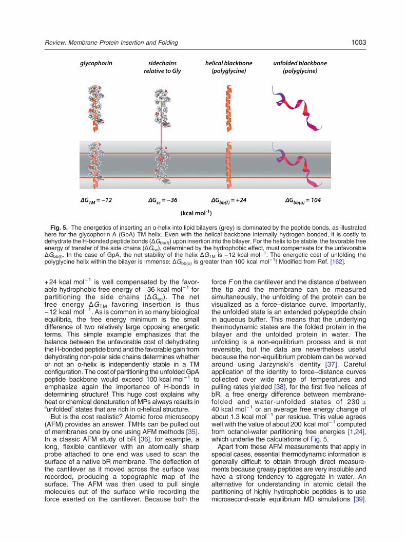
Fig. 5. The energetics of inserting an α-helix into lipid bilayers (grey) is dominated by the peptide bonds, as illustratedhere for the glycophorin A (GpA) TM helix. Even with the helical backbone internally hydrogen bonded, it is costly todehydrate the H-bonded peptide bonds (ΔGbb(f)) upon insertion into the bilayer. For the helix to be stable, the favorable freeenergy of transfer of the side chains (ΔGsc), determined by the hydrophobic effect, must compensate for the unfavorableΔGbb(f). In the case of GpA, the net stability of the helix ΔGTM is −12 kcal mol−1. The energetic cost of unfolding thepolyglycine helix within the bilayer is immense: ΔGbb(u) is greater than 100 kcal mol−1! Modified from Ref. [162].
1003Review: Membrane Protein Insertion and Folding
+24 kcal mol−1 is well compensated by the favor-able hydrophobic free energy of −36 kcal mol−1 forpartitioning the side chains (ΔGsc). The netfree energy ΔGTM favoring insertion is thus−12 kcal mol−1. As is common in so many biologicalequilibria, the free energy minimum is the smalldifference of two relatively large opposing energeticterms. This simple example emphasizes that thebalance between the unfavorable cost of dehydratingtheH-bondedpeptide bondand the favorable gain fromdehydrating non-polar side chains determines whetheror not an α-helix is independently stable in a TMconfiguration. The cost of partitioning the unfoldedGpApeptide backbone would exceed 100 kcal mol−1 toemphasize again the importance of H-bonds indetermining structure! This huge cost explains whyheat or chemical denaturation of MPs always results in“unfolded” states that are rich in α-helical structure.But is the cost realistic? Atomic force microscopy
(AFM) provides an answer. TMHs can be pulled outof membranes one by one using AFM methods [35].In a classic AFM study of bR [36], for example, along, flexible cantilever with an atomically sharpprobe attached to one end was used to scan thesurface of a native bR membrane. The deflection ofthe cantilever as it moved across the surface wasrecorded, producing a topographic map of thesurface. The AFM was then used to pull singlemolecules out of the surface while recording theforce exerted on the cantilever. Because both the
force F on the cantilever and the distance d betweenthe tip and the membrane can be measuredsimultaneously, the unfolding of the protein can bevisualized as a force–distance curve. Importantly,the unfolded state is an extended polypeptide chainin aqueous buffer. This means that the underlyingthermodynamic states are the folded protein in thebilayer and the unfolded protein in water. Theunfolding is a non-equilibrium process and is notreversible, but the data are nevertheless usefulbecause the non-equilibrium problem can be workedaround using Jarzynski's identity [37]. Carefulapplication of the identity to force–distance curvescollected over wide range of temperatures andpulling rates yielded [38], for the first five helices ofbR, a free energy difference between membrane-folded and water-unfolded states of 230 ±40 kcal mol−1 or an average free energy change ofabout 1.3 kcal mol−1 per residue. This value agreeswell with the value of about 200 kcal mol−1 computedfrom octanol-water partitioning free energies [1,24],which underlie the calculations of Fig. 5.Apart from these AFM measurements that apply in
special cases, essential thermodynamic information isgenerally difficult to obtain through direct measure-ments because greasy peptides are very insoluble andhave a strong tendency to aggregate in water. Analternative for understanding in atomic detail thepartitioning of highly hydrophobic peptides is to usemicrosecond-scale equilibrium MD simulations [39].

1004 Review: Membrane Protein Insertion and Folding
Simulation technology has not yet advanced sufficient-ly to examine the folding of multi-span MPs, but thetechnology is good enough to examine the folding ofsingle-span polyleucine segments. Such simulationsare initiated by placing a single unfolded polyleucinepeptide into a simulation cell containing a lipid bilayerand allowing the simulation to run until all accessiblestates have been sampled many times and cataloged.In simulations lasting several microseconds, polyleu-cine peptides are observed to adsorb into themembrane interface where they fold into a helicalconformation and make numerous excursions into themembrane as a TM helix. Figure 6 shows a represen-tation of the folding and insertion of a Leu10 peptide inwhich the insertion depth and helicity of the peptide aresampled during the course of the simulation. For eachsampled time point, the insertion depth (relative thebilayer center) is plotted against the helicity, and thepoints are connected sequentially by lines.Two observations are significant. First, the peptide
rapidly adsorbs at the bilayer interface. Second, afterthe first 50 ns, the peptide becomes α-helical andmoves repeatedly between a helical surface configu-ration and a helical TM configuration. This behavior isexactly what one would expect from considerations ofthe cost of peptide bond partitioning described above(Figs. 4 and 5). During the time course of thesimulation, the peptide is never seen to leave themembrane and return to the aqueous phase, which isthe behavior expected from extensive studies of theinteractions of melittin with bilayers (above). Indeed,microsecond-scale simulations of melittin folding atthe membrane interface are in quantitative agreementwith experiments [40,41].These considerations of the partitioning of peptides
into membranes provide an important perspective on
Fig. 6. Equilibrium microse-cond-scale simulations of the fold-ing and membrane insertion ofpolyleucine sequences (here 10leucines) reveal only two states[39]. The simulation shown herebegins with the unfolded peptide(U) in water (W) about 10 Å from thephosphatidylcholine bilayer (grey).Within a few nanoseconds (ns), itabsorbs to the membrane interfaceand never returns to the bulk waterphase. After the next 40 ns, thepeptide becomes α-helical and fluc-tuates between being on the sur-face (S) and across the membrane(TM) during the ensuing severalmicroseconds. The trajectory ofthe simulation is represented as aplot of the insertion depth of the
eptide's center of mass against its helicity. The sampled time points are connected sequentially by blue lines. Modifiedom Ref. [39]. This simulation reveals the importance of the membrane interface in MP folding and suggests that the
pfr
interface may play a role in translocon-guided insertion of TMhow translocons may manage the MP assemblyreaction pathway. They lead inexorably to theconclusion that the membrane interface in the vicinityof the translocon likely participates in assembly.
MP intrinsic interactions: Helix–helix interactions
There is much more to intrinsic interactions thanjust the folding and insertion of single TMHs into lipidbilayers because so many biologically importantMPs are multi-spanning proteins. The other part ofthe MP folding problem is therefore the energetics ofhelix–helix interactions that cause single helices tocome together to form functional three-dimensionalstructures in the lipid bilayer. Popot and Engelmanfirst focused attention on this problem through their“2-stage model” of folding [42], which was anoutgrowth of their work on the reassembly offunctional bR from proteolytically cleaved fragmentsseparately reconstituted into lipid vesicles. Ourclearest understanding of helix–helix interactionscomes from studies of glycophorin A (GpA) thatforms dimers when solubilized in sodium dodecylsulfate (SDS) [43] and lipid bilayers [44]. Engelmanet al. took advantage of this observation to discoverthe location and properties of the dimerizationdomain, –L75IxxG79V80xxG83V84xxT87–, within theGpA TMH [45,46] and to determine the structure ofthe dimer in detergent micelles using NMR [47](Fig. 7). Langosch et al. extended GpA dimerizationmeasurements to E. coli inner membranes [48].The GpA dimer is held together by van der Waals
interactions and tight shape complementarity,called “knobs-into-holes packing” [49,50]. TheGpA data led to the identification of the first
helices.

ITLIIFGVMAGVIGTILLISYGI
(b)(a)
Fig. 7. Helix–helix interactions of the glycophorin A(GpA) dimer (blue and orange helices) based upon thestructure of GpA in SDS [47]. (a) Glycine (or other smallresidues) separated by three residues allows the helices topack tightly. (b) Other amino acids in the structural vicinityof the GXXXG motif can facilitate or inhibit specific bindingof complementary surfaces [120].
1005Review: Membrane Protein Insertion and Folding
widespread interaction motif for TMHs, the so-calledGXXXG motif [51,52]. The interaction free energiesof GpA dimer formation are readily determined fordetergent-solubilized helices using analytical ultracen-trifugation to measure the equilibrium amounts ofmonomersanddimers [53], but the relationshipbetweeninteraction free energies in detergents and in lipidbilayers has been unclear. A recent study shows thathelix dimer stability in lipid bilayers depends strongly onthe type of lipid, ranging from −12 kcal mol−1 in purephosphatidylcholine bilayers to about −3 kcal mol−1 inE. coli lipids [54], the latter value being similar to thatobserved in natural membrane “blebs” [55].H-bonds between TMHs can also be important for
stabilization. For example, TM segments composedonly of leucine have relatively little tendency todimerize in vivo in E. coli unless a polar residue,especially Asn, Asp, or Glu, is also present in thesequence to form H-bonds [56]. However, a dou-ble-mutant thermodynamic cycle analysis of bR hasshown that the stabilization free energy of singleside-chain H-bonds contributes only about0.6 kcal mol−1. Of course, if multiple H-bonds arepresent between side chains in an MP, the additivestabilization could become significant.Because of the simple thermodynamic consider-
ations discussed earlier, MPs are absolutely con-strained to have their native helical structure inbilayer membranes. The first stage of the 2-stagemodel [42]—the establishment of TMHs acrossmembranes—is accomplished through co-transla-tional insertion and the catalytic activity of thetranslocon (below). Cells thus avoid the thermody-namic challenges associated with managing highlynon-polar proteins in the aqueous environment.The second stage of folding—helix association
within the bilayer environment—should be theprimary focus of MP stability rather than completedenaturation of MPs using heat or detergents,which are problematic. Just as we measure thestability of soluble proteins in their native aqueousenvironment, we should really measure the stabil-ity of MPs within their native membrane environ-ment. The question is then simply the energeticcost of separating the TMHs from one anotherwithin a lipid bilayer. This challenging task has nowbeen accomplished for bR in a bicelle environmentusing so-called steric trapping [57]. Steric trappingis based upon engineering two streptavidin bindingsites into the protein that are close together in thenative state. For folded proteins, only a singlemonovalent streptavidin can bind due to stericoverlap of the binding sites. However, when theprotein is unfolded in the membrane, then strepta-vidin can bind to both sites. The free energy ofunfolding can be determined by measuring of theamount of doubly bound protein during unfolding.In this way, Chang et al. determined a value ofapproximately −11 kcal mol−1 for the unfolding ofbR [58], which is within range of values observedfor water-soluble proteins. This is not a marginalvalue of stability. If other MPs are eventually foundto have values similar to those of water-solubleproteins and membrane-bound bR, then one cansafely assume that native three-dimensional struc-ture will follow once TMHs are inserted intomembranes.
Biological Boundaries of MP Biogenesis
How does the cell exploit and control the physicalinteractions that underlie the spontaneous insertionof hydrophobic peptides into lipid bilayers in order toproduce properly folded helix-bundle MPs whileavoiding the ever-present risk of protein aggrega-tion? The basic solution to this problem that naturehas come up with is co-translational insertion: Withfew exceptions [59], helix-bundle MPs are inserteddirectly into a target membrane as they come out ofthe ribosome. Co-translational insertion is mediatedby one or other type of translocon, that is, a proteinconduction channel in the membrane that is con-structed such that it can both shield polar parts of thepolypeptide from lipid contact as they are translo-cated across the membrane and expose at the sametime hydrophobic segments in the protein to thesurrounding lipid bilayer, facilitating the formation ofTMHs.In this section, we first review recent advances in our
understanding of how ribosomes from different kinds oforganisms handle nascent polypeptide chains and howribosomes synthesizing MPs are specifically targetedto translocons. We then discuss current models oftranslocon-mediated insertion and folding of MPs.

1006 Review: Membrane Protein Insertion and Folding
Ribosomes
Ribosomes are highly conserved RNA–proteincomplexes that translate mRNA into protein in allliving cells. For our present purposes, the mostimportant parts of the ribosome are the polypeptidetransferase center (PTC) where the incoming tRNAdelivers its amino acid to the growing polypeptide,the tunnel in the large ribosomal subunit throughwhich the nascent chain exits the ribosome, and thepolypeptide exit site on the “backside” of the largesubunit (Fig. 8). Peptide bond formation at the PTC iscatalyzed by ribosomal RNA (“The ribosome is aribozyme” [60,61]). Large sections of the tunnel wallsare also formed by ribosomal RNA, creating a polar,negatively charged environment for the nascentchain. There is a higher density of ribosomal proteinsaround the exit site, and many accessory proteinssuch as chaperones and targeting factors bind there.The area around the exit site is also involved indocking the ribosome to the translocon, such that thenascent chain can pass directly from the ribosomaltunnel into the translocon channel.The chief differences between prokaryotic and
eukaryotic ribosomes are found in their proteincomposition, with eukaryotic ribosomes having alarger complement of proteins than prokaryotic ones.There are also some extra “expansion loops” in theRNA present in eukaryotic ribosomes [62–64]. Thecore ribosomal structure is highly conserved, how-
Fig. 8. High-resolution structure of a mammalianribosome–Sec61 complex. The structure was obtainedusingadvancedcryo-EMmethods. PTC indicates thepeptidyltransferase center. The image is from Voorhees et al. [64].
ever, and the basic operational principles—includingthe biosynthesis of integral MPs—are very similarfrom bacteria to man.The key player in the early steps of MP biosyn-
thesis is the signal recognition particle (SRP). Ineubacteria such as E. coli, SRP is composed of asmall 4.5S RNA and a single protein subunit, Ffh[65,66]. Ffh contains both a signal-peptide bindingdomain and a GTPase domain, and it can bind nearthe polypeptide exit site on the ribosome. When asufficiently hydrophobic polypeptide segment,typically a signal peptide or a segment that willeventually form a TMH, appears at the exit site ofthe ribosome-nascent chain complex, it binds toSRP with subnanomolar affinity [67]. Binding willcause a major structural rearrangement of the SRP[66,68], priming the ribosome-nascent chain–SRPcomplex for binding to the SRP receptor FtsYlocated on the surface of the inner membrane.The ribosome–SRP–FtsY complex then binds tothe SecYEG translocon (see the next section). FtsYalso has a GTPase domain, and proper docking ofSRP to FtsY brings the SRP and FtsY GTPasedomains into juxtaposition such that the two completeactive sites are formed and two GTPs can behydrolyzed. This process, which serves as a qualitycontrol mechanism by assuring that the ribosome iscorrectly docked to SecYEG [69], leads to the releaseof the nascent chain from SRP, allowing it to insert intothe translocon.The mammalian SRP is more complex, with a
larger 7S RNA and five additional proteins. The Ffhhomologue is called SRP54 and has a similarsignal-peptide binding groove [65,66,70]. A majordifference between the bacterial SRP and themammalian SRP is that the latter not only bindshydrophobic segments in the nascent chain andmediates targeting to the translocon but also slowsdown or halts translation when bound to a ribo-some-nascent chain complex [65]. Presumably, thisgives the ribosome more time to find and dock to atranslocon in the complex cytosolic environment of aeukaryotic cell, ensuring the tight coupling betweentranslation and membrane insertion.Residues in the nascent polypeptide chain can
interact with the tunnel wall, especially in its upperparts. Such interactions can induce translationalstalling, presumably by subtly altering the geometryof the PTC. Specific peptide sequences, called arrestpeptides (AP), can stall translation on their own(intrinsic APs) or in combination with a small moleculesuch as an amino acid or an antibiotic (inducible APs)[71]. The latter are important in regulation of antibioticresistancegenes,which haveAPs that are activated inthe presence of the antibiotic [72]. As will be explainedbelow, translational APs have recently beenemployedas “force sensors” to detect dynamic interactionsbetween a growing nascent chain and its immediatesurroundings.

1007Review: Membrane Protein Insertion and Folding
Translocon-mediated membrane insertion
In bacteria, membrane insertion of nearly allhelix-bundle MPs is mediated by two translocons:SecYEG and the structurally unrelated YidC. Someproteins require only one or the other, and somerequire both. YidC is about 5-fold more abundant inE. coli than SecYEG [73,74]. SecYEG and YidC canform a 1:1:1:1 super-complex (the holo-translocon)together with SecDF (4-fold lower cellular abun-dance than SecYEG) and YajC (3-fold higher cellularabundance than SecYEG) [75].The eukaryotic Sec61 translocon in the ER is
homologous to SecYEG, but there are no YidC,SecDF, or YajC homologues in the ER. YidC homo-logues are, however, found in the inner mitochondrialmembrane (Oxa1) and the chloroplast thylakoidmembrane (Alb3/Alb4) [74]. On the other hand, theSec61 super-complex involves additional MPs such asTRAM, TRAP, and the oligosaccharyl transferase thathave no counterparts in E. coli. Eukaryotic Seccomplexes also include Sec62 and Sec63, thatfacilitate post-translational transport (reviewed inRef. [75]). Mitochondria in most organisms do nothave an equivalent to Sec61 in the inner membrane;the only translocase there is Oxa1. Because of theabsence of a Sec-type translocon, mitochondrialribosomes are specialized to bind directly to the innermembrane [76].The SecYEG/Sec61 translocon can mediate both
co- and post-translational translocations of proteins; inbacteria, most secreted proteins are translocatedpost-translationally with the help of the SecA motorprotein, whereas MPs are nearly always inserted intothe membrane co-translationally [77]. In contrast,
Fig. 9. Structure of the SecYE translocon from Pyrococcus fmembrane, and the right panel shows a view from the cytophydrophobic ring are shown as van der Waals spheres, indicabetween TMH2b and TMH7. SecE is shown in red, and SecY is
YidCmediates post-translationalmembrane insertion,except in mitochondria where a C-terminal domain inOxa1 can bind to mitochondrial ribosomes in aco-translational fashion [74]. In eukaryotic cells,insertion of proteins into the ER membrane is nearlyalways co-translational, while translocation of secre-tory proteins into the lumen of the ER can be bothco-translational and post-translational [77].High-resolution three-dimensional structures
(Fig. 9) are known for several SecYEG transloconsfrom different prokaryotic organisms [78–80] and forthe Sec61 complex from pig [64]. The centraltranslocation channel is formed by the SecY subunit(Sec61α in eukaryotes), a protein with 10 TMHs. Asmall plug domain closes the channel from theperiplasmic side, presumably in order to prevent ionleakage through the inactive translocon. The SecYsubunit is buttressed by SecE that partly encirclesSecY and enhances its stability [81]. SecG is moreperipherally located, and its precise function isunclear [82].A lateral gate located between TMH2b and TMH7 in
SecY can open up the channel toward the surroundingmembrane, as seen in Fig. 9. The simplest conceptualmodel for how TMHs in the nascent polypeptide areinserted into the membrane is that they first enter thecentral SecYEG channel, then exit it via the lateralgate and insert into the lipid bilayer one by one (Fig. 1).In essence, they partition between the translocon andthemembrane.Aswill be shownbelow, this is certainlyan oversimplification, but it serves as a good startingpoint for the discussion.The idea of translocon/membrane partitioning can
be examined experimentally by suitably designed testproteins in which a hydrophobic segment is placed far
uriosus [80]. The left panel shows a view in the plane of thelasm. The plug domain is circled, and the residues in theted by asterisks (*). The arrow points into the lateral gatecolor coded fromN-terminus (blue) to C-terminus (orange).
1008 Review: Membrane Protein Insertion and Folding
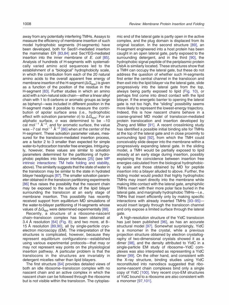
away from any potentially interfering TMHs. Assays tomeasure the efficiency of membrane insertion of suchmodel hydrophobic segments (H-segments) havebeen developed, both for Sec61-mediated insertionthe mammalian ER [83,84] and SecYEG-mediatedinsertion into the inner membrane of E. coli [85].Analysis of hundreds of H-segments with systemati-cally varied amino acid sequences led to theestablishment of a “biological” hydrophobicity scale,in which the contribution from each of the 20 naturalamino acids to the overall apparent free energy ofmembrane insertion of theH-segment (ΔGapp) is givenas a function of the position of the residue in theH-segment [83]. Further studies in which an aminoacid with a non-natural side chain—either a linear alkylchain with 1 to 8 carbons or aromatic groups as largeas biphenyl—was included in different position in theH-segment made it possible to measure the contri-bution of apolar surface area (i.e., hydrophobiceffect with solvation parameter σ) to ΔGapp: For analiphatic surface, σ was determined to be −10cal mol−1 Å−2, and for aromatic surface, the valuewas −7 cal mol−1 Å−2 [86] when at the center of theH-segment. These solvation parameter values, mea-sured for the translocon-mediated insertion process,are a factor 2 to 3 smaller than expected for simplewater-to-hydrocarbon transfer free energies. Intriguing-ly, however, these values are similar to solvationparameters determined for the partitioning of hydro-phobic peptides into bilayer interfaces [25] (see MPintrinsic interactions: TM helix folding and stability,above). The similarity suggests that the state ofwater inthe translocon may be similar to the state in hydratedbilayer headgroups [87]. The smaller solvation param-eter obtained in the translocon partitioning experiments[86] thus raises the possibility that the nascent chainmay be exposed to the surface of the lipid bilayersurrounding the translocon at an early stage ofmembrane insertion. This possibility has recentlyreceived support from equilibrium MD simulations ofthe water-to-bilayer partitioning of H-segments whosevalues of ΔGapp were determined experimentally [88].Recently, a structure of a ribosome-nascent
chain–translocon complex has been obtained at3.4 Å resolution [64] (Fig. 8); and two more, at 8–15 Å resolution [89,90], all by single-particle cryo-electron microscopy (EM). The interpretation of thestructures is complicated, however, because theyrepresent equilibrium configurations—establishedusing various experimental protocols—that may ormay not represent way points on the physiologicalinsertion pathway. A particular problem is that thetranslocons in the structures are invariably indetergent micelles rather than lipid bilayers.The first structure [64] provides detailed views of
both an idle ribosome–translocon complex with nonascent chain and an active complex in which thenascent chain can be followed through the ribosomebut is not visible within the translocon. The cytoplas-
mic end of the lateral gate is partly open in the activecomplex, and the plug domain is displaced from itsoriginal location. In the second structure [89], anH-segment engineered into a host protein has beencaught in an open lateral gate, party exposed to thesurrounding detergent, and in the third [90], thehydrophobic signal peptide of the periplasmic proteinDsbA is similarly located. These structures show thata TMH can occupy the lateral gate, but these do notaddress the question of whether such H-segmentsfirst enter the central channel in the translocon andthen exit into the lipid bilayer via the lateral gate, slideprogressively into the lateral gate from the top,always being partly exposed to lipid (Fig. 10), orperhaps first come into direct contact with the lipidbilayer. If the energetic barrier to opening the lateralgate is not too high, the “sliding” possibility seemsmore likely to represent the lowest-energy trajectory.Indeed, this is how nascent chains behave in acoarse-grained MD model of translocon-mediatedprotein translocation and insertion developed byZhang and Miller [91]. A recent crosslinking studyhas identified a possible initial binding site for TMHsat the top of the lateral gate and in close proximity tosurrounding lipid [92], from which the TMH couldconceivably slide deeper into the membrane within aprogressively expanding lateral gate. In the slidingmodel, a TMH would be partially exposed to lipidalready at an early stage during insertion, possiblyexplaining the coincidence between insertion freeenergies calculated from the biological hydrophobic-ity scale and those obtained by MD for peptideinsertion into a bilayer alluded to above. Further, thesliding model would predict that highly hydrophobicTMHs may insert directly into the membrane whilemaking little contact with the lateral gate, amphiphilicTMHs insert with their more polar face buried in thelateral gate, and marginally hydrophobic TMHs—i.e.,TMHs that insert efficiently only by making specificinteractions with already inserted TMHs [93–95]—would insert largely through the translocon channeland only expose a limited surface through the lateralgate.A high-resolution structure of the YidC translocon
has just been published [96], as has an accuratestructural model [97]. Somewhat surprisingly, YidCis a monomer in the crystal, while a previousprojection structure obtained by electron crystallog-raphy of two-dimensional crystals showed it as adimer [98], and the density attributed to YidC in asingle-particle EM study of ribosome–YidC com-plexes was also interpreted as representing a YidCdimer [99]. On the other hand, and consistent withthe X-ray structure, binding studies using YidCreconstituted into nanodiscs suggested that ribo-some-nascent chain complexes bind only a singlecopy of YidC [100]. Very recent cryo-EM structuresof YidC bound to a ribosome are also consistent witha monomer [97,101].

(a)
(b)
gate
cytoplasm
cytoplasm
90°
Fig. 10. Twomodels for translocon-mediated insertion of a Nout–Cin orientated TMH in a single-span (type I) MP. (a) The“in–out” model. The TMH (in black) first moves all the way into the central translocon channel and then exits sidewaysthrough the lateral gate. (b) The “sliding” model. The TMH slides along the lateral gate into the membrane, with one sideexposed to lipid at all times. The leading polar segment penetrates through the lateral gate and is shielded from lipidcontact. SecE is shown in red, and SecY is color coded from N-terminus (blue) to C-terminus (orange). On the right-handside, a schematic of the translocon (blue) interacting with an MP (green) and a membrane inserting TMH (red) are shown.
1009Review: Membrane Protein Insertion and Folding
The X-ray structure and accompanying MDsimulations show a deep, hydrated cleft withinYidC that extends halfway across the membraneand is open toward both the cytoplasm and thelipid bilayer (Fig. 11a). The cleft is capped on theperiplasmic side by what appears to be a tightly
Fig. 11. Structure of the YidC translocon from Bacillus halointo the hydrated cleft in the center of the protein. The left panewalls of the cavity, with the rest of the protein in stick represen
packed, stable structural domain. When Trp244at the top of the cleft is substituted by thephoto-activatable amino acid analog p-benzoyl-L-phenylalanine, it can be crosslinked in vivo to aco-expressed substrate protein, showing that sub-strate has access to the deep cleft [96]. There is also a
durans [96] viewed in the plane of the membrane, lookingl shows a surface representation; the right panel shows thetation.

1010 Review: Membrane Protein Insertion and Folding
rather mobile part, composed of two α-helices that lieflat on the cytoplasmic face of the membrane, in frontof the entrance to the cleft. The role of this part isunclear; perhaps it facilitates membrane insertion ofsubstrate TMHs by locally distorting the lipid bilayer.The structure [97,101] is suggestive of an insertion
mechanism in which a short polar tail or loop in thesubstrate protein penetrates at least halfway acrossthe membrane within the water-filled cleft, leavingthe adjoining hydrophobic TMHs in the lipid bilayer,similar to the sliding model proposed for SecYEGabove. The resulting intermediate state would be oflower free energy than an initial state where the TMHis embedded in the lipid headgroup region, but itwould be of higher free energy than a final statewhere the TMH completely spans the membrane.According to this model, YidC is designed to lowerthe energy barrier for the initial, partial insertion ofthe TMH. A speculative possibility is that the outerlipid monolayer becomes sufficiently perturbed in thevicinity of the intermediate YidC–substrate complexto make it possible for the polar tail or loop totranslocate fully across the membrane at a reason-able rate.A comparative analysis of the insertion of model
TMHs into the inner membrane of E. coli of YidC-and SecYEG-dependent MPs showed that thethreshold hydrophobicity required for 50% insertionof a hydrophobic segment of composition nL/(19-n)Ais similar (n50% ≈ 1–2) for the two translocons[85,102]. Likewise, the individual contributions tothe overall ΔGapp for YidC- and SecYEG-dependentinsertions are similar for the non-polar residues,whereas polar and charged residues have roughly2-fold larger ΔGapp values when membrane insertionismediated by the YidC translocon (Fig. 12). Possibly,a TMH is more lipid exposed during YidC-mediated
SecYEG, ΔGapp (kcal mol-1)
Sec
61, Δ
Gap
p (k
cal m
ol-1
)
(a) (
Fig. 12. Amino acid ΔGapp values for Sec61-, SecYEG-, an[84,85,102]. The SecYEG (a) and YidC (b) data are plotted aga(with the equations given in the panels), while the broken line inand weakly polar residues (slope = 0.8) [85].
insertion than during insertion mediated by theSecYEG translocon (where polar residues may besequestered into the lateral gate region, away fromlipid contact).Because some fraction of SecYEG and YidC are
bound together in the holo-translocon, they canconceivably act sequentially on MP substrates ormay even act simultaneously on different parts of amulti-spanning MP. Within the context of theholo-translocon, where the lateral gates in SecYEGand YidC conceivably are not far apart, one canimagine that an incoming N-terminal TMH, or aninternal “helical hairpin” composed of two TMHs witha short connection loop, can preferentially partitioninto the membrane via YidC, while TMHs flanked bylonger polar segments that cannot fit into the YidCcavity will preferentially be inserted via SecYEG.
Ribosome–translocon–membraneinsertion pathways
The progression of a hydrophobic TM segmentalong the ribosome–translocon–membrane insertionpathway has been followed mainly by chemicalcrosslinking experiments and, more recently, byusing translational APs as in vivo force sensors.Many crosslinking experiments have been carriedout by in vitro generation of stalled ribosome-nascentchain–translocon complexes using dog pancreasrough microsomes, that is, ER-derived membranevesicles that contain Sec61 translocons. Early studiesshowed that signal peptides in stalled nascent chainscould be crosslinked both to translocon componentsand to lipid [103], suggesting that the signal peptide istransiently held in an interfacial location between thetranslocon and surrounding lipid. This is in fullagreement with the EM structures discussed above.
SecYEG, ΔGapp (kcal mol-1)
Yid
C, Δ
Gap
p (k
cal m
ol-1
)
b)
d YidC-dependent membrane insertions of a model TMHinst the Sec61 data. Full lines indicate linear fits to the data(b) is the linear fit obtained when including only non-polar

1011Review: Membrane Protein Insertion and Folding
Asnoted above, a recent crosslinking study has furtheridentified an early interaction between N-terminalTMHs in a substrate protein with residues located atthe cytoplasmic tip of the lateral gate in Sec61 [92], aswould be expected if hydrophobic segments start topartition into the lateral gate region immediately uponentering the translocon rather than first moving into thecentral channel and exiting through the lateral gateonly at a later stage (cf., Fig. 10b).The development of a novel technique where
translational APs are used to measure forces actingon a nascent chain during co-translational processessuch as membrane insertion now makes it possibleto study the kinetics of MP insertion and folding invivo. APs are short stretches of polypeptide, typically~10–15 residues long, that bind in the upper parts ofthe ribosomal tunnel and induce ribosomal stalling ata specific codon in the mRNA [71]. Stalling can beprevented if a sufficiently strong “pulling force” actson the nascent chain at the precise point when theribosome reaches the critical codon [104]; presum-
(a)
(b)
Fig. 13. Using arrest peptides (APs) as in vivo force sensortwo natural TMHs (TM1, TM2; in black) and a model TMH compon the length L of the tether between the H-segment and the APtranslocon at the time when the ribosome reaches the last codoon the nascent chain will determine the fraction of stalled verprotein can be determined by [35S]Met pulse-labeling of growconstruct and analysis by SDS-PAGE, as shown on the rightwhich a critical proline in the AP has been mutated to alanine,protein as a function of L for a set of constructs designed as s
ably, pulling on the nascent chain breaks theinteractions between the AP and the ribosome thatcontrol stalling.APs from the SecM protein have proven particu-
larly useful. The AP from E. coli SecM is 17 residueslong and rather weak, but stronger APs have beenfound in SecM proteins from other bacteria [105].The strongest AP known to date is a mutated versionof the SecM AP from Mannheimia succiniciprodu-cens with sequence HPPIRGSP (called Ms-Sup1)[106]. This short AP can be introduced into anyprotein and will report on the tension in the nascentchain at the precise point during translation when theribosome reaches the proline codon at the 3′ end ofthe AP.The SecYEG-mediated insertion of polypeptide
segments (H-segments) of varying hydrophobicityinto the inner membrane of live E. coli cells wasrecently analyzed using the approach detailed inFig. 13a [106]. Strong pulling forces proportional tothe hydrophobicity of the H-segment were recorded at
L
s [106]. (a) An AP (AP; in blue) is inserted into an MP withosed of six leucines and 13 alanines (H; in red). Depending, the H-segment will be in different locations relative to then in the AP, as shown in the cartoon. The pulling force F(L)sus full-length protein produced. The fraction of full-lengthing E. coli, followed by immunoprecipitation of the proteinfor a construct with L = 63 residues (a control construct inpreventing stalling, is also shown). (b) Fraction full-lengthhown in (a) (top).

1012 Review: Membrane Protein Insertion and Folding
tether lengths L ≈ 30 and L ≈ 40 residues (Fig. 13b).From the known dimensions of the ribosome–translocon complex, the peak in the force profile atL ≈ 40 residues most likely corresponds to the finalinsertion of the H-segment into the membrane. Theinteraction responsible for the peak at L ≈ 30 residuesis more difficult to pinpoint: It depends only on thehydrophobicity of the N-terminal end of theH-segmentand could represent an interaction between theH-segment and the cytoplasmic faceof the lipid bilayeror possibly an interaction akin to the one identifiedbetween an N-terminal TMH and the tip of the lateralgate in Sec61 discussed above [92]. Regardless, thedata show that APs can be used to measure forcesacting co-translationally on a nascent chain with highsensitivity and high spatial precision (a force profilerecordedwith single-residue precision suchas the oneshown in Fig. 13b has a spatial resolution correspond-ing to one residue in an extended conformation, i.e.,~3 Å).Segments that flank the TMHs can also affect
membrane insertion. In particular, positively chargedresidues at the ends of a hydrophobic segment caneither increase or decrease its insertion propensity, inaccordance with the “positive-inside” rule [107–110].
Folding of Polytopic MPs In Vivo
Translocon complexes mediate the insertion of TMsegments into membranes. Although this process isguided by the amino acid composition and structureof the segment, its interaction with the transloconand translocon accessory components can becritical. As the polypeptide chain moves throughthe translocon, it is continuously scanned. A ring ofhydrophobic residues is present in the middle of thechannel in both SecYEG and Sec61 translocons[78,111] (Fig. 10). The interaction of the nascentchain with this ring of hydrophobic residues might beinvolved in the opening of the lateral gate and thetransfer of TM segments between the translocon andmembrane [112,113], where they can be exposed tothe lipid environment. If a segment is hydrophobicenough, it will insert into the membrane, and currentmodels assume that the TM segments in polytopicMPs are inserted sequentially [114]. One wouldexpect that replacement of the hydrophobic residuesin the ring would destabilize the translocon, but thatis not the case. Replacement with polar, evencharged, residues were not destabilizing [111]. Thereplacement does, however, affect ΔGapp for mem-brane insertion of H-segments, consistent with thering playing a role in translocon/membrane partition-ing [112].As discussed earlier in MP intrinsic interactions,
the α-helical structure of a nascent TMH forms at thelatest during the exposure of the segment to the lipidbilayer, driven by the necessity to shield the polarbackbone from an unfavorable exposure to the
hydrocarbon core of the membrane. In case ofinteractions with the translocon channel, a helicalstructure apparently can be induced even before theexposure to a lipid bilayer [92]. Helical structure mayeven be formed in the ribosome exit tunnel [115–118].The stage atwhich anα-helical structure develops canbe important for the formation of tertiary structure,because amino acid side chains are positioned ondifferent faces of an α-helix, thereby allowing favor-able alignment with residues in other TM segments toform tertiary structure.In polytopic MPs, individual TMHs can form
interactions that determine the final fold and functionof the mature protein. As discussed earlier, theinteractions between TM segments can consist ofdistinct interaction motifs, such as the GxxxGmotif ofGpA discussed above (Fig. 7). More generally, smallresidues spaced four residues apart (small-XXX-s-mall) represent a prominent motif that allows TMHsto approach each other closely and pack tightly dueto van der Waals attraction and steric constraints.Extensions of this motif (small-XXX-small-XXX-s-mall) allows for flexibilities of two helices as multiplesmall residues, each spaced in the distance of four,can form a groove in the surface of α-helices [119].While any two helices containing such a motif couldapproach each other in principle, residues surround-ing these motifs can provide certain specificity to agiven interaction [120–122].Although these motifs are highly abundant in MPs
[123], many other types of interactions that requiremore specificity can be formed between TMHs,interactions with multiple TMHs at the same time or ahigh degree of flexibility. However, many interactionsbetween TMHs cannot be explained by these simpleinteraction motifs [124]. Highly specific interactionnetworks between multiple TMHs that determine thefinal structure can be formed, and the flexibilitybetween these interactions allows for tertiary struc-ture changes that are critical for function. It istherefore not surprising that simple interaction motifsoccur mainly in rather simple function contexts suchas the on/off function of some receptors [125].Many MPs have more complex TM domains, for
example, TM channels. Here, the TMHs must forman aqueous pore, lined by hydrophilic residueswithin the channel. Based upon the biologicalhydrophobicity scale, a significant number of thesesegments are predicted not to insert efficiently into amembrane due to their low hydrophobicity [126]. Itwas therefore a long-standing question how suchH-segments insert [127,128]. One answer is thatcharged residues, especially arginine, have veryfavorable interactions with the phosphates of phos-pholipids [87,129].Charged and polar residues can face a high
energetic barrier when inserting into a lipid bilayer.Nevertheless, positive and negative charges withinthe same [130] and different TMHs [131] can interact

1013Review: Membrane Protein Insertion and Folding
with each other, thereby drastically reducing thisbarrier. This concept can be extended to polar sidechains, which are partially masked from the lipidbilayer by interacting with each other [56,132]. Infact, it was proposed early on that MPs are“inside-out” proteins, in the sense of having a polarcore and a hydrophobic exterior, opposite to water-soluble proteins [133,134]. Although this idea isoversimplified, it suggests how the energy barrier formembrane insertion of hydrophilic side chains mightbe overcome and how specific tertiary structureinteractions formed at the same time. It has beensuggested that polar residues within TMHs can beused to predict the tertiary structure of polytopic MPs[126,135]. Individual TMHs can associate well afterinsertion into a membrane to form a functionallyfolded protein [11,12]. This implies that the specificinteractions between TMHs that determine the finalfold do not strictly require the action of a complexfolding apparatus, as expected for equilibriumfolding.When, then, are specific interactions between
TMHs formed? The simplistic and sequential inser-tion of TMHs one by one as discussed earlier impliesthat these interactions form after the TMHs havebeen inserted. While this might be the case forsufficiently hydrophobic TMHs, marginally hydro-phobic segments would not insert into a membrane
Fig. 14. Folding spaces during MP structure formation. Durinsegments (shown in blue and orange) can associate already ichannel (2a, green) [138], where polar residues can be shielhydrophobic side chains (orange spheres) form a membrane induring insertion into amembrane (3, grey)whereas one segmentof a less hydrophobic segment by shielding hydrophilic residueswithin different proteins can also occur co-translationally (5) and
efficiently; thus, more complex models of membraneinsertion and folding are required. One couldimagine that these interactions between TMHsoccur early, before insertion into a membrane, eitheronce a TMH exits the ribosome or once it enters thetranslocon. It has been shown that helical hairpinscomposed of two closely spaced TMHs can form inthe ribosome exit vestibule as the TMHs of a voltagesensor domain exit the ribosome [136] (1 in Fig. 14).Although many TMHs risk aggregation when asso-ciating in an aqueous environment, the exit vestibuleof the ribosome could form a confined compartmentthat promotes tertiary structure formation [137]. It ispresently unclear whether the helix–helix interac-tions observed by Tu et al. [136] in the ribosome exitvestibule are native contacts or whether theyrepresent a folding intermediate, although initialresults suggest native contacts. These and otherresults provide a new perspective on co-translationalfolding of TMHs. It will be interesting to learn if otherproteins use this early folding space.Remarkably, the translocon can accommodate
more than two nascent chain segments; thus, specificinteractions could also form before the segmentsinsert into a membrane [138]. In this case, helicalhairpins could form as a minimal prefolding unit andbury hydrophilic interactions within the helix–helixinterface before entering the lipid bilayer (2a and 2b in
g nascent chain synthesis by the ribosome (brown), helicaln the ribosomal vestibule (1) [136] or within the transloconded within the helix–helix interface (2b, red spheres) andsertion competent surface. Helices can furthermore interactis already inserted into themembraneanddrives the insertionwithin the interaction interface [143]. Interactions of heliceswas shown do drive MP complex assembly [149].

1014 Review: Membrane Protein Insertion and Folding
Fig. 14). It is unclear how the formation of suchinteractions is induced within the translocon, howwater is expelled from the helix–helix interface, or ifsuch early interactions are merely the result of tertiaryinteractions already formed in the ribosome's exitvestibule.Currently, a widely discussed mode of the co-tran-
slational formation of helix–helix interactions is theformation during the sequential exit of successiveTMHs through the lateral gate, where previouslyinserted TMHs that are in close vicinity of the lateralgate can conceivably mediate membrane insertionand tertiary structure folding at the same time (3 inFig. 14). Sadlish et al. have shown that multipleTMHs of the same polypeptide chain can beassociated with the eukaryotic translocon andmediate the efficient insertion of more C-terminallylocated TMHs [114]. This has also been confirmedfor other MPs [139], suggesting that the transloconitself might be a foldase. However, it is unclear howand where TMHs associate with the exterior of thetranslocon complex. Although a site has beenidentified by crosslinking studies, it is unclear if themajority of TMHs occupy the same site [114]. Anyspecific interactions with a translocon complex thatmanages thousands of different TM segments wouldhave to be replaced by stronger interactions with theprotein's own TMHs as the newly inserted segmentsemerge from the complex. In such a scheme, a stricthierarchy of interaction strengths in the form of aninteraction and folding blueprint would have to be inplace for all MPs using the pathway. This might beachieved by the strict coupling of correct tertiarystructure formation and membrane insertion. How-ever, this blueprint would have to be at least partiallyencoded in the nascent chain itself, possibly in theform of conserved polar residues within TMHs [140].If this hierarchical mechanism of tertiary structureformation is a common one, it will be interesting inthe future to decipher the individual strengths ofthese interactions and thereby come closer to an abinitio understanding of MP folding. As pointed outabove, other factors such as the point of secondarystructure formation, tertiary structure formation, andretention within the translocon are critical factors aswell.An interesting special case is the folding of mem-
brane channels that are composed of evolutionarilyrelated halves [141,142]. Here, each half-channel is (oroncewas) a separate folding entity; otherwise, the highlevel of similarity between the halves could createinterchangeability between helix–helix interactionsfrom one half with the other that subsequently wouldresult in protein misfolding.In bacteria, the formation of specific interactions
between side chains from interacting TMHs hasbeen observed by means of APs (see above) [143]for several polytopic MPs during their insertion by thebacterial translocon. Mutating interacting residues in
either a membrane inserting segment or an alreadyinserted segment in the same polypeptide chaindecreased the pulling force detected by a C-termi-nally located AP. APs thus offer a highly sensitive invivo tool for studying the co-translational formation oftertiary structure contacts, similar to in vitro AFM(see MP intrinsic interactions: TM helix folding andstability).A recent publication offers unprecedented struc-
tural insight into the biogenesis of a polytopic MP inbacteria [144]. The structure of a ribosome-nascentchain–translocon complex shows the first two TMHsof proteorhodopsin (PR) inserted into the membranejust outside the lateral gate. Although the resolutionof the structure is too low to reveal side-chaininteractions between the PR TMHs themselves orwith the translocon, an interaction of positivelycharged residues in the PR loop between TMH1and TMH2 with the ribosomal RNA helix 59 could beobserved. This interaction was verified by mutagen-esis of the charged residues, which reduced theamount of co-purified SecY/nascent chain complex.Interestingly, the lateral gate was in between the fullyopen and closed conformation, which would allow atranslocated hydrophilic segment to probe for tertiaryinteractions with previously inserted TM segments. Itis tempting to speculate about possible mechanismsof membrane insertion and folding based on theseresults. Nevertheless, the structure represents, atbest, a snapshot of the biogenesis of PR. Futureinvestigations and more structural snapshots of thebiogenesis of polytopic MPs might yield a moregeneralized and conclusive picture.The insertion of more hydrophilic TMHs can occur
at a later stage of assembly as well, which requires alarger structural reorganization of the maturingprotein (4 in Fig. 14). This could be observed forseveral MPs [93,145–147]. In these cases, TMH3 isinserted in the opposite topology and TMH2 andTMH4 are initially not inserted into the membrane.Upon the reorientation of TMH3 at a later stage,TMH2 and TMH4 are inserted into the membraneand the water channel folds into its final functionalstructure. In order to form the tertiary structure of theprotein, large-scale rearrangements involve induc-tion of large tilts during folding, as well as topologyreversals [148]. In a particularly impressive case, thetopology of a bacterial MP was found to be in aflexible state until a specific C-terminal chargedresidue was synthesized [109].These examples demonstrate that the formation of
the tertiary structure that allows more hydrophilicTMHs to insert into a membrane can occur before,during, and after membrane insertion of the newlysynthesized protein. It is currently unknown which isthe preferred mode of folding in polytopic MPsbecause only a few examples have been investigatedto date. In order to achieve a broader understandingand gain a more general view, it is necessary to follow

1015Review: Membrane Protein Insertion and Folding
closely the folding of many more polytopic MPs in ahigh-throughput manner.The concept of specific interactions between TMHs
allowing more hydrophilic parts of a protein to insertinto a membrane can also be extended to thequaternary structure, when it comes to the homo-oligomerization of the same subunit. Furthermore,interactions between different subunits of a newlysynthesized MP complex can form during membraneinsertion (hetero-oligomerization; 5 in Fig. 14) as wasrecently demonstrated for the subunits of the T cellreceptor [149]. Feige and Hendershot found that amarginally hydrophobic TMH that is not assembledinto a hetero-oligomeric T cell receptor is translocatedinto the ER lumen, rapidly recognized by the ERquality control machinery, and subsequently degrad-ed. However, polar residues in already inserted,interacting subunits guided the membrane insertionandassembly of individual T cell receptor subunits andthereby prevented degradation. This marks an impor-tant step toward a mechanistic understanding of howMP complexes are assembled in cells. It will be bothvery challenging and interesting to understand mech-anistically the assembly of complexes located in theER membrane, complexes located in the mitochon-drial inner membrane, and complexes located in thethylakoid membrane.MP secondary and tertiary structures can form at
different stages: in the ribosome exit tunnel, theribosome exit vestibule [136], within the transloconchannel [115–118], during membrane insertion[39,88], and within a membrane. Each of theseenvironments is characterized by unique propertiesand thereby jointly offer an extended folding space forthe many different MPs that are chaperoned by theco-translational MP synthesis and insertion com-plexes. After exploring basic principles of MP synthe-sis, membrane insertion, and folding using modelMPs, it is now important to venture into the widelyunexplored folding space of cellular MPs and to followand understand mechanistically their synthesis withina cell. Not only are these misfolding pathways likely tooffer new insights into the biogenesis of MPs, but theywill provide us also with a more quantitative view ofwhich modes of folding are the major ones and whichrepresent the exceptions.
Perspectives and Outlook
Where do we stand in our understanding of MPbiogenesis in mid-2014? First, it is clear that ourknowledge is much more advanced for the SecYEG/Sec61 translocons than for systems such as themitochondrial TOM–TIM translocons, the peroxi-somal PEX translocons, and the chloroplast TIC–TOC translocons (e.g., see Refs. [150–152]). TheSecYEG-associated YidC translocon is also slowly
yielding its secrets. In recent years, the field hasmoved forward in a major way thanks to structuralstudies of the Sec and YidC translocons, and thereare now many structures of Sec translocases incomplex with ribosomes. In parallel, a biophysicaldescription of the basic insertion and foldingprocesses is emerging through the confrontation ofthermodynamic and kinetic studies of highly simpli-fied systems (peptide–bilayer interactions, in vitrofolding of purified proteins, MD calculations) withquantitative in vivo studies of the kinds describedabove (“biological” hydrophobicity scales, co-tran-slational force measurements). It is satisfying to seethat we are finally moving beyond the simplecartoons (Fig. 1) to more detailed structure–functiondescriptions.Still, many open questions remain, even for the
Sec-type translocons. How are holo-translocons—-both prokaryotic and eukaryotic—put together andhow do the different components cooperate? It iscurrently unclear how the numerous accessorycomponents of the holo-translocons are recruitedand what the dynamics of complex assembly/disassembly are. It is also unknown how thebacterial translocons, YidC and SecYEG, cooperateto insert MPs. What are the precise roles of thelateral gate and the plug domain in SecYEG, whatcontrols their movements, and how do they interactwith polar and non-polar segments in a nascentchain? How strong is the functional couplingbetween the ribosome and the translocon; that is,can conformational changes resulting from interac-tions between a nascent chain and the ribosome betransmitted to the translocon?Lipids appear to play an important role in protein
translocation, but possible mechanisms are largelyspeculative (reviewed in Ref. [153]). From the pointof view of a substrate protein, it is not clear to whatextent a nascent chain can interact with membranelipids as it passes from the ribosome into thetranslocon or at which point during translation thata TMH first starts to integrate into the bilayer. Itseems clear that an incoming TMH can makespecific interactions with already inserted TMHs atvery early stages of membrane insertion, but howare the upstream TMHs “chaperoned” by theholo-translocon while waiting for downstreamTMHs to appear? Further, what is the physicalbasis for the late reorientation of TMHs that has beenseen with, for example, AQP1 [154], EmrE [109], andBand III [147], and when the membrane lipidcomposition is drastically changed [155]?We tried to capture in Fig. 1 the idea deeply
embedded in the literature that the nascent chain,powered by the GTP-based elongation energy of theribosome, enters the translocon whereupon thehydrophobic ring opens, the plug domain moves outof the way, and the nascent chain passes through. If asuitable hydrophobic segment arrives, it is diverted into

1016 Review: Membrane Protein Insertion and Folding
the membrane by simple partitioning between translo-con and bilayer [83,84,87,156]. Reflecting on theliterature we have reviewed here, we wonder if thisscheme is entirely correct. The translocon is absolutelyessential for the assembly of most multi-spanMPs, butthere is a notable exception: the transmembraneprotein KdpD.KdpD, which acts as a potassium sensor in E. coli
[59,157], has several features that are intriguing: Theprotein is tethered to the cytoplasmic surface of theinner membrane by four TM segments (residues 401–498) with very short interhelical loops that occur fardownstream from the amino-terminus. It is targeted tothe inner membrane by SRP recognition of a cytoplas-mic amphipathic helix (residues 22–48) [158], andmembrane insertion does not require SecA, SecE, orYidC [59]. Because KdpD does not apparently engageSecA or the SecYEG channel, a reasonable explana-tion is spontaneous insertion following targeting by theSRP to, presumably, the SRP receptor FtsY. In E. coli,FtsY is not permanently anchored to the innermembrane, but rather, it partitions between membraneand cytoplasm via an amphipathic helix [159]. Thissuggests that SRP recognizes the TMHs of KdpD and
NN
cytoplasm
lateral gate
(a)
(b)
NN
cytoplasm
lateral gate
N
Fig. 15. An alternative view of translocon-aided insertion ofidea of this alternative view is shown in a cartoon fashion in (anascent chain (green, with red TMHs) is with the membrane inchain does not immediately thread into the translocon. Rather, tfor polar components of MPs to cross the membrane. This ismembrane, but we suggest that it does this only for polar poshown in (b).
targets them to FtsY on the membrane, which allowsthe TMHs to partition spontaneously. Such a scheme,while highly unusual, is completely consistent with whatwe know about lipid–protein interactions described atthe outset in Biophysical Boundaries of MP Insertionand Folding.In order to be provocative, which was our charge
from the editors of this issue, we propose analternative view of the translocon-aided insertion ofmulti-span MPs and the secretion of soluble proteins(Fig. 15). Keeping in mind the huge thermodynamicdriving forces for MP folding and assembly, wesuggest that a TMH initially contacts the cytoplasmicmembrane interface in the vicinity of the transloconand that it never fully enters the translocon channelbut rather slides into the membrane along the lateralgate. This is not to say that the translocon cannotform a proper channel through the membrane, butwe suggest that it does this only for secreted proteinsand soluble domains or loop regions in MPs, asshown in Fig. 15b. The view is based upon fourimportant observations. First, most hydrophobic andamphipathic helices have a very strong affinity forthe interface region of lipid bilayers; it would be
NN
SPasecleaves
×
NN
multi-span MPs and the secretion of soluble proteins. The) (also see Fig. 10). We suggest that initial contact of theterface in the vicinity of the translocon (blue) and that thehe translocon has YidC-like behavior; it provides a pathwaynot to say that it cannot form a passageway through thelypeptide segments in MPs and for secreted proteins, as

1017Review: Membrane Protein Insertion and Folding
thermodynamically surprising if nascent peptidechains of MPs did not interact with the membraneinterface at some point during insertion. Second, sofar, no structural data of active ribosome–transloconcomplexes reveal TM segments within the translo-con channel; they are only seen at lateral gate exitsite. Third, ribosome–translocon complexes are notapparently shielded from the cytoplasm and innermembrane surface; consistent with earlier observa-tions, the recent high-resolution structure of amammalian ribosome–Sec61 complex shows signif-icant gap between the ribosome and the surfaceof the membrane [64]. Fourth, mitochondrialinner membranes in general do not have a SecYapparatus, only the YidC-equivalent Oxa1, suggest-ing that direct contact with the membrane is anessential feature of MP insertion and that nascentmitochondrial proteins insert and fold spontaneouslyinto the membrane aided by Oxa1 to move chargedand or highly polar residues across the membrane.An important consideration is that we do not know
when or how nascent MP chains make initial contactwith translocons or in what manner. Do they movedirectly from the exit tunnel straight into the transloconwithout intervening steps, as current cartoons suggest(Fig. 1)? Given relative target sizes, it seems possiblethat nascent chains could make first contact with themembrane in the vicinity of the translocon. Thisprovides a logical explanation for the membraneinsertion of the potassium channel S3b–S4 helicesthat apparently assemble in the ribosome vestibule[136]. Furthermore, it was shown recently that theribosome detached from the translocon when ahydrophobic segment was placed in the ribosomeexit tunnel and only hydrophilic stretches within thetunnel provided a strong attachment of the ribosome tothe translocon [144]. The scheme would also explainwhy changes in E. coli inner membrane lipid compo-sition affect the topology of LacY [155]. Consistent withthe importance of thermodynamic driving forces in MPfolding, Vitrac et al. have shown that so-called topologyswitching of LacY can occur in vitro in lipid vesicles [15].We are attracted to the scheme depicted in Figs.
10b and 15a because of its simplicity: A TM segmentinitially associates with the top of the lateral gate andthe surrounding membrane interface and strongthermodynamic forces then drive membrane parti-tioning. Such a processwould be consistent with bothcrosslinking results and the AP force measurements(Fig. 13). It is not necessary in this scheme for theelongating chain to be threaded back and forthacross the membrane as in Fig. 1. For the four-TMHprotein shown in the Fig. 15a schematic, thetranslocon is mainly required for only two steps: thepassage of the TM1–TM2 and TM3–TM4 connectingloops across the membrane, reminiscent of thehelical hairpin insertion hypothesis [23]. Thermody-namic forces are sufficient for membrane partitioning,with the translocon acting as a catalyst.
Why do we need translocons at all if insertion andfolding are driven by thermodynamics? The answer,of course, is that targeting and coordinated assem-bly of MPs are essential in the extraordinarilycomplicated environment of cells where thousandsof biologically important reactions occur simulta-neously in a crowded environment. Without translo-cons and all the other members of the MP assemblyapparatus, chaos would prevail.
Acknowledgements
This work was supported in part by grants from theNational Institutes of General Medical Sciences(RO1 GM074637) and from the National Institute ofGeneral Medical Sciences and the National Instituteof Neurological Disorders and Stroke jointly (PO1GM86685) to S.H.W.; by grants from the SwedishCancer Foundation, the Swedish Research Council,the Swedish Foundation for Strategic Research, theEuropean Research Council (ERC-2008-AdG232648), and the Knut and Alice WallenbergFoundation to G.v.H.; and by a research fellowshipfrom the Deutsche Forschungsgemeinschaft (CY74/1-1) to F.C.
Q&A with Stephen White and Gunnar von Heijne
Q1
FJD: In your rather provocative model a TM spandoes not enter the translocon but slides along thelateral gate. How can this model explain that somemutations of residues in the constriction ring of thetranslocon have an impact on TM span integration?
FC/GvH/SW: The crucial issues are (1) the time scaleof the integration and (2) the dynamics of thetranslocon. Presumably, integration of TM segmentsoccurs on the same timescale as the elongation rate ofnascent polypeptide chains, which is roughly oneamino acid per 50 msec or about 1 second for a singleTMsegment or signal sequence.Wedonot knowwhatconformational states are available to a transloconinteracting with a nascent chain, but we suggest thaton such long time scales the nascent chain has ampleopportunity to explore rather thoroughly the surround-ing lipid bilayer and interior of the translocon, includingthe so-called constriction ring. The standard view thatthe nascent chain passes through the center of thetranslocon and exits through the lateral gate impliesthat the reverse process is also possible. The slow
1018 Review: Membrane Protein Insertion and Folding
time scale likely permits the chain to explore all of thestates energetically available to it, including onesseparated by significant kinetic barriers. Thus, wesuggest that even if the primary entrance path of thechain is via the lateral gate, the aminoacid compositionof the constriction ringmatters in the fateof the nascentchain. What is missing in our understanding of the roleof the passive translocon apparatus is knowledge ofthe conformational states available to the transloconand the energy barriers that separate the states.Molecular dynamics simulations could help clarifyconformational states available; unfortunately timesscales of tens of milliseconds are not technicallyfeasible at present.
Q2
FJD: How does your model account for the positiveinside rule for integration of membrane proteins witha type II topology? In your model Sec61 should notbe sensitive to the N-terminal flanking segment,while genetic evidence indicates that point mutationsin Sec61 can affect the topology of reporter proteins.
FC/GvH/SW: Our response to question 1 aboveapplies in this case as well. Sec61/SecY mutationsthat affect topology presumably affect translocon/nascent-chain conformational states and energy land-scape. In simplistic terms, the thermodynamicallyfavored location of a translocating chain segment inthe translocon may be affected by its hydrophobicity:strongly hydrophobic segments tend to translocate (orinsert into the membrane, as the case may be) in amore lipid-exposed location on the outside of thelateral gate, while less hydrophobic segments maytend to be more buried within the translocon channelas they move through the membrane. Topology-de-termining charged residues that flank a hydrophobictransmembrane segment could therefore tend to belocated more deeply in the lateral gate, or even fullywithin the translocon channel, when in transit. As theyexit the ribosome, they may also transiently beexposed to lipid headgroups.
Q3
FJD: According to your model, the signal sequence orfirst TM span of a membrane protein positions thenascent chain in the vicinity of the translocon, and theC-terminal flanking segment slides into the lateral gatein a hairpin conformation. How does a protein segmentfrom the mature region promote channel opening?
FC/GvH/SW: Our view of the translocon as adynamic molecule with multiple conformation states
(response 1) eliminates the need to think of TM helixinsertion as series of discrete steps involving distinctopening and closing events. We suggest thatintegration is a continuous process in which thegrowing nascent chain persistently explores thestates available to it. We suppose that transloconmutations (response 2) affect these states and thekinetic barriers that separate them.
Received 25 June 2014;Received in revised form 13 September 2014;
Accepted 22 September 2014Available online 30 September 2014
Keywords:membrane protein folding;
membrane protein biogenesis;transmembrane helix;
lipid–protein interactions
†F.C., G.v.H., and S.H.W. contributed equally to this workand are listed alphabetically.
Abbreviations used:TM, transmembrane; TMH, transmembrane helix; AP,arrest peptide; bR, bacteriorhodopsin; MP, membrane
protein; ER, endoplasmic reticulum; MD, moleculardynamics; AFM, atomic force microscopy; PTC, polypep-tide transferase center; SRP, signal recognition particle;
PR, proteorhodopsin.
References
[1] White SH, Wimley WC. Membrane protein folding andstability: Physical principles. Annu Rev Biophys BiomolStruct 1999;28:319–65.
[2] Popot JL, Engelman DM. Helical membrane protein folding,stability, and evolution. Annu Rev Biochem 2000;69:881–922.
[3] MacKenzie KR. Folding and stability of α-helical integralmembrane proteins. Chem Rev 2006;106:1931–77.
[4] Goldenberg DP. Mutational analysis of protein folding andstability. In: Creighton TE, editor. Protein Folding. New York:W. H. Freeman; 1992. p. 353–403.
[5] Yang AS, Sharp KA, Honig B. Analysis of the heat capacitydependence of protein folding. J Mol Biol 1992;227:889–900.
[6] Hong H, Joh NH, Bowie JU, Tamm LK. Methods formeasuring the thermodynamic stability of membraneproteins. Methods Enzymol 2009;455:213–36.
[7] Jackson MB, Sturtevant JM. Phase transitions of the purplemembranes of Halobacterium halobium. Biochemistry1978;17:911–5.
[8] Lau FW, Bowie JU. A method for assessing the stability of amembrane protein. Biochemistry 1997;36:5884–92.
[9] Popot J-L, Gerchman S-E, Engelman DM. Refolding ofbacteriorhodopsin in lipid bilayers. A thermodynamicallycontrolled two-stage process. J Mol Biol 1987;198:655–76.
[10] Popot JL, Trewhella J, Engelman DM. Reformation ofcrystalline purple membrane from purified bacteriorhodop-sin fragments. EMBO J 1986;5:3039–44.

1019Review: Membrane Protein Insertion and Folding
[11] Ridge KD, Lee SSJ, Yao LL. In vivo assembly of rhodopsinfrom expressed polypeptide fragments. Proc Natl Acad SciU S A 1995;92:3204–8.
[12] Bibi E, Kaback H. In vivo expression of the lacY gene in twosegments leads to functional lac permease. Proc Natl AcadSci U S A 1990;87:4325–9.
[13] Groves JD, Tanner MJA. Topology studies with biosyntheticfragments identify interacting transmembrane regions of thehuman red-cell anion exchanger (band 3; AE1). Biochem J1999;344:687–97.
[14] Koebnik R. In vivo membrane assembly of split variants ofthe E.coli outer membrane protein OmpA. EMBO J 1996;15:3529–37.
[15] Vitrac H, Bogdanov M, Dowhan W. In vitro reconstitution oflipid-dependent dual topology and post-assembly topologicalswitching of a membrane proteint. Proc Natl Acad Sci U S A2013;110:9338–43.
[16] Bayburt TH, Sligar SG. Membrane protein assembly intonanodiscs. FEBS Lett 2010;584:1721–7.
[17] Kalmbach R, Chizhov I, Schumacher MC, Friedrich T,Bamberg E, EngelhardM. Functional cell-free synthesis of aseven helix membrane protein: In situ insertion of bacter-iorhodpsin into liposomes. J Mol Biol 2007;371:639–48.
[18] Goren MA, Nozawa A, Makino SI, Wrobel RL, Fox BG. Cell-ree translation of integral membrane proteins in unilamelarliposomes. Methods Enzymol 2009;463:647–73.
[19] Long AR, O'Brian CC, Alder NN. The cell-free integration ofa polytopic mitochondrial membrane protein into liposomesoccurs cotranslationally and in a lipid-dependent manner.PLoS One 2012;7:e46332.
[20] Roos C, Zocher M, Müller D, Münch D, Schneider T, SahlHG, et al. Characterization of co-translationally formednanodisc complexes with small multidrug transporters,proteorhodopsin, and with the E. coli MraY translocase.Biochim Biophys Acta 1818;2012:3098–106.
[21] Yildiz AA, Knoll W, Gennis RB, Sinner EK. Cell-freesynthesis of cytochrome bo3 ubiquinol oxidase in artificialmembranes. Anal Biochem 2012;423:39–45.
[22] Gloge F, Becker AH, Kramer G, Bukau B. Co-translationalmechanisms of protein maturation. Curr Opin Struct Biol2014;24:24–33.
[23] EngelmanDM,SteitzTA.On the foldingand insertionof globularmembrane proteins. In: Wetlaufer DB, editor. The ProteinFolding Problem. Boulder: Westview Press; 1984. p. 87–113.
[24] Wimley WC, Creamer TP, White SH. Solvation energiesof amino acid sidechains and backbone in a family of host-guest pentapeptides. Biochemistry 1996;35:5109–24.
[25] Wimley WC, White SH. Experimentally determined hydro-phobicity scale for proteins at membrane interfaces. NatStruct Biol 1996;3:842–8.
[26] Wimley WC, Gawrisch K, Creamer TP, White SH. A directmeasurement of salt-bridge solvation energies using apeptide model system: Implications for protein stability. ProcNatl Acad Sci U S A 1996;93:2985–90.
[27] Reynolds JA, Gilbert DB, Tanford C. Empirical correlationbetween hydrophobic free energy and aqueous cavitysurface area. Proc Natl Acad Sci U S A 1974;71:2925–7.
[28] WimleyWC, Hristova K, Ladokhin AS, Silvestro L, Axelsen PH,White SH. Folding of beta-sheet membrane proteins: Ahydrophobic hexapeptide model. J Mol Biol 1998;277:1091–110.
[29] Ladokhin AS, White SH. Folding of amphipathic alpha-helices on membranes: Energetics of helix formation bymelittin. J Mol Biol 1999;285:1363–9.
[30] Ben-Tal N, Sitkoff D, Topol IA, Yang AS, Burt SK, Honig B.Free energy of amide hydrogen bond formation in vacuum,in water, and in liquid alkane solution. J Phys Chem B 1997;101:450–7.
[31] Ben-Tal N, Ben-Shaul A, Nicholls A, Honig B. Free energydeterminants of alpha-helix insertion into lipid bilayers.Biophys J 1996;70:1803–12.
[32] Almeida PF, Ladokhin AS, White SH. Hydrogen-bondenergetics drive helix formation in membrane interfaces.Biochim Biophys Acta 1818;2012:178–82.
[33] Jayasinghe S, Hristova K, White SH. Energetics, stability,and prediction of transmembrane helices. J Mol Biol 2001;312:927–34.
[34] Segrest JP, Jackson RL, Marchesi VT, Guyer RB, Terry W.Red cell membrane glycoprotein: Amino acid sequence ofan intramembranous region. Biochem Biophys Res Com-mun 1972;49:964–9.
[35] Sapra K, Besir S, Oesterhelt D, Muller D. Characterizingmolecular interactions in different bacteriorhodopsin as-semblies by single-molecule force spectroscopy. J Mol Biol2006;355:640–50.
[36] Oesterhelt F, Oesterhelt D, Pfeiffer M, Engel A, Gaub HE,Muller DJ. Unfolding pathways of individual bacteriorho-dopsins. Science 2000;288:143–6.
[37] Jarzynski C. Equilibrium free-energy differences from non-equilibrium measurements: A master-equation approach.Phys Rev E 1997;56:5018–35.
[38] Müller DJ, Kessler M, Oesterhelt F, Möller C, Oesterhelt D,Gaub H. Stability of bacteriorhodopsin α-helices and loopsanalyzed by single-molecule force spectroscopy. Biophys J2002;83:3578–88.
[39] Ulmschneider JP, Smith JC, White SH, Ulmschneider MB.In silico partitioning and transmembrane insertion ofhydrophobic peptides under equilibrium conditions. J AmChem Soc 2011;133:15487–95.
[40] Andersson M, Ulmschneider JP, Ulmschneider MB, WhiteSH. Conformational states of melittin at a bilayer interface.Biophys J 2013;104:L12–4.
[41] Chen CH, Wiedman G, Khan A, Ulmschneider MB.Absorption and folding of melittin onto lipid bilayermembranes via unbiased atomic detail microsecondmolecular dynamics simulation. Biochem Biophys Acta2014;1838:2243–9.
[42] Popot JL, Engelman DM. Membrane protein folding andoligomerization: The 2-stage model. Biochemistry 1990;29:4031–7.
[43] Furthmayr H, Marchesi VT. Subunit structure of humanerythrocyte glycophorin A. Biochemistry 1976;15:1137–44.
[44] Bormann B-J, Knowles WJ, Marchesi VT. Syntheticpeptides mimic the assembly of transmembrane glycopro-teins. J Biol Chem 1989;264:4033–7.
[45] Lemmon MA, Flanagan JM, Treutlein HR, Zhang J,Engelman DM. Sequence specificity in the dimerization oftransmembrane α-helices. Biochemistry 1992;31:12719–25.
[46] LemmonMA, Flanagan JM,Hunt JF, Adair BD, BormannB-J,Dempsey CE, et al. Glycophorin A dimerization is driven byspecific interactions between transmembrane α-helices. J BiolChem 1992;267:7683–9.
[47] MacKenzie KR, Prestegard JH, Engelman DM. A trans-membrane helix dimer: Structure and implications. Science1997;276:131–3.
[48] Langosch D, Brosig B, Kolmar H, Fritz HJ. Dimerisation ofthe glycophorin A transmembrane segment in membranes

1020 Review: Membrane Protein Insertion and Folding
probed with the ToxR transcription activator. J Mol Biol1996;263:525–30.
[49] Eriksson AE, Baase WA, Zhang XJ, Heinz DW, Blaber M,Baldwin EP, et al. Response of a protein structure to cavity-creating mutations and its relation to the hydrophobic effect.Science 1992;255:178–83.
[50] Lemmon MA, Engelman DM. Specificity and promiscuity inmembrane helix interactions. Q Rev Biophys 1994;27:157–218.
[51] RussWP, Engelman DM. TheGxxxGmotif: A framework fortransmembrane helix–helix association. J Mol Biol 2000;296:911–9.
[52] Senes A, Gerstein M, Engelman DM. Statistical analysis ofamino acid patterns in transmembrane helices: The GxxxGmotif occurs frequently and in association with beta-branchedresidues at neighboring positions. J Mol Biol 2000;296:921–36.
[53] Doura AK, Fleming KG. Complex interactions at the helix–helix interface stabilize the glycophorin A transmembranedimer. J Mol Biol 2004;343:1487–97.
[54] Hong H, Bowie JU. Dramatic destabilization of transmem-brane helix interactions by features of natural membraneenvironments. J Am Chem Soc 2011;133:11389–98.
[55] Chen L, Novicky L, Merzlyakov M, Hristov T, Hristova K.Measuring the energetics of membrane protein dimerization inmammalian membranes. J AmChem Soc 2010;132:3628–35.
[56] Zhou FX, Merianos HJ, Brunger AT, Engelman DM. Polarresidues drive association of polyleucine transmembranehelices. Proc Natl Acad Sci U S A 2001;98:2250–5.
[57] Hong H, Blois T, Cao Z, Bowie JU. Method to measurestrong protein–protein interactions in lipid bilayers using asteric trap. Proc Natl Acad Sci U S A 2010;107:19802–7.
[58] Chang Y-C, Bowie JU. Measuring membrane proteinstability under native conditions. Proc Natl Acad Sci U S A2014;111:219–24.
[59] Facey SJ, Kuhn A. The sensor protein KdpD inserts into theEscherichia coli membrane independent of the Sec translo-case and YidC. Eur J Biochem 2003;270:1724–34.
[60] Cech TR. Structural biology. The ribosome is a ribozyme.Science 2000;289:878–9.
[61] Ban N, Nissen P, Hansen J, Moore PB, Steitz TA. Thecomplete atomic structure of the large ribosomal subunit at2.4 Å resolution. Science 2000;289:905–20.
[62] Ben-Shem A, Garreau de Loubresse N, Melnikov S, JennerL, Yusupova G, Yusupov M. The structure of the eukaryoticribosome at 3.0 Å resolution. Science 2011;334:1524–9.
[63] Armache JP, Jarasch A, Anger AM, Villa E, Becker T,Bhushan S, et al. Cryo-EM structure and rRNA model of atranslating eukaryotic 80S ribosome at 5.5-Å resolution.Proc Natl Acad Sci U S A 2010;107:19748–53.
[64] Voorhees RM, Fernández IS, Scheres SHW, Hegde RS.Structure of the mammalian ribosome-Sec61 complex to3.4 Å resolution. Cell 2014;157:1632–43.
[65] Keenan RJ, Freymann DM, Stroud RM,Walter P. The signalrecognition particle. Annu Rev Biochem 2001;70:755–75.
[66] Akopian D, Shen K, Zhang X, Shan SO. Signal recognitionparticle: An essential protein-targeting machine. Annu RevBiochem 2013;82:693–721.
[67] Flanagan JJ, Chen JC, Miao Y, Shao Y, Lin J, Bock PE,et al. Signal recognition particle binds to ribosome-boundsignal sequences with fluorescence-detected subnanomo-lar affinity that does not diminish as the nascent chainlengthens. J Biol Chem 2003;278:18628–37.
[68] Ataide SF, Schmitz N, Shen K, Ke A, Shan SO, DoudnaJA, et al. The crystal structure of the signal recognition
particle in complex with its receptor. Science 2011;331:881–6.
[69] Saraogi I, Akopian D, Shan SO. A tale of two GTPases incotranslational protein targeting. Protein Sci 2011;20:1790–5.
[70] Janda CY, Li J, Oubridge C, Hernandez H, Robinson CV,Nagai K. Recognition of a signal peptide by the signalrecognition particle. Nature 2010;465:507–10.
[71] Ito K, Chiba S, Pogliano K. Divergent stalling sequencessense and control cellular physiology. Biochem BiophysRes Commun 2010;393:1–5.
[72] Ramu H, Mankin A, Vazquez-Laslop N. Programmed drug-dependent ribosome stalling. Mol Microbiol 2009;71:811–24.
[73] Drew D, Fröderberg L, Baars L, de Gier J-WL. Assemblyand overexpression of membrane proteins in Escherichiacoli. Biochim Biophys Acta 2003;1610:3–10.
[74] Dalbey RE, Kuhn A, Zhu L, Kiefer D. The membraneinsertase YidC. Biochim Biophys Acta 1843;2014:1489–96.
[75] Denks K, Vogt A, Sachelaru I, Petriman NA, Kudva R, KochHG. The Sec translocon mediated protein transport inprokaryotes and eukaryotes. Mol Membr Biol 2014;31:58–84.
[76] Greber BJ, Boehringer D, Leitner A, Bieri P, Voigts-Hoffmann F, Erzberger JP, et al. Architecture of the largesubunit of the mammalian mitochondrial ribosome. Nature2014;505:515.
[77] Rapoport TA. Protein translocation across the eukaryoticendoplasmic reticulum and bacterial plasma membranes.Nature 2007;450:663–9.
[78] van den Berg B, Clemons WM, Collinson I, Modis Y,Hartmann E, Harrison SC, et al. X-ray structure of a protein-conducting channel. Nature 2004;427:36–44.
[79] Tsukazaki T, Mori H, Fukai S, Ishitani R, Mori T, Dohmae N,et al. Conformational transition of Sec machinery inferredfrom bacterial SecYE structures. Nature 2008;455:988–91.
[80] Egea PF, Stroud RM. Lateral opening of a transloconupon entry of protein suggests the mechanism of insertioninto membranes. Proc Natl Acad Sci U S A 2010;107:17182–7.
[81] Kihara A, Akiyama Y, Ito K. FtsH is required for proteolyticelimination of uncomplexed forms of SecY, an essentialprotein translocase subunit. Proc Natl Acad Sci U S A 1995;92:4532–6.
[82] van der Sluis EO, van der Vries E, Berrelkamp G, NouwenN, Driessen AJ. Topologically fixed SecG is fully functional.J Bacteriol 2006;188:1188–90.
[83] Hessa T, Meindl-Beinker NM, Bernsel A, Kim H, Sato Y,Lerch-Bader M, et al. Molecular code for transmembrane-helix recognition by the Sec61 translocon. Nature 2007;450:1026–30.
[84] Hessa T, Kim H, Bihlmaier K, Lundin C, Boekel J, AnderssonH, et al. Recognition of transmembrane helices by theendoplasmic reticulum translocon. Nature 2005;433:377–81.
[85] Öjemalm K, Botelho SC, Studle C, von Heijne G.Quantitative analysis of SecYEG-mediated insertion oftransmembrane alpha-helices into the bacterial innermembrane. J Mol Biol 2013;425:2813–22.
[86] Öjemalm K, Higuchi T, Jiang Y, Langel U, Nilsson I, WhiteSH, et al. Apolar surface area determines the efficiency oftranslocon-mediated membrane-protein integration into theendoplasmic reticulum. Proc Natl Acad Sci U S A 2011;108:E359–64.
[87] Schow EV, Freites JA, Cheng P, Bernsel A, von Heijne G,White SH, et al. Arginine in membranes: The connection

1021Review: Membrane Protein Insertion and Folding
between molecular dynamics simulations and translocon-mediated insertion experiments. JMembr Biol 2011;239:35–48.
[88] Ulmschneider MB, Ulmschneider JP, Schiller N, WallaceBA, von Heijne G, White SH. Spontaneous transmembranehelix insertion thermodynamically mimics translocon-guidedinsertion. Nat Commun 2014;5:4863.
[89] Gogala M, Becker T, Beatrix B, Armache1 J-P, Barrio-Garcia C, Berninghausen O, et al. Cryo-EM structures of theSec61-complex engaged in nascent peptide translocationor membrane insertion. Nature 2014;506:107–10.
[90] Park E, Menetret JF, Gumbart JC, Ludtke SJ, Li W, WhynotA, et al. Structure of the SecY channel during initiation ofprotein translocation. Nature 2013;506:102–6.
[91] Zhang B, Miller TF. Long-timescale dynamics and regulation ofSec-facilitated protein translocation. Cell Rep 2012;2:927–37.
[92] Mackinnon AL, Paavilainen VO, Sharma A, Hegde RS,Taunton J. An allosteric Sec61 inhibitor traps nascenttransmembrane helices at the lateral gate. eLife 2014;3:e01483.
[93] Kauko A, Hedin LE, Thebaud E, Cristobal S, Elofsson A,von Heijne G. Repositioning of transmembrane alpha-helices during membrane protein folding. J Mol Biol 2010;397:190–201.
[94] Hedin LE, Öjemalm K, Bernsel A, Hennerdal A, Illergard K,Enquist K, et al. Membrane insertion of marginallyhydrophobic transmembrane helices depends on sequencecontext. J Mol Biol 2010;396:221–9.
[95] Öjemalm K, Halling KK, Nilsson I, von Heijne G. Orienta-tional preferences of neighboring helices can drive ERinsertion of a marginally hydrophobic transmembrane helix.Mol Cell 2012;45:529–40.
[96] Kumazaki K, Chiba S, Takemoto M, Furukawa A,Nishiyama K, Sugano Y, et al. Structural basis of Sec-independent membrane protein insertion by YidC. Nature2014;509:516–20.
[97] Wickles S, Singharoy A, Andreani J, Seemayer S, BischoffL, Berninghausen O, et al. A structural model of the activeribosome-bound membrane protein insertase YidC. eLife2014. http://dx.doi.org/10.7554/eLife.03035.
[98] Lotz M, Haase W, Kuhlbrandt W, Collinson I. Projectionstructure of YidC: A conserved mediator of membraneprotein assembly. J Mol Biol 2008;375:901–7.
[99] Kohler R, Boehringer D, Greber B, Bingel-Erlenmeyer R,Collinson I, Schaffitzel C, et al. YidC and Oxa1 form dimericinsertion pores on the translating ribosome. Mol Cell 2009;34:344–53.
[100] Kedrov A, Sustarsic M, de Keyzer J, Caumanns JJ, Wu ZC,Driessen AJ. Elucidating the native architecture of theYidC:ribosome complex. J Mol Biol 2013;425:4112–24.
[101] Seitl I, Wickles S, Beckmann R, Kuhn A, Kiefer D. The C-terminal regions of YidC from Rhodopirellula baltica andOceanicaulis alexandrii bind to ribosomes and partiallysubstitute for SRP receptor function in Escherichia coli. MolMicrobiol 2014;91:408–21.
[102] Xie K, Hessa T, Seppälä S, Rapp M, von Heijne G, DalbeyRE. Features of transmembrane segments that promote thelateral release from the translocase into the lipid phase.Biochemistry 2007;46:15153–61.
[103] Martoglio B, Hofmann MW, Brunner J, Dobberstein B. Theprotein-conducting channel in the membrane of the endo-plasmic reticulum is open laterally toward the lipid bilayer.Cell 1995;81:207–14.
[104] Butkus ME, Prundeanu LB, Oliver DB. Translocon “pulling”of nascent SecM controls the duration of its translational
pause and secretion-responsive secA regulation. J Bacter-iol 2003;185:6719–22.
[105] Yap MN, Bernstein HD. The plasticity of a translation arrestmotif yields insights into nascent polypeptide recognitioninside the ribosome tunnel. Mol Cell 2009;34:201–11.
[106] Ismail N, Hedman R, Schiller N, von Heijne G. A biphasicpulling force acts on transmembrane helices duringtranslocon-mediated membrane integration. Nat Struct MolBiol 2012;19:1018–22.
[107] von Heijne G. The distribution of positively charged residuesin bacterial inner membrane proteins correlates with thetrans-membrane topology. EMBO J 1986;5:3021–7.
[108] Gafvelin G, von Heijne G. Topological “frustration” in multi-spanning E. coli inner membrane proteins. Cell 1994;77:401–12.
[109] Seppälä S, Slusky JS, Lloris-Garcera P, Rapp M, vonHeijne G. Control of membrane protein topology by a singleC-terminal residue. Science 2010;328:1698–700.
[110] Fujita H, Kida Y, Hagiwara M, Morimoto F, Sakaguchi M.Positive charges of translocating polypeptide chain retrievean upstream marginal hydrophobic segment from theendoplasmic reticulum lumen to the translocon. Mol BiolCell 2010;21:2045–56.
[111] Demirci E, Junne T, Baday S, Berneche S, Spiess M.Functional asymmetry within the Sec61p translocon. ProcNatl Acad Sci U S A 2013;110:18856–61.
[112] Junne T, Kocik L, Spiess M. The hydrophobic core of theSec61 translocon defines the hydrophobicity threshold formembrane integration. Mol Biol Cell 2010;21:1662–70.
[113] Pitonzo D, Yang Z, Matsumura Y, Johnson AE, SkachWR. Sequence-specific retention and regulated integra-tion of a nascent membrane protein by the endoplasmicreticulum Sec61 translocon. Mol Biol Cell 2009;20:685–98.
[114] Sadlish H, Pitonzo D, Johnson AE, Skach WR. Sequentialtriage of transmembrane segments by Sec61a duringbiogenesis of a native multispanning membrane protein.Nat Struct Mol Biol 2005;12:870–8.
[115] Lu J, Deutsch C. Secondary structure formation of atransmembrane segment in Kv channels. Biochemistry2005;44:8230–43.
[116] Lu J, Deutsch C. Folding zones inside the ribosomal exittunnel. Nat Struct Mol Biol 2005;12:1123–9.
[117] Woolhead CA, Johnson AE, Bernstein HD. Translation arrestrequires two-way communication between a nascent poly-peptide and the ribosome. Mol Cell 2006;22:587–98.
[118] Daniel CJ, Conti B, Johnson AE, Skach WR. Control oftranslocation through the Sec61 translocon by nascentpolypeptide structure within the ribosome. J Biol Chem2008;283:20864–73.
[119] Kim S, Jeon TJ, Oberai A, Yang D, Schmidt JJ, Bowie JU.Transmembrane glycine zippers: Physiological and patho-logical roles in membrane proteins. Proc Natl Acad Sci U S A2005;102:14278–83.
[120] Fleming KG, Ackerman AL, Engelman DM. The effect ofpoint mutations on the free energy of transmembrane α-helix dimerization. J Mol Biol 1997;272:266–75.
[121] Melnyk RA, Kim S, Curran AR, Engelman DM, Bowie JU,Deber CM. The affinity of GXXXG motifs in transmembranehelix–helix interactions is modulated by long-range com-munication. J Biol Chem 2004;279:16591–7.
[122] Schneider D, Engelman DM. Motifs of two small residuescan assist but are not sufficient to mediate transmembranehelix interactions. J Mol Biol 2004;343:799–804.

1022 Review: Membrane Protein Insertion and Folding
[123] Liu Y, Engelman DM, Gerstein M. Genomic analysis ofmembrane protein families: Abundance and conservedmotifs. Genome Biol 2002;3:0054 research.
[124] Li E, Wimley WC, Hristova K. Transmembrane helixdimerization: Beyond the search for sequence motifs.Biochim Biophys Acta 1818;2012:183–93.
[125] Fleishman SJ, Schlessinger J, Ben-Tal N. A putativemolecular-activation switch in the transmembrane domainof erbB2. Proc Natl Acad Sci U S A 2002;99:15937–40.
[126] Elofsson A, von Heijne G. Membrane protein structure:Prediction versus reality. AnnuRevBiochem2007;76:125–40.
[127] Bowie JU. Solving the membrane protein folding problem.Nature 2005;438:581–9.
[128] von Heijne G. Formation of transmembrane helices in vivo—Ishydrophobicity all thatmatters? JGenPhysiol 2007;129:353–6.
[129] Freites JA, Tobias DJ, von Heijne G, White SH. Interfaceconnections of a transmembrane voltage sensor. Proc NatlAcad Sci U S A 2005;102:15059–64.
[130] Chin CN, von Heijne G. Charge pair interactions in a modeltransmembrane helix in the ER membrane. J Mol Biol 2000;303:1–5.
[131] Johnson ET, Parson WW. Electrostatic interactions in anintegral membrane protein. Biochemistry 2002;41:6483–94.
[132] Gratkowski H, Lear JD, DeGrado WF. Polar side chainsdrive the association of model transmembrane peptides.Proc Natl Acad Sci U S A 2001;98:880–5.
[133] Rees DC, DeAntonio L, Eisenberg D. Hydrophobic organi-zation of membrane proteins. Science 1989;245:510–3.
[134] Stevens TJ, Arkin IT. Are membrane proteins “inside-out”proteins? Proteins Struct Funct Genet 1999;36:135–43.
[135] Koehler J, Woetzel N, Staritzbichler R, Sanders CR, MeilerJ. A unified hydrophobicity scale for multispan membraneproteins. Proteins 2009;76:13–29.
[136] Tu L, Khanna P, Deutsch C. Transmembrane segmentsform tertiary hairpins in the folding vestibule of the ribosome.J Mol Biol 2014;426:185–98.
[137] O'Brien EP, Christodoulou J, Vendruscolo M, Dobson CM.New scenarios of protein folding can occur on the ribosome.J Am Chem Soc 2011;133:513–26.
[138] Zhang L, Sato Y, Hessa T, von Heijne G, Lee JK, Kodama I,et al. Contribution of hydrophobic and electrostatic interactionsto themembrane integration of the Shaker K+ channel voltagesensor domain. Proc Natl Acad Sci U S A 2007;104:8263–8.
[139] Cross BC, High S. Dissecting the physiological role ofselective transmembrane-segment retention at the ERtranslocon. J Cell Sci 2009;122:1768–77.
[140] Illergård K, Kauko A, Elofsson A. Why are polar residueswithin the membrane core evolutionary conserved? Pro-teins 2011;79:79–91.
[141] Lloris-Garcera P, Seppala S, Slusky JS, Rapp M, vonHeijne G. Why have small multidrug resistance proteins notevolved into fused, internally duplicated structures? J MolBiol 2014;426:2246–54.
[142] Duran AM, Meiler J. Inverted topologies in membraneproteins: A mini-review. Comput Struct Biotechnol J 2013;8:e201308004.
[143] Cymer F, von Heijne G. Cotranslational folding of membraneproteins probed by arrest-peptide-mediated force measure-ments. Proc Natl Acad Sci U S A 2013;110:14640–5.
[144] Bischoff L, Wickles S, Berninghausen O, van der Sluis EO,Beckmann R. Visualization of a polytopic membrane proteinduring SecY-mediated membrane insertion. Nat Commun2014;5:4103.
[145] Lu Y, Turnbull IR, Bragin A, Carveth K, Verkman AS, SkachWR. Reorientation of aquaporin-1 topology during maturationin the endoplasmic reticulum. Mol Biol Cell 2000;11:2973–85.
[146] Watson HR, Wunderley L, Andreou T, Warwicker J, High S.Reorientation of the first signal-anchor sequence duringpotassium channel biogenesis at the Sec61 complex.Biochem J 2013;456:297–309.
[147] Kanki T, Sakaguchi M, Kitamura A, Sato T, Mihara K,Hamasaki N. The tenth membrane region of band 3 isinitially exposed to the luminal side of the endoplasmicreticulum and then integrated into a partially folded band 3intermediate. Biochemistry 2002;41:13973–81.
[148] Virkki M, Boekel C, Illergard K, Peters C, Shu N, Tsirigos KD,et al. Large tilts in transmembrane helices can be inducedduring tertiary structure formation. J Mol Biol 2014;426:2529–38.
[149] Feige MJ, Hendershot LM. Quality control of integralmembrane proteins by assembly-dependent membraneintegration. Mol Cell 2013;51:297–309.
[150] Rucktaschel R, Girzalsky W, Erdmann R. Protein importmachineries of peroxisomes. Biochim Biophys Acta 1808;2011:892–900.
[151] Sokol AM, Sztolsztener ME, Wasilewski M, Heinz E,Chacinska A. Mitochondrial protein translocases for survivaland wellbeing. FEBS Lett 2014;588:2484–95.
[152] Benz JP, Soll J, Bolter B. Protein transport in organelles:The composition, function and regulation of the Tic complexin chloroplast protein import. FEBS J 2009;276:1166–76.
[153] van Voorst F, de Kruijff B. Role of lipids in the translocation ofproteins across membranes. Biochem J 2000;347:601–12.
[154] Buck TM, Wagner J, Grund S, Skach WR. A novel tripartitemotif involved in aquaporin topogenesis, monomer foldingand tetramerization. Nat Struct Mol Biol 2007;14:762–9.
[155] Dowhan W, Bogdanov M. Lipid-dependent membraneprotein topogenesis. Annu Rev Biochem 2009;78:515–40.
[156] Heinrich S, Mothes W, Brunner J, Rapoport T. The Sec61pcomplex mediates the integration of a membrane protein byallowing lipid partitioning of the transmembrane domain.Cell 2000;102:233–44.
[157] Zimmann P, Puppe W, Altendorf K. Membrane topologyanalysis of the sensor kinase KdpD of Escherichia coli. JBiol Chem 1995;270:28282–8.
[158] Maier KS, Hubich S, Liebhart H, Krauss S, KuhnA, Facey SJ.An amphiphilic region in the cystoplasmic domain of KdpD isrecognized by the signal recognition particle and targeted totheEscherichia colimembrane.Mol Microbiol 2008:1471–84.
[159] Parlitz R, Eitan A, Stjepanovic G, Bahar I, Bange G, Bibi E,et al.Escherichia coli signal recognition particle receptor FtsYcontains an essential and autonomous membrane-bindingamphipathic helix. J Biol Chem 2007;282:32176–84.
[160] Wimley WC, White SH. Membrane partitioning: distinguish-ing bilayer effects from the hydrophobic effect. Biochemistry1993;32:6307–12.
[161] Luecke H, Schobert B, Richter H-T, Cartaillr J-P, Lanyi J.Structure of bacteriorodopsin at 1.55 Å resolution. J MolBiol 1999;291:899–911.
[162] White SH, Ladokhin AS, Jayasinghe S, Hristova K. Howmembranes shape protein structure. J Biol Chem 2001;276:32395–8.
[163] White SH, Wimley WC. Hydrophobic interactions ofpeptides with membrane interfaces. Biochim Biophys Acta1998;1376:339–52.