mechanism of endosomal tlr inhibition by antimalarial ... of ph acidification in the presence of...
TRANSCRIPT

of June 20, 2018.This information is current as
Antimalarial Drugs and ImidazoquinolinesMechanism of Endosomal TLR Inhibition by
Jeras, Blaz Rozman and Roman JeralaAlenka Kuznik, Mojca Bencina, Urban Svajger, Matjaz
http://www.jimmunol.org/content/186/8/4794doi: 10.4049/jimmunol.1000702March 2011;
2011; 186:4794-4804; Prepublished online 11J Immunol
MaterialSupplementary
2.DC1http://www.jimmunol.org/content/suppl/2011/03/11/jimmunol.100070
Referenceshttp://www.jimmunol.org/content/186/8/4794.full#ref-list-1
, 16 of which you can access for free at: cites 36 articlesThis article
average*
4 weeks from acceptance to publicationFast Publication! •
Every submission reviewed by practicing scientistsNo Triage! •
from submission to initial decisionRapid Reviews! 30 days* •
Submit online. ?The JIWhy
Subscriptionhttp://jimmunol.org/subscription
is online at: The Journal of ImmunologyInformation about subscribing to
Permissionshttp://www.aai.org/About/Publications/JI/copyright.htmlSubmit copyright permission requests at:
Email Alertshttp://jimmunol.org/alertsReceive free email-alerts when new articles cite this article. Sign up at:
Print ISSN: 0022-1767 Online ISSN: 1550-6606. Immunologists, Inc. All rights reserved.Copyright © 2011 by The American Association of1451 Rockville Pike, Suite 650, Rockville, MD 20852The American Association of Immunologists, Inc.,
is published twice each month byThe Journal of Immunology
by guest on June 20, 2018http://w
ww
.jimm
unol.org/D
ownloaded from
by guest on June 20, 2018
http://ww
w.jim
munol.org/
Dow
nloaded from

The Journal of Immunology
Mechanism of Endosomal TLR Inhibition by AntimalarialDrugs and Imidazoquinolines
Alenka Kuznik,* Mojca Bencina,*,† Urban Svajger,‡ Matjaz Jeras,‡,x Blaz Rozman,{
and Roman Jerala*,†,‖
Endosomal TLRs play an important role in innate immune response as well as in autoimmune processes. In the therapy of systemic
lupus erythematosus, antimalarial drugs chloroquine, hydroxychloroquine, and quinacrine have been used for a long time. Their
suppression of endosomal TLR activation has been attributed to the inhibition of endosomal acidification, which is a prerequisite for
the activation of these receptors. We discovered that chloroquine inhibits only activation of endosomal TLRs by nucleic acids,
whereas it augments activation of TLR8 by a small synthetic compound, R848.We detected direct binding of antimalarials to nucleic
acids by spectroscopic experiments and determined their cellular colocalization. Further analysis revealed that other nucleic acid-
binding compounds, such as propidium iodide, also inhibited activation of endosomal TLRs and colocalized with nucleic acids to
endosomes. We found that imidazoquinolines, which are TLR7/8 agonists, inhibit TLR9 and TLR3 even in the absence of TLR7 or
TLR8, and their mechanism of inhibition is similar to the antimalarials. In contrast to bafilomycin, none of the tested antimalarials
and imidazoquinolines inhibited endosomal proteolysis or increased the endosomal pH, confirming that inhibition of pH acidifi-
cation is not the underlying cause of inhibition. We conclude that the direct binding of inhibitors to nucleic acids mask their TLR-
binding epitope and may explain the efficiency of those compounds in the treatment of autoimmune diseases. The Journal of
Immunology, 2011, 186: 4794–4804.
The innate immune response is the first line of defense againstpathogens and has also been implicated in autoimmune-mediated inflammatory disorders. This response is trig-
gered by activating TLRs with molecules of pathogenic origin or inimmune-mediated inflammatory disorders with molecules of hostorigin. Endosomal TLRs, comprising type I membrane receptorsTLR3, TLR7, TLR8, and TLR9, recognize different classes of bac-terial, viral, and endogenous nucleic acids. TLR3 is a receptor fordsRNA, TLR7 and -8 are activated by ssRNAs and imidazoqui-noline compounds, whereas TLR9 is activated by unmethylatedssDNA (1).The recognition of pathogen-associated molecular patterns leads
to immune defense, whereas the recognition of endogenous Agsthrough endosomal TLRs can contribute significantly to autoim-mune diseases, such as systemic lupus erythematosus (SLE) (2, 3).Patients with SLE have circulating DNA- and RNA-containingimmune complexes in the blood (4), which activate plasmacy-
toid dendritic cells (pDCs) through TLR9 and TLR7, inducingproinflammatory cytokine production and disease development (2,3). Inhibition of endosomal TLRs has a great therapeutic potentialfor the treatment of autoimmune diseases (5). The antimalarialdrugs chloroquine, hydroxychloroquine, and quinacrine have beenused for a long time to treat immune-mediated inflammatorydisorders such as SLE, rheumatoid arthritis, and Sjogren’s syn-drome (5).Chloroquine and its analog quinacrine inhibit CpG DNA-driven
cellular activation (6). Both compounds are weak bases that canpartition into acidic vesicles. Therefore, their inhibitory activityhas been attributed to the inhibition of endosomal acidification,because acidic pH is a prerequisite of endosomal TLR activation(6–8). CpG-DNA signaling is also efficiently blocked by dominantnegative Rab5 and bafilomycin A1, which interfere with endo-somal trafficking or acidification, respectively (7, 9). However,although chloroquine, quinacrine, and bafilomycin A1 dose-dependently inhibited CpG-DNA–driven NF-kB activation, SPRbiosensor experiments showed that only chloroquine and quina-crine inhibit binding of CpG to the TLR9 ectodomain, whereasbafilomycin A1 does not affect this interaction (10). These find-ings suggested that chloroquine and quinacrine may act as TLR9antagonists.With the aim to clarify the mechanism of endosomal TLR in-
hibition, we therefore investigated direct binding of antimalarialsto nucleic acid TLR ligand. Our results show that interaction be-tween chloroquine or quinacrine and nucleic acids affected theirconformation and availability for TLR binding sites. Further an-alysis revealed that other nucleic acid-binding compounds alsoinhibit activation of endosomal TLRs. Because of structuralsimilarity between antimalarials and imidazoquinolines, TLR7/8agonists, which also have been reported to inhibit TLR9 (11),we also investigated the potential similarities in their mechanismof endosomal TLR inhibition. Tested compounds did not inhibitendosomal proteolysis in contrast to bafilomycin, confirming that
*Department of Biotechnology, National Institute of Chemistry, Ljubljana, Slovenia;†Excellent NMR→Future Innovation for Sustainable Technologies Centre of Ex-cellence, Ljubljana, Slovenia; ‡Blood Transfusion Centre of Slovenia, Ljubljana,Slovenia; xCelica, Biomedical Center, Ljubljana, Slovenia; {Department of Rheuma-tology, University Medical Centre Ljubljana, Ljubljana, Slovenia; and ‖Faculty ofChemistry and Chemical Technology, University of Ljubljana, Ljubljana, Slovenia
Received for publication March 2, 2010. Accepted for publication February 9, 2011.
This work was supported by the Slovenian Research Agency, International Centre forGenetic Engineering and Biotechnology, and Excellent NMR→Future Innovation forSustainable Technologies Centre of Excellence.
Address correspondence and reprint requests to Prof. Dr. Roman Jerala, Departmentof Biotechnology, National Institute of Chemistry, Hajdrihova 19, P.O. Box 660, 1000Ljubljana, Slovenia. E-mail address: [email protected]
The online version of this article contains supplemental material.
Abbreviations used in this article: CD, circular dichroism; HEK, human embryonickidney cell; HMVEC-dLy, human dermal lymphatic neonatal microvascular endo-thelial cell; ODN, oligodeoxynucleotide; pDC, plasmacytoid dendritic cell; poly(I:C),polyinosinic-polycytidylic acid; SLE, systemic lupus erythematosus.
Copyright� 2011 by TheAmericanAssociation of Immunologists, Inc. 0022-1767/11/$16.00
www.jimmunol.org/cgi/doi/10.4049/jimmunol.1000702
by guest on June 20, 2018http://w
ww
.jimm
unol.org/D
ownloaded from
inhibition of pH acidification in the presence of antimalarials andimidazoquinolines are not the cause for their inhibitory action.Our findings provide a novel insight into the mechanism of anti-malarials and imidazoquinoline’s inhibition of endosomal TLRsignalization and indicate new therapeutic strategies to inhibit en-dosomal TLR activation.
Materials and MethodsSLE sera collection
Sera of SLE patients with high titers of antinuclear and anti-DNA Abs weretested on the induction of cytokines. Antinuclear Abs were determined byindirect immunofluorescence on HEp-2 cell line substrate (Immuno Con-cepts). Anti-DNA Abs were detected by the Farr RIA (14C DNA; Amer-sham Biosciences). Only sera with the titer of antinuclear Abs .1:640 andwith the DNA-binding activity .0.70 according to the Farr technique wereselected for positive samples. The informed consent of patients to use theirsera for research purposes was obtained (National Ethics Committee, grantP3-0314).
Human cell isolation
Buffy coats from the venous blood of normal healthy volunteers wereobtained from the Blood Transfusion Centre of Slovenia according to in-stitutional guidelines. PBMCs were isolated using Lympholyte-H (Cedar-lane Laboratories, Ontario, Canada). pDCs were enriched from PBMCs bypositive immunoselection using anti-BDCA4 microbeads (Miltenyi Biotec,Bergisch Gladbach, Germany) strictly following the manufacturer’s pro-tocol. The cells were washed twice with Dulbecco’s PBS, counted, and usedfor further experiments.
Cell cultures and reagents
Human embryonic kidney cells (HEK) 293 and HEK293Twere cultivated inDMEM (Invitrogen) supplemented with 10% (v/v) FBS (BioWhittaker,Walkersville, MD) at 37˚C in 5% CO2. Primary human dermal lymphaticneonatal microvascular endothelial cells (HMVEC-dLy; Lonza) were cul-tivated in microvascular endothelial cell growth medium-2 (EGM2-MVBulletKit; Lonza) according to the supplier’s recommendations. Mousemacrophages RAW264.7 and immortalized mouse macrophage cell linegenerated from wild-type C57BL/6 mouse were cultured in DMEM (Invi-trogen) supplemented with 10% (v/v) FBS (BioWhittaker).
Cells were treated with different CpG-oligodeoxynucleotides (ODN):ODN10104 (Coley), ODN2395 (Coley), ODN2006 (InvivoGen), ODN2006n(InvivoGen), polyinosinic-polycytidylic acid [poly(I:C)], and ssRNA40 (bothInvivoGen), R848 (Alexis), and A-DNA1 (Operon), either alone or in com-bination with gardiquimod (Alexis), chloroquine, quinacrine, hydroxychlo-roquine (Sigma-Aldrich), bafilomycin A1 (LC Laboratories/Sigma-Aldrich),Hoechst 34580 (Invitrogen), and propidium iodide (Fluka; Sigma-Aldrich).
Cytokine detection
PBMCs were cultured at 53 106 cells/well and pDCs at 13 105 cells/wellin 100 ml X-vivo15 Medium (BioWhittaker) in 96-well plates. The cellswere treated for 24 h with 20% serum of SLE patients, either negative(negative controls) or positive for anti-DNA Abs (positive samples), in theabsence or presence of chloroquine and quinacrine at the concentration of10 mg/ml. Protein level of cytokines (TNF-a, IL-8, IL-6, and IFN- a) inthe supernatants was measured by sandwich ELISA according to themanufacturer’s protocol (Bender MedSystems).
HMVEC-dLy cells were cultured at 13 105 cells/well in 100 ml EGM2-MV in 96-well plates. The cells were treated for 24 h with poly(I:C) in theabsence or presence of hydroxychloroquine (10 mg/ml), gardiquimod (10mg/ml), or bafilomycin A1 (0.5 mM). Human IL-6 protein level in thesupernatants of HMVEC-dLy cultures was measured by sandwich ELISAaccording to the manufacturer’s protocol (Bender MedSystems).
Transfection and reporter gene assay
HEK293 cells were harvested from an actively growing culture and platedinto CoStar White 96-well plates (Corning) at 6 3 104 cells/well. After24 h at 90% confluency, they were transfected with a mixture of theLipofectamine 2000 reagent and plasmid DNA according to the manu-facturer’s instructions (Invitrogen). For TLR9-expressing cells, we used50 ng pUNO-hTLR9-HA plasmid (Invivogen), 200 ng ELAM1-luc re-porter plasmid expressing firefly luciferase induced with NF-kB, and 5 ngphRL-TK (Promega) transfection control plasmid expressing Renilla lu-ciferase. For TLR8- and TLR7-expressing cells, we used 50 ng pUNO-
hTLR8-HA or pUNO-hTLR7-HA (Invivogen), 150 ng ELAM1-luc re-porter plasmid, and 5 ng phRL-TK control plasmid. For TLR3-expressingcells, we used 30 ng pUNO-hTLR3-HA (Invivogen), 50 ng pIFN-b-Flucreporter plasmid expressing firefly luciferase induced with IFN-b, and 5ng phRL-TK transfection control plasmid. For TLR5-expressing cells, weused 30 ng pUNO-hTLR5 plasmid (Invivogen), 50 ng ELAM1-luc re-porter plasmid, and 5 ng phRL-TK transfection control plasmid. Twenty-four hours posttransfection, the culture medium was replaced with freshDMEM supplemented with 10% FBS, and the cells were treated withdifferent CpG-ODN (ODN2006, ODN2006n, ODN10104), R848, ssRNAor poly(I:C), and other compounds: quinacrine, chloroquine, gardiquimod,propidium iodide, Hoechst 34580, or A-DNA1. After the cell treatment,6 h for cell transfected with TLR3 or 18–20 h for cells transfected withTLR8 or TLR9, the cells were lysed in Passive Lysis Buffer (Promega).The expression of the firefly luciferase reporter gene was analyzed usingDual Glo Luciferase Assay System reagents (Promega) and the MithrasLB940 luminometer plate reader (Berthold Technologies). The relativeluciferase activity was calculated by normalizing each sample’s luciferaseactivity for constitutive Renilla luciferase activity measured within thesame sample.
Western blot analysis
Actively growing HEK293T cells were plated into 12-well plates (TPP) at23 105 cells/well. After 24 h at 50% confluency, they were transfected withTLR9, TLR3, or TLR8 plasmid and with control vector pcDNA3 (1000 ngDNA/well) using Lipofectamine 2000 reagent according to manufacturer’sinstructions (Invitrogen). Twenty-four hours posttransfection, the culturemedium was replaced with fresh DMEM with 10% FBS, and cells werestimulated with ODN10104 (3 mM), poly(I:C) (10mg/ml), or with R848 (10mg/ml) in the absence or presence of chloroquine or quinacrine (10 mg/ml).The cells were lysed 6 h after stimulation (TLR3-transected cells) or 20 hafter stimulation (TLR9- and TLR8-transfected cells) using RIPA buffer (50mM Tris [pH 7.5], 150 mM NaCl, 1% (v/v) Triton X-100, 0.1% SDS, and0.5% deoxycholate) with Complete Mini protease inhibitors (Roche) andsonicated for 20 min with 60% amplitude. Samples were centrifuged for 30min at 13,200 rpm at 4˚C. The protein-containing supernatant was har-vested, and the remaining pellet was resuspended in fresh RIPA buffer andsubjected to a second round of sonication and centrifugation as above. Thetotal protein amount was quantified using the BCA Protein Assay (Pierce).Cell extracts were added sample buffer (SDS with 2-ME) and loaded ontoa 10% SDS-PAGE gel. After electrophoresis, proteins were transferred ontonitrocellulose membranes (Hybond-ECL; Amersham Biosciences) and de-tected with the following Abs: rabbit anti-HA (H6908; Sigma-Aldrich),mouse monoclonal anti-TLR3 (IMG-315A; Imgenex), mouse anti–b-actin(3700; Cell Signaling Technology), goat anti-rabbit IgG-HRP (ab6721;Abcam), and goat anti-mouse IgG-HRP (sc-2005; Santa Cruz Biotech-nology). The blots were developed using ECL substrate (Thermo Scien-tific).
Confocal microscopy
HEK293T cells (1 3 106 cells/well) were plated in eight-well tissueculture chambers (Ibidi; Integrated BioDiagnostics). The following day,cells were transfected with the pcDNA3-TLR9-YFP plasmid (300 ng;Addgene) using the GeneJuice transfection reagent according to themanufacturer’s instructions (Novagen; Merck). Twenty-four hours post-transfection, ODN10104-Alexa Fluor 633 (2 mM), quinacrine (1 mg/ml),or propidium iodide (10 mg/ml) was added to the cells and incubated for18 h. Lysosomes were stained with LysoTracker Red DND-99 (50 nM;Molecular Probes) for 45 min.
DQ-OVA was used to determine the activity of endosomes togetherwith dextran-Alexa Fluor 647 as a control for endocytosis. The nontrans-fected HEK293T cells were treated with chloroquine (3 mM), hydroxy-chloroquine (3mM), gardiquimod (3mM), R848 (3mM), or bafilomycin A1(0.2 mM) for 15–30 min, then DQ-OVA (10 mg/ml; Molecular Probes) anddextran-Alexa Fluor 647 (100 mg/ml; Molecular Probes) were added.Hoechst 34580 (1 mg/ml) was used for labeling cells nuclei, with an in-cubation period of 15 min.
Images of HEK293T or PBMC cells were acquired using a Leica TCSSP5 inverted laser-scanning microscope on a Leica DMI 6000 CS module(Leica Microsystems) equipped with a HCX Plane-Apochromat l blue 633oil-immersion objective with numerical aperture 1.4. A 458-nm laser lineof the multi-ion argon laser was used for the excitation of quinacrine, andthe emission was detected between 500 and 530 nm. The LysoTracker RedDND-99 and propidium iodide were excited with a 1-mW, 543-nm HeNelaser. The light emission was detected at 600–650 nm. TLR9-YFP andDQ-OVA were excited with the 514-nm laser line of the multi-ion argonlaser, and their fluorescence was detected between 520 and 550 nm. The
The Journal of Immunology 4795
by guest on June 20, 2018http://w
ww
.jimm
unol.org/D
ownloaded from

ODN10104-Alexa Fluor 633 and dextran-Alexa Fluor 647 were excitedwith a 10-mW, 633-nm HeNe laser, and their fluorescence was detectedbetween 650 and 700 nm. A 50-mW, 405-nm diode laser was used forHoechst 34580 excitation, and fluorescence emission was detected at 500to 530 nm.
Fluorescence spectroscopy
Fluorescence measurements were performed at 20˚C in a 5 3 5 mm quartzcuvette (Hellma) with an LS55 spectrofluorimeter (PerkinElmer). Inter-actions of quinacrine, gardiquimod, or R848 (10 mM) with nucleic acidsODN10104, ODN2006, or A-DNA1 were expressed as a change in fluo-rescence emission intensity at maximum peak using excitation at 430 nmfor quinacrine and 300 nm for gardiquimod and R848. The emissionfluorescence spectra for quinacrine and gardiquimod or R848 were re-corded between 450 and 580 nm and between 310 and 400 nm, re-spectively. All compounds were dissolved in 0.01 M phosphate bufferswith pH 5, 5.5, 6, 6.5, or 7. A concentrated solution of nucleic acid wasadded to a 1-ml solution of the compounds, which increased the volume ofthe solution by ,0.03%.
Circular dichroism measurements
For the nonfluorescent compound chloroquine, its interaction with nucleicacid was determined using circular dichroism (CD) measurements. The CDspectra were taken between 190 and 310 nm for ODN10104, between 210and 310 nm for poly(I:C), and between 300 and 600 nm for R848 ona Chirascan CD spectrometer (Applied Photophysics) fused with nitrogengas and equipped with a temperature controlled cuvette holder. A cell pathlength of 1 mm was used with sample concentrations of 100 mg/mlODN10104 and 50 mg/ml poly(I:C). All samples were dissolved in 0.01M phosphate buffer (pH 7). A fixed concentration of ODN10104 and poly(I:C) was titrated with an increasing concentration of chloroquine untilsaturation was achieved. A cell path length of 10 mm was used for CDspectra measurements of R848 with sample concentrations of 50 mg/mldissolved in 0.01 M phosphate buffer (pH 5). A fixed concentration ofR848 was titrated with an increasing concentration of quinacrine untilsaturation was achieved. Results are the average of three spectra measuredat 20˚C.
Endosomal pH measurement
The changes of endosomal pH were measured using silica nanoparticles(200 nm) with covalently bound pH-sensitive Oregon Green 514 dye andpH-insensitive Alexa Fluor 633 dye. Different primary cell lines (mousemacrophages, RAW 264.7, and HMVEC) were plated in eight-well tissueculture chambers (Ibidi; Integrated BioDiagnostics). After 24 h, cells weretreated with pH-sensitive nanoparticles for at least 3 h. The cells loaded withpH-sensitive nanoparticles were treated with bafilomycin (200 nM) orchloroquine (4 mM) and imaged using a sequential excitation of pH probe.For sequential excitation, a 50-mW, 514-nm line of a 25-mW argon laserand the 633-nm HeNe laser were used. Successive images excited at 514and 633 nm were captured. Fluorescence emission was detected at 525–570 nm (excitation 514 nm) and 650–700 nm (excitation 633 nm). Theratio between fluorescence intensity of Oregon Green 514 and Alexa Fluor633 was used to validate changes of endosomal pH. Increased ratio indi-cates higher endosomal pH in the cells treated with bafilomycin. A LeicaTCS SP5 laser scanning microscope mounted on a Leica DMI 6000 CSinverted microscope (Leica Microsystems) with an HCX plan apo 633(numerical aperture 1.4) oil immersion objective was used for imaging.
Flow cytometry
HEK293T cells (2,5 3 105 cells/well) were seeded in a six-well plate. Thefollowing day, cells were transfected with the pcDNA3-TLR9-YFP plas-mid (2 mg; Addgene) using the GeneJuice transfection reagent accordingto the manufacturer’s instructions (Novagen; Merck). After 48 h, the cellswere treated with ODN10104 (3 mM) in the absence or presence ofchloroquine (10 mg/ml). After 20 h, the cells were harvested, washed twicewith PBS, and resuspended in 500 ml PBS. Flow cytometry analysis wasperformed on a PICS ALTRA flow cytometer (Beckman Coulter). In eachsample, 10,000 cells were analyzed. Collected data were analyzed usinga WinMDI flow cytometry application.
Statistical analysis
Statistical significance was determined using the Student t test (*p , 0.05,**p , 0.005, and ***p , 0.001).
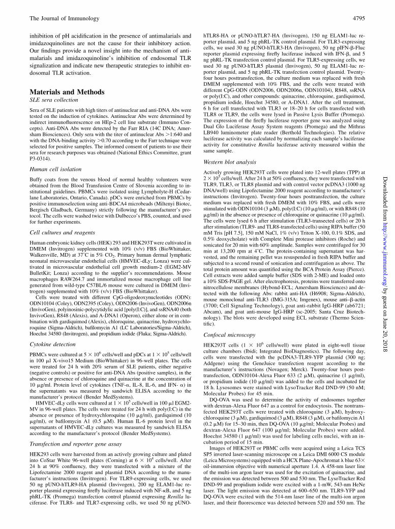
FIGURE 1. Chloroquine and quinacrine reduce the production of proinflammatory cytokines in PBMCs treated with anti-DNA Ab-positive sera of SLE
patients. A, PBMCs were treated for 24 h with 20% serum of SLE patients, negative (negative controls C1, C2, C3, C4) or positive for anti-DNA Abs
(positive samples S1, S2, S3, S4), for which binding activities were .0.70 according to the Farr technique. Human TNF-a protein levels in the PBMC
supernatants were measured by ELISA. Graphs show mean 6 SD (n = 3). B, PBMCs were treated for 24 h with 20% serum of SLE patients, negative
(negative controls C1, C2) or positive for anti-DNA Abs (positive samples S1, S2, S3, S4), for which binding activities were .0.70 according to the Farr
technique. Chloroquine (CQ) was added to anti-DNA Ab positive samples at a concentration 10 mg/ml. Human TNF-a protein level in the supernatants of
PBMC cultures was measured by ELISA. Graphs show mean 6 SD (n = 3). C and D, PBMCs were treated for 24 h with 20% serum of SLE patients,
negative (negative control C) or positive for anti-DNA Abs (positive samples S1, S2), for which binding activities were .0.70 according to the Farr
technique. CQ or quinacrine (Q) was added at final concentration of 5 or 10 mg/ml. Human IL-8 and IL-6 protein level in the supernatants of PBMC
cultures was measured by ELISA. M, X-vivo15 medium.
4796 INHIBITION OF ENDOSOMAL TLRs BY ANTIMALARIALS
by guest on June 20, 2018http://w
ww
.jimm
unol.org/D
ownloaded from
ResultsAntimalarials reduce cytokine synthesis in PBMCs exposed tosera of SLE patients
Self-recognition through endosomal TLRs can cause autoimmunediseases, such as SLE (3). Circulating immune complexes con-
sisting of Abs and endogenous nucleic acids activate pDCs and
B cells through activation of TLR7 or TLR9 (12–14). Stimulation
with nucleic acids leads to high levels of IFN-a production by
pDCs (3, 15) and causes proliferation and differentiation of au-
toreactive B cells into plasma cells, producing pathogenic auto-
antibodies against nuclear Ags (16). Anti-DNA Abs isolated from
sera of SLE patients, when combined with DNA from apoptotic
cells to form immune complexes, were shown to induce IFN-a as
well as TNF-a production in PBMCs (16, 17). Normal PBMCs
were stimulated with 20% serum from SLE patients, either neg-
ative (negative controls) or positive for anti-DNA Abs (positive
samples). Only SLE sera with very high DNA-binding activity(.0.70 according to the Farr technique) induced TNF-a expres-sion in human PBMCs (Fig. 1A; p , 0.0001). Chloroquine in-hibited the production of TNF-a (Fig. 1B; p , 0.001), whereasthe serum from the SLE patient undergoing hydroxychloroquinetherapy induced TNF-a expression in human PBMCs in thesimilar range as sera obtained from healthy volunteers (data notshown). In our experiments, we also detected a dose-dependentdecrease in IL-8 (Fig. 1C) and IL-6 (Fig. 1D) production in thepresence of chloroquine and quinacrine. Additional experimentson pDCs also revealed inhibitory effect of chloroquine and quin-acrine on IFN-a production, which was induced by some SLE sera(Supplemental Fig. 1A). Our results showed that the immune com-plexes of SLE sera were able to induce the production of differenttypes of proinflammatory cytokines in PBMC containing B cellsand pDCs (Supplemental Fig. 1C), which was in all cases inhib-ited with antimalarial agents.
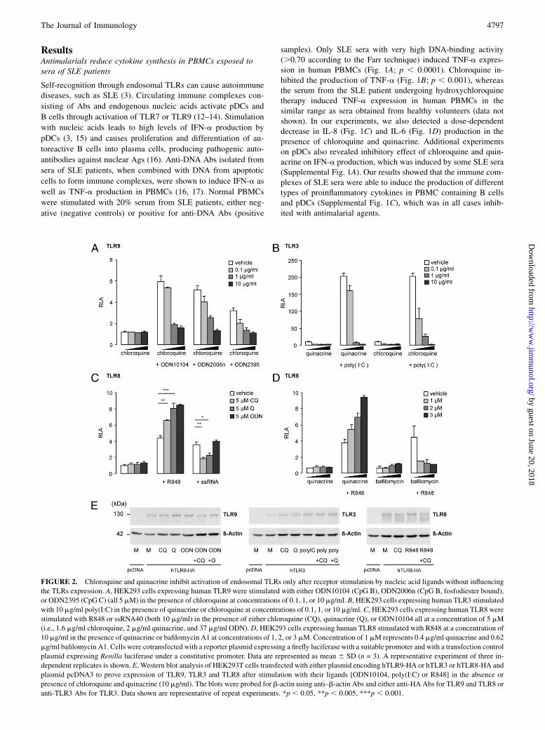
FIGURE 2. Chloroquine and quinacrine inhibit activation of endosomal TLRs only after receptor stimulation by nucleic acid ligands without influencing
the TLRs expression. A, HEK293 cells expressing human TLR9 were stimulated with either ODN10104 (CpG B), ODN2006n (CpG B, fosfodiester bound),
or ODN2395 (CpG C) (all 5mM) in the presence of chloroquine at concentrations of 0.1, 1, or 10mg/ml. B, HEK293 cells expressing human TLR3 stimulated
with 10 mg/ml poly(I:C) in the presence of quinacrine or chloroquine at concentrations of 0.1, 1, or 10 mg/ml. C, HEK293 cells expressing human TLR8 were
stimulated with R848 or ssRNA40 (both 10 mg/ml) in the presence of either chloroquine (CQ), quinacrine (Q), or ODN10104 all at a concentration of 5 mM
(i.e., 1.6 mg/ml chloroquine, 2 mg/ml quinacrine, and 37 mg/ml ODN). D, HEK293 cells expressing human TLR8 stimulated with R848 at a concentration of
10 mg/ml in the presence of quinacrine or bafilomycin A1 at concentrations of 1, 2, or 3 mM. Concentration of 1 mM represents 0.4 mg/ml quinacrine and 0.62
mg/ml bafilomycin A1. Cells were cotransfected with a reporter plasmid expressing a firefly luciferase with a suitable promoter and with a transfection control
plasmid expressing Renilla luciferase under a constitutive promoter. Data are represented as mean 6 SD (n = 3). A representative experiment of three in-
dependent replicates is shown. E, Western blot analysis of HEK293T cells transfected with either plasmid encoding hTLR9-HA or hTLR3 or hTLR8-HA and
plasmid pcDNA3 to prove expression of TLR9, TLR3 and TLR8 after stimulation with their ligands [ODN10104, poly(I:C) or R848] in the absence or
presence of chloroquine and quinacrine (10 mg/ml). The blots were probed for b-actin using anti–b-actin Abs and either anti-HA Abs for TLR9 and TLR8 or
anti-TLR3 Abs for TLR3. Data shown are representative of repeat experiments. *p , 0.05, **p , 0.005, ***p , 0.001.
The Journal of Immunology 4797
by guest on June 20, 2018http://w
ww
.jimm
unol.org/D
ownloaded from
Effect of antimalarials on endosomal TLR inhibition depend onthe type of TLR ligand
Activation of endosomal TLRs plays an important role in theautoimmune process in SLE patients (14, 18–20). To determine the
effect of quinacrine and chloroquine on separate endosomal TLRs,
we used HEK293 cells transfected with each of the endosomal
TLR receptors and stimulated them with different TLR agonists in
the presence or absence of quinacrine and chloroquine. TLR9 acti-
vation with agonists ODN10104 (B-type CpG, phosphorotioate
backbone), ODN2006n (B-type CpG, phosphodiester backbone),
and ODN2395 (C-type CpG) was inhibited in a dose-dependent
manner with chloroquine (Fig. 2A). The presence of 1 mg/ml
chloroquine inhibited TLR9 activation by .50%. Flow cytom-
etry confirmed that chloroquine had no effect on the level of TLR9
expression (Supplemental Fig. 2). In a similar experiment, quin-
acrine and chloroquine inhibited TLR3 activation with poly(I:C)
(Fig. 2B). However, when we analyzed the activation of TLR8
by two different TLR8 ligands, ssRNA and R848, we demon-
strated that chloroquine and quinacrine inhibited TLR8 activation
by ssRNA, but unexpectedly observed a costimulatory effect incombination with R848 (Fig. 2C). In contrast, bafilomycin A1,which inhibits vacuolar ATPase and consequently causes a de-crease in endosomal pH (21), had an inhibitory effect on TLR8activation by R848 (Fig. 2D). These results showed that chloro-quine and quinacrine most likely did not inhibit endosomal TLRactivation due to its effect on endosomal pH. Additionally,Western blot analysis showed that chloroquine and quinacrine hadno influence on the expression of the receptors TLR9, TLR3, andTLR8 (Fig. 2E). Together, these data demonstrated that the effectof the antimalarials is dependent on the type of TLR ligand usedfor stimulation of the receptor and suggest that antimalarials donot influence the endosomal pH and expression of TLRs.
Direct interaction of antimalarials with endosomal TLRligands
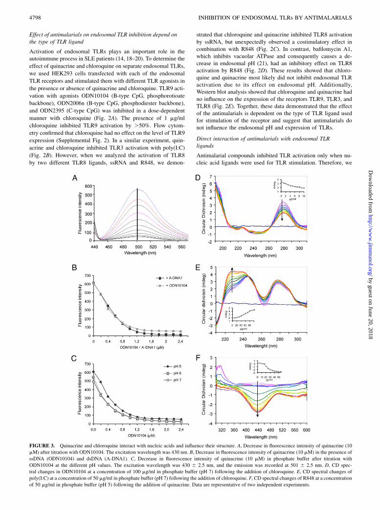
Antimalarial compounds inhibited TLR activation only when nu-cleic acid ligands were used for TLR stimulation. Therefore, we
FIGURE 3. Quinacrine and chloroquine interact with nucleic acids and influence their structure. A, Decrease in fluorescence intensity of quinacrine (10
mM) after titration with ODN10104. The excitation wavelength was 430 nm. B, Decrease in fluorescence intensity of quinacrine (10 mM) in the presence of
ssDNA (ODN10104) and dsDNA (A-DNA1). C, Decrease in fluorescence intensity of quinacrine (10 mM) in phosphate buffer after titration with
ODN10104 at the different pH values. The excitation wavelength was 430 6 2.5 nm, and the emission was recorded at 501 6 2.5 nm. D, CD spec-
tral changes in ODN10104 at a concentration of 100 mg/ml in phosphate buffer (pH 7) following the addition of chloroquine. E, CD spectral changes of
poly(I:C) at a concentration of 50 mg/ml in phosphate buffer (pH 7) following the addition of chloroquine. F, CD spectral changes of R848 at a concentration
of 50 mg/ml in phosphate buffer (pH 5) following the addition of quinacrine. Data are representative of two independent experiments.
4798 INHIBITION OF ENDOSOMAL TLRs BY ANTIMALARIALS
by guest on June 20, 2018http://w
ww
.jimm
unol.org/D
ownloaded from
proposed that the inhibition of endosomal TLR activation could bea result of interactions between nucleic acids and quinacrine orchloroquine. Molecular interactions were determined using eitherfluorescence spectroscopy for fluorescent quinacrine or CD in thecase of chloroquine. Quinacrine emits strong fluorescence by ex-citation at 430 nm. A titration of quinacrine with ODN10104 froma 0–1.5 mM final concentration resulted in a gradual fall in fluo-rescent intensity (Fig. 3A). The drop in fluorescence intensity isobserved both in ssDNA and dsDNA (Fig. 3B) and is independentof pH (Fig. 3C). This fluorescence quenching indicates the forma-tion of interactions between quinacrine and nucleic acids.The interactions of the tested antimalarials with nucleic acids
cause a change in the chemical environment of the nucleic acidchain and can affect their secondary structure. We used CD spectro-scopy to provide evidence of structural changes in DNA and RNAupon titration with chloroquine. A positive-induced CD band withsignificant ellipticity was developed around 280 nm by the addi-tion of chloroquine to the ODN10104 solution, whereas a smallnegative-induced CD band appeared around 245 nm (Fig. 3D).Addition of bafilomycin A1 to ODN10104 resulted in no changein the CD spectra, confirming that bafilomycin A1 does not in-teract with ssDNA (data not shown). Titration of poly(I:C) withchloroquine initiated a large enhancement in the positive (225 nm)and small enhancement in the negative (310 nm) CD bands of poly(I:C) in phosphate buffer at pH 7 (Fig. 3E). Those modifications
show the effect of antimalarials on the conformation of nucleicacids, which could make nucleic acid ligands unavailable to TLRreceptor binding sites.The unexpected costimulatory activity of quinacrine and R848
is also likely to be caused by direct interactions between bothcompounds and TLR8. Indeed, we observed an induced CD effectfor the interaction between R848 and quinacrine in solution (Fig.3F), providing a new insight into the mechanism of TLR8 acti-vation by small nucleoside analogs. It is likely that quinacrineforms a complex with R848, stabilized by stacking of aromaticrings, which bind cooperatively to the neighboring binding sites ofTLR8, thus triggering dimerization of its ectodomains and in-creasing the TLR8 activation.
Inhibition of endosomal TLRs by other nucleic acid-bindingcompounds
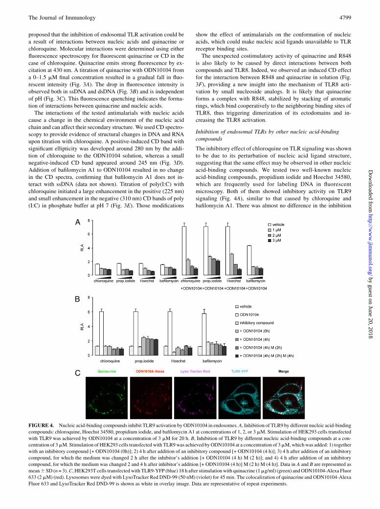
The inhibitory effect of chloroquine on TLR signaling was shownto be due to its perturbation of nucleic acid ligand structure,suggesting that the same effect may be observed in other nucleicacid-binding compounds. We tested two well-known nucleicacid-binding compounds, propidium iodide and Hoechst 34580,which are frequently used for labeling DNA in fluorescentmicroscopy. Both of them showed inhibitory activity on TLR9signaling (Fig. 4A), similar to that caused by chloroquine andbafilomycin A1. There was almost no difference in the inhibition
FIGURE 4. Nucleic acid-binding compounds inhibit TLR9 activation by ODN10104 in endosomes.A, Inhibition of TLR9 by different nucleic acid-binding
compounds: chloroquine, Hoechst 34580, propidium iodide, and bafilomycin A1 at concentrations of 1, 2, or 3 mM. Stimulation of HEK293 cells transfected
with TLR9 was achieved by ODN10104 at a concentration of 3 mM for 20 h. B, Inhibition of TLR9 by different nucleic acid-binding compounds at a con-
centration of 3mM. Stimulation of HEK293 cells transfected with TLR9was achieved byODN10104 at a concentration of 3mM,whichwas added: 1) together
with an inhibitory compound [+ ODN10104 (0h)]; 2) 4 h after addition of an inhibitory compound [+ ODN10104 (4 h)]; 3) 4 h after addition of an inhibitory
compound, for which the medium was changed 2 h after the inhibitor’s addition [+ ODN10104 (4 h) M (2 h)]; and 4) 4 h after addition of an inhibitory
compound, for which the medium was changed 2 and 4 h after inhibitor’s addition [+ ODN10104 (4 h)] M (2 h) M (4 h)]. Data in A and B are represented as
mean6 SD (n= 3).C, HEK293T cells transfected with TLR9-YFP (blue) 18 h after stimulationwith quinacrine (1mg/ml) (green) andODN10104-Alexa Fluor
633 (2 mM) (red). Lysosomes were dyed with LysoTracker Red DND-99 (50 nM) (violet) for 45 min. The colocalization of quinacrine and ODN10104-Alexa
Fluor 633 and LysoTracker Red DND-99 is shown as white in overlay image. Data are representative of repeat experiments.
The Journal of Immunology 4799
by guest on June 20, 2018http://w
ww
.jimm
unol.org/D
ownloaded from

when the inhibitor was added before ODN10104 or if the mediumwas changed before the addition of ODN (Fig. 4B). The DNA-binding compound probably accumulates in endosomes and canthere form a complex either before endocytosis or afterward.
Antimalarials colocalize in the endosomes with nucleic acids
To confirm the interactions between quinacrine and TLR ligandsinside living cells, we used confocal fluorescent microscopy.Quinacrine enters the cells very rapidly, because it can be detectedin the endosomal vesicles after only 10 s, which was confirmed byLyso Tracker Red costaining (not shown). After 18 h of incubationof TLR9-YFP–transfected HEK293T cells with ODN10104-AlexaFluor 633 and quinacrine, we found colocalization of ODNs andquinacrine inside lysosomes (Fig. 4C, Supplemental Fig. 3A). Wealso detected colocalization of ODN10104-Alexa Fluor 633 andquinacrine in human PBMCs (Supplemental Fig. 3B). A similarexperiment was performed using propidium iodide, which wecould detect inside cells only in the presence of ODN10104, thusdemonstrating the formation of a complex within the endosomes,because propidium iodide exhibits fluorescence only when itis intercalated with nucleic acids (Supplemental Fig. 3C). Wedetermined colocalization of ODN10104-Alexa Fluor 633 andTLR9-YFP. Therefore, we can conclude that in endosomes, TLR9coexists together with ODN and quinacrine.
Inhibition of TLR9 and TLR3 activation by TLR7/8 agonisticimidazoquinolines
Because of reports that imidazoquinolines, the TLR7/8 ligands,also cause the inhibition of TLR9 receptors, we were interested
in determining if their mechanism of action was similar to thatof the antimalarial compounds. It has been proposed that thecoexpression of TLR7 and TLR9 in B cells and pDCs creates thepotential for intracellular physical interactions of these receptors,resulting in a tight control of TLR7 signaling by TLR9 (11). It hasalso been assumed that TLR7-dependent inhibition of TLR9-induced IFN-g production may occur by direct interaction ofthe TLR7 receptor with TLR9 ligands (22). These studies in-volved experiments on cells that express both TLR9 and TLR7receptors. To decouple the potential interaction between the tworeceptors, we used HEK293 cells expressing only the TLR9receptor and investigated whether both receptors are indeednecessary for the inhibition of TLR9 signaling by TLR7/8agonists.When TLR9-transfected HEK293 cells were treated with TLR9
ligand (ODN2006) in the presence of imidazoquinolines, we ob-served a decrease in TLR9 activity (Fig. 5A). These experimentsrevealed that TLR7/8 ligands R848 and gardiquimod inhibitedTLR9 signalization directly, without any indirect effect throughTLR7 costimulation. Inhibition by imidazoquinolines was alsoobserved for TLR3-transfected HEK cells activated by poly(I:C)(Fig. 5B). The inhibition of TLR9 and TLR3 with gardiquimodwas dose dependent and comparable to that caused by quinacrineand chloroquine (not shown). To further confirm the relevance ofthese findings, we made experiments with two different ODNs(ODN10104 and ODN2006) (Fig. 5C) and showed a decrease inIL-8 synthesis using TLR9-transfected HEK293 cells treated withODN2006 in the presence of gardiquimod (Supplemental Fig. 4A).We also looked at the inhibition of IL-6 synthesis by using pri-mary HMVEC-dLy cells, which do not express TLR7 (23), treated
FIGURE 5. Imidazoquinolines R848 and gardiquimod inhibit TLR9 and TLR3 activation in a similarmanner as chloroquine and quinacrine.A, Inhibition of
TLR9 with gardiquimod and R848. HEK293 cells expressing TLR9 were treated with 3 mMODN10104 in the absence or presence of R848 and gardiquimod,
both at concentrations of 1 and 10mg/ml.B, Inhibition of TLR3with gardiquimod. HEK293 cells expressing TLR3were treatedwith 10mg/ml poly(I:C) in the
absence or presence of gardiquimod (0.1, 1, and 10mg/ml).C, Inhibition of TLR9, stimulated by ODN10104 (3mM) or ODN2006 (3mM), with gardiquimod.
HEK293 cells expressing TLR9 were treated with ODNs in the absence or presence of gardiquimod (0.1, 0.5, 1, and 5 mg/ml).D, Inhibition of IL-6 synthesis.
HMVEC-dLy were incubated with poly(I:C) (10 mg/ml) in the absence or presence of hydroxychloroquine (10 mg/ml), gardiquimod (10 mg/ml), and bafi-
lomycin A1 (0.5 mM). After 24, human IL-6 protein level in the supernatants was measured by ELISA. Data are represented as mean6 SD (n = 3).
4800 INHIBITION OF ENDOSOMAL TLRs BY ANTIMALARIALS
by guest on June 20, 2018http://w
ww
.jimm
unol.org/D
ownloaded from

with poly(I:C) in the presence of hydroxychloroquine, gardiquimod,and bafilomycin A1 (Fig. 5D). Additionally, we showed thatgardiquimod and resiquimod inhibited TLR9 similarly to chloro-quine and propidium iodide and did not require preincubation toexhibit their activity (Supplemental Fig. 4B). None of inhibitorycompounds, including chloroquine, propidium iodide, gardiqui-mod, and resiquimod, inhibited TLR5 activation by flagellin usedas a control (Supplemental Fig. 4C), thereby confirming that theinhibition is limited to ligand-mediated endosomal TLR ectodo-main dimerization.
Direct interactions of imidazoquinolines with nucleic acids
We presumed that the underlying mechanism for the inhibition ofTLR9 activation by imidazoquinolines could be the interactionbetween ODN and TLR7/8 ligands, much like the inhibition byantimalarial drugs, which share the structure of condensed het-eroaromatic rings. To check our hypothesis, we investigated thebinding of R848 and gardiquimod to ODN. Fluorescence spectro-scopic experiments showed fluorescence quenching of the in-trinsic fluorescence of gardiquimod after the addition of ssDNA(ODN10104) (Fig. 6A), which was even enhanced in the case ofdsDNA. The chemical structure of the nucleotide backbone inODNs did not have much influence on these interactions (Fig. 6B).In contrast to the experiments with quinacrine, pH affected the in-teractions of gardiquimod with ODN (Fig. 6C). Fluorescencequenching was more efficient at a lower pH, which indicates thatinteractions between gardiquimod and ODN are stronger in endo-somes than outside the cells. Quinacrine bound more strongly toODNs compared with gardiquimod (Fig. 6D), whereas interactionswith R848 were the weakest, which also explains why R848 hadless inhibitory effect on TLR9 activation within the cells thangardiquimod.
Antimalarials and imidazoquinolines do not inhibit endosomalproteolytic processing
The importance of proteolytic processing of endosomal TLRs hasbeen demonstrated for TLR9 (24, 25). This process could, in prin-ciple, be affected by antimalarial compounds at concentrationsthat do not affect acidification. We wanted to exclude the influenceof antimalarials and imidazoquinolines on the proteolytic pro-cessing of endosomal TLRs that was shown to be required forTLR9 activation (24, 25). As a positive control, we used bafilo-mycin, which is a specific inhibitor of the vacuolar H+-ATPaseresponsible for the acidification of vacuolar compartments.Endosomal acidification is required for the activity of proteasesthat function optimally at low pH and also for the ordered pro-gression of cargo along the endocytic pathway. HEK293T cellswere therefore loaded with fluorescent dyes DQ-OVA and Alexa647-dextran following their preincubation with DNA-bindingcompounds to compare the influence of hydroxychloroquine andbafilomycin A1 on proteolysis and endocytosis. The confocalmicroscopy imaging showed that bafilomycin A1 inhibited pro-teolytic degradation of DQ-OVA at a concentration of 0.2 mM(Fig. 7A, 7B). The presence of red-colored Alexa 467-dextranshowed that endocytosis was not affected. In contrast, the presenceof 3 mM chloroquine did not decrease proteolytic degradation ofDQ-OVA (Fig. 7C). Moreover, when we investigated other anti-malarials and imidazoquinolines, the results indicated that hydro-xychloroquine, gardiquimod, and R848 (Fig. 7D–F) did not inhibitproteolysis of fluorescently-tagged OVA inside endosomes either.Further experiments applying 10-fold higher concentration (30 mM)also showed no inhibition of proteolysis. Results of pH assess-ment in endosomes showed that bafilomycin increased pH in en-dosomes of RAW cells, whereas chloroquine at a concentrationthat inhibited TLR signaling did not influence the pH value. Ex-
FIGURE 6. Imidazoquinolines interact with nucleic acids in a similar manner as antimalarials. A, Decrease in the fluorescence intensity of gardiquimod
(10 mM) after titration with ssDNA (ODN10104) and dsDNA (A-DNA1). B, Effect of the phosphodiester (ODN2006n) and phosphotioate (ODN10104)
nucleic acid backbone on the decrease in fluorescence values after titration of gardiquimod (10 mM). C, Decrease in fluorescence intensity of gardiquimod
(10 mM) in phosphate buffer after titration with ODN10104 at the different pH values. D, Decrease in fluorescence intensity of gardiquimod, R848, and
quinacrine (all 10 mM) in phosphate buffer (pH 5) after titration with ODN10104.
The Journal of Immunology 4801
by guest on June 20, 2018http://w
ww
.jimm
unol.org/D
ownloaded from

periments on C57BL/6 mouse macrophages and human lung mi-crovascular endothelial cells showed quite similar results (Fig.7G). The evidence that antimalarials do not affect TLR via actingon endosomal pH additionally supports our proposed mechanismof endosomal TLR inhibition (Fig. 8).
DiscussionThe antimalarial drugs hydroxychloroquine, chloroquine, and quin-acrine are widely used in the therapy of autoimmune diseases suchas rheumatoid arthritis and SLE (26). They can promote a remis-sion in non-major organ SLE, especially in variants with skin andjoint affliction (27). However, despite the common use of anti-malarials as therapeutic drugs, very little is known about theirmechanism of action.We described that antimalarial agents efficiently inhibited pro-
duction of inflammatory cytokines in pDC and PBMCs incubatedwith immune complexes of sera from SLE patients. The inhibitory
activity of quinacrine on the activation of endosomal TLRs has beengenerally ascribed to be a consequence of blocking of endosomalacidification and maturation (7) as strong interactions of nucleicacids with TLR9 occur only under acidic conditions (pH 4.5–6.5)(28). However, we showed that the effect of antimalarials on ve-sicular pH at concentrations required to suppress the signal trans-duction was negligible, as also reported before (6, 29). We proposethat antimalarials affect endosomal TLR activation by their directinteraction with TLR ligands. Further results confirmed the in-hibitory effect of chloroquine and quinacrine on TLR9, TLR3, andTLR8 signaling, when these receptors were stimulated with nucleicacids. In contrast, we observed that antimalarials increased theactivation of TLR8 by imidazoquinoline R848. The costimulatoryeffect of CpG-ODN has been previously shown for TLR8 activa-tion by imidazoquinolines (30).Additional experiments proved direct interactions between anti-
malarials and nucleic acid TLR ligands. We detected the inter-
FIGURE 7. Antimalarials and imidazoquinolines have a different mechanism of endosomal TLR inhibition than bafilomycin. A, Control HEK293T cells
3 h after the addition of DQ-OVA (10 mg/ml) and dextran-Alexa Fluor 647 (100 mg/ml). B, HEK293T cells preincubated with bafilomycin A1 (0.2 mM) for
30 min before the addition of DQ-OVA (10 mg/ml) and Alexa Fluor 647-dextran (100 mg/ml). HEK 293T cells preincubated with chloroquine (C; 3 mM),
hydroxychloroquine (D; 3 mM), gardiquimod (E; 3 mM), or R848 (F; 3 mM) for 30 min before the addition of DQ-OVA (10 mg/ml) and Alexa Fluor 647-
Dextran (100 mg/ml). Hoechst 34580 (1 mg/ml) was used to detect nuclei. Images are representative of 10 random fields of view from two separate
experiments (1 cm on the figure means 25 mm). G, Changes of endosomal pH in primary cells treated with bafilomycin or chloroquine. Primary cell lines
were loaded with pH sensitive nanoparticles and treated with bafilomycin (0.2 mM) or chloroquine (4 mM). The change in pH was determined from ratios
between emission intensities of pH-dependent Oregon Green 514 and pH-independent Alexa Fluor 633, covalently bound to the same nanoparticles. Higher
ratio represents more alkaline pH, and lower ratio represents more acidic pH.
4802 INHIBITION OF ENDOSOMAL TLRs BY ANTIMALARIALS
by guest on June 20, 2018http://w
ww
.jimm
unol.org/D
ownloaded from

actions by fluorescence quenching of quinacrine after the additionof ODN, whereas CDwas applied to detect conformational changesin ODN10104 and poly(I:C) caused by the presence of chloroquine.Changes in the conformation of nucleic acids as a consequence ofinteractions with chloroquine can explain the inability of ODN orpoly(I:C) to trigger activation of TLR3 or TLR9. Additionally, theobserved interaction between R848 and quinacrine may causecooperative binding of their complex to the binding sites of TLR8,causing increased activation of the receptor instead of inhibition aswith ssRNA. This sheds additional light on the enigmatic activationof TLR7/8 by small molecules (31, 32). It seems likely that imi-dazoquinolines self-aggregate at the receptor binding sites, mim-icking binding of polynucleotide and bridging the two TLR8/7ectodomains.We confirmed our hypothesis on the mechanism of endosomal
TLR inhibition with other nucleic acid-binding compounds, such aspropidium iodide and Hoechst 34580. Both compounds inhibitedTLR9 activation much like chloroquine. The inhibition was notdependent on the preincubation of nucleic acids with the inhibitoras the complex formed within the endosomes, thus supporting ourhypothesis that quinacrine–nucleic acid interactions are indeedrelevant inside the living cells. We detected propidium iodidefluorescence in living cells when ODN was added, indicating thatpropidium iodide forms a fluorescent complex with ODN withinendosomes.We also investigated the effect of TLR7/8 ligands imidazo-
quinolines, which had been shown in our experiments to preventTLR9 signaling. Their inhibitory effect on TLR9 has already beenreported, but the mechanism of inhibition was not clear. It hadbeen proposed that physical interactions between TLR7 andTLR9 underlay this effect (11). However, our experiments withTLR9-transfected HEK293 cells showed that the presence of TLR7was not necessary for the inhibition of TLR9 signaling caused byresiquimod or gardiquimod. Moreover, we observed inhibition ofIL-6 synthesis by gardiquimod on TLR3-triggered stimulationof primary human lung microvascular endothelial cells, similarto the inhibition by hydroxychloroquine and bafilomycin A1.Again, we detected direct interactions between ODN and
imidazoquinolines, suggesting that the mechanism of TLR9 andTLR3 inhibition by those compounds is similar to that of an-
timalarials. We observed that the interactions between ODNand gardiquimod were stronger than those between ODN andR848 but weaker than in the case of quinacrine. The differencesin the strength of interaction could be a consequence of inter-calation properties of compounds guided by properties such asplanarity and the number and position of nitrogen atoms. NMRstudies have previously demonstrated a physical associationbetween DNA oligomers and imidazoquinolines (30). Prefer-ential binding of quinacrine to adenine- and thymine-rich DNAregions has also been reported (33), which is in agreement withour results (data not shown). Aminoacridine molecules, such asquinacrine, intercalate between base pairs and cause a slightunfolding and elongation of nucleic acid chains (34). Therefore,such nucleic acids with a changed conformation may not be ableto bind to the TLR binding sites. We already demonstrated theeffect of the nucleic acid conformation on activation of TLR3,which, due to the distance between the binding sites, binds nu-cleic acid duplex in an A- rather than in a B-type conforma-tion (35).The role of proteolysis and endosomal acidification connected
to it has been reported for TLR9 activation (24, 25). We wereable to demonstrate that antimalarials and imidazoquinolinesdo not inhibit endosomal proteolytic processing (Fig. 6). Asexpected, bafilomycin A1 with its selective inhibition of H+-ATPase prevented the acidification of endosomes (21, 36, 37)and inhibited the activity of lysosomal proteases requiringacidic pH. In contrast, quinacrine, chloroquine, hydroxychlo-roquine, gardiquimod, and R848 did not inhibit proteolysis ofDQ-OVA, thus demonstrating that those compounds did notaffect endosomal and lysosomal acidification. Additionally, wemeasured the pH in endosomes and confirmed that chloroquinedid not influence the endosomal pH in different types of primarycells, whereas bafilomycin increased the value of pH in endo-somes.Our proposed mechanistic model of endosomal TLR inhibition
by antimalarial drugs and imidazoquinolines is illustrated in Fig. 8.Although TLR9 is activated by binding of stimulatory DNA result-ing in rearrangement of the receptor (38), the addition of nucleicacid-binding compounds leads to the masking of TLR binding siteson nucleic acids and consequently preventing TLR ligand binding.Conformational modification of nucleic acid could retain bindingto single TLR binding site but would prevent the productive rear-rangement of the ectodomains. Therefore, interactions betweenantimalarial drugs or imidazoquinolines and nucleic acids do notrule out the possibility of antagonistic effect caused by the inhib-itors on endosomal TLRs.Altogether, our results suggest that the molecular mechanism of
action of antimalarial drugs in the therapy of autoimmune diseases,such as SLE, involves binding to nucleic acids, which changes thechemical environment and masks TLR ligand-binding epitopes.Moreover, the direct interactions between ODNs or poly(I:C) andimidazoquinolines cause inhibition of TLR9 and TLR3 by a mech-anism, similar to the mechanism by antimalarials. Understandingthe mechanism of endosomal TLR inhibition by antimalarials andimidazoquinolines represents a promising potential for the designof improved drugs for the therapy of autoimmune diseases.
AcknowledgmentsWe thank K.A. Fitzgerald (University of Massachusetts Medical School,
Worcester, MA) for kindly providing the immortalized mouse macrophage
cell line generated from the wild-type C57BL/6 mouse, M. Bele and
P. Nadrah for preparing fluorescently labeled silica nanoparticles, H. Gra-
disar for help in CD spectrometry experiments, and M. Mancek Keber for
help in flow cytometry experiments.
FIGURE 8. Mechanistic model of endosomal TLR inhibition by nucleic
acid-binding compounds (i.e., antimalarial drugs and imidazoquinolines).
Nucleic acid-binding to endosomal TLR induces activation of the signaling
pathway. When a nucleic acid-binding compound (inhibitor) is present,
there is no activation of the endosomal TLR by the nucleic acid ligand.
Antimalaric drugs and imidazoquinolines interact with nucleic acids, and
the interactions consequently cause structural modifications of the TLR
ligand that prevent their binding to TLR.
The Journal of Immunology 4803
by guest on June 20, 2018http://w
ww
.jimm
unol.org/D
ownloaded from

DisclosuresThe authors have no financial conflicts of interest.
References1. Panter, G., A. Kuznik, and R. Jerala. 2009. Therapeutic applications of nucleic
acids as ligands for Toll-like receptors. Curr. Opin. Mol. Ther. 11: 133–145.2. Barrat, F. J., T. Meeker, J. Gregorio, J. H. Chan, S. Uematsu, S. Akira, B. Chang,
O. Duramad, and R. L. Coffman. 2005. Nucleic acids of mammalian origin canact as endogenous ligands for Toll-like receptors and may promote systemiclupus erythematosus. J. Exp. Med. 202: 1131–1139.
3. Marshak-Rothstein, A. 2006. Toll-like receptors in systemic autoimmune dis-ease. Nat. Rev. Immunol. 6: 823–835.
4. Vallin, H., A. Perers, G. V. Alm, and L. Ronnblom. 1999. Anti-double-strandedDNA antibodies and immunostimulatory plasmid DNA in combination mimicthe endogenous IFN-alpha inducer in systemic lupus erythematosus. J. Immunol.163: 6306–6313.
5. Sun, S., N. L. Rao, J. Venable, R. Thurmond, and L. Karlsson. 2007. TLR7/9antagonists as therapeutics for immune-mediated inflammatory disorders.Inflamm. Allergy Drug Targets 6: 223–235.
6. Macfarlane, D. E., and L. Manzel. 1998. Antagonism of immunostimulatoryCpG-oligodeoxynucleotides by quinacrine, chloroquine, and structurally relatedcompounds. J. Immunol. 160: 1122–1131.
7. Hacker, H., H. Mischak, T. Miethke, S. Liptay, R. Schmid, T. Sparwasser,K. Heeg, G. B. Lipford, and H. Wagner. 1998. CpG-DNA-specific activation ofantigen-presenting cells requires stress kinase activity and is preceded by non-specific endocytosis and endosomal maturation. EMBO J. 17: 6230–6240.
8. Yi, A. K., R. Tuetken, T. Redford, M. Waldschmidt, J. Kirsch, and A. M. Krieg.1998. CpG motifs in bacterial DNA activate leukocytes through the pH-dependent generation of reactive oxygen species. J. Immunol. 160: 4755–4761.
9. Ahmad-Nejad, P., H. Hacker, M. Rutz, S. Bauer, R. M. Vabulas, and H. Wagner.2002. Bacterial CpG-DNA and lipopolysaccharides activate Toll-like receptorsat distinct cellular compartments. Eur. J. Immunol. 32: 1958–1968.
10. Rutz, M., J. Metzger, T. Gellert, P. Luppa, G. B. Lipford, H. Wagner, andS. Bauer. 2004. Toll-like receptor 9 binds single-stranded CpG-DNA in a se-quence- and pH-dependent manner. Eur. J. Immunol. 34: 2541–2550.
11. Wang, J. P., P. Liu, E. Latz, D. T. Golenbock, R. W. Finberg, and D. H. Libraty.2006. Flavivirus activation of plasmacytoid dendritic cells delineates key ele-ments of TLR7 signaling beyond endosomal recognition. J. Immunol. 177:7114–7121.
12. Krug, A. 2008. Nucleic acid recognition receptors in autoimmunity. In Hand-book of Experimental Pharmacology. S. Bauer and G. Hartmann, eds. Springer-Verlag Berlin, Heidelberg, Germany, p. 129–151.
13. Lamphier, M. S., C. M. Sirois, A. Verma, D. T. Golenbock, and E. Latz. 2006.TLR9 and the recognition of self and non-self nucleic acids. Ann. N. Y. Acad. Sci.1082: 31–43.
14. Means, T. K., E. Latz, F. Hayashi, M. R. Murali, D. T. Golenbock, andA. D. Luster. 2005. Human lupus autoantibody-DNA complexes activate DCsthrough cooperation of CD32 and TLR9. J. Clin. Invest. 115: 407–417.
15. Ronnblom, L., and G. V. Alm. 2003. Systemic lupus erythematosus and the typeI interferon system. Arthritis Res. Ther. 5: 68–75.
16. Leadbetter, E. A., I. R. Rifkin, A. M. Hohlbaum, B. C. Beaudette,M. J. Shlomchik, and A. Marshak-Rothstein. 2002. Chromatin-IgG complexesactivate B cells by dual engagement of IgM and Toll-like receptors. Nature 416:603–607.
17. Bave, U., G. V. Alm, and L. Ronnblom. 2000. The combination of apoptotic U937cells and lupus IgG is a potent IFN-alpha inducer. J. Immunol. 165: 3519–3526.
18. Boule, M. W., C. Broughton, F. Mackay, S. Akira, A. Marshak-Rothstein, andI. R. Rifkin. 2004. Toll-like receptor 9-dependent and -independent dendritic cell
activation by chromatin-immunoglobulin G complexes. J. Exp. Med. 199: 1631–1640.
19. O’Neill, L. A. 2008. Primer: Toll-like receptor signaling pathways–what dorheumatologists need to know? Nat. Clin. Pract. Rheumatol. 4: 319–327.
20. Krieg, A. M., and J. Vollmer. 2007. Toll-like receptors 7, 8, and 9: linking innateimmunity to autoimmunity. Immunol. Rev. 220: 251–269.
21. Yoshimori, T., A. Yamamoto, Y. Moriyama, M. Futai, and Y. Tashiro. 1991.Bafilomycin A1, a specific inhibitor of vacuolar-type H(+)-ATPase, inhibitsacidification and protein degradation in lysosomes of cultured cells. J. Biol.Chem. 266: 17707–17712.
22. Berghofer, B., G. Haley, T. Frommer, G. Bein, and H. Hackstein. 2007. Naturaland synthetic TLR7 ligands inhibit CpG-A- and CpG-C-oligodeoxynucleotide-induced IFN-alpha production. J. Immunol. 178: 4072–4079.
23. Pegu, A., S. Qin, B. A. Fallert Junecko, R. E. Nisato, M. S. Pepper, andT. A. Reinhart. 2008. Human lymphatic endothelial cells express multiplefunctional TLRs. J. Immunol. 180: 3399–3405.
24. Ewald, S. E., B. L. Lee, L. Lau, K. E. Wickliffe, G. P. Shi, H. A. Chapman, andG. M. Barton. 2008. The ectodomain of Toll-like receptor 9 is cleaved to gen-erate a functional receptor. Nature 456: 658–662.
25. Park, B., M. M. Brinkmann, E. Spooner, C. C. Lee, Y. M. Kim, and H. L. Ploegh.2008. Proteolytic cleavage in an endolysosomal compartment is required foractivation of Toll-like receptor 9. Nat. Immunol. 9: 1407–1414.
26. Rynes, R. I. 1997. Antimalarial drugs in the treatment of rheumatological dis-eases. Br. J. Rheumatol. 36: 799–805.
27. Wallace, D. J. 1994. Antimalarial agents and lupus. Rheum. Dis. Clin. North Am.20: 243–263.
28. Mellman, I., R. Fuchs, and A. Helenius. 1986. Acidification of the endocytic andexocytic pathways. Annu. Rev. Biochem. 55: 663–700.
29. Manzel, L., L. Strekowski, F. M. Ismail, J. C. Smith, and D. E. Macfarlane. 1999.Antagonism of immunostimulatory CpG-oligodeoxynucleotides by 4-amino-quinolines and other weak bases: mechanistic studies. J. Pharmacol. Exp. Ther.291: 1337–1347.
30. Gorden, K. K., X. Qiu, J. J. Battiste, P. P. Wightman, J. P. Vasilakos, andS. S. Alkan. 2006. Oligodeoxynucleotides differentially modulate activation ofTLR7 and TLR8 by imidazoquinolines. J. Immunol. 177: 8164–8170.
31. Hemmi, H., T. Kaisho, O. Takeuchi, S. Sato, H. Sanjo, K. Hoshino, T. Horiuchi,H. Tomizawa, K. Takeda, and S. Akira. 2002. Small anti-viral compounds ac-tivate immune cells via the TLR7 MyD88-dependent signaling pathway. Nat.Immunol. 3: 196–200.
32. Gorden, K. B., K. S. Gorski, S. J. Gibson, R. M. Kedl, W. C. Kieper, X. Qiu,M. A. Tomai, S. S. Alkan, and J. P. Vasilakos. 2005. Synthetic TLR agonistsreveal functional differences between human TLR7 and TLR8. J. Immunol. 174:1259–1268.
33. Comings, D. E. 1978. Mechanisms of chromosome banding and implications forchromosome structure. Annu. Rev. Genet. 12: 25–46.
34. Peacocke, A. R. 1973. The interaction of acridines with nucleic acids. In Acri-dines, 2nd Ed. R. M. Acheson, ed. John Wiley & Sons, New York, p. 723–757.
35. Pirher, N., K. Ivicak, J. Pohar, M. Bencina, and R. Jerala. 2008. A secondbinding site for double-stranded RNA in TLR3 and consequences for interferonactivation. Nat. Struct. Mol. Biol. 15: 761–763.
36. Clague, M. J., S. Urbe, F. Aniento, and J. Gruenberg. 1994. Vacuolar ATPaseactivity is required for endosomal carrier vesicle formation. J. Biol. Chem. 269:21–24.
37. Umata, T., Y. Moriyama, M. Futai, and E. Mekada. 1990. The cytotoxic action ofdiphtheria toxin and its degradation in intact Vero cells are inhibited by bafilo-mycin A1, a specific inhibitor of vacuolar-type H(+)-ATPase. J. Biol. Chem. 265:21940–21945.
38. Latz, E., A. Verma, A. Visintin, M. Gong, C. M. Sirois, D. C. Klein,B. G. Monks, C. J. McKnight, M. S. Lamphier, W. P. Duprex, et al. 2007.Ligand-induced conformational changes allosterically activate Toll-like receptor9. Nat. Immunol. 8: 772–779.
4804 INHIBITION OF ENDOSOMAL TLRs BY ANTIMALARIALS
by guest on June 20, 2018http://w
ww
.jimm
unol.org/D
ownloaded from