mechanism efficacy of oleic acid glutaraldehyde...
TRANSCRIPT
323
Mechanism of Efficacy of 2-Amino OleicAcid for Inhibition of Calcification ofGlutaraldehyde-Pretreated Porcine
Bioprosthetic Heart Valves
Weiliam Chen, PhD; Frederick J. Schoen, MD, PhD; Robert J. Levy, MD
Background Calcification is a frequent cause of the clinicalfailures of glutaraldehyde-pretreated bioprosthetic heartvalves (BPHV) fabricated from glutaraldehyde-cross-linkedporcine aortic valves. 2-Amino oleic acid (AOA) has beenshown in previous in vivo studies to be a promising anticalci-fication agent. Our objective was to investigate the mechanismof calcification inhibition mediated by AOA pretreatment ofporcine aortic valve bioprostheses.Methods and Results BPHV tissues were treated with an
AOA solution for 72 hours before experimentation. Thediffusion of AOA across both cusp and aortic wall wasevaluated. The lag time for AOA to diffuse across the aorticwall was prolonged compared with that of the cusp. Anextraction study was performed to determine the stability ofAOA binding; the results indicated that the binding wasrelatively stable regardless of solvent extraction conditions.The interaction between ionic calcium and AOA on treatedtissue also was investigated by evaluating the patterns ofcalcium diffusion across both treated and untreated tissues.
C alcific degeneration causes most clinical failuresof bioprosthetic heart valve (BPHV) replace-ments fabricated from porcine aortic valves
cross-linked with glutaraldehyde.1 Procedures for inhib-iting BPHV calcification have been investigated in ani-mal model studies involving either subdermal implantsin rats2 or circulatory implants in large animals (sheepor calves).3-8 Thus far, of all the inhibition strategiesstudied including pretreatments with diphosphonate ormetallic salt solutions, polymer incorporation into leaf-let structures, and others, only detergent pretreatmentshave inhibited calcification in both subdermal and cir-culatory animal model studies. The mechanism of ac-tion of detergent pretreatment for inhibiting biopros-thetic calcification is incompletely understood atpresent. The present investigations focused on a bio-
Received January 25, 1994; revision accepted March 1, 1994.From the Division of Pediatric Cardiology (W.C., R.J.L.),
University of Michigan Medical Center, Ann Arbor, and theDepartments of Pathology (F.J.S.), Brigham and Women's Hos-pital and Harvard Medical School, Boston.
Presented in part at the meeting of the Association for theAdvancement of Medical Instrumentation, Bethesda, Md, 1992,and presented in part at the 1993 American Society for ArtificialInternal Organs Meeting, New Orleans, La.
Correspondence to Robert J. Levy, MD, R5014 Kresge II, Divi-sion of Pediatric Cardiology, C.S. Mott Children's Hospital, Univer-sity of Michigan Medical Center, Ann Arbor, MI 48109-0576.© 1994 American Heart Association, Inc.
The results showed that AOA significantly reduced the diffu-sion of calcium. AOA inhibition of aortic valve calcification(calcium level, 5.5±3.0 mg/g of tissue compared with control;calcium level, 91.2±19.5 mg/g of tissue) but not aortic wall(calcium level, 158.7±10.3 mg/g of tissue compared withcontrol; calcium level, 157.5±7.9 mg/g of tissue) was demon-strated on representative specimens from valves implanted inleft ventricular apicoaortic shunts explanted after 150 days.
Conclsions AOA covalently binds to glutaraldehyde-pre-treated bioprosthetic heart valve tissue, presumably as the resultof an aldehyde-amino reaction. Covalently bound AOA dimin-ishes Ca2+ diffusion compared with non-AOA-pretreated bio-prosthetic tissues. This may explain in part the anticalcificationmechanism of AOA. Furthermore, AOA inhibits calcification ofporcine BPHV cusps in the circulation. (Circulation.1994;90.323-329.)Key Words * calcification * glutaraldehyde * aortic cusp
* aortic wall
prosthetic pretreatment procedure using a detergent,2-amino oleic acid (AOA), which has been hypothe-sized to covalently bond to aldehyde residuals and thusremain within pretreated bioprosthetic tissue, sustain-ing its calcification inhibition effects.
It was previously demonstrated in a study of porcinebioprosthetic leaflets implanted subdermally in rats7and a series of sheep orthotopic mitral valve replace-ments8 that AOA mitigates calcification of BPHV. AOAis hypothesized to bind by its 2-amino group to freealdehyde groups of tissue bound glutaraldehyde (aSchiff base reaction9-11), thus allowing AOA to remainin the tissue. Although AOA was shown to be effectivein reducing the level of bioprosthetic mineralization inanimal models, its long-term binding stability might notbe adequate because of the known reversibility of theSchiff base reaction.9-1"The objective of this study was to investigate the
mechanism of antimineralization efficacy of AOA pre-incubation of BPHV tissues. In vitro studies assessedthe kinetics of AOA diffusion through either aortic cuspor aortic wall. Since calcium ion influx into bioprosthetictissue immediately after implantation is hypothesized tobe one of the important steps in the calcification ofBPHV,12 calcium ion diffusion across AOA-pretreatedtissues (cusp or aortic wall) was studied. AOA inhibi-tion of bioprosthetic valve calcification was assessed inexplanted specimens of aortic leaflets and aortic wall
by guest on May 22, 2018
http://circ.ahajournals.org/D
ownloaded from
324 Circulation Vol 90, No 1 July 1994
from 150-day, left ventricle-to-descending aorta valvegrafts in sheep.
MethodsMaterialsAmino oleic acid was provided to our laboratory by the
Medtronic Heart Valve Division (Irvine, Calif) as a saturatedsolution (0.171% AOA) prepared under proprietary condi-tions also used to prepare the clinically implantable heartbioprostheses used in the animal models described below.Control bioprosthetic incubations used the identical solventused to prepare the saturated AOA solution. ['4C]-labeled(specific activity, 1.6 mCi/mM, labeled at the beta-carbon)AOA was synthesized at American Radio Chemical Inc (StLouis, Mo) using procedures established by Girardot.13 Non-radioactive AOA was synthesized by the same procedures byMedtronic Inc. Fresh and glutaraldehyde-cross-linked porcineaortic valve tissues were supplied by the Heart Valve Divisionof Medtronic Inc. Glutaraldehyde-pretreated porcine aorticvalves were prepared according to the clinical pretreatmentprotocol used by Medtronic Heart Valve Inc, following pro-prietary procedures. Non-AOA-exposed porcine biopros-thetic valve tissues were used in all control studies. Frozenbovine serum and Tween-80 (for use in extraction studies, seebelow) were purchased from Sigma Chemical Co and FisherChemical Inc, respectively. Porcine aortic valve bioprostheses(valved grafts, without stents) with and without AOA pretreat-ment,813 also exposed either to AOA plus proprietary solventor proprietary solvent alone, were provided to Crawford LongHospital (Emory University, Atlanta, Ga) for sheep implantstudy by the Heart Valve Division of Medtronic Inc. A 0.1% ofsodium azide (Sigma Chemical Co) was added to the bovineserum as a preservative. All other chemicals were reagentgrade; distilled and deionized water was used.A saturated AOA solution (0.171%) was prepared under
proprietary conditions by the Heart Valve Division ofMedtronic Inc following the method established by Girar-dot.8,13 A 0.1% [`4C]-AOA stock solution was prepared by thesame procedures.8'13 The [`4C]-AOA stock solution was spikeddirectly into AOA solution for all experiments. Porcine bio-prosthetic methodology for AOA linking followed establishedmethodology at 37°C for 72 hours under proprietary condi-tions (as above).813 Tissues pretreated with [`4C]-AOA werewashed extensively with an excess of normal saline until theradioactivity of the wash liquid was indistinguishable frombackground. Similar methodologies were applied to treat freshtissues.
Determination of AOA Content in["4C]-AOA-Treated TissuesSample tissues were lyophilized separately, weighed, and
incubated in Solvable (New England Nuclear Research Prod-ucts Inc) for 24 hours at 600 to 65°C in a water bath. Eachdigested sample then was combined with an adequate volumeof Ecolume (ICN Biomedical Inc), and ["4C] radioactivity wasdetermined by liquid scintillation counting (Beckman LS3801liquid scintillation counter). The AOA content of tissue wasassessed by comparing its dpm value with that of the ['4C]-AOA solution prepared for tissue treatment. The amount ofAOA incorporated was expressed as nanomoles per milligramof dried tissue.
Diffusion of AOA Across TissuesRepresentative thicknesses of all tissues were measured at
four sites using a micrometer (Bel-Art Instruments) beforeexperimentation, and data were reported as mean+SEM. Fordiffusion studies, each piece of cusp tissue was mounted on amicrodiffusion cell. One milliliter of ["4C]-spiked AOA solu-tion was pipetted into the donor chamber, and 1 mL ofnon-AOA-containing buffer was immediately added to the
recipient chamber. The diffusion cell was placed on a shakerrotating at 150 rpm; temperature was maintained at 37°C.After 15 minutes, the recipient chamber was evacuated andreplenished with 1 mL of fresh AOA buffer. Additionalsamples were withdrawn at 30 and 45 minutes and 1, 2, 3, 4, 5,6, and 7 hours. Each sample was combined with 19 mL ofEcolume, and ["4C] radioactivity was assessed by liquid scintil-lation counting. The AOA level was determined by comparingthe sample dpm value with that of the [`Cl-spiked AOAsolution prepared for the diffusion study. The AOA leveldetected was expressed as the cumulative percent AOA dif-fused into the recipient half-cell. The lag time for AOAdiffusion was estimated by back-extrapolating the linear phaseof the plot (cumulative % AOA diffused across versus time) tothe time axis. Similar methodologies were applied to deter-mine the diffusion parameters of glutaraldehyde-cross-linkedaortic wall tissue. The sampling intervals were 1, 2, 3, 4, 6, and7 hours and 1, 2, 3, 5, 6, and 7 days. Their diffusivities werecalculated based on steady-state equilibrium as described byJohnston et al,14 using the equation15
D=I2/6(tjag)where D is diffusivity, I is thickness of tissue, and tlag is lagtime.
Diffusion of Calcium Across TissuesThe thicknesses of all cusp samples were measured before
experimentation as described above. A piece of AOA-treatedcusp tissue was mounted on a microdiffusion cell. One millili-ter of 0.2% (18 mol/L) calcium chloride solution was pipettedinto the donor chamber, and 1 mL of water was immediatelyadded to the recipient chamber. The diffusion cell was placedon a shaker rotating at 150 rpm; temperature was maintainedat 37°C. After 5 minutes, the recipient chamber was evacuatedand replenished with 1 mL of water. Additional samples werewithdrawn at 10, 15, 20, 30, 45, and 60 minutes. The calciumcontents of all samples collected (after proper dilutions) weredetermined by atomic absorption spectroscopy (Perkin-Elmermodel 2380 spectrophotometer) at a wavelength of 422.8 nm.Similar methodologies were applied to evaluate the pattern ofcalcium diffusion across aortic wall. The sampling intervalswere 4, 8, 12, 24, 28, and 32 hours. Diffusivity constants werecalculated as described above.'4,15
AOA Extraction Study['4C]-AOA extraction studies were carried out with an array
of solvents including pH 7.4 phosphate-buffered saline, 20%aqueous Tween-80 solution, and bovine serum. Samples ofAOA-treated tissue were transferred to a vial of pH 7.4phosphate buffer placed on a shaker (rotating at 150 rpm) at37°C for 1 hour. Each BPHV specimen was then transferred toanother vial containing fresh phosphate buffer. The sameprocedure was repeated at 2, 4, 6, 8, 12, and 24 hours, then 2,4, 6, 8, 12, 16, 24, and 30 days. Samples were withdrawn fromeach vial and combined with Ecolume, and the ['4CJ level wasassessed by liquid scintillation counting. AOA content wasdetermined by comparing its dpm value with that of the['4C]-spiked AOA solution used for treating tissue.At the conclusion of the extraction study, each tissue
specimen was retrieved and digested with Solvable; residual[14C] radioactivity was determined by the procedures previ-ously described involving saline washes to remove residualradioactivity followed by lyophilization. The residual dpmvalue was added to the total cumulative counts gatheredduring the study; this number was stipulated as the initial totalcounts on the tissue before the experiment. The extractionstudy results were thereby expressed as cumulative % AOAleached versus time. Similar methodologies were applied toconduct extraction studies using both bovine serum andTween-80 as extraction media. Unpaired t tests were used toassess the significance of statistical differences from controls.
by guest on May 22, 2018
http://circ.ahajournals.org/D
ownloaded from
Chen et al Prevention of Calcification by Amino Oleic Acid 325
15
0
W
U)
ILL
0
be
12
9
6
3
A
v
v
v
O
0 1 2 3 4 5TIME (HOURS)
30 r
0
LI)IL
ILL
0
R
25 F
6 7 8
B
20
15 F
10 F
5
0m *0 ,
A
30 60 90 120 150 180
TIME (HOURS)
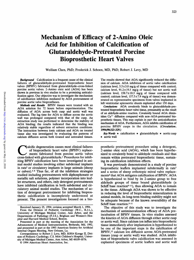
FIG 1. Plots show diffusion of 2-amino oleic acid (AOA, 0.146%wt/vol) across cusp tissue (A) and aortic wall tissue (B). Eachdata point represents the average of four replicates±SEM.
Calcium Analysis of ExplantedBioprosthetic Tissues
Fifteen sheep were used for valve implantation: seven ofthem received AOA-pretreated, stentless valves, and the restreceived control valves (ie, non-AOA-pretreated). Represen-tative samples (one sample from each leaflet and adjacentaortic wall tissues) from 150-day sheep valved grafts (implant-ed as left ventricular apicoaortic shunt) were assessed uponretrieval by one of us (F.J.S.) as part of an investigation of thehemodynamic performance and calcification inhibition ofAOA pretreated bioprosthetic heart valves.16 Retrieved aorticcusps and aortic wall samples were separately analyzed forcalcium as detailed below. Cuspal and aortic wall tissues werelyophilized, weighed, and acid hydrolyzed according to estab-lished procedures.17'18 Tissue calcium contents were deter-mined by atomic absorption spectroscopy at a wavelength of422.8 nm. ANOVA was used to test statistical significance.
ResultsAOA DiffusionWhen glutaraldehyde-cross-linked porcine aortic
valve tissue was used as a diffusion barrier (Fig 1A), a
significant amount of AOA (>1% of the AOA from thedonor chamber) penetrated the tissue after only 1 hour.The results of AOA diffusion across cusp tissue andaortic wall are illustrated in Fig 1A and 1B, respectively;the corresponding diffusion lag times and thicknessesare summarized in Table 1. The tissue thickness and thelag time determined were 0.73±0.11 mm and 0.56 hour,respectively. When aortic wall was used as a diffusionbarrier (Fig 1B), the time required to detect a significantamount of AOA (>1% of the AOA from the donorchamber) drastically increased to about 48 hours. Thecorresponding tissue thickness and extrapolated lag
TABLE 1. Diffusion of AOA in Glutaraldehyde-Pretreated Porcine Aortic Valve Tissues
Type of Thickness,* Lag Time, Diffusivity,*Tissue mm h mm2/hCusp 0.73±0.11 0.56 0.157±0.001Aortic wall 2.59+0.14 88.14 0.013±0.001AOA indicates 2-amino oleic acid.*Each number derived from four representative replicates
±SEM.
time were 2.59±0.14 mm and 88.14 hours, respectively.The diffusivities of cusp and aortic wall tissues are0.157±0.001 and 0.013±0.001 mm2/h, respectively. Theimplication is that AOA does not penetrate aortic wallas readily as cuspal tissue.
Stability of AOA BindingExtraction studies of AOA-treated tissues (cusp and
aortic wall) were performed to determine the stabilityof AOA binding. Initially, pH 7.4 phosphate buffer wasused as a medium for the study. To better simulate thein vivo environment that a BPHV valve would encounterafter implantation, an AOA extraction study was alsoconducted using bovine serum as an alternative me-dium. Furthermore, the poor solubility ofAOA in waterand hence in phosphate buffer may thus reflect itsdissolution characteristics rather than its binding stabil-ity. Therefore, Tween-80, a nonionic surfactant, wasused as a harsh dissociative medium to forcibly enhancenonbound AOA dissociation.The results of the 1-month extraction study using
both cusp and aortic wall are illustrated in Fig 2A and2B, respectively. In general, an initial burst followed bya greatly diminished rate of AOA extraction was ob-served in each study. The rate of extraction in phos-phate buffer began to plateau at 24 hours comparedwith that of Tween- 80, which leveled off within 8 hours.As illustrated in Fig 2A, using Tween-80 as the extrac-tion medium, approximately 60% of the AOA was lostfrom the cusps after the first 12 hours. Although asimilar pattern was observed with aortic wall (Fig 2B), asignificant amount of AOA was found to remain on thistissue as well at the conclusion of the study.
Table 2 summarizes the initial and residual AOAlevels on the tissues (both cusp and aortic wall) used forthe extraction study. For comparison, fresh tissue (afterAOA pretreatment) was used as a control. It should benoted that 42.2±2.3 nM of AOA was present on eachmilligram of fresh cusp tissue compared with 128.6±1.1nM/mg of its glutaraldehyde-cross-linked counterpart(before extraction). Therefore, the residual glutaralde-hyde appears to play an important role in linking AOAmolecules to tissues.13 Each milligram of cusp tissue,after leaching in phosphate buffer, retained 95.5±5.2nM of AOA; the high residual AOA level may simplyreflect poor aqueous solubility of AOA. The presence ofa surfactant (ie, Tween-80) greatly enhanced aqueoussolubility of AOA; thus, 35.6±1.0 nM of AOA re-mained on each milligram of tissue. When bovine serumwas used as an extraction medium, each milligram oftissue retained 52.1±2.2 nM of AOA. Although it ispossible that serum protein adsorption could have in-fluenced this result, this seems unlikely, given the
by guest on May 22, 2018
http://circ.ahajournals.org/D
ownloaded from

326 Circulation Vol 90, No 1 July 1994
80
60
% AOA
40
20
0
75
60
% AOA 45
30 _
15
A80
0 00
0Ca++
(sg/mI)R00.+0v
v v v v
60 F
40 F
20 F
00 5 10 15 20 25 30
TIME ( DAYS )
B30
25
20Ca++
(19/mI) 15
10
5
8
b v V V V V Vv v v
0 5 10 15 20 25
i
v
v
v. 57,v
0 20 40 60TIME ( minutes )
030
TIME (DAYS)
FIG 2. Plots show leaching of 2-amino oleic acid (AOA)-treatedcusp tissue (A) in pH 7.4 phosphate buffer (v), Tween-80solution (o), and bovine serum (,) and leaching of AOA-treatedaortic wall tissue (B) in pH 7.4 phosphate buffer (v), Tween-80solution (o), and bovine serum (*). Each data point representsthe average of at least five replicates±SEM.
B
{{
+
80
1 0 20 30 40 50
TIME (Hours)
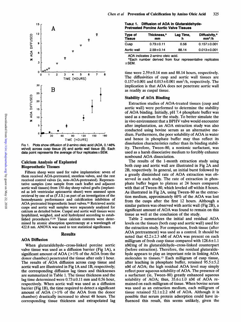
FIG 3. Plots show diffusion of calcium across 2-amino oleic acid(AOA)-treated (,) and untreated (v) cusp tissue (A) and diffusionof calcium across AOA-treated (.) and untreated (v) aortic walltissue (B). The concentration of calcium chloride solution is0.2%. Each data point represents the average of four replicates±SEM.
exhaustive washing protocol described above followedby freeze drying. Nevertheless, the results of the bovineserum studies were highly reproducible (as shown) andinternally consistent, suggesting high levels of AOAresidual tissue binding after serum exposure.
Calcium DiffusionThe results of calcium diffusion across cusp (AOA-
pretreated and untreated) and aortic wall (AOA-pre-TABLE 2. Bound and Residual AOA inGlutaraidehyde-Pretreated Porcine Aortic Valve Tissues
Amount of AOA,nM/mg of tissue
AorticMedium Cusp§ WallsControl* 128.6±1.1 74.6±1.1
Bovine serumt 52.1±2.2 31.8±1.4
20% Tween-80t 35.6±1.0 33.9+1.8
Phosphate buffert 95.5±5.2 70.4±0.1Fresh tissuet 42.2±2.3 26.6±1.2
AOA indicates 2-amino oleic acid.*No extraction study performed.tResults after 30 days of extraction.tNon-glutaraidehyde--cross-linked tissue subjected to AOA
pretreatment/no extraction study performed.§Each number represents at least five replicates ±SEM. AOA
levels on both types of tissue after the extraction study weresignificantly lower than those of controls (P<.001 in both cases).
treated and untreated) tissues are depicted in Fig 3. InFig 3A, after 1 hour of diffusion, the cumulativeamounts of calcium diffused across the AOA-pretreatedand untreated cusp tissues were 33.1±1.7 and 78.2± 3.8,ug, respectively. Their corresponding diffusivities are2.69±0.11 and 1.25±0.05 mm2/h. When aortic wall wasused as a diffusion barrier (Fig 3B), the cumulativeamount of calcium diffused across the AOA-pretreatedand untreated tissues was 9.0±1.7 and 14.0±1.6 pg,respectively, after 24 hours. Their corresponding diffu-sivities are 0.21±0.04 and 0.12±0.02 mm2/h. Overall,the presence of AOA on both tissues appears to signif-icantly retard the transport of calcium ions, and thisimplies that AOA does indeed interact with calcium.
Calcium Inhibition in the CirculationExplanted porcine bioprosthetic aortic valve cusps
had significantly less calcium (5.5±3.0 mg/g of tissue)than did cusps from untreated valves (91.2±19.5 mg/gof tissue). For comparison, unimplanted aortic valvecusps contained 0.6±0.2 mg of calcium per gram oftissue. However, porcine bioprosthetic aortic wall calci-fication was not significantly retarded by AOA pretreat-ment (calcium content, 158.7±10.0 mg/g of tissue)compared with untreated tissues (calcium content,157.5±7.9 mg/g of tissue). Unimplanted aortic wallcontained 0.7±0.2 mg of calcium per gram of tissue.
DiscussionThe long-term success of all types of BPHV (manu-
factured from glutaraldehyde-cross-linked porcine aor-
eQ9 v v l
nw_
F
by guest on May 22, 2018
http://circ.ahajournals.org/D
ownloaded from
Chen et al Prevention of Calcification by Amino Oleic Acid 327
tic valve or bovine pericardial tissues) is limited bycalcific degeneration.1 Calcification of glutaraldehyde-cross-linked BPHV is hypothesized to be a multifactoralprocess.12 The influx of calcium ions into bioprosthetictissue after BPHV implantation almost certainly playsan important role in the initial nucleation of calciumphosphate crystals intrinsically within devitalized cellsin the valve cusps.12"7-'9 Previous work has demon-strated that the initial calcium phosphate-forming eventin bioprosthetic leaflet calcification, which can occur asearly as 48 hours after a rat subdermal implant, involvescalcium diffusion into the phosphorus-rich ultrastruc-ture of glutaraldehyde-devitalized cells.19'20 Ultrastruc-tural calcium phosphate formation has been demon-strated to occur as a result of the coincidence of diffusedcalcium with immobilized phosphorus.19'20 Thus, anagent such as AOA, which significantly alters the kinet-ics of the calcium influx (see "Results"), could behypothesized to retard calcification by reducing thepotential rate of calcium phosphate formation.Could AOA interfere with the initial nucleation of
calcium phosphate deposits or prevent the long-termproliferation of calcification or both? The data in thepresent study demonstrate both inhibition of AOA in ashort-term rat model (21 days) and longer-term sheepimplants (5 months). Nevertheless, both types of animalmodels cannot be compared with clinical implants,which must be expected to function free of calcificationfor decades. The results of the present study do not shedany light as to the prognosis for the AOA-pretreatedheart valve on a long-term basis. However, other relatedresearch concerning detergent inhibition of calcificationmay be helpful in this regard. It has been shown byBosky et a121 that detergents interfere with nucleationby disrupting the calcium-phospholipid-phosphate com-plex necessary for initial calcium phosphate crystalliza-tion. Furthermore, detergents also interfere with crystalgrowth of preexisting calcium phosphate mineral.22Thus, previous research as well as the present resultsindicate that AOA would hypothetically be effective forinterfering with initial nucleation and for preventingcalcification on a long-term basis.Our investigation was focused on the anticalcification
efficacy of AOA on BPHV using an AOA preparation(or identical solvent in control studies) currently underconsideration for preparing bioprostheses for clinicaluse. The principal findings of this study were (1) tissue-bound AOA retarded the penetration of calcium ionsthrough tissue; (2) a finite and significant amount ofAOA remained bound to the tissue regardless of ag-gressive solvent extraction; (3) aortic wall was moreresistant to AOA penetration than cusp; and (4) therewas an anticalcification effect ofAOA on aortic cusp butnot aortic wall, as demonstrated by the sheep explantcalcium analyses.
Calcium DiffusionIn our calcium diffusion studies, the presence ofAOA
on bioprosthetic tissue greatly reduced the immediatecalcium ion influx. Therefore, covalently linked AOAcould hypothetically retard the kinetics of initiatingcrystalline calcium phosphate formation in part by asimilar mechanism. The low abundance of initial nucle-ation sites hinders further crystallization of calcium
Moreover, the phospholipids (and thus its phosphategroups) in cell membranes also could serve as sites forattracting calcium ion, which could in turn result ininitiating the process of calcification. AOA also couldfunction as a surfactant to extract and perhaps exchangethe phospholipid. This may thereby lessen the sitesavailable for calcification.
Stability of AOA Binding to TissuesThe results of the extraction study demonstrated that
a significant level of AOA remained bound despiteharsh solvent conditions. Thus, the hypothesized amino-residual aldehyde bonding mechanism is supported bythese results. Alternatively, AOA affinity (nonextract-ability) could be due to hydrogen bonding or nonspe-cific hydrophobic adsorption. Definitive studies on thepossible chemical bonds occurring between AOA andaldehyde have not yet been carried out.How can the actual covalent bonding of AOA to
residual aldehyde be proven? Definitive amino alde-hyde reaction studies have thus far only been possible inmodel systems using aqueous solutions of the com-pounds of interest.23-25 Cheung and Nimni23 have estab-lished the reactivity of glutaraldehyde with amino-containing model compounds and proteins. However,establishing covalent bonding with a cross-linked aorticvalve leaflet, which is resistant to enzymatic digestion,remains a challenge to overcome; thus far, no study byour group or others has addressed this issue. A numberof surface chemistry techniques have become availablein the last decade, such as electron spectroscopy forchemical analyses and attenuated total-reflectance Fou-rier-transformed infrared spectroscopy. However, thesetechniques cannot be applied to hydrated biomaterialsbecause dessication of a tissue such as a heart valveleaflet would distort structural and surface relations aswell as denature the protein matrix of interest. How-ever, some of the questions concerning the covalentreactions of glutaraldehyde with AOA can be ap-proached in model studies with purified chemical sys-tems, and these should be the subject of future research.Although the precise mechanism ofAOA binding has
not been fully elucidated in this study, a Schiff basecovalent linkage is one likely possibility. A similarmechanism was proposed in a previous study in whichaminopropanehydroxydiphosphonate (APD) waslinked to residual aldehyde groups on either porcine26or pericardial27 bioprosthetic tissue. Interestingly, APDcovalent binding, although stable in vitro, was less stablein vivo. Furthermore, APD pretreatment resulted adose-dependent inhibition of bioprosthetic calcificationin subdermal studies but not in sheep mitral valvereplacements,28 unlike AOA.8 Part of the previoussubdermal study involved treating a control group ofbioprosthetic cusps with lysine.27 Although lysine couldbe demonstrated to bind at higher levels than APD tobioprosthetic heart valve tissue, pretreatment with onlylysine was not effective in inhibiting calcification ofbioprosthetic tissue in subdermal implant studies. Thus,these prior studies demonstrate that simply reactingresidual aldehydes in bioprosthetic tissue with amino-containing compounds is not sufficient for preventBPHV calcification. Some other unique structural-func-tional relations must facilitate the efficacy of AOA inphosphate and thus the formation of hydroxyapatite.
by guest on May 22, 2018
http://circ.ahajournals.org/D
ownloaded from
328 Circulation Vol 90, No 1 July 1994
circulatory studies compared with the other aminocompounds investigated.
AOA DiffusionHypothetically, the amount of AOA interacting with
residual aldehyde groups is determined by the concen-tration ofAOA, the availability of free AOA molecules,and the extent of available free aldehyde groups. Thisphenomenon is in turn governed by the rate of AOAdiffusing into tissue. Compared with aortic walls, theaortic valve cusp is relatively thin and composed of lessdense collagenous tissue29 and thus AOA can appar-ently penetrate it easily. Therefore, AOA molecules andglutaraldehyde residuals react readily, and a high levelof AOA per unit weight of tissue can be achieved in arelatively short period of time. In contrast, the greaterthickness of aortic wall in conjunction with its denserand apparently less permeable composition for AOAmay affect the amount of AOA bound.
Differential Inhibition of Calcification of Cuspsand Aortic WallThe results from the in vivo study indicated that AOA
was effective in preventing calcification of cusp tissuebut not aortic wall. Further examination of AOA diffu-sion results (ie, the lag times for AOA penetration) inthe two types of tissues suggests a diffusion-basedexplanation. The longer lag time (>88 hours) requiredfor AOA to diffuse at equilibrium conditions across theaortic wall compared with cusp tissue indicated that theduration of tissue AOA treatment appeared to beinsufficient for achieving the desirable initial AOA levelto resist the immediate calcium influx into aortic walltissue soon after the implantation of a BPHV. Alterna-tively, AOA may not deter the pathophysiology of aorticwall mineralization (partially involving elastin calcifica-tion), which differs markedly from aortic cusp calcifica-tion, in which collagen rather elastin calcification is veryprominent.30
Furthermore, the level of cuspal calcification ob-served in the non-AOA-pretreated explants (control)from the present left ventricular-to-descending aortaconduit model was comparable to that noted in mitralorthotopic explants from sheep828 as well as in previousstudies of bovine left ventricular-to-descending aortaconduit.4 Thus, effective inhibition of calcification byAOA in both models was comparable in terms of thelevel of bulk mineral that would have been otherwisedeposited.
AOA Comparisons With Other DetergentsPrevious investigations by others have demonstrated
a number of detergents to be efficacious for inhibitingcalcification in BPHV implants either as orthotopicvalve replacements or in left ventricular-to-descendingaorta conduits. Of these other detergents, only polysor-bate 80 pretreatment28 inhibited calcification at compa-rable levels to those reported in the present study.However, polysorbate 80 is not chemically bonded tobioprosthetic heart valve tissue, unlike AOA, and thustheir similar efficacy in short-term animal implants, suchas those described in the present study, may not reflector predict comparable efficacy in long-term clinical
A number of detergents found to be effective pre-treatments for preventing BPHV calcification in animalmodel circulatory studies have been reported to causedeleterious effects on valve cusp integrity, such asdelamination.28 Initial studies with AOA, while demon-strating efficacy for preventing calcification, also showedthat the AOA pretreatment procedures caused dam-ages to cuspal surface integrity.8 However, this effecthas been eliminated by a filtration step in the propri-etary preparation procedures used by Medtronic HeartValve for eliminating particulate material from theAOA solutions, thus avoiding material degeneration.8"16Morphology results related to these considerations maybe found in these previous studies816 and were beyondthe scope of the present experiments.
Future ConsiderationsThe effectiveness ofAOA also should be viewed with
the perspective of other experimental anticalcificationstrategies. Pretreating bioprostheses with sodium dode-cyl sulfate (SDS) in sheep circulatory implants,31 alumi-num chloride and ferric chloride20 in rat subdermalstudies, and diphosphonate compounds (eg, amino-diphosphonate) in rat subdermal studies22 has proven tobe efficacious. Interestingly, aortic wall calcification hasnot been assessed in prevention studies except forsubdermal implant experiments in which A3I and Fe3`ion and aminodiphosphonate all inhibited aortic wallcalcification.32 In comparison to other detergents, thelevel of inhibition by AOA of cuspal calcification waseither comparable or superior to the most optimalresults reported previously,28 which were obtained usingeither SDS- or polysorbate 80-pretreated bioprostheticheart valves as orthotopic mitral valve replacement insheep.28 Furthermore, other detergents effective forpreventing calcification such as n-lauryl-sarcosine orTriton X-100 resulted in primary material failurecaused by destabilization of bioprosthetic tissue.28
Thus, it is possible to speculate that a sequentialapplication of optimal anticalcification agents such asAOA (and others) to treat BPHV before implantationcould be a rational approach for compensation oflimitation(s) of each agent, and this multiagent ap-proach of BPHV treatment may result in a synergisticanticalcification effect for both aortic wall and valve.
ConclusionsAOA was demonstrated to be tightly associated to
glutaraldehyde-pretreated BPHV tissue, possibly theresult of an aldehyde-amino reaction, and AOA associ-ation stability was demonstrated despite various stren-uous solvent extractions. AOA pretreatment diminishedCa2' diffusion into bioprosthetic tissues. This may ex-plain in part the anticalcification mechanism of AOA.Furthermore, sheep explant analyses demonstrated thatAOA preincubation was an effective means for inhibit-ing the calcification of glutaraldehyde-pretreated por-cine aortic valve cusps but not aortic wall.
AcknowledgmentThese studies were supported by grants and contracts to the
University of Michigan (Dr Chen and Dr Levy) from the HeartValve Division of Medtronic Inc (Irvine, Calif). We thankDavid Myers (Heart Valve Division, Medtronic Inc) for pro-
implants. vision of materials. We also thank Dr Robert Giiyton- for
by guest on May 22, 2018
http://circ.ahajournals.org/D
ownloaded from
Chen et al Prevention of Calcification by Amino Oleic Acid 329
cooperating by providing specimens from his sheep studies ofAOA-pretreated bioprostheses. We appreciate the support ofFrance Dixon-Helfer (Heart Valve Division, Medtronic Inc).
References1. Schoen FJ, Levy RL, Piehler HR. Pathological considerations in
replacement cardiac valves. Cardiovasc Pathol. 1992;1:29-52.2. Levy RJ, Schoen FJ, Levy JT, Nelson AC, Howard SL, Oshry LJ.
Biological determinants of dystrophic calcification and osteocalcindeposition in glutaraldehyde-preserved porcine aortic valveleaflets implanted subcutaneously in rats. Am J Pathol. 1983;113:143-155.
3. Barnhart GR, Jones M, Ishihara T, Chavez AM, Rose DM,Ferrans VJ. Failure of porcine aortic and bovine pericardial pros-thetic valves: an experimental investigation in young sheep.Circulation. 1982;66(suppl I):I-150-I-153.
4. Levy RJ, Zenker JA, Bernhard WF. Porcine bioprosthetic valvecalcification in bovine left ventricle aorta shunt: studies of depo-sition of vitamin K-dependent proteins. Ann Thorac Surg. 1983;36:87-192.
5. Thubrikar MJ, Deck JD, Aouad J, Nolan SP. Role of mechanicalstress in calcification of aortic bioprosthetic valves. J Thorac Car-diovasc Surg. 1983;86:115-125.
6. Gallo I, Nistal F, Artinano E, Fernandez D, Cayon R, Carrion M,Garcia-Martinez V. The behavior of pericardial versus porcinevalve xenografts in the growing sheep model. J Thorac CardiovascSurg. 1987;93:281-290.
7. Girardot MN, Girardot JM, Schoen FJ. Alpha amino oleic acid, anew compound, prevents calcification of bioprosthetic heart valve.Trans Soc Biomater. 1991;14:114. Abstract.
8. Gott JP, Pan-Chih, Dorsey LMA, Jay JL, Jett GK, Schoen FJ,Girardot J-M, Guyton RA. Calcification of porcine valves: a suc-cessful new method of antimineralization. Ann Thorac Surg. 1992;53:207-216.
9. Woodroof EA. Use of glutaraldehyde and formaldehyde to processtissue heart valve. J Bioeng. 1978;2:1-9.
10. Cheung DT, Nimni ME. Mechanism of cross-linking of proteins byglutaraldehyde, I: reaction of model compounds. Connect TissueRes. 1982;10:187-199.
11. Korn AH, Feairheller SH, Filachione EM. Glutaraldehyde: natureof the reagent. J Mol Biol. 1972;65:525-529.
12. Schoen FJ, Levy RJ. Calcification of bioprosthetic heart valves. In:Bodnar E, Frater R, eds. Replacement Heart Valves. Elmsford, NY:Pergamon Press; 1989:124-148.
13. Girardot JM. A method for Retarding or Preventing the Calcificationof a Prosthesis Implanted in a Mammal. US Patent No. 4,976,733.
14. Johnston TP, Boyd JA, Ciesliga BL, Schoen FJ, Amidon GL, LevyRJ. Controlled release of ethanehydroxy diphosphonate from poly-urethane reservoirs to inhibit calcification of bovine pericardiumused in bioprosthetic heart valves. Intl J Pharm. 1990;59:95-104.
15. Baker R. Diffusion controlled systems. In: Baker R, ed. ControlledRelease of BiologicallyActive Agents. New York: Wiley Press; 1987:39-83.
16. Hall JD, Whitlark J, Horsley S, Dorsey L, Pan-Chih, Girardot JM,Girardot N, Schoen FJ, Gott JP, Guyton RA. Antimineralization
of bioprostheses: an improved amino oleic acid technique. ASAIOMeeting, 1993.
17. Schoen FJ, Tsao JW, Levy RJ. Calcification of bovine pericardiumused in cardiac valve bioprostheses: implication for the mech-anisms of bioprosthetic tissue mineralization. Am J Pathol. 1986;123:134-145.
18. Schoen FJ, Levy RJ, Nelson AC, Bernhard WF, Nashef A, HawleyM. Onset and progression of experimental bioprosthetic heartvalve calcification. Lab Invest. 1985;52:523-532.
19. Webb CL, Schoen FJ, Flowers WE, Alfrey AC, Horton C, Levy RJ.Inhibition of mineralization of glutaraldehyde-pretreated bovinepericardium by AlCl3: mechanism and comparisons with FeCl3,LaCl3 and Ga(N03)3 in rat subdermal model studies.Am J Pathol.1991;138:971-981.
20. Levy RJ, Schoen FJ, Flowers WB, Staelin ST. Initiation of miner-alization in bioprosthetic heart valves: studies of alkaline phos-phatase activity and its inhibition by AlCl3 or FeCl3 preincubations.J Biomed Mater Res. 1991;25:905-935.
21. Bosky AL, Goldberg MR, Posner AS. Effect of diphosphonates onhydroxyapatite formation induced by calcium-phospholipid-phosphate complexes. Calcif Tissue Int. 1979;27:83-88.
22. Hidaka S, Abe K. The effects of sodium lauryl sulphate and itsoxidative breakdown products on calcium phosphate precipitationand transformation. Arch Oral BioL 1992;37:159-165.
23. Cheung DT, Nimni ME. Mechanism of crosslinking of proteins byglutaraldehyde, I: reaction with model compounds. Connect TissueRes. 1982;10:187-199.
24. Cheung DT, Nimni ME. Mechanism of crosslinking of proteins byglutaraldehyde, II: reaction with monomeric and polymericcollagen. Connect Tissue Res. 1982;10:201-216.
25. Cheung DT, Perelman N, Ko EC, Nimni ME. Mechanism ofcrosslinking of proteins by glutaraldehyde, III: reaction withcollagen in tissue. Connect Tissue Res. 1985;13:109-115.
26. Webb CL, Benedict JJ, Schoen FJ, Linden JA, Levy RJ. Inhibitionof heart valve calcification with covalently bound aminopropane-hydroxydiphosphonate. ASAIO. 1987;10:592-595.
27. Webb CL, Benedict JJ, Schoen FJ, Linden JA, Levy RJ. Inhibitionof bioprosthetic heart valve calcification with aminodiphosphonatecovalently bound to residual aldehyde groups. Ann Thorac Surg.1988;46:309-316.
28. Jones M, Eidbo EE, Hilbert SL, Ferrans VJ, Clark RE. Antical-cification treatments of bioprosthetic heart valves: in vivo studies insheep. J Cardiovasc Surg. 1989;4:69-73.
29. Weiss L. Cardiovascular system. In: Krstic RV, ed. Human Micro-scopic Anatomy: An Atlas for Students of Medicine and Biology.Berlin: Springer-Verlag; 1991:40-66.
30. Webb CL, Nguyen NM, Schoen FJ, Levy RJ. Calcification ofallograft aortic wall in a rat subdermal model: pathophysiology andinhibition by A3l and aminodiphosphonate preincubations. Am JPathol. 1992;141:487-496.
31. Jones M, Eidbo EE, Hilbert SL, Ferrans VJ, Clark RE. The effectsof anticalcification treatments of bioprosthetic valves implanted insheep. Trans Am Soc Artif Intern Organs. 1988;34:1027-1030.
32. Webb CL, Phelps LL, Schoen FJ, Levy RJ. Aminodiphosphonateor A3l preincubation inhibits calcification of homografts in the ratsubdermal model. Trans Am Soc Artif Intern Organs. 1988;34:851-854.
by guest on May 22, 2018
http://circ.ahajournals.org/D
ownloaded from
W Chen, F J Schoen and R J Levyglutaraldehyde-pretreated porcine bioprosthetic heart valves.
Mechanism of efficacy of 2-amino oleic acid for inhibition of calcification of
Print ISSN: 0009-7322. Online ISSN: 1524-4539 Copyright © 1994 American Heart Association, Inc. All rights reserved.
is published by the American Heart Association, 7272 Greenville Avenue, Dallas, TX 75231Circulation doi: 10.1161/01.CIR.90.1.323
1994;90:323-329Circulation.
http://circ.ahajournals.org/content/90/1/323World Wide Web at:
The online version of this article, along with updated information and services, is located on the
http://circ.ahajournals.org//subscriptions/
is online at: Circulation Information about subscribing to Subscriptions:
http://www.lww.com/reprints Information about reprints can be found online at: Reprints:
document. Permissions and Rights Question and Answer this process is available in the
click Request Permissions in the middle column of the Web page under Services. Further information aboutOffice. Once the online version of the published article for which permission is being requested is located,
can be obtained via RightsLink, a service of the Copyright Clearance Center, not the EditorialCirculationin Requests for permissions to reproduce figures, tables, or portions of articles originally publishedPermissions:
by guest on May 22, 2018
http://circ.ahajournals.org/D
ownloaded from