mechanical properties of dna origami nanoassemblies are
TRANSCRIPT
6574–6582 Nucleic Acids Research, 2016, Vol. 44, No. 14 Published online 7 July 2016doi: 10.1093/nar/gkw610
Mechanical properties of DNA origaminanoassemblies are determined by Holliday junctionmechanophoresPrakash Shrestha1, Tomoko Emura2, Deepak Koirala1, Yunxi Cui1, Kumi Hidaka2, WilliamJ Maximuck1, Masayuki Endo3,*, Hiroshi Sugiyama2,3,* and Hanbin Mao1,*
1Department of Chemistry and Biochemistry, Kent State University, Kent, OH 44242, USA, 2Department ofChemistry, Graduate School of Science, Kyoto University, Kitashirakawa-oiwakecho, Sakyo-ku, Kyoto 606-8502,Japan and 3Institute for Integrated Cell-Material Sciences (WPI-iCeMS), Kyoto University, Yoshida-ushinomiyacho,Sakyo-ku, Kyoto 606-8501, Japan
Received November 29, 2015; Revised June 23, 2016; Accepted June 24, 2016
ABSTRACT
DNA nanoassemblies have demonstrated wide ap-plications in various fields including nanomaterials,drug delivery and biosensing. In DNA origami, single-stranded DNA template is shaped into desired nanos-tructure by DNA staples that form Holliday junctionswith the template. Limited by current methodolo-gies, however, mechanical properties of DNA origamistructures have not been adequately characterized,which hinders further applications of these materi-als. Using laser tweezers, here, we have describedtwo mechanical properties of DNA nanoassembliesrepresented by DNA nanotubes, DNA nanopyramidsand DNA nanotiles. First, mechanical stability ofDNA origami structures is determined by the effec-tive density of Holliday junctions along a particularstress direction. Second, mechanical isomerizationobserved between two conformations of DNA nan-otubes at 10–35 pN has been ascribed to the collec-tive actions of individual Holliday junctions, whichare only possible in DNA origami with rotational sym-metric arrangements of Holliday junctions, such asthose in DNA nanotubes. Our results indicate thatHolliday junctions control mechanical behaviors ofDNA nanoassemblies. Therefore, they can be con-sidered as ‘mechanophores’ that sustain mechani-cal properties of origami nanoassemblies. The me-chanical properties observed here provide insightsfor designing better DNA nanostructures. In addi-tion, the unprecedented mechanical isomerization
process brings new strategies for the developmentof nano-sensors and actuators.
INTRODUCTION
Because of the highly stable and specific recognition be-tween two complementary DNA strands, DNA has beenused as an attractive component in nanoassembly. In theDNA origami nanoassembly, a long ssDNA serves as a tem-plate to fold into nanostructures through hundreds of Hol-liday junctions formed between short DNA staples comple-mentary to the template sequences at particular locations(1). The simple, robust and highly efficient synthesis strategyof DNA origami has established this structure as a highlypotent nanomaterial (2–10) yet to be fully characterized forits properties. Among these unknown territories, mechan-ical property is certainly a notable missing link with highsignificance. Mechanical stability of the connecting regionsin DNA nanoassemblies is essential to sustain robust in-teractions between biomolecules (11) and nanoassemblies.Likewise, mechanical rigidity of DNA nanocavities is crit-ical to define the morphology of inorganic nanoparticlescontained within (12,13). In the sensing applications usingDNA nanoassemblies, (14–18), the mechanical rigidity andstability of the DNA nanoassemblies can directly affect theaccuracy in the signal output and the sensitivity in the ana-lyte recognition (5).
So far, only a handful of investigations have been re-ported with a main focus on the mechanical functionalitiesof the DNA nanoassemblies (14,19–22). The insufficient in-formation on the mechanical rigidity and stability of DNAnanoassemblies in general and DNA origami in particularhinders rational design of DNA nanomaterials for applica-tions that exploit their mechanical properties. One reasonfor this lack of knowledge lies in the difficulty in the char-
*To whom correspondence should be addressed. Tel: +1 330 672 9380; Fax: +1 330 672 3816; Email: [email protected] may also be addressed to Masayuki Endo. Email: [email protected] may also be addressed to Hiroshi Sugiyama. Tel: +81 075 753 4002; Email: [email protected]
C© The Author(s) 2016. Published by Oxford University Press on behalf of Nucleic Acids Research.This is an Open Access article distributed under the terms of the Creative Commons Attribution License (http://creativecommons.org/licenses/by-nc/4.0/), whichpermits non-commercial re-use, distribution, and reproduction in any medium, provided the original work is properly cited. For commercial re-use, please [email protected]
Downloaded from https://academic.oup.com/nar/article-abstract/44/14/6574/2468220by gueston 05 April 2018
Nucleic Acids Research, 2016, Vol. 44, No. 14 6575
acterization of individual nano-objects for their mechanicalproperties. Special tools with high resolution for mechani-cal force measurement, such as AFM and optical tweezers,must be employed to carry out the characterization (22,23)after immobilization of nanoparticles with different shapes,which is another challenging practice.
We reasoned that due to the flexible nature in the designof a DNA origami with the single-nucleotide precision, itis possible to introduce two duplex DNA handles to tethera DNA origami. These handles are then linked to the twooptically trapped polystyrene beads in laser tweezers, whichallow the quantification of mechanical properties of DNAnanoassembly. Previously, with the aim to develop origamibased nanomechanical devices, Sugiyama and coworkershave found that tubular designs of origami structures canhave two stable conformations: a short and a long tubu-lar forms that may stem from different isomers of Holl-iday junctions contained inside the origami devices (10).Given the potential applications of controlling conforma-tions of DNA origami structures by mechanical factors asdiscussed above, here we wish to understand whether it isa unique feature for the tubular-shaped origami to demon-strate different structural isomers. To this purpose, we ap-plied two duplex DNA handles to the tubular DNA origamistructures for the comparison of their mechanical proper-ties with those from other nanoassemblies, DNA nanopy-ramids and DNA nanotiles. We found mechanical stabilitiesof DNA origami structures are correlated with the effectivedensity of Holliday junctions in a particular nanoassem-bly. The mechanical stability is anisotropic in nature withthe short axis of a DNA nanotube resisting higher externalstress than the long axis. Interestingly, mechanical isomer-ization between two conformations of a DNA nanoassembyat 10–35 pN external force was observed only in DNA nan-otubes, which have unique symmetric arrangements of Hol-liday junctions. Similar to the mechanical stability, the me-chanical isomerization also showed anisotropic behavior.Given that individual Holliday junctions have isomeriza-tion force in the sub-picoNewton range (24) and they areanisotropically arranged in DNA nanotubes, we have at-tributed the mechanical isomerization of the DNA nan-otubes to the collective actions of many Holliday junctionsthat experience similar microenvironment. All these resultsindicate that Holliday junctions in DNA origami structuresserve as mechanophores (25,26), which determine the me-chanical property of DNA nanoassemblies, similar to chro-mophores and fluorophores that carry spectroscopic infor-mation in a molecule. We anticipate these new findings areinstrumental to optimize the mechanical strength of DNAnanoassemblies.
RESULTS AND DISCUSSION
Mechanical isomerization and mechanical disassembly ofDNA tubes
Optical tweezers have been used to apply and measureforces in picoNewtons (27–29). Compared to AFM, ithas better force resolution and therefore is particularlysuitable to characterize mechanical stability of macro-molecules such as proteins and nucleic acids. Here, we used
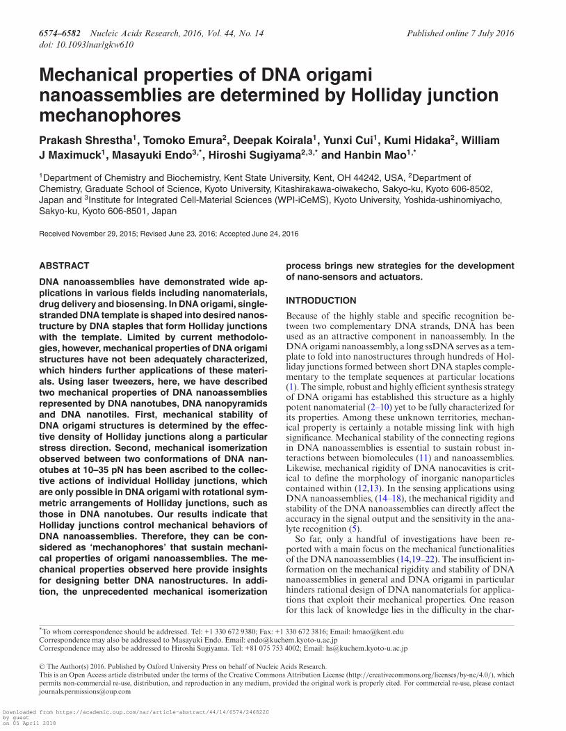
Figure 1. Mechanoanalytical characterization of DNA origami nan-otubes. (A) Schematic of the experimental set up for the mechanical iso-merization of DNA origami tubes. DNA origami tube is sandwichedbetween two long dsDNA handles and the whole construct is teth-ered between two optically trapped beads with affinity linkages. Themechanochemical property of DNA tubes is revealed by moving one ofthe beads away from another at a loading rate of ∼5.5 pN/s. AFM imageof the eight-tube DNA for longitudinal (B) and horizontal stretching (C).Schematic pictures are shown to the right of corresponding AFM images.
a home-built laser tweezers instrument (30,31) to character-ize the mechanical properties of individual DNA origaminanoassemblies. To this end, we introduced two double-stranded (ds) DNA handles to DNA origami structures (seeFigure 1A, Supplementary Figure S1A and B and Mate-rials and Methods) (14). DNA handles were inserted intothe origami structure via overhang single-stranded staplesequence, which contains 40 nucleotides complementaryto the single-stranded M13mp18 template (it has been re-ported that the shearing force of 30 bp duplex DNA is >60pN) (32). The other end of the handle was labeled with adigoxigenin or biotin molecule. These DNA handles weremixed and incubated with rest of the DNA components forthe origami construction (see Materials and Methods andSupplementary Figures S1–S7). AFM images have revealedsuccessful incorporation of the handles (see Figure 1B andC, Supplementary Figures S8 and S9 for the handles at-tached to the DNA nanotubes; see Supplementary FiguresS6 and S7 for handle incorporation of other origami struc-tures).
Using the digoxigenin and the biotin molecules labeledat the free end of the DNA handles, the modified DNAorigami construct was tethered to the two optically trappedparticles coated with digoxigenin antibody and streptavidinthrough respective affinity interactions (14). The mechan-ical stability of the origami was probed by force rampingexperiments in which one of the optically trapped particleswas moved away from the other at a loading rate of ∼5.5pN/s in laser tweezers.
Due to the unique behavior of isomerization displayedin DNA origami tubes (10), we chose these origami struc-tures as our first samples to evaluate mechanical proper-ties of DNA origami structures. Using the reported pro-cedures (10), we prepared eight-tube and six-tube DNA
Downloaded from https://academic.oup.com/nar/article-abstract/44/14/6574/2468220by gueston 05 April 2018
6576 Nucleic Acids Research, 2016, Vol. 44, No. 14
origami nanoassemblies that respectively consist of eightand six Holliday junctions in each of the circular layers thatare spirally arranged into desired tubes. Previously, AFMimages (10) have revealed that each type of tube has twoequilibrated isomers, long and short tubes (similar to thoseshown in Figure 1B and C, Supplementary Figures S8 andS9). Due to the limited time resolution in AFM, the isomer-ization between the two conformers has not been observedin real time. The dynamic nature of mechanical unfoldingat the single nanostructure level may provide an unprece-dented opportunity to investigate this isomerization pro-cess. To this end, by using the procedures reported earlier(14), either eight-tube (short and long) or six-tube (shortand long) DNA origami structures were obtained and teth-ered to two optically trapped polystyrene beads as describedabove.
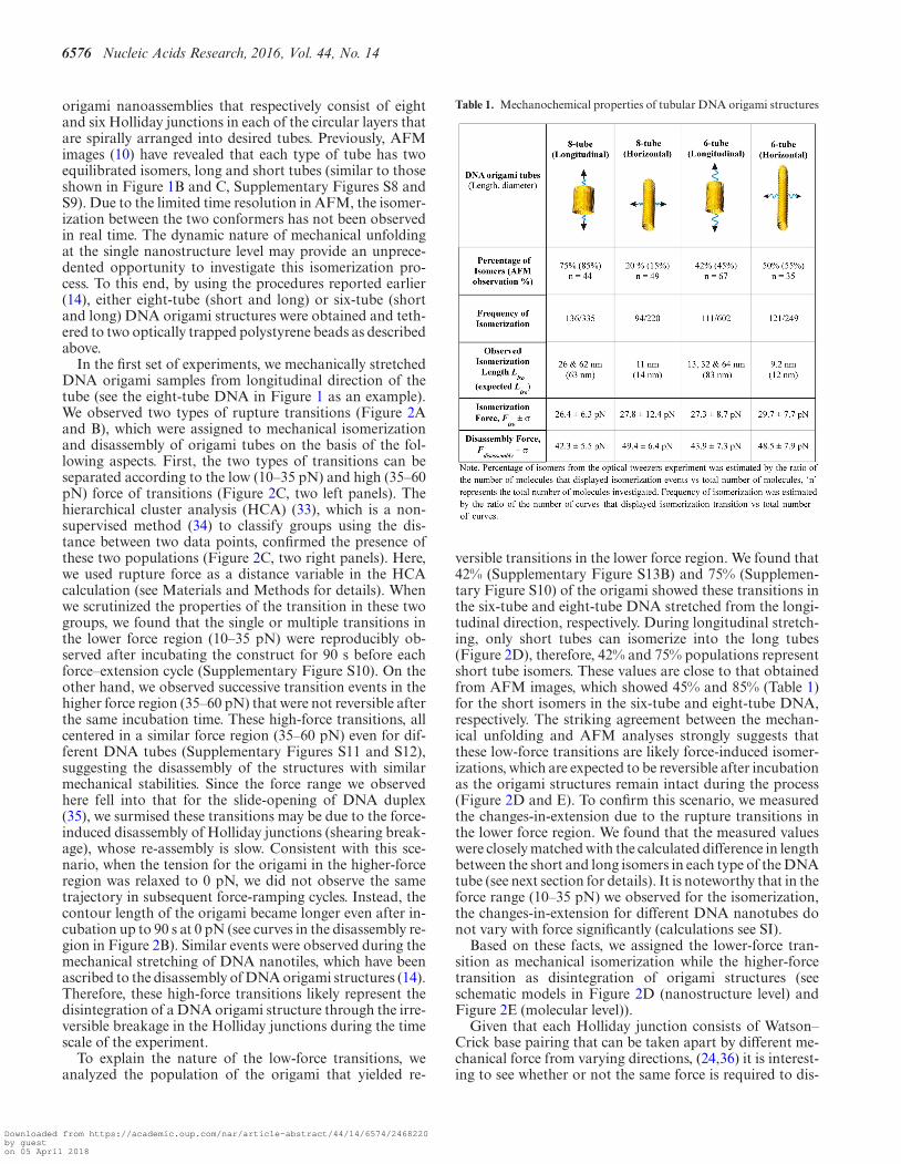
In the first set of experiments, we mechanically stretchedDNA origami samples from longitudinal direction of thetube (see the eight-tube DNA in Figure 1 as an example).We observed two types of rupture transitions (Figure 2Aand B), which were assigned to mechanical isomerizationand disassembly of origami tubes on the basis of the fol-lowing aspects. First, the two types of transitions can beseparated according to the low (10–35 pN) and high (35–60pN) force of transitions (Figure 2C, two left panels). Thehierarchical cluster analysis (HCA) (33), which is a non-supervised method (34) to classify groups using the dis-tance between two data points, confirmed the presence ofthese two populations (Figure 2C, two right panels). Here,we used rupture force as a distance variable in the HCAcalculation (see Materials and Methods for details). Whenwe scrutinized the properties of the transition in these twogroups, we found that the single or multiple transitions inthe lower force region (10–35 pN) were reproducibly ob-served after incubating the construct for 90 s before eachforce–extension cycle (Supplementary Figure S10). On theother hand, we observed successive transition events in thehigher force region (35–60 pN) that were not reversible afterthe same incubation time. These high-force transitions, allcentered in a similar force region (35–60 pN) even for dif-ferent DNA tubes (Supplementary Figures S11 and S12),suggesting the disassembly of the structures with similarmechanical stabilities. Since the force range we observedhere fell into that for the slide-opening of DNA duplex(35), we surmised these transitions may be due to the force-induced disassembly of Holliday junctions (shearing break-age), whose re-assembly is slow. Consistent with this sce-nario, when the tension for the origami in the higher-forceregion was relaxed to 0 pN, we did not observe the sametrajectory in subsequent force-ramping cycles. Instead, thecontour length of the origami became longer even after in-cubation up to 90 s at 0 pN (see curves in the disassembly re-gion in Figure 2B). Similar events were observed during themechanical stretching of DNA nanotiles, which have beenascribed to the disassembly of DNA origami structures (14).Therefore, these high-force transitions likely represent thedisintegration of a DNA origami structure through the irre-versible breakage in the Holliday junctions during the timescale of the experiment.
To explain the nature of the low-force transitions, weanalyzed the population of the origami that yielded re-
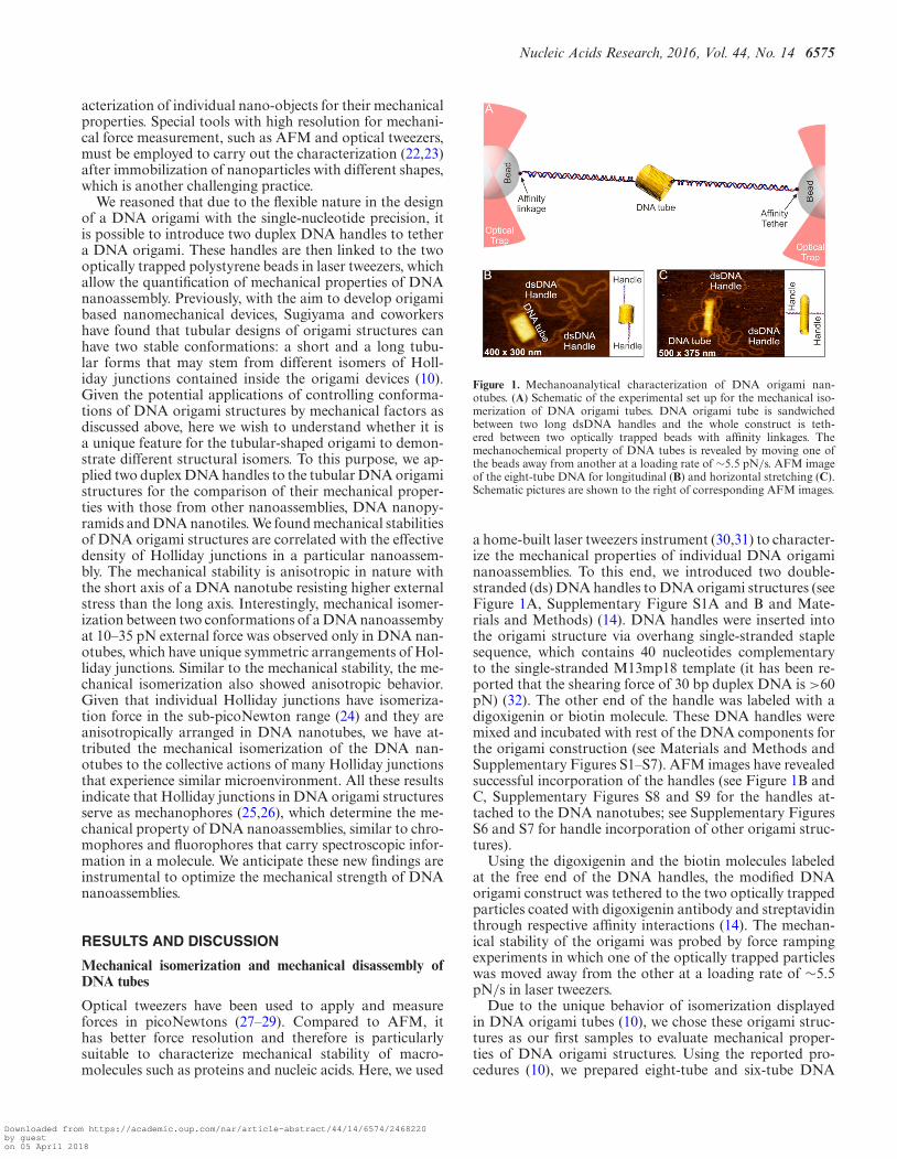
Table 1. Mechanochemical properties of tubular DNA origami structures
versible transitions in the lower force region. We found that42% (Supplementary Figure S13B) and 75% (Supplemen-tary Figure S10) of the origami showed these transitions inthe six-tube and eight-tube DNA stretched from the longi-tudinal direction, respectively. During longitudinal stretch-ing, only short tubes can isomerize into the long tubes(Figure 2D), therefore, 42% and 75% populations representshort tube isomers. These values are close to that obtainedfrom AFM images, which showed 45% and 85% (Table 1)for the short isomers in the six-tube and eight-tube DNA,respectively. The striking agreement between the mechan-ical unfolding and AFM analyses strongly suggests thatthese low-force transitions are likely force-induced isomer-izations, which are expected to be reversible after incubationas the origami structures remain intact during the process(Figure 2D and E). To confirm this scenario, we measuredthe changes-in-extension due to the rupture transitions inthe lower force region. We found that the measured valueswere closely matched with the calculated difference in lengthbetween the short and long isomers in each type of the DNAtube (see next section for details). It is noteworthy that in theforce range (10–35 pN) we observed for the isomerization,the changes-in-extension for different DNA nanotubes donot vary with force significantly (calculations see SI).
Based on these facts, we assigned the lower-force tran-sition as mechanical isomerization while the higher-forcetransition as disintegration of origami structures (seeschematic models in Figure 2D (nanostructure level) andFigure 2E (molecular level)).
Given that each Holliday junction consists of Watson–Crick base pairing that can be taken apart by different me-chanical force from varying directions, (24,36) it is interest-ing to see whether or not the same force is required to dis-
Downloaded from https://academic.oup.com/nar/article-abstract/44/14/6574/2468220by gueston 05 April 2018
Nucleic Acids Research, 2016, Vol. 44, No. 14 6577
Figure 2. Mechanochemical properties of DNA origami tubes. (A) A typical force–extension (F–X) trace for the longitudinal stretching of a eight-tubeDNA in optical tweezers experiment. Dotted circles depict the isomerization and disassembly processes. (B) A set of F–X traces for another eight-tubeDNA origami. The first transition event in the red curve depicts mechanical isomerization. The relaxing curve after mechanical isomerization is shownin black. After incubation, the next stretching curve is identical with the first red curve. After mechanical disassembly, the traces do not overlap with thefirst red curve after incubation at 0 pN. Instead, they depict longer contour lengths (blue). (C) Distribution plots for the transition forces (left two panels)and the hierarchical cluster analyses (right two panels) clearly indicate the presence of two populations. The red population with lower transition force isdue to isomerization of the nanotube while the blue population with higher transition force is due to nanotube disassembly. Solid curves in the left panelrepresent Gaussian fittings. The green data points are identified as stand-alone groups in HCA. We assigned these points to each of the populations basedon force. (D) Schematic of mechanical isomerization of the short to long eight-tube DNA origami structures followed by disassembly. Magnified sectionsshow the rearrangement of Holliday junctions before and after mechanical isomerization. (E) Schematic of the molecular rearrangements of Hollidayjunctions in a DNA tube during mechanical isomerization and disassembly processes. Numbers in black and green respectively indicate the duplex layersand the Holliday junctions in DNA origami tubes.
Downloaded from https://academic.oup.com/nar/article-abstract/44/14/6574/2468220by gueston 05 April 2018
6578 Nucleic Acids Research, 2016, Vol. 44, No. 14
integrate the DNA origami tube from different directions.To test this scenario, we stretched the same type of tubefrom horizontal direction by using two dsDNA handles fac-ing each other in the middle of the eight-tube DNA (Fig-ure 1C and Supplementary Figure S3). Similar to what wasobserved for the longitudinal stretching, two regions werefound for the horizontal stretching. However, the disassem-bly process was abrupt. While the lower force region (10-35 pN) showed reproducible features after incubation at 0pN, the higher force region (35–60 pN) again revealed irre-versible disassembly, likely due to disintegration of staples.Similar characteristics were observed during the longitudi-nal and horizontal stretchings of the six-tube DNA (see be-low for detailed discussion).
Mechanical disassembly (or stability) of a DNA origamistructure is correlated with the effective density of Hollidayjunctions
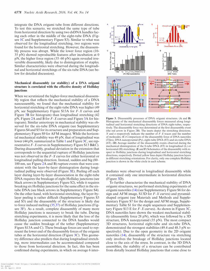
When we scrutinized the higher-force mechanical disassem-bly region that reflects the mechanical stability of a DNAnanoassembly, we found that the mechanical stability forhorizontal stretching of the eight-tube DNA was higher (49pN, see Supplementary Figure S13A for F–X curves andFigure 3B for histogram) than longitudinal stretching (42pN, Figure 2A and B for F–X curves and Figure 3A for his-togram). Similar anisotropy in mechanical stability was ob-served for the six-tube DNA origami (see SupplementaryFigures S4 and S5 for its structure and preparation and Sup-plementary Figure S9 for AFM images). While the horizon-tal mechanical stability was 49 pN, that of longitudinal waslower (44 pN, summarized in Table 1 and Figure 3C, see rep-resentative F–X curves in Supplementary Figure S13 B&C).During disassembly, gradual deviation in the extension thatcorresponds to the sequential rupture of Holliday junctionswas not observed in the F–X curves for either horizontal orlongitudinal pulling direction. Instead, sudden and big (80–100 nm, see Figure 2A and B) rupture events that were con-sistent with the layer-by-layer disintegration during longi-tudinal pulling were observed (Figure 3E). Peeling off eachlayer during layer-by-layer disassociation in the eight-tubeDNA requires the breakage of eight Holliday junctions (seeblack arrows in Supplementary Figure S2), while it requiresbreaking six Holliday junctions for the same effect in the six-tube DNA (see black arrows in Supplementary Figure S4).On the other hand, with horizontal stretching, the layer-by-layer peeling is rather difficult (Supplementary Figures S3and S5) and the disassembly of the structure is likely dueto force induced melting (35,37) of Holliday junctions (Fig-ure 3F). As a result, complete disassembly of many moreHolliday junctions is necessary to break the tube. Duringstretching experiments, it is more likely that the loss of theHolliday junction connected to the DNA handles wouldcause the sudden breakage of the tether (SupplementaryFigure S13A and C). These breakage forces are used to rep-resent the lower end of the disassembly forces of the origamitubes at the horizontal direction (Figure 3C). This geome-try difference also predicts that during longitudinal stretch-ing, more intermediates can be accommodated comparedto those from horizontal direction. In fact, this has beenconfirmed during experiments, in which on average 6 inter-
Figure 3. Disassembly processes of DNA origami structures. (A and B)Histograms of the mechanical disassembly forces measured along longi-tudinal and horizontal stretching directions of DNA eight-tubes, respec-tively. The disassembly force was determined at the first disassembly event(the red arrow in Figure 2B). The insets depict the stretching directions.N and n respectively indicate the number of F–X traces and the numberof molecules. (C) Comparison of the disassembly force of DNA nanotiles(Tiles), DNA nanopyramid (Py), eight-tube DNA (8T) and six-tube DNA(6T). (D) Average number of the disassembly events observed during themechanical disintegration of the 8-tube DNA (8T) in longitudinal (L) orhorizontal (H) stretching. (E and F) Schematics of the disassembly with re-spect to Holliday junctions during longitudinal and horizontal stretchingdirections, respectively. Dotted yellow lines depict Holliday junction layersin different stretching orientations. For clarity, only one complete Hollidayjunction is shown in the white circle in each scheme.
mediates were observed in longitudinal disassembly whileit contained only one intermediate in horizontal direction(Figure 3D).
To further characterize the mechanical stability of DNAorigami structures, we performed stretching experiments oforigami nanotiles (14) (see Supplementary Figure S6 for de-sign and AFM image, S13D for F–X curves) and pyramid-shaped origami (see Materials and Methods and Supple-mentary Figure S7 for the design and AFM image, Supple-mentary Table S1 for the staple sequences and Supplemen-tary Figure S13 E for F–X curves). As shown in Figure 3C,DNA nanotiles have shown the weakest mechanical stabil-ity (disassembly force 28 pN), which was followed by a 3Dstructure, DNA nanopyramid (35 pN). The most compact3D structures, horizontal eight-tube and six-tube DNA,demonstrated the strongest stabilities (49.4 and 48.5 pN re-spectively). Due to the open geometry in the 2D origaminanotiles (14), disassembly of the structure via stretchingonly involves the breakage of Holliday junctions locatedclose to the axis of the stress. In contrast, in the 3D DNAassemblies, the stability of a structure can be contributedfrom distally located Holliday junctions that come close to
Downloaded from https://academic.oup.com/nar/article-abstract/44/14/6574/2468220by gueston 05 April 2018
Nucleic Acids Research, 2016, Vol. 44, No. 14 6579
the stress axis through the long-range, 3D arrangement. Infact, the density of Holliday junctions along the longitu-dinal pulling for the 3D tubular structure increases signifi-cantly as the Holliday junctions in the same layer becomesmore compact after isomerization (see Supplementary Fig-ure S15). Therefore, the effective density of the Hollidayjunctions along the stretching direction becomes a decisivefactor for the mechanical stability of an origami structurealong that direction. Here, we defined the density of Holli-day junctions as the number of contributing Holliday junc-tions per nanometer of the force axis in the nanostructure.For nanotiles and nanopyramids, the effective Hollidayjunction densities are 0.17 and 0.18 HJ/nm, respectively.For the eight-tube and the six-tube DNA along the longi-tudinal pulling direction, due to the symmetric geometries,all the Holliday junctions contribute equally to the mechan-ical property of the origami. Therefore, they are all countedin the density calculation, which leads to much higher den-sities (1.43 HJ/nm for the eight-tube and 1.09 HJ/nm forthe six-tube). Corresponding to this trend, the disassem-bly forces of the isomerizable structures along the longitu-dinal direction for both six- and eight-tube DNA origamiare higher (35–60 pN) than those of the non-isomerizablestructures such as nanotiles and nanopyramids (28–40 pN).This clearly shows that the mechanical stability of DNAorigami structures may depend on the effective density ofthe Holliday junctions. Therefore, the observed data of me-chanical stability suggest that Holliday junctions serve asmechanophores (26,38,39) to impart the mechanical stabil-ity to the entire DNA origami nanoassemblies.
Holliday junctions in DNA origami tubes are responsible formechanical isomerization between nanotube isomers
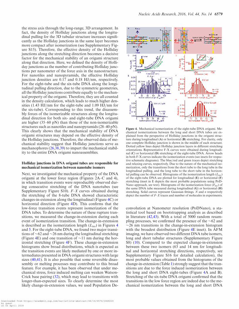
Next, we investigated the mechanical property of the DNAorigami at the lower force region (Figures 2A–C and 4),in which transition events were reproducibly observed dur-ing consecutive stretching of the DNA nanotubes (seeSupplementary Figure S10). F–X curves obtained duringthe stretching of the 8-tube DNA showed characteristicchanges-in-extension along the longitudinal (Figure 4C) orhorizontal direction (Figure 4D). This confirms that thelow-force transition events represent isomerization of theDNA tubes. To determine the nature of these rupture tran-sitions, we measured the change-in-extension during eachevent of isomerization transition. The change-in-extensionis described as the isomerization length (Liso) in Figures 4and 5. For the eight-tube DNA, we found two major transi-tions of ∼62 and ∼26 nm during the longitudinal stretching(Figure 4E) and one transition of ∼11 nm during the hor-izontal stretching (Figure 4F). These change-in-extensionhistograms show broad distributions, which is expected asthe transition events are likely mediated by one or more in-termediates presented in DNA origami structures with largesizes (40,41). It is also possible that some reversible disas-sembly or melting processes may contribute to this broadfeature. For example, it has been observed that under me-chanical stress, force induced melting can weaken Watson-Crick base pairing (32), which may lead to transitions withlonger-than-expected sizes. To clearly determine the mostlikely change-in-extension values, we used Population De-
Figure 4. Mechanical isomerization of the eight-tube DNA origami. Me-chanical isomerizations between the long and short DNA tubes are ex-plained from the perspective of Holliday junctions in the origami struc-ture during longitudinal (A) or horizontal (B) stretching. For clarity, onlyone complete Holliday junction is shown in the middle of each structure.Dotted yellow lines depict Holliday junction layers in different stretchingorientations. Representative F-X curves were obtained during longitudi-nal (C) or horizontal (D) stretching of the eight-tube DNA. Arrow headsin both F-X curves indicate the isomerization events (see insets for respec-tive schematic diagrams). The blue/red and green traces depict stretchingand relaxing curves, respectively. Due to the nature of the mechanical iso-merization, only the transitions from the short tube to the long tube in thelongitudinal pulling, and the long tube to the short tube in the horizon-tal pulling can be observed. Histograms of the isomerization length (Liso)of the eight-tube DNA are plotted for longitudinal (E) or horizontal (F)stretching (inset in E depicts the most probable populations using PoD-Nano approach, see text). Histograms of the isomerization force (Fiso) ofthe same DNA tube measured during longitudinal (G) or horizontal (H)stretching. Solid curves represent Gaussian fittings. N and n respectivelydepict the number of F–X traces and number of molecules in experiments.
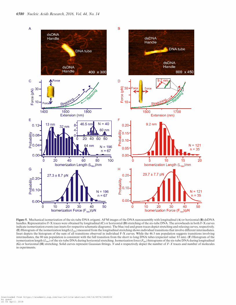
convolution at Nanometer resolution (PoDNano), a sta-tistical tool based on bootstrapping analysis as describedin literature (42,43). With a total of 5000 random resam-pling processes, we confirmed the presence of the ∼62 and∼26 nm transitions in the change-in-extension histogramwith the broadest distribution (Figure 4E inset). In AFMimaging, we have observed two different DNA tube isomers,long and short tubular structures (Supplementary FigureS8) (10). Compared to the expected change-in-extensionbetween these two isomers (63 and 14 nm for longitudi-nal and horizontal stretching directions, respectively, seeSupplementary Figure S16 for detailed calculation), themost probable values obtained from the histograms of thechange-in-extension (Table 1) strongly suggest that the tran-sitions are due to the force induced isomerization betweenthe long and short DNA eight-tubes (Figure 4A and B).Stretching of the six-tube DNA origami confirmed that thetransitions in the low force region are indeed due to the me-chanical isomerization between the long and short DNA
Downloaded from https://academic.oup.com/nar/article-abstract/44/14/6574/2468220by gueston 05 April 2018
6580 Nucleic Acids Research, 2016, Vol. 44, No. 14
Figure 5. Mechanical isomerization of the six-tube DNA origami. AFM images of the DNA nanoassembly with longitudinal (A) or horizontal (B) dsDNAhandles. Representative F-X traces were obtained by longitudinal (C) or horizontal (D) stretching of the six-tube DNA. The arrowheads in both F-X curvesindicate isomerization events (see insets for respective schematic diagrams). The blue/red and green traces depict stretching and relaxing curves, respectively.(E) Histogram of the isomerization length (Liso) measured from the longitudinal stretching shows individual transitions that involve different intermediates.Inset depicts the histogram of the sum of all transitions observed in individual F-X curves. While the 46.5 nm population suggests transitions involvingintermediates, the 80 nm population is consistent with the full transition from the short to long DNA tubes (expected value: 83 nm). (F) Histogram of theisomerization length (Liso) of the six-tube DNA during horizontal stretching. Isomerization force (Fiso) histograms of the six-tube DNA during longitudinal(G) or horizontal (H) stretching. Solid curves represent Gaussian fittings. N and n respectively depict the number of F–X traces and number of moleculesin experiments.
Downloaded from https://academic.oup.com/nar/article-abstract/44/14/6574/2468220by gueston 05 April 2018
Nucleic Acids Research, 2016, Vol. 44, No. 14 6581
tubes (80 and 9.2 nm in sum of all transition events vs ex-pected values of 83 and 12 nm for the longitudinal and hor-izontal stretching directions, respectively, see Figure 5, Sup-plementary Figures S16 and S17). It is noteworthy that mul-tiple transitions were observed in every longitudinal stretch-ing experiment. This suggests the presence of intermediatesduring isomerization. For example, a stable intermediate at∼46 nm (Figure 5E) can be attributed to a conformationtrapped during incomplete isomerization (SupplementaryFigure S18).
Interestingly, during horizontal stretching, the mechan-ical isomerization involves fewer intermediate states com-pared to the longitudinal stretching (compare between Fig-ure 4E and F; and between Figure 5E and F). This re-flects different origami arrangements along various stretch-ing orientations. During longitudinal stretching, it travelslonger (62 nm for the eight-tube and 80 nm for the six-tube)from the shorter DNA tube to the longer tube with respectto the horizontal stretching, in which the distance is muchshorter between the two isomers (11 nm for the eight-tubeand 9.2 nm for the six-tube). The extra transition distancein the former case (longitudinal) renders a more elastic en-ergetic profile that allows more intermediates to populate.In the latter case (horizontal), however, the entire processbecomes more cooperative due to the shorter and there-fore more rigid isomerization pathways that cannot hostas many intermediate populations. Such an observation isconsistent with the disassembly of the origami structuresat the high force region discussed above. While the DNAtube experiences more intermediates during the longitudi-nal, layer-by-layer disintegration, it has fewer intermediatesduring the horizontal disassembly with increased cooper-ativity (Figure 3D). Similar observations have been previ-ously obtained in the mechanical unfolding of short andlong DNA duplexes. While the short DNA duplexes such ashairpins almost do not present intermediates, (44) the saw-teeth features observed in force–extension traces demon-strate the existence of many intermediates in long DNA du-plexes (41).
During the longitudinal isomerization of the eight-tubeDNA origami, the isomerization force (Fiso = 26.4 pN, Fig-ure 4G and Table 1) is comparable to that of the horizontalFiso (27.8 pN, Figure 4H and Table 1). Similar forces be-tween the two stretching orientations have been observed inthe isomerization of the six-tube DNA origami (Fiso = 27.3pN for longitudinal and 29.7 pN for horizontal, see Figure5G and H, respectively). We reasoned that the isomerizationof the DNA nanotubes can be a result of collective isomer-ization of individual Holliday junctions at the microscopiclevel. Based on this assumption, we calculated the isomer-ization force for individual Holliday junctions as 0.12–0.14pN (see SI). This value is in agreement with the isomeriza-tion force of a single Holliday junction (0.1–0.3 pN) pre-dicted by Hohng et al. (24).
It is noteworthy that mechanical isomerization was notobserved in DNA nanotiles or nanopyramid structures.Close inspection on all three types of origami nanoassem-blies shows that the latter two structures are rather differ-ent from the tubular structures. While the DNA nanotileshave a planar geometry, nanopyramids consisting of fourtriangular tiles fold into an object in which the perimeter
of each layer gradually reduces from the base to the apexof the pyramid. In DNA nanotubes, the circular geometrywith the same tube diameter gives rise to less hierarchicalcontributions between the distal and proximal regions withrespect to the force axis, which bring a similar microenviron-ment to all Holliday junctions. However, due to the loss ofsymmetry in either DNA nanotiles or nanopyramids, indi-vidual Holliday junctions experience different environmentand therefore, behave differently. As a result, the mechanicalisomerization between two origami isomers can only be car-ried out by many identical Holliday junctions as a collectiveaction in DNA nanotubes, but not in other structures.
CONCLUSIONS
Serving as essential components inside DNA origaminanoassemblies, DNA staples offer stability to DNAnanoassemblies by forming Holliday junctions with thesingle-stranded DNA template. Using optical tweezers, wehave quantified and explained two emergent properties ofDNA nanoassemblies not seen in their individual compo-nents from mechanical perspective. First, we have found themechanical stability of DNA origami structures can be ge-ometry dependent, which is determined by the effective den-sity of Holliday junctions along a particular stretching di-rection. Second, we have quantitatively ascribed the coop-erative transition between short and long DNA nanotubesto the collective mechanical isomerizations of many indi-vidual Holliday junctions. These observations indicate thatthe Holliday junctions serve as mechanophores in DNAorigami nanoassemblies investigated here. Further testingis required to validate this point for more complex DNAnanoassemblies such as DNA helix bundles (2,21,22,45).We anticipate our new findings can provide unprecedentedguidelines to design DNA nanoassemblies with better me-chanical properties in both thermodynamic and kinetic as-pects.
SUPPLEMENTARY DATA
Supplementary Data are available at NAR Online.
FUNDING
NSF [CHE-1026532, CHE-1415883 to H.M.]; JSPS KAK-ENHI [15H03837, 24104002, 24225005, 26620133 toM.E. and H.S.]. Funding for open access charge: NSF[CHE-1026532, CHE-1415883 to H.M.]; JSPS KAKENHI[15H03837, 24104002, 24225005, 26620133 to M.E. andH.S.].Conflict of interest statement. None declared.
REFERENCES1. Rothemund,P.W.K. (2006) Folding DNA to create nanoscale shapes
and patterns. Nature, 440, 297–302.2. Douglas,S.M., Dietz,H., Liedl,T., Hogberg,B., Graf,F. and
Shih,W.M. (2009) Self-assembly of DNA into nanoscalethree-dimensional shapes. Nature, 459, 414–418.
3. Han,D., Pal,S., Nangreave,J., Deng,Z., Liu,Y. and Yan,H. (2011)DNA origami with complex curvatures in three-dimensional space.Science, 332, 342–346.
Downloaded from https://academic.oup.com/nar/article-abstract/44/14/6574/2468220by gueston 05 April 2018
6582 Nucleic Acids Research, 2016, Vol. 44, No. 14
4. Douglas,S.M., Marblestone,A.H., Teerapittayanon,S., Vazquez,A.,Church,G.M. and Shih,W.M. (2009) Rapid prototyping of 3DDNA-origami shapes with caDNAno. Nucleic Acids Res., 37,5001–5006.
5. Gerling,T., Wagenbauer,K.F., Neuner,A.M. and Dietz,H. (2015)Dynamic DNA devices and assemblies formed byshape-complementary, non–base pairing 3D components. Science,347, 1446–1452.
6. Maune,H.T., Han,S.-p., Barish,R.D., Bockrath,M., Goddard,I.I.A.,RothemundPaul,W.K. and Winfree,E. (2010) Self-assembly of carbonnanotubes into two-dimensional geometries using DNA origamitemplates. Nat. Nano, 5, 61–66.
7. Rajendran,A., Endo,M., Katsuda,Y., Hidaka,K. and Sugiyama,H.(2010) Programmed two-dimensional self-assembly of multiple DNAorigami jigsaw pieces. ACS Nano, 5, 665–671.
8. Andersen,E.S., Dong,M., Nielsen,M.M., Jahn,K., Subramani,R.,Mamdouh,W., Golas,M.M., Sander,B., Stark,H., Oliveira,C.L.P.et al. (2009) Self-assembly of a nanoscale DNA box with acontrollable lid. Nature, 459, 73–76.
9. Marras,A.E., Zhou,L., Su,H.-J. and Castro,C.E. (2015)Programmable motion of DNA origami mechanisms. Proc. Natl.Acad. Sci. U.S.A., 112, 713–718.
10. Endo,M., Yamamoto,S., Emura,T., Hidaka,K., Morone,N.,Heuser,J.E. and Sugiyama,H. (2014) Helical DNA origami tubularstructures with various sizes and arrangements. Angew. Chem. Int.Ed., 53, 7484–7490.
11. Fu,J., Yang,Y.R., Johnson-Buck,A., Liu,M., Liu,Y., Walter,N.G.,Woodbury,N.W. and Yan,H. (2014) Multi-enzyme complexes onDNA scaffolds capable of substrate channelling with an artificialswinging arm. Nat. Nano, 9, 531–536.
12. Sun,W., Boulais,E., Hakobyan,Y., Wang,W.L., Guan,A., Bathe,M.and Yin,P. (2014) Casting inorganic structures with DNA molds.Science, 346, 1258361.
13. Helmi,S., Ziegler,C., Kauert,D.J. and Seidel,R. (2014)Shape-controlled synthesis of gold nanostructures using DNAorigami molds. Nano Lett., 14, 6693–6698.
14. Koirala,D., Shrestha,P., Emura,T., Hidaka,K., Mandal,S., Endo,M.,Sugiyama,H. and Mao,H. (2014) Single-molecule mechanochemicalsensing using DNA origami nanostructures. Angew. Chem. Int. Ed.Engl., 53, 8137–8141.
15. Goodman,R.P., Heilemann,M., Doose,S., Erben,C.M.,Kapanidis,A.N. and Turberfield,A.J. (2008) Reconfigurable, braced,three-dimensional DNA nanostructures. Nat. Nano, 3, 93–96.
16. Pei,H., Liang,L., Yao,G., Li,J., Huang,Q. and Fan,C. (2012)Reconfigurable three-dimensional DNA nanostructures for theconstruction of intracellular logic sensors. Angew. Chem. Int. Ed.,124, 9154–9158.
17. Wen,Y., Liu,G., Pei,H., Li,L., Xu,Q., Liang,W., Li,Y., Xu,L., Ren,S.and Fan,C. (2013) DNA nanostructure-based ultrasensitiveelectrochemical microRNA biosensor. Methods, 64, 276–282.
18. Ge,Z., Lin,M., Wang,P., Pei,H., Yan,J., Shi,J., Huang,Q., He,D.,Fan,C. and Zuo,X. (2014) Hybridization chain reaction amplificationof microRNA detection with a tetrahedral DNA nanostructure-basedelectrochemical biosensor. Z. Anal. Chem., 86, 2124–2130.
19. Zhou,L., Marras,A.E., Su,H.-J. and Castro,C.E. (2013) DNAorigami compliant nanostructures with tunable mechanicalproperties. ACS Nano, 8, 27–34.
20. Chen,H., Weng,T.-W., Riccitelli,M.M., Cui,Y., Irudayaraj,J. andChoi,J.H. (2014) Understanding the mechanical properties of DNAorigami tiles and controlling the kinetics of their folding andunfolding reconfiguration. J. Am. Chem. Soc., 136, 6995–7005.
21. Kauert,D.J., Kurth,T., Liedl,T. and Seidel,R. (2011) Directmechanical measurements reveal the material properties ofthree-dimensional DNA origami. Nano Lett., 11, 5558–5563.
22. Pfitzner,E., Wachauf,C., Kilchherr,F., Pelz,B., Shih,W.M., Rief,M.and Dietz,H. (2013) Rigid DNA beams for high-resolutionsingle-molecule mechanics. Angew. Chem. Int. Ed., 52, 7766–7771.
23. Zhipeng,M., Young-Joo,K., Seongsu,P., Hirai,Y., Tsuchiya,T.,Kim,D.N. and Tabata,O. (2015) Nano/Micro Engineered andMolecular Systems (NEMS). IEEE, 581–584.
24. Hohng,S., Zhou,R., Nahas,M.K., Yu,J., Schulten,K., Lilley,D.M.J.and Ha,T. (2007) Fluorescence-force spectroscopy mapstwo-dimensional reaction landscape of the Holliday junction.Science, 318, 279–283.
25. Caruso,M.M., Davis,D.A., Shen,Q., Odom,S.A., Sottos,N.R.,White,S.R. and Moore,J.S. (2009) Mechanically-induced chemicalchanges in polymeric materials. Chem. Rev., 109, 5755–5798.
26. Mandal,S., Koirala,D., Selvam,S., Ghimire,C. and Mao,H. (2015) Amolecular tuning fork in single-molecule mechanochemical sensing.Angew. Chem. Int. Ed., 54, 7607–7611.
27. Moffitt,J.R., Chemla,Y.R., Smith,S.B. and Bustamante,C. (2008)Recent advances in optical tweezers. Annu. Rev. Biochem., 77,205–228.
28. Wang,M.D., Yin,H., Landick,R., Gelles,J. and Block,S.M. (1997)Stretching DNA with optical tweezers. Biophys. J., 72, 1335–1346.
29. Mehta,A.D., Rief,M., Spudich,J.A., Smith,D.A. and Simmons,R.M.(1999) Single-molecule biomechanics with optical methods. Science,283, 1689–1695.
30. Luchette,P., Abiy,N. and Mao,H. (2007) Microanalysis of cloudingprocess at the single droplet level. Sens. Actuators B: Chem., 128,154–160.
31. Mao,H. and Luchette,P. (2008) An integrated laser-tweezersinstrument for microanalysis of individual protein aggregates. Sens.Actuators B, 129, 764–771.
32. Morfill,J., Kuhner,F., Blank,K., Lugmaier,R.A., Sedlmair,J. andGaub,H.E. (2007) B-S transition in short oligonucleotides. Biophys.J., 93, 2400–2409.
33. Abraham Punnoose,J., Cui,Y., Koirala,D., Yangyuoru,P.M.,Ghimire,C., Shrestha,P. and Mao,H. (2014) Interaction ofG-quadruplexes in the full-length 3′ human telomeric overhang. J.Am. Chem. Soc., 136, 18062–18069.
34. Zhao,Y., Karypis,G. and Fayyad,U. (2005) Hierarchical clusteringalgorithms for document datasets. Data Min. Knowl. Disc., 10,141–168.
35. Williams,M.C., Rouzina,I. and Bloomfield,V.A. (2002)Thermodynamics of DNA interactions from single moleculestretching experiments. Accounts Chem. Res., 35, 159–166.
36. Amit,R., Gileadi,O. and Stavans,J. (2004) Direct observation ofRuvAB-catalyzed branch migration of single Holliday junctions.Proc. Natl. Acad. Sci. U.S.A., 101, 11605–11610.
37. Koirala,D., Yu,Z., Dhakal,S. and Mao,H. (2011) Detection of singlenucleotide polymorphism using tension-dependent stochasticbehavior of a single-molecule template. J. Am. Chem. Soc., 133,9988–9991.
38. May,P.A. and Moore,J.S. (2013) Polymer mechanochemistry:techniques to generate molecular force via elongational flows. Chem.Soc. Rev., 42, 7497–7506.
39. Brantley,J.N., Wiggins,K.M. and Bielawski,C.W. (2013) Polymermechanochemistry: the design and study of mechanophores. Polym.Int., 62, 2–12.
40. Bae,W., Kim,K., Min,D., Ryu,J.-K., Hyeon,C. and Yoon,T.-Y. (2014)Programmed folding of DNA origami structures throughsingle-molecule force control. Nat. Commun., 5, 5654.
41. Bockelmann,U., Thomen,P., Essevaz-Roulet,B., Viasnoff,V. andHeslot,F. (2002) Unzipping DNA with optical tweezers: highsequence sensitivity and force flips. Biophys. J., 82, 1537–1553.
42. Yu,Z., Gaerig,V., Cui,Y., Kang,H., Gokhale,V., Zhao,Y., Hurley,L.H.and Mao,H. (2012) Tertiary DNA structure in the single-strandedhTERT promoter fragment unfolds and refolds by parallel pathwaysvia cooperative or sequential events. J. Am. Chem. Soc., 134,5157–5164.
43. Yu,Z. and Mao,H. (2013) Non-B DNA structures show diverseconformations and complex transition kinetics comparable to RNAor proteins | a perspective from mechanical unfolding and refoldingexperiments. Chem. Rec., 13, 102–116.
44. Woodside,M.T., Anthony,P.C., Behnke-Parks,W.M., Larizadeh,K.,Herschlag,D. and Block,S.M. (2006) Direct measurement of the full,sequence-dependent folding landscape of a nucleic acid. Science, 314,1001–1004.
45. Mathieu,F., Liao,S., Kopatsch,J., Wang,T., Mao,C. and Seeman,N.C.(2005) Six-helix bundles designed from DNA. Nano Lett., 5, 661–665.
Downloaded from https://academic.oup.com/nar/article-abstract/44/14/6574/2468220by gueston 05 April 2018