m.d. degree examination branch ii – obstetrics
TRANSCRIPT

DISSERTATION ON VISUAL INSPECTION OF CERVIX AFTER APPLICATION OF ACETIC ACID AND LUGOL’S IODINE IN CERVICAL CANCER
SCREENING
M.D. DEGREE EXAMINATION BRANCH II – OBSTETRICS & GYNAECOLOGY
Stanley Medical CollegeChennai
Dissertation Submitted toTHE TAMIL NADU Dr. M.G.R. MEDICAL UNIVERSITY
CHENNAI
MARCH 2009

CERTIFICATE
This is to certify that this dissertation entitled "VISUAL INSPECTION OF
CERVIX AFTER APPLICATION OF ACETIC ACID AND LUGOL’S
IODINE IN CERVICAL CANCER SCREENING" is a bonafide original work
of Dr.K.S.SRI DEEPA Post Graduate Student (2006-2009) in the department of
Obstetrics and Gynaecology, Stanley Medical College, Chennai in partial
fulfilment of the regulations laid down by the Tamil Nadu Dr.M.G.R.Medical
University, Chennai for M.D. (Branch II) Obstetrics and Gynaecology examination
held in March 2009.
Dr. A.MOHANASUNDHARAMM.D., D.N.B., Ph.D.
DeanStanley Medical College, Chennai – 600 001.
Prof. M.MOHANAMMAL,M.D., D.G.O.,
Superintendent,Department of Obstetrics and
Gynaecology,Govt. RSRM Lying in Hospital,Govt. Stanley Medical College,Chennai – 600 013.

ACKNOWLEDGEMENT
I express my sincere thanks to the Dean, Stanley Medical College, Chennai,
Dr.MOHANASUNDARAM, M.D., D.N.B., Ph.D., for permitting me to utilize all the resources at
this hospital.
I express my sincere and heartfelt gratitude to Prof. Dr.M.MOHANAMBAL, M.D., D.G.O.,
Superintendent, Government RSRM Hospital, for her encouragement, support and guidance during my
study.
I also express my heartfelt deep sense of gratitude to Prof.Dr.AMRITA PRICILLA NALINI,
M.D., D.G.O., Deputy Superintendent, Government RSRM Hospital, for her constant encouragement,
support, valuable comments and timely suggestions to shape this work.
I am extremely grateful to Prof.Dr.P.SASIREKHA, M.D., D.G.O., Additional Professor of
Obstetrics & Gynaecology, Government RSRM Hospital, for her encouragement, support and guidance
during my study.
I am extremely grateful to Prof.Dr.C.R.ANURADHA, M.D., D.G.O., Additional Professor of
Obstetrics & Gynaecology, Government RSRM Hospital, for her constant encouragement, support,
valuable comments and timely suggestions to shape this work.
I express my sincere and heartfelt gratitude to Prof.Dr.C.K.RAJINI, M.D., D.G.O., Additional
Professor of Obstetrics & Gynaecology, Government RSRM Hospital, for her guidance and valuable
support.
I express my sincere and heartfelt gratitude to Prof.N.HEPHZIBAH KIRUBAMANI, M.D.,
D.G.O., Ph.D., RMO Government RSRM Hospital for her guidance and valuable support throughout
the course and the present study.
I express my sincere thanks to Prof.Dr.A.SUNDARAM, M.D. Pathology., Professor and HOD

of Department of Pathology, Stanley Medical College for his valuable support to this study.
I am extremely grateful to Dr. D.M. CHRISTE, Research Medical Officer in ICMR,
Government RSRM Hospital, for her constant encouragement, support and suggestions to shape this
work.
Above all, I thank all my patients without them the study would not have been possible.

CONTENTS
S.NO. TITLE PAGE NO.
1. INTRODUCTION 1
2. AIM OF THE STUDY 3
3. MATERIALS AND METHODS 4
4. REVIEW OF LITERATURE 13
5. RESULTS 47
6. DISCUSSION 60
7. SUMMARY 70
8. CONCLUSION 71
9. BIBLIOGRAPHY
10. PROFORMA
11. MASTER CHART
INTRODUCTION
Worldwide, cervical cancer comprises 12% of all cancers in women and 2,31,000 women die of
cervical cancer every year, over 80% of whom live in developing countries. South east Asia contributes
about 25% of the total disease burden.
Cervical cancer is the most common genital tract cancer in Indian women with 1,26,000 new
cases and 70,000 deaths each year. Incidence is higher than in Eastern Asia. Across India cervical
cancer is the commonest cancer reported from all cancer registries except those in Mumbai & Delhi
where breast cancer is the commonest.
Cervical cancer is a preventable disease because of its long preinvasive state of over 10 to 15
years, availability of various screening programmes, effective treatment for preinvasive lesions. World
wide, successful cervical cancer prevention is based on an organized screening program. Cervical
cytology is presently considered to be the only test known to reduce cervical cancer incidence in
organized screening programs. The goal of periodic cervical cancer screening is to detect the
preinvasive state of the disease and treat it appropriately before it progresses to cervical cancer. In
developed countries initiation and sustenance of cervical cytology programs, involving screening of
sexually active women yearly or once every 2-5 years have resulted in a large decline in cervical cancer
incidence, mortality and morbidity there by saving women’s life. However an organized screening
program is difficult to implement in developing countries where resources are scarce.
Community based screening programs require a relatively sophisticated infrastructure,
including highly trained personnel, adequately equipped laboratories and good referral systems to
communicate the results of the test to the women. In view of these requirements, an alternative
screening methods like VIA,VIAM,VILI is needed in developing countries with very limited resources
and infrastructure because it is inexpensive, requires supplies usually locally obtainable and can be
competently performed by non physician.

Prior to the use of pap smear and cytology based screening programs, health care providers
relied on looking at the cervix to detect abnormalities. After 1950, cytology became the standard for
cervical screening and the colposcope was used to further investigate the cervix. Unaided visual
inspection (down staging) was evaluated by three cross sectional studies in India. Though the accuracy
of down staging alone in the early detection of cervical carcinoma and precursor lesions was found to
be inadequate, a combination of downstaging, VIA, VILI, VIAM as a cancer screening tool was very
effective.
Pap smear, colposcopy and cervix biopsy are the other methods by which the cervix can be
studied for the evidence of early malignant disease. These are out patient procedure and requires no
anaesthesia.
The accuracy of detection and diagnosis may be increased by a systematic combination of the
above screening procedures.

AIM OF THE STUDY
1. To do cervical cancer screening by VIA and VILI.
2. To determine the sensitivity and specificity of VIA, VILI.
3. To assess the reliability of VIA and VILI as a cancer screening tool in the
detection of precancerous lesions of cervix by comparing its sensitivity and
specificity with pap smear keeping colposcopy and colposcopy directed
biopsy as reference standard.
4. To study the efficacy of combined screening programs.
8
MATERIALS AND METHODS
500 patients were selected from the Gynaecology OPD of Government R.S.R.M.
lying in Hospital Chennai – 13, considering the following criteria.
INCLUSION CRITERIA
1. Women with history of sexual activity for more than three years with
Intact uterus and cervix.
2. Non pregnant.
3. No past / present history suggestive of CIN / Cancer cervix.
4. No bleeding P/V at the time of examination.
5. Not had any treatment for cervical lesion (like Cryo, cautery,laser
etc.)
EXCLUSION CRITERIA
1. Unmarried woman not exposed to sexual activity.
2. Pregnant women.
3. Women who have had therapy for cervical lesion.
4. Patient in periods / bleeding PV.
5. Previous colposcopy done.
6. Prior hysterectomy.
7. Obvious growth on cervix.
9

All 500 women were subjected to a questionnaire addressing clinical and
epidemiological risk factors of cervical disease (eg. Socio economic class, age of
marriage, parity, birth spacing, occupation, travelling jobs etc.). Then all women were
subjected to down staging, pap smear, VIA,VILI and colposcopy. The decision to take a
histological specimen was based upon the abnormal colposcopic findings and by grading
(combined colposcopic index). Normal cervix by colposcopy was accepted as truly
normal cervix.
DOWN STAGING
Materials - Sim’s Speculum
Light Source
Sterile gloves
Examination table.
Patient was put in lithotomy position, speculum examination preceded bimanual
pelvic examination in all cases to prevent.
1. Removal of desquamated epithelium from the surface of cervix.
2. The lubricant may disturb the discharge obtained for bacteriological and
cytological study.
3. A bleed from surface lesion may prevent inspection.
10

PAP SMEAR
Materials
Cusco’s bivalved self retaining speculum
Nulliparous - 28 mm
Postmenopause - 28 mm
Multiparous - 36 mm
Light Source
Ayre’s spatula
Endo cervical brush
Glass slide
Marker Pencil
Sterile glove
Fixative 95% ethanol / cytofix spray
SAMPLING AND PREPARATION METHODS
Patient Instructions
1. Schedule the examination, two weeks after the first day of last menstrual period – more
specifically it is preferable to avoid examination during menses because blood may
obscure significant findings.
2. Do not use vaginal medication, vaginal contraceptives or douches for 48 hours before
the appointment.
3. Intercourse is not recommended the night before the appointment.
11

SPECIMEN COLLECTION
1. Specimen should be obtained after a non-lubricated speculum (moistened only with
warm water if needed) is inserted, prior to vaginal examination to prevent removal of
desquamated cervical cells thereby preventing false negative reports
2. Excess mucus or other discharge should be removed gently with ring forceps holding a
folded gauze pad.
3. Sample should be obtained before the application of acetic acid or Lugol’s iodine.
4. An optimal sample includes cells from the ectocervix and endocervix.
Patient was put in lithotomy position and a suitable sized Cusco’s speculum was
introduced without lubricant. The cervix was visualized with good light source. The
cervical smear was taken with an Ayre’s spatula rotating it through 360 degrees over the
squamo columar junction. Sample from the endocervix was taken using endocervical
brush rotated gently only one quarter turn. The smear should be applied and fixed over
the slide marked with the pap smear number for that patient. Immediate fixation (within
seconds) is critical in order to prevent air drying artifact which distorts the cells and
hinders interpretation. It was then subjected to modified papanicolaou staining in the
laboratory and studied. The fixed slides are transferred directly from the fixative into the
following solutions:
1. 80% ethyl alcohol - 10 dips
2. 70% ethyl alcohol - 10 dips
3. 50% ethyl alcohol - 10 dips
4. Distilled water - 10dips
5. Harris haematoxylin - 3mts.
12

6. Running tap water - 1mt.
7. HCl (0.5%) - 5 dips
8. Again running tap water - 1dip
9. 50% ethyl alcohol - 10dips
10. 70% ethyl alcohol - 10dips
11. 80% ethyl alcohol - 10dips
12. 95% ethyl alcohol - 10dips
13. Orange G6 - 1mt.
14. 90% ethyl alcohol - 10dips
15. G.A 36 - 4mts.
16. 95% ethyl alcohol - 10dips
17. Absolute alcohol - 10dips
18. Xylene - 3dips
19. Clear in Xylol - 3dips
Slides are mounted with DPX.
Results: Nucleus – Blue colour
Cytoplasm of superficial cells – pink
Cytoplasm of intermediate cells – Bluish green
13

VISUAL INSPECTION WITH ACETIC ACID AND LUGOL’S IODINE
Materials
Private examination room
Examination table with stirrups
Good light source (100 Watts lamp)
Sterile cusco’s speculum
Pair of gloves
Cotton swabs
Ring forceps
Plastic bucket with plastic bag
Preparation of 5% freshly prepared Acetic acid solution
5ml glacial acetic acid + 95ml distilled water.
Preparation of Lugol’s Iodine
6gm potassium iodide + 100ml distilled water + 4gms of Iodine crystals.
Get informed consent about the procedure. Reassure patient that the procedure is
painless. Ensure that patient is fully relaxed. Put the patient in modified lithotomy
position. Introduce and fix unlubricated bivalved cusco’s speculum under good light
source. Down staging of cervix was done. Conventional pap smear was taken using
Ayre’s spatula and endocervical brush. Wash away excess mucus with saline soaked
swab. Apply 3-5 % acetic acid on the cervix with cotton tipped applicator. Read after
14
1minute. Followed by the application of Lugol’s Iodine.
Aceto whitening + - VIA Positive
No Aceto whitening - VIA Negative
Mahogany brown or black - VILI Negative
Mustard yellow or saffron coloured - VILI Positive
COLPOSCOPY
Materials
Colposcope
Bivalved Cusco’s speculum
Cotton tipped swabs
Sterile glove
Normal saline
3% acetic acid
Lugol’s Iodine
Examination table
With the patient in lithotomy position cervix exposed with bivalved Cusco’s
speculum and colposcope focused on external os at a distance of 20 cms. Cervix and
vagina are gently cleaned with saline to remove mucus taking care not to provoke
bleeding. Cervix inspected for lesions like leukoplakia , viral condylomata and
carcinoma. Then a solution of 3-5 % acetic acid was applied over the cervix gently and
liberally. The solution is mucolytic changes the colour and vascular pattern after an
15

interval of 10-30 seconds. Cervix inspected for colour, surface, columnar epithelium,
transformation zone, squamo columnar junction and vascular pattern. The vascular
pattern was again studied using a green filter. Schiller’s Iodine test was done for patient
with suspicious lesions.
The colposcopy findings were reported based on the terminology of 4th
International Congress of cervical pathology and colposcopy in London 1981 and grading
of atypical colposcopy appearances were done.
The special symbols for the different colposcopical patterns are used by the
colposcopists to document the colposcopic findings which imitate as closely as possible
the picture observed in the colposcope.
The two recording systems in Vogue are:
1. Odell diagram - colposcopic lesions may be represented in a circular diagram in
relation to the OS.
2. Modified Hammond’s graph of cervix.
It consists of 3 concentric circles with 12 radial lines in clockwise fashion. The
innermost represents endocervix, intermediate one is the transformation zone and the
outermost is the ectocervix. In this graph the colposcopic findings can be recorded
accordingly. Exact location of the specific lesions can thus be documented.
16

Biopsy Cervix
Materials
Sterile glove
Vulsellum
Sim’s Speculum
Light source
Tischler biopsy forceps
Container
10% formalin
The management of abnormal lesion was finally dependent upon the
histopathological diagnosis. Biopsies were taken from the iodine negative areas or areas
of atypical colposcopic findings.
Techniques
1. Punch biopsy
2. Excision biopsy
3. Wedge biopsy
4. Diathermy loop biopsy
5. Curettage
6. Conization
The specimens were put in 10% formalin and sent to pathology lab, where
paraffin block of tissue were made, sectioned, stained with eosin, haematoxylin and
examined under microscope for evidence of dysplasia or malignancy.
17

REVIEW OF LITERATURE
In 1851, Robert Hull marvelled that with the introduction of the vaginal speculum
a veritable epidemic of uterine disease had appeared.
The existence of a preinvasive stage in the development of cervical cancer has
been known since Sir John Williams in the Harverian lectures in 1886 presented a use of
symptomless cancer cervix which is now known as carcinoma in situ.
In 1910 Rubin described non-invasive change at the margins of invasive
carcinoma then came the word carcinoma in situ.
Hinselmann from Germany first published an account of colposcopy in 1925. It
has become possible to observe cancer at its very earliest stage, by finding changes in the
cervix which are invisible to the naked eye. He also combined colposcopy with acetic
acid application.
The Schillers test invented in 1928 because of its simplicity has been in
widespread use to distinguish between normal and abnormal epithelium of portio
vaginalis of the cervix.
Despite the introduction of colposcope and Schiller’s iodine test, it was not until
Papanicolaou and Traut described a simple technique of cytology in 1945. The Clinicians
recognized that at last they have an effective and simple way of detecting premalignant
lesions of cervix.
The concept of preinvasive disease of cervix was introduced in 1947. Boyes and
Worth (1979) declared that introduction of cytologic screening for cancer cervix in
developed countries has resulted in considerable reduction in morbidity and mortality
from the disease, when compared to developing countries like India that don’t have mass
18

cytologic screening. In this context down staging was introduced there by emphasizing
the value of speculum examination than no screening at all.
Frisch LE et al (1995) showed that combination of cytologic screening and naked
– eye inspection of the cervix (NIC) increased the screening yield as compared with a
Pap smear alone but with some loss of positive predictive value. NIC significantly
improved the predictive value of negative cytologic screening results (1).
Belinson JL et al (2002) showed that the sensitivity of visual inspection equaled
or exceeded reported rates for conventional cervical cytology. Visual inspection and
colposcopy have similar specificity profiles for CIN II and greater (2).
Mandelblatt JS et al (2002) concluded that well organized screening programs can
reduce the cervical cancer mortality in less-developed countries at low costs (3).
Tayyeb R et al (2003) concluded that higher sensitivity, accuracy, low cost, easy
applicability and immediate results make VIA, a useful screening test in developing
countries as compared to pap smear (4).
Basu PS et al (2003) by conducting a study in kolkata concluded that VIA and
VIAM had significantly higher sensitivity than cytology but the specificity of cytology
was higher than that of VIA and VIAM (5).
Ferreccio C et al (2003) showed that as a single test, either liquid – based
cytology or HPV DNA testing was significantly more accurate than conventional
cytology or cervicography. Paired tests incorporating either liquid – based cytology or
HPV DNA testing were not substantially more accurate than either of those two test
strategies alone. However a possibly useful synergy was observed between the
conventional smear and cervicography (6).
19

Sankaranarayanan R et al (2003) showed that VIA and VILI are suitable atternate
screening tests to cytology for detecting cervical neoplasia in low resource settings (7).
Bhatla N et al (2004) showed that visual inspection can be performed reliably by
trained paramedical workers and doctors and is an effective screening options in low
resource settings (8).
Denny L et al (2004) concluded that DVI is a low cost, simple primary screening
test in low resource settings (9).
Sankaranarayanan R et al (2004) by conducting cross – sectional studies in
Mumbai and Kolkata concluded that low level magnification (2-4 x) did not improve the
test performance of naked eye visualization of acetic acid impregnated uterine cervix
(10).
Ghaemmaghami F et al (2004) concluded that the sensitivity and specificity of
VIA is high and comparable with that of cytology. Hence VIA can be undertaken as a
feasible method of screening in cervical cancer in countries where access to
cytopathology is limited (11).
Sankaranarayanan R et al (2004) showed that VILI had a significantly higher
sensitivity than VIA in detecting HSIL, but specificity was similar. VILI appears to be a
more accurate visual test for use in screening and treatment programs in low – resource
settings (12).
Winkler JL et al (2005) conducted a study in Rural Mexico and showed that i)
VIAM is more sensitive but less specific than VIA.
ii) Training of clinical personnel in visual inspection is critical to improve
the effectiveness of these screening methods (13).
20
Derchain SF et al (2005) concluded that VIA and Hybrid capture II (HC II)
contributed to the screening of cervical neoplasia in a group of Brazilian women, but the
cost effectiveness of conjoint screening modalities is still debatable (14).
De Vuyst H et al (2005) showed that the pap smear had the highest specificity
(94.6%) and HPV testing the highest sensitivity (94.4%). The visual methods, VIA and
cervicography, were similar and showed an accuracy in between the former two tests
(15).
Wu S et al (2005) showed that DNA hybridization assay (HPV) is the best choice
in primary screening if available. Screening should begin at the age of 20 years (16).
Shastri SS et al (2005) showed that visual tests are promising in low – resource
settings like India. The use of both VIA and VILI may be considered where good quality
cytology or HPV testing are not feasible. The sensitivity of cytology and HPV testing
increased significantly when combined with VIA and VILI (17).
LAMS (Latin American Screening Study) (2005) compared PAP smear, Aided
visual inspection, Colposcopy, Cervicography and HPV testing as an optional screening
tests in Latin America. This study concluded that variation in cervical cancer incidence is
due to i) different natural history of the precursor lesions, or ii) Due to different
levels of exposure to the known risk factors (18).
Valdespino VM et al (2006) showed the papanicolaou test is the best method of
screening in high resource settings. Visual inspection using cervical dyes is more useful
method in low resource settings. The challenge for the future is more dependent on local
finances and screening policies (19).
Elit L et al (2006) concluded that VIA has an acceptable test parameter for
21

population based – cervical screening in Mongolia compared to cervical cytology (20).
In study by Denny L et al (2006) organized and quality assured cytology – based
screening programs have substantially reduced cervical cancer incidence in many
developed countries. However, there are considerable barriers to setting up cytology –
based screening programs, particularly in developing countries. This has stimulated the
search for novel and alternate approaches to cytology for cervical cancer prevention.
These approaches generally perform as well as cytology, but have lower specificity
resulting in higher false positive rates (21).
Sangwa – Lugoma G et al (2006) showed that VIA and VILI, performed by
nurses and physicians are slightly more sensitive but less specific than Pap cytology.
Given their lower cost and easy deployment, visual inspection methods merit further
assessment as cervical cancer screening methods for low resource countries (22).
Sodhani P et al (2006) concluded that VIA, VIAM can be used as a mass
screening tool for cervical cancer in resource poor settings due to greater sensitivity (23).
Muwonge R et al (2007), studied the gain in diagnostic performance when two
visual inspection methods were combined and showed that settings already using VIA
would advocate combined testing and for settings using VILI to opt for the single test due
to greater sensitivity of VILI (91.5%) alone and combined testing (92.9%) compared to
VIA alone (81.3%) (24).
Arbyn M et al (2008) showed that liquid based cervical cytology is neither more
sensitive nor more specific for detection of high grade CIN compared with the
conventional pap test (25).
Dhaubhadel P et al (2008) showed that VIA as a screening test for cervical
22

neoplasia did not miss any lesion detected by pap smear and confirmed by cervical
biopsy (26).
El – Shalakany AH et al (2008) showed that VILI in feasible, easy to perform
with superior sensitivity to cervical cytology and VIA in detecting cervical premalignant
and malignant lesions. VILI can be used as an efficient primary screening tool with a
satisfactory low biopsy rate in low resource setting (27).
Jun JK et al (2008) showed that women with a normal or benign pap smear had a
statistically significantly lower risk of invasive cervical cancer and CIS of cervix
compared with those never screened and also that regular screening of cervical cancer
reduces invasive cervical cancer incidence and CIS of the cervix among Korean women
(28).
Davis – Dao CA et al (2008) showed that women with cervicitis were twice as
likely to have a positive VIA result as women without cervicitis. Presence of cervicitis
may influence the accuracy of results obtained from colposcopy and VIA (29).
In study by Arbyn M et al (2008) five screening methods VIA, VILI, VIAM, Pap
smear, HPV testing with the high risk probe of Hybrid capture -2 assay, were evaluated
in 11 studies in India and Africa in women aged 25 -64 years. Verification was based on
colposcopy and colposcopy directed biopsy. Negative colposcopy was accepted as truly
negative outcome (30). In this study
VIA showed sensitivity of 79% and 83% specificity of 85% and 84% for CIN 2 +
and CIN 3+ lesions respectively.
VILI – 10% more sensitive and equally effective.
VIAM – Similar results as VIA.
23

Pap smear showed lowest sensitivity 57% with high specificity 93% for CIN 2
lesion.
HC-2 showed sensitivity of 62% and specificity of 94% for CIN II lesion.
In study by Sankaranarayanan R et al (2008) cryotherapy and large loop excision
of the transformation zone are effective and safe treatment methods for cervical
intraepithelial neoplasia. The clinical stage of cancer is the single most important
prognostic factor and should be carefully evaluated in choosing optimal treatment
between surgery and radiotherapy, with or without chemotherapy (31).
24

HISTOLOGY OF VAGINA AND CERVIX
The wall of vagina is made up of outer fibrous layer in criss cross special fashion
and subepithelial connective tissue (elastic) on which the stratified squamous epithelium
rest. The epithelium also covers that part of the cervix which projects into the vagina
known as ectocervix. The squamous epithelium in a sexually matured woman has four
layer of cells.
Basal
Parabasal
Intermediate
Superficial
EPITHELIUM OF CERVIX
Endocervix – the epithelium is tall columnar type with basal solid nucleus. They
are thrown into folds and ciliated by but not in the crypts and glands. Beneath this layer
are cubical basal or the reserve cells from which new surface cells are believed to
develop and which undergo squamous metaplasia.
Ectocervix – Epithelium of portio vaginalis is stratified squamous epithelium
although sub-epithelial papillae are less marked and may be absent.
Squamo-columnar Junction - The point were the stratified squamous epithelium
meets the glandular epithelium is termed as the squamo-columnar junction. It’s normally
situated at the external OS. There is abrupt change in cellular type here.
Transformation Zone – Extends from original squamo columnar junction to active
squamo columnar junction
25
ANATOMIC ZONE
1. Portio Vaginalis which lies beyond the external os lined by stratified squamous
epithelium.
2. Endocervix bounded by Internal os and External os.
HISTOLOGICAL ZONE
Histological Portio
Cervical stroma without glands lined by squamous epithelium.
Transitional Zone
Originally covered by columnar epithelium gradually transformed to squamous
epithelium.
Endocervix
Endocervix consists of stroma, glandular epithelium and columnar cells.
HISTOGENESIS OF TRANSITIONAL ZONE
SITES
Basal cells of Portio epithelium
Basal cells of Portio epithelium at the margin of old erosion
Basal cells of squamous epithelium
Sub cylindrical cells of endocervix adjacent to histological portio.
26

STAFL MATTINGLY REF AMERICAN JOURNAL OF OBST AND GYN 1974
Pathogenesis of Cancer Cervix
Columnar epithelium
Exposed to Vaginal environment (Low ph of vagina)
Early metaplasia
Physiological Atypical
metaplasia metaplasia
Normal Abnormal
Transformation Transformation Zone
Zone
Adequate Inadequate
host response host response
Well
differentiated No progression Dysplasia
squamous epithelium Carcinoma in situ
Invasive carcinoma
27

GOVAN’S CLASSIFICATION OF CERVICAL LESIONS
BENIGN
Squamous hyperplasia
Reactive hyperplasia
Basal cell hyperplasia
Reserve cell proliferation
Metaplaia
1. Complete
2. Incomplete
MALIGNANT
Cervical intra epithelial neoplasia
Mild dysplasia
Moderate dysplasia
Severe dysplasia
Micro invasive carcinoma
Invasive carcinoma
METHODS OF EARLY DIAGNOSIS OF CERVICAL LESIONS
DOWN STAGING IN MASS SCREENING
Naked eye visualization of the cervix without acetic acid nor magnification to
identify early stages of cancer. Also known as unaided visual inspection.
28

In visual Inspection of cervix following are noted :
Colour, surface, contour of cervix
Healthy / Unhealthy cervix
Cervical polyp
Ectropion
Leukoplakia / condyloma
Cervical warts
Nabothian follicle
Old scars
Bleeds on touch, stippled cervix
Hypertrophied elongated irregular edematous cervix
Hard indurated cervix
Bleeding cervical erosion
Suspected growth / ulcer
Rural health workers are taught to conduct speculum examination for all women
at reproductive age group irrespective of their complaints.
DRAW BACK OF DOWN STAGGING
Lesions are not detected early enough to prevent invasion, because a large
proportion of the cancers detected are relatively advanced, requiring complex medical
therapy that is difficult to provide in many settings.
29
VISUAL INSPECTION WITH ACETIC ACID (VIA)
VIA was first described by ottaviano and La Torre in 1981
Naked eye visualization of uterine cervix without magnification after application
of diluted 3-5 % acetic acid solution to screen for cervical abnormalities. Also known as
Cervicoscopy or Direct visual inspection.
MAGNIFIED VISUAL INSPECTION AFTER APPLICATION OF ACETIC
ACID (VIAM)
Visualization of uterine cervix using low power maginification after application of
3-5% acetic acid is known as VIAM. Also known as Gynoscopy or Aided visual
inspection.
VISUAL INSPECTION WITH LUGOL’S IODINE (VILI)
Visualization of uterine cervix after application of Lugol’s iodine
MOLECULAR BASIS OF VIA
Acetic acid causes dehydration of cells and surface coagulation of cellular
proteins thereby reducing the transparency of cervical epithelium. These changes are
more pronounced in abnormal epithelium due to greater nuclear density and consequent
higher concentration of proteins enabling much earlier detection and treatment of pre
cancerous lesions.
30
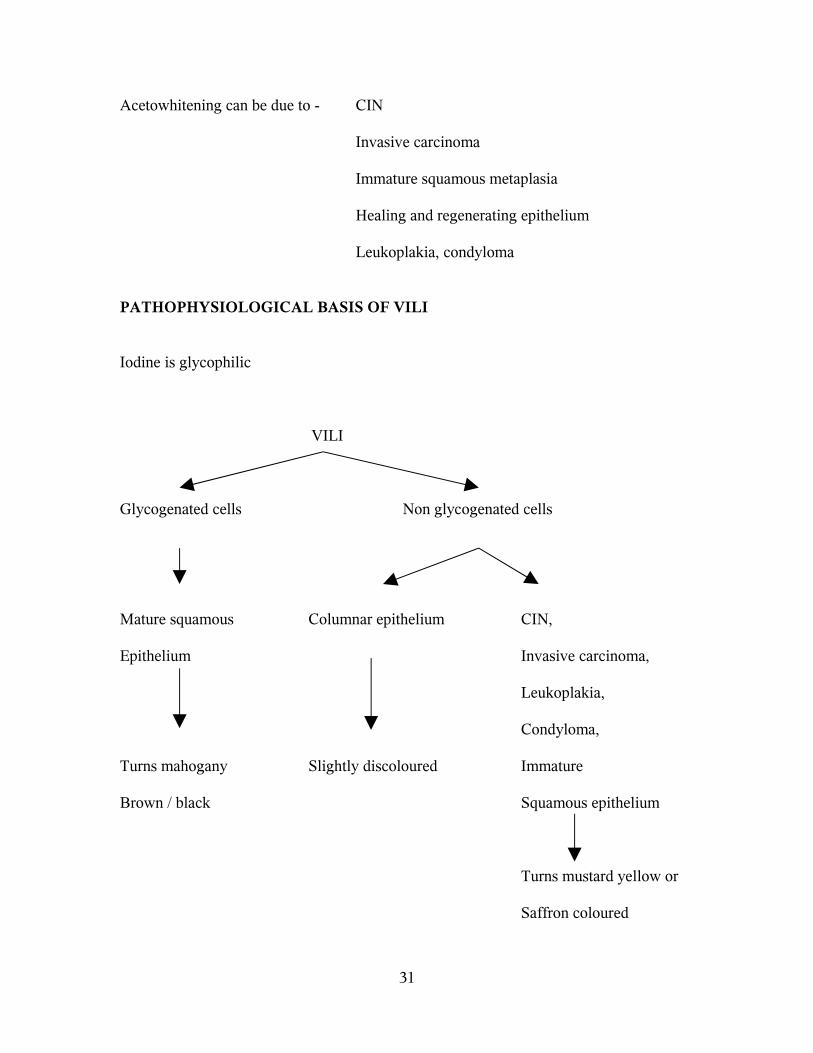
Acetowhitening can be due to - CIN
Invasive carcinoma
Immature squamous metaplasia
Healing and regenerating epithelium
Leukoplakia, condyloma
PATHOPHYSIOLOGICAL BASIS OF VILI
Iodine is glycophilic
VILI
Glycogenated cells Non glycogenated cells
Mature squamous Columnar epithelium CIN,
Epithelium Invasive carcinoma,
Leukoplakia,
Condyloma,
Turns mahogany Slightly discoloured Immature
Brown / black Squamous epithelium
Turns mustard yellow or
Saffron coloured
31
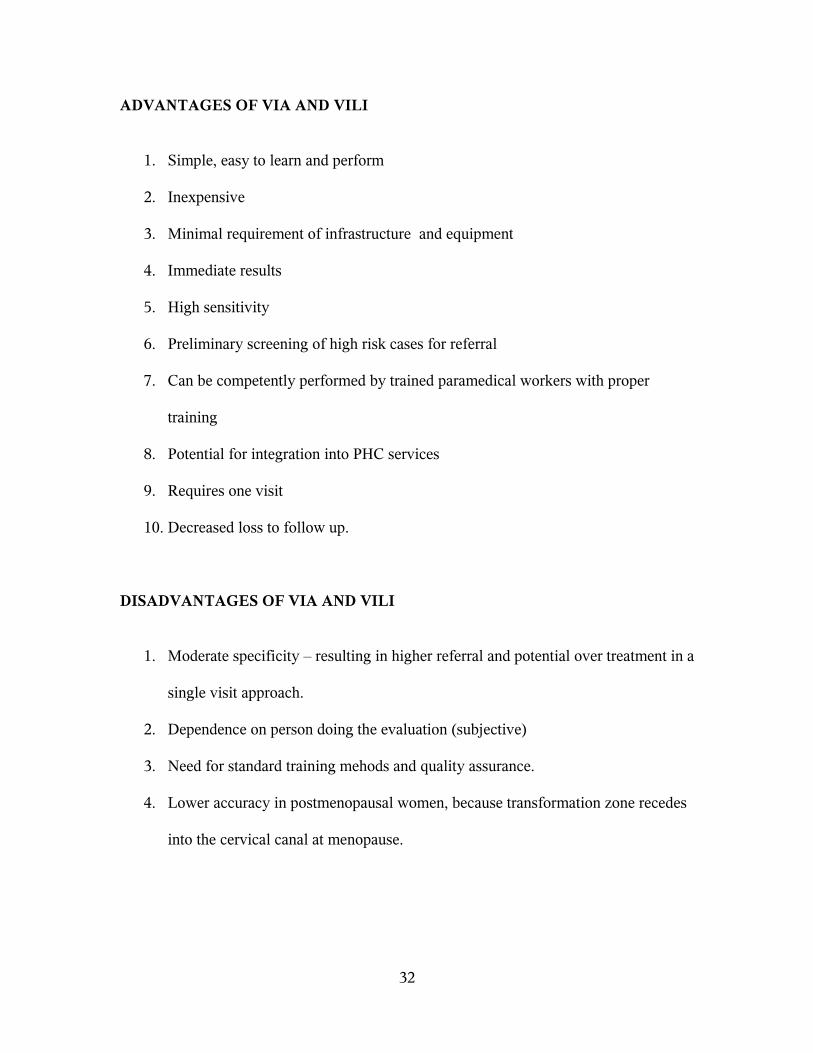
ADVANTAGES OF VIA AND VILI
1. Simple, easy to learn and perform
2. Inexpensive
3. Minimal requirement of infrastructure and equipment
4. Immediate results
5. High sensitivity
6. Preliminary screening of high risk cases for referral
7. Can be competently performed by trained paramedical workers with proper
training
8. Potential for integration into PHC services
9. Requires one visit
10. Decreased loss to follow up.
DISADVANTAGES OF VIA AND VILI
1. Moderate specificity – resulting in higher referral and potential over treatment in a
single visit approach.
2. Dependence on person doing the evaluation (subjective)
3. Need for standard training mehods and quality assurance.
4. Lower accuracy in postmenopausal women, because transformation zone recedes
into the cervical canal at menopause.
32

IARC CRITERIA FOR INTERPRETATION OF VIA AND VILI
VIA Positive
Well defined, sharp, distinct, dense acetowhite areas with or without
raised margins, abutting the squamo-columnar junction in the
transformation zone
Strikingly dense acetowhite area in the columnar epithelium
Condyloma and leukoplakia occurring closer to the squamo-columnar
junction turning intensely white after application of acetic acid
VIA Negative
No acetowhite lesions on the cervix
Polyps protruding from the cervix with bluish-white acetowhite
areas
Nabothian cysts appearing as button-like areas / whitish acne, or pimples
Faint line-like or ill-defined acetowhitening at squamocolumnar junction
Shiny, pinkish-white, cloudy-white, faint patchy, or doubtful lesions with
ill-defined, indefinite margins, blending with the rest of the cervix
Angular, irregular, digitating, acetowhite lesions resembling geographical
regions far away from the transformation zone (satellite lesions)
Ill – defined, patchy, pale acetowhite areas in the inflamed, unhealthy,
ulcerated cervix with bleeding and mucopurulent discharge
Streak –like acetowhitening in the columnar epithelium
Red spots on cervix against pinkish white background after applying
33

acetic acid
Dot-like areas in the endocervix, which are due to grape-like columnar
epithelium staining with acetic acid
VILI Positive
Dense, thick, bright, mustard-yellow or saffron yellow iodine non- uptake
areas abutting the squamo-columnar junction in the transformation zone
VILI Negative
Normal cervix where squamous epithelium turns mahogany brown or
black and the columnar epithelium does not change colour; no yellow
areas seen;
In ectropion, when an extensive area of columnar epithelium with regular
margins on the ectocervix remains without colour change
Patchy, indistinct, ill-defined, colourless or partially brown areas are seen
in the cervix
Non- or partial-iodine uptake, pale areas corresponding to pre-existing
nabothian follicles and /or polyps are seen
Stippling or leopard skin appearance associated with T. vaginalis infection
When pepper-like non-iodine uptake areas seen in the squamous
epithelium far away from the squamo-columnar junction
When satellite, thin, yellow, non-iodine uptake areas with angular, or
digitating margins resembling geographical areas are seen far away from
34
the squamo-columnar junction.
OTHER VISUAL INSPECTION APPROACHES
CERVICOGRAPHY
Cervicography entails photographing the cervix after application of 3-5% diluted
acetic acid, with a uniquely designed camera. The photographs called Cervigrams, are
viewed as projected slides by colposcopists trained in their interpretation. It is useful
when a colposcopist is not available for spot evaluation.
SPECULOSCOPY
Speculoscopy is a technique where acetic acid is applied to the cervix, but a
chemical or chemiluminescent light-source and magnifying lens are used to visualize the
acetowhite lesions of the cervix.
35

PAP SMEAR (SURFACE BIOPSY)
Dr. George N. Papanicolaou in 1928 reported malignant cells from the cervix in
vaginal smear taken from vagina using pipette (“Exfoliative cervical cytology”).
Dr. Herbert Trait (Gynaecologist) helped Papanicolaou by providing clinical
samples.
Dr. J. Ernest Ayre (Canadian Gynaecologist) took smear from the cervix using
wooden spatula.
Pap is the screening test that detects 98% of cancer cervix and 70% endometrial
cancer (shaw text book).
False positive result is reported in the presence of infection.
Types of Pap Smear
1. Conventional smear
2. Liquid based preparation
- Thin prep
- Sure path
3. Auto pap screening system
36

REPORTING SYSTEM
Papanicolaou Class System (1943)
I - Normal cells
II - Slightly abnormal, suggestive of inflammatory change, repeat smear
after treating the infection
III - A more serious type of abnormality, usually indicative of the need
for biopsy
IV - Distinctly abnormal, possibly malignant and definitely requiring
biopsy
V - Malignant cells seen
WHO CIN BETHESEDA (III) 2001Negative Negative With in normal limitsAtypical squamous cells Inflammatory Inflammatory
- infection
- reactive / reparative
changes
ASCUS (Atypical squamous cell of
undetermined significance)Mild dysplasia CIN I Low grade squamous intraepithelial
lesion (LSIL)Moderate dysplasia
Severe dysplasia
Carcinoma in situ
CIN II
CIN III
High grade squamous intraepithelial
lesion (HSIL)
Invasive Carcinoma Invasive Carcinoma Invasive Carcinoma
37

I - Normal pap smear
- Basal layer cells (small rounded basophilic with large nuclei)
- Middle layer cells (squamous cells, transparent, basophilic with vesicular
nuclei)
- Superficial layer cells (acidophilic with pyknotic nuclei)
Nuclear cytoplasmic ratio is increased in malignant cells.
II – Cervical Intraepithelial Neoplasia
CIN I
Mild dysplasia, undifferentiated cells involving the lower 1/3 of epithelium.
Smears have predominantly superficial and intermediate cells with few dysplastic cells.
CIN II
Moderate dysplasia, undifferentiated cells occupying the lower and middle third
layer and smear have cells intermediate between CIN I and CIN III.
CIN III
Severe dysplasia, undifferentiated cells almost reaches the surface except a few
mature cells in the superficial layer. Smears have predominantly dysplatic cells with very
few superficial cells.
38

CIS
The entire thickness is replaced by undifferentiated cells with no mature
superficial cells. Basement membrane is intact without any breach.
IN SMEAR IT HAS TWO CLASSICAL TYPE
1. Undifferentiated type
Have syncytial sheets with poorly defined outline. Indistinct or absent
intercellular border. Cyanophilic cytoplasm with gauge like texture. Closely packed
nuclei with no cytoplasm. Nuclear chromatin is irregular in distribution and increased in
amount finely stippled and condensed into coarse aggregates.
2. Malignant parabasal cells
Discrete round or oval have a disproportionately large hyperchromatic nuclei with
coarse condensed chromatin. Degenerative changes seen.
III – Invasive Squamous Cell Carcinoma
1. Well differentiated
Pleomorphic tad pole with bulbous head and long tail and contain keratin.
2. Moderately differentiated
Recognizable squamous cells with intercellular bridges but without keratin.
Pleomorphic cells with considerable variation in size and shape.
39
3. Undifferentiated
Small and immature nuclei, large oval or spindle shaped and in syncitial sheets
smaller than in carcinoma in situ.
Adenocarcinoma
The cytoplasm tend to be abundant and textured frequently blue lavender tinge
more granular and vacuolated. Nuclei and nucleoli are always present and may be
multiple. The non secreting cells show increased nuclear cytoplasmic ratio.
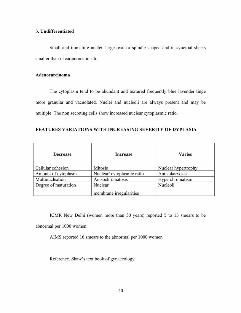
FEATURES VARIATIONS WITH INCREASING SEVERITY OF DYPLASIA
Decrease Increase Varies
Cellular cohesion Mitosis Nuclear hypertrophyAmount of cytoplasm Nuclear/ cytoplasmic ratio AntisokaryosisMultinucleation Anisochromatosis Hyperchromatism Degree of maturation Nuclear
membrane irregularities
Nucleoli
ICMR New Delhi (women more than 30 years) reported 5 to 15 smears to be
abnormal per 1000 women.
AIMS reported 16 smears to the abnormal per 1000 women
Reference. Shaw’s text book of gynaecology
40

SCREENING GUIDELINES
Guidelines American Cancer Society ACOGInitial screening Age 21 or 3 years after vaginal sex Age 21 or 3 years after vaginal
sexInterval -Every year for conventional pap
-Every 2 years for liquid based pap
-Every 2-3 years after age 30 with 3
consecutives normal
-Every year for either
liquid based pap or
conventional pap
-Every 2-3 years after age
30 with 3 consecutives
normalDiscontinue Age 70 if 3 consecutives normal in 10
years
No upper limit of age
IARC SCREENING GUIDELINES
An organized screening programme should cover women aged 25-65
years.
IARC advises that annual screening smears are unnecessary even with
conventional cytology.
Screening of women less than 25 years of age offers minimal benefit.
For woman 25 – 49 years of age three yearly pap smears are
recommended, five yearly where resources are limited.
Five yearly smears from 50-65 years of age are recommended.
Screening can cease after 65 years of age provided there are no suspicious
results in the previous two tests.
PROGRESSION OF CIN TO CIS AND INVASIVE CARCINOMA
41

ASCUS – 10 – 20% risk of CIN I
3 – 5% risk of CIN II and III
LSIL (CIN I)
- Undergoes spontaneous regression in 60 – 85% of cases within 2
years of follow up with cytology and colposcopy.
- 25% risk of CIN II and CIN III with in two years.
- if lesions progresses during follow up or persist at two years treat with
ablation therapy.
HSIL (CIN II and III)
- Do colposcopic directed biopsy. If biopsy positive for HSIL, do Large
Loop Electrosurgical Excision Procedure (LEEP).
- CIN II and III progresses to CIS in 20% of the cases, invasive
carcinoma in 5% of the cases.
CIS – Progresses to invasive carcinoma in 5% of the cases
Reference. Novak’s Text book of Gynaecology
42

COLPOSCOPY
In 1925, Hinselman in Germany devised the first colposcope from which the
modern day instruments have evolved. These are binocular instruments giving a
stereoscopic magnification of 10-20 times.
Indication for Colposcopy
1. Abnormal pap smear cytology
2. To locate abnormal areas
3. To obtain directed biopsy
4. Conservative therapy under colposcopic guidance
5. Follow up of cases treated conservatively
COLPOSCOPIC FINDINGS
I Normal Colposcopic findings:
1. Normal Squamous Epithelium
- Smooth, pink, uniform, featureless epithelium
- Stains Positively for glycogen
2. Columnar Epithelium
- Appear as villi with capillary loops and covered by mucus. After
application of acetic acid it has a typical grape like appearance.
43

3. Normal Trasformation Zone
Components of transformation Zone may be islands of columnar epithelium
surrounded by metaplastic squamous epithelium, gland openings, and nabothian cysts.
Stromal vessel have a characteristic tree like branching pattern.
II. ABNORMAL COLPOSCOPIC FINDINGS
A. Atypical Transformation Zone
1. Leucoplakia
White epithelium that is present before the application of acetic acid.
Caused by a layer of keratin on the surface of the epithelium.
2. Acetowhite Epithelium
Seen after application of acetic acid. Metaplastic and dysplastic epithelium
appear as white or grayish white due to increased cellular nuclear density. Normal cells
appear pink.
3. Iodine Negative Epithelium
Doesn’t stain with Lugol’s or Schiller’s Iodine. Normal native squamous
epithelium stains brown.
44
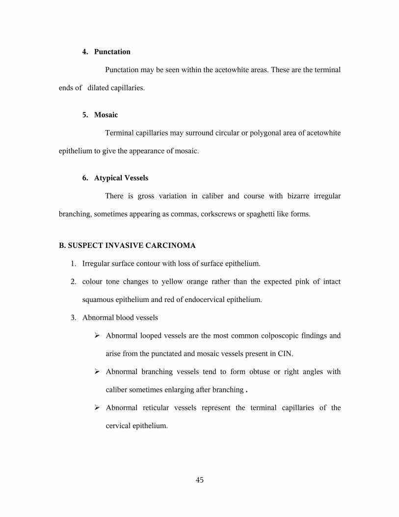
4. Punctation
Punctation may be seen within the acetowhite areas. These are the terminal
ends of dilated capillaries.
5. Mosaic
Terminal capillaries may surround circular or polygonal area of acetowhite
epithelium to give the appearance of mosaic.
6. Atypical Vessels
There is gross variation in caliber and course with bizarre irregular
branching, sometimes appearing as commas, corkscrews or spaghetti like forms.
B. SUSPECT INVASIVE CARCINOMA
1. Irregular surface contour with loss of surface epithelium.
2. colour tone changes to yellow orange rather than the expected pink of intact
squamous epithelium and red of endocervical epithelium.
3. Abnormal blood vessels
Abnormal looped vessels are the most common colposcopic findings and
arise from the punctated and mosaic vessels present in CIN.
Abnormal branching vessels tend to form obtuse or right angles with
caliber sometimes enlarging after branching .
Abnormal reticular vessels represent the terminal capillaries of the
cervical epithelium.
45

III. UNSATISFACTORY COLPOSCOPIC FINDINGS
If the entire squamocolumnar junction is not visible it’s judged unsatisfactory.
IV. MISCELLANEOUS COLPOSCOPIC FINDINGS
Seen in inflammations, atrophic changes, true erosion, condyloma and papilloma.
They are not related to cervical neoplasia and are present both in the transformation zone
and in original squamous epithelium.
COLPOSCOPIC GRADING
BASED ON COPPLESON AND REID
1. Grade I (insignificant, not suspicious)
Flat white epithelium, fine caliber and regular vessels with small inter capillary
distance – Normal epithelium to minor dysplasia.
2. Grade II (significant, suspicious)
Flat, white epithelium, vessels with dilated caliber and regular shape absence of
atypical vessels and increased inter capillary distance – moderate dysplasia to carcinoma
in situ.
46

3. Grade III ( Highly significant, highly suspicious)
Markedly white epithelium irregularly shaped and dilated vessels with variable
intercapillary distance and irregular surface – carcinoma in situ to early invasive
carcinoma.
Reid Scalzi Score
Based on four colposcopic signs
- Margin
- Colour
- Vessels
- Iodine test
Score 0-2 – CIN I
3-5 – CIN I to II
6-8 – CIN II to III
COLPOMICROSCOPY
Magnification is 100 – 300 times. Looks at the structure at the cellular level.
Interpretation is not very easy hence its lack of popularity.
47

BIOPSY CERVIX
Indication for Biopsy
1. All cases of leukoplakia even in the presence of negative smear
2. Lesions of squamous epithelium which whiten after acetic acid application.
3. All other abnormal lesions including those which are difficult to interpret at
colposcopy.
In the presence of obvious cervical lesions punch biopsy or wedge biopsy may be
taken.
Colposcopic directed biopsy is more specific and avoids lot of false negative
biopsies.
Indications for Cone Biopsy
1. When the area of abnormality is large.
2. When the squamo columnar junction is not visible completely on colposcopy.
3. When the inner margin of the lesion has receded into the cervical canal.
4. When there is discrepancy between colposcopy, cytology and biopsy.
5. Endocervical curettings positive for CIN II or III.
6. Colposcopically directed biopsy positive for micro invasion.
7. Colposcopy is unable to rule out invasive cancer.
Complications of Cone Biopsy
Bleeding, infection, cervical stenosis and incompetance.
48

NEWER TECHNIQUE
HPV DETECTION
By
1. Cytology (pap smear)
2. Histology
3. Electron microscopy
4. Immuno histochemistry (Identification of group specific antigen)
5. Molecular test
a. Insitu hybridization
b. Dot blot test
c. By amplification of viral DNA
I. Target amplification (PCR)
II. Signal amplification (hybrid capture II)
6. Serology (Detection of viral capsid proteins)
49

EPIDEMIOLOGY, AETIOLOGY, RISK FACTOR OF CANCER CERVIX
1. Coitus before 18 years of age.
2. Multiple sexual partners.
3. Delivery of first baby before 20 years.
4. Multi parity with poor birth spacing between pregnancies.
5. Poor personal hygiene.
6. Poor socio economic status.
7. Smoking, alcohol, drug abuse.
8. History of sexually transmitted diseases, TB, HIV, HPV, HSV2, condyloma.
9. Immuno suppressed.
10. OC pills intake.
11. History of prior genital tract dysplasia.
12. Women who do not come for regular health check up / lack of prior pap smear
screening.
50

The two Canadian task forces considered that the adult female population can be
classified into two groups.
1. Group that is not at risk and should not be included in the screening
program for cancer cervix comprising.
- Women who had never been sexually active.
- Women over 60 who had been screened in the past and had no atypia in
their smears.
- Women who had a hysterectomy for benign disease with complete
removal of cervical epithelium.
2. The group at risk for cancer cervix (ie.) women over 18 years and
under 60 years who are or have been sexually active.
51

RESULTS
The study was conducted on 500 women attending the Gynaecology OPD of
GOVERNMENT RSRM LYING IN HOSPITAL CHENNAI – 13 for a period of two
years (2006-08). A detailed history was taken. General examination and vital signs
assessment was done. All 500 women were subjected to down staging, pap smear,
VIA,VILI, and colposcopic examination. Colposcopy directed biopsy was taken in
women with abnormal colposcopic findings and the results were tabulated as follows.
AGE DISTRIBUTION
TABLE – 1
Age group No. of Women Percentage20 – 30 years
31 – 40 years
41 – 50 years
51 – 60 years
241
185
58
16
48.2%
37%
11.6%
3.2%85.2% of the women in the study group were aged between 20 – 40 years.
AGE IN YEARS
31-4020-30
PE
RC
EN
T
60
50
40
30
20
10
0
51-6041-50
A G E
DI
S T RI
B U TI
O N
52

PARITY DISTRIBUTION
TABLE- 2
Parity No. of Women Percentage 1
2
3
4
5
6
8
40
290
117
33
11
8
1
8%
58%
23.4%
6.6%
2.2%
1.6%
0.2%
88% of the women in the study group belong to para 2 – 4.
Though the incidence of unhealthy cervix was similar to healthy cervix in para 2 –
4, number of women with unhealthy cervix was considerably lower in para 1 and
considerably higher in para 4.
PARITY DISTRIBUTION
PARITY
8654321
Pe
rce
nt
70
60
50
40
30
20
10
0
53

MARRIED YEARS DURATION
TABLE-3
Married Years Duration No. of Women Percentage03 – 10
11 – 20
21 – 30
31 – 40
> 40
168
229
84
16
03
33.6%
45.8%
16.8%
3.2%
0.6%
Mean married years duration in the study group was 15.28 years.
MARRIED YEARS DURATION (YEARS)
>4031-4021-3011-203-10
Pe
rce
nt
50
40
30
20
10
0
MARRIED YEARS DURATION
54
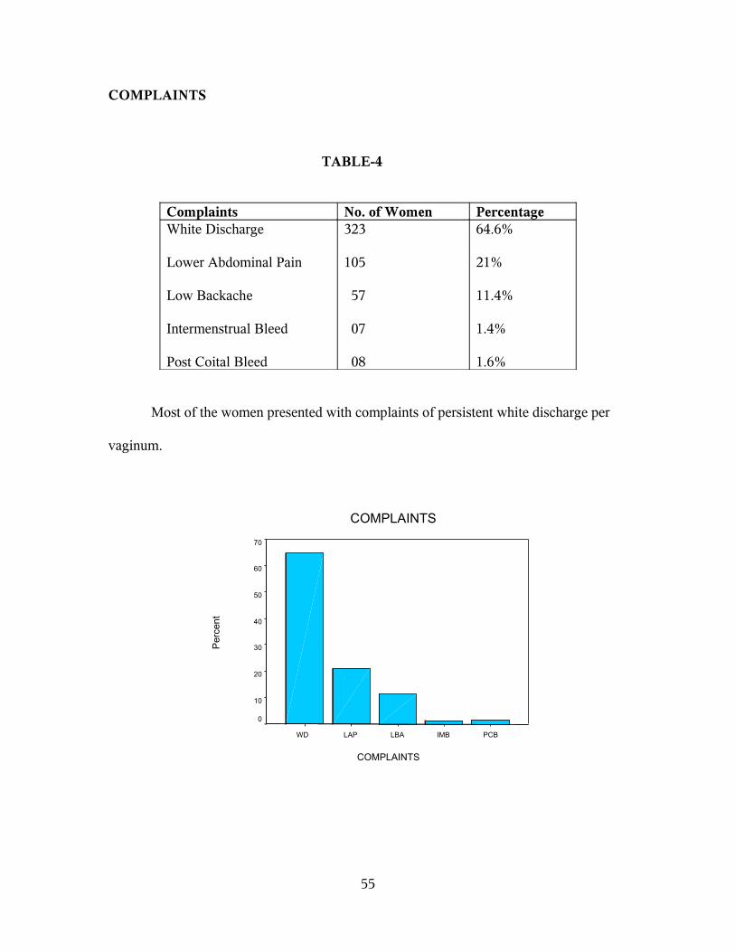
COMPLAINTS
TABLE-4
Complaints No. of Women PercentageWhite Discharge
Lower Abdominal Pain
Low Backache
Intermenstrual Bleed
Post Coital Bleed
323
105
57
07
08
64.6%
21%
11.4%
1.4%
1.6%
Most of the women presented with complaints of persistent white discharge per
vaginum.
COMPLAINTS
COMPLAINTS
PCBIMBLBALAPWD
Per
cen
t
70
60
50
40
30
20
10
0
55

DOWN STAGING OF CERVIX
TABLE-5
Down Staging of Cervix No. of women Percentage
Healthy Cervix 263 52.6%
Unhealthy Cervix
1. Erosion
2. Hypertrophy
3. Ectropion
4. Congestion
5. Old Cervical Tear
6. Polyp
7. Ulcer
133
37
23
21
11
8
4
26.6%
7.4%
4.6%
4.2%
2.2%
1.6%
0.8%
In speculum examination 263 women had healthy cervix and 237 women had
unhealthy cervix of which erosion cervix is the commonest.
SPECULUM EXAMINATION
SPECULUM EXAMINATION
ULCERPOLYPOCTCONECHYERHT
Pe
rce
nt
60
50
40
30
20
10
0
56

RESULTS OF VIA
TABLE-6
VIA Biopsy Positive for
CIN/CA Cervix
Biopsy Negative for
CIN/CA Cervix
Total
Positive 64 70 134Negative 23 343 366Total 87 413 500
VIA was positive in 134 women, negative in 366 women. VIA was
positive in 64 women with biopsy report positive for CIN/CA Cervix and 70 women with
biopsy report negative for CIN/CA Cervix. VIA was negative in 23 women with biopsy
report positive for CIN/CA Cervix and 343 women with biopsy report negative for CIN/
CA Cervix.
57

VIA vs BIOPSY
TABLE-7
Biopsy VIA Positive VIA NegativeCIN I
CIN II
CIN III
Squamous Cell Carcinoma
34
16
07
07
18
05
-
-Total 64 23
VIA was positive in 64 women with biopsy report positive for CIN and CA
cervix. Of which VIA was positive in 34 women with CIN I, 16 women with CIN II,
7 women with CIN III, and 7 women with squamous cell carcinoma.
VIA was negative in 23 women with biopsy report positive for CIN and CA
cervix. Of which VIA was negative in 18 women with CIN I and 5 women with CIN II.
VIA detected all cases of CIN III and squamous cell carcinoma.
58

RESULTS OF VILI
TABLE-8
VILI Biopsy Positive for
CIN/CA Cervix
Biopsy Negative for
CIN/CA Cervix
Total
Positive 61 66 127Negative 26 347 373Total 87 413 500
VILI was positive in 127 women and negative in 373 women. VILI was positive
in 61 women with biopsy report positive for CIN/CA cervix and 66 women with biopsy
report negative for CIN/CA cervix. VILI was negative in 26 women with biopsy report
positive for CIN/CA cervix and 347 women with biopsy report negative for CIN/CA
cervix.
59

VILI vs BIOPSY
TABLE-9
Biopsy VILI Positive VILI NegativeCIN I
CIN II
CIN III
Squamous Cell Carcinoma
33
16
06
06
19
05
01
01Total 61 26
VILI was positive in 61 women with biopsy report positive for CIN and CA
cervix. Of which VILI was positive in 33 women with CIN I, 16 women with CIN II, 6
women with CIN III, and 6 women with squamous cell carcinoma.
VILI was negative in 26 women with biopsy report positive for CIN and CA
cervix. Of which VILI was negative in 19 women with CIN I, 5 women with CIN II, 1
women with CIN III and 1 women with squamous cell carcinoma.
60

PARITY vs VIA and VILI
TABLE-10
Parity No. of Women VIA Positive VILI Positive1
2
3
4
> 5
40
290
117
33
20
02
54
42
23
13
03
51
42
18
13Total 500 134 127
As the parity increases VIA and VILI positivity increases.
MARRIED YEARS DURATION VS VIA AND VILI
TABLE-11
Married Years Duration No. of Women VIA Positive VILI Positive03-10
11-20
21-30
31-40
> 40
168
229
84
16
03
13
59
48
11
03
14
64
38
08
03Total 500 134 127
As the married years duration increases VIA and VILI positivity increases
61

PAP SMEAR GRADING
TABLE-12
Grade No. of WomenI (Normal)
II (Inflammatory)
III (CIN-I)
IV (CIN-II & III)
V (SCC)
222
200
56
22
---Total 500
Pap smear study revealed 222 women with normal cytology (Grade I), 200
women had inflammatory smear (Grade II), CIN I (Grade III) in 56 women, CIN II and
III (Grade IV) in 22 women.
62

COLPOSCOPIC FINDINGS
TABLE-13
Grade No. of WomenNormal
I
II
III
356
88
42
14Total 500
A detailed colposcopic examination was done in 500 women. 356 women had
normal colposcopic findings. 144 women had abnormal colposcopic findings, of this 88
women had grade I lesions, 42 women had grade II lesions, 14 women had grade III
lesions.
63

COLPOSCOPY DIRECTED BIOPSY CERVIX
TABLE-14
Biopsy Findings No. of WomenBiopsy not taken
Normal
Chronic cervicitis
CIN I
CIN II
CIN III
Squamous cell carcinoma
356
29
28
52
21
07
07Total 500
Colposcopy directed biopsy was not taken in 356 women with normal colposcopic
findings. Colposcopy directed biopsy was taken in 144 women with abnormal
colposcopic findings. Of these histology was normal in 29 women, 28 women had
chronic cervicitis, CIN I was seen in 52 women, CIN II in 21 women, CIN III in 7
women and squamous cell carcinoma in 7 women.
64

DISCUSSION
The concept of screening has been defined as the search for unrecognized disease
or defect by means of rapidly applied tests, examination or other procedures in apparently
healthy individual. Today screening is considered a preventive care function and a logical
extension of health care. The basic purpose of screening is to sort out from a large group
of apparently healthy persons those likely to have the diseases and to bring those who are
apparently abnormal under medical supervision and treatment.
The case detection, control of disease, research and education are the 4 main uses
of the screening programme. A mass screening when backed up by suitable treatment
reduces the duration of illness or alters its final outcome.
Selective screening is more productive, for example cancer cervix tends to occur
relatively less often in the upper socio economic groups. Therefore screening of cancer
cervix in the lower socio economic groups will increase the yield of more cases.
Acceptability and repeatability depending on the technique, observer variation, subject
variation and errors are to be taken into account in mass campaigns.
AGE AND PARITY
MEAN AGE OF WOMEN
Our study - 33.11 years
Kasper et al., - 32.5 years
Kenneth Francus - 33.25 years
Dunn - 27 years
65

TABLE-15
Mean age of the women in biopsy negative group was 31.6 %
Mean age of the women in biopsy positive group was 40.2 %
P value < 0.001 hence highly statistically significant.
Based on a study in Eden Hospital Medical College, Calcutta by Roy Chowdry,
1975, cancer cervix is common in lower age group. In India 40% were between 36- 40
years and 50% married below 16 years with early onset of sexual activity. In our study
40% women were between 31 - 40 years.
He also points out that cancer cervix is common in multi para (95%). In our study
out of 87 women diagnosed as dysplasia none were para 1, 22 women were para 2,
65 women were more than para 3.
SOCIO ECONOMIC STATUS
According to two cases – control studies carried out in Columbia and Spain it was
found that cancer cervix is more common in women of low socioeconomic status than
women of high socio economic status. The reasons being sexual behavior of men
particularly contact with sex workers and HPV infection. The women of high socio-
economic status have other risk factors like smoking and oral contraceptive pill use.
In our study all women belong to socio economic status IV and V.
66
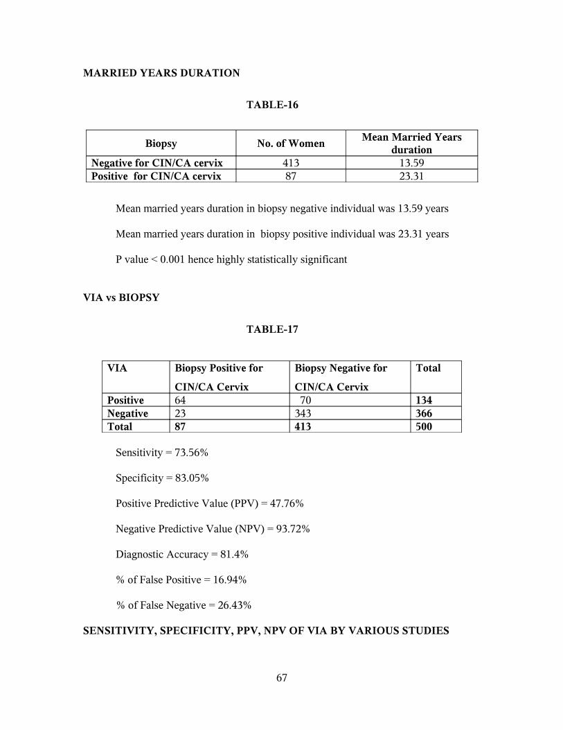
MARRIED YEARS DURATION
TABLE-16
Biopsy No. of WomenMean Married Years
durationNegative for CIN/CA cervix 413 13.59Positive for CIN/CA cervix 87 23.31
Mean married years duration in biopsy negative individual was 13.59 years
Mean married years duration in biopsy positive individual was 23.31 years
P value < 0.001 hence highly statistically significant
VIA vs BIOPSY
TABLE-17
VIA Biopsy Positive for
CIN/CA Cervix
Biopsy Negative for
CIN/CA Cervix
Total
Positive 64 70 134Negative 23 343 366Total 87 413 500
Sensitivity = 73.56%
Specificity = 83.05%
Positive Predictive Value (PPV) = 47.76%
Negative Predictive Value (NPV) = 93.72%
Diagnostic Accuracy = 81.4%
% of False Positive = 16.94%
% of False Negative = 26.43%
SENSITIVITY, SPECIFICITY, PPV, NPV OF VIA BY VARIOUS STUDIES
67

TABLE-18
Study Year Sensitivity Specificity PPV NPVEl-Shalakany AH et al.,
Arbyn M et al.,
Muwonge R et al.,
Elit L et al.,
De Vuyst H et al.,
Sankaranarayanan R et al.,
Our Study
2008
2008
2007
2006
2005
2004
2008
90.9%
83%
81.3%
82.9%
73.3%
76.8%
73.56%
94.6%
85%
87.3%
88.6%
80%
85.5%
83.05%
43.5%
47.76%
99.6%
93.72%
Our study showed sensitivity of about 73.56% which is comparable with that of
De Vuyst H et al., 2005 and Sankaranarayanan R et al., 2004 whose study showed
sensitivity of about 73.3% and 76.8% respectively. The sensitivity shown by Elit L et al.,
2006, Muwonge R et al., 2007, Arbyn M et al., 2008, El-Shalakany AH et al., 2008, is
some what higher when compared to our study.
Our study showed specificity of about 83.05% which is comparable with that of
De Vuyst H et al., 2005, Sankaranarayanan R et al., 2004 and Arbyn M et al.,
2008, whose study showed specificity of about 80%, 85.5%, 85% respectively. The
specificity shown by Elit L et al., 2006, Muwonge R et al., 2007, is some what higher
when compared to our study.
VILI vs BIOPSY
68

TABLE-19
VILI Biopsy Positive for
CIN/CA Cervix
Biopsy Negative
for CIN/CA Cervix
Total
Positive 61 66 127Negative 26 347 373Total 87 413 500
Sensitivity = 70.11%
Specificity = 84.02%
Positive Predictive Value (PPV) = 48.03%
Negative Predictive Value (NPV) = 93.03%
Diagnostic Accuracy = 81.6%
% of False Positive = 15.98%
% of False Negative = 29.88%
69

SENSITIVITY, SPECIFICITY, PPV, NPV OF VILI BY VARIOUS STUDIES
TABLE-20
Study Year Sensitivity Specificity PPV NPVEl-Shalakany AH et al.,
Arbyn M et al.,
Muwonge R et al.,
Sangwa Lugoma G et al.,
Shastri SS et al.,
Sankaranarayanan R et al.,
Our Study
2008
2008
2007
2006
2005
2003
2008
97.7%
93%
91.5%
68.3%
75%
87.2%
70.11%
94.6%
85%
86.9%
76.2%
84%
84.7%
84.02%
46.2%
48.03%
99.9%
93.03%
Our study showed sensitivity of about 70.11% which is comparable with that of
Sangwa Lugoma G et al., 2006 Shastri SS et al., 2005 whose study showed sensitivity of
about 68.3% and 75% respectively. The sensitivity shown by Sankaranarayanan R et al.,
2003, Muwonge R et al., 2007, Arbyn M et al., 2008, El-Shalakany AH et al., 2008, is
some what higher when compared to our study.
Our study showed specificity of about 84.02% which is comparable with that of
Sankaranarayanan R et al., 2004, Shastri SS et al., 2005, Muwonge R et al., 2007, Arbyn
M et al., 2008, whose study showed specificity of about 84.7%, 84%, 86.9%, 85%
respectively. The specificity shown by El-Shalakany AH et al., 2008 is some what higher
when compared to our study.
PAP SMEAR vs BIOPSY
70

TABLE-21
Pap Smear Biopsy Positive for
CIN/CA Cervix
Biopsy Negative for CIN/
CA Cervix
Total
Positive 69 9 78Negative 18 404 422Total 87 413 500
Sensitivity = 79.31%
Specificity = 97.82%
Positive Predictive Value (PPV) = 88.46%
Negative Predictive Value (NPV) = 95.73%
Diagnostic Accuracy = 94.6%
% of False Positive = 2.17%
% of False Negative = 20.6%
71

SENSITIVITY, SPECIFICITY, PPV, NPV OF PAP SMEAR BY VARIOUS
STUDIES
TABLE-22
Study Year Sensitivity Specificity PPV NPVEl-Shalakany AH et al.,
Arbyn M et al.,
Sodhani P et al.,
Elit L et al
Sangwa Lugoma G et al.,
Shastri SS et al.,
Sankaranarayanan R et al.,
Our Study
2008
2008
2006
2006
2006
2005
2003
2008
22.7%
57%
91.4%
88.6%
31-72%
57%
81.9%
79.31%
97.6%
98.6%
86.6%
98.5%
94-99%
98.6%
87.8%
97.82%
41.7%
51.7%
88.46%
96.6%
99.8%
95.73%
Our study showed sensitivity of about 79.31% which is comparable with that of
Sankaranarayanan R et al., 2003, whose study showed sensitivity of about 81.9%.
Our study showed specificity of about 97.82% which is comparable with that of ,
Shastri SS et al., 2005, Sangwa Lugoma G et al., 2006, Elit L et al., 2006, Arbyn M et al.,
2008, El-Shalakany AH et al., 2008 whose study showed specificity of about 98.6%,
94-99%, 98.5%, 98.6%, and 97.6% respectively.
72

COMPARING THE SENSITIVITY AND SPECIFICITY OF VIA, VILI, PAP
SMEAR IN THE DETECTION OF PREINVASIVE LESIONS AND INVASIVE
CARCINOMA OF CERVIX
TABLE-23
Screening Method Sensitivity Specificity Percentage of
False Positive
VIA
VILI
Pap Smear
73.56%
70.11%
79.31%
83.05%
84.02%
97.82%
16.94%
15.98%
2.17%
In our study, sensitivity of VIA (73.56%) and VILI (70.11%) is almost similar to
pap smear (79.31%) but the specificity of pap smear (97.82%) is some what high
compared to VIA (83.05%) and VILI (84.02%).
Hence VIA and VILI has similar sensitivity to pap smear but with lower
specificity in the detection of pre invasive lesions of cervix. This is associated with a high
number of false positive rates. The observed high number of false positive results of VIA
and VILI may lead to high rates of referral and may increase the rates of treatment which
may translate to higher costs. On the other hand high detection rate of VIA and VILI for
high grade pre-malignant lesions may prevent malignancies at a low cost.
73
CORRELATION OF VIA, VILI, PAP SMEAR, AND COLPOSCOPY WITH
COLPOSCOPY DIRECTED BIOPSY IN THE DETECTION OF CIN AND
INVASIVE CARCINOMA IN 144 CASES
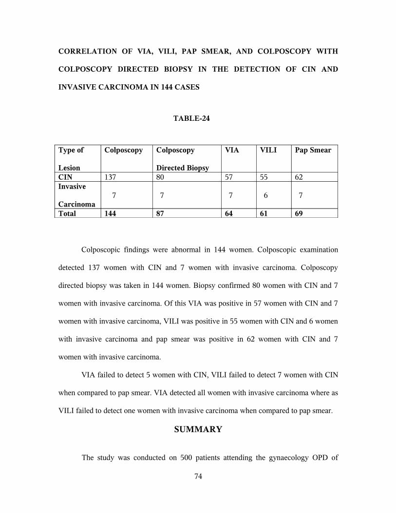
TABLE-24
Type of
Lesion
Colposcopy Colposcopy
Directed Biopsy
VIA VILI Pap Smear
CIN 137 80 57 55 62Invasive
Carcinoma 7 7 7 6 7
Total 144 87 64 61 69
Colposcopic findings were abnormal in 144 women. Colposcopic examination
detected 137 women with CIN and 7 women with invasive carcinoma. Colposcopy
directed biopsy was taken in 144 women. Biopsy confirmed 80 women with CIN and 7
women with invasive carcinoma. Of this VIA was positive in 57 women with CIN and 7
women with invasive carcinoma, VILI was positive in 55 women with CIN and 6 women
with invasive carcinoma and pap smear was positive in 62 women with CIN and 7
women with invasive carcinoma.
VIA failed to detect 5 women with CIN, VILI failed to detect 7 women with CIN
when compared to pap smear. VIA detected all women with invasive carcinoma where as
VILI failed to detect one women with invasive carcinoma when compared to pap smear.
SUMMARY
The study was conducted on 500 patients attending the gynaecology OPD of
74

GOVERNMENT RSRM LYING IN HOSPITAL – CHENNAI during a period of two
year (2006-08). All women were married from reproductive age group to post
menopausal age group, maximum between 20 – 40 years. Parity varies from primipara to
multipara. All women belong to low socio economic class. Most of the women presented
with more than one complaint, maximum being white discharge. Mean married year’s
duration was 15 years. Most of them were married at the age of 16 – 18 years with early
onset of sexual activity.
All these women were subjected to down staging, Pap smear, VIA, VILI and
colposcopic examination. Colposcopic examination was abnormal in 144 women. They
were subjected to colposcopy directed biopsy. Biopsy detected 80 women with CIN and 7
women with invasive carcinoma. Of this VIA, VILI, Pap smear detected 57, 55, 62
women with CIN and 7, 6, 7 women with invasive carcinoma respectively.
The sensitivity, specificity of VIA, VILI, Pap smear are as follows
Screening Method Sensitivity SpecificityVIA
VILI
Pap Smear
73.56%
70.11%
79.31%
83.05%
84.02%
97.82%
75

CONCLUSION
Visual inspection of the cervix after application of acetic acid and Lugol’s Iodine
can be used as one of the low cost screening tool in the detection of pre invasive lesions
of cervix. VIA and VILI has a similar sensitivity to cervical cytology but with lower
specificity. This is associated with high number of false positive rate leading to high rates
of referral. On the other hand high detection rate of VIA and VILI for high grade pre
malignant lesions may prevent malignancy at a low cost. Hence VIA, VILI can be under
taken as a feasible method of screening in cervical cancer in countries where access to
cytopathology is limited.
The sensitivity of cytology increased significantly when combined with VIA and
VILI.
Visual inspection can be performed easily by trained paramedical workers in rural
areas for early referral to higher centers. This may ultimately bring down the severity of
CIN and Cancer cervix in the long run.
The higher sensitivity, accuracy, low cost, easy applicability and immediate
results make VIA, VILI a useful screening test in developing countries like India. This
must go hand in hand with increasing the awareness of women about cervical cancer
screening programmes.
76

77

BIBLIOGRAPHY
1) Frisch LE, Milner FH, Ferris DG. Naked-eye inspection of the cervix after acetic acid
application may improve the predictive value of negative cytologic screening. J Fam
Pract. 1995 Apr;40(4):327.
2) Belinson JL, Pretorius RG, Zhang WH, Wu LY, Qiao YL, Elson P. Cervical cancer
screening by simple visual inspection after acetic acid. Obstet Gynecol. 2002 Mar;
99(3):517-8.
3) Mandelblatt JS, Lawrence WF, Gaffikin L, Limpahayom KK, Lumbiganon P,
Warakamin S, King J, Yi B, Ringers P, Blumenthal PD. Costs and benefits of
different strategies to screen for cervical cancer in less-developed countries. J Natl
Cancer Inst. 2002 Oct 2;94(19):1469-83.
4) Tayyeb R, Khawaja NP, Malik N. Comparison of visual inspection of cervix and Pap
smear for cervical cancer screening. J Coll Physicians Surg Pak. 2003 Apr;13(4):
201-3.
5) Basu PS, Sankaranarayanan R, Mandal R, Roy C, Das P, Choudhury D, Bhattacharya
D, Chatterjee R, Dutta K, Barik S, Tsu V, Chakrabarti RN, Siddiqi M; Calcutta
Cervical Cancer Early Detection Group. Visual inspection with acetic acid and
cytology in the early detection of cervical neoplasia in Kolkata, India. Int J Gynecol
Cancer. 2003 Sep-Oct; 13(5):626-32.
6) Ferreccio C, Bratti MC, Sherman ME, Herrero R, Wacholder S, Hildesheim A, Burk
RD, Hutchinson M, Alfaro M, Greenberg MD, Morales J, Rodriguez AC, Schussler J,
Eklund C, Marshall G, Schiffman M. A comparison of single and combined visual,
cytologic, and virologic tests as screening strategies in a region at high risk of

cervical cancer. Cancer Epidemiol Biomarkers Prev. 2003 Sep;12(9):815-23.
7) Sankaranarayanan R, Wesley R, Thara S, Dhakad N, Chandralekha B, Sebastian P,
Chithrathara K, Parkin DM, Nair MK. Test characteristics of visual inspection with
4% acetic acid (VIA) and Lugol's iodine (VILI) in cervical cancer screening in
Kerala, India. Int J Cancer. 2003 Sep 1;106(3):404-8.
8) Bhatla N, Mukhopadhyay A, Joshi S, Kumar A, Kriplani A, Pandey RM, Verma K.
Visual inspection for cervical cancer screening: evaluation by doctor versus
paramedical worker. Indian J Cancer. 2004 Jan-Mar; 41(1):32-6.
9) Denny L, Kuhn L, Pollack A, Wright TC Jr. Direct visual inspection for cervical
cancer screening: an analysis of factors influencing test performance. Cancer. 2004
Nov 15;101(10):2365; author reply 2366.
10) Sankaranarayanan R, Shastri SS, Basu P, Mahé C, Mandal R, Amin G, Roy C,
Muwonge R, Goswami S, Das P, Chinoy R, Frappart L, Patil S, Choudhury D,
Mukherjee T, Dinshaw K. The role of low-level magnification in visual inspection
with acetic acid for the early detection of cervical neoplasia. Cancer Detect Prev.
2004;28(5):345-51.
11) Ghaemmaghami F, Behtash N, Modares Gilani M, Mousavi A, Marjani M, Moghimi
R. Visual inspection with acetic acid as a feasible screening test for cervical neoplasia
in Iran. Int J Gynecol Cancer. 2004 May-Jun;14(3):465-9.
12) Sankaranarayanan R, Basu P, Wesley RS, Mahe C, Keita N, Mbalawa CC, Sharma R,
Dolo A, Shastri SS, Nacoulma M, Nayama M, Somanathan T, Lucas E, Muwonge R,
Frappart L, Parkin DM; IARC Multicentre Study Group on Cervical Cancer Early
Detection. Accuracy of visual screening for cervical neoplasia: Results from an IARC

multicentre study in India and Africa. Int J Cancer. 2004 Jul 20;110(6):907-13.
13) Pérez-Cruz E, Winkler JL, Velasco-Mondragón E, Salmerón-Castro J, García F,
Davis-Tsu V, Escandón-Romero C, Hernández-Avila M. [Screening and follow-up
for cervical cancer prevention in rural Mexico using visual inspection]. Salud Publica
Mex. 2005 Jan-Feb;47(1):39-48.
14) Bragança JF, Derchain SF, Sarian LO, Messias da Silva SM, Labatte S, Zeferino LC.
Aided visual inspection with acetic acid (VIA) and HPV detection as optional
screening tools for cervical cancer and its precursor lesions. Clin Exp Obstet Gynecol.
2005;32(4):225-9.
15) De Vuyst H, Claeys P, Njiru S, Muchiri L, Steyaert S, De Sutter P, Van Marck E,
Bwayo J, Temmerman M. Comparison of pap smear, visual inspection with acetic
acid, human papillomavirus DNA-PCR testing and cervicography. Int J Gynaecol
Obstet. 2005 May;89(2):120-6.
16) Wu S, Meng L, Wang S, Ma D. A comparison of four screening methods for cervical
neoplasia. Int J Gynaecol Obstet. 2005 Nov;91(2):189-93. Epub 2005 Sep 23.
17) Shastri SS, Dinshaw K, Amin G, Goswami S, Patil S, Chinoy R, Kane S, Kelkar R,
Muwonge R, Mahé C, Ajit D, Sankaranarayanan R. Concurrent evaluation of visual,
cytological and HPV testing as screening methods for the early detection of cervical
neoplasia in Mumbai, India. Bull World Health Organ. 2005 Mar;83(3):186-94. Epub
2005 Mar 16.
18) Syrjänen K, Naud P, Derchain S, Roteli-Martins C, Longatto-Filho A, Tatti S, Branca
M, Erzen M, Hammes LS, Matos J, Gontijo R, Sarian L, Braganca J, Arlindo FC,
Maeda MY, Lörincz A, Dores GB, Costa S, Syrjänen S. Comparing PAP smear

cytology, aided visual inspection, screening colposcopy, cervicography and HPV
testing as optional screening tools in Latin America. Study design and baseline data
of the LAMS study. Anticancer Res. 2005 Sep-Oct;25(5):3469-80.
19) Valdespino VM, Valdespino VE. Cervical cancer screening: state of the art. Curr
Opin Obstet Gynecol. 2006 Feb;18(1):35-40.
20) Elit L, Baigal G, Tan J, Munkhtaivan A. Assessment of 2 cervical screening methods
in Mongolia: cervical cytology and visual inspection with acetic acid. J Low Genit
Tract Dis. 2006 Apr;10(2):83-8.
21) Denny L, Quinn M, Sankaranarayanan R. Screening for cervical cancer in developing
countries. Vaccine. 2006 Aug 31;24 Suppl 3:S3/71-7.
22) Sangwa-Lugoma G, Mahmud S, Nasr SH, Liaras J, Kayembe PK, Tozin RR, Drouin
P, Lorincz A, Ferenczy A, Franco EL. Visual inspection as a cervical cancer
screening method in a primary health care setting in Africa. Int J Cancer. 2006 Sep
15;119(6):1389-95.
23) Sodhani P, Gupta S, Sharma JK, Parashari A, Halder K, Singh V, Sehgal A. Test
characteristics of various screening modalities for cervical cancer: a feasibility study
to develop an alternative strategy for resource-limited settings. Cytopathology. 2006
Dec;17(6):348-52.
24) Muwonge R, Walter SD, Wesley RS, Basu P, Shastri SS, Thara S, Mbalawa CG,
Sankaranarayanan R; IARC Multicentre Study Group on Cervical Cancer Early
Detection. Assessing the gain in diagnostic performance when two visual inspection
methods are combined for cervical cancer prevention. J Med Screen. 2007;14(3):
144-50.
25) Arbyn M, Bergeron C, Klinkhamer P, Martin-Hirsch P, Siebers AG, Bulten J. Liquid
compared with conventional cervical cytology: a systematic review and meta-
analysis. J Fam Pract. 2008 Apr;57(4):preceding 220. Obstet Gynecol. 2008 Jan;
111(1):2-3.
26) Dhaubhadel P, Vaidya A, Choudhary P. Early detection of precursors of cervical
cancer with cervical cytology and visual inspection of cervix with acetic Acid. JNMA
J Nepal Med Assoc. 2008 Apr-Jun;47(170):71-6.
27) El-Shalakany AH, Saeed MM, Abdel-Aal MR, El-Nakeeb AH, Noseirat N, Ayyad
SB, El Din ZS. Direct visual inspection of the cervix with Lugol iodine for the
detection of premalignant lesionsJ Low Genit Tract Dis. 2008 Jul;12(3):193-8.
28) Jun JK, Choi KS, Jung KW, Lee HY, Gapstur SM, Park EC, Yoo KY. Effectiveness
of an organized cervical cancer screening program in Korea: Results from a cohort
study. Int J Cancer. 2008 Sep 10.
29) Davis-Dao CA, Cremer M, Felix J, Cortessis VK. Effect of cervicitis on visual
inspection with acetic acid. J Low Genit Tract Dis. 2008 Oct;12(4):282-6.
30) Arbyn M, Sankaranarayanan R, Muwonge R, Keita N, Dolo A, Mbalawa CG,
Nouhou H, Sakande B, Wesley R, Somanathan T, Sharma A, Shastri S, Basu P.
Pooled analysis of the accuracy of five cervical cancer screening tests assessed in
eleven studies in Africa and India. Int J Cancer. 2008 Jul 1;123(1):153-60.
31) Sankaranarayanan R, Thara S, Esmy PO, Basu P. Cervical cancer: screening and
therapeutic perspectives. Med Princ Pract. 2008;17(5):351-64. Epub 2008 Aug 6.

PROFORMA
SERIAL NO.:
NAME
AGE
OP NO.
ADDRESS
RELIGION
OCCUPATION / INCOME
SOCIO ECONOMIC STATUS
MENSTRUAL HISTORY
AGE AT MENARCHE
CYCLE LENGTH
DURATION OF FLOW
AMOUNT OF FLOW
AGE AT MENOPAUSE
MARITAL HISTORY
MARRIED SINCE YEARS
AGE OF MARRIAGE
SEXUAL HISTORY
AGE AT FIRST COITUS
FREQUENCY OF COITUS
NUMBER OF PARTNERS

OBSTETRIC HISTORY
PARITY
LAST CHILD BIRTH
LACTATION
PERSONAL HISTORY
PID
OC PILL INTAKE
RT / CT / IMMUNO – SUPPRESSION
TB / DRUG INTAKE
COMPLAINTS
CLINICAL FINDINGS
DOWN STAGING
PAP SMEAR
VIA
VILI
COLPOSCOPY
COLPOSCOPY DIRECTED BIOPSY CERVIX
IMPRESSION

MASTER CHART
S.No. Name IP No. Age Mrd Yrs SEC Pty PC S/E Pap VIA VILI Colp Biop
1 Priya 21147 22 4 4 1 WD N I Neg Neg N NB
2 Kavitha 20460 25 6 4 2 WD N II Neg Neg N NB
3 Selvi 21146 28 10 4 2 LBA EC II Neg Neg N NB
4 Muniyammal 5709 27 15 5 3 WD EC II Pos Pos I CC
5 Ruth 22113 30 14 5 2 WD N III Pos Pos II CIN I
6 Lakshmi 14829 33 15 4 2 LBA N I Neg Neg N NB
7 Vasanthi 21071 30 12 4 1 LBA N I Neg Neg N NB
8 Uma 11522 30 17 4 1 WD N II Neg Neg N NB
9 Govindammal 14185 35 20 5 3 WD E III Pos Neg II CIN I
10 Malarvizhi 22215 37 20 5 4 WD E II Pos Pos I CC
11 Vani 8667 24 6 4 2 LAP N II Pos Neg N NB
12 Brinda 21285 29 10 4 2 LBA N II Neg Neg N NB
13 Ramani 8938 28 9 4 2 LBA N II Neg Neg I N
14 Devi 21285 42 26 5 2 WD E IV Pos Pos II CIN II
15 Geetha 8938 40 20 5 2 WD E IV Neg Pos II CIN II
16 Vadivu 21217 58 40 5 2 PCB E IV Pos Pos III SCC
17 Kanniammal 9217 42 24 5 2 PCB E IV Pos Pos III SCC
18 Sivabharathi 9250 30 16 4 3 WD E II Neg Pos II CIN I
19 Savitha 20627 40 20 4 3 WD E III Pos Pos II CIN I
20 Jamuna 23021 35 18 4 6 LBA N I Neg Neg N NB
21 Rajeswari 19350 40 21 4 2 LAP N I Neg Neg N NB
22 Umavathi 19351 29 13 4 3 LAP OCT II Neg Neg N NB
23 Begam 17824 60 44 5 8 WD E IV Pos Pos II SCC

S.No. Name IP No. Age Mrd Yrs SEC Pty PC S/E Pap VIA VILI Colp Biop
24 Sarala 24567 32 16 5 3 WD H II Pos Pos I CIN I
25 Renuka 23360 25 11 4 3 LBA E I Neg Neg N NB
26 Murugammal 24720 45 27 4 2 WD N II Neg Neg N NB
27 Padma 22522 32 15 4 3 LAP N I Neg Neg N NB
28 Meharun 25474 38 21 5 5 WD E IV Pos Pos III CIN II
29 Rabitha 8527 55 40 4 4 LBA N I Neg Neg N NB
30 Selvarani 27193 36 17 4 2 LBA N I Neg Neg N NB
31 Badma 26071 23 5 4 1 WD N I Neg Neg N NB
32 Banu 27050 26 9 4 2 LAP E II Neg Pos N NB
33 Geetha 12641 28 10 4 2 LAP H II Neg Neg N NB
34 Rajee 28362 32 14 4 2 WD N II Neg Neg N NB
35 Rathna 29611 30 12 4 2 IMB P II Neg Pos N NB
36 Dhanam 29388 48 30 5 6 WD U III Pos Pos II SCC
37 Suriya 21971 32 14 4 3 WD H III Neg Neg I CC
38 Vasanthi 28355 45 23 4 2 LAP N I Neg Neg N NB
39 Mari 29501 25 7 4 2 LAP N I Neg Neg N NB
40 Saranya 30849 22 4 4 1 WD N I Neg Neg N NB
41 Suganya 30981 24 5 4 2 WD E II Neg Neg N NB
42 Marieswari 30193 30 12 4 3 LBA C III Neg Neg I CINI
43 Vijayalakshmi 31146 23 7 4 2 LBA E I Neg Neg N NB
44 Rani 29080 29 10 4 2 WD C II Neg Neg N NB
45 Pavithra 31247 26 9 4 2 WD EC II Pos Neg N NB
46 Hema 28562 30 9 4 2 LAP N II Neg Neg N NB

S.No. Name IP No. Age Mrd Yrs SEC Pty PC S/E Pap VIA VILI Colp Biop
47 Kiliamma 31401 30 14 4 3 WD E II Neg Pos I CIN I
48 Anjali 27388 40 26 5 4 WD E III Pos Neg I CIN II
49 Ramani 32536 23 6 4 2 LBA E II Neg Neg N NB
50 Rukmani 30287 38 21 5 3 WD H III Pos Neg I CIN I
51 Shanthi 33724 37 20 4 2 LBA N I Neg Neg N NB
52 Rajakumari 28717 30 13 4 3 LAP N I Neg Neg N NB
53 Kaveri 26767 45 30 4 2 LBA N I Neg Neg N NB
54 Sarkunam 30808 40 21 5 4 WD H IV Pos Pos III CIN III
55 Lakshmi 40779 46 21 4 4 LBA OCT II Pos Neg N NB
56 Rani 40614 28 12 4 3 WD E II Neg Neg N NB
57 Manjula 40613 26 12 4 2 WD C II Pos Neg N NB
58 Lakshmi 40616 45 30 4 2 WD E II Pos Pos I CC
59 Mangalam 40560 25 8 4 2 WD N I Neg Neg N NB
60 Revathi 39129 27 11 4 3 LBA N I Neg Neg N NB
61 Kumutha 40739 29 12 4 3 LAP N I Neg Neg N NB
62 Vani 40763 26 9 4 2 WD N I Neg Neg N NB
63 Jeenath 40771 26 8 4 2 WD N I Neg Neg N NB
64 Mohana 40057 30 16 4 4 WD OCT III Pos Neg I CIN I
65 Mari 40756 30 14 4 2 WD N II Pos Pos N NB
66 Geetha 40785 32 16 4 2 LBA N I Neg Neg N NB
67 Muthu 40794 29 12 4 2 WD N II Neg Neg N NB
68 Kanchana 40778 28 10 4 2 LAP E II Pos Pos N NB
69 Usha 40798 26 6 4 2 LAP EC II Neg Neg N NB

S.No. Name IP No. Age Mrd Yrs SEC Pty PC S/E Pap VIA VILI Colp Biop
70 Valliammal 41666 24 17 4 2 WD C I Neg Neg N NB
71 Suseela 41662 26 7 4 2 WD N I Neg Neg N NB
72 Muniyammal 41637 30 22 4 4 WD E III Pos Pos II CIN III
73 Durka 41650 30 12 4 2 WD E II Pos Pos N NB
74 Glory 41627 38 22 4 2 LBA N I Neg Neg N NB
75 Valarmathi 41663 30 13 4 2 WD N I Neg Neg N NB
76 Rekha 31048 28 10 4 1 WD C II Neg Neg N NB
77 Rahmath Bi 31136 60 45 5 6 WD E IV Pos Pos III SCC
78 Latha 31064 30 10 4 1 LAP N I Neg Neg N NB
79 Rajeswari 31708 40 26 4 3 WD E II Pos Pos N NB
80 Malika 32118 30 13 4 2 LBA N I Neg Neg N NB
81 Samanthi 32106 31 17 4 2 LBA N II Neg Neg N NB
82 Kamala 32113 24 6 4 2 WD N I Neg Neg N NB
83 Roja 30156 26 6 4 1 LAP E I Neg Neg N NB
84 Kaveri 41870 32 17 4 2 WD E III Pos Pos I CC
85 Dhanalakshmi 38923 40 32 4 3 WD N I Neg Neg N NB
86 Pavazha 42022 28 7 4 1 PCB P II Neg Neg N NB
87 Tamimunisha 42007 36 20 5 4 WD E II Neg Pos I CC
88 Ammu 31091 38 20 5 3 PCB U IV Pos Neg III SCC
89 Jyothi 42000 25 8 4 2 LAP N I Neg Neg N NB
90 Alamalu 33567 27 11 4 2 LAP N I Neg Neg N NB
91 Durga 40690 30 13 4 2 WD N II Neg Neg N NB
92 Meena 20011 25 6 4 2 LAP N II Neg Neg N NB

S.No. Name IP No. Age Mrd Yrs SEC Pty PC S/E Pap VIA VILI Colp Biop
93 Oorvasi 42304 22 4 4 1 WD C I Neg Neg N NB
94 Kanaga 42132 29 10 4 2 IMB P II Neg Neg N NB
95 Amsa 42324 35 17 4 2 WD E III Pos Pos I CIN I
96 vijayalakshmi 42303 26 9 4 2 LAP N I Neg Neg N NB
97 Panchavarnam 42328 27 10 4 2 WD N I Neg Neg N NB
98 vijaya 46542 33 16 5 2 WD E II Pos Pos N NB
99 Seetha 42325 25 9 4 2 LAP N I Neg Neg N NB
100 Ambika 15798 29 12 4 3 LAP N I Neg Neg N NB
101 Jayanthi 15795 28 12 4 3 WD E II Pos Pos N NB
102 Komala 11755 26 10 4 2 LAP N I Neg Neg N NB
103 Jamuna 42375 32 15 4 2 WD EC II Neg Neg N NB
104 Parameswari 42309 34 18 4 3 WD E I Neg Neg N NB
105 Vanitha 42377 26 10 4 2 WD E II Pos Pos I CC
106 Kowsalya 42427 35 18 4 2 LAP H I Neg Neg N NB
107 Malika 42424 50 33 5 4 WD N IV Pos Pos III CIN II
108 Uma 42413 37 20 4 2 LAP N I Neg Neg N NB
109 Sujatha 42404 38 21 4 2 WD N II Neg Neg N NB
110 Lakshmi 32012 25 8 4 2 WD N I Neg Neg N NB
111 Govindammal 41614 28 10 4 2 WD E II Pos Pos I CC
112 Ammala 40806 28 12 4 2 WD N I Neg Neg N NB
113 Ponmathi 40827 29 12 4 3 WD E I Neg Neg N NB
114 Badmavathi 41689 33 15 4 2 WD EC II Neg Pos N NB
115 Devamani 42841 40 23 5 6 LAP C IV Pos Pos II CIN II

S.No. Name IP No. Age Mrd Yrs SEC Pty PC S/E Pap VIA VILI Colp Biop
116 Gunasundari 41806 29 12 4 3 LAP H II Neg Neg N NB
117 Amudha 42646 25 7 4 2 WD N I Neg Neg N NB
118 Murugammal 32361 45 27 4 2 WD N II Neg Neg N NB
119 Padma 31668 32 15 4 3 LAP N I Neg Neg N NB
120 Gajalakshmi 42421 34 17 5 3 LAP OCT II Pos Neg N NB
121 Rani 41740 38 22 4 3 LAP N I Neg Neg N NB
122 Neelu 31688 30 13 4 2 WD N II Neg Neg N NB
123 Sariba 42634 39 22 4 2 WD E III Pos Pos I CC
124 Ameena 42834 44 28 4 3 WD N III Pos Neg II CIN II
125 Valli 42624 25 8 4 2 WD N I Neg Neg N NB
126 Sasikala 42515 32 16 4 2 LAP N II Neg Neg N NB
127 Mary 34228 36 20 5 4 LAP OCT IV Pos Pos II CIN II
128 Athilakshmi 35002 33 16 4 2 WD E II Neg Neg N NB
129 Vani 34929 36 9 4 2 WD N I Neg Neg N NB
130 Lakshmi 34262 35 18 4 3 LAP C II Pos Pos N NB
131 Govindalakshmi 32598 29 12 4 4 LAP N II Neg Neg N NB
132 Janaki 35708 26 8 4 2 WD N I Neg Neg N NB
133 Rasulbee 34211 27 11 4 2 LAP N I Neg Neg N NB
134 Kalaiselvi 34957 32 15 4 3 WD E II Pos Pos N NB
135 Allirani 36028 45 28 4 3 WD N I Neg Neg N NB
136 Maheswari 35412 45 27 4 3 LAP OCT II Neg Neg N NB
137 Maragatham 31634 42 25 4 2 LBA H II Neg Pos N NB
138 Sasikala 29132 36 19 4 2 WD N I Neg Neg N NB
S.No. Name IP No. Age Mrd Yrs SEC Pty PC S/E Pap VIA VILI Colp Biop
139 Rani 33989 55 38 5 5 WD N IV Pos Neg III CIN III
140 Mallika 35682 29 7 4 2 WD N I Neg Neg N NB
141 Amaravathi 36477 45 27 4 3 LAP E II Pos Pos I CC
142 Vasanthi 35811 49 31 4 4 LAP N II Neg Neg N NB
143 Nirmala 36509 27 10 4 2 WD N I Neg Neg N NB
144 Ponnammal 34383 30 12 4 3 WD H II Neg Neg N NB
145 Parimala 31702 37 20 4 2 WD E II Neg Pos I CIN I
146 Shanmugavalli 37635 28 11 4 2 LAP N I Neg Neg N NB
147 Jasmine 33984 30 12 4 3 LAP N II Neg Neg N NB
148 Meena 37111 33 15 4 2 WD E II Pos Neg N NB
149 Suguna 38693 25 8 4 1 WD N I Neg Neg N NB
150 Kasiammal 37187 35 17 4 2 LAP N II Neg Neg N NB
151 Revathi 39530 28 10 4 2 WD N I Neg Neg N NB
152 Saraswathi 37924 26 9 4 2 WD N I Neg Neg N NB
153 Puspa 35469 28 11 4 3 LAP N II Neg Neg N NB
154 Dhanalakshmi 38465 45 27 5 6 WD E III Neg Pos II CIN II
155 Devi 38573 35 8 4 2 WD N I Neg Neg N NB
156 Nirmala 39238 31 14 4 3 LAP N I Neg Neg N NB
157 Muniammal 35118 27 10 4 2 WD N I Neg Neg N NB
158 Valliammal 38979 39 23 5 4 LAP N III Pos Neg I CIN I
159 Jayanthi 34309 26 9 4 2 LAP N I Neg Neg N NB
160 Nirmala 34635 29 12 4 3 LAP N II Neg Neg N NB
161 Meenatchi 34183 25 8 4 2 WD N I Neg Neg N NB
S.No. Name IP No. Age Mrd Yrs SEC Pty PC S/E Pap VIA VILI Colp Biop
162 Durga 41470 28 11 4 3 WD E II Pos Pos I CC

163 Saraswathi 41424 32 16 4 2 WD E I Neg Neg N NB
164 Kamatchi 40487 28 11 4 1 LAP N I Neg Neg N NB
165 Nagammal 40894 45 30 5 3 LAP N II Pos Pos I CC
166 Vimala 30115 31 15 4 3 WD N I Neg Neg N NB
167 Gowri 40479 25 8 4 2 WD E I Neg Neg N NB
168 Lakshmi 40453 35 15 4 3 WD H II Neg Pos N NB
169 Varalakshmi 38966 40 21 4 2 WD N I Neg Neg N NB
170 Inbarani 411374 48 33 4 3 WD E III Pos Pos I CC
171 Vijaya 40629 24 8 4 2 WD C I Neg Neg N NB
172 Fathima 39681 52 35 4 3 WD N III Pos Neg II CIN II
173 Samburanam 40468 26 9 4 2 LAP N I Neg Neg N NB
174 Valli 27497 34 18 4 2 WD E I Neg Neg N NB
175 Jyothi 37187 28 10 4 1 LAP N I Neg Neg N NB
176 Sengala 39530 35 18 4 3 WD E II Pos Pos N NB
177Palanimammal 37924 31 14 4 2 WD N I Neg Neg N NB
178 Kamatchi 35469 46 30 4 1 WD N II Pos Neg N NB
179 Akila 38475 30 11 4 2 LAP N I Neg Neg N NB
180 Lakshmi 38573 28 10 4 2 WD E II Neg Neg N NB
181 Sagayamary 39238 32 15 5 3 PCB E III Pos Pos I CIN I
182 Kirandevi 35118 35 18 4 3 WD E II Pos Neg I CC
183 Nagammaal 38979 50 32 4 3 LAP N I Neg Neg N NB
184 Susila 34039 42 24 4 6 WD N IV Pos Pos II CIN II
S.No. Name IP No. Age Mrd Yrs SEC Pty PC S/E Pap VIA VILI Colp Biop
185 Vijaya 34635 37 20 4 4 WD OCT I Neg Neg N NB
186 Kavitha 34183 27 9 4 2 WD N II Neg Neg N NB

187 Suvathi 41470 27 10 4 3 LAP N I Neg Neg N NB
188 Lalitha 41424 29 11 4 4 WD E II Pos Pos I CC
189 Kamatchi 40487 35 17 4 2 WD E III Pos Neg I N
190 Varalakshmi 40894 26 10 4 2 WD E I Neg Neg N NB
191 Suganthi 30115 26 8 4 3 WD N I Neg Neg N NB
192 Kanagavalli 40479 44 26 5 5 WD E II Neg Neg I CIN I
193 Shanthi 40452 40 24 4 3 WD E I Neg Neg N NB
194 Saratha 38966 38 20 4 2 LAP N I Neg Neg N NB
195 Nasima 41374 35 20 5 3 WD N II Pos Pos N NB
196 Kumutha 40629 32 15 4 3 LAP N I Neg Neg N NB
197 Savithri 39681 35 20 4 2 IMB E III Pos Pos I CIN I
198 Girija 40468 28 10 4 3 WD N I Neg Neg N NB
199 Pappa 27497 26 8 4 2 WD N I Neg Neg N NB
200 Punithavathi 27442 43 25 4 3 WD E II Pos Pos I CIN I
201 Suganthi 41236 34 18 4 2 LAP N I Neg Neg N NB
202 Sasikala 41502 31 14 4 2 WD N I Neg Neg N NB
203 Mala 41413 28 10 4 2 WD N I Neg Neg N NB
204 Kamala 34873 38 21 5 5 PCB E III Neg Pos II CIN II
205 Selvi 31972 26 8 4 2 LAP N I Neg Neg N NB
206 Amutha 31960 37 19 4 1 WD N I Neg Neg N NB
207 Manjula 40165 28 11 4 2 WD N II Neg Neg N NB
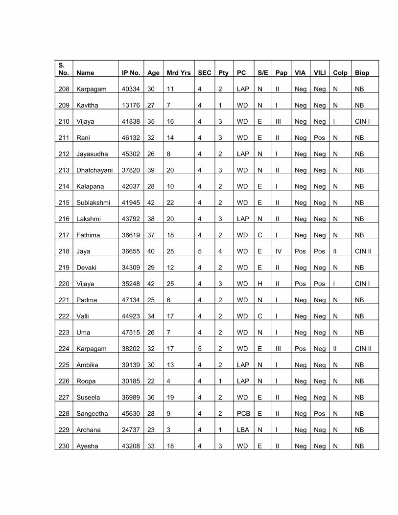
S.No. Name IP No. Age Mrd Yrs SEC Pty PC S/E Pap VIA VILI Colp Biop
208 Karpagam 40334 30 11 4 2 LAP N II Neg Neg N NB
209 Kavitha 13176 27 7 4 1 WD N I Neg Neg N NB
210 Vijaya 41838 35 16 4 3 WD E III Neg Neg I CIN I
211 Rani 46132 32 14 4 3 WD E II Neg Pos N NB
212 Jayasudha 45302 26 8 4 2 LAP N I Neg Neg N NB
213 Dhatchayani 37820 39 20 4 3 WD N II Neg Neg N NB
214 Kalapana 42037 28 10 4 2 WD E I Neg Neg N NB
215 Sublakshmi 41945 42 22 4 2 WD E II Neg Neg N NB
216 Lakshmi 43792 38 20 4 3 LAP N II Neg Neg N NB
217 Fathima 36619 37 18 4 2 WD C I Neg Neg N NB
218 Jaya 36655 40 25 5 4 WD E IV Pos Pos II CIN II
219 Devaki 34309 29 12 4 2 WD E II Neg Neg N NB
220 Vijaya 35248 42 25 4 3 WD H II Pos Pos I CIN I
221 Padma 47134 25 6 4 2 WD N I Neg Neg N NB
222 Valli 44923 34 17 4 2 WD C I Neg Neg N NB
223 Uma 47515 26 7 4 2 WD N I Neg Neg N NB
224 Karpagam 38202 32 17 5 2 WD E III Pos Neg II CIN II
225 Ambika 39139 30 13 4 2 LAP N I Neg Neg N NB
226 Roopa 30185 22 4 4 1 LAP N I Neg Neg N NB
227 Suseela 36989 36 19 4 2 WD E II Neg Neg N NB
228 Sangeetha 45630 28 9 4 2 PCB E II Neg Pos N NB
229 Archana 24737 23 3 4 1 LBA N I Neg Neg N NB
230 Ayesha 43208 33 18 4 3 WD E II Neg Neg N NB

S.No. Name IP No. Age Mrd Yrs SEC Pty PC S/E Pap VIA VILI Colp Biop
231 Rubela 42366 44 25 4 4 WD N III Pos Neg II CIN I
232 Punitha 40588 29 9 4 2 LBA N I Neg Neg N NB
233 Ganga 42603 21 3 4 1 WD N I Neg Neg N NB
234 Seetha 41118 27 9 4 2 WD E II Neg Neg N NB
235 Rajakumari 12917 25 8 4 1 LBA N I Neg Neg N NB
236 Arya 13382 32 15 4 2 LAP N II Neg Neg N NB
237 Varalakshmi 9396 24 6 4 2 WD N I Neg Neg N NB
238 Jyothi 12196 40 26 4 2 WD H III Pos Pos II CIN II
239 Rajathi 11625 37 20 4 2 LBA N I Neg Neg N NB
240 Indrani 42500 34 17 4 3 WD E II Neg Neg N NB
241 Maheswari 13116 30 12 4 2 LBA EC I Neg Neg N NB
242 Parimala 36231 25 4 4 2 WD N I Neg Neg N NB
243 Vijaya 33724 35 15 4 2 WD N II Pos Neg N NB
244 Vedavalli 15373 37 20 4 2 WD E II Neg Neg N NB
245 Manjula 24590 22 4 4 2 WD N I Neg Neg N NB
246 Kothai 26469 30 12 4 2 WD N II Neg Neg N NB
247 Savithri 17113 38 23 5 3 WD H III Pos Pos II CIN I
248 Athilakshmi 17099 45 30 5 3 WD N II Pos Pos N NB
249 Latha 18007 35 15 4 2 WD E II Pos Neg N NB
250 Prema 17101 32 12 4 2 WD E II Neg Neg N NB
251 Karpagam 16206 30 15 5 3 WD H III Pos Pos I CIN I
252 Kannagi 17598 34 15 4 1 WD N II Neg Pos N NB
253 Sundari 12865 40 25 4 2 WD E II Pos Neg II CIN I
S.No. Name IP No. Age Mrd Yrs SEC Pty PC S/E Pap VIA VILI Colp Biop

254 Deepa 12522 25 6 4 2 LAP N I Neg Neg N NB
255 Ranjitham 17922 36 20 4 2 LAP N II Neg Neg N NB
256 Pramila 38988 35 17 4 2 WD N II Neg Neg N NB
257 Kumutha 18228 31 11 4 1 LBA N I Neg Neg N NB
258 Meena 16686 33 12 4 2 WD E II Pos Pos N NB
259 Mehrunisha 38511 29 12 4 2 WD E II Pos Neg N NB
260 Murugammal 19049 30 13 4 2 WD N II Neg Neg N NB
261 Santhi 25699 43 25 4 2 LBA N III Pos Pos I N
262 Senthamarai 17052 28 8 4 2 LAP N I Neg Neg N NB
263 Athilakshmi 19385 51 35 4 2 LAP N III Pos Pos I CIN I
264 Rukmani 19722 40 22 4 2 WD E II Pos Pos I N
265 Annalakshmi 18454 25 7 4 1 WD E I Neg Neg N NB
266 Salamma 19317 55 40 5 3 WD E IV Pos Pos III SCC
267 Yasmin 19786 35 20 4 3 WD N I Neg Neg N NB
268 Rajeswari 20460 32 17 4 2 LAP H II Pos Pos N NB
269 Mary 21124 45 26 4 4 WD H II Pos Pos I CC
270 Sundari 21129 30 14 4 2 LAP H II Neg Neg N NB
271 Bavani 20640 32 15 4 2 WD H II Pos Pos I N
272 Priya 17959 24 5 4 1 LAP N I Neg Neg N NB
273 Ramya 21315 27 8 4 2 WD EC I Neg Neg N NB
274 Aarthy 17430 23 5 4 2 WD N I Neg Neg N NB
275 Annammal 29833 60 42 5 5 WD N III Pos Pos I CIN II
276 Santhanalakshmi 18733 36 15 4 3 IMB H II Neg Pos II CIN I
S.No. Name IP No. Age Mrd Yrs SEC Pty PC S/E Pap VIA VILI Colp Biop
277 Agavalli 18235 28 8 4 2 LAP N I Neg Neg N NB

278 Mumthaj 11764 36 19 4 2 WD H II Neg Neg N NB
279 Geetha 16454 30 9 4 2 WD N I Neg Neg N NB
280 Komala 11517 26 5 4 1 WD H II Pos Pos N NB
281 Fathima 16527 33 11 4 3 WD N I Neg Neg N NB
282 Santhi 16494 41 25 4 2 WD H III Neg Neg I CIN I
283 Kalyani 27328 45 30 4 3 IMB H III Neg Pos I CIN I
284 Chellamai 13844 51 35 4 3 WD N II Pos Neg II CIN I
285 Periyanayagi 22972 35 19 4 2 LAP EC II Pos Pos I N
286 Sujatha 24332 42 26 4 2 WD N II Pos Pos I N
287 Puvitha 25534 29 15 4 2 WD N II Neg Neg N NB
288 Rajeswari 23178 28 10 4 2 WD N I Neg Neg N NB
289 Jaya 22473 40 25 4 2 LBA N II Pos Pos I CC
290 Lakshmi 23289 22 4 4 2 WD N II Pos Neg N NB
291 Kala 22473 40 15 4 3 WD N III Pos Pos II CIN I
292 Selvi 28152 31 9 4 2 WD EC II Neg Neg N NB
293 Chitra 23952 25 7 4 2 WD N I Neg Neg N NB
294 Pooja 21045 26 7 4 2 WD E I Neg Neg N NB
295 Bhoopathi 44793 24 6 4 2 LAP E I Neg Neg N NB
296 Dhatchayini 22337 25 6 4 2 WD E II Pos Neg N NB
297 Vanitha 23579 27 10 4 2 WD E III Pos Pos I CIN I
298 Deepa 23370 24 7 4 2 WD N II Neg Pos N NB
299 Meenatchi 23479 32 4 4 2 WD E I Neg Neg N NB
S.No. Name IP No. Age Mrd Yrs SEC Pty PC S/E Pap VIA VILI Colp Biop
300 Padmavathi 23501 30 15 4 3 WD N III Neg Pos II CIN II
301 Ravanammal 22560 30 14 4 2 LAP EC II Neg Neg N NB

302 Lavanya 42891 28 7 4 2 LBA EC I Neg Neg N NB
303 Malliga 42851 36 13 4 2 WD N I Neg Neg N NB
304 Ruth 42628 42 24 4 2 WD E II Pos Pos I CC
305 Fathima be 24200 35 20 4 2 WD E II Neg Neg N NB
306 Parvathi 45843 44 30 4 2 LBA N I Neg Neg N NB
307 Banu 24341 26 9 4 2 WD C I Neg Neg N NB
308 Kannagi 41749 30 13 4 3 LAP E I Neg Neg N NB
309 Pachiamma 46569 47 27 4 3 LBA H IV Pos Pos III CIN III
310 Uma 46615 26 8 4 3 WD N I Neg Neg N NB
311 Kalaivani 43094 28 7 4 1 WD E I Neg Neg N NB
312 Lakshmi 44366 26 7 4 2 WD N I Neg Neg N NB
313 Rani 42863 37 20 4 3 WD E III Neg Pos II CIN II
314 Thulasi 25734 37 20 5 4 WD E III Pos Pos II CIN I
315 Selvi 42526 35 15 4 2 LBA N I Neg Neg N NB
316 Shanthi 44175 33 20 5 3 WD N I Neg Neg N NB
317 Devi 41946 25 7 4 2 WD N I Neg Neg N NB
318 Nagarathinam 43944 46 30 5 4 WD C III Pos Pos I CIN I
319 Jagatha 30659 32 15 4 4 LBA H II Pos Pos I N
320 Santha 16921 36 13 4 2 LBA N I Neg Neg N NB
321 Puspa 43094 26 8 4 2 WD N I Neg Neg N NB
322 Saraswathi 39643 30 9 4 1 WD N I Neg Neg N NB
S.No. Name IP No. Age Mrd Yrs SEC Pty PC S/E Pap VIA VILI Colp Biop
323 Prema 43053 30 13 4 2 WD H III Pos Pos I CIN I
324 Mariamma 38081 32 18 4 2 WD N I Neg Neg N NB
325 Fathima 31625 23 5 44 2 WD N I Neg Neg N NB

326 Prabavathi 43220 30 14 4 3 WD E II Pos Pos I CC
327 Meenatchi 43160 30 15 4 2 WD N I Neg Neg N NB
328 Shanthi 43168 37 20 4 3 WD N II Neg Neg I CIN I
329 Prabhavathi 14653 30 14 4 3 WD N I Neg Neg N NB
330 Arasi 42325 36 15 4 2 LBA N I Neg Neg N NB
331 Suseela 42240 35 15 4 4 WD E II Pos Pos I N
332 Geetha 43014 30 12 4 2 WD N I Neg Neg N NB
333 Muthulakshmi 42309 57 40 4 5 WD E IV Pos Pos II CIN II
334 Komala 45001 23 4 4 1 WD N I Neg Neg N NB
335 Lakshmi 42000 50 30 4 6 WD U IV Pos Pos III CIN III
336 Kasdoori 31091 36 20 4 2 WD N I Neg Neg N NB
337 Malathi 42023 36 19 4 2 WD E I Neg Neg N NB
338 Anitha 41806 30 8 4 2 WD H III Pos Pos I CC
339 Marry 30901 42 17 4 3 WD H III Pos Pos II CIN I
340 Lalitha 42200 56 20 4 2 WD N I Neg Neg N NB
341 Nagavalli 15609 38 17 4 2 WD N II Pos Pos N NB
342 Rajakumari 41627 32 13 4 3 LAP N I Neg Neg N NB
343 Gandhimathi 40051 48 26 4 3 WD E I Neg Neg N NB
344 Devi 41662 28 12 4 2 WD C II Neg Neg N NB
345 Valli 41374 35 19 4 3 WD N II Pos Pos I CIN I
S.No. Name IP No. Age Mrd Yrs SEC Pty PC S/E Pap VIA VILI Colp Biop
346 Vijaya 19175 33 16 4 2 WD E II Pos Pos N NB
347 Malarvizhi 20011 50 30 4 3 WD N I Neg Neg N NB
348 Ragavi 40559 25 7 4 2 LAP N I Neg Neg N NB
349 Bakkiam 45590 23 4 4 2 WD N I Neg Neg N NB

350 Mary 43269 43 23 4 2 WD E II Pos Neg N NB
351 Thilagavathi 33087 30 14 4 3 WD E III Neg Pos II CIN I
352 Kala 38750 25 8 4 2 WD C I Neg Neg N NB
353 Kavitha 38021 28 11 4 3 WD N II Neg Neg N NB
354 Devi 37953 30 11 4 2 LAP N I Neg Neg N NB
355 Maha 38634 35 15 4 3 LBA EC I Neg Neg N NB
356 Padma 38082 40 24 4 2 LBA N I Neg Neg N NB
357 Sathya 22653 50 30 4 3 WD OCT III Neg Neg I CIN I
358 Amala 14978 35 19 4 3 LAP N II Neg Neg N NB
359 Saroja 13627 35 13 4 2 WD P I Neg Neg N NB
360 Suguna 38292 32 16 5 5 WD OCT II Neg Pos N NB
361 Chandra 32305 40 22 4 4 LBA E II Pos Pos I CC
362 Anjali 14024 26 8 4 2 LAP N I Neg Neg N NB
363 Muthulakshmi 12660 38 16 4 2 LAP N I Neg Neg N NB
364 Maheswari 12152 45 25 4 4 WD N III Pos Pos I CIN I
365 Kasthuri 12193 40 18 4 3 LAP OCT I Neg Neg N NB
366 Meera 10969 35 10 4 2 WD N I Neg Neg N NB
367 Amaravathi 32370 45 27 4 3 WD E II Pos Pos I CC
368 Victoria 26033 47 30 4 4 LAP EC II Neg Neg N NB
S.No. Name IP No. Age Mrd Yrs SEC Pty PC S/E Pap VIA VILI Colp Biop
369 Marry 27879 45 25 5 4 WD H III Pos Neg II CIN II
370 Sasikala 10600 26 6 4 2 WD N I Neg Neg N NB
371 Reena 19486 32 15 4 2 WD E I Neg Neg N NB
372 Saraswathi 27164 40 25 4 2 WD N II Neg Neg N NB
373 Savithri 27151 40 24 4 3 PCB E IV Pos Pos II CIN II

374 Latha 22598 24 4 4 2 WD N I Neg Neg N NB
375 Rani 17982 28 9 4 2 WD C I Neg Neg N NB
376 Sujatha 22964 38 20 5 2 LBA E II Neg Neg N NB
377 Padmavathi 20332 27 8 4 2 WD N I Neg Neg N NB
378 Malliga 22182 29 10 4 1 IMB P II Neg Neg N NB
379 Egavalli 41565 37 20 4 3 WD E II Pos Pos N NB
380 Meenatchi 20561 28 11 4 2 WD N I Neg Neg N NB
381 Thanam 28108 48 30 4 3 LAP OCT II Pos Neg N NB
382 Jeyanthi 20180 33 12 5 3 WD E I Neg Neg N NB
383 Meena 14172 36 20 4 2 WD H II Pos Pos I CC
384 Thilothama 40817 26 8 4 2 WD N I Neg Neg N NB
385 Suguna 40823 37 22 4 3 LAP N III Pos Pos I CIN I
386 Radha 41234 28 10 4 2 LAP N I Neg Neg N NB
387 Sumathi 41362 30 12 5 3 WD E II Pos Pos I CC
388 Yasodha 18200 30 13 4 3 WD N I Neg Neg N NB
389 Kasthoori 23322 40 21 4 1 WD N I Neg Neg N NB
390 Komala 21254 22 6 4 2 WD E II Neg Pos N NB
391 Latha 23952 36 20 4 2 LBA C II Neg Neg N NB

S.No. Name IP No. Age Mrd Yrs SEC Pty PC S/E Pap VIA VILI Colp Biop
392 Prabavathi 22641 42 17 5 2 LAP N I Neg Neg N NB
393 Chitra 26057 25 10 5 3 WD N I Neg Neg N NB
394 Rajeswari 25732 34 18 5 3 WD H II Neg Neg I CIN I
395 Nirmala 26273 48 30 5 6 WD E II Neg Neg N NB
396 Mangai 25919 46 30 4 2 LBA EC I Neg Neg N NB
397 Varalakshmi 22903 40 20 5 2 WD E II Pos Pos II CC
398 Parveen 26761 35 19 5 4 WD EC II Neg Neg N NB
399 Kumari 21231 25 4 4 2 WD N I Neg Neg N NB
400 Jaya 25784 40 25 4 2 LAP N II Neg Neg N NB
401 Jayamma 22598 35 10 4 2 LAP N I Neg Neg N NB
402 Kumari 12440 24 7 4 2 WD N I Neg Neg N NB
403 Sarasu 24132 27 6 4 2 LBA C II Neg Neg N NB
404 Parameswari 27330 40 21 4 2 LBA EC I Neg Neg N NB
405 Kalai 22646 42 23 5 5 WD E III Pos Neg II CIN I
406 Ramani 13387 38 12 4 2 WD N II Neg Pos II CIN I
407 Nagammaal 27591 28 8 4 2 LAP N II Pos Neg N NB
408 Shantha 37591 35 17 4 3 LBA EC I Neg Neg N NB
409 Stella 37850 28 6 4 2 WD N I Neg Neg N NB
410 Thilagavathi 31856 38 20 4 3 WD E II Neg Neg N NB
411 Rajeswari 37953 45 30 5 5 WD E IV Pos Pos III CIN III
412 Indhira 37903 23 5 4 2 WD N I Neg Neg N NB
413 Shanthi 37512 27 8 4 2 WD E I Neg Neg N NB
414 Rajee 37706 45 24 4 2 WD N II Neg Neg N NB
S.No. Name IP No. Age Mrd Yrs SEC Pty PC S/E Pap VIA VILI Colp Biop

415 Valli 37656 55 40 5 4 WD U IV Pos Pos III CIN III
416 Marry 37710 36 16 4 4 WD N II Neg Neg N NB
417 Nageswari 37645 32 15 4 2 WD P II Neg Pos N NB
418 Vani 38225 38 20 5 5 WD E III Pos Pos I CC
419 Selvi 38272 28 10 4 3 WD E II Neg Neg N NB
420 Thanam 16002 30 14 4 2 LAP N I Neg Neg N NB
421 Kasthoori 17780 35 15 4 3 WD H II Neg Neg N NB
422 Ellammal 25425 23 4 4 1 LAP N I Neg Neg N NB
423 Nirmala 22564 40 25 4 3 WD N I Neg Neg N NB
424 Lakshmi 13922 30 11 4 2 LBA N I Neg Neg N NB
425 Uma 25194 26 5 4 1 WD N II Neg Neg N NB
426 Vasanthi 28563 34 18 4 2 WD E III Pos Pos II CIN I
427 Shanthi 12211 30 12 4 2 LAP N II Pos Neg N NB
428 Ranganayagi 23975 32 10 4 3 LBA N II Neg Pos N NB
429 Thagira 23897 30 12 4 2 WD N II Neg Neg N NB
430 Gayathri 23695 28 10 4 2 WD N II Pos Neg N NB
431 Jamuna 32305 32 15 4 2 WD N I Neg Neg N NB
432 Shanthi 27164 26 6 4 2 LAP N I Neg Neg N NB
433 Vijaya 22964 42 23 4 3 WD N III Pos Neg II CIN I
434 Mahalakshmi 30659 30 15 4 2 WD N II Pos Neg N NB
435 Roja 39675 21 3 4 1 LAP N II Neg Neg N NB
436 Shankari 33478 38 22 5 5 WD E II Neg Neg N NB
437 Latha 32170 38 20 4 2 WD E III Pos Pos I CIN I

S.No. Name IP No. Age Mrd Yrs SEC Pty PC S/E Pap VIA VILI Colp Biop
438 Selvi 32790 39 23 4 3 WD C II Pos Neg N NB
439 Vijayalakshmi 23367 30 9 4 2 LAP N I Neg Neg N NB
440 Yasodha 25693 30 8 4 2 LAP E II Neg Neg N NB
441 Allirani 17998 40 23 4 3 WD EC II Pos Pos I CC
442 Jyothi 23121 23 6 4 2 WD E I Neg Neg N NB
443 Mohana 24753 30 14 4 2 LAP N II Neg Neg N NB
444 Malar 43213 28 10 4 2 WD E II Neg Neg N NB
445 Thangam 42569 28 10 4 2 LAP N I Neg Neg N NB
446 Maha 43511 33 15 4 2 WD E II Neg Neg N NB
447 Nabeesha 28977 25 6 4 1 WD N II Neg Pos N NB
448 Chellammal 32667 28 10 5 2 WD P II Neg Neg N NB
449 Vanaja 46677 38 18 4 2 WD N II Neg Pos II CIN I
450 Beevi 45534 28 10 4 4 LBA H II Pos Pos I CC
451 kannagi 40700 28 7 4 1 WD E I Neg Neg N NB
452 Shanthi 41872 35 15 5 2 LBA N I Neg Neg N NB
453 Poovitha 32413 24 7 4 2 WD N II Neg Neg N NB
454 Kanchana 37834 28 10 4 2 WD N II Neg Pos N NB
455 Anandavalli 22561 28 13 5 4 WD H III Pos Neg I CIN I
456 Rani 46611 28 9 4 2 WD C I Neg Neg N NB
457 Parimala 23423 25 4 4 2 WD N I Neg Neg N NB
458 Kalai 43268 28 7 4 1 WD E I Neg Neg N NB
459 Manju 38750 25 7 4 1 WD N I Neg Neg N NB
460 Kavitha 37953 22 4 4 1 LBA N II Neg Neg N NB
S.No. Name IP No. Age Mrd Yrs SEC Pty PC S/E Pap VIA VILI Colp Biop

461 Amirtham 42981 34 17 4 2 WD E III Pos Neg II CIN I
462 Lavanya 23844 24 7 4 2 LAP EC I Neg Neg N NB
463 Rathna 23178 26 10 4 2 LBA N I Neg Neg N NB
464 Pratheepa 43273 32 14 5 2 WD E II Neg Neg N NB
465 Malarkodi 29722 33 15 4 2 WD E II Neg Pos N NB
466 Ramya 20466 27 10 4 2 WD N I Neg Neg N NB
467 Savitha 22141 35 19 4 4 WD EC II Neg Neg N NB
468 Kumari 21937 25 4 4 2 WD N I Neg Neg N NB
469 Jayamma 17114 35 10 4 2 LAP N I Neg Neg N NB
470 Kanaga 17598 34 18 5 3 WD H III Neg Pos I CIN I
471 Latha 28988 24 4 4 2 WD N I Neg Neg N NB
472 Padmavathi 29049 27 8 4 2 LAP N I Neg Neg N NB
473 Rajathi 45637 37 20 4 2 LBA N I Neg Neg N NB
474 Indrani 40833 34 17 4 3 WD E II Neg Neg N NB
475 Maheswari 22178 30 12 4 2 LBA EC I Neg Neg N NB
476 Parimala 42356 25 4 4 2 WD N I Neg Neg N NB
477 Vijaya 20647 35 15 4 2 WD N II Neg Neg N NB
478 Vedavalli 38512 37 20 4 2 WD E II Neg Neg I N
479 Manjula 28988 22 4 4 2 WD N I Neg Neg I N
480 Kothai 37922 30 12 4 2 WD N II Neg Neg I N
482 Rabitha 31711 55 40 4 4 LBA N I Neg Neg I N
483 Selvarani 26469 36 17 4 2 LBA N I Neg Neg I N

S.No. Name IP No. Age Mrd Yrs SEC Pty PC S/E Pap VIA VILI Colp Biop
484 Badma 22182 23 5 4 1 WD N I Neg Neg I N
485 Banu 20332 26 9 4 2 LAP E II Neg Neg I N
486 Geetha 31092 28 10 4 2 LAP H II Neg Neg I N
487 Rajee 45008 32 14 4 2 WD N II Neg Neg I N
488 Rathna 46569 30 12 4 2 IMB P II Neg Neg I N
489 Kala 41749 45 25 5 3 WD N III Neg Neg III CIN I
490 Devi 22457 29 13 4 2 WD E II Neg Pos II CIN I
491 Lalitha 31934 56 20 4 2 WD N I Neg Neg I N
492 Nagavalli 41121 38 17 4 2 WD N II Neg Neg I N
493 Rajakumari 40088 32 13 4 3 LAP N I Neg Neg I N
494 Gandhimathi 41098 48 26 4 3 WD E I Neg Neg I N
495 Devi 34532 28 12 4 2 WD C II Neg Neg I N
496 Rajathi 41338 37 20 4 2 LBA N I Neg Neg I N
497 Indrani 42578 34 17 4 3 WD E II Neg Neg I N
498 Maheswari 48923 30 12 4 2 LBA EC I Neg Neg I N
499 Parimala 32338 25 4 4 2 WD N I Neg Neg I N
500 Vijaya 35987 35 15 4 2 WD N II Neg Neg I N

KEY WORDS TO MASTER CHART
Mrd Yrs = Married Years Duration
PC = Presenting Complaint
Biop = Biopsy
Colp = Colposcopy
Pty = Parity
WD = White Discharge
LAP = Lower Abdominal Pain
LBA = Low Backache
IMB = Inter Menstrual Bleed
PCB = Post Coital Bleed
N = Normal
E = Erosion

EC = Ectropion
H = Hypertrophy
OCT = Old Cervical Tear
P = Polyp
U = Ulcer
C = Congestion
NB = No Biopsy
CC = Chronic Cervicitis
SCC = Squamous Cell Carcinoma