materials in drug delivery applications - mdpi
TRANSCRIPT

marine drugs
Review
Sulfated Seaweed Polysaccharides as MultifunctionalMaterials in Drug Delivery Applications
Ludmylla Cunha 1,2 and Ana Grenha 1,2,*
1 Centre for Marine Sciences, University of Algarve, 8005-139 Faro, Portugal; [email protected] Drug Delivery Laboratory, Centre for Biomedical Research (CBMR), Faculty of Sciences and Technology,
University of Algarve, Gambelas Campus, 8005-139 Faro, Portugal* Correspondence: [email protected]; Tel.: +351-289-244-441; Fax: +351-289-800-066
Academic Editor: Paola LaurienzoReceived: 14 January 2016; Accepted: 15 February 2016; Published: 25 February 2016
Abstract: In the last decades, the discovery of metabolites from marine resources showing biologicalactivity has increased significantly. Among marine resources, seaweed is a valuable source ofstructurally diverse bioactive compounds. The cell walls of marine algae are rich in sulfatedpolysaccharides, including carrageenan in red algae, ulvan in green algae and fucoidan in brownalgae. Sulfated polysaccharides have been increasingly studied over the years in the pharmaceuticalfield, given their potential usefulness in applications such as the design of drug delivery systems.The purpose of this review is to discuss potential applications of these polymers in drug deliverysystems, with a focus on carrageenan, ulvan and fucoidan. General information regarding structure,extraction process and physicochemical properties is presented, along with a brief reference toreported biological activities. For each material, specific applications under the scope of drug deliveryare described, addressing in privileged manner particulate carriers, as well as hydrogels and beads.A final section approaches the application of sulfated polysaccharides in targeted drug delivery,focusing with particular interest the capacity for macrophage targeting.
Keywords: carrageenan; drug delivery; fucoidan; macrophage targeting; ulvan;sulfated polysaccharides
1. Introduction
Marine environment and the associated wide diversity of organisms offer a rich source of valuablematerials. Among marine resources, algae, which are sometimes referred as seaweeds, are well knownnatural sources of polysaccharides. Sulfated polysaccharides are of the most common in the cell wallsof seaweeds. The number and chemical structure of these polymers vary according to the specific algalspecies [1].
Marine algae can be classified into three main groups based on the exhibited photosyntheticpigments: red, brown and green. Botanists refer to these groups as Rhodophyceae, Phaeophyceae andChlorophyceae, respectively. Brown seaweeds are usually large and range from the giant kelp that isoften 20 m long, to thick, leather-like seaweeds of 2–4 m long, to smaller species 30–60 cm long. Redseaweeds are usually smaller, generally ranging from a few centimeters to about a meter in length.Curiously, red seaweeds are not always red, sometimes being purple, even brownish red, but stillbeing classified by botanists as Rhodophyceae because of other characteristics. Green seaweeds arealso small, with a size range similar to that of red seaweeds [2].
In the last decades, sulfated polysaccharides of algal origin have attracted much attention asfunctional additives in the pharmaceutical field, but also in food and cosmetic industries. The major
Mar. Drugs 2016, 14, 42; doi:10.3390/md14030042 www.mdpi.com/journal/marinedrugs

Mar. Drugs 2016, 14, 42 2 of 41
sulfated polysaccharides found in marine algae include carrageenan from red algae, ulvan isolatedfrom green algae and fucoidan from brown algae [3]. Carrageenan is the most used of the three,with wide application as emulsifier, stabilizer or thickener. Fucoidan, in turn, is available commerciallyfrom various cheap sources and has been investigated in recent years to develop novel drugs, medicinesand functional foods. Ulvan is the less known of the group. It displays several physicochemical andbiological features of potential interest for food, pharmaceutical, agricultural and chemical applications,but needs deeper investigation. Various studies have revealed that sulfated polysaccharides isolatedfrom marine algae exhibit a variety of biological activities [4–6], potentiating their use in pharmaceuticalapplications. These polymers have been increasingly studied over the years in this context, given theirpotential usefulness in applications that mainly involve the design of drug delivery systems. Figure 1depicts the number of publications retrieved on ISI Web of Knowledge with the keywords “name ofthe polymer” and “drug delivery”, showing the increasing interest in carrageenan and fucoidan in therecent years, with the undoubted prevalence of the former. Additionally, it confirms that ulvan is theless explored of the three polymers.
Mar. Drugs 2016, 14, x 2 of 40
wide application as emulsifier, stabilizer or thickener. Fucoidan, in turn, is available commercially
from various cheap sources and has been investigated in recent years to develop novel drugs,
medicines and functional foods. Ulvan is the less known of the group. It displays several
physicochemical and biological features of potential interest for food, pharmaceutical, agricultural
and chemical applications, but needs deeper investigation. Various studies have revealed that
sulfated polysaccharides isolated from marine algae exhibit a variety of biological activities [4–6],
potentiating their use in pharmaceutical applications. These polymers have been increasingly studied
over the years in this context, given their potential usefulness in applications that mainly involve the
design of drug delivery systems. Figure 1 depicts the number of publications retrieved on ISI Web of
Knowledge with the keywords “name of the polymer” and “drug delivery”, showing the increasing
interest in carrageenan and fucoidan in the recent years, with the undoubted prevalence of the former.
Additionally, it confirms that ulvan is the less explored of the three polymers.
Figure 1. Number of scientific publications published on the topic “name of polymer” and “drug
delivery” as a function of publication years. Taken from ISI Web of Knowledge. The colors allude to
the colors of algae (red: carrageenan, brown: fucoidan, green: ulvan).
The purpose of this review is to discuss potential applications of sulfated polysaccharides, with
a special emphasis on carrageenan, ulvan and fucoidan, in designing drug delivery systems and also
address their suitability for specific applications, such as cell targeting. A summary of basic
characteristics of the three carbohydrates is displayed in Table 1.
Figure 1. Number of scientific publications published on the topic “name of polymer” and “drugdelivery” as a function of publication years. Taken from ISI Web of Knowledge. The colors allude tothe colors of algae (red: carrageenan, brown: fucoidan, green: ulvan).
The purpose of this review is to discuss potential applications of sulfated polysaccharides, witha special emphasis on carrageenan, ulvan and fucoidan, in designing drug delivery systems andalso address their suitability for specific applications, such as cell targeting. A summary of basiccharacteristics of the three carbohydrates is displayed in Table 1.

Mar. Drugs 2016, 14, 42 3 of 41
Table 1. Description of several characteristics of carrageenan, fucoidan and ulvan.
SulfatedPolysaccharide
Marine AlgaeGroup Main Genera Molecular Weight
(kDa)Solubility in
WaterViscosity
(cps, in Water)pH in Aqueous
Solution References
Carrageenan Rhodophyceae
ChondrusEuchema
FurcellariaGigartinaHypneaIridae
Kappaphycus
100–1000* κ-, ι- and
λ-carrageenansoluble at 80 ˝C
5–800(1.5% w/v, 75 ˝C) 7.0–10.0 [2,7–10]
Fucoidan Phaeophyceae
AnalipusChorda
DictyotaFucus
KjellmaniellaPelvetia
SargassumUndaria
10–950 10 mg/mL(F. vesiculosus) n.a. n.a. [11–19]
Ulvan Chlorophyceae EnteromorphaUlva 1.14 to > 2 ˆ 106 n.a. 18–100
(1.6% w/v, Ulva spp.) 7.5 (Ulva spp.) [20–22]
* Further solubility conditions in Reference [10]; n.a.: not available.
Mar. Drugs 2016, 14, 42 4 of 41
2. Carrageenan: Sulfated Polysaccharide of Red Seaweeds
Historically, red seaweeds (Rhodophyta) have been harvested and consumed as foods for at least2800 years. Although red algae are consumed by humans, the carrageenan extracted from seaweedis not assimilated by the human body, merely providing bulk. However, these algae do providefunctional properties that are exploited on a commercial scale [23]. As one of its main properties relieson the ability to form thermoreversible gels or highly viscous solutions, carrageenan is commonly usedas gelifying, stabilizing and emulsifying agent in food, pharmaceutical and cosmetic industry [24].A broad and recent review on the industrial applications of carrageenan, including in food andpharmaceutical related areas, is available on [25].
2.1. Origin, Extraction and Processing
The original source of carrageenan was the red seaweed Chondrus crispus (also known as IrishMoss). With the expansion of the carrageenan industry over time, the increasing demand for the rawmaterial led to the introduction of the cultivation of species of Eucheuma, originally E. cottonii andE. spinosum, now referred to as Kappaphycus alvarezii and Eucheuma denticulatum, respectively [2,23].The advantage thereof compared to the natural Chondrus crispus, is a predominant content ofkappa- and iota-carrageenan, respectively, while Chondrus crispus contains a mixture of kappa and lambdacarrageenan that cannot be separated during commercial extraction. Therefore, most of carrageenan isnow extracted from K. alvarezii and E. denticulatum but several species of Gigartina, Iridae, Hypnea andFurcellaria genera have been exploited, providing different types of carrageenan extracts [2,26,27].
Carrageenan manufacture consists of extraction, purification, concentration, precipitation anddrying, although the basic process may vary according to the red algae family from which thepolysaccharide is extracted. In some cases, pretreatment to remove excess of color or alkalinemodification can be also performed prior to extraction [23]. The specific details of the extractionprocess are secured as trade secrets by several carrageenan manufacturers. There are two mainmethods for producing carrageenan based on different principles. Briefly, in the first and originalmethod carrageenan is extracted from the seaweed in an aqueous solution. After filtration to removethe remaining residues, carrageenan is usually recovered from the solution by addition of an alcoholto induce precipitation. Finally, the precipitate is separated, dried and milled, resulting in a refinedcarrageenan. In the second method, carrageenan is actually not extracted from the seaweed. Instead,the principle is to wash out residual minerals, soluble protein and fat from the seaweed, leaving behindcarrageenan and other insoluble matter. This insoluble residue, consisting largely of carrageenanand cellulose, is then dried and sold as semi-refined carrageenan, usually for non-food gellingapplications. Although the process is much shorter and cheaper than the first one, its purity isnecessarily lower [2,28–31]. Other methods can be found in the literature such as enzyme-treatedor fungal-treated extractions [32–35]. Enzymatic extraction, for example, can enable the productionof specific gelation properties, since the physicochemical properties of carrageenan depend on thecomposition of the polysaccharide and on the number of counterions [36].
2.2. Chemical Structure and Types of Carrageenan
The seaweeds that biosynthesize this polysaccharide are called carrageenophytes. Carrageenanis in fact a general name for a family of galactans, the commonest and most abundant cell wallconstituents encountered in red algae. The backbone structure of this polysaccharide is based on linearchains of repeating galactose units in D configuration (D-sugar) and 3,6-anhydro-galactose copolymer,joined by alternating α-(1Ñ3) and β-(1Ñ4) linkages, as shown in Figure 2. In terms of chemicalstructure, this polygalactan is classified into various types, including but not limited to κ-, λ-, ι-, µ-,θ-, β- and ν-carrageenans, all containing 15%–40% ester sulfate with the exception of β-carrageenan,which is devoid of sulfate content [1,26].

Mar. Drugs 2016, 14, 42 5 of 41Mar. Drugs 2016, 14, x 5 of 40
Figure 2. Carrageenan structures. Linear chains of repeating galactose units in D configuration and
3,6‐anhydro‐galactose copolymer, joined by alternating α‐(1→3) and β‐(1→4) glycosidic linkages.
At least 15 different carrageenan structures are reported, with κappa (κ), ιota (ι) and lambda (λ)
forms being the most industrially relevant. The major difference among the various forms of
carrageenan is related to structural characteristics, including the number and the position of sulfate
groups and the occurrence of 3,6‐anhydro‐D‐galactose in the chain [37]. For instance, κ‐, ι‐ and λ‐
carrageenans are distinguished by the presence of one, two and three ester‐sulfate groups per
repeating disaccharide unit, respectively [38]. The chemical structures of carrageenans are, thus, very
heterogeneous and are correlated to the algal sources, the life stage of the seaweed (i.e., gametophyte)
and the extraction procedures of the polysaccharide [27].
2.3. Physicochemical Properties
The particular composition and the conformation of a given polysaccharide constitute the basis
of its physicochemical and biological properties. Self‐assembling and gel formation ability, for
instance, are related with specific conformations resulting from primary polysaccharide
structures [38].
The average molecular weight of commercially available carrageenan ranges from 100 to 1000
kDa [9]. The structure of κ‐carrageenan was reported as alternating 3‐linked β‐D‐galactose and
4‐linked anhydro‐galactose (AG) units. It has an ester sulfate content of about 25%–30% and a
Figure 2. Carrageenan structures. Linear chains of repeating galactose units in D configuration and3,6-anhydro-galactose copolymer, joined by alternating α-(1Ñ3) and β-(1Ñ4) glycosidic linkages.
At least 15 different carrageenan structures are reported, with κappa (κ), ιota (ι) and lambda(λ) forms being the most industrially relevant. The major difference among the various forms ofcarrageenan is related to structural characteristics, including the number and the position of sulfategroups and the occurrence of 3,6-anhydro-D-galactose in the chain [37]. For instance, κ-, ι- andλ-carrageenans are distinguished by the presence of one, two and three ester-sulfate groups perrepeating disaccharide unit, respectively [38]. The chemical structures of carrageenans are, thus, veryheterogeneous and are correlated to the algal sources, the life stage of the seaweed (i.e., gametophyte)and the extraction procedures of the polysaccharide [27].
2.3. Physicochemical Properties
The particular composition and the conformation of a given polysaccharide constitute the basis ofits physicochemical and biological properties. Self-assembling and gel formation ability, for instance,are related with specific conformations resulting from primary polysaccharide structures [38].
The average molecular weight of commercially available carrageenan ranges from 100 to1000 kDa [9]. The structure of κ-carrageenan was reported as alternating 3-linked β-D-galactoseand 4-linked anhydro-galactose (AG) units. It has an ester sulfate content of about 25%–30% and

Mar. Drugs 2016, 14, 42 6 of 41
a 3,6-AG content of about 28%–35%. Iota carrageenan has an additional sulfate group on C-2 of theAG residue, resulting in two sulfates per disaccharide repeating unit. It has an ester sulfate contentof 28%–30% and about 25%–30% content of 3,6-AG. Lambda carrageenan has three sulfate groups perdisaccharide unit with the third sulfate group of this form at the C-6 position of the 4-linked residue.There is an ester sulfate content of about 32% to 39% and no 3,6-AG content [38].
Carrageenan exhibits the solubility characteristics normally shown by hydrophilic colloids. It iswater soluble and insoluble in most organic solvents such as alcohol, ether and oil. Aqueous solubilityis influenced by a number of factors, including the type of carrageenan, temperature, pH, numberof counterions and the presence of other solutes. The numerous forms of carrageenan, showingvariations in the chemical structure, as described above, provide much variability regarding solubilityproperties [8,23]. In general, hydrophilicity is structurally provided by the hydrophilic sulfateand hydroxyl groups, while 3,6-anhydro-D-galactose residues (3,6-AG) are more hydrophobic [39].Therefore, λ-carrageenan, being highly sulfated and having no 3,6-AG content, is easily soluble undermost conditions, whereas κ-carrageenan, with 3,6-AG residue and fewer sulfate groups, is relativelyless hydrophilic and, thus, less soluble. The intermediate ι-carrageenan, is more hydrophilic dueto the presence of the two sulfate groups, which counteract the slight hydrophobic character of the3,6-AG residue. Solubility characteristics are also affected by the salt form of the sulfated ester groups.The free acid is unstable and, therefore, commercial carrageenans are available as sodium, potassiumand calcium salts or, most commonly, as a mixture of these. These salts afford the needed stability.In general, the sodium forms of carrageenan are more easily soluble, while potassium forms dissolvewith more difficulty. For instance, the potassium salt of both κ- and ι-carrageenan is insoluble in coldwater, requiring the application of temperature to bring the polysaccharide into solution, whereas inthe sodium form it dissolves readily. However, λ-carrageenan is soluble in all its salt forms. Moreover,both the dissolving rate and solubility of carrageenan are affected by the presence of other soluteswhich may compete for available water, thus altering the state of hydration of the polysaccharide [8].
The products emerging from the manufacturing process are inherently variable. Differencesin algal sources and adjustments in processing conditions produce different carrageenans.The standardization of the product is determined by the functionality required in the specificapplication. The product is sold as a powder, which dissolves slowly at room temperature, producinga highly viscous aqueous solution. Viscosity depends on the type of carrageenan, its molecular weight,the used polymer concentration, applied temperature and the presence of other solutes (such as salts).The viscosity of a carrageenan solution decreases with decreasing concentration, sulfation or molecularweight and by increasing temperature. In addition, due to its swelling ability, carrageenan is difficultto disperse in water due to the formation of a film layer around each carrageenan particle. This leads tothe formation of large agglomerates which make it very difficult for the water molecules to penetrate.Efficient dispersion can be achieved by high-speed mixing or by premixing the powder with inertmatter, such as a sugar in a carrageenan/sugar mass ratio of 1:10. The presence of acid and oxidizingagents in solution may induce carrageenan hydrolysis leading to loss of physical properties throughcleavage of glycosidic linkages. Such depolymerization is greatly accelerated by the presence ofdissolved oxygen, high temperature and low pH. Therefore, in order to ensure minimum degradationduring processing, high temperature short time processes are preferred. In this context, optimumstability of carrageenan occurs at pH 9, while severe degradation is known to occur at pH below3.5 [8,23].
As previously mentioned, commercial carrageenans are available as stable sodium, potassiumand calcium salts. The associated cations together with the conformation of the galactose units in thepolymer chain, which results in different carrageenan types, determine the gelling properties of thesecarbohydrates [8]. Thermal gelation is a valuable property of carrageenans that is determinantin diverse applications, including in food and pharmaceutical industries. The functionality ofcarrageenans in various applications depends on the rheological properties. Carrageenan solutionsare non-Newtonian fluids and show pseudoplastic behavior [23]. At a certain range of temperatures

Mar. Drugs 2016, 14, 42 7 of 41
and cation concentrations, carrageenan solutions may gel and the viscoelastic properties of thesegels vary depending on these parameters [40]. The available carrageenans differ in their ability toundergo gelation. While κ- and ι-carrageenans form gels, λ-carrageenan does not gel and behaves as acommon polyelectrolyte in solution [41]. There is general agreement on the mechanism of gelation ofthe polymer. It is assumed that, in solution and at high temperature, carrageenans exist as randomcoils. When dissolved by heating, followed by cooling below certain temperatures, the reduction intemperature induces the formation of double helices. The structure of κ- and ι-carrageenan allowssegments of the two molecules to form the so-called double helices, which bind the chain molecules ina three-dimensional network that is in fact a gel. In turn, λ-carrageenan has a structure that does notallow the formation of such double helix, presumably due to its high degree of sulfate substitution,which hampers gel formation [40]. In other words, the gelation of carrageenan solutions occurs asa result of coil-to-helix conformational transition and the subsequent aggregation among orderedhelices [42], as depicted in Figure 3. By itself, the formation of the helical structure does not lead tocarrageenan gelation, though. The associated counterions such as Na+, K+ and Ca2+, mentioned before,are those responsible for the final sol-gel transition of the polysaccharide. The role of various cationsin promoting cross-links and inducing a gelation was previously studied. In the particular case ofcarrageenan, the formation of the double helix corresponds to a limited number of chains that arelinked together through intermolecular forces. However, this occurs into small domains that requirefurther association by cation-mediated helix-helix aggregation to develop a cohesive network. Onlythe latter structure corresponds to the gel.
Mar. Drugs 2016, 14, x 7 of 40
vary depending on these parameters [40]. The available carrageenans differ in their ability to undergo
gelation. While κ‐ and ι‐carrageenans form gels, λ‐carrageenan does not gel and behaves as a
common polyelectrolyte in solution [41]. There is general agreement on the mechanism of gelation of
the polymer. It is assumed that, in solution and at high temperature, carrageenans exist as random
coils. When dissolved by heating, followed by cooling below certain temperatures, the reduction in
temperature induces the formation of double helices. The structure of κ‐ and ι‐carrageenan allows
segments of the two molecules to form the so‐called double helices, which bind the chain molecules
in a three‐dimensional network that is in fact a gel. In turn, λ‐carrageenan has a structure that does
not allow the formation of such double helix, presumably due to its high degree of sulfate substitution,
which hampers gel formation [40]. In other words, the gelation of carrageenan solutions occurs as a
result of coil‐to‐helix conformational transition and the subsequent aggregation among ordered
helices [42], as depicted in Figure 3. By itself, the formation of the helical structure does not lead to
carrageenan gelation, though. The associated counterions such as Na+, K+ and Ca2+, mentioned before,
are those responsible for the final sol‐gel transition of the polysaccharide. The role of various cations
in promoting cross‐links and inducing a gelation was previously studied. In the particular case of
carrageenan, the formation of the double helix corresponds to a limited number of chains that are
linked together through intermolecular forces. However, this occurs into small domains that require
further association by cation‐mediated helix‐helix aggregation to develop a cohesive network. Only
the latter structure corresponds to the gel.
Figure 3. Scheme of carrageenan gel formation. The structure of κ‐ and ι‐carrageenan allows segments
of the two molecules to form the so‐called double helices which bind the chain molecules in a three‐
dimension network. The associated counterions such as Na+, K+ and Ca2+, are also required to induce
the sol‐gel transition of the referred types of carrageenan.
Of all carrageenans, kappa is the one providing the strongest gels. The strength of carrageenan
gels is greatly dependent on the carrageenan concentration and is also related to the type and
concentration of cation. The effectiveness of salts in influencing gel strength has been evaluated and
it was shown that the addition of salts such as NaCl, KCl, CaCl2 and BaCl2 at an adequate
concentration apparently improves the gel strength of κ‐carrageenan gels by means of an
enhancement of conformational ordering and subsequent aggregation. Several works devoted to the
study of the influence of salts on carrageenan gel properties (κ‐ and ι‐carrageenan) should be
consulted for further details [23,39,40,42].
Figure 3. Scheme of carrageenan gel formation. The structure of κ- and ι-carrageenan allowssegments of the two molecules to form the so-called double helices which bind the chain molecules ina three-dimension network. The associated counterions such as Na+, K+ and Ca2+, are also required toinduce the sol-gel transition of the referred types of carrageenan.
Of all carrageenans, kappa is the one providing the strongest gels. The strength of carrageenan gelsis greatly dependent on the carrageenan concentration and is also related to the type and concentrationof cation. The effectiveness of salts in influencing gel strength has been evaluated and it was shownthat the addition of salts such as NaCl, KCl, CaCl2 and BaCl2 at an adequate concentration apparentlyimproves the gel strength of κ-carrageenan gels by means of an enhancement of conformationalordering and subsequent aggregation. Several works devoted to the study of the influence of salts oncarrageenan gel properties (κ- and ι-carrageenan) should be consulted for further details [23,39,40,42].
Mar. Drugs 2016, 14, 42 8 of 41
The gelling temperature of a carrageenan solution is a function of the concentration and typeof gelling cations present in the system. When removing the gelation-inducing cations from themedium, as well as from the polysaccharide, the obtained polymeric solution does not form a gelirrespective of the applied temperature. As long as gelling cations are present, the carrageenansolution will gel at a specific temperature. In fact, the higher the cation concentration, the greater is thegelling temperature [42]. The gelling temperature of κ-carrageenan ranges from 35 to 65 ˝C. Iota typecarrageenan has a higher gelling temperature than κ-carrageenan at the same equivalent concentrationof their respective strongest gelling cations [41].
Apart from the various types of carrageenan, the literature also describes several hybridcarrageenans. The term hybrid refers to the co-occurrence of different disaccharide units in thepolymer, which are found in native or unprocessed κ- and ι-carrageenan chains [43]. However,these molecules have been used as gelling agents mainly in applications of food industry [44–47],thus being out of the scope of this review.
2.4. Biological Activity
Apart from the physicochemical properties, which justify most of the interest on this carbohydrate,the inherent biological activities of the polymer have also increased its biomedical interest. In thisregard, several biological activities have been reported for carrageenan over the last years, mainlyincluding anticoagulant, antiviral and antitumor activities [8]. Based on this, carrageenans have beentested in therapeutic approaches of respiratory weaknesses, ranging from the common cold [48]to the infection with influenza virus H1N1 [49]. Other viral infections, such as hepatitis A,herpes and dengue [50] have also been addressed. Other studies have shown the antitumor andimmunomodulation activities of carrageenan [51,52], as well as anticoagulant properties [53,54].In addition, cholesterol- and lipid-lowering effects of carrageenan have been demonstrated in a clinicaltrial, showing that carrageenan can significantly reduce serum cholesterol and triglyceride levels [55].Furthermore, carrageenan is well known for its inflammatory capacity, being inclusively used in one ofthe most frequent protocols for inflammation induction regarding the assessment of anti-inflammatorysubstances [25]. A very complete review on carrageenan biological activities has been recentlypublished [56] and these activities were also reviewed in [25].
3. Fucoidan: Sulfated Polysaccharide of Brown Seaweeds
Fucoidan, first isolated by Kylin in 1913, designates a family of sulfated polysaccharides extractedfrom marine brown algae (Phaeophycophyta) and some echinoderms (sea urchin and sea cucumber).It was first called “fucoidin”, but it is now named fucoidan, according to the International Union ofPure and Applied Chemistry (IUPAC) recommendations. The term fucoidan is commonly applied forcomplex sulfated polysaccharides, often isolated from marine algae, mainly containing fucose residues,but also many other monosaccharides. In turn, the term sulfated fucan is reserved for polysaccharideswith a regular structure, containing a majority of sulfated fucose, which are often extracted frommarine invertebrates such as sea cucumber and sea urchin. However, not all authors consider thesedenominations and, therefore, the polymer is usually indistinctly termed fucoidan or fucan, while otherterms like fucosan might also be referred [57,58].
Over the last years, there has been a growing interest among producers and consumers in usingnew functional ingredients in the diets, due to various beneficial health effects. In this context, researchfrom the past decade has provided extensive scientific evidence on the health benefits of fucoidan.The biological properties of this polymer (to be mentioned later on in the section) have supported itsapplication as functional food for disease prevention and health promotion [59]. Besides, fucoidanhas revealed potential applications in nutraceutical, cosmeceutical and pharmaceutical industriesas well [60], the latter counting with several applications in therapeutic approaches [61]. However,determining how this active polysaccharide may retain its functional properties in different processingsteps of the particular industrial applications remains to be demonstrated and studied [60].

Mar. Drugs 2016, 14, 42 9 of 41
3.1. Origin, Extraction and Processing
Brown algae (Phaeophyta), the second most abundant group of algae, produce a range of activecomponents. The structure of their cell walls consists of an amorphous matrix of acid polysaccharides,linked to each other by proteins. These acid polysaccharides are mainly composed of fucoidan andalginic acid, which confer structural toughness and flexibility to seaweed [60]. Fucoidan usuallyconstitutes about 5%–10% of dry algal biomass, depending on the species, the part of the thallus beingused and the harvesting period [62]. In the recent years, these fucose-containing polysaccharideshave been isolated from different sources (see Table 1). Before choosing the source, it is important toconsider that fucoidan can differ in structure among algal species and may even vary within the samespecies. Because of the heterogeneity of fucoidan structures, different extraction conditions may leadto the isolation of distinct fucoidan forms [38].
Isolation and purification of fucoidan from marine algae are generally carried out throughthe following steps: collecting, washing, drying and milling of the raw material; pretreatment ofalgae; fucoidan extraction with extracting agents such as hot water, dilute acid or alkali; isolationand purification of fucoidan by fractional precipitation with ethanol [63], lead salts [64], calciumsalts [65], quaternary ammonium salts [66] or by the use of anion exchanger columns [67]; and finallyfreeze-drying of fucoidan extracts. It must be considered that some extraction methods may alter thenatural structure and destroy the sulfation pattern and, thus, the bioactivity and physicochemicalproperties of fucoidan may be affected [68].
3.2. Chemical Structure
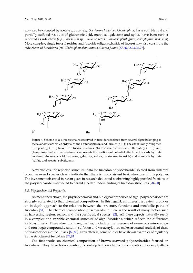
Although fucoidan has been known for over a century, its chemical structure is still incompletelydetermined, owing to its heterogeneity and irregularity. This is due to the fact that marine brown algaesynthesize highly branched polysaccharides, which structures and proportion vary in dependenceof the specific taxonomic position. For instance, it has been shown that fucoidan obtained fromrepresentatives of Chordariales and Laminariales may display different backbone structure comparedwith that isolated from algae belonging to the order Fucales [58,67,69]. In addition, more than one typeof fucoidan may occur simultaneously in the same algal species [70]. The major sulfated polysaccharideof brown seaweed differs from red algae polysaccharides in the main sugar backbone which is galactosefor carrageenan and fucose for fucoidan [64]. In fact, fucoidan essentially consists of α-L-fucose units(usually referred as α-L-fucopyranose). Sulfation of α-L-fucose residues may occur at positions C-2and/or C-4 and, though it is rare, also at position C-3; but the structure and sulfation pattern of thesugar-backbone are species-related [71,72].
Besides fucose and sulfate, fucoidan may also contain additional sugar constituents, includingmannose, galactose, glucose, xylose, uronic acids and yet acetyl groups [14,65,73,74]. The presenceof these additional components, sometimes in appreciable amounts, has not yet been establishedas a regular phenomenon. On the other hand, a certain similarity in the backbone structure ofdifferent fucoidan molecules has been observed, regarding the positions of inter-glycosidic linkages.Many studies show that several representatives of the orders Chordariales and Laminariales containfucoidan with a linear backbone composed of (1Ñ3)-linked α-L-fucose residues. However, fucoidanisolated from algae belonging to the order Fucales mostly display a backbone composed of alternating(1Ñ3)- and (1Ñ4)-linked α-L-fucose residues [66,72,73,75]. Representative backbone structures ofthese fucoidans are depicted in Figure 4.
These findings may not be considered a pattern, though. For instance, fucoidan isolated frombrown seaweed species of the order Fucales have also been reported to have fucose and galactosein comparable amounts; these structures are generally referred to as sulfated galactofucans. Theseare mainly composed of (1Ñ6)-β-D-galactose and/or (1Ñ2)-β-D-mannose units [14]. In these cases,fucoidan molecules not only differ in composition, but also in terms of glycosidic bond positions.In general, fucoidan polysaccharides may be branched, presenting a variety of substituting groups andside chain compositions. The typical positions referred to link sulfate groups (C-2, C-3 and/or C-4)
Mar. Drugs 2016, 14, 42 10 of 41
may also be occupied by acetate groups (e.g., Saccharina latissima, Chorda filum, Fucus sp.). Neutral andpartially sulfated residues of glucuronic acid, mannose, galactose and xylose have been furtherreported as side chain (e.g., Sargassum sp., Fucus serratus, Punctaria plantaginea, Ascophyllum nodosum).More complex, single fucosyl residue and fucoside (oligosaccharide of fucose) may also constitute theside chain of fucoidans (ex. Cladosiphon okamuranus, Chorda filum) [57,66,72,73,76,77].Mar. Drugs 2016, 14, x 10 of 40
Figure 4. Scheme of α‐L‐fucose chains observed in fucoidans isolated from several algae belonging to
the taxonomic orders Chordariales and Laminariales (a) and Fucales (b); (a) The chain is only
composed of repeating (1→3)‐linked α‐L‐fucose residues; (b) The chain consists of alternating (1→3)‐
and (1→4)‐linked α‐L‐fucose residues. R represents the positions of potential attachment of
carbohydrate residues (glucuronic acid, mannose, galactose, xylose, α‐L‐fucose, fucoside) and non‐
carbohydrate (sulfate and acetate) substituents.
These findings may not be considered a pattern, though. For instance, fucoidan isolated from
brown seaweed species of the order Fucales have also been reported to have fucose and galactose in
comparable amounts; these structures are generally referred to as sulfated galactofucans. These are
mainly composed of (1→6)‐β‐D‐galactose and/or (1→2)‐β‐D‐mannose units [14]. In these cases,
fucoidan molecules not only differ in composition, but also in terms of glycosidic bond positions. In
general, fucoidan polysaccharides may be branched, presenting a variety of substituting groups and
side chain compositions. The typical positions referred to link sulfate groups (C‐2, C‐3 and/or C‐4)
may also be occupied by acetate groups (e.g., Saccharina latissima, Chorda filum, Fucus sp.). Neutral
and partially sulfated residues of glucuronic acid, mannose, galactose and xylose have been further
reported as side chain (e.g., Sargassum sp., Fucus serratus, Punctaria plantaginea, Ascophyllum nodosum).
More complex, single fucosyl residue and fucoside (oligosaccharide of fucose) may also constitute
the side chain of fucoidans (ex. Cladosiphon okamuranus, Chorda filum) [57,66,72,73,76,77].
Nevertheless, the reported structural data for fucoidan polysaccharide isolated from different
brown seaweed species clearly indicate that there is no consistent basic structure of this polymer. The
investment observed in recent years in research dedicated to obtaining highly purified fractions of
the polysaccharide, is expected to permit a better understanding of fucoidan structures [78–80].
3.3. Physicochemical Properties
As mentioned above, the physicochemical and biological properties of algal polysaccharides are
strongly correlated to their chemical composition. In this regard, an interesting review provides an
in‐depth approach to the relations between the structure, functions and metabolic paths of fucoidan
[81]. The chemical composition of seaweeds, in turn, is the result of many factors such as harvesting
region, season and the specific algal species [82]. All these aspects naturally result in a complex and
variable chemical structure of algal fucoidans, which reflects the differences in biosynthesis. These
structural irregularities, including the presence of numerous minor sugar and non‐sugar compounds,
random sulfation and/or acetylation, make structural analysis of these polysaccharides a difficult task
Figure 4. Scheme of α-L-fucose chains observed in fucoidans isolated from several algae belonging tothe taxonomic orders Chordariales and Laminariales (a) and Fucales (b); (a) The chain is only composedof repeating (1Ñ3)-linked α-L-fucose residues; (b) The chain consists of alternating (1Ñ3)- and(1Ñ4)-linked α-L-fucose residues. R represents the positions of potential attachment of carbohydrateresidues (glucuronic acid, mannose, galactose, xylose, α-L-fucose, fucoside) and non-carbohydrate(sulfate and acetate) substituents.
Nevertheless, the reported structural data for fucoidan polysaccharide isolated from differentbrown seaweed species clearly indicate that there is no consistent basic structure of this polymer.The investment observed in recent years in research dedicated to obtaining highly purified fractions ofthe polysaccharide, is expected to permit a better understanding of fucoidan structures [78–80].
3.3. Physicochemical Properties
As mentioned above, the physicochemical and biological properties of algal polysaccharides arestrongly correlated to their chemical composition. In this regard, an interesting review providesan in-depth approach to the relations between the structure, functions and metabolic paths offucoidan [81]. The chemical composition of seaweeds, in turn, is the result of many factors suchas harvesting region, season and the specific algal species [82]. All these aspects naturally resultin a complex and variable chemical structure of algal fucoidans, which reflects the differencesin biosynthesis. These structural irregularities, including the presence of numerous minor sugarand non-sugar compounds, random sulfation and/or acetylation, make structural analysis of thesepolysaccharides a difficult task [62,83]. Nevertheless, some studies have shown examples of regularityin the structure of fucoidans [75,84].
The first works on chemical composition of brown seaweed polysaccharides focused onfucoidans. They have been classified, according to their chemical composition, as ascophyllans,

Mar. Drugs 2016, 14, 42 11 of 41
glycuronofuco-galactans sulfate and fucoidans (homofucans). The latter has the simplest chemicalstructure, i.e., homofucans are polysaccharides consisting of sulfated fucose only [85]. However,the term “fucoidan” is commonly used to describe the other fucose-containing heteropolysaccharides.
In addition, structural modifications, such as desulfation, oversulfation, acetylation andbenzoylation, allow the development of derivatives of fucoidans. In general, the natural sulfation graderanges between 4% and 8%, depending on the site and season of collection of the algae [86]. Fucoidanhas been demonstrated to bind to a large number of compounds, including proteins. The bindingaffinity appears to be mainly determined by the negative charge of the polymer, the molecular weightand degree of sulfation, rather than by any specific structure of the carbohydrate [87–89].
Fucoidans are not only highly heterogeneous polysaccharides regarding the sugar compositionand sulfate content, but also concerning their molecular weight. This may vary from 10 [89] toapproximately 2000 kDa [61]. Lower molecular weight fucoidans can be prepared by chemical, physicalor enzymatic means to obtain oligosaccharides with more diverse bioactivities [38]. Acidic hydrolysisof fucoidan leads to sulfated fucoses and oligosaccharide fragments [80], which may be also obtainedby autohydrolysis [90,91], by a radical process involving a hydrogen peroxide-cupric redox system [92]or by enzymatic cleavage [69,93]. A disadvantage of the chemical hydrolysis is that it is quite unspecific.Additionally, high acid concentrations may destroy the sulfation pattern and the polysaccharide chain,which may lead to inactive monosaccharides [58]. Oppositely, enzymatic modifications of fucoidanscan be done by a group of hydrolases, the so called fucoidanases or α-L-fucosidases. These are able tospecifically cleave glycosidic bonds in the polysaccharide chain, while preserving the sulfation patternand, thus, the basic physicochemical properties of fucoidan [68].
Knowledge on the solubility and rheological properties of fucoidan is important to understand andestablish different applications. Fucoidan is very soluble once extracted and the solubility is related tothe level of branching, depending on the content of sulfate groups. There are, however, very few reportsin the literature on the rheological characteristics of fucoidan isolated from brown seaweeds [94–96].Despite of its hygroscopic behavior, fucoidan does not develop highly viscous solutions [85], so thepolymer is not industrially used as thickening or gelling agent, as many other polysaccharides.In fact, a study showed that partially purified fucoidan from Laminaria religiosa, Undaria pinnatifida,Hizikia fusiforme and Sargassum fulvellum produced aqueous solutions of low apparent viscositywith pseudoplastic flow behavior [94]. Complementarily, it was reported that fucoidan fromF. vesiculosus exhibited Newtonian behavior and had the highest viscosity, when compared to thespecies Saccharina longicruris and Ascophyllum nodosum [97]. Moreover, it was reported that the dynamicviscoelasticity of fucoidan isolated from commercially cultured Cladosiphom okamuranus increasedlinearly with an increase of fucoidan concentration up to 2% (w/w) and decreased gradually withincrease in temperature. Additionally, fucoidan viscoelasticity increases with addition of NaCl, CaCl2and sugar [94,95]. Besides, the dynamic viscoelasticity of the polymer was stable over a wide pH range(5.8 to 9.5), indicating that fucoidan molecules are stable under acidic and alkaline conditions [95,98].In this perspective, it seems that the viscosity of fucoidan is influenced by algae species, concentration,molecular weight, presence of sulfate groups, branching, pH and temperature. More extensive researchis however needed to enable the establishment of a straight relationship between viscosity and fucoidanstructure [97].
In contrast to carrageenan and ulvan (the latter being another sulfated polysaccharide to bedescribed in the next section), there is little evidence about gelling and film forming properties offucoidan [83]. In fact, gelation of fucoidan was not observed up to 25% concentration [41]. It hasbeen reported, however, that upon mixing with other polymers, particularly those of opposite netcharge, the formation of gels and films is enabled based on electrostatic interactions between negativelycharged sulfate groups of fucoidan and positively charged groups of the other polymers. Examples ofthese structures have been evidenced with chitosan and poly(2-hydroxyethyl methacrylate) [99,100].

Mar. Drugs 2016, 14, 42 12 of 41
3.4. Biological Activity
As mentioned for carrageenan, fucoidan and its oligosaccharides have been extensively studiedregarding the evidence of diverse biological activities. These include antitumor effect [101],antiviral [102], anticoagulant [89] and anti-inflammatory activities [103]. Based on the reports, theseproperties are related to molecular size, type of sugar content, sulfation degree and molecular structure.From all the reported biological activities, the potent anticoagulant property of fucoidan is by farthe most widely investigated [104–106]. Many studies showed that this anticoagulant activity ispossibly related to the sulfate content and the position of sulfate groups, molecular weight and sugarcomposition. Furthermore, fucoidan requires an enough long sugar-chain and a certain conformation tobind to thrombin, so apparently a relatively large molecular weight is needed to achieve anticoagulantactivity. However, it was also demonstrated that branched structures are not always necessary for ananticoagulant action [57].
Additionally, it has been shown that the carbohydrate further has potent antiviral effect againstherpes simplex virus type 1 (HSV-1), HSV-2 and human cytomegalovirus [102]. Yet, the anti-metastasisand anti-lymphangiogenesis activities of fucoidan, as well as its immunomodulatory effect, have beendemonstrated [107,108].
The biological activities of fucoidan were recently reviewed, with a particular focus on antitumoractivity [109]. For all these properties, fucoidan has been finding applications in the biopharmaceuticalindustry [60] and, in the recent years, the interest in this sulfated carbohydrate has also been extendedto biomedical-related fields, including tissue engineering [6].
4. Ulvan: Sulfated Polysaccharide of Green Seaweeds
Classification of algae has not always been an easy task and, specifically regarding green algae,the complexity increased when recent genetic studies revealed, for instance, that the green seaweedsEnteromorpha and Ulva are not of distinct genera [110]. Despite of the fact that researchers extractbiopolymers from different genus and species of green algae using many distinct extraction methods,it is now generally accepted that ulvan designates a group of sulfated polysaccharides extracted fromgreen seaweed. A relevant aspect to highlight is the fact that, from the sulfated polysaccharidespresented in this review, ulvan is by far the less studied.
Green marine algae are distributed worldwide and considered an important food source in manyparts of the world as a marine vegetable. Ulva spp. (commonly known as sea lettuce) is a rich naturalsource of carbohydrates, vitamins, essential amino acids, minerals and dietary fibers [111,112]. Ulvan iscurrently receiving a great deal of attention, owing to physicochemical and biological properties ofpotential interest for agriculture [113] and pharmaceutical applications [114–117]. These properties arehighly dependent on the chemical composition, charge density and molecular weight of ulvan [118],as also referred for the other polysaccharides. Furthermore, as observed in other seaweed divisions,the yield and specific composition of polysaccharides from green algae depend on environmentalfactors, such as the species from which they are obtained, [20] the season of collection [119] and theemployed extraction method [120].
4.1. Origin, Extraction and Processing
After its first identification in the early 1940s–1950s, researchers have been struggling with theprocessing and characterization of ulvan. As described for carrageenan and fucoidan, the overallprocedure to obtain ulvan from green algae initiates with selection, collection and identification ofthe raw material. This step is followed by algae stabilization and grinding. The stabilization can beperformed by several alternative procedures, including freezing, drying methods, brining and drysalting, a selection that has a considerable impact on the final yield of extraction [120].
Ulvan extraction is mostly performed with hot water solutions [121,122] and might be furtherimproved by the presence of calcium chelating agents [118], acidic or alkaline solutions [123].

Mar. Drugs 2016, 14, 42 13 of 41
The purification of the polymer to eliminate pigments, lipids, amino acids and peptides has beenreported using various procedures [115,119,123–128] and organic solvents [22,121,123,129]. Generally,a polysaccharide with improved purity is obtained by precipitation with organic solvents, frequentlyethanol [22,115,126]. Finally, ulvan aqueous extract can be concentrated in a rotary evaporator [125] ordried by freeze-drying or hot air-drying [129,130]. The removal of impurities, as well as the drying ofulvan extract, may favor the modification of the polysaccharide conformation and properties [131].In fact, a study reported the effects of time and temperature on ulvan degradation, indicating thattemperature was the main factor affecting the rate of depolymerization [21]. Besides, the use ofdifferent solvents to extract ulvan will result in extracts with varying composition and, thus, differentbiological and physicochemical properties [120].
4.2. Chemical Structure
Ulvan corresponds to the major biopolymeric fraction isolated from green seaweed cellwalls, showing a structure of great complexity and variability [123]. The pioneering works fromBrading et al. [123] and Percival et al. [121] established that sulfate, rhamnose, xylose and glucuronicacid are the main constituents of ulvan, showing the structure depicted in Figure 5. However, it wasonly after the work of Quemener et al. [128] that iduronic acid was recognized as a constituentcarbohydrate unit in ulvan.
Mar. Drugs 2016, 14, x 13 of 40
Ulvan extraction is mostly performed with hot water solutions [121,122] and might be further
improved by the presence of calcium chelating agents [118], acidic or alkaline solutions [123]. The
purification of the polymer to eliminate pigments, lipids, amino acids and peptides has been reported
using various procedures [115,119,123–128] and organic solvents [22,121,123,129]. Generally, a
polysaccharide with improved purity is obtained by precipitation with organic solvents, frequently
ethanol [22,115,126]. Finally, ulvan aqueous extract can be concentrated in a rotary evaporator [125]
or dried by freeze‐drying or hot air‐drying [129,130]. The removal of impurities, as well as the drying
of ulvan extract, may favor the modification of the polysaccharide conformation and properties [131].
In fact, a study reported the effects of time and temperature on ulvan degradation, indicating that
temperature was the main factor affecting the rate of depolymerization [21]. Besides, the use of
different solvents to extract ulvan will result in extracts with varying composition and, thus, different
biological and physicochemical properties [120].
4.2. Chemical Structure
Ulvan corresponds to the major biopolymeric fraction isolated from green seaweed cell walls,
showing a structure of great complexity and variability [123]. The pioneering works from Brading et
al. [123] and Percival et al. [121] established that sulfate, rhamnose, xylose and glucuronic acid are the
main constituents of ulvan, showing the structure depicted in Figure 5. However, it was only after
the work of Quemener et al. [128] that iduronic acid was recognized as a constituent carbohydrate
unit in ulvan.
Figure 5. Structure of the main repeating disaccharides in ulvan isolated from Ulva sp. (a)
Ulvanobiuronic acid type A3s disaccharide is composed of glucuronic acid and sulfated rhamnose,
whereas type B3s consists of iduronic acid and sulfated rhamnose; (b) Ulvanobiose acids in which
xylose or sulfated xylose residues occur in place of uronic acids.
The major repeating disaccharide in the ulvan extracted from different ulva samples was found
to comprise two different types of aldobiouronic acid. These were named ulvanobiuronic acid 3‐
sulfate type A and type B (A3s and B3s, respectively). The A3s disaccharide is composed of glucuronic
acid and sulfated rhamnose, while type B3s consists of iduronic acid and sulfated rhamnose, mainly
associated via (1→4) glycosidic linkages. Rhamnose residues are sulfated mainly at position C‐3 or
at both positions C‐2 and C‐3. In some ulva extracts, xylose or sulfated xylose residues may occur in
place of uronic acids, as shown in Figure 4. In this case, the disaccharides are called ulvanobiose acids
and symbolized as U3s (ulvanobiose acid 3‐sulfate) and U2’s3s (ulvanobiose acid 2,3‐disulfate). Low
proportions of galactose, glucose and mannose have been reported, but their real integration in ulvan
structure has been questioned [20,132–134].
Figure 5. Structure of the main repeating disaccharides in ulvan isolated from Ulva sp.(a) Ulvanobiuronic acid type A3s disaccharide is composed of glucuronic acid and sulfated rhamnose,whereas type B3s consists of iduronic acid and sulfated rhamnose; (b) Ulvanobiose acids in whichxylose or sulfated xylose residues occur in place of uronic acids.
The major repeating disaccharide in the ulvan extracted from different ulva samples was found tocomprise two different types of aldobiouronic acid. These were named ulvanobiuronic acid 3-sulfatetype A and type B (A3s and B3s, respectively). The A3s disaccharide is composed of glucuronicacid and sulfated rhamnose, while type B3s consists of iduronic acid and sulfated rhamnose, mainlyassociated via (1Ñ4) glycosidic linkages. Rhamnose residues are sulfated mainly at position C-3 orat both positions C-2 and C-3. In some ulva extracts, xylose or sulfated xylose residues may occurin place of uronic acids, as shown in Figure 4. In this case, the disaccharides are called ulvanobioseacids and symbolized as U3s (ulvanobiose acid 3-sulfate) and U2’s3s (ulvanobiose acid 2,3-disulfate).Low proportions of galactose, glucose and mannose have been reported, but their real integration inulvan structure has been questioned [20,132–134].

Mar. Drugs 2016, 14, 42 14 of 41
4.3. Physicochemical Properties
Generally, ulvan exhibits some of the hydroxyl groups of the sugar residues substituted bysulfate groups (Figure 4). As explained before, these biopolymers are constituted by complex highlybranched molecules which do not appear to have a defined backbone or simple repeating unit. Nor dothey appear to have long chains of a single sugar [1]. The sugar composition of ulvans is extremelyvariable, being the most frequent rhamnose (16.8%–45.0%), xylose (2.1%–12.0%), glucose (0.5%–6.4%),glucuronic acid (6.5%–19.0%) and iduronic acid (1.1%–9.1%). Mannose, galactose and arabinosehave also been found in ulvan from some Ulva species. Determining the sugar sequence in ulvanthus represents a major challenge. Oligosaccharides and oxidation products released after mildacid hydrolysis of native and chemically modified ulvan suggested the presence of rhamnose, xylose,glucuronic acid or glucose, all present in the same chain. Moreover, it was also indicated that glucuronicacid can occur as branches on C-2 of rhamnose [20]. Anyhow, the polysaccharide composition may beeven more complex and is known to be influenced by seaweed species, algal seasonality and the modeof preservation of algae [135], as is usually described for many algal polysaccharides.
The heterogeneous chemical composition of ulvan leads to an essentially disordered conformationof the biopolymer. In spite of this disordered structure, the local regularity given by the repeatingaldobiouronic units, for instance, is believed to be sufficient for the formation of transient “junctionzones” responsible for the formation of the weak gel that ulvan is known to produce in nativestate [127].
Many characteristics and properties of ulvan remain unknown when the extraction conditionsvary, namely the rheological and textural properties. According to the literature, there are fewreports on the rheological properties of ulvan polysaccharides extracted under particular conditions.However, the impact of extraction procedures on the chemical, textural and rheological properties ofulvan extracts from Ulva lactuca was recently assessed. Regarding rheological characteristics, resultsdemonstrated a great contribution of the extraction method on these properties. Ulvan extracts havegenerally demonstrated a pseudoplastic behavior, a viscosity decrease being observed as the shear rateincreased [136]. Furthermore, as observed for carrageenan, ulvan has been shown to produce viscoussolutions when dissolved in water. It also forms gels in presence of B+ and Ca2+ ions at basic pH byyet an unclear mechanism; and the gelling ability was shown to depend on the presence of divalentcations [137,138]. Lahaye and Axelos [130] studied the influence of pH, buffer type and the amountof added ions (Ca2+ and B+) on the gelling characteristics of ulvan extracted with boiling water afterenzymatic treatment of algae. In a subsequent work, the thermo-reversibility of the gel formed byulvan from Ulva rigida, during heating and cooling was studied. According to the authors, ulvan yieldsviscous aqueous solutions that can form thermo-reversible gels in the presence of Ca2+ and B+ ionsat basic pH. It was also shown that gel formation is a time-dependent process and the viscoelasticbehavior of the ulvan solution in the presence of ions (pH 7.5) indicated that the investigated extractsled to systems with properties close to a solid, rather than liquid material. Other authors showed theimpact of stabilization treatments (freezing, freeze-drying, hot-air drying, brining and dry salting) ofUlva rotundata on the physicochemical and rheological properties of ulvan that was extracted usingoxalate sodium, followed by water extraction [120]. As a whole, these studies revealed that ulvan gelsare thermo-reversible and that high or low ion concentration, as well as pH variations, may influenceulvan conformation, and, thus, the gel formation.
The various forms of ulvan markedly differ in the intrinsic viscosity and molecular weight.Regarding the latter, different molecular weights and molecular weight distributions have beenreported. Sedimentation measurements indicated molecular weights ranging from 530 kDa to3.6 ˆ 103 kDa for ulvans obtained from U. pertusa, U. conglobata and E. prolifera. Great variations werefound in ulvans extracted from U. conglobata depending on the temperature at which the extractionwas performed. This indicates that different molecular weight ulvans can be obtained by changingthe temperature of extraction, a high temperature being required to extract high molecular weight

Mar. Drugs 2016, 14, 42 15 of 41
ulvan [20]. It is worth noting that ulvan extracts with lower molecular weights (28.2–151.7 kDa ) canbe obtained by using H2O2 treatment [21].
Ulvan is considered to have a high charge density, which determines its water-solubility. However,it has a certain hydrophobic character, possibly determined by the presence of a great amount of methylgroups in the rhamnose repeating unit. Notwithstanding the aqueous solubility of the polysaccharide,a study performing ultrastructural analysis revealed the presence of spherical shaped aggregates ofulvan in aqueous solution. As a polyelectrolyte, both the ionic strength and the pH of the used solventplay a role on the solubility and morphology of ulvan, since the type and amount of counterions insolution could contribute to the condensation of the polymer [131]. Therefore, it is very importantto bear in mind that using this polymer, as any other with polyelectrolyte character, implies a goodoptimization of conditions regarding the final objective of its application.
4.4. Biological Activity
As for the previously reviewed sulfated polysaccharides, different bioactivities have beenattributed to ulvan. The polysaccharide has been demonstrating to have antioxidant activity, which isapparently dependent on molecular weight, since low molecular weight ulvan shows strongerantioxidant activity compared to larger fractions [21]. Antilipidemic effect has also been registered.In this regard, ulvan has been reported to reduce total serum cholesterol, triglycerides and low densitylipoprotein (LDL) cholesterol, while elevating high density lipoprotein (HDL) cholesterol levels.This effect was identified to depend on the molecular weight of ulvan fractions, as high molecularweight fraction is more effective on total serum and LDL-cholesterol, whereas low molecular weightfractions are more effective on triglycerides and HDL-cholesterol [114,139].
Ulvan has also been studied for antiviral activity in vitro against a number of human and avianinfluenza viruses. In fact, Ivanova et al. [140] described that ulvan polysaccharides isolated from greenalgae had good inhibitory effect on influenza A virus, the inhibition effect being dose-dependentand strain-specific. Likewise, ulvan has been shown to have high and specific activity against herpessimplex virus [141].
As for carrageenan and fucoidan, the reported biological activities depend on sugar composition,molecular weight and sulfate content of ulvan and thus, as above-mentioned, on genus, species andecological and environmental factors. Anyhow, an attractive use and exploitation of green algae wouldtake advantage of these biological properties and apply them in pharmaceutical and biomedical fields.However, among the three main divisions of macroalgae, green algae remain a rather underexploitedbiomass, particularly in areas where other sulfated polysaccharides of algal origin have already proventheir value [6].
5. Drug Delivery Systems Based on Seaweed Sulfated Polysaccharides
Drug substances are usually not administered as they are in pure state, but rather as partof a dosage form where they are frequently combined with other agents (excipients). In manyoccasions, excipients act as simple inert supports of the active molecule(s) [142], but it is also truethat multifunctional excipients have been being increasingly used. The pharmaceutical industryuses excipients from a wide variety of sources, both synthetic and natural. Among those of naturalorigin, polysaccharide-based excipients have been registering increased application, because of theirability to produce a wide range of materials, carriers and devices, owing to specific properties,including molecular weights. Besides, one of the properties most often referred for polysaccharidesis their structural flexibility. This feature enables the chemical modification of the polymers to fulfillthe requirements of specific drug delivery systems, thus allowing a direct competition with thesynthetic excipients available in the market [143]. The enormous orientation of pharmaceuticalindustry towards naturally derived polymers has become a subject of increasing interest, driving thecontinuous exploitation of such compounds [142]. Furthermore, the biomedical field, including tissueengineering, regenerative medicine and drug delivery, is constantly looking for new biomaterials with

Mar. Drugs 2016, 14, 42 16 of 41
innovative properties. Polysaccharides are potential candidates, not only because of their propensityfor biocompatibility and biodegradability, but also due to their high availability at relatively low cost.In this context, sulfated polysaccharides present in different marine algae species have been gatheringgreat interest, which is a reflex of the continuous growing of knowledge on chemical and biologicalactivities of these compounds [6].
The specific application of polysaccharides in pharmaceutical formulations include their use in themanufacture of solid monolithic matrix systems, implants, films, beads, microparticles, nanoparticles,inhalable and injectable systems, as well as hydrogel formulations. It happens frequently thatcarriers like nanoparticles, microparticles and beads prepared with polymers having gelling abilityare called hydrogels. However, for the effects of this review, all carriers exhibiting a particulatemorphology/structure were treated as such, independently of the terminology originally used inthe primary references. Within the dosage forms listed above, the carbohydrate polymers mightserve rather different functions, including the use as binders, coatings, matrix materials, drug releasemodifiers, thickeners, stabilizers, disintegrants, solubilizers, emulsifiers, suspending agents, gellingagents and bioadhesives [144]. Occasionally, some of the referred functions are cumulative.
As mentioned before, the emphasis of this review is placed on the three algae-derivedsulfated polysaccharides carrageenan, fucoidan and ulvan. Owing to their particular features,described in detail in the previous section, a growing interest is being observed regarding abiopharmaceutical application in drug delivery. Particularly, carrageenan-based pellets [145–147],beads [148–157], nanoparticles [158–167], microparticles [168–175], hydrogels [176–185], films [186–193],matrices [194,195] and other devices [196–200] have been extensively investigated as drug deliverycarriers. In turn, the research on fucoidan for this purpose has also increased in recent years.Fucoidan-based drug carriers, such as nanoparticles [201–212], microparticles [213–217] andhydrogels [99,218] have also been successfully developed. Differently, ulvan remains a ratherunexploited biomaterial for an application in the design of drug delivery systems. Despite its chemicaland biological versatility, very few studies on ulvan biomedical applications have been reported todate [133,134,219,220], although some address drug delivery approaches [221–223].
There are two major elements contributing for the importance and relevance of biomaterials basedon sulfated polysaccharides with application in pharmaceutical biotechnology: (1) the glycosidic bonds,which can be easily cleaved by hydrolase enzymes and, thus, contribute for biodegradability; and(2) the presence of the negatively charged sulfate groups that potentiate polyelectrolyte behaviorand permit functionalization for specific applications [224], apart from a privileged interactionwith negatively charged epithelia. Additionally, the presence of hydroxyl groups (OH) on thestructure of these polymers provides the necessary moieties for several chemical modifications. In thisregard, the introduction of hydrophobic, acidic or basic groups, or even other functionalities intopolysaccharide structures might alter the properties of biopolymers, enabling specific tailoring towardsthe devised objectives. Taking benefit from these features, the use of these carbohydrates in drugdelivery applications has been proposed frequently. From the three, carrageenan is by far the mostreported, as indicated in Figure 1, which is certainly a result of its first isolation, easy purification andwell-defined chemical structure.
For an easier structuration of the review, the application of the polysaccharides in the design ofdifferent carriers is arranged and presented according to the carrier-types.
5.1. Nano and Microparticles
Particulate carriers have been developed as a physical approach to alter and improve thepharmacokinetic and pharmacodynamic properties of various types of drug molecules. Compared withconventional dosage forms, particulate delivery systems offer many advantages, such as availability fordelivery through various routes of administration, tailoring of particle size and surface characteristicsand, in some cases, possibility to offer controlled and sustained release of the drug at specificsites [225,226].

Mar. Drugs 2016, 14, 42 17 of 41
Nano- and microparticles are the most referred of the particulate carriers, in the majority ofcases presenting a matrix composed by polymeric materials. Although the definition may not beconsensual under all instances, nanoparticle is the term frequently used for spherical particles withdiameters ranging from 10 to 1000 nm, whereas microparticles present diameters in the micrometerrange (typically from 1 µm to 1000 µm). Drugs can be dissolved, entrapped, encapsulated or attachedto the polymer matrix of the particulate carriers. Structurally, these systems are divided in twocategories: nanocapsules/microcapsules in which the drug is mainly confined to a cavity surroundedby a polymer membrane (shell); and nanospheres/microspheres in which the drug is dispersed withinthe polymeric matrix, according to a classification that is now widely accepted [227].
Sulfated seaweed polysaccharides have been finding applications in the production ofnanoparticles and microparticles, mainly owing to their ionic nature. This enables the formationof complexes with oppositely charged polyelectrolytes, which has been found very useful regardingthe design of drug carriers, since polyelectrolyte complexes allow the association of drugs in thepolymer matrix at a molecular level. Such structures permit drug entrapment during precipitation ofthe complex, or through absorption to the already formed complexes. The drug can also be chemicallybound to one of the polymers and be incorporated during the complexation. Afterwards, the drug isreleased from the polyelectrolyte complex either by ion exchange mechanism or by charge interaction,as well as by polymer breakdown and dissolution of the complex [228].
5.1.1. Nanoparticles
Polymeric nanoparticles have been extensively studied for drug delivery purposes and variedmethods have been developed for their production, including emulsification, coacervation, ionicgelation and polyelectrolyte complexation, among others. All these methods comprise bottom-upfabrication processes, which involve the assembly of molecules in solution to form defined structures,in this case, nanoparticles. Readers interested in a detailed analysis of these methodological approachesare directed to the reviews [229,230].
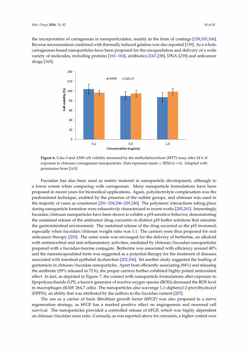
The use of sulfated polysaccharides has been explored in the design of polymeric nanoparticles,mainly taking advantage of the sulfate content, which directly results in the exhibited negative charge.The complexation with cationic polymers is, thus, frequently used as the driving force towards theformation of nano-sized carriers. In this regard, carrageenan has been mostly referred to be complexedwith chitosan [160–163,169,231–236], but the use of other counterions such as protamine [237] anda cationized pullulan [162], or a direct complexation with drugs [238] were also reported. A recent workreviewed the application of carrageenan in drug delivery, including the production of nanoparticles [9]and, therefore, we will focus on works not comprised in that review or which are considered relevant.Our group was one of the first to report chitosan/κ-carrageenan nanoparticles [160,161,163], testingcross-linkers, demonstrating the ability to encapsulate proteins, evaluating the storage stability andthe stability in presence of lysozyme and assessing the cytotoxicity of the complexes. Regardingthe latter, a methyltetrazolium (MTT) assay performed in two respiratory cell lines (A549 andCalu-3) revealed an absence of toxicity in concentrations up to 1 mg/mL exposed for 24 h (Figure 6).Additionally, a strategy was proposed for the delivery of the nanoparticles by inhalation, mediated bymicroparticles-containing-nanoparticles [163].
The effect of different types of carrageenan (κ-, ι-, λ-) and varied polymeric charge ratioson the final characteristics of chitosan/carrageenan nanoparticles was explored by other authors.Nanoparticles formulated with κ-carrageenan were those showing the higher encapsulation efficiencies(up to 79%) of glucose oxidase, used as model molecule, while the charge ratio also played a rolein the association capacity. A controlled release was observed when the nanoparticles were treatedwith different physiological and enzyme solutions; κ-carrageenan/chitosan nanoparticles being thoseshowing the lowest release rate [164]. A controlled release was also reported for erythropoietin(48% encapsulation efficiency), which released 50% over a two week period [233]. Notwithstandingthe predomination of polyelectrolyte complexation, other methodological approaches have provided
Mar. Drugs 2016, 14, 42 18 of 41
the incorporation of carrageenan in nanoparticulates, mainly in the form of coatings [158,165,166].Reverse microemulsion combined with thermally induced gelation was also reported [159]. As a whole,carrageenan-based nanoparticles have been proposed for the encapsulation and delivery of a widevariety of molecules, including proteins [161–163], antibiotics [167,238], DNA [239] and anticancerdrugs [165].
Mar. Drugs 2016, 14, x 18 of 40
Figure 6. Calu‐3 and A549 cell viability measured by the methyltetrazolium (MTT) assay after 24 h of
exposure to chitosan/carrageenan nanoparticles. Data represent mean ± SEM (n = 6). Adapted with
permission from [163].
Fucoidan has also been used as matrix material in nanoparticle development, although to a
lower extent when comparing with carrageenan. Many nanoparticle formulations have been
proposed in recent years for biomedical applications. Again, polyelectrolyte complexation was the
predominant technique, enabled by the presence of the sulfate groups, and chitosan was used in the
majority of cases as counterion [201–204,206–209,240]. The polymeric interactions taking place during
nanoparticle formation were exhaustively characterized in recent works [205,241]. Interestingly,
fucoidan/chitosan nanoparticles have been shown to exhibit a pH‐sensitive behavior, demonstrating
the sustained release of the antitumor drug curcumin in distinct pH buffer solutions that simulate
the gastrointestinal environment. The sustained release of the drug occurred as the pH increased,
especially when fucoidan/chitosan weight ratio was 1:1. The carriers were thus proposed for oral
anticancer therapy [203]. The same route was envisaged for the delivery of berberine, an alkaloid
with antimicrobial and anti‐inflammatory activities, mediated by chitosan/fucoidan nanoparticles
prepared with a fucoidan‐taurine conjugate. Berberine was associated with efficiency around 40%
and the nanoencapsulated form was suggested as a potential therapy for the treatment of diseases
associated with intestinal epithelial dysfunction [202,206]. Yet another study suggested the loading
of gentamicin in chitosan/fucoidan nanoparticles. Apart from efficiently associating (94%) and
releasing the antibiotic (99% released in 72 h), the proper carriers further exhibited highly potent
antioxidant effect. In fact, as depicted in Figure 7, the contact with nanoparticle formulations after
exposure to lipopolysaccharide (LPS, a known generator of reactive oxygen species (ROS)) decreased
the ROS level in macrophages (RAW 264.7 cells). The nanoparticles also scavenge 1,1‐diphenyl‐2‐
picrylhydrazyl (DPPH), an ability that was attributed by the authors to the fucoidan content [207].
The use as a carrier of basic fibroblast growth factor (bFGF) was also proposed in a nerve
regeneration strategy, as bFGF has a marked positive effect on angiogenesis and neuronal cell
survival. The nanoparticles provided a controlled release of bFGF, which was highly dependent on
chitosan/fucoidan mass ratio. Curiously, as was reported above for curcumin, a higher control over
the release rate was obtained for mass ratios of 1/1 (Figure 8), which showed a continuous release for
four days. In turn, the other tested ratios tended for a plateau. This work also demonstrated that
nanoparticles protect bFGF from heat and enzymatic deactivation, and decrease the amount of
growth factor needed for neurite extension [240].
Figure 6. Calu-3 and A549 cell viability measured by the methyltetrazolium (MTT) assay after 24 h ofexposure to chitosan/carrageenan nanoparticles. Data represent mean ˘ SEM (n = 6). Adapted withpermission from [163].
Fucoidan has also been used as matrix material in nanoparticle development, although toa lower extent when comparing with carrageenan. Many nanoparticle formulations have beenproposed in recent years for biomedical applications. Again, polyelectrolyte complexation was thepredominant technique, enabled by the presence of the sulfate groups, and chitosan was used inthe majority of cases as counterion [201–204,206–209,240]. The polymeric interactions taking placeduring nanoparticle formation were exhaustively characterized in recent works [205,241]. Interestingly,fucoidan/chitosan nanoparticles have been shown to exhibit a pH-sensitive behavior, demonstratingthe sustained release of the antitumor drug curcumin in distinct pH buffer solutions that simulatethe gastrointestinal environment. The sustained release of the drug occurred as the pH increased,especially when fucoidan/chitosan weight ratio was 1:1. The carriers were thus proposed for oralanticancer therapy [203]. The same route was envisaged for the delivery of berberine, an alkaloidwith antimicrobial and anti-inflammatory activities, mediated by chitosan/fucoidan nanoparticlesprepared with a fucoidan-taurine conjugate. Berberine was associated with efficiency around 40%and the nanoencapsulated form was suggested as a potential therapy for the treatment of diseasesassociated with intestinal epithelial dysfunction [202,206]. Yet another study suggested the loading ofgentamicin in chitosan/fucoidan nanoparticles. Apart from efficiently associating (94%) and releasingthe antibiotic (99% released in 72 h), the proper carriers further exhibited highly potent antioxidanteffect. In fact, as depicted in Figure 7, the contact with nanoparticle formulations after exposure tolipopolysaccharide (LPS, a known generator of reactive oxygen species (ROS)) decreased the ROS levelin macrophages (RAW 264.7 cells). The nanoparticles also scavenge 1,1-diphenyl-2-picrylhydrazyl(DPPH), an ability that was attributed by the authors to the fucoidan content [207].
The use as a carrier of basic fibroblast growth factor (bFGF) was also proposed in a nerveregeneration strategy, as bFGF has a marked positive effect on angiogenesis and neuronal cellsurvival. The nanoparticles provided a controlled release of bFGF, which was highly dependenton chitosan/fucoidan mass ratio. Curiously, as was reported above for curcumin, a higher control over

Mar. Drugs 2016, 14, 42 19 of 41
the release rate was obtained for mass ratios of 1/1 (Figure 8), which showed a continuous releasefor four days. In turn, the other tested ratios tended for a plateau. This work also demonstrated thatnanoparticles protect bFGF from heat and enzymatic deactivation, and decrease the amount of growthfactor needed for neurite extension [240].Mar. Drugs 2016, 14, x 19 of 40
Figure 7. Lipopolysaccharide‐induced RAW 264.7 cells detected by flow cytometry. Data are mean ±
SD of values calculated on 5 distinct batches (n = 5). Statistical analysis was performed by one‐way
ANOVA. * p < 0.01 versus control. *** p < 0.001 versus control. ### p < 0.001 versus LPS. Adapted with
permission from [207].
Figure 8. The release kinetics of basic fibroblast growth factor (bFGF) from chitosan/fucoidan
nanoparticles, as measured by ELISA. Adapted with permission from [240].
Still in the field of regenerative medicine, chitosan/fucoidan nanoparticles added of the cross‐
linker tripolyphosphate were described as adequate to deliver stromal cell‐derived factor, which is
an important chemokine in stem cells mobilization [208]. In a different approach, multilayer
nanoparticles were successfully prepared through layer‐by‐layer assembly of fucoidan and chitosan,
over a core of polystyrene. The fucoidan‐chitosan particles showed ability for the encapsulation of
poly‐L‐lysine, which was found to be released by a pH‐dependent mechanism [209], further
reinforcing the pH‐sensitivity described in previous works.
One work was referred above describing a specific effect for the proper chitosan/fucoidan
carriers, in that case a scavenging and antioxidant effect [207]. Interestingly, there are some other
works on these nanosystems that do not envisage carrying a specific molecule, but instead explore
the proper biological activities of fucoidan. In one such work, the known ability of fucoidan to inhibit
angiogenesis was explored. An oversulfated fucoidan was synthesized to improve the anti‐
angiogenic effect and complexed with chitosan to produce nanoparticles intended for oral delivery.
A more prolonged release of the oversulfated fucoidan was observed in simulated intestinal
conditions comparing with gastric medium. Additionally, formulations with a positive surface
charge provided the transient opening of Caco‐2 cell tight junctions, an effect known to be featured
Figure 7. Lipopolysaccharide-induced RAW 264.7 cells detected by flow cytometry. Data are mean˘ SDof values calculated on 5 distinct batches (n = 5). Statistical analysis was performed by one-way ANOVA.* p < 0.01 versus control. *** p < 0.001 versus control. ### p < 0.001 versus LPS. Adapted with permissionfrom [207].
Mar. Drugs 2016, 14, x 19 of 40
Figure 7. Lipopolysaccharide‐induced RAW 264.7 cells detected by flow cytometry. Data are mean ±
SD of values calculated on 5 distinct batches (n = 5). Statistical analysis was performed by one‐way
ANOVA. * p < 0.01 versus control. *** p < 0.001 versus control. ### p < 0.001 versus LPS. Adapted with
permission from [207].
Figure 8. The release kinetics of basic fibroblast growth factor (bFGF) from chitosan/fucoidan
nanoparticles, as measured by ELISA. Adapted with permission from [240].
Still in the field of regenerative medicine, chitosan/fucoidan nanoparticles added of the cross‐
linker tripolyphosphate were described as adequate to deliver stromal cell‐derived factor, which is
an important chemokine in stem cells mobilization [208]. In a different approach, multilayer
nanoparticles were successfully prepared through layer‐by‐layer assembly of fucoidan and chitosan,
over a core of polystyrene. The fucoidan‐chitosan particles showed ability for the encapsulation of
poly‐L‐lysine, which was found to be released by a pH‐dependent mechanism [209], further
reinforcing the pH‐sensitivity described in previous works.
One work was referred above describing a specific effect for the proper chitosan/fucoidan
carriers, in that case a scavenging and antioxidant effect [207]. Interestingly, there are some other
works on these nanosystems that do not envisage carrying a specific molecule, but instead explore
the proper biological activities of fucoidan. In one such work, the known ability of fucoidan to inhibit
angiogenesis was explored. An oversulfated fucoidan was synthesized to improve the anti‐
angiogenic effect and complexed with chitosan to produce nanoparticles intended for oral delivery.
A more prolonged release of the oversulfated fucoidan was observed in simulated intestinal
conditions comparing with gastric medium. Additionally, formulations with a positive surface
charge provided the transient opening of Caco‐2 cell tight junctions, an effect known to be featured
Figure 8. The release kinetics of basic fibroblast growth factor (bFGF) from chitosan/fucoidannanoparticles, as measured by ELISA. Adapted with permission from [240].
Still in the field of regenerative medicine, chitosan/fucoidan nanoparticles added of thecross-linker tripolyphosphate were described as adequate to deliver stromal cell-derived factor,which is an important chemokine in stem cells mobilization [208]. In a different approach, multilayernanoparticles were successfully prepared through layer-by-layer assembly of fucoidan and chitosan,over a core of polystyrene. The fucoidan-chitosan particles showed ability for the encapsulation ofpoly-L-lysine, which was found to be released by a pH-dependent mechanism [209], further reinforcingthe pH-sensitivity described in previous works.
One work was referred above describing a specific effect for the proper chitosan/fucoidan carriers,in that case a scavenging and antioxidant effect [207]. Interestingly, there are some other works onthese nanosystems that do not envisage carrying a specific molecule, but instead explore the proper

Mar. Drugs 2016, 14, 42 20 of 41
biological activities of fucoidan. In one such work, the known ability of fucoidan to inhibit angiogenesiswas explored. An oversulfated fucoidan was synthesized to improve the anti-angiogenic effect andcomplexed with chitosan to produce nanoparticles intended for oral delivery. A more prolongedrelease of the oversulfated fucoidan was observed in simulated intestinal conditions comparing withgastric medium. Additionally, formulations with a positive surface charge provided the transientopening of Caco-2 cell tight junctions, an effect known to be featured by chitosan since a long timeago, improving the paracellular transport of fucoidan [210]. In another work, the nanoparticles wereused to obtain an anticoagulant effect, which was two-fold higher than that provided by a fucoidansolution [242].
Other relevant works reported the preparation of fucoidan nanoparticles by other methodsnot involving the complexation with an oppositely charged molecule. An acetylated fucoidanformed nanoparticles containing doxorubicin by dialysis and the drug was shown to releaseaccording to first-order kinetics for 5 days. Macrophages treated with these fucoidan nanoparticlesoverexpressed various antitumor cytokines (tumor necrosis factor-alpha and granulocyte-macrophagecolony-stimulating factor) [211]. Another work reported the grafting of fucoidan with hexadecylamine,which leads to self-assembled nanostructures that demonstrated anti-proliferative effect (inhibition ofproliferation between 2% and 44%) in various tumor cells [212].
As reported above for carrageenan, in some cases fucoidan was used as coating of nanoparticlescomposed of other materials, with several objectives. The coating of iron nanoparticles was reportedto improve the affinity by platelets, thus permitting the visualization of platelet-rich thrombus bymagnetic resonance imaging [243], while that of poly(isobutylcyanoacrylate) nanoparticles improvedcell uptake (macrophages and fibroblasts) [244]. In turn, the coating of pDNA/polyethyleneiminecomplexes was reported to reduce cytotoxicity towards B16-F10 cells (murine melanoma cells) [239].As referred in Section 3.4, fucoidan has been reported to have antitumor effect and this featurehas been explored in the design of fucoidan-based nanoparticles. In fact, there are several worksreporting the cytotoxic effect of fucoidan nanoparticles, either when fucoidan is part of the nanoparticlematrix [211,212] or when it is used as coating material [245].
Considering its polyelectrolyte properties, ulvan may also establish ionic interactions with cationiccompounds, being able to interact in this way with drugs and/or polymers, forming differentcomplexes which have found application in the biomedical field [246,247]. Although some ofthe referred works consist in the development of ulvan-based biomaterials for tissue engineering,the application of this carbohydrate in the design of nano drug delivery systems is not described.As detailed in the previous section, this material has a structure that is still not perfectly definedand seems to be complex, thus certainly restraining the interest of researchers in investigatingits applications.
5.1.2. Microparticles
As stated before, polymeric microparticles including microspheres and microcapsules may beemployed to deliver drugs in a rate-controlled and, sometimes, targeted manner. To date, ulvan has noreports in this regard, while carrageenan and fucoidan microparticles have found some applications.The use of carrageenan to prepare microparticles for drug delivery was reported in a limited number ofoccasions. To our knowledge, a first report appeared in 2000, when ι-carrageenan microparticles wereproposed to encapsulate horseradish peroxidase, used as model protein. Microparticles formed byinteraction with amines (mono-, di- or oligoamines), but the performed studies were no furtherthan the association of the molecule and the evaluation of its stability [170]. Some years later,λ-carrageenan was combined with gelatin to obtain microspheres for ophthalmic delivery of timololmaleate, an anti-glaucoma drug. Different polymeric ratios proved to be useful in modulating drugrelease profile, a higher content of gelatin providing faster release. Yet, in vivo tests performed in rabbitsrevealed that the drug concentration and bioavailability in the aqueous humor were significantlyhigh in comparison with commercial formulations [171]. The same group reported recently the

Mar. Drugs 2016, 14, 42 21 of 41
ocular delivery of ciprofloxacin mediated by λ-carrageenan microparticles, but chondroitin sulfatemicroparticles revealed a better performance, comparatively [172]. Carrageenan microspheres werealso reported to encapsulate allopurinol and local anesthetic agents, such as lidocaine hydrochloride,dibucaine hydrochloride and tetracaine hydrochloride. Kappa- and ι-carrageenan were used to preparemicroparticles by spray-drying. The drug loading efficiency was over 70% and allopurinol and thelocal anesthetic agents released from the microspheres for at least 400–600 min, depending on the typeof carrageenan [168]. The encapsulation of insulin regarding an oral therapy of diabetes has also beenapproached with κ-carrageenan microparticles, using a lectin functionalization. Insulin associationefficiency was as high as 94% and the oral administration of insulin entrapped in the microparticlesled to a prolonged duration of the hypoglycemic effect, up to 12–24 h, in diabetic rats [173]. A furtherstudy proposed the use of κ-carrageenan/calcium carbonate microparticles functionalized with folicacid to deliver doxorubicin to cancer cells. Highly porous microparticles were obtained which reducedcell viability in 87% in a human osteosarcoma cell line (MG-63) [175].
Regarding fucoidan, its application is much scarcer when compared with that of carrageenan.In fact, in the ambit of drug delivery the almost totality of carriers relies on the so-called fucospheres.These are microspheres obtained by polyelectrolyte complexation with chitosan and were first proposedfor protein delivery. Bovine serum albumin (BSA) was used as model protein and encapsulated into themicrospheres with efficiency up to 90%. The rate of drug release from the microspheres was dependenton the concentration of polymers involved in the particle production and also on the concentrationof BSA [213]. Fucospheres were also shown to encapsulate and release ofloxacin, a broad-spectrumantibiotic [214] and a plasmid-encoded granulocyte colony-stimulating factor [215]. Furthermore,as also described for nanoparticles, in certain cases the polymeric carriers are used because of theirproper characteristics, without encapsulating any drug. In this regard, fucospheres demonstratedin vitro and in vivo the ability to treat dermal burns, benefiting from an intrinsic capacity of fucoidanin tissue healing. Fucospheres shortened the treatment period of burns existing in rabbits, providinga fast and effective healing by improving regeneration and re-epithelization [99,216]. With this set ofresults, fucospheres were presented as a potential delivery system for different applications and forthe encapsulation of different bioactive molecules.
Another work reported the preparation of fucoidan/chitosan microparticles using the coaxialelectro-spray drying technique. Lipoic acid was used as model drug, releasing according to a profilecomprising an initial burst until 4 h (50%–70%) and a more sustained release up to 48 h. Because of thedesign of the spray-drying apparatus, the microparticles were described to have a fucoidan core anda chitosan shell. The composite particles underwent physical gelation upon contact with water andshowed a unique drug release pattern [217].
5.2. Hydrogel and Beads
Hydrogels are 3D networks of water-soluble polymers. They have received considerable attentionas drug delivery systems, mainly because their highly porous structure permits loading of drugs intothe gel matrix and subsequent drug release at a rate dependent on the diffusion coefficient of themolecule through the gel network. Furthermore, hydrogels swell without dissolving when in contactwith water or other biological fluids and can easily be tuned by controlling the density of cross-links inthe gel matrix [248].
Several works on carrageenan-based hydrogels for drug delivery applications have beenpublished. Very different uses that include the production of ophthalmic [176] and floating drugdelivery systems [177], antimicrobial wound dressings [178] and combined carrier systems suchas lipid-hydrogel films [185], have been reported. In fact, given the physicochemical propertiesof this carbohydrate, which were already discussed, carrageenan is frequently combined withother compounds to develop hydrogel systems. For instance, the combination of carrageenan withgelatin [179,180], poloxamer 407 [181] and sodium carboxymethyl cellulose [182] have been reportedregarding the production of hydrogel composites for drug delivery applications. As well, the use
Mar. Drugs 2016, 14, 42 22 of 41
of carrageenan-based hydrogels in tissue engineering has been evaluated. A study reported thatcarrageenan hydrogel provided an adequate support for culture and differentiation of encapsulatedhuman-adipose-derived stem cells in the regeneration of cartilage [183]. Another report showedthat the injection of human nerve growth factor β delivered by collagen/nano-hydroxyapatite/κ-carrageenan gels to sites of new bone formation can appreciably improve bone consolidation [184].
Carrageenan beads were also described as potential drug carriers systems [151,152].Carrageenan/chitosan beads were reported to efficiently deliver sodium diclofenac in gastrointestinalfluid [153]. Interestingly, a formulation of solid-lipid beads based on carrageenan was shownto have the potential to mask the bitter taste of enrofloxacin and extend its release rate [154].Kappa-carrageenan/polyacrylamide beads have shown a pH-responsive behavior and were proposedfor targeting ketoprofen to the intestine. The particles assembled by electrostatic interaction andketoprofen release was significantly increased when pH of the medium was changed from acidic(maximum of 10% release) to alkaline (about 90% release) [155]. Besides, the use of κ-carrageenanbeads might also be effective in the field of tissue engineering, being reported to incorporate plateletderived growth factor [157].
While fucoidan beads have not been reported, the application of this polysaccharide in theproduction of hydrogels used for drug delivery purposes was reported to address the therapy ofischemic disease. Nakamura and co-workers [218] developed an injectable chitosan/fucoidan microcomplex-hydrogel which was found effective at releasing fibroblast growth factor-2 in vitro and in vivo.After subcutaneous injection in mice, significant neovascularization and fibrous tissue formation wereinduced near the site of injection at 1 week, and the hydrogel was biodegraded and disappeared after4 weeks. Another work reported the functionalization of 3D scaffolds with fucoidan. The scaffoldswere loaded with vascular endothelial growth factor, which is intended to provide angiogenic activityin ischemic tissues. Functionalized scaffolds induced higher neovessel area and density, comparingwith scaffolds without fucoidan [249]. Other studies have investigated the pharmaceutical/biomedicalapplication of fucoidan-based hydrogels, mainly regarding the treatment of dermal burns [99,250]or generally addressing wound healing [251]. However, in those cases no drug molecules wereencapsulated, the proper hydrogels being used for the intrinsic properties of fucoidan.
Ulvan has been demonstrated to enable the production of hydrogels, although the majorityof works proposes applications such as ion exchange [252] and tissue engineering [133,219], whichdo not specifically address the association and release of drugs. One sole study reports an ulvanhydrogel aimed at drug delivery. Ulvan was cross-linked with 1,4-butanediol diglycidyl ether to forma 2D structure, which was loaded with dexamethasone as model drug. It was observed an initial steadyrelease of the drug (~49% in 8 h) followed by slower and sustained release up to 14 days [223]. The samegroup has reported the preparation of ulvan beads for a posterior incorporation into polylactic acidhydrogels in a strategy of bone engineering. The beads were produced by complexation with chitosanand also associated dexamethasone. The production of the hydrogel and the incorporation of thebeads involved supercritical fluids technology and, although drug release studies and cytotoxicityevaluation were performed, these comprised the whole system and not only the ulvan beads [246].
5.3. Other Drug Delivery Systems
Apart from the described delivery systems, which are considered those of more advancedtechnology in the field, other classes of systems also report the use of the polysaccharides beingfocused in this review. Conventional systems like tablets have been widely explored but an applicationas fibers, films and wafers also deserves reference.
Matrix tablets are widely accepted for sustained release, as they are simple and easy to formulate,although they are one of the most conventional formulations and usually are not the focus of thelatest drug delivery developments. Ulvan has no reports as part of the composition of tablets,but fucoidan was included in one work. Nevertheless, its inclusion envisaged benefiting from theanti-inflammatory, anti-coagulant and anti-tumor activities of the polymer and not an application as

Mar. Drugs 2016, 14, 42 23 of 41
matrix material [253]. Contrarily, controlled-release tablet matrices containing carrageenan have longbeen studied. A very complete assessment in this regard was recently performed [9] and the readersare directed to that review for further details. Since then, some other works have come to light, some ofthem with interesting approaches. As general information, there are several aspects influencing therelease of drugs from carrageenan matrix tablets. These include the type of carrageenan, the useddrugs, which establish different interaction with the polymer, the presence of other excipients in thematrix, pH and ionic strength of the dissolution medium, type of diluent used, the compression forceand tablet dimensions [254–256]. The release mechanism from tablets with different composition(including carrageenan) and associating different drugs (theophylline and metoprolol succinate) wasinvestigated in a recent work [257]. Yet another work was devoted to studying the behavior of severalpolysaccharides, including λ-carrageenan, in the release of atenolol from drug tablets [258]. The roleplayed by drug/polymer interaction on the water uptake, swelling, drug dissolution and drug releasewas investigated in a study using λ-carrageenan. It was observed that different drugs give complexeswith quite different characteristics of solubility and drug release kinetics. In another approach,carrageenan was associated with two drug models, diltiazem HCl and metoprolol tartrate, and the twostudied complexes released the drug with different mechanism indicating two different drug/polymerinteraction strengths (stronger interaction with diltiazem) [259]. Other recent developments includedthe use of carrageenan, combined with other matrix materials, to associate and release vitamin B2 [260]and lisinopril [261].
The use of carrageenan was also tested to produce oral granules, but those composed of gelatin anda derivative of this protein exhibited better properties regarding elasticity and swelling ability [262].
Another application deserving reference, which is also a nano-sized carrier, consists of fibers.To date, only carrageenan and ulvan have such a reported application in the context of drugdelivery. Iota-carrageenan fibers demonstrated to associate a wide range of low solubility drugmolecules, including benzocaine, furosemide, griseofulvin, hydrocortisone, ibuprofen, indomethacin,phenylephrine HCl, sulfapyridine and thymol. The selected compounds cover a range of structuralvariety and therapeutic indications. The carrageenan matrix was found to protect drugs from thermaldegradation. Griseofulvin was the drug selected to perform release assays, evidencing a burst release(80% in 20 min) and a plateau reached up to 2 h [263]. Although no direct application in drug deliverywas reported, this was suggested by authors preparing ulvan nanofibers from a poly(vinyl alcohol)(PVA)-ulvan blend, using an electrospinning technique. The fibers were described as having a highdegree of orientation, which was attributed to the ulvan component [221].
In the same line, another work also proposed electrospinning as a technique to produce nanofibersfrom a blend of ulvan and polycaprolactone or polyethylene oxide. Again, the association of a drugwas not reported, but only suggested [222]. In general, nanofibers find application at the level of tissueengineering and thus the encapsulation and release of drugs, growth factors or other molecules mightbe of interest.
Other systems deserving a reference are films and wafers, which have been produced withcarrageenan and applied in buccal drug delivery. To our knowledge, neither fucoidan nor ulvan havesimilar applications. Films prepared by combining κ-carrageenan, poloxamer® 407 and polyethyleneglycol were loaded with ibuprofen (up to 0.8%, w/w) as model hydrophobic drug. Drug dissolutionat a pH simulating that of saliva showed that amorphous ibuprofen released from the films (65%in 120 min) at a faster rate than the pure crystalline drug [188]. Another work reported thatmucosal films based on κ-carrageenan, carboxymethyl cellulose and glycerol (1:2:3, w/w) providedsustained release of paracetamol (85%) and amoxicillin (71%) for 8 h in pH 6.5 buffer [190]. Similarworks have demonstrated the ability of carrageenan-based buccal films to incorporate and releasedrugs [189,193]. Wafers prepared with κ-carrageenan and pluronic® were also proposed for buccaldelivery of paracetamol or ibuprofen. Drugs remained stable over 6 months and the release ina medium resembling salivary pH was gradual within 2 h (ibuprofen reached 75% after 120 minwhereas paracetamol released 50% in the same time [198].

Mar. Drugs 2016, 14, 42 24 of 41
Similar systems have also been proposed for wound healing. Wound dressings are traditionallyused to protect the wound from contamination, but they can be exploited as platforms to deliverbioactive molecules to wound sites. Unlike traditional dressings such as gauze and cotton wool thatare passive agents in the wound healing process, advanced therapeutic dressings are designed tohave biological activity either on its own or by releasing bioactive components incorporated withinthe dressing. The incorporated drugs can play an active role in the wound healing process, such asantimicrobial or anti-inflammatory agents, or by removing necrotic tissue and promoting tissueregeneration. In chronic wound management, where patients usually undergo long treatments andfrequent dressing changes, a system that delivers drugs to a wound site in a controlled manner canimprove patient compliance and therapeutic outcomes [264]. Advanced therapeutic dressings foreffective wound healing, including films and wafers based on biopolymers, are described in detail ina review [265].
On this matter, a group reported polyethylene oxide (Polyox®) and κ-carrageenan based filmsas dressings for drug delivery to wounds [191,192]. The films were loaded with streptomycin anddiclofenac for enhanced healing effects in chronic wounds. Drug loaded films showed a high capacityto absorb simulated wound fluid and also significant mucoadhesion, which is expected to alloweffective adherence to and protection of the wound. As well, the films showed controlled release ofboth streptomycin and diclofenac for 72 h. Besides, these drug loaded films produced higher zonesof inhibition against Staphylococcus aureus, Pseudomonas aeruginosa and Escherichia coli compared tothe individual drugs zones of inhibition. The same research group also developed wafers of similarcomposition and loading the same drugs and a sustained release of both drugs was also observed in72 h [199].
Although a few studies have reported the development of films [266–269] containing fucoidan aswound dressings, these systems were not addressed to deliver drugs. Actually, fucoidan was used toactively participate in the process of wound healing for its intrinsic properties.
6. The Application of Sulfated Polysaccharides in Targeted Drug Delivery
The targeted or site-specific delivery of drugs, as the name suggests, is a method of deliveringdrugs to a patient in a very specific manner that allows concentrating the drugs in the site of interest,while reducing its concentration in the remaining tissues [270]. This not only improves the inherentefficacy of drugs, but also reduces side effects. Targeted drug delivery is, therefore, a very attractiveattainment, because it provides one of the most potential ways to improve the therapeutic effectof drugs.
Since it is very difficult for a drug molecule to reach its destination in the complex cellularnetwork of an organism, the assistance of a drug carrier is usually required for this end. Drug deliverysystems offer an intelligent approach for carrying and, at the same time, modulating the release andthe absorption of the drug. In summary, they may tailor the response. However, their success isfrequently limited by short residence times at the site of absorption or action. For this reason, it wouldbe advantageous to have means to provide an intimate contact between the drug delivery system andthese sites. In this context, microstructural design and chemical composition can be used to adapt thestructure-activity relationship and tailor improved polymeric matrices. Various polymer architectures(linear, branched backbones) and combinations of polymers physically mixed (polymer blends orinterpenetrating networks) or chemically bonded (copolymers) offer tremendous scope as carriersystems [271]. Moreover, because of the versatility of preparation methods and chemical structureof polymers, surface functionalities may sometimes be incorporated in the carriers. This facilitatesadditional attractive properties, such as the attachment of ligands that prolong the circulation of thedrug carrier system in the blood stream, or the targeting of ligands for interaction with specific cellreceptors [272]. Therefore, when designing a drug delivery system, it is important to consider, not onlythe polymer characteristics, but also the specific properties of target cells [273].

Mar. Drugs 2016, 14, 42 25 of 41
Various drug delivery systems at the nano- and micro-scale have been designed with specificfeatures, enabling the targeting of different cell types. For an active targeting approach, carriers musteither have a matrix composed of materials that act as targeting moieties themselves or incorporatesurface ligands with selective affinity for specific receptors. Carriers comprising polysaccharidesor simpler carbohydrates in their structure have been referred for this end [274], for instance totarget cancer cells [275,276], cells at epithelial surfaces [277,278] or macrophages [279–281]. Sulfatedpolysaccharides have been shown to be endowed with specific features that enable this targeting ability.The targeting of macrophages has been one of the most referred activities for this class of materials.It is well-known that macrophages express a variety of cell surface receptors, including those specificfor mannose, fucose, galactose and N-acetylglucosamine residues [282]. Therefore, carbohydratesmay be used as specific recognition signals to target macrophages and trigger immune responses.As a matter of fact, several studies have investigated the targeting of the macrophage mannose receptor,using carbohydrate-based agents [283–286]. Besides surface ligands/moieties, macrophage targetingmight be favored by adapting the size, surface charges and hydrophobicity of the carriers [287–290].A recent review from our group has addressed these aspects [282].
Particulate systems are engulfed by macrophages usually via two main endocytic pathways,phagocytosis or pinocytosis [291]. There is a long list of macrophage surface receptors which arefrequently categorized in three main groups: Toll-like receptors (TLR), non-TLR and opsonic receptors.The latter include complement receptors (integrins) and Fc receptors (immunoglobulin superfamily).The mentioned opsonic receptors are those working towards the phagocytosis and endocytosis ofcomplement- or antibody-opsonized particles, respectively. Other complementary receptors, such asC-type lectin receptors, also play a role in endocytosis and phagocytosis [282], recognizing conservedcarbohydrate structures, including mannose and galactose. Moreover, the mannose receptor onmacrophage surface is reported as being capable of recognizing mannose, fucose, N-acetylglucosamineunits and sulfated sugars [282,289,292]. Nevertheless, the scavenger receptors are those playingthe major role in macrophage recognition of carrageenan and fucoidan, particularly scavengerreceptor class A [282]. Interestingly, a study evaluated the effects of the functional groups locatedon microsphere surfaces upon the uptake by alveolar macrophages. Both polystyrene microspheresand cellulose microspheres were modified to exhibit sulfate, hydroxyl or carboxyl residues on thesurface. All microsphere surfaces were found to be negatively charged and were effectively takenup by macrophages. Nevertheless, the functionalization of cellulose microspheres with the samenegatively charged groups enhanced particle internalization by peritoneal macrophage cells comparedto those microspheres with non-ionic hydrophilic surface [293].
Considering all the above, sulfated polysaccharides extracted from marine algae may beof potential use in stimulating the immune system or controlling macrophage activity. In fact,the importance of sulfate groups on the macrophage-stimulating activities of ascophyllan extractedfrom the brown algae Ascophyllum nosodum has been recently reported [294]. Regarding polymers,carrageenan and fucoidan appear to exert immunomodulatory activities in mammals, as they havebeen reported to modify the activity of macrophages [295,296]. Actually, carrageenan has beeninvestigated to activate macrophages. This carbohydrate is known to have inflammatory activityand, thus, induce the recruitment of monocytes and macrophages [297]. Moreover, it was foundthat carrageenans may increase binding and killing activities of macrophages [298], which might bevery useful in therapies requiring the elimination of macrophage intracellular pathogens, such asin tuberculosis and leishmaniasis. Likewise, the immunomodulatory effect of fucoidan was alsoreported. In this regard, a study performed on macrophages infected with strains of Leishmania donovanidemonstrated that fucoidan significantly enhanced the production of pro-inflammatory cytokinesIL-12 and TNF-α, while markedly attenuated the level of IL-10 and TGF-β (anti-inflammatorycytokines) [299]. Another study revealed that fucoidan functionalized with acetyl residue inducesmacrophage activation through membrane receptors (toll-like receptor 4, CD14, scavenger andmitogen-activated protein kinase receptors) signaling pathways. The unique structural features

Mar. Drugs 2016, 14, 42 26 of 41
of acetyl-fucoidan were suggested to play a relevant role in the activation process [300]. Nevertheless,several reports have suggested that fucoidan appears to modulate macrophage functions by inhibitionrather than activation, acting as an anti-inflammatory agent [104,301,302]. The literature thusapparently provides contradictory information, but it is important to recall that, apart from thecomplexity of macrophage systems and actions, different fucoidan molecules might be used thatresult in rather different (and inclusive opposite) outcomes. Other research groups have reportedthe immunomodulating activities exhibited by fucoidan depending on variations of their structuralfeatures [104,105].
The structural characteristics of ulvan also suggest a potential immunomodulating propertyfor this polysaccharide, which has been observed both in nature [303] and towards a macrophagecell line [304]. As mentioned in Section 4.2, ulvan is mainly formed by glucuronic acid andsulfated rhamnose, a disaccharide that resembles glycosaminoglycans, such as hyaluronan andchondroitin sulfate, regarding the content in glucuronic acid and, in the latter, also sulfate groups.All this makes ulvan worthy of investigation. Indeed, ulvan polysaccharides extracted from theseaweed Ulva rigida induced the expression of IL-1β, an inflammatory marker, in turbot peritonealleucocytes. The overall results indicated that sulfate groups were required to induce this activationresponse [305]. These results were confirmed in another study, where the activity of murinemacrophages (RAW264.7), evaluated through the production of inflammatory cytokines and receptors,and nitric oxide, was demonstrated to be modulated by the same ulvan polysaccharides. Furthermore,the presence of sulfate groups in the molecules was suggested to be determinant to obtain the activationeffects, as the desulfation of the molecules decreased considerably the stimulatory capacity of theacidic polysaccharides [116].
In most of the current approaches of drug delivery, drug molecules are incorporated in carriers.Taking into account the information exposed above, the application of sulfated polysaccharides incarrier structures could be beneficial regarding the interaction with cells. However, only few reportsaimed at a cell targeting strategy propose carriers based on sulfated polysaccharides in general,and the number becomes even narrower when the three considered polysaccharides are focused.In this regard, although not addressing the delivery of a drug molecule, a study on the interaction offucoidan-coated isobutylcyanoacrylate nanoparticles with both macrophages (J774) and fibroblasts(NIH-3T3), indicated that the interaction is modulated by the presence of fucoidan in macrophages,but not in fibroblasts [244].
The potential for cell targeting strategies is identified, mainly owing to the unique structuralfeatures of the addressed sulfated polysaccharides. Nevertheless, to our knowledge, this potentialhas never materialized into the design of carriers based on carrageenan, fucoidan or ulvan aimed atproviding cell targeting in a drug delivery strategy. A higher investment of researchers in this regardis expected in the coming years, as the physicochemical properties of these carbohydrates enable theability for interaction with other compounds, either drugs, proteins or other polymers. Moreover,the presence of sulfate groups along with the proper carbohydrate-based structure may be used asparticular recognition signal to specifically deliver biological active components.
7. Conclusions
Usually, the chemical structure of polymers is the driving force for the many applicationsdescribed. With sulfated polysaccharides and, namely carrageenan, fucoidan and ulvan, the sameobservation applies, as their biological and physicochemical properties, naturally resulting from thespecific chemical structure, have dictated the registered advancements in drug delivery strategies.While in the past excipients were traditionally included in the formulations as inert substances whoserole only consisted of aiding production processes, in the last decades the paradigm has changed andthey are now approached as multifunctional excipients. This means that several functions apply and,in the case of the specific polysaccharides under review, these entail from the stabilization and controlof the release, to providing biocompatible properties and targeting moieties, just to mention some.

Mar. Drugs 2016, 14, 42 27 of 41
Applications in the development of particulate carriers, either at the micro- or nanoscale, have beengathering the greatest interest. However, the production of hydrogels and beads, and also matrixtablets fibers and films, has also been described and explored in this review. Carrageenan is the mostdescribed polymer in all the applications, while ulvan is by far that with lower number of reporteduses. This is certainly a result of the difficult characterization of the polymer, because of its irregularcomposition, along with its apparent complexity. Applications of these polysaccharides at the levelof cell targeting will certainly be further explored in the future, as therapeutic approaches to localand systemic diseases might be envisaged. However, in order to take the maximum benefit from thepotential of these materials, there is a strong need to dedicate research to a deeper knowledge on thechemical structure, permitting the rigorous definition of the basic structures, as well as the inherentphysicochemical properties. In parallel, the optimization of extraction procedures, providing more purebiomaterials, is also a desired achievement. In fact, the uncertainty about structures and the difficultiesin extraction are definitely the strongest limitations regarding the proposal of applications, thuspreventing the progression of these materials to more advanced therapeutic solutions. Additionally,long-term toxicity assays will be needed to effectively evaluate the possibility of using the materialsin drug delivery approaches. As a whole, the potential of the materials in several applications hasbeen indicated. Basic characteristics remain, however, to be clarified and characterized, so that theapplications can be materialized into more intense progression.
Acknowledgments: This work was supported by National Portuguese funding through FCT—Fundaçãopara a Ciência e a Tecnologia, through the projects PTDC/DTP-FTO/0094/2012, UID/Multi/04326/2013 andUID/BIM/04773/2013. The Ph.D. scholarship to Ludmylla Cunha (BEX 1168/13-4), attributed by CoordinationHigher Education Personnel Training—Brazil is also acknowledged.
Conflicts of Interest: The authors declare no conflicts of interest.
References
1. Percival, E. The polysaccharides of green, red and brown seaweeds: Their basic structure, biosynthesis andfunction. Br. Phycol. J. 1979, 14, 103–117. [CrossRef]
2. McHugh, D.J. A Guide to the Seaweed Industry; FAO Fisheries Technical Paper 441; Food and AgricultureOrganization of the United Nations: Rome, Italy, 2003.
3. Mclachlan, J. Macroalgae (seaweeds): Industrial resources and their utilization. Plant Soil 1985, 157, 137–157.[CrossRef]
4. Dai-Hung, N.; Se-Kwon, K. Sulfated polysaccharides as bioactive agents from marine algae. Int. J.Biol. Macromol. 2013, 62, 70–75.
5. Wijesekara, I.; Pangestuti, R.; Se-Kwon, K. Biological activities and potential health benefits of sulfatedpolysaccharides derived from marine algae. Carbohydr. Polym. 2011, 84, 14–21. [CrossRef]
6. Silva, T.H.; Alves, A.; Popa, E.G.; Reys, L.L.; Gomes, M.E.; Sousa, R.A.; Silva, S.S.; Mano, J.F.; Reis, R.L.Marine algae sulfated polysaccharides for tissue engineering and drug delivery approaches. Biomatter 2012,2, 278–289. [CrossRef] [PubMed]
7. Carragenan Properties and Specifications. Available online: http://www.webcitation.org/6eT9TLtpS(accessed on 12 January 2016).
8. Necas, J.; Bartosikova, L. Carrageenan: A review. Vet. Med. 2013, 58, 187–205.9. Li, L.; Ni, R.; Shao, Y.; Mao, S. Carrageenan and its applications in drug delivery. Carbohydr. Polym. 2014, 103,
1–11. [CrossRef] [PubMed]10. Rowe, R.C. Handbook of Pharmaceutical Excipients; Pharmaceutical Press: London, UK, 2006; pp. 124–126.11. Fucoidan Product. Available online: http://www.webcitation.org/6eTAI2JT1 (accessed on 12 January 2016).12. Descamps, V.; Colin, S.; Lahaye, M.; Jam, M.; Richard, C.; Potin, P.; Barbeyron, T.; Yvin, J.C.; Kloareg, B.
Isolation and culture of a marine bacterium degrading the sulfated fucans from marine brown algae.Mar. Biotechnol. 2006, 8, 27–39. [CrossRef] [PubMed]
13. Tsutomu, F.; Yusuke, S.; Shigeru, M.; Kazue, T.; Takashi, K.; Tomohiko, S.; Yoshinori, N.; Yoshinori, T.Fucoidan is the active component of Fucus vesiculosus that promotes contraction of fibroblast-populatedcollagen gels. Biol. Pharm. Bull. 2000, 23, 1180–1184.
Mar. Drugs 2016, 14, 42 28 of 41
14. Duarte, M.E.R.; Cardoso, M.A.; Noseda, M.D.; Cerezo, A.S. Structural studies on fucoidans from the brownseaweed Sargassum stenophyllum. Carbohydr. Res. 2001, 333, 281–293. [CrossRef]
15. Bakunina, I.I.; Nedashkovskaia, O.I.; Alekseeva, S.A.; Ivanova, E.P.; Romanenko, L.A.; Gorshkova, N.M.;Isakov, V.V.; Zviagintseva, T.N.; Mikhaılov, V.V. Degradation of fucoidan by the marine proteobacteriumPseudoalteromonas citrea. Mikrobiologiia 2002, 71, 49–55. [PubMed]
16. Woo-Jung, K.; Sung-Min, K.; Hyun Guell, K.; Hye-Rim, O.; Kyung-Bok, L.; Yoo-Kyung, L.; Yong-Il, P.Purification and anticoagulant activity of a fucoidan from Korean Undaria pinnatifida sporophyll. Algae 2007,22, 247–252.
17. Marais, M.F.; Joseleau, J.P. A fucoidan fraction from Ascophyllum nodosum. Carbohydr. Res. 2001, 336, 155–159.[CrossRef]
18. Albuquerque, I.R.L.; Queiroz, K.C.S.; Alves, L.G.; Santos, E.A.; Leite, E.L.; Rocha, H.A.O. Heterofucansfrom Dictyota menstrualis have anticoagulant activity. Braz. J. Med. Biol. Res. 2004, 37, 167–171. [CrossRef][PubMed]
19. Bilan, M.I.; Zakharova, A.N.; Grachev, A.A.; Shashkov, A.S.; Nifantiev, N.E.; Usov, A.I. Polysaccharidesof algae: 60. Fucoidan from the Pacific brown alga Analipus japonicus (Harv.) Winne (Ectocarpales,Scytosiphonaceae). Bioorg. Khim. 2007, 33, 44–53. [CrossRef] [PubMed]
20. Lahaye, M.; Robic, A. Structure and function properties of Ulvan, a polysaccharide from green seaweeds.Biomacromolecules 2007, 8, 1765–1774. [CrossRef] [PubMed]
21. Huimin, Q.; Tingting, Z.; Quanbin, Z.; Zhien, L.; Zengqin, Z.; Ronge, X. Antioxidant activity of differentmolecular weight sulfated polysaccharides from Ulva pertusa Kjellm (Chlorophyta). J. Appl. Phycol. 2005, 17,527–534.
22. Siddhanta, A.K.; Goswami, A.M.; Ramavat, B.K.; Mody, K.H.; Mairh, O.P. Water soluble polysaccharides ofmarine algal species of Ulva (Ulvales, Chlorophyta) of Indian waters. Indian J. Mar. Sci. 2001, 30, 166–172.
23. Therkelsen, G.H. Carrageenan. In Industrial Gums: Polysaccharides and Their Derivatives; Whistler, R.L.,Bemiller, J.M., Eds.; Academic Press: West Lafayette, IN, USA, 1993; pp. 145–180.
24. Thomas, W.R. Production, properties and uses of carrageenan. In Thickening and Gelling Agents for Food;Imeson, A., Ed.; Chapman and Hall: London, UK, 1997; pp. 45–59.
25. Prajapati, V.D.; Maheriya, P.M.; Jani, G.K.; Solanki, H.K. Carrageenan: A natural seaweed polysaccharideand its applications. Carbohydr. Polym. 2014, 105, 97–112. [CrossRef] [PubMed]
26. De Ruiter, G.A.; Rudolph, B. Carrageenan biotechnology. Trends Food Sci. Technol. 1997, 8, 389–395. [CrossRef]27. Hawkes, M.W. Reproductive strategies. In Biology of the Red Algae; Cole, K.M., Sheath, R.G., Eds.;
Press Syndicate of the University of Cambridge: New York, NY, USA, 1990; pp. 455–476.28. Stanley, N. Production, properties and uses of carrageenan. In Production and Utilization of Products from
Commercial Seaweeds; McHugh, D.J., Ed.; Food and Agriculture Organization of the United Nations: Rome,Italy, 1987; pp. 116–146.
29. Normah, O.; Nazarifah, I. Production of semi-refined carrageenan from locally available red seaweed,Eucheuma cottonii on a laboratory scale. J. Trop. Agric. Food Sci. 2003, 31, 207–213.
30. Hilliou, L. Hybrid carrageenan: Isolation, chemical structure and gel properties. In Advances in Foodand Nutrition Research, Marine Carbohydrates: Fundamentals and Application, Part A; Se-Kwon, K., Ed.;Elsevier Science: Whaltman, MA, USA, 2014; pp. 17–44.
31. Dewi, E.N.; Java, C. Characterization and quality of semi refined carrageenan (SCR) products from differentcoastal waters based on fourier transform infrared technique. J. Coast. Dev. 2012, 16, 25–31.
32. Varadarajan, S.A.; Nazaruddin, R.; Arbakariya, A.; Mamot, S. Development of high yielding carragenanextraction method from Eucheuma cotonii using cellulase and Aspergillus niger. In Proceedings of ProsidingSeminar Kimia Bersama UKM-ITB VIII9, Bangi, Malaysia, 11 June 2009; pp. 461–469.
33. Barbeyron, T.; Michel, G.; Potin, P.; Henrissat, B.; Kloareg, B. ι-Carrageenases constitute a novel family ofglycoside hydrolases, unrelated to that of κ-carrageenases. J. Biol. Chem. 2000, 275, 35499–35505. [CrossRef][PubMed]
34. Blanco-Pascual, N.; Alemán, M.C.; Gómez-Guillén, M.C.; Monteiro, M. Enzyme-assisted extraction ofκ/ι-hybrid carrageenan from Mastocarpus stellatus for obtaining bioactive ingredients and their applicationfor edible active films development. Food Funct. 2014, 5, 319–329. [CrossRef] [PubMed]
35. Rhein-Knudsen, N.; Ale, M.T.; Meyer, A.S. Seaweed hydrocolloid production: An update on enzyme assistedextraction and modification technologies. Mar. Drugs 2015, 13, 3340–3359. [CrossRef] [PubMed]

Mar. Drugs 2016, 14, 42 29 of 41
36. Tuvikene, R.; Truus, K.; Vaher, M.; Kailas, T.; Martin, G.; Kersen, P. Extraction and quantification of hybridcarrageenans from the biomass of the red algae Furcellaria lumbricalis and Coccotylus truncatus. Proc. EstonianAcad. Sci. Chem. 2006, 55, 40–53.
37. Lahaye, M. Developments on gelling algal galactans, their structure and physico-chemistry. J. Appl. Phycol.2001, 13, 173–184. [CrossRef]
38. Guangling, J.; Guangli, Y.; Junzeng, Z.; Ewart, H.S. Chemical structures and bioactivities of sulfatedpolysaccharides from marine algae. Mar. Drugs 2011, 9, 196–233.
39. CP Kelco ApS, GENU® Carrageenan Book; CP Kelco—A Huber Company: Lille Skensved, Denmark, 2002;pp. 1–26.
40. Thrimawithana, T.R.; Young, S.; Dunstan, D.E.; Alany, R.G. Texture and rheological characterization of kappaand iota carrageenan in the presence of counter ions. Carbohydr. Polym. 2010, 82, 69–77. [CrossRef]
41. Venugopal, V. Polysaccharide from seaweed and microalgae. In Marine Polysaccharides: Food Applications;Zollo, S., Ed.; Taylor and Francis Group: Boca Raton, FL, USA, 2011; pp. 111–122.
42. Lai, V.M.F.; Wong, P.A.L.; Li, C.Y. Effects of cation properties on sol-gel transition and gel properties ofκ-carrageenan. J. Food Sci. 2000, 65, 1332–1337. [CrossRef]
43. Jouanneau, D.; Guibet, M.; Boulenguer, P.; Mazoyer, J.; Smietana, M.; Helbert, W. New insights intothe structure of hybrid κ-/µ-carrageenan and its alkaline conversion. Food Hydrocoll. 2010, 24, 452–461.[CrossRef]
44. Van de Velde, F. Structure and function of hybrid carrageenans. Food Hydrocoll. 2008, 22, 727–734. [CrossRef]45. Hilliou, L.; Larotonda, F.D.S.; Abreu, P.; Ramos, A.M.; Sereno, A.M.; Gonçalves, M.P. Effect of extraction
parameters on the chemical structure and gel properties of κ/ι-hybrid carrageenans obtained fromMastocarpus stellatus. Biomol. Eng. 2006, 23, 201–208. [CrossRef] [PubMed]
46. Bo, Y.; Guangli, Y.; Xia, Z.; Weina, R.; Guangling, J.; Lianhua, F.; Yuehua, W.; Guanhua, D.; Tiller, C.;Girouard, G.; et al. Structural characterisation and bioactivities of hybrid carrageenan-like sulphated galactanfrom red alga Furcellaria lumbricalis. Food Chem. 2011, 124, 50–57.
47. Van de Velde, F.; Antipova, A.S.; Rollema, H.S.; Burova, T.V.; Grinberg, N.V.; Pereira, L.; Gilsenan, P.M.;Tromp, R.H.; Rudolph, B.; Grinberg, V.Y. The structure of κ/ι-hybrid carrageenans II. Coil-helix transition asa function of chain composition. Carbohydr. Res. 2005, 340, 1113–1129. [CrossRef] [PubMed]
48. Eccles, R.; Meier, C.; Jawad, M.; Weinmüllner, R.; Grassauer, A.; Prieschl-Grassauer, E. Efficacy and safety ofan antiviral iota-carrageenan nasal spray: A randomized, double-blind, placebo-controlled exploratory studyin volunteers with early symptoms of the common cold. Respir. Res. 2010, 11, 1–10. [CrossRef] [PubMed]
49. Wang, W.; Zhang, P.; Hao, C.; Zhang, X.-E.; Cui, Z.-Q.; Guan, H.-S. In vitro inhibitory effect of carrageenanoligosaccharide on influenza A H1N1 virus. Antivir. Res. 2011, 92, 237–246. [CrossRef] [PubMed]
50. De S.F-Tischera, P.C.; Talarico, L.; Noseda, M.; Guimaraes, S.M.P.; Damonte, E.; Duarte, M. Chemical structureand antiviral activity of carrageenans from Meristiella gelidium against herpes simplex and dengue virus.Carbohydr. Polym. 2006, 63, 459–465.
51. Yuan, H.; Song, J.; Li, X.; Li, N.; Dai, J. Immunomodulation and antitumor activity of kappa-carrageenanoligosaccharides. Cancer Lett. 2006, 243, 228–234. [CrossRef] [PubMed]
52. Zhou, G.; Sun, Y.; Xin, H.; Zhang, Y.; Li, Z.; Xu, Z. In vivo antitumor and immunomodulation activities ofdifferent molecular weight lambda-carrageenans from Chondrus ocellatus. Pharmacol. Res. 2004, 50, 47–53.[CrossRef] [PubMed]
53. Güven, K.C.; Özsoy, Y.; Ulutin, O.N. Anticoagulant, fibrinolytic and antiaggregant activity of carrageenansand alginic acid. Bot. Mar. 1991, 34, 429–432. [CrossRef]
54. Carlucci, M.J.; Pujol, C.A.; Ciancia, M.; Noseda, M.D.; Matulewicz, M.C.; Damonte, E.B.; Cerezo, A.S.Antiherpetic and anticoagulant properties of carrageenans from the red seaweed Gigartina skottsbergii andtheir cyclized derivatives: Correlation between structure and biological activity. Int. J. Biol. Macromol. 1997,20, 97–105. [CrossRef]
55. Panlasigui, L.N.; Baello, O.Q.; Dimatangal, J.M.; Dumelod, B.D. Blood cholesterol and lipid-lowering effectsof carrageenan on human volunteers. Asia Pac. J. Clin. Nutr. 2003, 12, 209–214. [PubMed]
56. Pangestuti, R.; Kim, S. Biological activities of carrageenan. In Advances in Food and Nutrition Research;Se-Kwon, K., Ed.; Waltham, MA, USA, 2014; Volume 72, pp. 113–124.
57. Bo, L.; Fei, L.; Xinjun, W.; Ruixiang, Z. Fucoidan: Structure and bioactivity. Molecules 2008, 13, 1671–1695.

Mar. Drugs 2016, 14, 42 30 of 41
58. Holtkamp, A.D.; Kelly, S.; Ulber, R.; Lang, S. Fucoidans and fucoidanases-focus on techniques for molecularstructure elucidation and modification of marine polysaccharides. Appl. Microbiol. Biotechnol. 2009, 82, 1–11.[CrossRef] [PubMed]
59. Vo, T.-S.; Kim, S.-K. Fucoidans as a natural bioactive ingredient for functional foods. J. Funct. Foods 2013, 5,16–27. [CrossRef]
60. Wijesinghe, W.A.J.P.; Jeon, Y. Biological activities and potential industrial applications of fucose rich sulfatedpolysaccharides and fucoidans isolated from brown seaweeds: A review. Carbohydr. Polym. 2012, 88, 13–20.[CrossRef]
61. Fitton, J.H.; Stringer, D.N.; Karpiniec, S.S. Therapies from fucoidan: An update. Mar. Drugs 2015, 13,5920–5946. [CrossRef] [PubMed]
62. Usov, A.I.; Zelinski, N.D. Chemical structures of algal polysaccharides. In Functional Ingredients from Algaefor Foods and Nutraceuticals; Domínguez, H., Ed.; Elsevier Science: Cambridge, UK, 2013; pp. 45–49.
63. Bernardi, G.; Springer, F. Properties of highly purified fucan. J. Biol. Chem. 1962, 237, 75–80. [PubMed]64. Percival, E.G.V.; Ross, A.G. Fucoidin. Part I. The isolation and purification of fucoidin from brown seaweeds.
J. Chem. Soc. 1950, 717–720. [CrossRef]65. Ponce, N.M.A.; Pujol, C.A.; Damonte, E.B. Fucoidans from the brown seaweed Adenocystis utricularis:
Extraction methods , antiviral activity and structural studies. Carbohydr. Res. 2003, 338, 153–165. [CrossRef]66. Bilan, M.I.; Grachev, A.A.; Ustuzhanina, N.E.; Shashkov, A.S.; Nifantiev, N.E.; Usov, A.I. A highly regular
fraction of a fucoidan from the brown seaweed Fucus distichus L. Carbohydr. Res. 2004, 339, 511–517.[CrossRef] [PubMed]
67. Chizhov, A.O.; Dell, A.; Morris, H.R.; Haslam, S.M.; McDowell, R.A.; Shashkov, A.S.; Nifant’ev, N.E.;Khatuntseva, E.A.; Usov, A.I. A study of fucoidan from the brown seaweed Chorda filum. Carbohydr. Res.1999, 320, 108–119. [CrossRef]
68. Xiangdong, Q.; Amarasekara, A.; Doctor, V. Effect of oversulfation on the chemical and biological propertiesof fucoidan. Carbohydr. Polym. 2006, 63, 224–228.
69. Kusaykin, M.I.; Chizhov, A.O.; Grachev, A.A.; Alekseeva, S.A.; Bakunina, I.Y.; Nedashkovskaya, O.I.;Sova, V.V.; Zvyagintseva, T.N. A comparative study of specificity of fucoidanases from marinemicroorganisms and invertebrates. J. Appl. Phycol. 2006, 18, 369–373. [CrossRef]
70. Skriptsova, A.V. Fucoidans of brown algae: Biosynthesis, localization, and physiological role in thallus.Russ. J. Mar. Biol. 2015, 41, 145–156. [CrossRef]
71. Hahn, T.; Lang, S.; Ulber, R.; Muffler, K. Novel procedures for the extraction of fucoidan from brown algae.Process Biochem. 2012, 47, 1691–1698. [CrossRef]
72. Ale, M.T.; Mikkelsen, J.D.; Meyer, A.S. Important determinants for fucoidan bioactivity: A critical review ofstructure-function relations and extraction methods for fucose-containing sulfated polysaccharides frombrown seaweeds. Mar. Drugs 2011, 9, 2106–2130. [CrossRef] [PubMed]
73. Bilan, M.I.; Grachev, A.A.; Ustuzhanina, N.E.; Shashkov, A.S.; Nifantiev, N.E.; Usov, A.I. Structure ofa fucoidan from the brown seaweed Fucus evanescens. Carbohydr. Res. 2002, 337, 719–730. [CrossRef]
74. Li, B.; Wei, X.J.; Sun, J.L.; Xu, S.Y. Structural investigation of a fucoidan containing a fucose-free core fromthe brown seaweed, Hizikia fusiforme. Carbohydr. Res. 2006, 341, 1135–1146. [CrossRef] [PubMed]
75. Chevolot, L.; Mulloy, B.; Ratiskol, J.; Foucault, A.; Colliec-Jouault, S. A disaccharide repeat unit is the majorstructure in fucoidans from two species of brown algae. Carbohydr. Res. 2001, 330, 529–535. [CrossRef]
76. Bilan, M.I.; Shashkov, A.S.; Usov, A.I. Structure of a sulfated xylofucan from the brown algaPunctaria plantaginea. Carbohydr. Res. 2014, 393, 1–8. [CrossRef] [PubMed]
77. Bilan, M.I.; Grachev, A.A.; Shashkov, A.S.; Nifantiev, N.E.; Usov, A.I. Structure of a fucoidan from the brownseaweed Fucus serratus L. Carbohydr. Res. 2006, 341, 238–245. [CrossRef] [PubMed]
78. Usui, T.; Asari, K.; Takashi, M. Isolation of highly purified “fucoidan” from Eisenia bicyclis and itsanticoagulant and antitumor activities. Agric. Biol. Chem. 1980, 44, 1965–1966. [CrossRef]
79. Xing, R.; Liu, S.; Yu, H.; Chen, X.; Qin, Y.; Li, K.; Li, P. Extraction and separation of fucoidan fromLaminaria japonica with chitosan as extractant. Biomed. Res. Int. 2013, 1, 1–4.
80. Colliec, S.; Boisson-Vidal, C.; Jozefonvicz, J. A low molecular weight fucoidan fraction from the brownseaweed Pelvetia canaliculata. Phytochemistry 1994, 35, 697–700. [CrossRef]
81. Pomin, V.H. Fucanomics and galactanomics: Marine distribution, medicinal impact, conceptions,and challenges. Mar. Drugs 2012, 10, 793–811. [CrossRef] [PubMed]
Mar. Drugs 2016, 14, 42 31 of 41
82. Zvyagintseva, T.N.; Shevchenko, N.M.; Chizhov, A.O.; Krupnova, T.N.; Sundukova, E.V.; Isakov, V.V.Water-soluble polysaccharides of some far-eastern brown seaweeds. Distribution, structure, and theirdependence on the developmental conditions. J. Exp. Mar. Biol. Ecol. 2003, 294, 1–13. [CrossRef]
83. Synytsya, A.; Copicová, J.; Woo, J.K.; Yong, P., II. Marine algal biotechnology. In Springer Handbook of MarineBiotechnology; Se-kwon, K., Ed.; Springer: London, UK; New York, NY, USA, 2015; pp. 1–8.
84. Mulloy, B.; Ribeiro, A.C.; Aves, A.P.; Vieira, R.P.; Mourão, P.A.S. Sulfated fucans from Echinoderms havea regular tetrasaccharide repeating unit defined by specific patterns of sulfation at the 0–2 and 0–4 positions.J. Biol. Chem. 1994, 269, 22113–22123. [PubMed]
85. Rupérez, P.; Ahrazem, O.; Leal, J.A. Potential antioxidant capacity of sulfated polysaccharides from theedible marine brown seaweed Fucus vesiculosus. J. Agric. Food Chem. 2002, 50, 840–845. [CrossRef] [PubMed]
86. Black, W.A.P. The seasonal variation in the combined L-fucose content of the common British Laminariaceaeand Fucaceae. J. Sci. Food Agric. 1954, 5, 445–448. [CrossRef]
87. Soeda, S.; Ohmagari, Y.; Shimeno, H.; Nagamatsu, A. Preparation of oversulfated fucoidan fragments andevaluation of their antithrombotic activities. Thromb. Res. 1993, 72, 247–256. [CrossRef]
88. Ellouali, M.; Boisson-Vidal, C.; Durand, P.; Jozefonvicz, J. Antitumor activity of low molecular weight fucansextracted from brown seaweed Ascophyllum nodosum. Anticancer Res. 1993, 13, 2011–2019. [PubMed]
89. Taylor, P.; Nishino, T.; Aizu, Y.; Nagumo, T. The relationship between the molecular weight andthe anticoagulant activity of two types of fucan sulfates from the brown seaweed Ecklonia kurome.Agric. Biol. Chem. 1991, 55, 791–796.
90. Anastyuk, S.D.; Shevchenko, N.M.; Nazarenko, E.L.; Imbs, T.I.; Gorbach, V.I.; Dmitrenok, P.S.;Zvyagintseva, T.N. Structural analysis of a highly sulfated fucan from the brown alga Laminaria cichorioidesby tandem MALDI and ESI mass spectrometry. Carbohydr. Res. 2010, 345, 2206–2212. [CrossRef] [PubMed]
91. Rodríguez-Jasso, R.M.; Mussatto, S.I.; Pastrana, L.; Aguilar, C.N.; Teixeira, J.A. Extraction of sulfatedpolysaccharides by autohydrolysis of brown seaweed Fucus vesiculosus. J. Appl. Phycol. 2013, 25, 31–39.[CrossRef]
92. Nardella, A.; Boisson-vidal, C.; Blondin, C.; Durand, P. Anticoagulant low molecular weight fucans producedby radical process and ion exchange chromatography of high molecular weight fucans extracted from thebrown seaweed Ascophyllum nodosum. Carbohydr. Res. 1996, 289, 201–208. [CrossRef]
93. Silchenko, A.S.; Kusaykin, M.I.; Kurilenko, V.V.; Zakharenko, A.M.; Isakov, V.V.; Zaporozhets, T.S.;Gazha, A.K.; Zvyagintseva, T.N. Hydrolysis of fucoidan by fucoidanase isolated from the marine bacterium,Formosa algae. Mar. Drugs 2013, 11, 2413–2430. [CrossRef] [PubMed]
94. Jae-Geun, K.; Kil-Suk, J.; Jin-Hee, P. Rheological properties of fucoidans from Laminaria religiosa, Sporophyllsof Undaria pinnatifida, Hizikia fusiforme and Sagassum fulvellum in Korea. Korean J. Fish. Aquat. Sci. 1997, 30,329–333.
95. Tako, M. Rheological characteristics of fucoidan isolated from commercially cultured Cladosiphon okamuranus.Bot. Mar. 2003, 46, 465. [CrossRef]
96. MyoungLae, C.; Won-Seok, C.; Sangguan, Y. Steady and dynamic shear rheology of fucoidan-buckwheatstarch mixtures. Starch J. 2009, 61, 282–290.
97. Rioux, L.; Turgeon, S.L.; Beaulieu, M. Rheological characterisation of polysaccharides extracted from brownseaweeds. J. Sci. Food Agric. 2007, 87, 1630–1638. [CrossRef]
98. Do-Yeong, K.; Weon-Sun, S. Roles of fucoidan, an anionic sulfated polysaccharide on BSA-stabilizedoil-in-water emulsion. Macromol. Res. 2009, 17, 128–132.
99. Sezer, A.D.; Cevher, E.; Hatıpoglu, F.; Ogurtan, Z.; Bas, A.L.; Akbuga, J. Preparation of fucoidan-chitosanhydrogel and its application as burn healing accelerator on rabbits. Biol. Pharm. Bull. 2008, 31, 2326–2333.[CrossRef] [PubMed]
100. Lee, H.M.; Kim, J.-K.; Cho, T.-S. Applications of ophthalmic biomaterials embedded with fucoidan. J. Ind.Eng. Chem. 2012, 18, 1197–1201. [CrossRef]
101. Kwak, J. Fucoidan as a marine anticancer agent in preclinical development. Mar. Drugs 2014, 12, 851–870.[CrossRef] [PubMed]
102. Jung-Bum, L.; Hayashi, K.; Hashimoto, M.; Nakano, T.; Hayashi, T. Novel antiviral fucoidan from Sporophyllof Undaria pinnatifida (Mekabu). Chem. Pharm. Bull. 2004, 52, 1091–1094.

Mar. Drugs 2016, 14, 42 32 of 41
103. Young, H.; Ho, M.; Park, C.; Jin, C.; Gi-Young, K.; Il-Whan, C.; Deuk, N.; Taek-Jeong, N.; Kyu, T.;Hyun, Y. Anti-inflammatory effects of fucoidan through inhibition of NF-κB, MAPK and Akt activation inlipopolysaccharide-induced BV2 microglia cells. Food Chem. Toxicol. 2011, 49, 1745–1752.
104. Cumashi, A.; Ushakova, N.A.; Preobrazhenskaya, M.E.; D’Incecco, A.; Piccoli, A.; Totani, L.; Tinari, N.;Morozevich, G.E.; Berman, A.E.; Bilan, M.I.; et al. A comparative study of the anti-inflammatory,anticoagulant, antiangiogenic, and antiadhesive activities of nine different fucoidans from brown seaweeds.Glycobiology 2007, 17, 541–552. [CrossRef] [PubMed]
105. Croci, D.O.; Cumashi, A.; Ushakova, N.A.; Preobrazhenskaya, M.E.; Piccoli, A.; Totani, L.;Ustyuzhanina, N.E.; Bilan, M.I.; Usov, A.I.; Grachev, A.A.; et al. Fucans, but not fucomannoglucuronans,determine the biological activities of sulfated polysaccharides from Laminaria saccharina brown seaweed.PLoS ONE 2011, 6, e17283. [CrossRef] [PubMed]
106. Kuznetsova, T.A.; Besednova, N.N.; Mamaev, A.N.; Momot, A.P.; Shevchenko, N.M.; Zvyagintseva, T.N.Anticoagulant activity of fucoidan from brown algae Fucus evanescens of the Okhotsk Sea. Bull. Exp. Biol.Med. 2003, 136, 471–473. [CrossRef] [PubMed]
107. Raghavendran, H.R.B.; Srinivasan, P.; Rekha, S. Immunomodulatory activity of fucoidan againstaspirin-induced gastric mucosal damage in rats. Int. Immunopharmacol. 2011, 11, 157–163. [CrossRef][PubMed]
108. Teng, H.; Yang, Y.; Wei, H.; Liu, Z.; Liu, Z.; Ma, Y.; Gao, Z.; Hou, L.; Zou, X. Fucoidan suppresseshypoxia-induced lymphangiogenesis and lymphatic metastasis in mouse hepatocarcinoma. Mar. Drugs 2015,13, 3514–3530. [CrossRef] [PubMed]
109. Senthilkumar, K.; Manivasagan, P.; Venkatesan, J.; Kim, S.-K. Brown seaweed fucoidan: Biological activityand apoptosis, growth signaling mechanism in cancer. Int. J. Biol. Macromol. 2013, 60, 366–374. [CrossRef][PubMed]
110. Hayden, H.S.; Blomster, J.; Maggs, C.A.; Silva, P.C.; Stanhope, M.J.; Waaland, J.R. Linnaeus was right allalong: Ulva and Enteromorpha are not distinct genera. Eur. J. Phycol. 2003, 38, 277–294. [CrossRef]
111. Fleurence, J. L’habilitation des algues en alimentation humaine: Le point sur la réglementation française.Ind. Aliment. Agric. 1991, 108, 501–502.
112. Abd-El-Baky, H.H.; El-Baz, F.K.; El-Baroty, G.S. Potential biological properties of sulphated polysaccharidesextracted from the macroalgae Ulva lactuca L. Acad. J. Cancer Res. 2009, 2, 1–11.
113. Stadnik, M.J.; De Freitas, M.B. Algal polysaccharides as source of plant resistance inducers. Trop. Plant Pathol.2014, 39, 111–118. [CrossRef]
114. Qia, H.; Huang, L.; Liu, X.; Liu, D.; Zhang, Q.; Liu, S. Antihyperlipidemic activity of high sulfate contentderivative of polysaccharide extracted from Ulva pertusa (Chlorophyta). Carbohydr. Polym. 2012, 87,1637–1640. [CrossRef]
115. Feng Wang, F.; Wang, X.; Liu, X.; Hou, Y.; Zhang, Q. Extraction of the polysaccharides from five algae andtheir potential antioxidant activity in vitro. Carbohydr. Polym. 2010, 82, 118–121.
116. Leiro, J.M.; Castro, R.; Arranz, J.A.; Lamas, J. Immunomodulating activities of acidic sulphatedpolysaccharides obtained from the seaweed Ulva rigida C. Agardh. Int. Immunopharmacol. 2007, 7, 879–888.[CrossRef] [PubMed]
117. Wenjun, M.; Xiaoxue, Z.; Yi, L.; Huijuan, Z. Sulfated polysaccharides from marine green algae Ulva conglobataand their anticoagulant activity. J. Appl. Phycol. 2006, 18, 9–14.
118. Hernández-Garibay, E.; Zertuche-González, J.A.; Pacheco-Ruíz, I. Isolation and chemical characterizationof algal polysaccharides from the green seaweed Ulva clathrata (Roth) C. Agardh. J. Appl. Phycol. 2011, 23,537–542. [CrossRef]
119. Robic, A.; Sassi, J.F.; Dion, P.; Lerat, Y.; Lahaye, M. Seasonal variability of physicochemical and rheologicalproperties of ulvan in two ulva species (chlorophyta) from the Brittany coast. J. Phycol. 2009, 45, 962–973.[CrossRef]
120. Robic, A.; Sassi, J.F.; Lahaye, M. Impact of stabilization treatments of the green seaweed Ulva rotundata(Chlorophyta) on the extraction yield, the physico-chemical and rheological properties of ulvan.Carbohydr. Polym. 2008, 74, 344–352. [CrossRef]
121. Percival, E.; Wold, J.K. The acid polysaccharide from the green seaweed Ulva lactuca. Part II. The xite of theester sulphate. J. Chem. Soc. 1963, 5459–5468. [CrossRef]

Mar. Drugs 2016, 14, 42 33 of 41
122. Yanomoto, M. Physicochemical studies on sulfated polysaccharides extracted from seaweeds at varioustemperatures. Agric. Biol. Chem. 1980, 44, 589–593. [CrossRef]
123. Brading, J.W.E.; Georg-Plant, M.M.T.; Hardy, D.M. The polysaccharide from the alga Ulva lactuca. Purification,hydrolysis, and methylation of the polysaccharide. J. Chem. Soc. 1954, 319–324. [CrossRef]
124. Sarker, S.D.; Latif, Z.; Gray, A.I. Natural products isolation. In Methods in Biotechnology; Sarker, S.D., Latif, Z.,Gray, A.I., Eds.; Humana Press: Totowa, NJ, USA, 2005; pp. 1–26.
125. Costa, C.; Alves, A.; Pinto, P.R.; Sousa, R.A.; Da Silva, E.A.B.; Reis, R.L.; Rodrigues, A.E. Characterization ofulvan extracts to assess the effect of different steps in the extraction procedure. Carbohydr. Polym. 2012, 88,537–546. [CrossRef]
126. Alves, A.; Caridade, S.G.; Mano, J.F.; Sousa, R.A.; Reis, R.L. Extraction and physico-chemical characterizationof a versatile biodegradable polysaccharide obtained from green algae. Carbohydr. Res. 2010, 345, 2194–2200.[CrossRef] [PubMed]
127. Paradossi, G.; Cavalieri, F.; Pizzoferrato, L.; Liquori, A.M. A physico-chemical study on the polysaccharideulvan from hot water extraction of the macroalga Ulva. Int. J. Biol. Macromol. 1999, 25, 309–315. [CrossRef]
128. Quemener, B.; Lahaye, M.; Bobin-Dubigeon, C. Sugar determination in ulvans by a chemical-enzymaticmethod coupled to high performance anion exchange chromatography. J. Appl. Phycol. 1997, 9, 179–188.[CrossRef]
129. Chattopadhyay, K.; Mandal, P.; Lerouge, P.; Driouich, A.; Ghosal, P.; Ray, B. Sulphated polysaccharidesfrom Indian samples of Enteromorpha compressa (Ulvales, Chlorophyta): Isolation and structural features.Food Chem. 2007, 104, 928–935. [CrossRef]
130. Lahaye, M.; Axelos, M.A.V. Gelling properties of water-soluble polysaccharides from proliferating marinegreen seaweeds (Ulva spp.). Carbohydr. Polym. 1993, 22, 261–265. [CrossRef]
131. Robic, A.; Gaillard, C.; Sassi, J.F.; Leral, Y.; Lahaye, M. Ultrastructure of ulvan: A polysaccharide from greenseaweeds. Biopolymers 2009, 91, 652–664. [CrossRef] [PubMed]
132. Robic, A.; Bertrand, D.; Sassi, J.F.; Lerat, Y.; Lahaye, M. Determination of the chemical composition of ulvan,a cell wall polysaccharide from Ulva spp. (Ulvales, Chlorophyta) by FT-IR and chemometrics. J. Appl. Phycol.2009, 21, 451–456. [CrossRef]
133. Alves, A.; Sousa, R.A.; Reis, R.L. Processing of degradable ulvan 3D porous structures for biomedicalapplications. J. Biomed. Mater. 2012, 101, 998–1006. [CrossRef] [PubMed]
134. Barros, A.A.A.; Alves, A.; Nunes, C.; Coimbra, M.A.; Pires, R.A.; Reis, R.L. Carboxymethylation of ulvanand chitosan and their use as polymeric components of bone cements. Acta Biomater. 2013, 9, 9086–9097.[CrossRef] [PubMed]
135. Robic, A.; Rondeau-Mouro, C.; Sassi, J.F.; Lerat, Y.; Lahaye, M. Structure and interactions of ulvan in the cellwall of the marine green algae Ulva rotundata (Ulvales, Chlorophyceae). Carbohydr. Polym. 2009, 77, 206–216.[CrossRef]
136. Yaich, H.; Garna, H.; Besbes, S.; Barthélemy, J.-P.; Paquot, M.; Blecker, C.; Attia, H. Impact of extractionprocedures on the chemical, rheological and textural properties of ulvan from Ulva lactuca of Tunisia coast.Food Hydrocoll. 2014, 40, 53–63. [CrossRef]
137. Haug, A. The influence of borate and calcium on the gel formation of a sulfated polysaccharide fromUlva lactuca. Acta Chem. Scand. B 1976, 30, 562–566. [CrossRef] [PubMed]
138. Lahaye, M.; Ray, B.; Baumberger, S.; Quemener, B.; Axelos, M.A.V. Chemical characterisation and gellingproperties of cell wall polysaccharides from species of ulva (Ulvales, Chlorophyta). Hydrobiologia 1996, 326,473–480. [CrossRef]
139. Pengzhan, Y.; Li, N.; Liu, X.; Zhou, G.; Zhang, Q.; Li, P. Antihyperlipidemic effects of different molecularweight sulfated polysaccharides from Ulva pertusa (Chlorophyta). Pharmacol. Res. 2003, 48, 543–549.[CrossRef]
140. Ivanova, V.; Rouseva, R.; Kolarova, M.; Serkedjieva, J.; Rachev, R.; Manolova, N. Isolation of a polysaccharidewith antiviral effect from Ulva lactuca. Prep. Biochem. 1994, 24, 83–97. [CrossRef] [PubMed]
141. Cassolato, J.E.F.; Noseda, M.D.; Pujol, C.A.; Pellizzari, F.M.; Damonte, E.B.; Duarte, M.E.R. Chemical structureand antiviral activity of the sulfated heterorhamnan isolated from the green seaweed Gayralia oxysperma.Carbohydr. Res. 2008, 343, 3085–3095. [CrossRef] [PubMed]
142. Arsul, V.A.; Lahoti, S.R. Natural polysaccharides as pharmaceutical excipients. World J. Pharm. Res. 2014, 3,3776–3790.

Mar. Drugs 2016, 14, 42 34 of 41
143. Rajamma, A.; Yogesha, H.; Sateesha, S. Natural gums as sustained release carriers: Development ofgastroretentive drug delivery system of ziprasidone HCl. DARU J. Pharm. Sci. 2012, 20. [CrossRef]
144. Beneke, C.E.; Viljoen, A.M.; Hamman, J.H. Polymeric plant-derived excipients in drug delivery. Molecules2009, 14, 2602–2620. [CrossRef] [PubMed]
145. Ghanam, D.; Kleinebudde, P. Suitability of κ-carrageenan pellets for the formulation of multiparticulatetablets with modified release. Int. J. Pharm. 2011, 409, 9–18. [CrossRef] [PubMed]
146. Kranz, H.; Jürgens, K.; Pinier, M.; Siepmann, J. Drug release from MCC- and carrageenan-based pellets:Experiment and theory. Eur. J. Pharm. Biopharm. 2009, 73, 302–309. [CrossRef] [PubMed]
147. Ige, P.P.; Rajput, P.; Pardeshi, C.; Kawade, R.; Swami, B.; Mahajan, H.; Nerkar, P.; Belgamwar, V.; Surana, S.;Gattani, S. Development of pellets of nifedipine using HPMC K15 M and κ-carrageenan as mucoadhesivesustained delivery system and in vitro evaluation. Iran. Polym. J. 2013, 22, 911–921. [CrossRef]
148. Zhang, Y.; Zhang, Y. Preparation of kappa-carrageenan-chitosan polyelectrolyte gel beads. Zhongguo ZhongYao Za Zhi 2012, 37, 466–470. (In Chinese). [PubMed]
149. Sankalia, M.G.; Mashru, R.C.; Sankalia, J.M.; Sutariya, V. Stability improvement of alpha-amylase entrappedin kappa-carrageenan beads: Physicochemical characterization and optimization using composite index.Int. J. Pharm. 2006, 312, 1–14. [CrossRef] [PubMed]
150. Ozsoy, Y.; Bergisadi, N. Preparation of mefenamic acid sustained release beads based on kappa-carrageenan.Boll. Chim. Farm. 1999, 139, 120–123.
151. Mohamadnia, Z.; Zohuriaan-Mehr, M.J.; Kabiri, K.; Jamshidi, A.; Mobedi, H. Ionically cross-linkedcarrageenan-alginate hydrogel beads. J. Biomater. Sci. Polym. Ed. 2008, 19, 47–59. [CrossRef] [PubMed]
152. Mahdavinia, G.R.; Rahmani, Z.; Karami, S.; Pourjavadi, A. Magnetic/pH-sensitive κ-carrageenan/sodiumalginate hydrogel nanocomposite beads: Preparation, swelling behavior, and drug delivery. J. Biomater. Sci.Polym. Ed. 2014, 25, 1891–1906. [CrossRef] [PubMed]
153. Piyakulawat, P.; Praphairaksit, N.; Chantarasiri, N.; Muangsin, N. Preparation and evaluation ofchitosan/carrageenan beads for controlled release of sodium diclofenac. AAPS PharmSciTech 2007, 8, 1–11.[CrossRef] [PubMed]
154. Kim, E.-H.; Choi, H.-K. Preparation of various solid-lipid beads for drug delivery of enrofloxacin. Drug Deliv.2004, 11, 365–370. [CrossRef] [PubMed]
155. Kulkarni, R.V.; Boppana, R.; Krishna Mohan, G.; Mutalik, S.; Kalyane, N.V. pH-responsive interpenetratingnetwork hydrogel beads of poly(acrylamide)-G-carrageenan and sodium alginate for intestinal targeteddrug delivery: Synthesis, in vitro and in vivo evaluation. J. Colloid Interface Sci. 2012, 367, 509–517. [CrossRef][PubMed]
156. Popa, E.G.; Gomes, M.E.; Reis, R.L. Cell delivery systems using alginate–carrageenan hydrogel beads andbibers for regenerative medicine applications. Biomacromolecules 2011, 12, 3952–3961. [CrossRef] [PubMed]
157. Santo, V.E.; Frias, A.M.; Carida, M.; Cancedda, R.; Gomes, M.E.; Mano, J.F.; Reis, R.L. Carrageenan-basedhydrogels for the controlled delivery of PDGF-BB in bone tissue engineering applications. Biomacromolecules2009, 10, 1392–1401. [CrossRef] [PubMed]
158. Rosas-Durazo, A.; Lizardi, J.; Higuera-Ciapara, I.; Argüelles-Monal, W.; Goycoolea, F.M. Developmentand characterization of nanocapsules comprising dodecyltrimethylammonium chloride and κ-carrageenan.Colloids Surf. B Biointerfaces 2011, 86, 242–246. [CrossRef] [PubMed]
159. Daniel-da-Silva, A.L.; Ferreira, L.; Gil, A.M.; Trindade, T. Synthesis and swelling behavior of temperatureresponsive κ-carrageenan nanogels. J. Colloid Interface Sci. 2011, 355, 512–517. [CrossRef] [PubMed]
160. Rodrigues, S.; da Costa, A.M.R.; Grenha, A. Chitosan/carrageenan nanoparticles: Effect of cross-linkingwith tripolyphosphate and charge ratios. Carbohydr. Polym. 2012, 89, 282–289. [CrossRef] [PubMed]
161. Grenha, A.; Gomes, M.E.; Rodrigues, M.; Santo, V.E.; Mano, J.F.; Neves, N.M.; Reis, R.L. Development ofnew chitosan/carrageenan nanoparticles for drug delivery applications. J. Biomed. Mater. Res. A 2010, 92,1265–1272. [CrossRef] [PubMed]
162. Dionísio, M.; Cordeiro, C.; Remuñán-López, C.; Seijo, B.; Rosa-da-Costa, A.M.; Grenha, A. Pullulan-basednanoparticles as carriers for transmucosal protein delivery. Eur. J. Pharm. Sci. 2013, 50, 102–113. [CrossRef][PubMed]
163. Rodrigues, S.; Cordeiro, C.; Seijo, B.; Remuñán-López, C.; Grenha, A. Hybrid nanosystems based on naturalpolymers as protein carriers for respiratory delivery: Stability and toxicological evaluation. Carbohydr. Polym.2015, 123, 369–380. [CrossRef] [PubMed]

Mar. Drugs 2016, 14, 42 35 of 41
164. Briones, A.V.; Sato, T. Encapsulation of glucose oxidase (GOD) in polyelectrolyte complexes ofchitosan-carrageenan. React. Funct. Polym. 2010, 70, 19–27. [CrossRef]
165. D’Souza, A.A.; Devarajan, P.V. Bioenhanced oral curcumin nanoparticles: Role of carbohydrates.Carbohydr. Polym. 2016, 136, 1251–1258. [CrossRef] [PubMed]
166. Raman, M.; Devi, V.; Doble, M. Biocompatible ι-carrageenan-γ-maghemite nanocomposite for biomedicalapplications—Synthesis, characterization and in vitro anticancer efficacy. J. Nanobiotechnol. 2015, 13, 1–13.[CrossRef] [PubMed]
167. Devi, N.; Maji, T.K. Genipin crosslinked chitosan-κ-carrageenan polyelectrolyte nanocapsules for thecontrolled delivery of isoniazid. Int. J. Polym. Mater. 2010, 59, 828–841. [CrossRef]
168. Tomoda, K.; Asahiyama, M.; Ohtsuki, E.; Nakajima, T.; Terada, H.; Kanebako, M.; Inagi, T.; Makino, K.Preparation and properties of carrageenan microspheres containing allopurinol and local anesthetic agentsfor the treatment of oral mucositis. Colloids Surf. B Biointerfaces 2009, 71, 27–35. [CrossRef] [PubMed]
169. Devi, N.; Maji, T.K. Microencapsulation of isoniazid in genipin-crosslinked gelatin-A-κ-carrageenanpolyelectrolyte complex. Drug Dev. Ind. Pharm. 2010, 36, 56–63. [CrossRef] [PubMed]
170. Patil, R.T.; Speaker, T.J. Water-based microsphere delivery system for proteins. J. Pharm. Sci. 2000, 89, 9–15.[CrossRef]
171. Bonferoni, M.C.; Chetoni, P.; Giunchedi, P.; Rossi, S.; Ferrari, F.; Burgalassi, S.; Caramella, C.Carrageenan-gelatin mucoadhesive systems for ion-exchange based ophthalmic delivery: In vitro andpreliminary in vivo studies. Eur. J. Pharm. Biopharm. 2004, 57, 465–472. [CrossRef] [PubMed]
172. Gavini, E.; Bonferoni, M.C.; Rassu, G.; Sandri, G.; Rossi, S.; Salis, A.; Porcu, E.P.; Giunchedi, P. Engineeredmicroparticles based on drug-polymer coprecipitates for ocular-controlled delivery of ciprofloxacin:Influence of technological parameters. Drug Dev. Ind. Pharm. 2015, 19, 1–9. [CrossRef] [PubMed]
173. Leong, K.H.; Chung, L.Y.; Noordin, M.I.; Onuki, Y.; Morishita, M.; Takayama, K. Lectin-functionalizedcarboxymethylated kappa-carrageenan microparticles for oral insulin delivery. Carbohydr. Polym. 2011, 86,555–565. [CrossRef]
174. Suzuki, S.; Lim, J.K. Microencapsulation with carrageenan-locust bean gum mixture in a multiphaseemulsification technique for sustained drug release. J. Microencapsul. 1994, 11, 197–203. [CrossRef] [PubMed]
175. Bosio, V.E.; Cacicedo, M.L.; Calvignac, B.; León, I.; Beuvier, T.; Boury, F.; Castro, G.R. Synthesisand characterization of CaCO3-biopolymer hybrid nanoporous microparticles for controlled release ofdoxorubicin. Colloids Surf. B Biointerfaces 2014, 123, 158–169. [CrossRef] [PubMed]
176. Fernández-Ferreiro, A.; González Barcia, M.; Gil-Martínez, M.; Vieites-Prado, A.; Lema, I.; Argibay, B.; BlancoMéndez, J.; Lamas, M.J.; Otero-Espinar, F.J. In vitro and in vivo ocular safety and eye surface permanencedetermination by direct and magnetic resonance imaging of ion-sensitive hydrogels based on gellan gumand kappa-carrageenan. Eur. J. Pharm. Biopharm. 2015, 94, 342–351. [CrossRef] [PubMed]
177. Selvakumaran, S.; Muhamad, I.I.; Abd Razak, S.I. Evaluation of kappa carrageenan as potential carrier forfloating drug delivery system: Effect of pore forming agents. Carbohydr. Polym. 2016, 135, 207–214. [CrossRef][PubMed]
178. Singh, D.; Singh, A.; Singh, R. Polyvinyl pyrrolidone/carrageenan blend hydrogels with nanosilver preparedby gamma radiation for use as an antimicrobial wound dressing. J. Biomater. Sci. Polym. Ed. 2015, 26,1269–1285. [CrossRef] [PubMed]
179. Varghese, J.S.; Chellappa, N.; Fathima, N.N. Gelatin-carrageenan hydrogels: Role of pore size distributionon drug delivery process. Colloids Surf. B Biointerfaces 2014, 113, 346–351. [CrossRef] [PubMed]
180. Jonganurakkun, B.; Nodasaka, Y.; Sakairi, N.; Nishi, N. DNA-based gels for oral delivery of probiotic bacteria.Macromol. Biosci. 2006, 6, 99–103. [CrossRef] [PubMed]
181. Chenxi, L.; Chunyan, L.; Zheshuo, L.; Qiuhong, L.; Xueying, Y.; Yu, L.; Lu, W. Enhancement in bioavailabilityof ketorolac tromethamine via intranasal in situ hydrogel based on poloxamer 407 and carrageenan.Int. J. Pharm. 2014, 474, 123–133.
182. Hezaveh, H.; Muhamad, I.I. Impact of metal oxide nanoparticles on oral release properties of pH-sensitivehydrogel nanocomposites. Int. J. Biol. Macromol. 2012, 50, 1334–1340. [CrossRef] [PubMed]
183. Popa, E.; Reis, R.; Gomes, M. Chondrogenic phenotype of different cells encapsulated in κ-carrageenanhydrogels for cartilage regeneration strategies. Biotechnol. Appl. Biochem. 2012, 59, 132–141. [CrossRef][PubMed]

Mar. Drugs 2016, 14, 42 36 of 41
184. Wang, L.; Cao, J.; Lei, D.L.; Cheng, X.B.; Zhou, H.Z.; Hou, R.; Zhao, Y.H.; Cui, F.Z. Application of nervegrowth factor by gel increases formation of bone in mandibular distraction osteogenesis in rabbits. Br. J.Oral Maxillofac. Surg. 2010, 48, 515–519. [CrossRef] [PubMed]
185. Kulkarni, C.V.; Moinuddin, Z.; Patil-Sen, Y.; Littlefield, R.; Hood, M. Lipid-hydrogel films for sustained drugrelease. Int. J. Pharm. 2015, 479, 416–421. [CrossRef] [PubMed]
186. Pascalau, V.; Popescu, V.; Popescu, G.L.; Dudescu, M.C.; Borodi, G.; Dinescu, A.; Perhaita, I.; Paul, M.The alginate/k-carrageenan ratio’s influence on the properties of the cross-linked composite films.J. Alloys Compd. 2012, 536, 418–423. [CrossRef]
187. Bajpai, S.K.; Dehariya, P.; Singh Saggu, S.P. Investigation of moisture sorption, permeability, cytotoxicityand drug release behavior of carrageenan/poly vinyl alcohol films. J. Macromol. Sci. A 2015, 52, 243–251.[CrossRef]
188. Kianfar, F. Formulation development of a carrageenan based delivery system for buccal drug delivery usingibuprofen as a model drug. J. Biomater. Nanobiotechnol. 2011, 2, 582–595. [CrossRef]
189. Khan, S.; Boateng, J.S.; Mitchell, J.; Trivedi, V. Formulation, characterisation and stabilisation of buccal filmsfor paediatric drug delivery of omeprazole. AAPS PharmSciTech 2015, 16, 800–810. [CrossRef] [PubMed]
190. Boateng, J.; Mani, J.; Kianfar, F. Improving drug loading of mucosal solvent cast films using a combination ofhydrophilic polymers with amoxicillin and paracetamol as model drugs. Biomed. Res. Int. 2013, 2013, 1–8.[CrossRef] [PubMed]
191. Boateng, J.S.; Pawar, H.V.; Tetteh, J. Polyox and carrageenan based composite film dressing containinganti-microbial and anti-inflammatory drugs for effective wound healing. Int. J. Pharm. 2013, 441, 181–191.[CrossRef] [PubMed]
192. Pawar, H.V.; Tetteh, J.; Boateng, J.S. Preparation, optimisation and characterisation of novel wound healingfilm dressings loaded with streptomycin and diclofenac. Colloids Surf. B Biointerfaces 2013, 102, 102–110.[CrossRef] [PubMed]
193. Kianfar, F.; Chowdhry, B.Z.; Antonijevic, M.D.; Boateng, J.S. Novel films for drug delivery via the buccalmucosa using model soluble and insoluble drugs. Drug Dev. Ind. Pharm. 2012, 38, 1207–1220. [CrossRef][PubMed]
194. Naim, S.; Samuel, B.; Chauhan, B.; Paradkar, A. Effect of potassium chloride and cationic drug on swelling,erosion and release from kappa-carrageenan matrices. AAPS PharmSciTech 2004, 5, 1–8. [CrossRef] [PubMed]
195. Panyoyai, N.; Bannikova, A.; Small, D.M.; Kasapis, S. Controlled release of thiamin in a glassyκ-carrageenan/glucose syrup matrix. Carbohydr. Polym. 2015, 115, 723–731. [CrossRef] [PubMed]
196. Li, B.; Zaveri, T.; Ziegler, G.R.; Hayes, J.E. User preferences in a carrageenan-based vaginal drug deliverysystem. PLoS ONE 2013, 8, e54975. [CrossRef] [PubMed]
197. Kulkarni, R.V.; Nagathan, V.V.; Biradar, P.R.; Naikawadi, A.A. Simvastatin loaded composite polyspheresof gellan gum and carrageenan: In vitro and in vivo evaluation. Int. J. Biol. Macromol. 2013, 57, 238–244.[CrossRef] [PubMed]
198. Kianfar, F.; Antonijevic, M.; Chowdhry, B.; Boateng, J.S. Lyophilized wafers comprising carrageenan andpluronic acid for buccal drug delivery using model soluble and insoluble drugs. Colloids Surf. B Biointerfaces2013, 103, 99–106. [CrossRef] [PubMed]
199. Pawar, H.V.; Boateng, J.S.; Ayensu, I.; Tetteh, J. Multifunctional medicated lyophilised wafer dressing foreffective chronic wound healing. J. Pharm. Sci. 2014, 103, 1720–1733. [CrossRef] [PubMed]
200. Boateng, J.S.; Auffret, A.D.; Matthews, K.H.; Humphrey, M.J.; Stevens, H.N.E.; Eccleston, G.M.Characterisation of freeze-dried wafers and solvent evaporated films as potential drug delivery systems tomucosal surfaces. Int. J. Pharm. 2010, 389, 24–31. [CrossRef] [PubMed]
201. Liu, Y.; Yao, W.; Wang, S.; Di, G.; Zheng, Q.; Chen, A. Preparation and characterization of fucoidan-chitosannanospheres by the sonification method. J. Nanosci. Nanotechnol. 2014, 14, 3844–3849. [CrossRef] [PubMed]
202. Yu, S.; Wu, S.; Wu, J.Y.; Wen, D.; Mi, F. Preparation of fucoidan-shelled and genipin-crosslinked chitosanbeads for antibacterial application. Carbohydr. Polym. 2015, 126, 97–107. [CrossRef] [PubMed]
203. Huang, Y.-C.; Lam, U.-I. Chitosan/fucoidan pH sensitive nanoparticles for oral delivery system. J. Chin.Chem. Soc. 2011, 58, 779–785. [CrossRef]
204. Lee, E.J.; Lim, K.H. Polyelectrolyte complexes of chitosan self-assembled with fucoidan: An optimumcondition to prepare their nanoparticles and their characteristics. Korean J. Chem. Eng. 2014, 31, 664–675.[CrossRef]
Mar. Drugs 2016, 14, 42 37 of 41
205. Lee, E.J.; Lim, K.-H. Formation of chitosan-fucoidan nanoparticles and their electrostatic interactions:Quantitative analysis. J. Biosci. Bioeng. 2016, 121, 73–83. [CrossRef] [PubMed]
206. Shao-Jung, W.; Trong-Ming, D.; Cheng-Wei, L.; Fwu-Long, M. Delivery of berberine usingchitosan/fucoidan-taurine conjugate nanoparticles for treatment of defective intestinal epithelial tightjunction barrier. Mar. Drugs 2014, 12, 5677–5697.
207. Huang, Y.-C.; Li, R.-Y. Preparation and characterization of antioxidant nanoparticles composed of chitosanand fucoidan for antibiotics delivery. Mar. Drugs 2014, 12, 4379–4398. [CrossRef] [PubMed]
208. Huang, Y.-C.; Liu, T.J. Mobilization of mesenchymal stem cells by stromal cell-derived factor-1 released fromchitosan/tripolyphosphate/fucoidan nanoparticles. Acta Biomater. 2012, 8, 1048–1056. [CrossRef] [PubMed]
209. Pinheiro, A.C.; Bourbon, A.I.; Cerqueira, M.A.; Maricato, É.; Nunes, C.; Coimbra, M.A.; Vicente, A.A.Chitosan/fucoidan multilayer nanocapsules as a vehicle for controlled release of bioactive compounds.Carbohydr. Polym. 2015, 115, 1–9. [CrossRef] [PubMed]
210. Yu, S.H.; Tang, D.W.; Hsieh, H.Y.; Wu, W.S.; Lin, B.X.; Chuang, E.Y.; Sung, H.W.; Mi, F.L. Nanoparticle-inducedtight-junction opening for the transport of an anti-angiogenic sulfated polysaccharide across Caco-2 cellmonolayers. Acta Biomater. 2013, 9, 7449–7459. [CrossRef] [PubMed]
211. Lee, K.; Jeong, D.; Na, K. Doxorubicin loading fucoidan acetate nanoparticles for immune and chemotherapyin cancer treatment. Carbohydr. Polym. 2013, 94, 850–856. [CrossRef] [PubMed]
212. Dantas-Santos, N.; Almeida-Lima, J.; Vidal, A.A.J.; Gomes, D.L.; Oliveira, R.M.; Pedrosa, S.S.; Pereira, P.;Gama, F.M.; Rocha, H.A.O. Antiproliferative activity of fucan nanogel. Mar. Drugs 2012, 10, 2002–2022.[CrossRef] [PubMed]
213. Sezer, A.D.; Akbuga, J. Fucosphere-new microsphere carriers for peptide and protein delivery: Preparationand in vitro characterization. J. Microencapsul. 2006, 23, 513–522. [CrossRef] [PubMed]
214. Sezer, A.D.; Akbuga, J. The design of biodegradable ofloxacin-based core-shell microspheres: Influence ofthe formulation parameters on in vitro characterization. Pharm. Dev. Technol. 2012, 17, 118–124. [CrossRef][PubMed]
215. Sezer, A.D.; Akbuga, J. Comparison on in vitro characterization of fucospheres and chitosan microspheresencapsulated plasmid DNA (pGM-CSF): Formulation design and release characteristics. AAPS PharmSciTech2009, 10, 1193–1199. [CrossRef] [PubMed]
216. Sezer, A.D.; Cevher, E.; Hatipoglu, F.; Ogurtan, Z.; Baš, A.L.; Akbuga, J. The use of fucosphere in thetreatment of dermal burns in rabbits. Eur. J. Pharm. Biopharm. 2008, 69, 189–198. [CrossRef] [PubMed]
217. Park, S.; Hwang, S.; Lee, J. pH-responsive hydrogels from moldable composite microparticles prepared bycoaxial electro-spray drying. Chem. Eng. J. 2011, 169, 348–357. [CrossRef]
218. Nakamura, S.; Nambu, M.; Ishizuka, T.; Hattori, H.; Kanatani, Y.; Takase, B.; Kishimoto, S.; Amano, Y.;Aoki, H.; Kiyosawa, T.; et al. Effect of controlled release of fibroblast growth factor-2 from chitosan/fucoidanmicro complex-hydrogel on in vitro and in vivo vascularization. J. Biomed. Mater. Res. A 2008, 85, 619–627.[CrossRef] [PubMed]
219. Dash, M.; Sangram, K.K.; Bartoli, C.; Morelli, A.; Smet, P.F.; Dubruel, P.; Chiellini, F. Biofunctionalizationof ulvan scaffolds for bone tissue engineering. ACS Appl. Mater. Interfaces 2014, 6, 3211–3218. [CrossRef][PubMed]
220. Toskas, G.; Heinemann, S.; Heinemann, C.; Cherif, C.; Rolf-Dieter, H.; Roussis, V.; Hanke, T. Ulvan andulvan/chitosan polyelectrolyte nanofibrous membranes as a potential substrate material for the cultivationof osteoblasts. Carbohydr. Polym. 2012, 89, 997–1002. [CrossRef] [PubMed]
221. Toskas, G.; Rolf-Dieter, H.; Laourine, E.; Cherif, C.; Smyrniotopoulos, V.; Roussis, V. Nanofibers based onpolysaccharides from the green seaweed Ulva rigida. Carbohydr. Polym. 2011, 84, 1093–1102. [CrossRef]
222. Kikionis, S.; Ioannou, E.; Toskas, G.; Roussis, V. Electrospun biocomposite nanofibers of ulvan/PCL andulvan/PEO. J. Appl. Polym. Sci. 2015, 132, 1–5. [CrossRef]
223. Alves, A.; Pinho, E.D.; Neves, N.M.; Sousa, R.A.; Reis, R.L. Processing ulvan into 2D structures: Cross-linkedulvan membranes as new biomaterials for drug delivery applications. Int. J. Pharm. 2012, 426, 76–81.[CrossRef] [PubMed]
224. Raveendran, S.; Yoshida, Y.; Maekawa, T.; Kumar, D.S. Pharmaceutically versatile sulfated polysaccharidebased bionano platforms. Nanomedicine 2013, 9, 605–626. [CrossRef] [PubMed]
225. Mohanraj, V.J.; Chen, Y. Nanoparticles—A Review. Trop. J. Lournal Pharm. Res. 2006, 5, 561–573. [CrossRef]

Mar. Drugs 2016, 14, 42 38 of 41
226. Nikam, A.P.; Mukesh, P.R.; Haudhary, S.P. Nanoparticles—An overview. J. Drug Deliv. Ther. 2014, 3,1121–1127.
227. Srikanth, K.; Gupta, V.R.M.; Manvi, S.R.; Devanna, N. Particulate carrier systems. Int. Res. J. Pharm. 2012, 3,22–26.
228. Lankalapalli, S.; Kolapalli, V.R.M. Polyelectrolyte complexes: A review of their applicability in drug deliverytechnology. Indian J. Pharm. Sci. 2009, 71, 481–487. [CrossRef] [PubMed]
229. Chan, H.-K.; Kwok, P.C.L. Production methods for nanodrug particles using the bottom-up approach.Adv. Drug Deliv. Rev. 2011, 63, 406–416. [CrossRef] [PubMed]
230. Chiellini, F.; Piras, A.M.; Errico, C.; Chiellini, E. Micro/nanostructured polymeric systems for biomedicaland pharmaceutical applications. Nanomedicine 2008, 3, 367–393. [CrossRef] [PubMed]
231. Tapia, C.; Escobar, Z.; Costa, E.; Sapag-Hagar, J.; Valenzuela, F.; Basualto, C.; Gai, M.N.; Yazdani-Pedram, M.Comparative studies on polyelectrolyte complexes and mixtures of chitosan-alginate andchitosan-carrageenan as prolonged diltiazem clorhydrate release systems. Eur. J. Pharm. Biopharm.2004, 57, 65–75. [CrossRef]
232. Liu, Y.; Yang, J.; Zhao, Z.; Li, J.; Zhang, R.; Yao, F. Formation and characterization of natural polysaccharidehollow nanocapsules via template layer-by-layer self-assembly. J. Colloid Interface Sci. 2012, 379, 130–140.[CrossRef] [PubMed]
233. Bulmer, C.; Margaritis, A.; Xenocostas, A. Encapsulation and controlled release of recombinant humanerythropoietin from chitosan-carrageenan nanoparticles. Curr. Drug Deliv. 2012, 9, 527–537. [CrossRef][PubMed]
234. Luo, Y.; Wang, Q. Recent development of chitosan-based polyelectrolyte complexes with naturalpolysaccharides for drug delivery. Int. J. Biol. Macromol. 2014, 64, 353–367. [CrossRef] [PubMed]
235. Cheng, L.; Bulmer, C.; Margaritis, A. Characterization of novel composite alginate chitosan-carrageenannanoparticles for encapsulation of BSA as a model drug delivery system. Curr. Drug Deliv. 2015, 12, 351–357.[CrossRef] [PubMed]
236. Senthil, V.; Kumar, R.S.; Nagaraju, C.V.V.; Jawahar, N.; Ganesh, G.N.K.; Gowthamarajan, K. Design anddevelopment of hydrogel nanoparticles for mercaptopurine. J. Adv. Pharm. Technol. Res. 2010, 1, 334–337.[CrossRef] [PubMed]
237. Dul, M.; Paluch, K.J.; Kelly, H.; Healy, A.M.; Sasse, A.; Tajber, L. Self-assembled carrageenan/protaminepolyelectrolyte nanoplexes-Investigation of critical parameters governing their formation and characteristics.Carbohydr. Polym. 2015, 123, 339–349. [CrossRef] [PubMed]
238. Cheow, W.S.; Kiew, T.Y.; Hadinoto, K. Amorphous nanodrugs prepared by complexation withpolysaccharides: Carrageenan versus dextran sulfate. Carbohydr. Polym. 2015, 117, 549–558. [CrossRef][PubMed]
239. Kurosaki, T.; Kitahara, T.; Kawakami, S.; Nishida, K.; Nakamura, J.; Teshima, M.; Nakagawa, H.; Kodama, Y.;To, H.; Sasaki, H. The development of a gene vector electrostatically assembled with a polysaccharidecapsule. Biomaterials 2009, 30, 4427–4434. [CrossRef] [PubMed]
240. Huang, Y.-C.; Yang, Y.-T. Effect of basic fibroblast growth factor released from chitosan-fucoidan nanoparticleson neurite extension. J. Tissue Eng. Regen. Med. 2013. [CrossRef] [PubMed]
241. Lee, E.J.; Lim, K.-H. Relative charge density model on chitosan-fucoidan electrostatic interaction: Qualitativeapproach with element analysis. J. Biosci. Bioeng. 2015, 119, 237–246. [CrossRef] [PubMed]
242. Da Silva, L.C.; Garcia, T.; Mori, M.; Sandri, G.; Bonferoni, M.C.; Finotelli, P.V.; Cinelli, L.P.; Caramella, C.;Cabral, L.M. Preparation and characterization of polysaccharide-based nanoparticles with anticoagulantactivity. Int. J. Nanomed. 2012, 7, 2975–2986. [CrossRef] [PubMed]
243. Suzuki, M.; Bachelet-Violette, L.; Rouzet, F.; Beilvert, A.; Autret, G.; Maire, M.; Menager, C.; Louedec, L.;Choqueux, C.; Saboural, P.; et al. Ultrasmall superparamagnetic iron oxide nanoparticles coated withfucoidan for molecular MRI of intraluminal thrombus. Nanomedicine 2015, 10, 73–87. [CrossRef] [PubMed]
244. Lira, M.C.B.; Santos-Magalhães, N.S.; Nicolas, V.; Marsaud, V.; Silva, M.P.C.; Ponchel, G.; Vauthier, C.Cytotoxicity and cellular uptake of newly synthesized fucoidan-coated nanoparticles. Eur. J. Pharm. Biopharm.2011, 79, 162–170. [CrossRef] [PubMed]
245. Tengdelius, M.; Gurav, D.; Konradsson, P.; Påhlsson, P.; Griffith, M.; Oommen, O.P. Synthesis and anticancerproperties of fucoidan-mimetic glycopolymer coated gold nanoparticles. Chem. Commun. 2015, 51, 8532–8535.[CrossRef] [PubMed]

Mar. Drugs 2016, 14, 42 39 of 41
246. Alves, A.; Duarte, A.R.C.; Mano, J.F.; Sousa, R.A.; Reis, R.L. PDLLA enriched with ulvan particles as a novel3D porous scaffold targeted for bone engineering. J. Supercrit. Fluids 2012, 65, 32–38. [CrossRef]
247. Covis, R.; Vives, T.; Gaillard, C.; Benoit, M.; Benvegnu, T. Interactions and hybrid complex formation ofanionic algal polysaccharides with a cationic glycine betaine-derived surfactant. Carbohydr. Polym. 2015, 121,436–448. [CrossRef] [PubMed]
248. Hoare, T.R.; Kohane, D.S. Hydrogels in drug delivery: Progress and challenges. Polymer 2008, 49, 1993–2007.[CrossRef]
249. Purnama, A.; Aid-Launais, R.; Haddad, O.; Maire, M.; Mantovani, D.; Letourneur, D.; Hlawaty, H.;Le Visage, C. Fucoidan in a 3D scaffold interacts with vascular endothelial growth factor and promotesneovascularization in mice. Drug Deliv. Transl. Res. 2015, 5, 187–197. [CrossRef] [PubMed]
250. Sezer, A.D.; Hatipoglu, F.; Ogurtan, Z.; Bas, A.L.; Akbuga, J. Evaluation of fucoidan-chitosan hydrogels onsuperficial dermal burn healing in rabbit: An in vivo study. J. Biotechnol. 2005, 118, S77–S77.
251. Murakami, K.; Aoki, H.; Nakamura, S.; Nakamura, S.; Takikawa, M.; Hanzawa, M.; Kishimoto, S.; Hattori, H.;Tanaka, Y.; Kiyosawa, T.; et al. Hydrogel blends of chitin/chitosan, fucoidan and alginate as healing-impairedwound dressings. Biomaterials 2010, 31, 83–90. [CrossRef] [PubMed]
252. Kanno, K.; Akiyoshi, K.; Nakatsuka, T.; Watabe, Y.; Yukimura, S.; Ishihara, H.; Shin, N.; Kawasaki, Y.; Yano, D.Biocompatible hydrogel from a green tide-forming chlorophyta. J. Sustain. Dev. 2012, 5, 38–45. [CrossRef]
253. Tran, T.T.-D.; Ngo, D.K.-P.; Vo, T.V.; Tran, P.H.-L. Design of sustained release tablet containing fucoidan.Curr. Drug Deliv. 2015, 12, 231–237. [CrossRef] [PubMed]
254. Bonferoni, M.C.; Rossi, S.; Ferrari, F.; Caramella, C. Development of oral controlled-release tablet formulationsbased on diltiazem-carrageenan complex. Pharm. Dev. Technol. 2004, 9, 155–162. [CrossRef] [PubMed]
255. Rosario, N.L.; Ghaly, E.S. Matrices of water-soluble drug using natural polymer and direct compressionmethod. Drug Dev. Ind. Pharm. 2002, 28, 975–988. [CrossRef] [PubMed]
256. Gupta, V. Controlled-release tablets from carrageenans: Effect of formulation, storage and dissolution factors.Eur. J. Pharm. Biopharm. 2001, 51, 241–248. [CrossRef]
257. Liang, L.; Linlin, W.; Jinfeng, L.; Shan, J.; Yitong, W.; Xin, Z.; Ding, J.; Yu, T.; Mao, S. Insights into themechanisms of chitosan-anionic polymers-based matrix tablets for extended drug release. Int. J. Pharm. 2014,476, 253–265.
258. Elviri, L.; DeRobertis, S.; Baldassarre, S.; Bettini, R. Desorption electrospray ionization high-resolution massspectrometry for the fast investigation of natural polysaccharide interactions with a model drug in controlledrelease systems. Rapid Commun. Mass Spectrom. 2014, 28, 1544–1552. [CrossRef] [PubMed]
259. Bettini, R.; Bonferoni, M.C.; Colombo, P.; Zanelotti, L.; Caramella, C. Drug release kinetics and frontmovement in matrix tablets containing diltiazem or metoprolol/λ-carrageenan complexes. Biomed. Res. Int.2014, 2014, 671532. [CrossRef] [PubMed]
260. Buchholcz, G.; Kelemen, A.; Sovány, T.; Pintye-Hódi, K. Matrix tablets based on a carrageenan with themodified-release of sodium riboflavin 5’-phosphate. Pharm. Dev. Technol. 2015, 20, 676–683. [CrossRef][PubMed]
261. Jagdale, S.C.; Suryawanshi, V.M.; Pandya, S.V.; Kuchekar, B.S.; Chabukswar, A.R. Development ofpress-coated, floating-pulsatile drug delivery of lisinopril. Sci. Pharm. 2014, 82, 423–440. [CrossRef][PubMed]
262. Ito, I.; Ito, A.; Unezaki, S. Preparation and evaluation of gelling granules to improve oral administration.Drug Discov. Ther. 2015, 9, 213–220. [CrossRef] [PubMed]
263. Janaswamy, S.; Gill, K.L.; Campanella, O.H.; Pinal, R. Organized polysaccharide fibers as stable drug carriers.Carbohydr. Polym. 2013, 94, 209–215. [CrossRef] [PubMed]
264. Boateng, J.; Catanzano, O. Advanced therapeutic dressings for effective wound healing—A Review.J. Pharm. Sci. 2015, 104, 3653–3680. [CrossRef] [PubMed]
265. Mayet, N.; Choonara, Y.E.; Kumar, P.; Tomar, L.K.; Tyagi, C.; Du Toit, L.C.; Pillay, V. A comprehensive reviewof advanced biopolymeric wound healing systems. J. Pharm. Sci. 2014, 103, 2211–2230. [CrossRef] [PubMed]
266. Pielesz, A. Temperature-dependent FTIR spectra of collagen and protective effect of partially hydrolysedfucoidan. Spectrochim. Acta A Mol. Biomol. Spectrosc. 2014, 118, 287–293. [CrossRef] [PubMed]
267. Sezer, A.D.; Hatipoglu, F.; Cevher, E.; Ogurtan, Z.; Bas, A.L.; Akbuga, J. Chitosan film containing fucoidanas a wound dressing for dermal burn healing: Preparation and in vitro/in vivo evaluation. APPS Pharm.Sci. Tech. 2007, 8, 1–8. [CrossRef] [PubMed]

Mar. Drugs 2016, 14, 42 40 of 41
268. Murakami, K.; Ishihara, M.; Aoki, H.; Nakamura, S.; Nakamura, S.I.; Yanagibayashi, S.; Takikawa, M.;Kishimoto, S.; Yokoe, H.; Kiyosawa, T.; et al. Enhanced healing of mitomycin C-treated healing-impairedwounds in rats with hydrosheets composed of chitin/chitosan, fucoidan, and alginate as wound dressings.Wound Repair Regen. 2010, 18, 478–485. [CrossRef] [PubMed]
269. Yanagibayashi, S.; Kishimoto, S.; Ishihara, M.; Murakami, K.; Aoki, H.; Takikawa, M.; Fujita, M.; Sekido, M.;Kiyosawa, T. Novel hydrocolloid-sheet as wound dressing to stimulate healing-impaired wound healing indiabetic db/db mice. Biomed. Mater. Eng. 2012, 22, 301–310. [PubMed]
270. Manish, G.; Vimukta, S. Pulsatile drug delivery system: A review. Int. Res. J. Pharm. 2011, 1, 135–138.271. Pillai, O.; Panchagnula, R. Polymers in drug delivery. Curr. Opin. Chem. Biol. 2001, 5, 447–451. [CrossRef]272. Fahmy, T.M.; Fong, P.M.; Goyal, A.; Saltzman, W.M. Targeted for drug delivery. Mater. Today 2005, 8, 18–26.
[CrossRef]273. Rani, K.; Paliwal, S. A review on targeted drug delivery: Its entire focus on advanced therapeutics and
diagnostics. Sch. J. App. Med. Sci. 2014, 2, 328–331.274. Kang, B.; Opatz, T.; Landfester, K.; Wurm, F.R. Carbohydrate nanocarriers in biomedical applications:
Functionalization and construction. Chem. Soc. Rev. 2015, 44, 8301–8325. [CrossRef] [PubMed]275. Medina, S.H.; Tekumalla, V.; Chevliakov, M.V.; Shewach, D.S.; Ensminger, W.D.; El-Sayed, M.E.H.
N-acetylgalactosamine-functionalized dendrimers as hepatic cancer cell-targeted carriers. Biomaterials2011, 32, 4118–4129. [CrossRef] [PubMed]
276. Luo, Y.; Ziebell, M.R.; Prestwich, G.D. A hyaluronic acid-taxol antitumor bioconjugate targeted to cancercells. Biomacromolecules 2000, 1, 208–218. [CrossRef] [PubMed]
277. He, C.; Yin, L.; Song, Y.; Tang, C.; Yin, C. Optimization of multifunctional chitosan-siRNA nanoparticlesfor oral delivery applications, targeting TNF-α silencing in rats. Acta Biomater. 2015, 17, 98–106. [CrossRef][PubMed]
278. Fievez, V.; Plapied, L.; des Rieux, A.; Pourcelle, V.; Freichels, H.; Wascotte, V.; Vanderhaeghen, M.-L.;Jerôme, C.; Vanderplasschen, A.; Marchand-Brynaert, J.; et al. Targeting nanoparticles to M cells withnon-peptidic ligands for oral vaccination. Eur. J. Pharm. Biopharm. 2009, 73, 16–24. [CrossRef] [PubMed]
279. Chono, S.; Tanino, T.; Seki, T.; Morimoto, K. Efficient drug targeting to rat alveolar macrophages bypulmonary administration of ciprofloxacin incorporated into mannosylated liposomes for treatment ofrespiratory intracellular parasitic infections. J. Control. Release 2008, 127, 50–58. [CrossRef] [PubMed]
280. Shepherd, V.L.; Lee, Y.C.; Schlesinger, P.H.; Stahl, P.D. L-Fucose-terminated glycoconjugates are recognizedby pinocytosis receptors on macrophages. Proc. Natl. Acad. Sci. USA 1981, 78, 1019–1022. [CrossRef][PubMed]
281. Hwang, S.M.; Kim, D.D.; Chung, S.J.; Shim, C.K. Delivery of ofloxacin to the lung and alveolar macrophagesvia hyaluronan microspheres for the treatment of tuberculosis. J. Control. Release 2008, 129, 100–106.[CrossRef] [PubMed]
282. Rodrigues, S.; Grenha, A. Activation of macrophages: Establishing a role for polysaccharides in drug deliverystrategies envisaging antibacterial therapy. Curr. Pharm. Des. 2015, 21, 4869–4887. [CrossRef] [PubMed]
283. Mahor, S.; Dash, B.C.; O’Connor, S.; Pandit, A. Mannosylated polyethyleneimine-hyaluronan nanohybridsfor targeted gene delivery to macrophage-like cell lines. Bioconjug. Chem. 2012, 23, 1138–1148. [CrossRef][PubMed]
284. Nimje, N.; Agarwal, A.; Saraogi, G.K.; Lariya, N.; Rai, G.; Agrawal, H.; Agrawal, G.P. Mannosylatednanoparticulate carriers of rifabutin for alveolar targeting. J. Drug Target. 2015, 17, 777–787. [CrossRef][PubMed]
285. Zhu, L.; Chen, L.; Cao, Q.-R.; Chen, D.; Cui, J. Preparation and evaluation of mannose receptor mediatedmacrophage targeting delivery system. J. Control. Release 2011, 152, 190–191. [CrossRef] [PubMed]
286. Asthana, G.S.; Asthana, A.; Kohli, D.V.; Vyas, S.P. Mannosylated chitosan nanoparticles for delivery ofantisense oligonucleotides for macrophage targeting. Biomed. Res. Int. 2014, 2014, 1–17. [CrossRef] [PubMed]
287. Makino, K.; Yamamoto, N.; Higuchi, K.; Harada, N.; Ohshima, H.; Terada, H. Phagocytic uptake ofpolystyrene microspheres by alveolar macrophages: Effects of the size and surface properties of themicrospheres. Colloids Surf. B Biointerfaces 2003, 27, 33–39. [CrossRef]
288. Maretti, E.; Rossi, T.; Bondi, M.; Croce, M.A.; Hanuskova, M.; Leo, E.; Sacchetti, F.; Iannuccelli, V. Inhaledsolid lipid microparticles to target alveolar macrophages for tuberculosis. Int. J. Pharm. 2014, 462, 74–82.[CrossRef] [PubMed]

Mar. Drugs 2016, 14, 42 41 of 41
289. Chong, A.S.; Parish, C.R. Cell surface receptors for sulphated polysaccharides: A potential marker formacrophage subsets. Immunology 1986, 58, 277–284. [PubMed]
290. Hirota, K.; Terada, H. Endocytosis of particle formulations by macrophages and its application to clinicaltreatment. In Molecular Regulation of Endocytosis; Ceresa, B., Ed.; InTech: Rijeka, Croatia, 2012; pp. 413–428.
291. Geiser, M. Update on macrophage clearance of inhaled micro- and nanoparticles. J. Aerosol Med. Pulm.Drug Deliv. 2010, 23, 207–217. [CrossRef] [PubMed]
292. Aderem, A.; Underhill, D.M. Mechanisms of phagocytosis in macrophages. Annu. Rev. Immunol. 1999, 17,593–623. [CrossRef] [PubMed]
293. Tabata, Y.; Ikada, Y. Effect of the size and surface charge of polymer microspheres on their phagocytosis bymacrophage. Biomaterials 1988, 9, 356–362. [CrossRef]
294. Jiang, Z.; Ueno, M.; Nishiguchi, T.; Abu, R.; Isaka, S.; Okimura, T.; Yamaguchi, K.; Oda, T. Importanceof sulfate groups for the macrophage-stimulating activities of ascophyllan isolated from the brown algaAscophyllum nodosum. Carbohydr. Res. 2013, 380, 124–129. [CrossRef] [PubMed]
295. Heinzelmann, M.; Polk, H.C., Jr.; Frederick, N. Modulation of lipopolysaccharide-induced monocyteactivation by heparin-binding protein and fucoidan. Infect. Immun. 1998, 66, 5842–5847. [PubMed]
296. Bhattacharyya, S.; Gill, R.; Ling Chen, M.; Zhang, F.; Linhardt, R.J.; Dudeja, P.K.; Tobacman, J.K. Toll-likereceptor 4 mediates induction of the Bcl10-NFκB-interleukin-8 inflammatory pathway by carrageenan inhuman intestinal epithelial cells. J. Biol. Chem. 2008, 283, 10550–10558. [CrossRef] [PubMed]
297. Nacife, V.P.; Soeiro, M.D.; Araújo-Jorge, T.C.; Castro-Faria Neto, H.C.; Meirelles, M.D. Ultrastructural,immunocytochemical and flow cytometry study of mouse peritoneal cells stimulated with carrageenan.Cell Struct. Funct. 2000, 25, 337–350. [PubMed]
298. Sugita-Konishi, Y.; Yamashita, S.; Amano, F.; Shimizu, M. Effects of carrageenans on the binding, phagocytotic,and killing abilities of macrophages to salmonella. Biosci. Biotechnol. Biochem. 2003, 67, 1425–1428. [CrossRef][PubMed]
299. Kar, S.; Sharma, G.; Das, P.K. Fucoidan cures infection with both antimony-susceptible and-resistant strainsof Leishmania donovani through Th1 response and macrophage-derived oxidants. J. Antimicrob. Chemother.2011, 66, 618–625. [CrossRef] [PubMed]
300. Teruya, T.; Tatemoto, H.; Konishi, T.; Tako, M. Structural characteristics and in vitro macrophage activationof acetyl fucoidan from Cladosiphon okamuranus. Glycoconj. J. 2009, 26, 1019–1028. [CrossRef] [PubMed]
301. Zedong, J.; Okimura, T.; Yamaguchi, K.; Oda, T. The potent activity of sulfated polysaccharide, ascophyllan,isolated from Ascophyllum nodosum to induce nitric oxide and cytokine production from mouse macrophageRAW264.7 cells: Comparison between ascophyllan and fucoidan. Nitric Oxide Biol. Chem. 2011, 25, 407–415.
302. Yang, J.W.; Yonn, S.Y.; Oh, S.J.; Kim, S.K.; Kang, K.W. Bifunctional effects of fucoidan on the expression ofinducible nitric oxide synthase. Biochem. Biophys. Res. Commun. 2006, 346, 345–350. [CrossRef] [PubMed]
303. Dumas, B.; Jaulneau, V.; Lafitte, C.; Jacquet, C.; Fournier, S.; Salamagne, S.; Briand, X.; Esquerré-Tugayé, M.T.Ulvan, a sulfated polysaccharide from green algae, activates plant immunity through the jasmonic acidsignaling pathway. J. Biomed. Biotechnol. 2010, 2010, 525291. [CrossRef]
304. Lee, D.-G.; Hyun, J.-W.; Kang, K.-A.; Lee, J.-O.; Lee, S.-H.; Ha, B.-J.; Ha, J.-M.; Lee, E.Y.; Lee, J.-H. Ulvalactuca: A potential seaweed for tumor treatment and immune stimulation. Biotechnol. Bioprocess Eng. 2004,9, 236–238. [CrossRef]
305. Castro, R.; Piazzon, M.C.; Zarra, I.; Leiro, J.; Noya, M.; Lamas, J. Stimulation of turbot phagocytes byUlva rigida C. Agardh polysaccharides. Aquaculture 2006, 254, 9–20. [CrossRef]
© 2016 by the authors; licensee MDPI, Basel, Switzerland. This article is an open accessarticle distributed under the terms and conditions of the Creative Commons by Attribution(CC-BY) license (http://creativecommons.org/licenses/by/4.0/).