materials chemistry and physicsmartinbritto.zohosites.com/files/56.sabari - v.pdf · nonlinear...
TRANSCRIPT

lable at ScienceDirect
Materials Chemistry and Physics 212 (2018) 224e229
Contents lists avai
Materials Chemistry and Physics
journal homepage: www.elsevier .com/locate/matchemphys
Nonlinear optical properties of Ag@SiO2 core-shell nanoparticlesinvestigated by continuous wave He-Ne laser
A. Sakthisabarimoorthi, S.A. Martin Britto Dhas, M. Jose*
Department of Physics, Sacred Heart College (Autonomous), Tirupattur, Tamilnadu, India
h i g h l i g h t s
� The Ag@SiO2 core-shell NPs were grown by a simple and effective approach.� The red shifting of the SPR band suggests the complete coverage of Ag NPs with SiO2 shell.� The surface functionality revealed that silica networks formed an outer shell.� The crystallinity of Ag NPs was not affected by the formation of the amorphous SiO2 shell.� z-scan profile showed interesting optical nonlinearity and exhibited the switching behaviour.
a r t i c l e i n f o
Article history:Available online 17 March 2018
Keywords:Core-shell nanoparticlesLinear optical behaviourTEM analysisNonlinear optical parametersSaturated absorption
* Corresponding author.E-mail address: [email protected] (M. Jose).
https://doi.org/10.1016/j.matchemphys.2018.03.0470254-0584/© 2018 Elsevier B.V. All rights reserved.
a b s t r a c t
A facile one step synthesis approach for preparing Ag@SiO2 core-shell nanoparticles was reported and itsthird-order nonlinear optical analysis was discussed. The linear optical behaviour of the synthesizedcore-shell structured Ag@SiO2 nanoparticles was studied by UV-vis spectral analysis. The red shifting ofthe surface plasmon resonance peak affirmed the core-shell structure of the products. The surfacefunctionality of the product was probed by the FTIR spectral investigations and the results revealed thatsilica networks formed an outer shell covering Ag nanoparticles. The crystallinity and chemicalcomposition of the products are analyzed by powder XRD pattern and confirmed that the crystallinity ofAg nanoparticles was not affected by the formation of the amorphous SiO2 shell. The particle size,morphology and the formation of uniform SiO2 shell over the Ag core are visualized by the TEM analysis.Moreover, the optical nonlinearity of the Ag and Ag@SiO2 core-shell nanoparticles is examined by z-scantechnique using continuous wave He-Ne laser which showed interesting optical nonlinearity andexhibited switching behaviour from reverse saturated absorption to saturated absorption, which couldbe useful for numerous photonic applications like, Q-switching and optical limiting.
© 2018 Elsevier B.V. All rights reserved.
1. Introduction
There has been enormous interest in the fabrication of core-shell nanoparticles (NPs) for their unique and tailored functional-ities arising from the core and shell for the wide range of applica-tions [1]. The optical properties of metal NPs originates from thesurface plasmon resonance effect and it is well known that metalNPs coated by transparent dielectric materials exhibit novel andenhanced linear and nonlinear optical properties due to the localfield enhancement of the metal NPs. These local field enhance-ments play a key role in the observation of large third order
nonlinearities [2]. Indeed, SiO2 coating over the Ag NPs has manyadvantages such as high stability against conglomeration, thetunability of optical properties due to its high dielectric constantand its versatile surface provide a good platform for furtherconjugation of biomolecules [3,4]. Nevertheless, the synthesis ofsimple metal NPs can be very easy and many successful synthesisapproaches have been developed but the challenge arises whentrying to develop core-shell structured NPs with the uniformcoating, size and shape. However, several approaches have beenreported in the literature to fabricate Ag@SiO2 core-shell NPs whichincludes, flame spray pyrolysis [5], one step synthesis process [6],microwave irradiation [7], reverse micelle method [1] and ultra-short pulsed laser ablation method [8]. Among many routesinvestigated for the synthesis of Ag@SiO2 core-shell NPs, the ver-satile one step synthesis process is unique due to its several
A. Sakthisabarimoorthi et al. / Materials Chemistry and Physics 212 (2018) 224e229 225
advantages such as high yield, reduced time and steered clear of thedisadvantage of the post modification approaches [9e13].
Moreover, most of the investigations of Ag@SiO2 core-shell NPshave been focused on tuning the thickness of shell over the coreand studied their usefulness in several applications such as, surfaceenhanced raman scattering (SERS) imaging and sensing [5], anti-bacterial activity and drug delivery [14], catalysis [15], opticalsensing [16] and so on. Notably, this is the first report on the third-order nonlinear optical analysis of Ag@SiO2 core-shell NPs. Third-order nonlinear optical behaviours of the novel core-shell struc-tured optical materials are the physical basis of various applicationsin high capacity communication networks. Indeed, the growth ofoptoelectronic based applications like optical communications,high-speed electro-optical information processing, short optical-pulse generation and optical switching necessitate the demandfor novel nonlinear optical materials [17]. In the earlier in-vestigations, authors have reported enhanced nonlinear absorptioncoefficient and refraction of Cu@Ag core-shell NPs compared withAg NPs and they conclude that these NPs have considerable impacton the resulting nonlinear optical properties [18]. Interestingly,authors have modified the nonlinear absorption coefficient ofSiO2@Ag core-shell NPs from two photon absorption into reversesaturated absorption when the Ag NPs are used as shell materialover the SiO2 core [19]. Furthermore, Tom et al. have demonstrateda strong optical limiting with high laser damage threshold behav-iour of Ag@TiO2 core-shell NPs [20].
The work discussed herein describes the synthesis of core-shellstructured Ag@SiO2 NPs and its third-order nonlinear optical in-vestigations. The UV-visible absorption spectrum of Ag@SiO2 core-shell NPs demonstrates very similar features of the Ag NPs abet aslight red shift of the SPR peak. FTIR spectroscopy establishes thecore-shell formation and the surface environment of Ag@SiO2 core-shell NPs. The powder XRD analysis distinctly evidences thechemical composition and core-shell structure of the products. Theenhanced nonlinear optical parameters such as absorption andrefraction were evaluated by an effective z-scan technique underlow power continuous wave He-Ne laser. The results suggest thatthe nonlinear absorption coefficient was mainly caused by satu-rated absorption in the continuous excitation regime.
2. Experimental
2.1. Materials
Silver nitrate (AgNO3), Cetyltrimethylammonium bromide(CTAB) (C19H42BrN), Ascorbic Acid (C6H8O6), Tetraethoxysilane(TEOS) (C6H20O4Si), ammonia (NH3), absolute ethanol (C6H5OH),and Sodium Hydroxide pellets (NaOH) were used for the prepara-tion of core-shell NPs. All the chemicals used are analytical gradeand used as received without further purification. Throughout thesynthesis, high purity double distilled water was used.
2.2. Preparation of Ag@SiO2 core-shell NPs
The core-shell structured Ag@SiO2 NPs are prepared through atypical synthesis route similar to the work reported elsewhere [6].The coating was successfully achieved by a one step synthesisprocedure at ambient condition following the well establishedStober process. The Stober process involves hydrolysis of TEOSunder the alkaline environment. The synthesized Ag NPs wastreated with 0.1M of NaOH to speed up the reaction rate followedby coating with the addition of 5mL ethanol and 0.05mL of TEOS.After adding few drops of NaOH, the colour of the solution changedfrom yellowish brown into green due to change in the pH of thereaction medium. Further, the solution was stirred for another 3 h
for the growth of uniform SiO2 NPs over the Ag NPs. After beingstirred, the prepared Ag@SiO2 core-shell NPs is segregated from thecolloidal solution by the centrifugation process at the rate of10000 rpm for 20min and washed few times with ethanol andwater for removing the uncoated particles. The possible core-shellformation can be described as follows: Upon the addition of TEOS tothe freshly formed Ag NPs, the electrostatic interaction among theCTAB capped Ag NPs and silicate molecules of TEOS quickly facili-tated the pre-nucleation of SiO2 particles and the subsequent ag-gregation led to the growth of uniform SiO2 shell over Ag NPs in thealkaline environment [1,6].
The SiO2 NPs are prepared by a modified version of the pro-cedure described elsewhere [21]. In brief, 1mL of 0.2M liquidammonia and 1mL of 1MH2O are mixed with 30mL of the abso-lute ethanol solution. Later on, 2.7mL of 0.2M tetraethoxysilanewas injected drop wise into the above mixed solution and the re-action was allowed to complete for one day with gentle stirring atambient temperature for the uniform growth of SiO2 NPs. Finally,the precipitate was harvested from the mother solution by centri-fugation process and washed few times with acetone and ethanolto remove the non-reacted species.
The Ag NPs are prepared by the reduction of metal precursorAgNO3 by a weak reductant ascorbic acid and a strong protectiveagent CTAB. A typical synthesis procedure used for the preparationof Ag NPs is as follows: 1mL of 0.1M AgNO3 was added underconstant stirring to 20mL of 2.5mM CTAB solution for 15min.Subsequently, the same volume of 0.1M Ascorbic acid was slowlyadded to the above mixture. The colour of the solution changedslowly from colourless into yellowish brown due to the formationof Ag NPs. The colour changes of Ag NPs is in conformity with theresonance oscillation of conduction electrons known as surfaceplasmon resonance (SPR), which is further evidenced from the UV-vis spectrum with the occurrence of SPR maximum at 415 nm.Finally, the colloids were centrifuged and washed few times withethanol and water for further uses.
2.3. Characterization
Cary Varian 50 Ls UV-visible spectrometer was employed toanalyse the optical behaviour of the core-shell NPs over thewavelength of 200e800 nm using a 1 cm path length suprasilquartz cuvette. Perkinn Elmer spectrum-2 FTIR spectrophotometerwas used to probe the surface coordination interaction of theproducts over the wave number region 4000 to 400 cm�1 at roomtemperature. Enraf Nonius CAD-F Powder X-ray diffractometerwith Cuka radiation (l¼ 1.540 Å) was employed to study thestructure and composition of the products over the wide angle of10�e90�. FEI QUANTA FEG-200 Field-Emission Scanning ElectronMicroscope (FE-SEM) and Tecni G2 (TF 20) Transmission ElectronMicroscope (TEM) were applied to acquire the size, shape and core-shell formation of the particles. The z-scan technique was appliedto examine the nonlinear optical properties of the samples underlow power continuous wave He-Ne laser at the excitation wave-length of 632.8 nm.
3. Results and discussion
3.1. Linear optical analysis
The UV-visible optical absorption spectra of SiO2 NPs, Ag NPsand Ag@SiO2 core-shell NPs are presented in Fig.1. As it is observed,the SiO2 NPs shows no characteristic SPR peak in the entire UV-visible region, since it is a transparent material due to its highband gap energy. The intense absorption peak maximum occurredat 415 nm in the spectrum of Ag NPs, is the well known

Fig. 1. UV-visible extinction spectra of SiO2 NPs, Ag NPs and Ag@SiO2 core-shell NPs.
Fig. 2. FTIR spectra of SiO2 NPs, Ag NPs and Ag@SiO2 core-shell NPs.
A. Sakthisabarimoorthi et al. / Materials Chemistry and Physics 212 (2018) 224e229226
characteristic SPR peak of Ag NPs. The single and the near-symmetric nature of the peak suggest the formation of uniformsized spherical Ag nanoparticles. However, the absorption spec-trum of the Ag@SiO2 core-shell NPs, the SPR peak maximum wasreasonably increased and shifted to the higher wavelength (redshift) and positioned at 428 nm. The increase of SPR maximum andred shifting is attributed to the decreased plasmon oscillation en-ergy and low refractive index of SiO2 which suggest the coating ofSiO2 over the Ag NPs [22].
Nevertheless, the SPR peak of Ag NPs is very sensitive to thesurrounding dielectric medium, surface adsorbed species and uponthe addition of SiO2, the interaction between the surface of Ag NPsand SiO2 led to the change in the SPR peak position. In common,SPR are oscillation modes arising when an electric field is coupledto the collective electronic excitations at the interface of a metaland a surrounding dielectric medium, which strongly relies on theparticles size and shape [19]. Besides, the absorption spectrum ofAg@SiO2 core-shell NPs shows very similar features as those of theAg NPs, however, the absorption spectrum becomes broader whichis attributed to the non-uniformity in the thickness of the SiO2 shellover the core and variation in the size distribution. The SPR shiftingin the spectrum strongly suggest the complete coverage of Ag NPswith SiO2 shell, which is further witnessed by the powder XRD andTEM micrographs.
3.2. Functional analysis
In order to understand the mechanism of Ag@SiO2 NPs forma-tion, Fourier Transform Infrared (FTIR) spectroscopy was used toprobe the surface environment of the products. Fig. 2 depicts theFTIR spectra of SiO2 NPs, Ag NPs and Ag@SiO2 core-shell NPs. TheFTIR spectra of SiO2 NPs clearly indicates the presence of silicanetwork in the synthesized SiO2 NPs. Indeed, the absorption bandsoriginating at 464, 800, 946 and 1096 cm�1 correspond to therocking vibration of ? (Si-O), bending vibration of d (Si-O), bendingvibration of d (Si-OH) and asymmetric vibration of yas (Si-OH)respectively [18]. In general, Silver oxides exhibit the absorptionbands below 1000 cm�1, however, the absence of any absorptionbands in the finger print region demonstrates that silver exist in itspure form of Ag instead of AgO and Ag2O. This exemplifies that AgNPs is well stabilized from agglomeration and oxidation by CTAB.Specifically, the absorption bands emerged at 1019 and 1379 cm�1
represents the bending vibrations of d (C-N) and d (C-H) and thebands originated at 2856 and 2917 cm�1 are due to the symmetric
and asymmetric vibrations of ? (CH2) and yas (CH2) respectively,which is due to the presence of CTAB capped Ag NPs [23]. The FTIRspectrum of the Ag@SiO2 core-shell NPs indicates the emergence ofnewabsorption band emerged at 464 and 1064 cm�1 due to rockingvibration of ? (Si-O) and asymmetric vibration of yas (Si-OH), whichis attributed to the presence of silica network in the product. Inaddition, the band emerged at 2080 cm�1 corrosponds to thestretching vibration of ? (N-H) due to the presence of NH3
þ group inthe CTAB. Moreover, the bands appeared at 1618 and 3438 cm�1aredue to the bending and stretching vibrations of d (O-H) and ʋ (O-H)respectively due to the presence of adsorbedwatermolecules in theproduct. Hence, it is reasonable to believe that the surface of the AgNPs fairly contains the silica groups and proved further the for-mation of core-shell NPs.
3.3. Structural analysis
The crystalline nature and chemical composition of the productsare identified by powder XRD analysis (Fig. 3). The powder XRDpattern of SiO2 shows a wide and strong reflection peak at 23�
indicating the amorphous structure of SiO2. The observed strongreflections located at 38.31�, 44.40�, 64.52� and 77.63� are attrib-uted to the crystalline planes of (111), (200), (220) and (311) of AgNPs, respectively. All the reflection peaks are perfectly indexedwithJCPDS No. 04e0783, suggesting the formation of Ag in the facecentred cubic (FCC) structure with good crystallinity. The appear-ance of high intensity reflection plane (111) indicates that Ag NPsare preferentially oriented in (111) direction. However, the powder

Fig. 3. Powder XRD pattern of SiO2 NPs, Ag NPs and Ag@SiO2 core-shell.
Fig. 4. (a) FESEM image of SiO2 NPs; (b) TEM images of Ag NPs and (c, d) Ag@SiO2
core-shell NPs.
A. Sakthisabarimoorthi et al. / Materials Chemistry and Physics 212 (2018) 224e229 227
XRD pattern of the Ag@SiO2 core-shell NPs shows the widereflection at the angle of 23� which could be attributed to theamorphous structure of SiO2 and all the other reflections areassigned to the FCC structure of metallic Ag. Apparently, thereduction in intensity of the reflection planes of Ag is caused by thesuccessful coating of SiO2 shell over the surface of Ag NPs. Appar-ently, the reduction in intensity of the reflection planes of Ag is dueto the successful coating of SiO2 shell over the surface of Ag NPs.Moreover, the crystallinity of Ag NPs is not affected by SiO2 coatingjudging from the emergence of sharp diffraction peaks. Further, theabsence of any other reflections in the spectrum clearly evidencesthe synthesized product is constituted from merely Ag and SiO2.This result is in harmony with the UV-vis analysis and FTIR spectralinvestigations.
3.4. Morphological analysis
The particles size, morphology and structure of the SiO2 NPs, AgNPs and Ag@SiO2 core-shell NPs are visualized by FESEM and TEMmicrographs (Fig. 4(aed)). The modified Stober process was used toprepare SiO2 NPs and the micrographs show the formation ofuniform and well dispersed SiO2 NPs in the size range of around100 nm (Fig. 4(a)). It is clearly seen from Fig. 4 (b) that mono-dispersed Ag NPs are formed with spherical morphology. The uti-lization of SiO2 NPs allowed the growth of uniform and welldispersed Ag NPs as presented in Fig. 4(c and d). The distinctboundary between the crystalline Ag NPs and the amorphous SiO2is apparently observable, owing to the large difference in electrondensity between the core and shell. Moreover, most of the particlesare nearly spherical with the size of core and shell being 14 nm and8 nm, respectively. Hence, these TEMmicrographs further evidencethe formation of core-shell structured Ag@SiO2 NPs.
3.5. Nonlinear optical analysis
In recent years, the optical nonlinearity of the novel core-shell
structured nanomaterials has received more attention owing totheir improved and enhanced optical properties, like two-photonabsorption, three-photon absorption, saturable and reverse satu-rable absorption arising from the nonlinear absorption and self-focusing and self-defocusing originating from nonlinear refraction[18,19,24]. It is well known that the enhancement of nonlinearscattering is attributed to the surface plasmon effect arising fromthe increase in the molecular density of the material in the core-shell NPs.
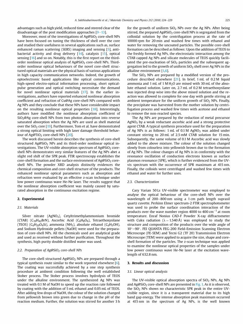
The nonlinear absorption coefficient (b) of the products wasevaluated under the open aperture z-scan mode (Fig. 5(a,c)). Theopen aperture profile (Fig. 5(a)) revealed the saturated absorptionbehaviour (peak shaped curve) as a result of interaction betweenthe Ag inner walls and the excitons in outer SiO2 walls. This syn-ergistic effects produces local field enhancement of both the innerand outer walls of Ag and SiO2. In general, saturated absorptionbehaviour occurs in core-shell nanostructures when a two photonabsorption process excites an electron from the valence band toconduction band of SiO2 and some other electron from the Fermilevel of Ag is excited to the conduction band of SiO2 through onephoton absorption [24]. The observed strong nonlinear absorptionbehaviour of Ag@SiO2 core-shell NPs may lead to the better opticallimiting performance. In contrast, the Ag NPs exhibits the reversesaturated absorption behaviour (valley shaped curve) as presentedin Fig. 5(c). The observed reverse saturated absorption behaviour ofthe Ag NPs is due to the fact that the absorption process wascontrolled by carrying an excited electron from the lower to higherconduction band through a secondary photon [25]. The switchingbehaviour from reverse saturated absorption into saturated ab-sorption is attributed to the interplay among the ground stateplasmon bleaching and saturation of two photon absorption orexcited state absorption [26]. Owing to the observed switchingfrom reverse saturated absorption to saturated absorption,Ag@SiO2 core-shell NPs could be useful for numerous photonicapplications like, Q-switching and optical limiting.
In order to evaluate the nonlinear refractive index (n2) of theproducts, we also carried out the closed aperture profile of z-scanmeasurements (Fig. 5)(b,d). The closed aperture investigation of
(a) (b)
(c) (d)
Fig. 5. (aed) Open aperture and closed aperture profiles of Ag@SiO2 core-shell NPs and Ag NPs.
A. Sakthisabarimoorthi et al. / Materials Chemistry and Physics 212 (2018) 224e229228
Ag@SiO2 core-shell NPs (Fig. 5(b)) shows the peak followed byvalley and the negative nonlinear refraction indicates the self-defocusing nature (n2< 0). It is long-familiar that the thermalnonlinearities (such as, high single photon absorption cross sec-tions) mainly contribute to the negative nonlinear refraction of thematerial [27]. The closed aperture profile of Ag NPs (Fig. 5(d))shows a dramatic reverse trend of valley followed by peak and thepositive nonlinear refraction indicates the self-focusing effect(n2> 0). Besides, the nonlinear refractive index (n2) and absorptioncoefficient (b) are estimated from the experimental data byemploying Shiek-bahae model [28]. Moreover, the nonlinearrefractive index and nonlinear absorption coefficient of the mate-rials are directly related to the real and imaginary parts of the thirdorder susceptibility (c3). The calculated value of n2 is in the order of10�6 cm2/W�1 and found that the value increased from 3.39 to4.05� 10�6 cm2/W�1 for Ag NPs and Ag@SiO2 core-shell NPs. The bcalculated from the open aperture profile has increased from 3.19 to11.38� 102 cmW�1 for Ag NPs and Ag@SiO2 core-shell NPs. Theobtained n2 and b values of the Ag@SiO2 core-shell NPs is compa-rable with the reported values in the literature [18e20,29].
4. Conclusions
In summary, the core-shell structured Ag@SiO2 NPs were pre-pared by the simple synthetic approach. The SPR red shifting in theUV-vis spectrum strongly indicated the complete coverage of AgNPs with SiO2 shell. The FTIR results are well corroborated with thesurface interaction between the Ag core and SiO2 shell. The powder
XRD results confirmed the successful formation of the Ag@SiO2
core-shell NPs and demonstrated that the crystallinity of Ag NPs isnot affected by the coating of SiO2 layer. TEMmicrographs revealedthe formation of Ag@SiO2 core-shell NPs. The core-shell structuredAg@SiO2 NPs exhibited considerable nonlinear absorption andrefractionwhich are comparable to the Ag NPs. The observed strongsaturated absorption behaviour suggested that the Ag@SiO2 core-shell NPs could be a potential material for optical limitingapplications.
Compliance with ethical standards
None.
Conflict of interest
The authors declare that they have no conflict of interest.
Acknowledgement
The authors thank Prof. D. Sasti Kumar, Department of Physics,NIT Trichy, India for providing z-scan analysis facility.
References
[1] Lu Han, Hao Wei, Bo Tu, Dongyuan Zhao, A facile one-pot synthesis of uniformcoreeshell silver nanoparticle@mesoporous silica nanospheres, Chem. Com-mun. 47 (2011) 8536e8538, https://doi.org/10.1039/c1cc12718g.
[2] Omar G. Morales-Saavedra, Rodolfo Zanella, Viridiana Maturano-Rojas,Vicente Torres-Zuniga, Jose O. Flores-Flores, Antonio A. Rodriguez-Rosales,
A. Sakthisabarimoorthi et al. / Materials Chemistry and Physics 212 (2018) 224e229 229
Roberto Ortega-Martinez, Preparation and z-scan nonlinear optical charac-terization of Au/SiO2- and Ag/SiO2-supported nanoparticles dispersed in silicasonogel films, J. Sol. Gel Sci. Technol. 63 (2012) 340e355, https://doi.org/10.1007/s10971-012-2793-8.
[3] Jessica Rodriguez- Fernandez, Isabel Pastoriza-Santos, Jorge Perez-Juste, F.Javier Garcia de Abajo, Luis M. Liz-Marzan, The effect of silica coating on theoptical response of sub-micrometer gold spheres, J. Phys. Chem. C 111 (2007)13361e13366, https://doi.org/10.1021/jp073853n.
[4] Chunyuan Song, Jun Chen, Justin L. Abell, Yiping Cui, Yiping Zhao, Ag�SiO2core�shell nanorod arrays: morphological, optical, SERS, and wetting prop-erties, Langmuir 28 (2012) 1488e1495, https://doi.org/10.1021/la203772u.
[5] Yanjie Hu, Yunli Shi, Hao Jiang, Guangjian Huang, Chunzhong Li, Scalablepreparation of ultrathin silica-coated Ag nanoparticles for SERS application,ACS Appl. Mater. Interfaces 5 (2013) 10643e10649, https://doi.org/10.1021/am402604h.
[6] Ke Xu, Jie-Xin Wang, Xu-Liang Kang, Jian-Feng Chen, Fabrication of antibac-terial monodispersed Ag-SiO2 coreeshell nanoparticles with high concentra-tion, Mater. Lett. 63 (2009) 31e33, https://doi.org/10.1016/j.matlet.2008.08.039.
[7] Masoud Karimipour, Elahe Shabani, Mohsen Mollaei, Mehdi Molaei, Micro-wave synthesis of Ag@SiO2 coreeshell using oleylamine, J. Nanoparticle Res.17 (2015) 2e8, https://doi.org/10.1007/s11051-014-2832-1.
[8] A. Santagata, A. Guarnaccio, D. Pietrangeli, A. Szegedi, J. Valyon, A. De Stefanis,A. De Bonis, R. Teghil, M. Sansone, D. Mollica, G.P. Parisi, Production of silver-silica core-shell nanocomposites using ultra-short pulsed laser ablation innanoporous aqueous silica colloidal solutions, J. Phys. D Appl. Phys. 48 (2015)205304e205315, https://doi.org/10.1088/0022-3727/48/20/205304.
[9] Jerome Leveneur, Geoffrey I.N. Waterhouse, John Kennedy, James B. Metson,David R.G. Mitchell, Nucleation and growth of Fe nanoparticles in SiO2: a TEM,XPS, and Fe L-edge XANES investigation, J. Phys. Chem. C 115 (2011)20978e20985, https://doi.org/10.1021/jp206357c.
[10] J. Leveneur, J. Kennedy, G.V.M. Williams, J. Metson, A. Markwitz, Large roomtemperature magnetoresistance in ion beam synthesized surface Fe nano-clusters on SiO2, Appl. Phys. Lett. 98 (2011) 053111e053113, https://doi.org/10.1063/1.3553274.
[11] K. Kaviyarasu, E. Manikandan, J. Kennedy, M. Maaza, A comparative study onthe morphological features of highly ordered MgO: AgO nanocube arraysprepared via a hydrothermal method, RSC Adv. 5 (2015) 82421e82428,https://doi.org/10.1039/c5ra15132e.
[12] N. Jayaprakash, J. Judith Vijaya, K. Kaviyarasu, K. Kombaiah, L. John Kennedy,R. Jothi Ramalingam, Murugan A. Munusamy, Hamad A. Al-Lohedan, Greensynthesis of Ag nanoparticles using Tamarind fruit extract for the antibacterialstudies, J. Photochem. Photobiol. B Biol. 169 (2017) 178e185, https://doi.org/10.1016/j.jphotobiol.2017.03.013.
[13] K. Anand, K. Kaviyarasu, Sudhakar Muniyasamy, Selvaraj Mohana Roopan,R.M. Gengan, A.A. Chuturgoon, Bio-synthesis of silver nanoparticles usingAgroforestry residue and their catalytic degradation for sustainable wastemanagement, J. Cluster Sci. 28 (2017) 2279e2291, https://doi.org/10.1007/s10876-017-1212-2.
[14] Philippe Saint-Cricq, Junzheng Wang, Ayae Sugawara-Narutaki,Atsushi Shimojima, Tatsuya Okubo, A new synthesis of well-dispersed,coreeshell Ag@SiO2 mesoporous nanoparticles using amino acids andsugars, J. Mater. Chem. B 1 (2013) 2451e2454, https://doi.org/10.1039/c3tb20210k.
[15] Ya Mao, Wanquan Jiang, Sheng Wang, Mei Liu, Shouhu Xuan, Xinglong Gong,Ken Cham-Fai Leung, Mesoporous SiO2 yolk shell confined core-satellite Ag
nanoparticles: preparation and catalytic activity, J. Alloy. Comp. 680 (2016)406e414, https://doi.org/10.1016/j.jallcom.2016.04.139.
[16] Michael P. Brandon, Deirdre M. Ledwith, V.M. Kelly, Preparation of saline-stable, silica-coated triangular silver nanoplates of use for optical sensing,J. Colloid Interface Sci. 415 (2014) 77e84, https://doi.org/10.1016/j.jcis.2013.10.017.
[17] Njemuwa Nwaji, David O. Oluwole, John Mack, Marcel Louzada,Samson Khene, Jonathan Britton, Tebello Nyokong, Improved nonlinear op-tical behaviour of ball type indium (III) phthalocyanine linked to glutathionecapped nanoparticles, Dyes Pigments 140 (2017) 417e430, https://doi.org/10.1016/j.dyepig.2017.01.066.
[18] A. Sakthisabarimoorthi, M. Jose, S.A. Martin Britto Dhas, S. Jerome Das,Fabrication of Cu@Ag coreeshell nanoparticles for nonlinear optical applica-tions, J. Mater. Sci. Mater. Electron. 28 (2016) 4545e4552, https://doi.org/10.1007/s10854-016-6090-0.
[19] A. Sakthisabarimoorthi, S.A. Martin Britto Dhas, M. Jose, Fabrication andnonlinear optical investigations of SiO2@Ag core-shell nanoparticles, Mater.Sci. Semicond. Process. 71 (2017) 69e75, https://doi.org/10.1016/j.mssp.2017.07.008.
[20] Renjis T. Tom, A Sreekumaran Nair, Singh Navinder, M. Aslam, C.L. Nagendra,Reji Philip, K. Vijayamohanan, T. Pradeep, Freely dispersible Au@TiO2, Au@ZrO2, Ag@TiO2, and Ag@ZrO2 core-shell nanoparticles: one-step synthesis,characterization, spectroscopy, and optical limiting properties, Langmuir 19(2003) 3439e3445.
[21] Werner Stober, Arthur Fink, Ernst Bohn, Controlled growth of monodispersesilica spheres in the micron size range, J. Colloid Interface Sci. 26 (1968)62e69, https://doi.org/10.1016/0021-9797(68)90272-5.
[22] Zhenhua Bai, Rui Chen, Peng Si, Youju Huang, Handong Sun, Dong-Hwan Kim,Fluorescent pH sensor based on Ag@SiO2 core�shell nanoparticle, ACS Appl.Mater. Interfaces (2013), https://doi.org/10.1021/am401528w.
[23] Moumita Chakraborty, Fang-Wei Hsiao, Bappaditya Naskar, Chien-Hsiang Chang, Amiya Kumar Panda, Surfactant-assisted synthesis and char-acterization of stable silver bromide nanoparticles in aqueous media, Lang-muir 28 (2012) 7282e7290, https://doi.org/10.1021/la300615b.
[24] M. Karimipour, M. Ebrahimi, Z. Abafat, M. Molaei, Synthesis of Ag@TiO2 core-shells using a rapid microwave irradiation and study of their nonlinear opticalproperties, Opt. Mater. 57 (2016) 257e263, https://doi.org/10.1016/j.optmat.2016.05.010.
[25] Shundong Guan, Xiuli Fu, Ying Tang, Zhijian Peng, AuAg@CdS double-wallednanotubes: synthesis and nonlinear absorption properties, Nanoscale 9(2017) 10277e10284, https://doi.org/10.1039/C7NR02861J.
[26] S. Venugopal Rao, N. Venkatram, L. Giribabu, D. Narayana Rao, Ultrafastnonlinear optical properties of alkyl-phthalocyanine nanoparticles investi-gated using Z-scan technique, J. Appl. Phys. 105 (2009) 053109e053114,https://doi.org/10.1063/1.3079801.
[27] M.S. Neo, N. Venkatram, G.S. Li, W.S. Chin, W. Ji, Synthesis of PbS/CdS core-shell QDs and their nonlinear optical properties, J. Phys. Chem. C 114 (2010)18037e18044, https://doi.org/10.1021/jp104311j.
[28] Mansoor Sheik-Bahae, Ali A. Said, Tai-huei wei, David J. Hagan, E.W. Vanstryland, Sensitive measurement of optical nonlinearities using a single beam,IEEE J. Quant. Electron. 26 (4) (1990) 760e769, https://doi.org/10.1109/3.53394.
[29] Seetharaman Amreetha, Dhanuskodi Sivasubramanian, Vinitha Gandhiraj,Venugopal Rao Soma, Tunable nanosecond and femtosecond nonlinear opticalproperties of C-N-S doped TiO2 nanoparticles, J. Phys. Chem. C 121 (43) (2017)24192e24205, https://doi.org/10.1021/acs.jpcc.7b08778.