materials and methods - springer static content server10.1007... · · 2016-11-04materials and...
TRANSCRIPT

Materials and methods
Cloning, expression, and purification
The genes for the SH32 and SH2 domain, which contain residues M1‐A178 or A73‐A178,
respectively, was cloned into the expression vector pTWIN1. The vectors were named
pTWIN1‐SH32 and pTWIN1‐SH2, and the corresponding protein products were named
SH32‐intein2‐CBD and SH2‐intein2‐CBD, respectively. The Csk kinase domain gene
containing residues A179‐L450 with an A179C mutation and C‐terminal His‐tag was
amplified and cloned into the expression vector pTWIN1. The plasmid was named
pTWIN1‐kinase‐His, and the designed fusion protein was named CBD‐intein1‐kinase‐His.
Overexpression and purification of proteins were described elsewhere (Liu et al., 2009).
The protein tyrosine phosphatase PEP proline‐rich peptide (PEP‐3BP1, residues 605‐629,
SRRTDDEIPPPLPERTPESFIVVEE) was expressed and purified as previously described
(Ghose et al., 2001).
Segmental labeling of NMR sample
Kinase protein mixture was added to the chitin column bound with SH32‐intein2‐CBD
with Buffer A (50 mM Tris‐HCl, pH 7.5, 200 mM NaCl). The molar ratio of SH32 or SH2 α‐
thioester and N‐terminal cysteine kinase was 1:1. The system was added with 0.5 mM
EDTA and 200 mM 2‐mercapto‐ethanesulfonic acid. Subsequently, the column was
stored at 15 °C for 72 h. During storage, three reactions proceeded: thiolysis of SH32‐
intein2‐CBD to form SH32 α‐thioester, degradation of fusion CBD‐intein1‐kinase‐His to
form kinase‐His, and reaction of SH32 α‐thioester with kinase‐His with the N‐terminal

cysteine (Figure 1C). On‐column ligation increases the ligation efficiency (Vitali et al.,
2006).
Ligation mixture was eluted from the chitin column and dialyzed against buffer A at 4 °C.
The solution was loaded on 10 ml of TALON beads charged with Co2+ and pre‐
equilibrated with buffer A. The column was washed with 50 ml of buffer A to remove
the unbound protein (mainly inteins, SH32, or SH2; Figures 2A and 2B). PEP‐3BP1 is used
to reduce the self‐association of the SH3 domain (Borchert et al., 1994; Ghose et al.,
2001). The column was washed with another 10 ml of buffer A with 1 mM PEP‐3BP1
peptide to remove the SH32 domain bound with the ligation product because of the
dimerization of SH3 domain. The bound protein was eluted and further purified by using
ion exchange chromatography.
NMR spectroscopy
All NMR experiments were performed at 25 °C on Bruker 900 MHz, 800 MHz, or 700
MHz spectrometers equipped with triple‐resonance cryoprobes. Protein solutions were
prepared in the following buffer conditions: 0.5–1.0 mM protein in 50 mM Tris‐HCl (pH
7.5), 10 mM DTT, 1.0 mM EDTA, 0.01% (w/v) NaN3, 5% D2O, and 0.1 mM DSS (4,4‐
dimethyl‐4‐silapentane‐1‐sulfonate). For the experiments containing SH32, a solution of
3BP1 peptide dissolved in NMR buffer was titrated into a SH32 or SH32‐kinase‐His
solution until the peptide was in a molar ratio of 1:2. All the NMR samples were sealed
in the NMR tube under nitrogen atmosphere. 3D triple‐resonance experiments, namely,
HNCA, HN(CO)CA, HNCO, HN(CA)CO, HNCACB and CBCA(CO)NH were performed to

determine backbone resonance assignments. 1H chemical shifts were referenced to
internal DSS. 13C and 15N chemical shifts were referenced indirectly using the 1H/13C or
1H/15N frequency ratios of the zero point 0.101329118 (15N) and 0.251449530 (13C) (Live
et al., 1984; Wishart et al., 1995). The combined chemical shift change of a particular
residue upon ligation with the kinase domain was calculated as:
Δδtotal=sqrt[(ΔδH)2+(0.154 ΔδN)2] (Mulder et al., 1999). ΔδH and ΔδN correspond to the
amide proton and nitrogen chemical shift changes upon kinase binding to SH32 or SH2,
respectively. The weight factor for 1H and 15N was determined from the ratio of the
average variances of amide nitrogen and proton chemical shifts observed for the 20
common amino acid residues in proteins as deposited with the BioMagResBank. The
NMR assignments of the SH32 domain of Csk were deposited to BMRB with the
accession number 16017.
Small‐Angle X‐Ray Scattering Experiments.
The small‐angle X‐ray scattering (SAXS) data for Csk with and without 3BP1 peptide
were collected using the X9 beamline at Brookhaven National Laboratory. The purified
proteins were stored in a buffer containing 50 mM Tris‐HCl (pH 7.5), 10 mM DTT, 1.0
mM EDTA. Each blank or sample was measured in triplicate, and the sample
measurements were adjusted by subtracting the scattering from buffer‐alone.

Accession numbers
The NMR assignments of the SH32 domain of Csk were deposited to BMRB with the accession number 16017.
Figure S1
Function, structure and segmental labeling procedure of Csk. (A) Regulation of SFKs by
Csk through Cbp binding in lipid rafts. (B)The monomeric structure of Csk with bound
3BP1 (to SH3) and CBP peptide (to SH2). The overall structure is plotted with active Csk
1K9A‐A. The position of PEP‐3BP1 and CBP peptide is modeled according to the
structure of 1JEG and 1SPS, respectively. (C) Schematic depiction of the segmental
labeling procedure of human Csk protein.
Figure S2
Size exclusion chromatography and small angle X‐ray scattering of Csk samples. Protein
solutions are prepared in conditions: 0.5–1.0 mM protein in 50 mM Tris‐HCl (pH 7.5).
Figure S3
Sedimentation equilibrium analysis of Csk: The plot shows the concentration (Y‐axis)
depending on the radius (cm), which is the distance to the rotor in the center. All graphs
show the experimental data with their fit and the plots for the sedimentation speed at
9,000 rpm. The experimental data collected at 15,000 rpm are shown by empty dots.
The panel on the upper left shows the aberration of the experimental data from the fit

using the monomer–dimer equilibrium model. (A) Full‐length Csk alone. (B) Full‐length
Csk in the presence of 3BP1. (C)The molecular weight (kDa) of Csk calculated from the
AU experimental results using different models are shown for different models and
dissociation constant for full‐length Csk.
Figure S4
Protein ligation, purification and chariazation of SH32‐kinase or SH2‐kinase.
(A)Procedure used for the purification of the SH32‐kinase ligation products. (B)
Expression and purification of CBD‐intein1‐kinase‐His (lanes 1, 2, 3, and 4) and SH32‐
intein2‐CBD (lanes 5, 6, 7, and 8). Lanes 1 and 2, insoluble and soluble fractions of cell
lysate; lanes 3 and 4, flow through and elution of TALON beads; lanes 5 and 6, insoluble
and soluble fractions of cell lysate; lanes 7 and 8, flow through and elution of chitin
beads. (C). Ligation and purification of SH2‐kinase‐His. Lanes 1 and 2, CBD‐intein1‐
kinase‐His before and after TALON bead purification; lane 3, flow through of chitin
beads; lane 4, on‐column cleavage and ligation of SH2‐kinase‐His; lane 5, elution of
ligation product from chitin beads; lanes 6 and 7, flow through and elution of TALON
beads; lane 8, molecular weight ladder. (D), (E) Selected mass results of the ligation
product (Table S1).
Figure S5
Structural perturbation of SH2 domain observed via segmental labeling methods. (A)
Overlay of 1H‐15N HSQC spectra of the Csk SH2 with and without ligated kinase. Red: 15N‐
SH2; blue: 15N‐SH2 ligated with unlabeled kinase. Both of the two samples are

concentrated to 0.1 mM. The residues that undergo significant chemical shift changes
are indicated by arrow on the spectrum. (B) Magnified view of the binding pocket in the
SH3‐SH2 and SH2‐kinase linker regions.
Figure S6
Structure perturbation of Csk identified by 1H‐15N‐HSQC spectra. Overlay of 1H‐15N‐HSQC
spectra of the 15N2H kinase with and without ligated SH32. Red: Csk 15N2H‐labeled
kinase; Green: 15N2H‐labeled kinase ligated with unlabeled SH32. The peaks indicated by
arrows show significant chemical shift perturbations.
Figure S7
Structure perturbation of Csk identified by 1H‐15N‐HSQC spectra. Overlay of 1H‐15N‐HSQC
spectra of the 15N2H kinase with and without ligated SH2. Red: Csk 15N2H‐labeled kinase;
Green: 15N2H‐labeled kinase ligated with unlabeled SH2. The peaks indicated by arrows
show significant chemical shift perturbations.

Figure S1
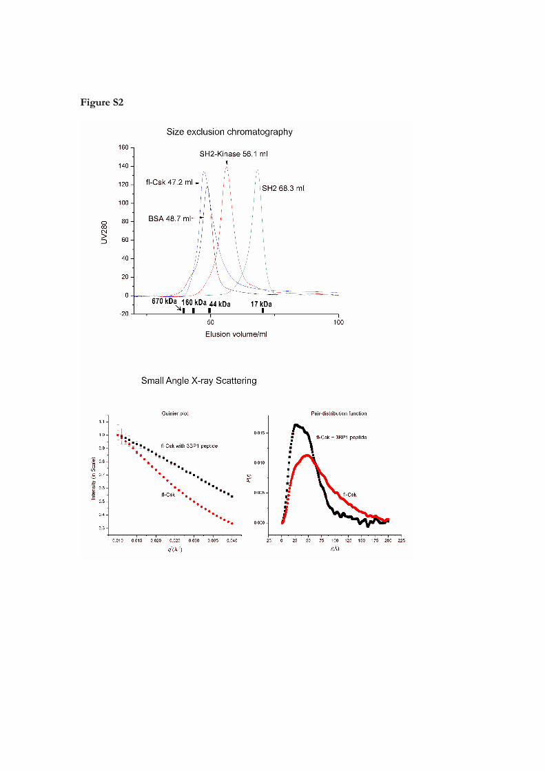
Figure S2

Figure S3
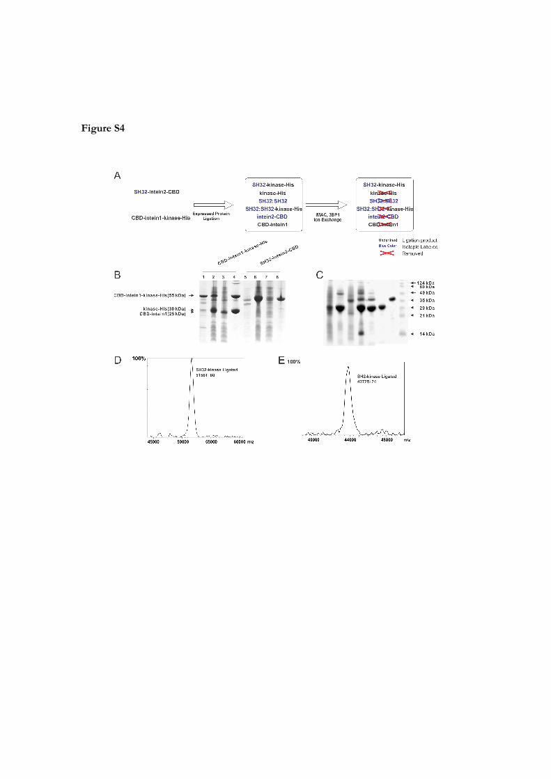
Figure S4

Figure S5

Figure S6

Figure S7
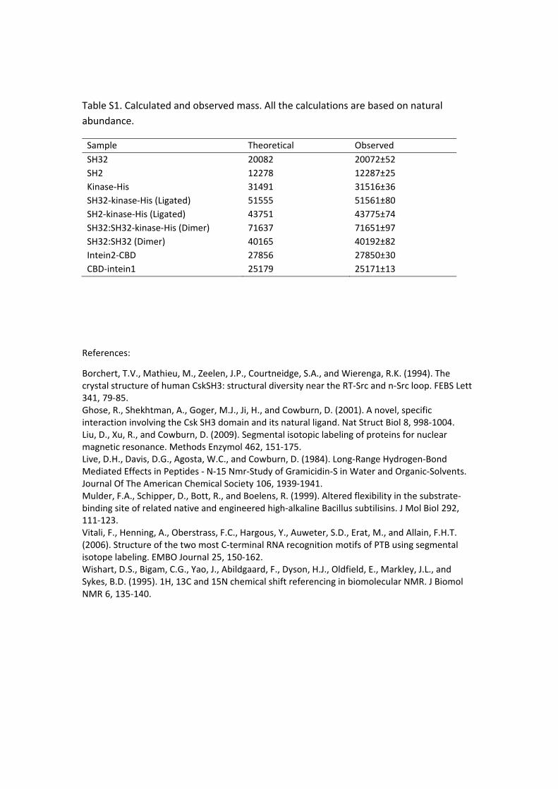
Table S1. Calculated and observed mass. All the calculations are based on natural
abundance.
Sample Theoretical Observed
SH32 20082 20072±52
SH2 12278 12287±25
Kinase‐His 31491 31516±36
SH32‐kinase‐His (Ligated) 51555 51561±80
SH2‐kinase‐His (Ligated) 43751 43775±74
SH32:SH32‐kinase‐His (Dimer) 71637 71651±97
SH32:SH32 (Dimer) 40165 40192±82
Intein2‐CBD 27856 27850±30
CBD‐intein1 25179 25171±13
References:
Borchert, T.V., Mathieu, M., Zeelen, J.P., Courtneidge, S.A., and Wierenga, R.K. (1994). The crystal structure of human CskSH3: structural diversity near the RT‐Src and n‐Src loop. FEBS Lett 341, 79‐85. Ghose, R., Shekhtman, A., Goger, M.J., Ji, H., and Cowburn, D. (2001). A novel, specific interaction involving the Csk SH3 domain and its natural ligand. Nat Struct Biol 8, 998‐1004. Liu, D., Xu, R., and Cowburn, D. (2009). Segmental isotopic labeling of proteins for nuclear magnetic resonance. Methods Enzymol 462, 151‐175. Live, D.H., Davis, D.G., Agosta, W.C., and Cowburn, D. (1984). Long‐Range Hydrogen‐Bond Mediated Effects in Peptides ‐ N‐15 Nmr‐Study of Gramicidin‐S in Water and Organic‐Solvents. Journal Of The American Chemical Society 106, 1939‐1941. Mulder, F.A., Schipper, D., Bott, R., and Boelens, R. (1999). Altered flexibility in the substrate‐binding site of related native and engineered high‐alkaline Bacillus subtilisins. J Mol Biol 292, 111‐123. Vitali, F., Henning, A., Oberstrass, F.C., Hargous, Y., Auweter, S.D., Erat, M., and Allain, F.H.T. (2006). Structure of the two most C‐terminal RNA recognition motifs of PTB using segmental isotope labeling. EMBO Journal 25, 150‐162. Wishart, D.S., Bigam, C.G., Yao, J., Abildgaard, F., Dyson, H.J., Oldfield, E., Markley, J.L., and Sykes, B.D. (1995). 1H, 13C and 15N chemical shift referencing in biomolecular NMR. J Biomol NMR 6, 135‐140.