maroteaux–lamy syndrome: functional characterization of pathogenic mutations and polymorphisms in...
TRANSCRIPT

Molecular Genetics and Metabolism 94 (2008) 305–312
Contents lists available at ScienceDirect
Molecular Genetics and Metabolism
journal homepage: www.elsevier .com/ locate/ymgme
Maroteaux–Lamy syndrome: Functional characterization of pathogenicmutations and polymorphisms in the arylsulfatase B gene
Elena Garrido a,b, Bru Cormand a,b, John J. Hopwood c, Amparo Chabás b,d,Daniel Grinberg a,b, Lluïsa Vilageliu a,b,*
a Departament de Genètica, Facultat de Biologia, Universitat de Barcelona, Av. Diagonal 645, Edifici Annex, 3ª Planta, E-08028 Barcelona, Spainb CIBERER, IBUB, Barcelona, Spainc Lysosomal Diseases Research Unit, Department of Genetic Medicine, Children, Youth and Women’s Health Service, North Adelaide, SA, Australiad Institut de Bioquímica Clínica, Hospital Clínic, Barcelona, Spain
a r t i c l e i n f o
Article history:Received 14 January 2008Received in revised form 27 February 2008Accepted 27 February 2008Available online 10 April 2008
Keywords:N-Acetylgalactosamine-4-sulfataseMaroteaux–Lamy syndromeTransient expressionCOS-7 cellsNonsense-mediated RNA decayIndirect immunofluorescence
1096-7192/$ - see front matter � 2008 Elsevier Inc. Adoi:10.1016/j.ymgme.2008.02.012
* Corresponding author. Address: Departament de GUniversitat de Barcelona, Av. Diagonal 645, EdificiBarcelona, Spain. Fax: +34 93 4034420.
E-mail address: [email protected] (L. Vilageliu).
a b s t r a c t
Mucopolysaccharidosis VI (MPS VI; Maroteaux-Lamy syndrome) is an autosomal recessive lysosomal dis-order caused by deficiency of N-acetylgalactosamine-4-sulfatase (ARSB), which is required for the degra-dation of dermatan sulfate. We recently reported mutational screening of 12 Spanish and 4 ArgentinianMPS VI patients. In the present study, seven missense mutations (c.245T > G [p.L82R], c.413A > G[p.Y138C], c.719C > T [p.S240F], c.922G > A [p.G308R], c.937C > G [p.P313A], c.1340G > T [p.C447F] andc.1415T > C [p.L472P]) were transiently expressed in COS-7 cells and 4-sulfatase activity was measuredin cell extracts. All mutations resulted in less than 6% of wild-type enzyme activity, in most cases unde-tectable. Mutations were expressed in their original haplotype context with respect to two non-synony-mous polymorphisms present in the ARSB protein, p.V358M and p.S384N. The three less frequenthaplotype combinations yielded an ARSB activity of 16%, 57% and 70%, when compared to the most fre-quent haplotype (p.358V and p.384S). Western blot analyses showed that the expressed mutations sig-nificantly reduced the amount of mature protein. Sub-cellular localization studies of mutant ARSBproteins in fibroblasts of MPS VI patients were performed. RNA analysis confirmed that nonsense-med-iated RNA decay had taken place for all mutant alleles (c.1143 � 1G > C, c.1143 � 8T > G, p.W322X,c.427delG and c.1142 + 2T > A) which were candidates for causing RNA degradation by this mechanism.In summary, all the ARSB mutations studied had a significant effect on enzyme activity, protein process-ing and/or mRNA stability.
� 2008 Elsevier Inc. All rights reserved.
Maroteaux–Lamy syndrome or mucopolysaccharidosis (MPS)VI, is caused by impaired activity of the lysosomal enzyme N-acet-ylgalactosamine-4-sulfatase (4-sulfatase, arylsulfatase B or ARSB,EC 3.1.6.1), resulting from mutations in the ARSB gene (MIM #253200). The enzyme deficiency leads to the accumulation ofharmful amounts of undegraded dermatan sulfate within the lyso-somes, which causes progressive damage in several tissues. Symp-toms include short stature, hepatosplenomegaly, dysostosismultiplex, joint stiffness, corneal clouding, cardiac abnormalitiesand coarse facies, without intellectual impairment. MPS VI patientscan present within a wide continuous clinical spectrum from rap-idly to slowly advancing pathology.
ll rights reserved.
enètica, Facultat de Biologia,Annex, 3ª Planta, E-08028
ARSB is a 533-amino-acid lysosomal soluble glycoprotein.Translocation through the ER membrane is accompanied by cleav-age of the signal peptide and glycosylation of four asparagine res-idues. During transport from the ER to the Golgi, 4-sulfatase isprocessed from a polypeptide precursor of 66 kDa to three disul-fide-linked polypeptides of 43, 8 and 7 kDa [1].
Like other mucopolysaccharidoses, MPS VI may be caused bymany different disease-causing mutations in the ARSB gene thathave been identified by mutational screening of patients fromNorth America, South America, Europe, and Australasia [2–23].Mutations with genotype–phenotype correlations have beenclearly identified for some genotypes [20]. The described mutantalleles include deletions, insertions, missense and nonsense muta-tions, as well as splice site mutations. In a previous paper, we re-ported mutations in Spanish and Argentinean patients [23]involving 19 different alleles, nine of which were novel. The mostprevalent mutation in this group of patients was the splice sitechange c.1143 � 1G > C, accounting for about 22% of the total
306 E. Garrido et al. / Molecular Genetics and Metabolism 94 (2008) 305–312
mutant alleles (7/32). Haplotype analysis pointed to a single originfor this change.
Here we expressed seven missense mutations in MPS VI pa-tients found in Spain and Argentina (c.245T > G [p.L82R],c.413A > G [p.Y138C], c.719C > T [p.S240F], c.922G > A [p.G308R],c. 937C > G [p.P313A], c.1340G > T [p.C447F] and c.1415 T > C[p.L472P]) in a heterologous transient expression system. Somesplice and frameshift mutations were characterized at the RNA le-vel and possible degradation by the nonsense-mediated RNA decay(NMD) mechanism was assessed. The possible effect of some muta-tions on protein trafficking in patients’ fibroblasts was also studied.
Materials and methods
Patients
The mutant alleles and fibroblasts used in this study correspond to the MPS VIpatients previously described [23]. Patient numbers refer to those in the samepublication.
Nonsense-mediated RNA decay
Skin fibroblasts from patients or their parents were cultured in 100-mmplates and maintained in DMEM supplemented with 10% bovine serum (BS)and 1% penicillin/streptomycin (Gibco, Invitrogen Life Technologies, Heidelberg,Germany) at 37 �C and 5% CO2. Cells were cultured in the presence or absenceof the protein synthesis inhibitor cycloheximide (Sigma-Aldrich, Steinheim, Ger-many) at 0, 500, 1000 or 1500 lg/ml for 8–24 h. Total RNA was isolated from thecultured cells using the QIAshredder� Kit and the RNeasy� Minikit (Qiagen, Hil-den, Germany). RT-PCR reactions were performed from total RNA to amplify anARSB cDNA fragment spanning exons 4–8 as described in Garrido et al. [23].Quantification of the relative intensity of the electrophoretic bands was per-formed using the Discovery Series Quantity One 1D Analysis Software (Bio-Rad,Hercules, CA, USA). Amplification of the GAPDH or b-actin cDNA, used as a con-trol, was performed using commercially available primers (BD Biosciences Clon-tech, Palo Alto, CA, USA).
RT-PCR, subcloning and site-directed mutagenesis
Total RNA was isolated from primary fibroblast cultures of a wild-type individ-ual using the QIAshredder and the RNeasy Minikit (Qiagen, Hilden, Germany). Areverse transcription reaction was carried out from total RNA using oligo-d(T) pri-mer and M-MLV Reverse Transcript RNAse-H Minus, following the manufacturer’srecommendations (Promega, Madison, WI, USA). The ARSB cDNA was PCR-amplifiedin two overlapping fragments using the primers 50-TTCCTCATTCTATCAGCGGTACAA-30 (forward) and 50-TTGCAACTTCTTCTTCGCCATCT-30 (reverse) for a 678-bpfragment including the ATG codon and 50-ACTTTGGATATCTCCTGGGTAGTG-30 (for-ward) and 50-AAAAGGCCTGAGGTCCAACT-30 (reverse) for a 1163-bp fragmentincluding the stop codon. The primers were designed using the Primer3 software(frodo.wi.mit.edu/cgi-bin/primer3/primer3_www.cgi). PCRs were performed usingthe Long Expand High Fidelity PCR System (Roche, Indianapolis, IN, USA) and con-sisted of 100 ng of template cDNA, 1 U of Long Expand Polymerase, 2 mM MgCl2
in the recommended buffer, 100 lM of dNTPs, 10% DMSO and 20 pmol of each pri-mer in a final volume of 50 ll. The reactions were subjected to 1 min at 98 �C, and35 cycles of denaturation at 96 �C for 15 s, annealing at 53 �C for 30 s and elongationat 68 �C for 80 s, with a final extension step at 68 �C for 5 min. The amplificationreactions were performed in a GeneAmp� PCR System 2700 Thermocycler (PEApplied Biosystems, Foster City, CA, USA). The two PCR fragments were purified withthe GFX PCR DNA and Gel Band purification Kit (Amersham Biosciences, Amersham,UK), dephosphorylated with alkaline phosphatase T4 Poly Kinase (Promega),blunted with DNA PolI Klenow (Promega) and cloned separately into SmaI-digestedpUC18 plasmid.
The two fragments were digested with EcoRI/DraIII and DraIII/XbaI, respec-tively, and cloned together into EcoRI/XbaI-digested pUC19 using the DNA LigationKit Ver 2.1 (Takara Bio Inc., Otsu, Shiga, Japan). The entire wild-type ARSB cDNA(1724 bp) was sequenced using the BigDye Terminator Cycle Sequencing v3.1 kit(PE Applied Biosystems, Foster City, CA, USA) in an ABI PRISM 3700 DNA analyser(PE Applied Biosystems). Sequence chromatograms were analysed with the Seq-ManTM II software (DNASTAR Inc., Madison, WI, USA).
Mutations p.L82R, p.Y138C, p.S240F, p.G308R, p.P313A, p.C447F and p.L472Pand the four possible combinations of the two polymorphic variants p.V358Mand p.S384N were introduced into the wild-type ARSB cDNA by site-directed muta-genesis using the QuickChange XL Site-Directed Mutagenesis Kit (Stratagene LifeTechnologies Co., La Jolla, CA, USA). The sequences of the mutagenic primers areavailable upon request. All products were sequenced in full to verify the presenceof the introduced change and the absence of undesired alterations. Mutant, wild-type and antisense cDNAs were transferred to the pcDNA3 expression vector (Invit-rogen, Heidelberg, Germany).
Cell culture and transfection
COS-7 cells were maintained in DMEM supplemented with 10% BS and 1% pen-icillin/streptomycin (Gibco) at 37 �C and 5% CO2. For transfection, 2 � 106 cells weregrown to 95% confluence on 100-mm plates. Cells were transfected with 2 lg ofplasmid DNA and 15 ll of LipofectamineTM 2000 (Invitrogen, Heidelberg, Germany)following the manufacturer’s instructions. Cells were harvested 48 h after transfec-tion and centrifuged, and pellets were washed once in PBS 1� and stored at �80 �Cuntil 4-sulfatase activity measurements or Western blot analyses were performed.Identical activity results were obtained when harvesting cells after 24 and 72 hpost-transfection. Transfection efficiency was assessed by GFP transfection underthe same conditions on separate plates.
Enzyme analysis
In vitro 4-sulfatase activity of cell extracts was determined by spectrophoto-metric quantification of p-nitrocathecol produced by hydrolysis of the substratep-nitrocathecol sulfate [24].
The activity of another lysosomal enzyme (b-hexosaminidase) was measured asa control (data not shown).
The average endogenous 4-sulfatase activity of untransfected COS-7 cells, cellstransfected with an empty pcDNA3 plasmid and cells transfected withp.358V;p.384S cDNA in antisense orientation, produced insignificant levels of activ-ity, as expected. The average activity of cells transfected with an empty pcDNA3plasmid was used to correct the results of cells expressing mutated ARSB cDNA.
SDS–PAGE and Western blot analysis
Proteins were subjected to SDS–PAGE (12.5% polyacrylamide) and electropho-retically transferred onto an Immobilon-P Transfer Membrane (Millipore, Billerica,Massachusetts, USA). About 30 lg of protein extract were loaded per lane. Westernblots were performed as described elsewhere [25]. The primary antibody was apolyclonal sheep anti-human recombinant 4-sulfatase IgG. The secondary antibodywas a polyclonal rabbit anti-sheep peroxidase-conjugated IgG (Sigma–Aldrich,Steinheim, Germany). The detection reaction was performed by incubating themembrane in a luminol solution (100 mM Tris–HCl pH 9, coumaric acid 0.2 mM,luminol 1.25 mM and 0.3% H2O2) for 2 min at RT. The membrane was exposed toautoradiography films (HyperfilmTM ECL High-performance chemiluminescencefilm, Amersham Biosciences, Buckinghamshire, UK). Quantification of the bandswas performed with The Discovery Series Quantity One 1D Analysis Software(Bio-Rad, Hercules, CA, USA) by measuring the relative intensity of western blotbands and subtracting the intensity of the background.
Subcellular localization of 4-sulfatase in fibroblasts from MPS VI patients
Fibroblasts were cultured on 13-mm coverslips in 12-well plates at 80–90%confluence. Cells were rinsed with 10% PBS pre-heated at 37 �C and fixed for15 min at RT with 3% paraformaldehyde in 0.1 M PB and 60 mM saccharose.After fixation, cells were washed in PBS Gly (1% PBS and 20 mM glycine). Cellswere then treated with a permeabilization and blocking solution (0.05% w/v Tri-ton X-100, 1% w/v BSA in PBS Gly) for 10 min at 37 �C and then washed again.The sheep antibody against 4-sulfatase was used as a primary antibody. Thelysosomes were labeled with the mouse anti-human Lamp-2 monoclonal anti-body H4B4 (Developmental Studies Hybridoma Bank, University of Iowa, Depart-ment of Biological Sciences, Iowa City, IA, www.uiowa.edu/dshbwww), which isknown to recognize the monkey Lamp-2 of COS-7 cells [26]. Primary antibodieswere diluted in blocking solution at 1/75 for anti-ARSB and 1/100 for H4B4. Forsecondary antibodies we used donkey FITC anti-sheep IgG (Jackson Immunore-search Laboratories, Inc., West Prove, PA, USA) and goat AlexaFluor� 660 anti-mouse IgG from Molecular Probes (Invitrogen). Mounting medium was Mowiol4–8 (Calbiochem, Merck Biosciences, Darmstadt, Germany). Images were cap-tured with an Olympus Fluoview FV300 confocal microscope and analyzed withthe Fluoview FV500 image software. All experiments were repeated at least threetimes to ensure reproducibility. Autofluorescence and secondary antibody controltests were performed.
3D molecule visualization
Mutated amino acids were located on a 3D ARSB protein graphic using the Ras-Mol v2.7.3.1 software (www.openrasmol.org, Fig. 1).
Results
Studies of nonsense-mediated RNA decay
In a previous paper we reported several ARSB mutations thatgenerated premature termination codons (PTC) [23]. Mutationsc.899_1142del (genomic deletion of exon 5), c.1142 + 2T > A (exon

Fig. 1. 3D model of the ARSB protein showing the localization of mutated amino acids analyzed in this study (represented as solid spheres; black for pathogenic mutationsand violet for non-synonymous polymorphisms). a-Helices are colored in red, b-sheets in yellow, turns are blue and all the other residues are depicted in white. Green, centralmotives involving the catalytic core of ARSB: Asp53, Asp54, FGly91, Arg95, Lys145, His147, His242, Asn301, Asp300 and Lys318.
E. Garrido et al. / Molecular Genetics and Metabolism 94 (2008) 305–312 307
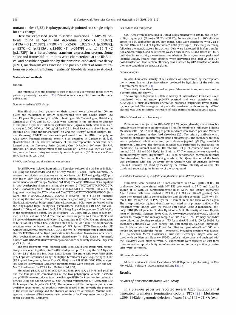
5 skipping), c.1143 � 1G > C (exon 6 skipping), c.1143 � 8T > G(exon 6 skipping), c.427delG (frameshift in exon 2) andc.966C > A (p.W322X) were candidates for inducing transcript deg-radation by the nonsense-mediated mRNA decay (NMD) mecha-nism. For some of these mutations, preliminary resultssuggesting that NMD had taken place were presented. In the pres-ent work we analysed the effect of cycloheximide (CHX), a well-known inhibitor of protein synthesis and therefore, of NMD, onML2, ML3, ML4, ML9f (father of patient ML9) and ML16 fibroblasts,to confirm the NMD process (Fig. 2). It should be noted that for theanalysis of patient ML9, fibroblasts from the father were used dueto lack of sample from the patient. The father was previouslyshown to be the carrier of mutation c.1142 + 2T > A [23].
The c.1143 � 1G > C allele was present in patients ML3 andML16 together with the missense mutations p.R160Q andp.S240F, respectively. As shown in Fig. 2a, the lower band, whichcorresponds to the skipping of exon 6 due to a splice site mutation,showed a clear increase in intensity upon treatment with 500 or1000 lg/ml CHX, both at 8 and 12 h. For example, for patientML3, the intensity ratio of the lower band with respect to the high-er one changes from 0.07:1 to 0.2:1 after 8 h of CHX treatment at500 lg/ml.
In patients ML2 (homozygous for c.1143 � 8T > G) and ML4(c.427delG/p.W322X), both alleles are assumed to be targets ofthe NMD process. Thus, amplification of the GAPDH gene by PCRmultiplex was performed for comparison. After CHX treatment(500 or 1000 lg/ml, 8 or 12 h), there is a clear increase in the ratioARSB:GAPDH for both patients in all conditions (Fig. 2b and c). Forexample, the ARSB:GAPDH ratio for patient ML4 (500 lg/ml, 8 h)rose from 0.02:1 to 0.4:1. Upon specific digestion for c.427delGin patient ML4, the CHX-mediated mRNA increase took place forboth alleles (lower panel).
Finally, weak RNA recovery was observed for the sample fromthe father of patient ML9, ML9f (c.1142 + 2T > A/+), only after16 h of CHX incubation and overloading the gel (Fig. 2d).
Expression of wild-type and mutated 4-sulfatase alleles in COS-7 cells
In order to evaluate the enzyme activity of 4-sulfatase proteinsbearing the amino acid changes p.L82R, p.Y138C, p.S240F, p.G308R,p.P313A, p.C447F, p.L472P, p.V358M and p.S384N, COS-7 cellswere transfected with the corresponding mutated cDNAs.
The mutations were expressed in their haplotype context, con-cerning the two polymorphic variants p.V358M (rs1065757,c.1072G > A, exon 5) and p.S384N (rs25414, c.1151G > A, exon 6),as this could be responsible for some degree of variation in enzymeactivity. For the sake of clarity, the haplotypes will be described asGG, GA, AG, and AA, referring to the nucleotide present in positionsc.1072 and c.1151, respectively. The haplotype context for patientsbearing the p.L82R, p.Y138C and p.P313A substitutions was GG,and mutations p.S240F, p.C447F and p.L472P were in an AG chro-mosome. In the case of mutation p.G308R, since the phase was un-known, it was expressed in the two possible haplotype contexts forthis patient: GG or GA. Additionally, for the cases in which the hap-lotype was different from the most active combination (GG), themutant alleles were also expressed in this context.
For the wild-type ARSB protein with the GG haplotype, theabsolute corrected values of activity ranged between 1368 and2898 nmol/h mg of protein. The haplotype combination AGshowed 70% ARSB activity compared to that of the GG haplo-type. The activity went down to 57% in the GA haplotype andto 16% in the AA combination. In comparison, all the analyzedmutations showed very low or negligible 4-sulfatase activity(Fig. 3).
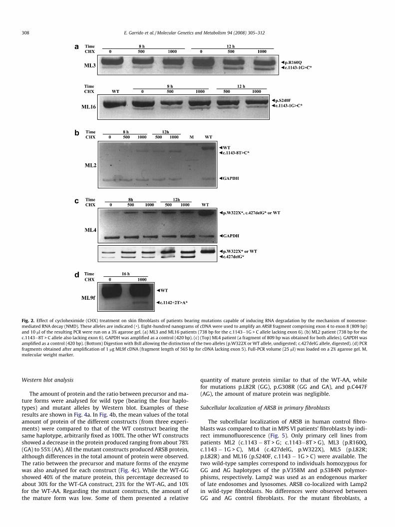
Fig. 2. Effect of cycloheximide (CHX) treatment on skin fibroblasts of patients bearing mutations capable of inducing RNA degradation by the mechanism of nonsense-mediated RNA decay (NMD). These alleles are indicated (*). Eight-hundred nanograms of cDNA were used to amplify an ARSB fragment comprising exon 4 to exon 8 (809 bp)and 10 ll of the resulting PCR were run on a 3% agarose gel. (a) ML3 and ML16 patients (738 bp for the c.1143�1G > C allele lacking exon 6). (b) ML2 patient (738 bp for thec.1143�8T > C allele also lacking exon 6). GAPDH was amplified as a control (420 bp). (c) (Top) ML4 patient (a fragment of 809 bp was obtained for both alleles). GAPDH wasamplified as a control (420 bp). (Bottom) Digestion with BslI allowing the distinction of the two alleles (p.W322X or WT allele, undigested; c.427delG allele, digested). (d) PCRfragments obtained after amplification of 1 lg ML9f cDNA (fragment length of 565 bp for cDNA lacking exon 5). Full-PCR volume (25 ll) was loaded on a 2% agarose gel. M,molecular weight marker.
308 E. Garrido et al. / Molecular Genetics and Metabolism 94 (2008) 305–312
Western blot analysis
The amount of protein and the ratio between precursor and ma-ture forms were analysed for wild type (bearing the four haplo-types) and mutant alleles by Western blot. Examples of theseresults are shown in Fig. 4a. In Fig. 4b, the mean values of the totalamount of protein of the different constructs (from three experi-ments) were compared to that of the WT construct bearing thesame haplotype, arbitrarily fixed as 100%. The other WT constructsshowed a decrease in the protein produced ranging from about 78%(GA) to 55% (AA). All the mutant constructs produced ARSB protein,although differences in the total amount of protein were observed.The ratio between the precursor and mature forms of the enzymewas also analysed for each construct (Fig. 4c). While the WT-GGshowed 40% of the mature protein, this percentage decreased toabout 30% for the WT-GA construct, 23% for the WT-AG, and 10%for the WT-AA. Regarding the mutant constructs, the amount ofthe mature form was low. Some of them presented a relative
quantity of mature protein similar to that of the WT-AA, whilefor mutations p.L82R (GG), p.G308R (GG and GA), and p.C447F(AG), the amount of mature protein was negligible.
Subcellular localization of ARSB in primary fibroblasts
The subcellular localization of ARSB in human control fibro-blasts was compared to that in MPS VI patients’ fibroblasts by indi-rect immunofluorescence (Fig. 5). Only primary cell lines frompatients ML2 (c.1143 � 8T > G; c.1143�8T > G), ML3 (p.R160Q,c.1143 � 1G > C), ML4 (c.427delG, p.W322X), ML5 (p.L82R;p.L82R) and ML16 (p.S240F, c.1143 � 1G > C) were available. Thetwo wild-type samples correspond to individuals homozygous forGG and AG haplotypes of the p.V358M and p.S384N polymor-phisms, respectively. Lamp2 was used as an endogenous markerof late endosomes and lysosomes. ARSB co-localized with Lamp2in wild-type fibroblasts. No differences were observed betweenGG and AG control fibroblasts. For the mutant fibroblasts, a
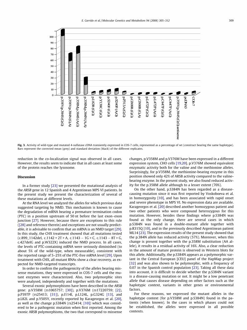
Fig. 3. Activity of wild-type and mutated 4-sulfatase cDNA transiently expressed in COS-7 cells, represented as a percentage of wt (construct bearing the same haplotype).Bars represent the corrected mean (grey) and standard deviation (black) of the different replicates.
E. Garrido et al. / Molecular Genetics and Metabolism 94 (2008) 305–312 309
reduction in the co-localization signal was observed in all cases.However, the results seem to indicate that in all cases at least someof the protein reaches the lysosome.
Discussion
In a former study [23] we presented the mutational analysis ofthe ARSB gene in 12 Spanish and 4 Argentinean MPS VI patients. Inthe present study we present the characterization of several ofthese mutations at different levels.
At the RNA level we analyzed the alleles for which previous datasuggested targeting by NMD. This mechanism is known to causethe degradation of mRNA bearing a premature termination codon(PTC) in a position upstream of 50 nt before the last exon–exonjunction [27]. However, because there are exceptions to this rule([28] and reference therein) and exceptions are not usually predict-able, it is advisable to confirm that an mRNA is an NMD target [29].In this study, the CHX treatment showed that all mutations tested(c.899_1142del, c.1142 + 2T > A, c.1143 � 1G > C, c.1143 � 8T > G,c.427delG and p.W322X) induced the NMD process. In all cases,the levels of PTC-containing mRNA were seriously diminished (toabout 5% of the wild-type, when measurable), consistent withthe reported range of 5–25% of the PTC-free mRNA level [29]. Upontreatment with CHX, all mutant RNAs show a clear recovery, as ex-pected for NMD-targeted transcripts.
In order to confirm the pathogenicity of the alleles bearing mis-sense mutations, they were expressed in COS-7 cells and the mu-tant enzymes were characterized. Also, two polymorphic siteswere analysed, independently and together with the mutations.
Several exonic polymorphisms have been described in the ARSBgene: p.V358M (rs1065757; [30]), p.V376M (rs17220759; [2]),p.P397P (rs25413; [31]), p.I114I, p.L124L, p.G324G [13], andp.L82L and p.Y505Y, recently reported by Karageorgos et al. [20],as well as the change p.S384N (rs25414; [10]) which was consid-ered to be a pathogenic mutation when first reported. Among theexonic ARSB polymorphisms, the two that correspond to missense
changes, p.V358M and p.V376M have been expressed in a differentexpression system, CHO cells [19,20]. p.V376M showed equivalentenzymatic activity both for the valine and the methionine alleles.Surprisingly, for p.V358M, the methionine-bearing enzyme in thisposition showed only 42% of ARSB activity compared to the valine-bearing enzyme. In the present study, we also found reduced activ-ity for the p.358M allele although to a lesser extent (70%).
On the other hand, p.S384N has been regarded as a disease-causing mutation since it was first reported by Voskoboeva et al.in homozygosity [10], and has been associated with rapid onsetand severe phenotype in MPS VI. No expression data are available.Karageorgos et al. [20] described another homozygous patient andtwo other patients who were compound heterozygous for thismutation. However, besides these findings where p.S384N wasfound as the only change, there are several cases in whichp.S384N was found in a double-mutant allele together withp.R315Q [10], and in the previously described Argentinean patientML14 [23]. The expression results of the present study showed thatthe p.384N allele has reduced activity (57%). Moreover, when thischange is present together with the p.358M substitution (AA al-lele), it results in a residual activity of 16%. Also, a clear reductionin the amount of mature protein is observed in Western blots forthis allele. Additionally, the p.S384N appears as a polymorphic var-iant in the Central European [CEU] panel of the HapMap project[32] and was also shown to be polymorphic, with a frequency of0.07 in the Spanish control population [23]. Taking all these datainto account, it is difficult to decide whether the p.S384N variantis a disease-causing mutation or not. It might be a low penetrantallele that causes disease depending on other factors such as thehaplotypic context, variants in other genes or environmentalfactors.
For these reasons, we expressed the mutant alleles in thehaplotype context (for p.V358M and p.S384N) found in the pa-tients (when known). In the cases in which phases could notbe established, the alleles were expressed in all possiblecontexts.
Fig. 4. (a) Western blot analysis of ARSB proteins from wild-type cDNA constructs and those bearing missense mutations expressed in COS-7 cells, showing the precursor(66 kDa) and mature form (43 kDa). COS-7 lane corresponds to untransfected cells. (b) Total amount of ARSB protein from each construct compared to that of wild-typeconstruct bearing the same haplotypic combination. Bars correspond to the mean of three different experiments. Standard deviations are indicated. (c) Relative amount ofprecursor (grey) and mature form (black) for each construct.
310 E. Garrido et al. / Molecular Genetics and Metabolism 94 (2008) 305–312
Mutations p.L82R, p.Y138C, p.S240F, p.G308R, p.P313A,p.C447F and p.L472P showed a null or very low level of activityindependently of the haplotype for the polymorphisms in thecases in which more than one haplotype context was tested.However, slight differences were observed for the amount of pro-tein and, particularly, for the ratio between precursor and matureprotein produced by the different constructs. Thus, for example,mutations p.L82R, p.G308R and p.C447F (the latter in the AG con-text), seem to have a stronger effect on the maturation of theprotein.
Mutations p.L82R, p.Y138C, p.S240F, p.G308R, p.C447F andp.L472P did not involve any amino acids defined by theconsensus sequences CTPSR (residues 91–95 in ARSB) andGKWHLG (residues 144–149) that are conserved within thehuman sulfatase family [33]. However, mutation p.S240F islocated close to one of the amino acids of the catalytic core,H242, as shown by localization of mutated amino acids inthe 3D model of the molecule (Fig. 1). A potential impact onthe interaction of the mutated enzyme with the substrate canbe assumed. p.Y138C is located very near to the second con-sensus sequence of the sulfatase family, which some authorshave extended to residues 137–147 [34], so this amino acid
may play a catalytic role. p.G308R is also located close to thecatalytic residues Asp300 and Asn301. Mutations p.C447F andp.L472P are located close to the protein surface and their inter-ference with the ARSB function remains unclear, along with thegeneral disturbance in the tertiary structure assumed to be pro-duced by changes in amino acids such as proline or cysteine. Adifferent missense mutation at position p.447 has been foundrecently [20]. Finally, mutation p.L82R may be in a relevant re-gion since a mutation (p.D83Y) was reported in the neighboringamino acid residue [20].
In summary, we show the effect of all the mutations, either onRNA stability/integrity, on the maturation of the protein or on en-zyme activity. The results confirm that all these changes found inthe MPS VI patients are, indeed, disease-causing mutations. Thecontroversial p.S384N change has been found in our series of pa-tients always together with a pathogenic change in the same allele.These data, together with the expression results, support thehypothesis that it is a non pathogenic polymorphism. However,this question remains open since other authors have described afew patients bearing alleles with only this change. It might be con-sidered a low penetrant allele, able to cause disease depending ondifferent genetic or environmental factors.
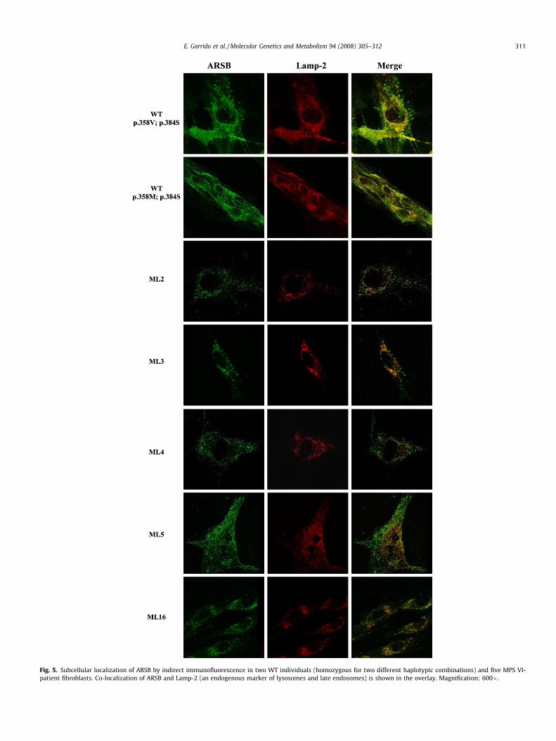
Fig. 5. Subcellular localization of ARSB by indirect immunofluorescence in two WT individuals (homozygous for two different haplotypic combinations) and five MPS VI-patient fibroblasts. Co-localization of ARSB and Lamp-2 (an endogenous marker of lysosomes and late endosomes) is shown in the overlay. Magnification: 600�.
E. Garrido et al. / Molecular Genetics and Metabolism 94 (2008) 305–312 311
312 E. Garrido et al. / Molecular Genetics and Metabolism 94 (2008) 305–312
Acknowledgments
The authors are grateful to R. Rycroft for revising the English.We thank Josep Jarque for the enzymatic measurements and Móni-ca Cozar for her technical support with Western blotting. We areindebted to Raquel García and Nieves Hernández from the ServeisCientificotècnics UB for their assistance with confocal microscopy.Financial support was provided by CICYT (SAF2003-00386 andSAF2006-12276), FIS Redes Temáticas, REDEMETH G03/054,PI051343 and PI051182), AGAUR (2005SGR00848) and CIBERER(INTRA/07/720.1).
References
[1] T. Kobayashi, K. Honke, T. Jin, S. Gasa, T. Miyazaki, A. Makita, Components andproteolytic processing sites of arylsulfatase B from human placenta, Biochim.Biophys. Acta 1159 (1992) 243–247.
[2] G. Wicker, V. Prill, D. Brooks, G. Gibson, J. Hopwood, K. von Figura, C. Peters,Mucopolysaccharidosis VI (Maroteaux–Lamy syndrome). An intermediateclinical phenotype caused by substitution of valine for glycine at position137 of arylsulfatase B, J. Biol. Chem. 266 (1991) 21386–21391.
[3] W.D. Jin, C.E. Jackson, R.J. Desnick, E.H. Schuchman, Mucopolysaccharidosistype VI: identification of three mutations in the arylsulfatase B gene of patientswith the severe and mild phenotypes provides molecular evidence for geneticheterogeneity, Am. J. Hum. Genet. 50 (1992) 795–800.
[4] T. Litjens, C.P. Morris, E.F. Robertson, C. Peters, K. von Figura, J.J. Hopwood, AnN-acetylgalactosamine-4-sulfatase mutation delta G238 results in a severeMaroteaux–Lamy phenotype, Hum. Mutat. 1 (1992) 397–402.
[5] T. Litjens, D.A. Brooks, C. Peters, G.J. Gibson, J.J. Hopwood, Identification,expression, and biochemical characterization of N-acetylgalactosamine-4-sulfatase mutations and relationship with clinical phenotype in MPS-VIpatients, Am. J. Hum. Genet. 58 (1996) 1127–1134.
[6] G. Arlt, D.A. Brooks, D. Isbrandt, J.J. Hopwood, J. Bielicki, T.M. Bradford, C.A.Bindloss-Petherbridge, K. von Figura, C. Peters, Juvenile form ofmucopolysaccharidosis VI Maroteaux–Lamy syndrome. A C-terminalextension causes instability but increases catalytic efficiency of arylsulfataseB, J. Biol. Chem. 269 (1994) 9638–9643.
[7] D. Isbrandt, G. Arlt, D.A. Brooks, J.J. Hopwood, K. von Figura, C. Peters,Mucopolysaccharidosis VI Maroteaux–Lamy syndrome: six uniquearylsulfatase B gene alleles causing variable disease phenotypes, Am. J. Hum.Genet. 54 (1994) 454–463.
[8] D. Isbrandt, J.J. Hopwood, K. von Figura, C. Peters, Two novel frameshiftmutations causing premature stop codons in a patient with the severe form ofMaroteaux–Lamy syndrome, Hum. Mutat. 7 (1996) 361–363.
[9] E. Voskoboeva, D. Isbrandt, K. von Figura, X. Krasnopolskaya, C. Peters, Fournovel mutant alleles of the arylsulfatase B gene in two patients withintermediate form of mucopolysaccharidosis VI (Maroteaux–Lamysyndrome), Hum. Genet. 93 (1994) 259–264.
[10] E. Voskoboeva, K.D. Krasnopol’skaia, K. Peters, K. von Figura, Identification ofmutations in the arylsulfatase B gene in Russian mucopolysaccharidosis typeVI patients, Genetika 36 (2000) 837–843.
[11] CM. Simonaro, E.H. Schuchman, N-Acetylgalactosamine-4-sulfatase:identification of four new mutations within the conserved sulfatase regioncausing mucopolysaccharidosis type VI, Biochim. Biophys. Acta 1272 (1995)129–132.
[12] G.R. Villani, N. Balzano, P. Di Natale, Two novel mutations of the arylsulfatase Bgene in two Italian patients with severe form of mucopolysaccharidosis.Mutation in brief 127, Hum. Mutat. 11 (1998) 410.
[13] G.R. Villani, N. Balzano, D. Vitale, M. Saviano, V. Pavone, P. Di Natale,Maroteaux–lamy syndrome: five novel mutations and their structurallocalization, Biochim. Biophys. Acta 1453 (1999) 185–192.
[14] J.Y. Wu, C.F. Yang, C.C. Lee, J.G. Chang, F.J. Tsai, A novel mutation (Q239R)identified in a Taiwan Chinese patient with type VI mucopolysaccharidosis(Maroteaux–Lamy syndrome), Hum. Mutat. 15 (2000) 389–390.
[15] C.F. Yang, J.Y. Wu, S.P. Lin, F.J. Tsai, Mucopolysaccharidosis type VI: report oftwo Taiwanese patients and identification of one novel mutation, J. FormosMed. Assoc. 100 (2001) 820–823.
[16] M.F. Petry, T. Dieter, M. Burin, R. Giugliani, S. Leistner, Identification of a novelmutation in the ARSB gene that is frequent among Brazilian MPS VI patients,Genet. Test. 7 (2003) 347–649.
[17] M.F. Petry, K. Nonemacher, J.C. Sebben, I.V. Schwartz, A.C. Azevedo, M.G. Burin,A.R. De Rezende, C.A. Kim, R. Giugliani, S. Leistner-Segal,Mucopolysaccharidosis type VI: Identification of novel mutations on thearylsulphatase B gene in South American patients, J. Inherit. Metab. Dis. 286(2005) 1027–1034.
[18] L. Karageorgos, P. Harmatz, J. Simon, A. Pollard, P.R. Clements, D.A. Brooks, J.J.Hopwood, Mutational analysis of mucopolysaccharidosis type VI patientsundergoing a trial of enzyme replacement therapy, Hum. Mutat. 23 (2004)229–233.
[19] L. Karageorgos, D.A. Brooks, P. Harmatz, D. Ketteridge, A. Pollard, E.L. Melville,E. Parkinson-Lawrence, P.R. Clements, J.J. Hopwood, Mutational analysis ofmucopolysaccharidosis type VI patients undergoing a phase II trail of enzymereplacement therapy, Mol. Genet. Metab. 90 (2007) 164–170.
[20] L. Karageorgos, D.A. Brooks, A. Pollard, E.L. Melville, L.K. Hein, P.R. Clements, D.Ketteridge, S.J. Swiedler, M. Beck, R. Giugliani, P. Harmatz, J.E. Wraith, N.Guffon, E. Leao Teles, M.C. Sa Miranda, J.J. Hopwood, Mutational analysis of105 mucopolysaccharidosis type VI patients, Hum. Mutat. 28 (2007) 897–903.
[21] D.A. Brooks, G.J. Gibson, L. Karageorgos, LK. Hein, E.F. Robertson, JJ. Hopwood,An index case for the attenuated end of the mucopolysaccharidosis type VIclinical spectrum, Mol. Genet. Metab. 853 (2005) 236–238.
[22] W. Dou, X. Gu, C. Peng, J. Zheng, J. Chen, W. Zhang, S. Huang, H.Z. Sheng, Twonovel mutations of the arylsulfatase B gene in a Chinese MPS VI childundergoing bone marrow transplantation therapy, Clin. Chim. Acta 374 (2006)171–172.
[23] E. Garrido, A. Chabás, M.J. Coll, M. Blanco, C. Domínguez, D. Grinberg, L.Vilageliu, B. Cormand, Identification of the molecular defects in Spanish andArgentinian mucopolysaccharidosis VI (Maroteaux–Lamy syndrome) patients,including 9 novel mutations, Mol. Genet. Metab. 92 (2007) 122–130.
[24] H. Baum, K.S. Dogson, B. Spencer, The assay of arylsulphatases A and B inhuman urine, Clin. Chim. Acta 4 (1959) 453–455.
[25] M. Montfort, E. Garrido, J.J. Hopwood, D. Grinberg, A. Chabas, L. Vilageliu,Expression and functional characterization of human mutant sulfamidase ininsect cells, Mol. Genet. Metab. 83 (2004) 246–251.
[26] B. Shen, S.J. Orlow, The ocular albinism type 1 gene product is an N-glycoprotein but glycosylation is not required for its subcellular distribution,Pigment Cell Res. 14 (2001) 485–490.
[27] E. Nagy, L.E. Maquat, A rule for termination-codon position within intron-containing genes: when nonsense affects RNA abundance, Trends Biochem.Sci. 23 (1998) 198–199.
[28] J.A. Holbrook, G. Neu-Yilik, M.W. Hentze, A.E. Kulozik, Nonsense-mediateddecay approaches the clinic, Nat. Genet. 36 (2004) 801–808.
[29] H.A. Kuzmiak, L.E. Maquat, Applying nonsense-mediated mRNA decayresearch to the clinic: progress and challenges, Trends Mol. Med. 12 (2006)306–316.
[30] W.D. Jin, R.J. Desnick, E.H. Schuchman, A common polymorphism in the humanarylsulfatase B (ARSB) gene at 5q13–q14, Nucleic Acids Res. 19 (1991) 4305.
[31] E.H. Schuchman, C.E. Jackson, R.J. Desnick, Human arylsulfatase B: MOPACcloning, nucleotide sequence of a full-length cDNA, and regions of amino acididentity with arylsulfatases A and C, Genomics 6 (1990) 149–158.
[32] G.A. Thorisson, A.V. Smith, L. Krishnan, L.D. Stein, The International HapMapProject web site, Genome Res. 15 (2005) 1592–1593.
[33] C.S. Bond, P.R. Clements, S.J. Ashby, C.A. Collyer, S.J. Harrop, J.J. Hopwood, J.M.Guss, Structure of a human lysosomal sulfatase, Structure 5 (2007) 277–289.
[34] S.R. Hanson, M.D. Best, C.H. Wong, Sulfatases: structure, mechanism, biologicalactivity, inhibition, and synthetic utility, Angew. Chem., Int. Ed. 43 (2004)5736–5763.