marks-hirschfeld museum of medical history
TRANSCRIPT
Marks-Hirschfeld Museumof Medical HistoryApril 2021
Curator’s IntroductionDear Readers,
My guess is that many of you have donned a pair of specs to read this newsletter, if you weren’t wearing them already. In this edition, Robert narrates the evolution of spectacles, from the concave emerald that Nero employed to watch gladiatorial fights to the classy frames and thin lenses we know today. The changes that glasses enabled for industry and the relationship between glasses and literacy are particularly interesting.
Like Robert, Jan Nixon is another of the Museum’s long-serving volunteers. She has been working diligently on our photographic collection, including sorting through our large archive of historical images from the Faculty. Jan’s contribution to this edition of the newsletter is a closer look at the medical school’s second year class photographs from years 1937 and 1942. See if you recognise any of the names!
Finally, in the next fortnight we will be emailing a short audience survey which I urge you to complete. We are hoping to gain a better understanding of what you look for in a newsletter, to ensure we are providing relevant, interesting and anticipated content in a format that is easy to enjoy. After all, where would we be without our readers!
Until I write again, take care.
Charla StrelanCurator, Marks-Hirschfeld Museum of Medical History
Mark Hirschfeld Museum of Medical History - newsletter April 2021 - 2
Eyes have always fascinated humans. They acquired metaphysical properties in antiquity, from” The Eye of Horus” which was used by ancient Egyptians to protect its wearer from evil, to classical Greek mythology which related how seeing the mythical gorgon’s face meant being turned to stone. Vision is central to awareness and consciousness. Modern neurophysiology shows that the optical cortex has many connections throughout the cerebral cortex to explain the deep integration of vision into emotions, memory, sensations and language.
Poor vision affects many humans, so it is unsurprising to find that there have been frequent attempts to improve human vision across history. Whilst myopia or short sightedness arises in childhood, presbyopia or the diminishing ability to focus on objects at close range affects us all in our fifth decade. As the development of reading and writing in the classical periods of both Asia and Europe progressed, so to did vision related problems and the need for corrective lenses. References in texts are sparse which makes it difficult to
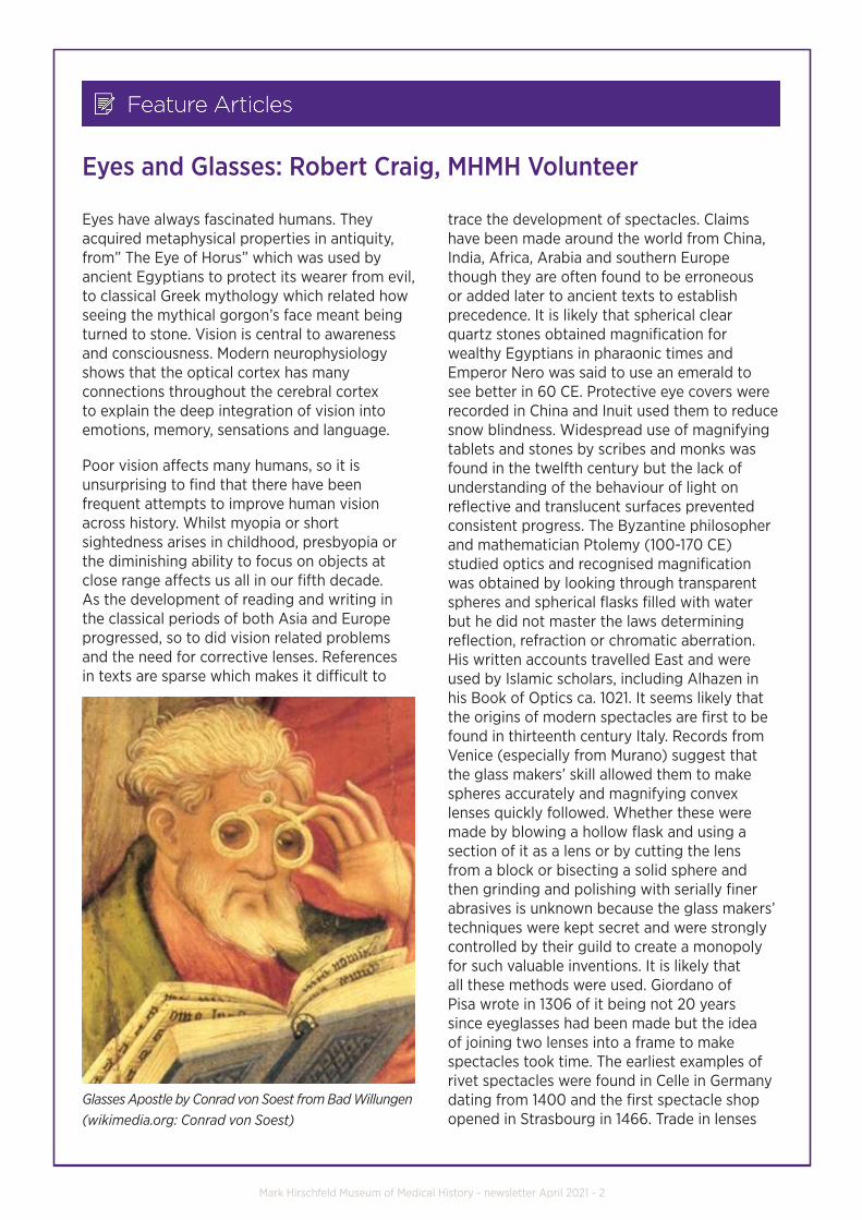
Glasses Apostle by Conrad von Soest from Bad Willungen(wikimedia.org: Conrad von Soest)
Eyes and Glasses: Robert Craig, MHMH Volunteer
trace the development of spectacles. Claims have been made around the world from China, India, Africa, Arabia and southern Europe though they are often found to be erroneous or added later to ancient texts to establish precedence. It is likely that spherical clear quartz stones obtained magnification for wealthy Egyptians in pharaonic times and Emperor Nero was said to use an emerald to see better in 60 CE. Protective eye covers were recorded in China and Inuit used them to reduce snow blindness. Widespread use of magnifying tablets and stones by scribes and monks was found in the twelfth century but the lack of understanding of the behaviour of light on reflective and translucent surfaces prevented consistent progress. The Byzantine philosopher and mathematician Ptolemy (100-170 CE) studied optics and recognised magnification was obtained by looking through transparent spheres and spherical flasks filled with water but he did not master the laws determining reflection, refraction or chromatic aberration. His written accounts travelled East and were used by Islamic scholars, including Alhazen in his Book of Optics ca. 1021. It seems likely that the origins of modern spectacles are first to be found in thirteenth century Italy. Records from Venice (especially from Murano) suggest that the glass makers’ skill allowed them to make spheres accurately and magnifying convex lenses quickly followed. Whether these were made by blowing a hollow flask and using a section of it as a lens or by cutting the lens from a block or bisecting a solid sphere and then grinding and polishing with serially finer abrasives is unknown because the glass makers’ techniques were kept secret and were strongly controlled by their guild to create a monopoly for such valuable inventions. It is likely that all these methods were used. Giordano of Pisa wrote in 1306 of it being not 20 years since eyeglasses had been made but the idea of joining two lenses into a frame to make spectacles took time. The earliest examples of rivet spectacles were found in Celle in Germany dating from 1400 and the first spectacle shop opened in Strasbourg in 1466. Trade in lenses
Mark Hirschfeld Museum of Medical History - newsletter April 2021 - 3
was extensive and widespread by this time and included a shipment of 24,000 Italian glasses found in Turkey dating from the early 1500s. These predated the earliest authenticated pictures and references in China or India.
The renaissance, the enlightenment and the demands of industrialisation accelerated the process and success was rewarded with improved efficiency, advances in scholarship and prolongation of the working life of artisans. The invention of the printing press in 1456 was pivotal in eyeglass history and improved lamp-making extended the working day. With the widespread printing of books, the use of reading glasses began trickling down through the ranks of society. However, it was not until 1620s in Spain that the problems of making graded lenses were overcome, allowing the prescriber to test the eyes and match the lens required. The study of optics and technical advance has resulted in improvement and demand for visual aids into the 21st century.
It took until ca 1750 for Antonie van Leeuwenhoek in the Netherlands (1632-1723) to make the first
compound microscope. He used both hollow and solid spherical lenses as the objectives, but he had to make them himself to enable him to be the first to see various microbes and cells. Roger Bacon knew about concave lenses for myopia in 1262 but an explanation was not forthcoming until Johannes Kepler (1571-1630), more famous for his astrological discoveries and elucidating the laws of planetary motion, published his work in 1604 on optics, Astronomiae Pars Optica, though his interest in vision was secondary to astrology and astronomy. He outlined the laws governing the behaviour of light, reflection and the principle of a pinhole camera but the law of refraction was absent from his work. Cylindrical lenses used for strabismus were not designed until 1825 when they may have been introduced by George Airy a British astronomer contemporaneously with John McAllister in Philadelphia. Benjamin Franklin (1706-1790) is said to have introduced bifocals by using half-moons of convex and concave lenses to correct his myopia and presbyopia but this was probably an English invention by Samuel Price in 1775. However,
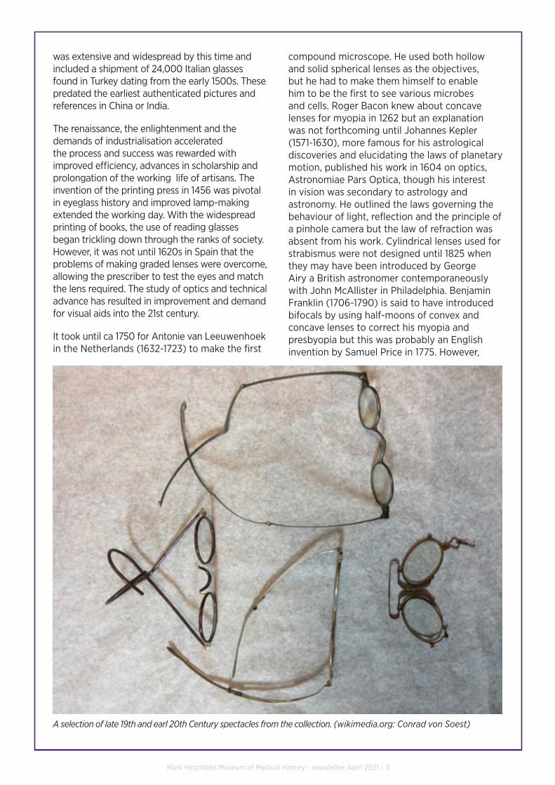
A selection of late 19th and earl 20th Century spectacles from the collection. (wikimedia.org: Conrad von Soest)
Mark Hirschfeld Museum of Medical History - newsletter April 2021 - 4
George Washington is credited for helping to reduce the prejudice that glasses indicate frailty by using them to read part of his speech to rally his troops who were on the point of mutiny in their encampment near New York in 1783. This was widely reported, and they responded with sympathy to his situation by withdrawing their complaints. In London, in 1727, Edward Scarlett made spectacles held comfortably in place with arms passing over the ears which slowly replaced monocles, pince-nez and lorgnettes. These continue to be improved using tough, light, alloys to make resilient frames with emphasis on comfort, personal image and fashion.
Further major developments came with the Zeiss and Moritz Von Rohr spherical point-focus lens in the early twentieth century. Plastics replaced glass from the 1960s after acrylic from the 1940s was found to be too brittle and yellowed with age. Television heralded a huge demand for distance vision correction in the 1950s. Testing of visual acuity and prescription of glasses was carried out by a variety of
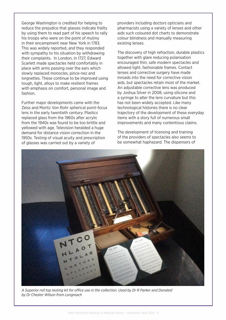
A Superior roll top testing kit for office use in the collection. Used by Dr R Parker and Donated by Dr Chester Wilson from Longreach
providers including doctors opticians and pharmacists using a variety of lenses and other aids such coloured dot charts to demonstrate colour blindness and manually measuring existing lenses.
The discovery of high refraction, durable plastics together with glare reducing polarisation encouraged thin, safe modern spectacles and allowed light, fashionable frames. Contact lenses and corrective surgery have made inroads into the need for corrective vision aids, but spectacles retain most of the market. An adjustable corrective lens was produced by Joshua Silver in 2008, using silicone and a syringe to alter the lens curvature but this has not been widely accepted. Like many technological histories there is no clear trajectory of the development of these everyday items with a story full of numerous small improvements and many contentious claims.
The development of licensing and training of the providers of spectacles also seems to be somewhat haphazard. The dispensers of
Mark Hirschfeld Museum of Medical History - newsletter April 2021 - 5
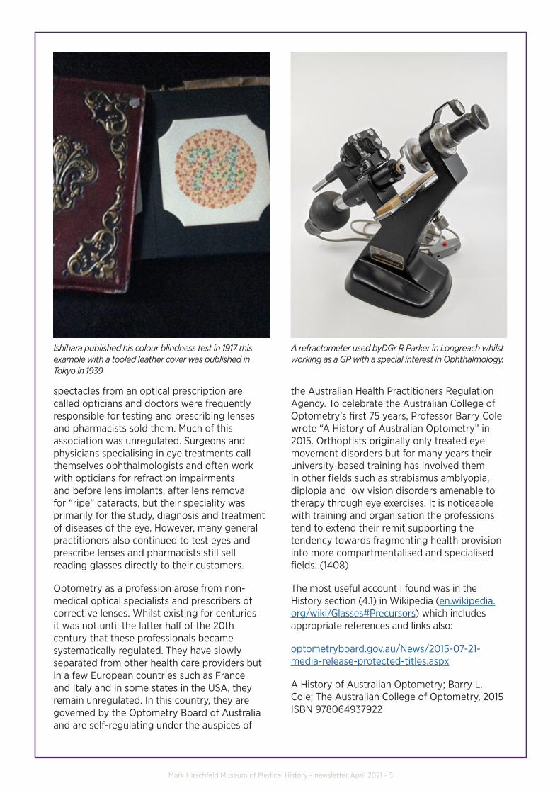
Ishihara published his colour blindness test in 1917 this example with a tooled leather cover was published in Tokyo in 1939
spectacles from an optical prescription are called opticians and doctors were frequently responsible for testing and prescribing lenses and pharmacists sold them. Much of this association was unregulated. Surgeons and physicians specialising in eye treatments call themselves ophthalmologists and often work with opticians for refraction impairments and before lens implants, after lens removal for “ripe” cataracts, but their speciality was primarily for the study, diagnosis and treatment of diseases of the eye. However, many general practitioners also continued to test eyes and prescribe lenses and pharmacists still sell reading glasses directly to their customers.
Optometry as a profession arose from non-medical optical specialists and prescribers of corrective lenses. Whilst existing for centuries it was not until the latter half of the 20th century that these professionals became systematically regulated. They have slowly separated from other health care providers but in a few European countries such as France and Italy and in some states in the USA, they remain unregulated. In this country, they are governed by the Optometry Board of Australia and are self-regulating under the auspices of
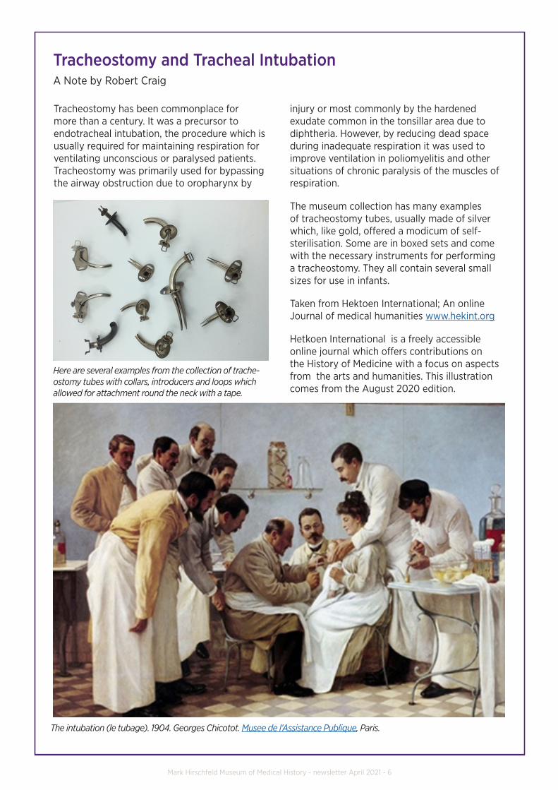
A refractometer used byDGr R Parker in Longreach whilst working as a GP with a special interest in Ophthalmology.
the Australian Health Practitioners Regulation Agency. To celebrate the Australian College of Optometry’s first 75 years, Professor Barry Cole wrote “A History of Australian Optometry” in 2015. Orthoptists originally only treated eye movement disorders but for many years their university-based training has involved them in other fields such as strabismus amblyopia, diplopia and low vision disorders amenable to therapy through eye exercises. It is noticeable with training and organisation the professions tend to extend their remit supporting the tendency towards fragmenting health provision into more compartmentalised and specialised fields. (1408)
The most useful account I found was in the History section (4.1) in Wikipedia (en.wikipedia.org/wiki/Glasses#Precursors) which includes appropriate references and links also:
optometryboard.gov.au/News/2015-07-21-media-release-protected-titles.aspx
A History of Australian Optometry; Barry L. Cole; The Australian College of Optometry, 2015 ISBN 978064937922
Mark Hirschfeld Museum of Medical History - newsletter April 2021 - 6
Tracheostomy and Tracheal IntubationA Note by Robert Craig
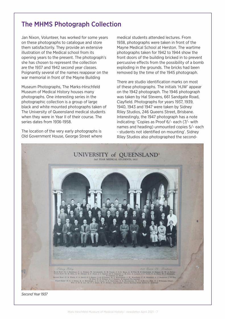
Here are several examples from the collection of trache-ostomy tubes with collars, introducers and loops which allowed for attachment round the neck with a tape.
Tracheostomy has been commonplace for more than a century. It was a precursor to endotracheal intubation, the procedure which is usually required for maintaining respiration for ventilating unconscious or paralysed patients. Tracheostomy was primarily used for bypassing the airway obstruction due to oropharynx by
injury or most commonly by the hardened exudate common in the tonsillar area due to diphtheria. However, by reducing dead space during inadequate respiration it was used to improve ventilation in poliomyelitis and other situations of chronic paralysis of the muscles of respiration.
The museum collection has many examples of tracheostomy tubes, usually made of silver which, like gold, offered a modicum of self-sterilisation. Some are in boxed sets and come with the necessary instruments for performing a tracheostomy. They all contain several small sizes for use in infants.
Taken from Hektoen International; An online Journal of medical humanities www.hekint.org
Hetkoen International is a freely accessible online journal which offers contributions on the History of Medicine with a focus on aspects from the arts and humanities. This illustration comes from the August 2020 edition.
The intubation (le tubage). 1904. Georges Chicotot. Musee de l’Assistance Publique, Paris.
Mark Hirschfeld Museum of Medical History - newsletter April 2021 - 7
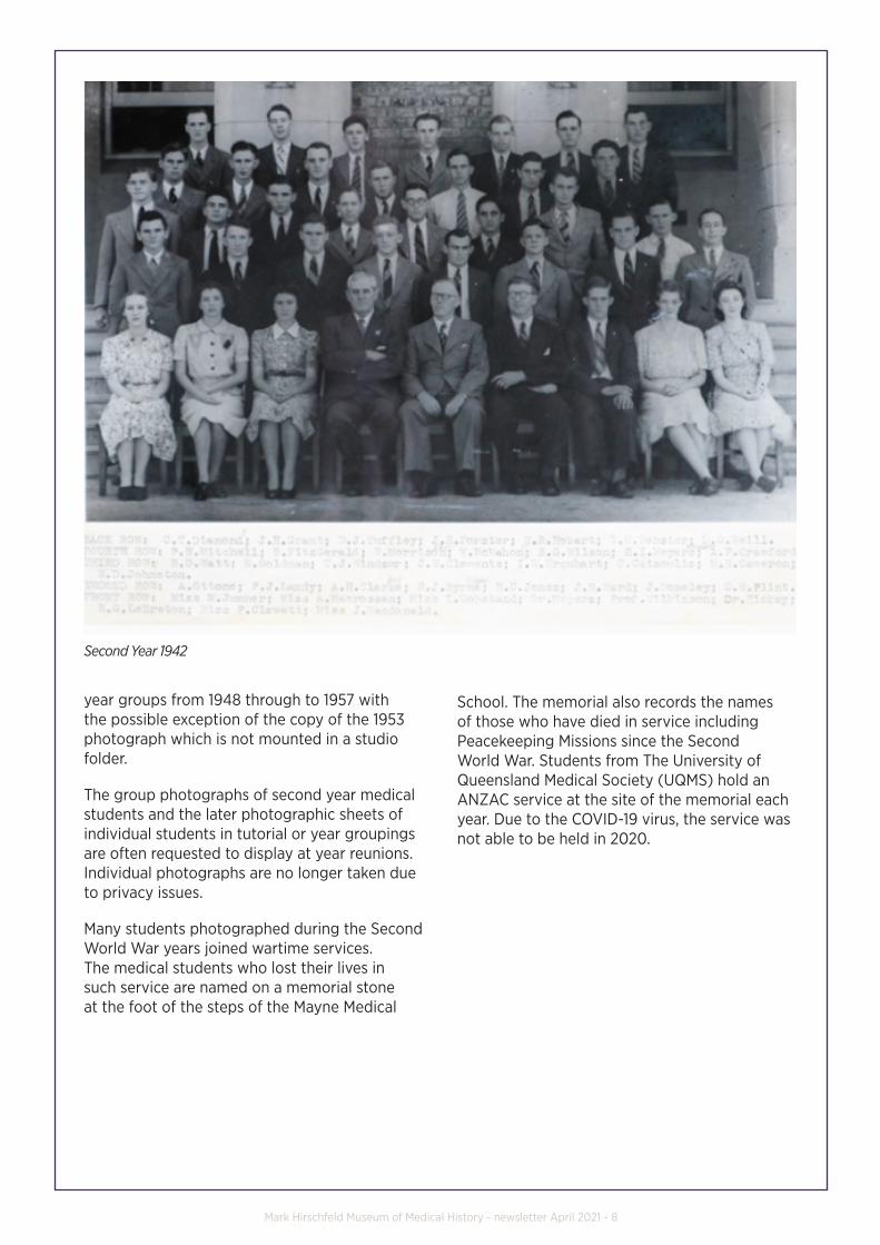
Jan Nixon, Volunteer, has worked for some years on these photographs to catalogue and store them satisfactorily. They provide an extensive illustration of the Medical school from its opening years to the present. The photograph’s she has chosen to represent the collection are the 1937 and 1942 second year classes. Poignantly several of the names reappear on the war memorial in front of the Mayne Building
Museum Photographs, The Marks-Hirschfeld Museum of Medical History houses many photographs. One interesting series in the photographic collection is a group of large black and white mounted photographs taken of The University of Queensland medical students when they were in Year II of their course. The series dates from 1936-1958.
The location of the very early photographs is Old Government House, George Street where
The MHMS Photograph Collection
Second Year 1937
medical students attended lectures. From 1938, photographs were taken in front of the Mayne Medical School at Herston. The wartime photographs taken for 1942 to 1944 show the front doors of the building bricked in to prevent percussive effects from the possibility of a bomb exploding in the grounds. The bricks had been removed by the time of the 1945 photograph.
There are studio identification marks on most of these photographs. The initials ‘HJW’ appear on the 1942 photograph. The 1946 photograph was taken by Hal Stevens, 661 Sandgate Road, Clayfield. Photographs for years 1937, 1939, 1940, 1943 and 1947 were taken by Sidney Riley Studios, 246 Queens Street, Brisbane. Interestingly, the 1947 photograph has a note indicating: ‘Copies as Proof 6/- each (7/- with names and heading) unmounted copies 5/- each - students not identified on mounting’. Sidney Riley Studios also photographed the second-
Mark Hirschfeld Museum of Medical History - newsletter April 2021 - 8
Second Year 1942
year groups from 1948 through to 1957 with the possible exception of the copy of the 1953 photograph which is not mounted in a studio folder.
The group photographs of second year medical students and the later photographic sheets of individual students in tutorial or year groupings are often requested to display at year reunions. Individual photographs are no longer taken due to privacy issues.
Many students photographed during the Second World War years joined wartime services. The medical students who lost their lives in such service are named on a memorial stone at the foot of the steps of the Mayne Medical
School. The memorial also records the names of those who have died in service including Peacekeeping Missions since the Second World War. Students from The University of Queensland Medical Society (UQMS) hold an ANZAC service at the site of the memorial each year. Due to the COVID-19 virus, the service was not able to be held in 2020.
Mark Hirschfeld Museum of Medical History - newsletter April 2021 - 9
MEDICAL STUDENTS WHO GAVE THEIR LIVES 1935-1945 AND IN MISSIONS SINCE THAT TIME
NAMES TAKEN FROM WAR MEMORIAL STONE AT FRONT OF MAYNE MEDICAL SCHOOL BUILDING.
AUSTIN, J. MED IV RAAF
DOUGLAS, H.B. MED II RAAF (1940 SECOND YEAR PHOTO)
GANNON, W.J. MED I AIF
HOOPER, B. GRAD AIF (1937 SECOND YEAR PHOTO)
KELLY, C.D. MED II RAAF
MACTAGGART, J. MED II RAAF
McGILL, J.A.D. MED II RAAF (1940 SECOND YEAR PHOTO)
MINCHIN-SMITH, G.G. MED I RAAF
RANDALL, N.P. MED I RAAF
RYDER, J.S. MED III RA VR (1940 SECOND YEAR PHOTO)
STAPLES, H.B. MED I AIF
1952 PURSSEY, I.G.S. MED II RAAF
1993 FELSCHE, SUSAN MBBS RAAN MC-UN (STUDENT PHOTO WAS SHOWN IN MUSEUM WOMEN AT WAR EXHIBITION)
1997 PAUL McCARTHY MBBS RAAF
Other historic group photographs held by the Museum include:
Inauguration of Faculty of Medicine, The University of Queensland, October 1936.
Teaching staff and first class. Photographer not named. Black and white photograph.
Graduates of Medicine 1952 with names listed, Regent Studios, 43 Queens Street, Brisbane. Black and white photograph.
The First Convention of Medical Students of Australia, May 1960. Courtesy of the Fryer Library, The University of Queensland. Black and white photograph.
Resident Medical Officers 1962 in a folder with names. ‘Casey’s Cameras’ is embossed on the folder. Black and white photograph.
Graduation Dinner 1968 with names listed. David McCarthy and Assoc. Black and white photograph. One copy framed and one copy laminated.
Surgical Professorial Unit, General Hospital, March 1975. Photographer not named. Black and white photograph.
Full-time Academic Staff and Alumni, Department of Child Health for the 75th Anniversary of The University of Queensland, 1985. Graham Jurott, (photograph in colour).
Faculty of Medicine, The University of Queensland Golden Jubilee 1986 with names listed. Graham Jurott, (Photograph in colour).
Staff of the Faculty 1991. Photographer not named. Black and white photograph.
1990/1991 and 1995 graduation photographs in colour (photographer/s not named)..
Many photographs of celebrations and events such as anniversaries and book launches were taken by Mr. Graham Jurott who was Senior Photographer at the Faculty of Medicine, 1964-1993. He also photographed a beautiful black and white series of the artistic details on the Medical School building. We sadly marked Graham’s passing in October 2017.
Mark Hirschfeld Museum of Medical History - newsletter April 2021 - 10
Would you like to share your experiences with medicine?We invite readers to share their personal memories and experiences of studying and practicing med-icine or other health disciplines in Queensland. These stories will form a new, regular column of the Mark-Hirschfeld Museum newsletter. Please email your story to [email protected].
Donate to the MuseumThe Museum is managed by a team of dedicated volunteers. Our generous philanthropic supporters are vital to the works of the Museum, and we welcome donations in support of our collection preser-vation and archival programs, exhibitions and educational activities.
Through your gift you will be playing a vital role in preserving medical history and building a signifi-cant collection to deliver inspiring and engaging learning opportunities to our students, researchers and the community.
You can support the Museum by donating online, contacting us on 07 3365 5081 or emailing [email protected]
Become a volunteer If you’d like to join the volunteer team, please contact us at [email protected]
Contribute to the Museum newsletterThe Marks-Hirschfeld Museum of Medical History newsletter is issued four times per year. We are always on the lookout for interesting materials that explore the rich tapestry of medical history. If you would like to contribute a story or have a topic that you would like to see included in future editions, please send an email to [email protected].
Our next newsletter will be distributed in July 2020. If you are interested in submitting an arti-cle, please send your story and photographs by no later than Monday 21 June.
Share your feedbackWhat do you think of our new newsletter format? Do you have ideas for new sections or subjects? Send through your thoughts or suggestions by clicking here.
The University of Queensland, Level 6, Oral Health Centre, Herston Rd, Herston Qld 4006
www.medicine.uq.edu.au CRICOS Provider Number: 00025B to [email protected]