mapping polypeptide interactions of the seca atpase during ... · mapping polypeptide interactions...
TRANSCRIPT

Mapping polypeptide interactions of the SecA ATPaseduring translocationBenedikt W. Bauer and Tom A. Rapoport1
Howard Hughes Medical Institute and Department of Cell Biology, Harvard Medical School, 240 Longwood Avenue, Boston, MA 02115
Contributed by Tom A. Rapoport, September 15, 2009 (sent for review September 8, 2009)
Many bacterial proteins, including most secretory proteins, aretranslocated across the plasma membrane by the interplay of thecytoplasmic SecA ATPase and a protein-conducting channel formedby the SecY complex. SecA catalyzes the sequential movement ofpolypeptide segments through the SecY channel. How SecA inter-acts with a broad range of polypeptide segments is unclear, butstructural data raise the possibility that translocation substratesbind into a ‘‘clamp’’ of SecA. Here, we have used disulfide bridgecross-linking to test this hypothesis. To analyze polypeptide inter-actions of SecA during translocation, two cysteines were intro-duced into a translocation intermediate: one that cross-links to theSecY channel and the other one for cross-linking to a cysteineplaced at various positions in SecA. Our results show that atranslocating polypeptide is indeed captured inside SecA’s clampand moves in an extended conformation through the clamp intothe SecY channel. These results define the polypeptide path duringSecA-mediated protein translocation and suggest a mechanism bywhich ATP hydrolysis by SecA is used to move a polypeptide chainthrough the SecY channel.
disulfide bridge cross-linking � protein translocation � SecY � secretion �SecA clamp
Many bacterial polypeptides, including most secretory pro-teins, are transported across the plasma membrane by a
process that is similar to protein translocation across the endoplas-mic reticulum membrane in eukaryotes (for review, see ref. 1). Thepolypeptide substrates contain hydrophobic signal sequences thatare usually located at the N terminus and cleaved off after mem-brane transfer. Translocation occurs through a hydrophilic channelformed by a conserved heterotrimeric membrane protein complex,called the SecY complex in bacteria and archaea and the Sec61complex in eukaryotes. The complex consists of a large �-subunit(SecY or Sec61p) that spans the membrane ten times, and twosmaller �- and �-subunits (called SecG and SecE in bacteria). Inbacteria, the SecY channel can either associate with the ribosometo translocate proteins during their synthesis (cotranslational trans-location), or it can cooperate with the cytosolic ATPase SecA totransport polypeptides after completion of their synthesis (post-translational translocation).
SecY forms an hourglass shaped translocation pathway withwater-filled funnels toward both sides of the membrane (2–4).The constriction of the pore is located approximately halfwayacross the membrane and consists of a pore ring of amino acidswhose hydrophobic, bulky side chains project radially toward theinterior of the channel. The cytoplasmic funnel is empty whereasthe external funnel is plugged by a short helix. The channel isopened by displacement of the plug helix, which is triggered bythe binding of the signal sequence of a substrate (5, 6).
The SecA ATPase uses the energy of ATP hydrolysis to pushpolypeptides through the SecY channel (7). SecA is a multido-main protein (8). It contains two RecA-like nucleotide-bindingdomains (NBD1 and NBD2), which bind the nucleotide at theirinterface and move relative to one another during the ATPhydrolysis cycle. SecA also contains a polypeptide-cross-linkingdomain (PPXD), a helical wing domain (HWD), and a helical
scaffold domain (HSD). The latter consists of a long helix andtwo shorter ones that form a two-helix finger (3).
A recent crystal structure of the SecA – SecYEG complexrevealed that SecA uses its PPXD and the long helix of the HSDto interact with the SecY channel and that SecA’s two- helixfinger inserts deeply into the cytoplasmic funnel of SecY (3).Disulfide cross-linking experiments indicate that the fingertipcontacts a translocating polypeptide chain right above the en-trance into the translocation pore, and mutagenesis experimentsshow that a tyrosine (or another bulky, hydrophobic residue) atthe fingertip is essential for protein translocation (9). These datasuggest that motions of the two-helix finger move a polypeptidechain into the SecY channel with the tyrosine providing themajor contact site. However, interactions of the fingertip with apolypeptide chain cannot explain how a translocation substrateis recognized and bound by SecA.
The mechanism by which SecA interacts with its translocationsubstrates is indeed an interesting problem. The initial bindinglikely involves the signal sequence, but ultimately, SecA mustinteract with a broad range of polypeptide segments to movethem sequentially into the SecY channel (10). One possiblepolypeptide-binding site is the ‘‘clamp’’ of SecA (3), which isformed by the PPXD, the NBD2, and parts of the HSD (Fig. S1).Structures of SecA in isolation show the clamp in various openconformations (8, 11–13), which would allow a polypeptide chainto enter it. Structures of SecA bound to the SecY channel showthe clamp in a closed conformation, in which the PPXD hasrotated all of the way toward the NBD2 (3) (Fig. S1). This motionmight capture a polypeptide chain and position it above the SecYpore. Although this model is attractive, other polypeptide bind-ing sites in SecA have been proposed (8, 12, 14, 15). In addition,the SecA-SecYEG structure shows that the closed clamp leavesonly little space for a translocating polypeptide chain (3), whichemphasizes the need for experimental testing of the proposedmodel. Recent spin-label perturbation experiments provide ev-idence that a polypeptide chain can indeed bind to the clamp ofnontranslocating SecA in solution, but several positions outsidethe clamp also showed interactions (16). In fact, our disulfidebridge cross-linking experiments with single cysteines in SecAand in the substrate indicate that SecA interacts rather promis-cuously with polypeptides when not engaged in translocation(see Results). It is therefore important to define substrateinteractions by analyzing translocating SecA molecules.
Here, we have tested the postulated role of SecA’s clamp usingdisulfide bridge cross-linking to analyze interactions of SecA thatis engaged in translocation. We show that a polypeptide chain iscaptured inside the clamp and moves through it into the SecYpore. Our data delineate the entire path of a translocating
Author contributions: B.W.B. and T.A.R. designed research; B.W.B. performed research;B.W.B. and T.A.R. analyzed data; and T.A.R. wrote the paper.
The authors declare no conflict of interest.
Freely available online through the PNAS open access option.
1To whom correspondence should be addressed. E-mail: tom�[email protected].
This article contains supporting information online at www.pnas.org/cgi/content/full/0910550106/DCSupplemental.
20800–20805 � PNAS � December 8, 2009 � vol. 106 � no. 49 www.pnas.org�cgi�doi�10.1073�pnas.0910550106
polypeptide chain during SecA-mediated protein translocationand shed light on the mechanism by which polypeptides aretranslocated by SecA.
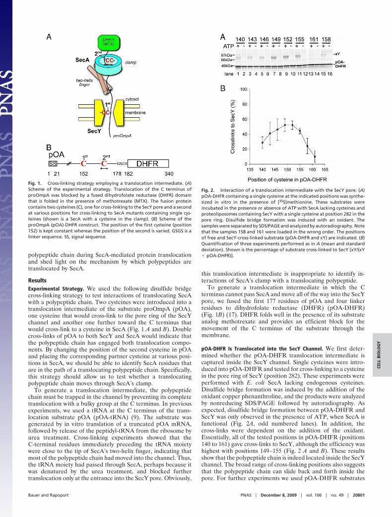
ResultsExperimental Strategy. We used the following disulfide bridgecross-linking strategy to test interactions of translocating SecAwith a polypeptide chain. Two cysteines were introduced into atranslocation intermediate of the substrate proOmpA (pOA),one cysteine that would cross-link to the pore ring of the SecYchannel and another one further toward the C terminus thatwould cross-link to a cysteine in SecA (Fig. 1 A and B). Doublecross-links of pOA to both SecY and SecA would indicate thatthe polypeptide chain has engaged both translocation compo-nents. By changing the position of the second cysteine in pOAand placing the corresponding partner cysteine at various posi-tions in SecA, we should be able to identify SecA residues thatare in the path of a translocating polypeptide chain. Specifically,this strategy should allow us to test whether a translocatingpolypeptide chain moves through SecA’s clamp.
To generate a translocation intermediate, the polypeptidechain must be trapped in the channel by preventing its completetranslocation with a bulky group at the C terminus. In previousexperiments, we used a tRNA at the C terminus of the trans-location substrate pOA (pOA-tRNA) (9). The substrate wasgenerated by in vitro translation of a truncated pOA mRNA,followed by release of the peptidyl-tRNA from the ribosome byurea treatment. Cross-linking experiments showed that theC-terminal residues immediately preceding the tRNA moietywere close to the tip of SecA’s two-helix finger, indicating thatmost of the polypeptide chain had moved into the channel. Thus,the tRNA moiety had passed through SecA, perhaps because itwas denatured by the urea treatment, and blocked furthertranslocation only at the entrance into the SecY pore. Obviously,
this translocation intermediate is inappropriate to identify in-teractions of SecA’s clamp with a translocating polypeptide.
To generate a translocation intermediate in which the Cterminus cannot pass SecA and move all of the way into the SecYpore, we fused the first 177 residues of pOA and four linkerresidues to dihydrofolate reductase (DHFR) (pOA-DHFR)(Fig. 1B) (17). DHFR folds well in the presence of its substrateanalog methotrexate and provides an efficient block for themovement of the C terminus of the substrate through themembrane.
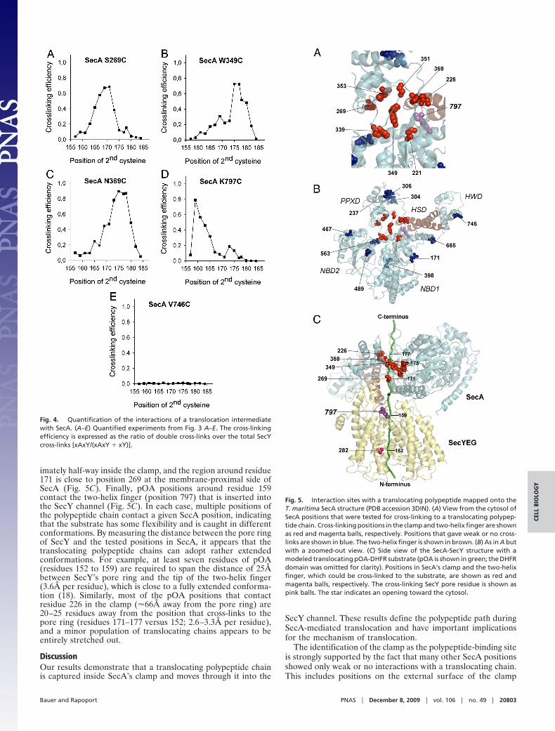
pOA-DHFR Is Translocated into the SecY Channel. We first deter-mined whether the pOA-DHFR translocation intermediate iscaptured inside the SecY channel. Single cysteines were intro-duced into pOA-DHFR and tested for cross-linking to a cysteinein the pore ring of SecY (position 282). These experiments wereperformed with E. coli SecA lacking endogenous cysteines.Disulfide bridge formation was induced by the addition of theoxidant copper phenanthroline, and the products were analyzedby nonreducing SDS/PAGE followed by autoradiography. Asexpected, disulfide bridge formation between pOA-DHFR andSecY was only observed in the presence of ATP, when SecA isfunctional (Fig. 2A, odd numbered lanes). In addition, thecross-links were dependent on the addition of the oxidant.Essentially, all of the tested positions in pOA-DHFR (positions140 to 161) gave cross-links to SecY, although the efficiency washighest with positions 149–155 (Fig. 2 A and B). These resultsshow that the polypeptide chain is indeed located inside the SecYchannel. The broad range of cross-linking positions also suggeststhat the polypeptide chain can slide back and forth inside thepore. For further experiments we used pOA-DHFR substrates
Fig. 1. Cross-linking strategy employing a translocation intermediate. (A)Scheme of the experimental strategy. Translocation of the C terminus ofproOmpA was blocked by a fused dihydrofolate reductase (DHFR) domainthat is folded in the presence of methotrexate (MTX). The fusion proteincontains two cysteines (C), one for cross-linking to the SecY pore and a secondat various positions for cross-linking to SecA mutants containing single cys-teines (shown is a SecA with a cysteine in the clamp). (B) Scheme of theproOmpA (pOA)-DHFR construct. The position of the first cysteine (position152) is kept constant whereas the position of the second is varied. GSGS is alinker sequence. SS, signal sequence.
Fig. 2. Interaction of a translocation intermediate with the SecY pore. (A)pOA-DHFR containing a single cysteine at the indicated positions was synthe-sized in vitro in the presence of [35S]methionine. These substrates wereincubated in the presence or absence of ATP with SecA lacking cysteines andproteoliposomes containing SecY with a single cysteine at position 282 in thepore ring. Disulfide bridge formation was induced with an oxidant. Thesamples were separated by SDS/PAGE and analyzed by autoradiography. Notethat the samples 158 and 161 were loaded in the wrong order. The positionsof free and SecY-cross-linked substrate (pOA-DHFR and xY) are indicated. (B)Quantification of three experiments performed as in A (mean and standarddeviation). Shown is the percentage of substrate cross-linked to SecY [xY/(xY� pOA-DHFR)].
Bauer and Rapoport PNAS � December 8, 2009 � vol. 106 � no. 49 � 20801
CELL
BIO
LOG
Y

with a cysteine at position 152, which gave prominent cross-linksto SecY (Fig. 2 A, lane 9).
Probing Interactions of pOA-DHFR by Double Cross-Linking to SecAand SecY. To determine whether a translocating polypeptidemoves through SecA’s clamp, we used pOA-DHFR constructsthat contained a cysteine at position 152 for cross-linking to thepore residue 282 in SecY and a second cysteine placed at moreC-terminal positions (positions 157–184) to probe for interactionwith SecA’s clamp. The pOA-DHFR substrates were incubatedin the presence or absence of ATP with proteoliposomes con-taining purified SecY (I282C) complex and SecA mutants thatcontained a single cysteine at different positions. Disulfidebridge formation was induced by addition of an oxidant.
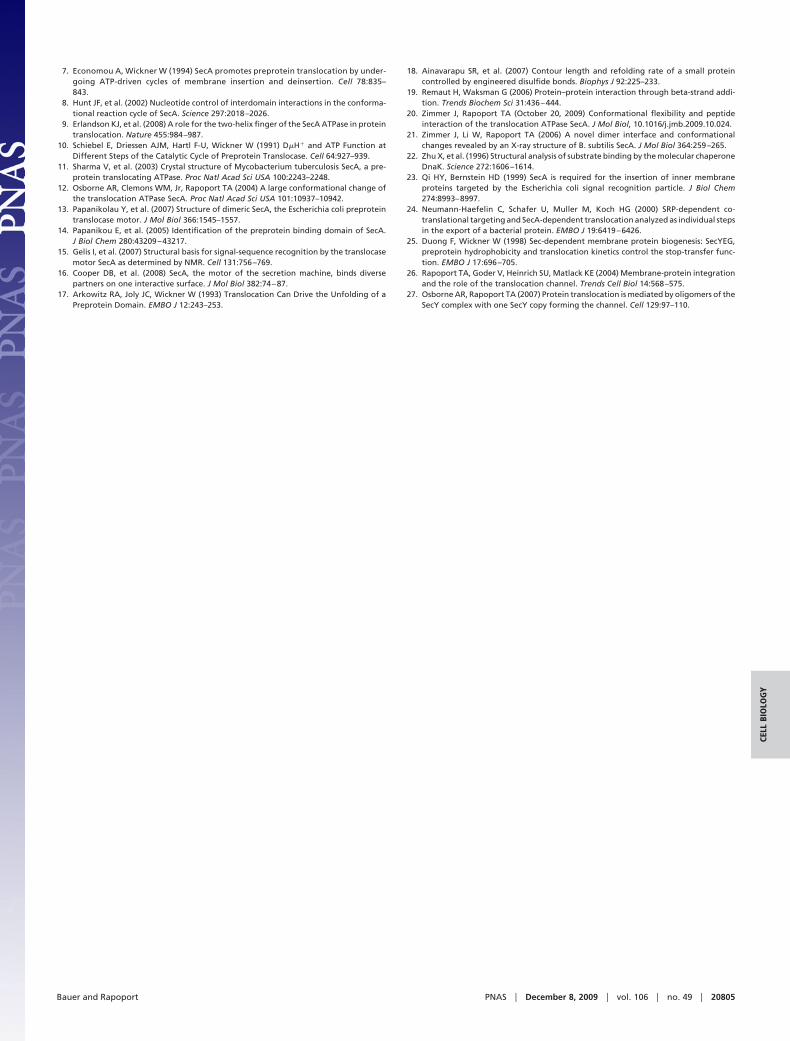
We first tested a SecA mutant that carried a cysteine atposition 269 (SecA-S269C), a position in the PPXD locatedinside the clamp (Fig. 3A). Several positions indeed gave prom-inent double cross-links of pOA-DHFR to both SecY and SecA(xAxY). The double cross-links had the expected size and couldbe immunoprecipitated by SecY or SecA antibodies (Fig. S2). Incontrast, the SecY single-cross-links were recognized only bySecY antibodies and the SecA-single cross-links by SecA anti-bodies, although a minor proportion of these cross-links was alsoprecipitated nonspecifically by SecY antibodies (Fig. S2). Thedouble cross-links and the single cross-links to SecY (xY) wereonly seen in the presence of ATP (Fig. 3A), whereas thecross-links to SecA (xA) were also seen in the absence of ATP,reflecting the promiscuous interaction of SecA with nontrans-locating substrate.
The analysis showed that positions 165 to 171 of the substrategave the most prominent double cross-links to SecA-S269C (Fig.3A); �70% of the pOA-DHFR molecules that cross-linked toSecY also cross-linked to SecA (Fig. 4A). At other positions ofthe substrate, the double cross-links were weaker, and instead,the SecY single cross-links were more intense (Figs. 3A and 4A).These results indicate that position 269 in the clamp of SecA isin the path of a translocating polypeptide, contacting a region inthe substrate that is centered around residue 170.
We next tested other positions in the clamp. Position 349 in thePPXD and position 369, which is located near the �-strands thatconnect the NBD1 with the PPXD, also gave efficient doublecross-links (Fig. 3 B and C); quantification indicated cross-linking yields of up to 90% at the optimum cysteine positions inthe substrate (Fig. 4 B and C). Other positions chosen inside theclamp also gave cross-linking yields exceeding 70% (Figs. S3 A–Dand S5 A–D), with the exception of position 339 (�55% yield)(Figs. S3E and S5E). In each case, the intensities of the doublecross-links were dependent on the position of the second cys-teine in the pOA-DHFR construct and displayed pronouncedmaxima. These results indicate that each position in the clampcontacts a certain region of the substrate. Because a substrateregion, rather than a single residue, contacts a given SecAposition, these data also indicate that the polypeptide chain iscaught at different stages during its movement through theclamp.
In agreement with previous results (9), prominent doublecross-links were also seen with a cysteine placed at the tip of thetwo-helix finger (position 797) (Figs. 3D and 4D). However, incontrast to the previous experiments in which the substratecarried a tRNA at the C terminus, the cross-links to the fingertipdisappeared when the second cysteine was placed further towardthe C terminus of pOA (Figs. 3D and 4D). These data areconsistent with the assumption that the DHFR domain preventsthe C terminus of the substrate from entering SecA’s clampwhereas the tRNA only blocks translocation at a later point (atthe entrance into the SecY pore).
Ten positions chosen outside the predicted path of a translo-cating polypeptide chain either did not give double cross-links at
all or cross-linked only weakly (Figs. 3E and 4E; the otherpositions are shown in Figs. S4 A–I and S5 F–N).
We mapped the tested positions onto SecA in the recentlydetermined SecA-SecYEG structure (3). Viewed from the cy-toplasm, the substrate-interacting positions are all localizedinside the central cavity of the clamp (shown in red in Figs. 5 Aand B), including positions in the PPXD and the two �-strandsthat connect the NBD1 with the PPXD. In a side view, thesepositions and the cross-linking position 797 of the two-helixfinger delineate the path of the polypeptide chain from the fusedDHFR domain all of the way into the SecY channel (Fig. 5C).The most C-terminal residues of pOA (approximately position177), which immediately precede the DHFR block (beginning atposition 182), contact the clamp on its membrane-distal side(position 226) (Fig. 5C). The preceding pOA residues aroundresidue 173–175 contact positions 349 and 369 located approx-
Fig. 3. Probing interactions of a translocation intermediate with SecA. (A)Interaction of a substrate with the SecA clamp. pOA-DHFR containing acysteine at positions 152 and a second cysteine at the indicated positions wassynthesized in vitro in the presence of [35S]methionine. The substrate wasincubated in the presence or absence of ATP with SecA containing a singlecysteine in the clamp at position 269 and with proteoliposomes containingSecY with a cysteine at position 282. The samples were treated with an oxidantand analyzed by nonreducing SDS/PAGE and autoradiography. The positionsof free, SecA-, SecY-, and double cross-linked substrate (pOA-DHFR, xA, xY,and xAxY) are indicated. (B) As in A but with a cysteine in the SecA clamp atposition 349. (C) As in A but with a cysteine in the SecA clamp at position 369.(D) Interaction of a substrate with the two-helix finger of SecA. The experi-ments were performed as in A but with a cysteine at the SecA fingertip(position 797). (E) A cysteine randomly placed into SecA does not interact withthe translocation intermediate. The experiments were performed as in A butwith a cysteine at position 746 of SecA.
20802 � www.pnas.org�cgi�doi�10.1073�pnas.0910550106 Bauer and Rapoport
imately half-way inside the clamp, and the region around residue171 is close to position 269 at the membrane-proximal side ofSecA (Fig. 5C). Finally, pOA positions around residue 159contact the two-helix finger (position 797) that is inserted intothe SecY channel (Fig. 5C). In each case, multiple positions ofthe polypeptide chain contact a given SecA position, indicatingthat the substrate has some flexibility and is caught in differentconformations. By measuring the distance between the pore ringof SecY and the tested positions in SecA, it appears that thetranslocating polypeptide chains can adopt rather extendedconformations. For example, at least seven residues of pOA(residues 152 to 159) are required to span the distance of 25Åbetween SecY’s pore ring and the tip of the two-helix finger(3.6Å per residue), which is close to a fully extended conforma-tion (18). Similarly, most of the pOA positions that contactresidue 226 in the clamp (�66Å away from the pore ring) are20–25 residues away from the position that cross-links to thepore ring (residues 171–177 versus 152; 2.6–3.3Å per residue),and a minor population of translocating chains appears to beentirely stretched out.
DiscussionOur results demonstrate that a translocating polypeptide chainis captured inside SecA’s clamp and moves through it into the
SecY channel. These results define the polypeptide path duringSecA-mediated translocation and have important implicationsfor the mechanism of translocation.
The identification of the clamp as the polypeptide-binding siteis strongly supported by the fact that many other SecA positionsshowed only weak or no interactions with a translocating chain.This includes positions on the external surface of the clamp
Fig. 4. Quantification of the interactions of a translocation intermediatewith SecA. (A–E) Quantified experiments from Fig. 3 A–E. The cross-linkingefficiency is expressed as the ratio of double cross-links over the total SecYcross-links [xAxY/(xAxY � xY)].
Fig. 5. Interaction sites with a translocating polypeptide mapped onto theT. maritima SecA structure (PDB accession 3DIN). (A) View from the cytosol ofSecA positions that were tested for cross-linking to a translocating polypep-tide chain. Cross-linking positions in the clamp and two-helix finger are shownas red and magenta balls, respectively. Positions that gave weak or no cross-links are shown in blue. The two-helix finger is shown in brown. (B) As in A butwith a zoomed-out view. (C) Side view of the SecA-SecY structure with amodeled translocating pOA-DHFR substrate (pOA is shown in green; the DHFRdomain was omitted for clarity). Positions in SecA’s clamp and the two-helixfinger, which could be cross-linked to the substrate, are shown as red andmagenta balls, respectively. The cross-linking SecY pore residue is shown aspink balls. The star indicates an opening toward the cytosol.
Bauer and Rapoport PNAS � December 8, 2009 � vol. 106 � no. 49 � 20803
CELL
BIO
LOG
Y

(positions 237, 304, and 306), which are located in a groove thatNMR experiments implicated in signal sequence interaction(15). This suggests that SecA has distinct binding sites for signalsequences and polypeptide segments that follow them. How asignal sequence would be transferred from the outside of theclamp into the SecY channel is unclear, particularly because theSecA-SecYEG structure shows that a direct path is blocked bythe interaction of the PPXD with loops of SecY (3). Perhaps, thesignal sequence is released from its binding site before SecAinteracts with SecY, or the hydrophobic interior of the clampprovides an additional or alternative binding site for the signalsequence.
At the beginning of translocation, SecA’s clamp must be open toallow the entry of a substrate. This requires that the PPXD rotatesaway from the NBD2, a conformation represented by x-ray struc-tures of SecA in isolation (8, 12) (see Fig. S1). We propose that theopen clamp interacts through the two �-strands that link the NBD1and PPXD with the backbone of a polypeptide chain. The inter-acting polypeptide segment would be induced to form a short�-strand that extends this �-sheet, a mechanism termed ‘‘�-strandaugmentation’’ (19). This sequence-independent mode of interac-tion is supported by a recent structure of a SecA-peptide complex(20), as well as by two other SecA structures in which a polypeptidesegment formed an additional short �-strand next to the �-sheetconnecting NBD1 and PPXD (8, 21).
Once the polypeptide chain is bound, the PPXD would rotatetoward the NBD2 and close the clamp, a conformation that isstabilized by an interaction of the PPXD with SecY (3). Thecapture of the polypeptide chain inside the clamp would con-tribute to the sequence-independent interaction of SecA with asubstrate. This mechanism of polypeptide binding is in factreminiscent of how many chaperones, such as Hsp70s, interactwith a broad range of peptide substrates (22); in these cases,extended polypeptide chains are embraced by the walls of a deepgroove, making side chain interactions less important. In therecently determined SecA-SecY structure, the interior of theclamp is partially occluded by a loop at the membrane-distal side(3). Our data now suggest that the loop is f lexible and likelymoves out of the way when a translocation substrate is present.In fact, positions 349 and 351 in this loop give efficient cross-links. Accommodation of a substrate in the clamp is facilitatedby the fact that the polypeptide chain is in a rather extendedconformation. Our results indicate that a translocating polypep-tide chain is in an unfolded conformation all of the way from itsentry into the SecA clamp to the pore ring of the SecY channel.Thus, any folding can only occur when the polypeptide chainemerges on the extracellular side of the SecY channel.
The closed clamp might simply position the polypeptide chainabove the channel, allowing a translocating polypeptide chain toslide back and forth within its central cavity. However, we considerit more likely that the clamp tightens and widens during the ATPhydrolysis cycle through movements of the PPXD relative to theNBD2. The �-strand augmentation mechanism may still be impor-tant because it might prevent complete dissociation of the polypep-tide. In this context, it should be noted that cysteines placed closeto the two �-strands connecting the NBD1 and PPXD gave thehighest cross-linking yields (positions 221, 226, 369).
The translocating polypeptide chain is fully surrounded by clampresidues over a distance of �26Å but has to cross a significant gapof �12Å from the clamp into the SecY pore. This opening towardthe cytosol is located underneath the mouth of the clamp, right
above the lateral gate of the SecY channel (indicated by a star in Fig.5C) and would be of sufficient size to accommodate two polypep-tide strands in an extended conformation. A polypeptide chaincould therefore loop out sideways into the cytosol, rather than moveinto the SecY channel. However, this would be energeticallyunfavorable and would not normally occur during the translocationof a secretory protein. Also, because SecA is required for thebiosynthesis of some membrane proteins (23–25), the cytosolic exitpathway may allow segments following transmembrane domains toemerge into the cytosol, similar to what has been proposed formembrane protein integration during cotranslational translocation(26).
Our results show that a polypeptide chain emerging from theclamp contacts the two-helix finger of SecA before it enters theSecY pore. The determined polypeptide path is consistent witha model in which, upon ATP binding by SecA, the finger wouldmove toward the SecY pore and drag the polypeptide chain withit. At the same time, the clamp would loosen its grip on thepolypeptide chain. Upon ATP hydrolysis, the clamp wouldtighten, the finger would disengage from the polypeptide andreset to ‘‘grab’’ the next polypeptide segment. This process wouldbe repeated until the polypeptide chain is all of the way throughthe SecY channel.
Materials and MethodsCloning, Mutagenesis, and Protein Purification. Full-length E. coli proOmpAwith a C-terminal hexa-histidine tag was cloned into pMAl p4E (NEB) usingNdeI and HindIII restriction sites. A SalI site was introduced after position 175and a NcoI site in front of the His-tag by site directed mutagenesis (Strat-agene). These sites were used to insert E.coli DHFR with N- and C-terminalGSGS-linkers. The two endogenous cysteines of DHFR were removed, andsingle cysteines were introduced into the new construct at positions 152 and157–184 by site-directed mutagenesis. All constructs were confirmed by se-quencing.
For translocation and cross-linking assays, a linear fragment of pOA-DHFRcontaining a 5� SP6 promoter and a 3� stop-codon was generated by PCR. Afterin vitro transcription (Promega), the mRNA was translated in rabbit reticulo-cyte lysate (Promega) in the presence of [35S]methionine. The crude lysate wasthen used for the experiments.
Point mutations in E. coli SecA and SecY were introduced by site directedmutagenesis. Both SecA and SecY complex were overexpressed in BL21 cellsand purified using Ni- affinity chromatography (12). Purified SecY complexwas reconstituted into liposomes made of E.coli polar lipids (27).
Cross-Linking. Cross-linking was performed in 20 �l reactions containing 50mM Hepes/KOH pH7.5, 50 mM KCl, 5 mM MgCl2, 2.5 mM ATP, 0.5 mg/ml BSA,40 �g/ml SecA, 20 �g/ml SecY complex in proteoliposomes, and 1 �l lysatecontaining in vitro synthesized pOA-DHFR (9). For cross-linking in the absenceof ATP, 0.5 units of hexokinase and 10 mM glucose were added to the reaction.After 15 min at 37 °C, cross-links were formed by the addition of 50 �M copperphenanthroline for 10min at 37 °C. Samples were treated with 10 mM N-ethylmaleimide (NEM) for 5 min on ice and then subjected to nonreducingSDS/PAGE on 4–20% Tris�HCl gels (Bio-Rad). The bands were visualized byphosphorimaging. QuantityOne (Bio-Rad) was used for local backgroundsubtraction and quantification. Cross-linking efficiencies were expressed asthe ratio of counts in double cross-linked bands (xAxY) to the sum of countsin double cross-linked and single SecY cross-linked bands [xAxY/(xAxY � xY)].
ACKNOWLEDGMENTS. We thank K. Erlandson for providing protocols, mu-tants, and helpful hints; J. Zimmer for help with protein purification andmodeling; E. Park for advice on cloning and mutagenesis; and J. Zimmer, A.Salic, Y. Chen, and A. Tripathi for critical reading of the manuscript. We alsothank P. Pohl for generously providing the opportunity for B.W.B.’s stay inBoston. This work was supported by National Institutes of Health GrantGM052586. B.W.B. was supported by a fellowship from the Austrian MarshallPlan Foundation. T.A.R. is a Howard Hughes Medical Institute investigator.
1. Rapoport TA (2007) Protein translocation across the eukaryotic endoplasmic reticulumand bacterial plasma membranes. Nature 450:663–669.
2. van den Berg B, et al. (2004) X-ray structure of a protein-conducting channel. Nature427:36–44.
3. Zimmer J, Nam Y, Rapoport TA (2008) Structure of a complex of the ATPase SecA andthe protein-translocation channel. Nature 455:936–943.
4. Tsukazaki T, et al. (2008) Conformational transition of Sec machinery inferred frombacterial SecYE structures. Nature 455:988–991.
5. Harris CR, Silhavy TJ (1999) Mapping an interface of SecY (PrlA) and SecE (PrlG) by usingsynthetic phenotypes and in vivo cross-linking. J Bacteriol 181:3438–3444.
6. Tam PC, Maillard AP, Chan KK, Duong F (2005) Investigating the SecY plug movementat the SecYEG translocation channel. EMBO J 24:3380–3388.
20804 � www.pnas.org�cgi�doi�10.1073�pnas.0910550106 Bauer and Rapoport
7. Economou A, Wickner W (1994) SecA promotes preprotein translocation by under-going ATP-driven cycles of membrane insertion and deinsertion. Cell 78:835–843.
8. Hunt JF, et al. (2002) Nucleotide control of interdomain interactions in the conforma-tional reaction cycle of SecA. Science 297:2018–2026.
9. Erlandson KJ, et al. (2008) A role for the two-helix finger of the SecA ATPase in proteintranslocation. Nature 455:984–987.
10. Schiebel E, Driessen AJM, Hartl F-U, Wickner W (1991) D�H� and ATP Function atDifferent Steps of the Catalytic Cycle of Preprotein Translocase. Cell 64:927–939.
11. Sharma V, et al. (2003) Crystal structure of Mycobacterium tuberculosis SecA, a pre-protein translocating ATPase. Proc Natl Acad Sci USA 100:2243–2248.
12. Osborne AR, Clemons WM, Jr, Rapoport TA (2004) A large conformational change ofthe translocation ATPase SecA. Proc Natl Acad Sci USA 101:10937–10942.
13. Papanikolau Y, et al. (2007) Structure of dimeric SecA, the Escherichia coli preproteintranslocase motor. J Mol Biol 366:1545–1557.
14. Papanikou E, et al. (2005) Identification of the preprotein binding domain of SecA.J Biol Chem 280:43209–43217.
15. Gelis I, et al. (2007) Structural basis for signal-sequence recognition by the translocasemotor SecA as determined by NMR. Cell 131:756–769.
16. Cooper DB, et al. (2008) SecA, the motor of the secretion machine, binds diversepartners on one interactive surface. J Mol Biol 382:74–87.
17. Arkowitz RA, Joly JC, Wickner W (1993) Translocation Can Drive the Unfolding of aPreprotein Domain. EMBO J 12:243–253.
18. Ainavarapu SR, et al. (2007) Contour length and refolding rate of a small proteincontrolled by engineered disulfide bonds. Biophys J 92:225–233.
19. Remaut H, Waksman G (2006) Protein–protein interaction through beta-strand addi-tion. Trends Biochem Sci 31:436–444.
20. Zimmer J, Rapoport TA (October 20, 2009) Conformational flexibility and peptideinteraction of the translocation ATPase SecA. J Mol Biol, 10.1016/j.jmb.2009.10.024.
21. Zimmer J, Li W, Rapoport TA (2006) A novel dimer interface and conformationalchanges revealed by an X-ray structure of B. subtilis SecA. J Mol Biol 364:259–265.
22. Zhu X, et al. (1996) Structural analysis of substrate binding by the molecular chaperoneDnaK. Science 272:1606–1614.
23. Qi HY, Bernstein HD (1999) SecA is required for the insertion of inner membraneproteins targeted by the Escherichia coli signal recognition particle. J Biol Chem274:8993–8997.
24. Neumann-Haefelin C, Schafer U, Muller M, Koch HG (2000) SRP-dependent co-translational targeting and SecA-dependent translocation analyzed as individual stepsin the export of a bacterial protein. EMBO J 19:6419–6426.
25. Duong F, Wickner W (1998) Sec-dependent membrane protein biogenesis: SecYEG,preprotein hydrophobicity and translocation kinetics control the stop-transfer func-tion. EMBO J 17:696–705.
26. Rapoport TA, Goder V, Heinrich SU, Matlack KE (2004) Membrane-protein integrationand the role of the translocation channel. Trends Cell Biol 14:568–575.
27. Osborne AR, Rapoport TA (2007) Protein translocation is mediated by oligomers of theSecY complex with one SecY copy forming the channel. Cell 129:97–110.
Bauer and Rapoport PNAS � December 8, 2009 � vol. 106 � no. 49 � 20805
CELL
BIO
LOG
Y