mapk pathways via he4 ovarian cancer by regulating the
TRANSCRIPT

Page 1/30
YWHAE In�uences the Malignant Behaviour ofOvarian Cancer by Regulating the PI3K/AKT andMAPK Pathways Via HE4Xiao Li
Obstetrics and Gynecology of Shengjing Hospital of China Medical University https://orcid.org/0000-0003-4447-194XCaixia Wang
Shengjing Hospital of China Medical UniversityShuang Wang
Shengjing Hospital of China Medical UniversityYuexin Hu
Shengjing Hospital of China Medical UniversityShan Jin
Shengjing Hospital of China Medical UniversityOuxuan Liu
Shengjing Hospital of China Medical UniversityRui Gou
Shengjing Hospital of China Medical UniversityXin Nie
Shengjing Hospital of China Medical UniversityJuanjuan Liu
Shengjing Hospital of China Medical UniversityBei Lin ( [email protected] )
Shengjing Hospital of China Medical University https://orcid.org/0000-0002-8881-9859
Primary research
Keywords: YWHAE, HE4, interacting protein, poor prognosis, malignant behaviour, ovarian cancer
Posted Date: April 12th, 2021
DOI: https://doi.org/10.21203/rs.3.rs-385473/v1
License: This work is licensed under a Creative Commons Attribution 4.0 International License. Read Full License

Page 2/30
AbstractBackground: Malignant tumours of the female reproductive system threaten the lives and health ofwomen worldwide, with ovarian cancer having the highest mortality rate among all malignant tumours.Based on previous work, this study analysed the expression and role of YWHAE in ovarian epithelialtumours.
Methods: The interaction between YWHAE and HE4 was evaluated by immunoprecipitation, western blotanalysis, and cellular immuno�uorescence. Immunohistochemistry was used to address the relationshipbetween YWHAE expression and clinicopathological parameters and patient prognosis. Changes in cellinvasion, epithelial–mesenchymal transition, migration, proliferation, cell cycle, and apoptosis before andafter differential expression of YWHAE were also explored in ovarian cancer cell lines and in vivoexperiments.
Results: YWHAE was found to directly interact with HE4, and its expression was positively correlated withHE4 expression. Moreover, YWHAE higher levels were associated with advanced ovarian cancer casesand with poorer patient outcome. YWHAE was found to enhance the invasion, migration, proliferation,and inhibition of apoptosis of ovarian cancer cells. These biological effects were found to be meditatedby the activity of the PI3K/AKT and MAPK signalling pathways.
Conclusions: Altogether, this study demonstrates that YWHAE is signi�cantly increased in ovarian cancertissues, representing a risk factor for the prognosis of ovarian cancer that is positively correlated withHE4 expression. Furthermore, YWHAE and its downstream signals may represent new therapeutic targetsto tackle ovarian cancer.
IntroductionOvarian cancer has the highest mortality rate among the most common malignant tumours of the femalereproductive system. Due to the lack of obvious or speci�c symptoms, as well as of ideal screeningmethods, the early detection and diagnosis rates of ovarian cancer are extremely low [1]. When thetumour progresses to an advanced stage, the tumour-free and overall survival times of patients arerapidly shortened [2]. Therefore, it is urgent to �nd tumour markers with high sensitivity and speci�city toguide early clinical screening, early diagnosis, and monitoring of ovarian cancer.
Human Epididymis Protein 4 (HE4) is an ovarian cancer marker that was identi�ed by genomics andproteomics screenings [3]. Its high sensitivity and speci�city has attracted the attention of researchers,who have proved that HE4 has more advantages than the common tumour marker CA125 for the earlydiagnosis of ovarian cancer and for monitoring disease progression [4]. In 2003, HE4 was designated asa serum marker for ovarian cancer and it was approved by the U.S. Food and Drug Administration in 2009for monitoring the recurrence and progression of epithelial ovarian cancer [5]. Currently, several ongoingclinical studies are addressing the diagnostic potential of HE4, but only few are exploring its underlyingmolecular mechanisms.

Page 3/30
Our previous work has proven that HE4 is highly expressed in ovarian cancer tissues, and that HE4 andAnnexin A2 interaction can promote the invasion and metastasis of ovarian cancer. This mechanism isaccomplished by activating adhesion signalling pathways such as MAPK and FOCAL [6–7]. Proteinsperform their biological functions mainly by interacting with other proteins to form complexes or byworking together with chaperone molecules. The tight coordination of proteins with different functions intime and space constitutes the basic process of life. Therefore, given the signi�cant role of HE4 inovarian cancer, it has become clear that recognition and improved knowledge on HE4-interacting proteinsis an important step for an overall better understanding of this disease. A previous study using the two-hybrid screening method and HE4 as bait, has identi�ed the YWHAE protein as a partner of HE4.
Tyrosine 3-monooxygenase/tryptophan 5-monooxygenase activation protein epsilon (YWHAE, alsoknown as 14-3-3ε) belongs to the YWHA protein family, which comprises at least seven highly conservedsubtypes of soluble acidic proteins encoded by different genes, namely β, ε, η, γ, τ, ξ, and σ [8]. They canbe widely combined with other proteins such as kinases, phosphatases, transmembrane receptors, andtranscription factors, among other target proteins [9–11], functioning as a protein interaction bridge in awide range of biological processes [12–14].
X-ray diffraction analysis showed that YWHAE protein monomers form homodimers or heterodimers [15],which are bound by some highly conserved hydrophobic amino acids. Different YWHA subtypes can bindto the same target, granting these proteins the ideal conditions to regulate a large number ofphysiological processes, such as cell proliferation, apoptosis, protein transport, metabolic regulation,signal transduction, among others [16–17]. The particular structure of YWHAE may be the basis for itsrole as a "bridge protein", as well as for its contribution towards disease incidence [18].
Based on previous preliminary �ndings, this study explored the relationship between YWHAE and HE4and analysed the expression of YWHAE in ovarian epithelial tumors and its mechanism of action.Enhanced knowledge on the underlying activity of YWHAE and HE4 may provide a basis to further explorethe development and progression of ovarian cancer and develop new diagnostic strategies.
Materials And MethodsPrimary samples
Ovarian tissue samples were derived from para�n specimens surgically collected between 2008 and2012 in the Department of Obstetrics and Gynecology, Shengjing Hospital, China Medical University. Atotal of 16 samples were derived from normal ovarian tissue removed due to uterine �broids or cervicalcancer (normal group), 18 samples were from benign cases, 24 samples were classi�ed as borderline,and 105 samples were from malignant ovarian cancers. The pathological types among the malignantsamples included 71 serous tumours, 7 mucinous tumours, 19 endometrioid tumours, and 8 clear cellcarcinomas. The malignant group was also classi�ed according to pathology assessment, with 51 casesidenti�ed as highly differentiated and 54 as poorly differentiated. The surgical pathological staging wasperformed in accordance with the International Union of Obstetrics and Gynecology (FIGO) standards: 44

Page 4/30
cases in stage I-II and 61 in stage III-IV, of which a comprehensive exploratory operation was performed inthe early stage and cytoreductive surgery was performed in the late stage. In the malignant group, 92patients underwent lymph node dissection, with lymph node metastasis being con�rmed in 28 cases.Protein samples for western blotting analysis were derived from tissue specimens collected in 2018–2019 in the Department of Obstetrics and Gynecology, Shengjing Hospital A�liated to China MedicalUniversity. A total of 33 specimens were surgically collected from 15 cases in the malignant group, 6cases in the borderline group, 6 cases in the benign group, and 6 cases in the normal group. All caseswere newly diagnosed and radiotherapy and chemotherapy naïve.
Immunochemistry
The histopathological specimens were �xed with 10% formalin solution, embedded in para�n, and thenserially sectioned into 5 μm slices. The para�n sections were depara�nised with xylene and re-hydratedwith gradient alcohol solutions, and the antigens were hot-recovered. Then, H2O2, goat serum blockingsolution, and an anti-YWHAE antibody (1:100, sc-23957, Santa Cruz Biotechnology, Santa Cruz, CA) or ananti-HE4 (1:1500, ab200828, Abcam, Cambridge, UK ) antibody were added dropwise sequentially, and thesolutions were left to incubate overnight at 4 °C. On the next day, the slices were incubated in horseradishperoxidase (HRP)-labelled goat anti-rabbit/mouse secondary antibodies and stained using 3,3-diaminobenzidine (Ultrasensitive TM SP Mouse/Rabbit IHC Kit, Maixin, Fuzhou, China). The cell nucleuswas stained blue using haematoxylin. The sections were then dehydrated, cleared by xylene, andmounted.
The results were evaluated by two pathologists who did not know in advance the clinical information ofthe patients, and they independently observed and scored each sample. If discordant scoring results wereobtained, a third pathologist would assess the sample for the �nal decision. The samples were classi�edas positive when presenting with brownish-yellow or brown colour in the cell cytoplasm and/ormembrane. If the proportion of positive cells was less than 5%, it was scored as 0 points, 5–25% wasscored as 1 point, 26–50% was 2 points, 51–75% was 3 points, and more than 75% was counted as 4points. According to the colour intensity, they were further scored with 3 points for brown, 2 points forbrownish-yellow, 1 point for light yellow, and 0 points for no staining. To reach a �nal score, the twoclassi�cations were multiplied: 0–2 points were recorded as negative expression (−), 3–4 points as weakpositive expression (+); 5–8 points as moderate positive expression (++), and 9–12 points as strongpositive expression (+++).
Cellular immuno�uorescence
The cells were seeded on a microscope slide and washed with PBS after they adhered to the glass. Goatserum blocking solution was added, followed by the anti-YWHAE (1:100, sc-23957, Santa CruzBiotechnology, Santa Cruz, CA) and anti-HE4 (1:200, DF8160, A�nity, OH, USA) primary antibody mix. Asecondary antibody mixture containing tetramethylrhodamine-labelled goat anti-rabbit IgG (SA00007-2,Proteintech, Wuhan, China) and �uorescein isothiocyanate-labelled goat anti-mouse IgG (SA00003-1,
Page 5/30
Proteintech, Wuhan, China) was dropped onto the slide, which was then incubated for 2 h in the dark. 4′,6-diamidino-2-phenylindole (4083S; Cell Signaling Technology, Beverly, MA, USA) was used to stain the cellnucleus, and an anti-quenching agent was added dropwise onto the slide immediately before assessingthe slices on a confocal microscope.
Establishment of stable over-expressing YWHAE cell lines and transient YWHAE-knockdown cell lines
The ovarian cancer cell lines CAOV3 and ES2 (Institute of Biochemistry and Cell Biology, ChineseAcademy of Sciences, Shanghai, China) were cultured in RPMI 1640 medium (Biological Industries, Beit-Haemek, Israel) containing 10% foetal bovine serum (Biological Industries, Beit-Haemek, Israel). When thecells reached 80–90% con�uence, the medium was discarded, the cells were washed with phosphate-buffered saline (PBS), trypsinised, and split to continue the culture.
YWHAE-siRNA and HE4-siRNA were transfected into CAOV3 and ES2 cells. YWHAE-siRNA(GAAGCAGGUUAGCGUUGAATTUUCAACGCUAACCUGCUUCTT), Mock-YWHAE-siRNA(GAAGCAGGUUAGCGUUAGATTUU), HE4-siRNA(UUCUCCGAACGUGUCACGUTTACGUGACACGUUCGGAGAATT), and Mock-HE4-siRNA(UUCUCCGAACGUGUACGUTT) working solutions were prepared according to the manufacturer’sinstructions (Gene Pharma, Suzhou, China). The cells were seeded in 6-well plates with serum-freemedium on the day of transfection. The transfection was performed using Lipofectamine 3000(Invitrogen, Waltham, MA, USA) according to the manufacturer’s instructions.
Lentivirus-mediated YWHAE over-expression vector was used to transfect OVCAR3 and A2780 cell lines,which had relatively low expression of YWHAE. To calculate the multiplicity of infection (MOI) of YWHAE-expressing lentivirus, 500 µL of complete medium were added to a 24-well plate, in addition to lentivirussupernatant and a corresponding volume of polybrene, to promote transfection. Puromycin at 50% lethalconcentration (LC50) was used to select the e�ciently transfected cells.
Western blotting
Total protein samples were separated by sodium dodecyl sulphate polyacrylamide gel electrophoresis.Brie�y, 5–10 µL of protein sample was placed on each well of the gel and subjected to 80–120 V ofconstant electrical current for 40–100 min. The proteins were transferred onto a polyvinylidene �uoridemembrane (Millipore, Burlington, MA, USA), which was then blocked with 5% milk/bovine serum albuminsolution for 2 h at 37 °C. The membrane was incubated overnight at 4 °C with primary antibodies(YWHAE, 1:1000, ab92311, Abcam, Cambridge, UK; HE4, 1:1500, DF8160, A�nity, OH, USA; Cyclin D1,1:1000, 2978S,Cell Signaling Technology, Beverly, MA Ki-67, 1:1000, 9449S, Cell Signaling Technology,Beverly, MA; Bax, 1:1000, 5023S, Cell Signaling Technology, Beverly, MA; Bcl-2, 1:1000, 12789-1-AP,Proteintech, Wuhan, China; MMP2, 1:1000, 10373-2-AP, Proteintech, Wuhan, China; MMP9, 1:1000, 10375-2-AP, Proteintech, Wuhan, China E-cadherin, 1:1000, 20874-1-AP, Proteintech, Wuhan, China; N-cadherin,1:1000, 4061S, Cell Signaling Technology, Beverly, MA; Vimentin, 1:3000, 10366-1-AP, Proteintech, Wuhan,China; PI3K, 1:1000, 4292S, Cell Signaling Technology, Beverly, MA; p-PI3K, 1:1000, 4228S, Cell Signaling

Page 6/30
Technology, Beverly, MA; AKT, 1:1000, 4691S, Cell Signaling Technology, Beverly, MA; p-AKT, 1:1000,4060S, Cell Signaling Technology, Beverly, MA; m-TOR, 1:1000, 2972S, Cell Signaling Technology, Beverly,MA; p-m-TOR, 1:1000, 2971S, Cell Signaling Technology, Beverly, MA; MEK, 1:1000, sc-81504, Santa CruzBiotechnology, Santa Cruz, CA p-MEK, 1:1000, 2338S, Cell Signaling Technology, Beverly, MA; ERK,1:1000, 9102S, Cell Signaling Technology, Beverly, MA; p-ERK, 1:1000, 9101S, Cell Signaling Technology,Beverly, MA). After washing, the membrane was incubated with HRP-labelled goat anti-rabbit (1:3000, ZB-2301, ZSGB-BIO, Beijing, China) or goat anti-mouse (1:3000, ZB-2305, ZSGB-BIO, Beijing, China)secondary antibodies for 1 h at 37℃ and then washed again. Lastly, western chemiluminescent HRPSubstrate (Thermo Fisher Scienti�c, Waltham, MA, USA) was added dropwise onto the membrane, andluminescent signals were detected in a luminometer at different exposure times.
Immunoprecipitation
Cells in exponential growth were collected and washed. Iced lysis buffer was added and the cellsuspension was sonicated. The supernatant was collected and total protein concentration wasdetermined. Total protein samples (500 μg) were incubated with 2 μg of YWHAE (1 100, sc-23957, SantaCruz Biotechnology, Santa Cruz, CA) or HE4 (1:1500, ab200828, Abcam, Cambridge, UK) primaryantibody. An IgG antibody (5145S, Cell Signaling Technology, Beverly, MA) of the same species of theprimary antibody was used as negative control. After mixing, the samples were left overnight at 4°C withslow rotation. Afterwards, beads (sc-2003, Santa Cruz Biotechnology, Santa Cruz, CA) were added to eachtube and incubated for 6 h. The bound proteins were collected by centrifugation, (2×) Loading Buffer wasadded, and the samples were heated to denature the proteins.
Invasion test
A transwell insert was placed in the culture plate. Matrigel (1:7.5 dilution, 356234, BD Biosciences, NewYork, USA) and serum-free 4 × 10⁴ cell suspension were added to the upper layer of the chamber, whereasthe lower chamber contained complete medium (with serum). After culturing for 48–72 h, the cells in thelower chamber were collected, �xed, and stained. Residual cells lefts on plate were observed under amicroscope. The experiment was repeated thrice.
Scratch test
Cells were seeded in a 6-well culture plate and maintained at 37 °C in a 5% CO2 incubator. Aftercon�rming under the microscope that the cells were at 90% con�uence, the cell layer was scratched witha 100 µl pipette tip. The cells were cultured in serum-free medium for 24 h, then washed with PBS andphotographed to monitor the healing of the scratches. The experiment was repeated thrice.
MTT assay
A total of 2,000 cells/well were seeded in a 96-well culture plate and maintained at 37 °C in a 5% CO2
incubator. After the cells were adhered, 20 µL of sterile MTT (M8180, Solarbio, Beijing, China) working

Page 7/30
solution was added to each well, mixed well, and the cells were incubated at 37 °C for 4h. Following this,the medium was aspirated and 150 µL of DMSO (D8370, Solarbio, Beijing, China) was added to eachwell. The absorbance of each well was measured in a microplate reader after shaking for 5 min. Theexperiment was repeated thrice.
Cell cycle analysis
Cells in log phase were collected and washed, and pre-cooled ethanol was slowly added to �x the cells forlater use. Before the analysis, a totle of 500 µl PI/ RNase A (KGA512, KeyGen Biotech, Nanjing, China)staining solution were added to the cell suspension and the cells were left in the dark for 20 min,according to the manufacturer’s instructions. The cells were analysed by �ow cytometry. The experimentwas repeated thrice.
Apoptosis assessment
The Annexin V/PI double staining method was used to evaluate the impact of YWHAE overexpression onovarian cancer cells. The cells were trypsinized without EDTA, and the suspension was centrifuged todiscard the supernatant. The cells were re-suspended in 500 µL of Binding Buffer added slowly andincubated with 5 µL of Annexin V-APC (550474, BD Biosciences, New York, USA) / FITC (KGA107, KeyGenBiotech, Nanjing, China) and PI in the dark at 37 ℃ for 15 minutes according to the manufacturer’sinstructions. The staining was evaluated by �ow cytometry.
In vivo xenograft model of ovarian cancer
Twenty female nude mice (Huafukang Biosciences, Beijing, China) were randomly divided into twogroups and injected with either OVCAR3-YWHAE-MOCK or OVCAR3-YWHAE-H ovarian cancer cell lines.Approximately 100 µL of cell suspension containing 1×10⁷ cells was injected subcutaneously into thearmpit of the right forelimb of each nude mouse. The tumour progression and overall health status of themice were observed every three days, and the diameter of the tumour and the weight of the mice weremeasured. The tumour volume was calculated as V = (a × b2)/2, where a represents the largest diameterand b the shortest diameter. On day 7 post-injection the newly formed tumours were detectable, while onday 21 the largest tumour was nearly 1 cm in diameter, and the mice had started to shown poor healthsymptoms.The tumor samples were then �xed in 4% paraformaldehyde and embedded in para�n.Continuous 5 μm-thick sections were cut and analyzed using hematoxylin and eosin (HE) orimmunohistochemical staining. The animal study was approved by the Institutional Animal ResearchCommittee of China Medical University.
Signalling pathway inhibition experiment
YWHAE-overexpressing and mock-transduced cells were cultured in the presence of thephosphatidylinositol-4,5-bisphosphate 3-kinase (PI3K) inhibitor (GDC-0941, Selleck, Houston, USA), or a

Page 8/30
selective inhibitor of mitogen-activated protein kinase 1 and 2 (MAPK1/2) inhibitor (PD98059, Selleck,Houston, USA), 25 and 20 µmol/L, respectively. 0.1% DMSO served as the negative control.
Bioinformation analysis
The YWHAE co-expression gene set was downloaded from cBioPortal (www.cbioportal.org), and the top300 genes were annotated according to the gene ontology (GO) (BP, biological process; CC, cellularcomponent; and MF, molecular function) and the Kyoto Encyclopaedia of Genes and Genomes (KEGG)pathway analysis using the tools available in DAVID (www.david.ncifcrf.gov). The interaction networkamong the top 100 genes was constructed using STRING (www.string-db.org) and Cytoscape(www.cytoscape.org).
The Oncomine database (http://www.oncomine.org), which is currently the world’s largest oncogene chipdatabase integrated with a data mining platform, was used to analyze the mRNA expression level ofYWHAE in the different cancer cell types.
The protein-protein interaction (PPI) network was constructed using the STRING (http://string.embl.de/)online tool. The visualization plot was generated using Cytoscape software with a con�dence score of≥0.1 de�ned as the cutoff. The core modules of the PPI network were screened using Molecular ComplexDetection (MCODE) with the following parameters: degree threshold = 2, node threshold = 0.2, kcore = 2,and maximum depth = 100.
Statistical analysis
Statistical differences between two groups were evaluated by the Student’s t test, and one-way analysisof variance was used for the comparison of more than two groups. The data were counted using the χ2
and Fisher’s exact probability tests, and measurements of the data were performed using the single factoranalysis of variance. P<0.05 was considered statistically signi�cant.
ResultsYWHAE and HE4 are interacting proteins in ovarian cancer
The levels of YWHAE and HE4 were evaluated in the CAOV3 and ES2 ovarian cancer cell lines by cellularimmunohistochemistry, revealing that both proteins were expressed in the cytoplasm and cell membrane(Fig. 1a). Co-localizing in the cytoplasm and cell membrane of CAOV3 and ES2 (Fig. 1b). Co-immunoprecipitation of both YWHAE and HE4 in these cell lines further demonstrated that they areinteracting proteins (Fig. 1c), siRNA-mediated knock-down of YWHAE led to reduced levels of HE4 in bothCAOV3 and ES2 transiently transfected cells, whereas HE4 knock-down had no signi�cant effect onYWHAE levels. These results suggest that YWHAE is upstream of HE4 (Fig. 1d) and it can regulate HE4expression.

Page 9/30
David analyzed the GO (BP/CC/MF) and KEGG pathways of the top 300 gene set, and enriched 36 BP,such as RNA scattering, Wnt signaling pathway, cell adhesion, 23 CC, 13 MF (all P<0.05), and 2 KEGGsignaling pathways (all P<0.05), such as spliceosome, RNA transport, etc., and drew the bubble diagramof the top 5 go (BP/cc/MF) and KEGG pathways. The results showed that YWHAE might interact withPAFAH1B1, EIF5A, PITPNA, TIMM22, PFN1, WFDC2(HE4) (Fig. 1e).
The expression and correlation analysis of YWHAE and HE4 in each group of ovarian tissue
YWHAE expression in ovarian tissues
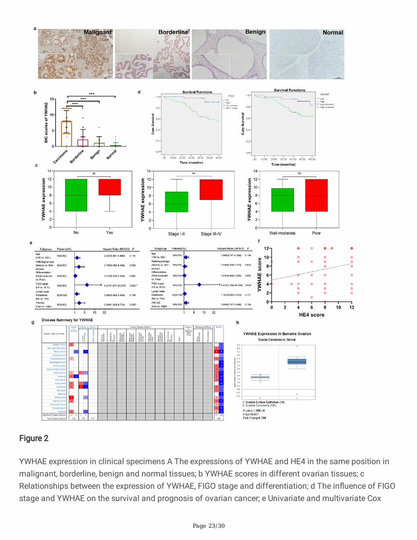
In agreement with the cell line results, analysis of primary tissue samples showed that YWHAE wasmainly located in the cytoplasm and cell membrane. Approximately 96.19%[101/105] of the malignantsamples were positive for YWHAE with a strong positive rate of 70.48%[74/105], whereas the borderlinegroup had a YWHAE-positive rate of 41.67%[10/24] and a strong positive rate of 16.67%[4/24]. Benignand normal ovarian samples had a positive rate of 16.67%[3/18] and 6.25%[1/16], and a strong positiverate of 11.11%[2/18] and 0.00%[0/16], respectively. Comparison between the different groups revealedthat YWHAE-positive expression was signi�cantly higher in the malignant group (P<0.05); however, thejunction group YWHAE-positive rate was also higher than that of the benign and normal groups (P<0.05).The expression rate of YWHAE in the benign group was higher than in the normal group, but thedifference was not statistically signi�cant (P>0.05) (Table 1, Fig. 2a,b)
Relationship between YWHAE expression and clinicopathological parameters of ovarian cancer
In order to compare clinicopathological parameters with the expression of YWHAE in ovarian tissue, wereviewed the clinical information of the 105 patients with primary ovarian epithelial malignant tumours.Analysis of the pathological data showed that YWHAE-strong positive expression rate was signi�cantlyhigher in FIGO III-IV stage ovarian epithelial malignancies than in the early stage group (80.33% versus56.82%, P<0.01). No statistical differences were observed in their other clinicopathological parameters(Table 2, Fig. 2c).
Relationship between YWHAE expression and survival prognosis of patients with ovarian cancer
Follow-up of the patients further showed that only four deaths occurred in the YWHAE low expressiongroup (n=31), while 21 deaths were recorded in the YWHAE high expression group (n=74). Kaplan-Meiersurvival analysis showed that the survival rate among patients with high YWHAE levels was signi�cantlylower compared with that in the YWHAE low expression group. This result followed the same trend asobserved when comparing patients with early or late FIGO staging (P<0.05) (Fig. 2d). Univariate andmultivariate Cox regression analysis of YWHAE expression with age, pathological type, degree ofdifferentiation, FIGO stage, and lymph node metastasis demonstrated that YWHAE expression and FIGOstaging are risk factors for the prognosis of epithelial ovarian malignancies (Fig. 2e).
YWHAE and HE4 expressions are related in ovarian cancer tissues

Page 10/30
Next, the co-expression of YWHAE and HE4 was evaluated in 80 cases of ovarian cancer. A total of 0, 3, 9,and 68 cases were YWHAE−/HE4−, YWHAE−/HE4+, YWHAE+/HE4−, and YWHAE+/HE4+, respectively.Spearman correlation analysis con�rmed that YWHAE and HE4 expressions are positively correlated inovarian cancer (correlation coe�cient Rs=0.277, P=0.013) (Table 3). Linear regression analysis showedthat the expression of YWHAE and HE4 can in�uence each other (P<0.05), and that the late FIGO stage isan important factor affecting the expression of HE4 (Table 4). Multivariate linear regression analysis alsoshowed that HE4 expression score was an independent in�uencing factor of YWHAE expression, as wellas YWHAE expression was an independent in�uencing factor of HE4 expression (Fig. 2e).
YWHAE and HE4 expressions are related in ovarian cancer tissues
Study of YWHAE expression in Oncomine database showed that YWHAE was highly expressed in themany cancer group compared to the normal tissue group, for ovarian cancer, YWHAE was signi�cantlyhighly expressed in 185 ovarian carcinoma tissues compared with 10 ovarian surface epithelium tissues(Fig. 2g,h).
YWHAE promotes ovarian cancer cell invasion, migration, and epithelial–mesenchymal transitionpotential
Analysis of the levels of YWHAE in several ovarian cancer cell lines revealed that it was higher in CAOV3and ES2 than in OVCAR3 and A2780. Based on these results, CAOV3 and ES2 cells with relatively highexpression of YWHAE were used to establish a cell line model with low YWHAE expression, whereasOVCAR3 and A2780 ovarian cancer cells with relatively low YWHAE were used to establish a stable over-expressing cell line model of YWHAE (Fig. 3a-f).
The effect on invasion and migration of ovarian cancer cells upon transient knock-down or stable over-expression of YWHAE was evaluated next by transwell and scratch experiments. Overall, the datarevealed that both OVCAR3 and A2780 cells overexpressing YWHAE had signi�cantly enhanced invasionand migration capacity than mock-transduced and untransduced cells. In contrast, CAOV3 and ES2 cellslacking YWHAE showed weaker invasion and migration abilities compared with control cells (all P<0.05)(Fig. 3g-n). To further explore the impact of YWHAE on the behaviour of ovarian cancer cells, the levels ofcell epithelial and mesenchymal markers were evaluated in these cells by western blot. YWHAE-overexpression was found to be associated with higher levels of the N-cadherin, Vimentin, MMP2, andMMP9 (cell mesenchymal markers), whereas the epithelial marker E-cadherin was found to be reducedcompared with that of cells of the control groups. The opposite trend was seen when YWHAE wasknocked-down (both P<0.05), with higher levels of E-cadherin and reduced levels of N-cadherin, Vimentin,MMP2, and MMP9 (Fig. 4a-d). Altogether, these results demonstrate that YWHAE promotes invasion,migration, and epithelial-mesenchymal transition of epithelial ovarian cancer cells.
YWHAEpromotes ovarian cancer via inducing cell proliferation, cell cycle progression, and apoptosisinhibition

Page 11/30
Additional assessment of the OVCAR3 and A2780 YWHAE-over-expressing cells further showed that moreof these cells were in G2/M phase, suggesting that they were actively proliferating compared with mock-transduced and untransduced cells (all P<0.05). Furthermore, these cells had higher expression of Ki67,Cyclin D1, and Bcl-2 and reduced levels of Bax (all P<0.05). CAOV3 and ES2 cells lacking YWHAE showedthe opposite results, with signi�cantly lower proportion of cells in the G2/M phase, reduced levels ofproliferation markers, and increased levels of the Bax apoptosis marker. Furthermore, �ow cytometryresults revealed that compared with the control group, the overall apoptosis of OVCAR3 and A2780 cellswas signi�cantly reduced after YWHAE overexpression (P<0.05) , while apoptosis was signi�cantlyincreased in OVCAR3 and A2780 cells compared to the control group when YWHAE expression wasinhibited (Fig. 4e-h, Fig. 5). Taken together, these results demonstrate that the expression of YWHAE canenhance the proliferation of ovarian cancer cells, while it also promotes cell cycle progression andinhibits cell apoptosis.
Effect of YWHAE on the in vivo tumorigenesis of ovarian cancer cells
In order to explore the effect of YWHAE on the tumorigenic ability of ovarian cancer cells, OVCAR3 stablyoverexpressing YWHAE or a mock control were injected into athymic nude mice. Assessment of thetumours formed 21 days after the cells were injected revealed that cells overexpressing YWHAE weresigni�cantly bigger and weighed about 2.83 times more than the tumours seen in the control group.Moreover, the growth rate of the tumours produced by YWHAE-overexpressing cells was also signi�cantlyhigher compared with those in the control group (Fig. 6a-c). Tumour biopsies collected and evaluated byimmunohistochemistry revealed that the signals of activated AKT and ERK (p-AKT and p-ERK,respectively) were stronger in OVCAR3 cells over-expressing YWHAE, further suggesting that YWHAE canpromote the proliferation of tumour cells in vivo (Fig. 6d).
YWHAE-induced cellular effects are mediated by the PI3K/AKT and MAPK signalling pathways
We used the STRING database to predict the relevant molecules of YWHAE by PPI, and the resultsshowed that YWHAE had direct or indirect interaction with PI3K, MAPK, ACTR1A, cAMP-dependent proteinkinase and other molecules (Fig. 7a).
To have a more detailed perspective on the underlying mechanisms triggered by YWHAE, the levels ofseveral critical signalling molecules were evaluated by western blotting. The results showed that the ratioof p-PI3K/PI3K, p-AKT/AKT, mTOR/p-m-TOR, p-ERK/ERK, and p-MEK/MEK increased signi�cantly in thepresence of high levels of YWHAE, but they were reduced upon YWHAE-knock-down (all P<0.05). Theabove results prove that YWHAE can activate PI3K/AKT and MAPK signal pathways (Fig. 7b-e).
To further explore the role of these two signalling pathways on YWHAE-induced cellular effects, PI3Kinhibitor (GDC-0941) or MEK inhibitor (PD98059) was used. The invasion and migration experimentswere repeated in the presence of these chemical inhibitors, revealing that blockage of both PI3K/AKT andMAPK signals signi�cantly weakened the pro-invasion and pro-migration effect promoted by YWHAEover-expression (P<0.05). Moreover, the results of the MTT experiment showed that the proliferation

Page 12/30
ability of OVCAR3 cells overexpressing YWHAE-H was signi�cantly reduced in the presence of GDC-0941or PD98059 (P<0.05) (Fig. 8, Fig. 9).
The above described results demonstrate that YWHAE can impact on the invasion, migration, andproliferation potentials of ovarian cancer cells, as well as other malignant biological behaviours, throughthe PI3K/AKT and the MAPK signalling pathways.
DiscussionOvarian cancer is the tumour of the female reproductive system with the highest mortality rate. Althougha variety of targeted drugs for ovarian cancer have been used in the clinical setting, its high mortality ratestill represents a serious threat to the lives and health of women worldwide.
YWHAE protein is widely expressed in eukaryote cells, and has been detected in wheat [19], gianttrematodes in goat blood cells [20], liver �ukes [21], and mosquitoes [22]. Moreover, in the physiologicalstate of the human body, YWHAE was described as an important element in retinal photoreceptor rodcells [23]. Some researchers have found that YWHAE is also involved in the differentiation of adipose-derived mesenchymal stem cells into osteoblasts, enhancing the body’s osteogenic ability [24]. Incontrast, some studies have detected a peak of YWHAE expression 168 h after partial liver resection,preventing cell cycle and negatively regulating liver regeneration [25]. Therefore, these discordant �ndingssuggest that YWHAE may have a two-way regulatory effect on the cell cycle.
Since YWHAE was originally identi�ed in the brain, its pathological effects were initially investigated inthe �eld of neurological diseases, such as Parkinson’s [26] and Alzheimer’s disease [27], brain excitotoxicinjury [28], and myocardial ischemia reperfusion [29], among others. These studies agreed that themechanism by which YWHAE could impact on nerve cells could be related to mitochondrial dysfunctionand regulation of apoptosis.
In recent years, studies have suggested that abnormal expression of YWHAE may also play an importantrole in the occurrence and development of tumours. Liang et al. found that YWHAE is highly expressed inkidney cancer tissues, and in vitro experiments demonstrated that YWHAE can promote the abnormalproliferation of tumour cells [30]. In gastric cancer cell lines, the expression of YWHAE is signi�cantlyincreased and it can inhibit cell proliferation, invasion, and migration by reducing the expression of MYCand CDC25B, whereas MYC induces cell proliferation, invasion, and migration by enhancing CDC25B andreducing YWHAE expression [31–32]. In breast cancer, YWHAE expression was also related to tumoursize, lymph node metastasis, and patient survival prognosis, as well as to breast cancer cell resistance tochemotherapy. Indeed, YWHAE overexpression signi�cantly increases breast cancer cell proliferation,migration, and invasion, whereas reduced YWHAE expression prevents Snail and Twist expression inbreast cancer cells [33]. Although high expression of YWHAE has been described in colorectal, liver,kidney, breast, gastric, and oesophageal cancers, its speci�c mechanism of action remains unclear.Among the malignant tumours of the female reproductive system, YWHAE is more commonly reportedupon genetic testing of the uterine sarcoma cells. Endometrial stromal sarcoma carrying the YWHAE–

Page 13/30
NUTM2 (or YWHAE–FAM22) fusion gene has obvious malignant biological effects, such as enhancedinvasion and drug resistance, and the prognosis of patients harbouring such genetic abnormality is worse[34–35]. Sylvain et al. [36] performed gene chip detection on matched tumour samples from six patientswith advanced high-grade epithelial ovarian cancer before and after chemotherapy. The results showedthat 54 genes that recurred after chemotherapy showed a down-regulation trend, whereas 121 genes,including YWHAE, showed an up-regulation trend. This change in the expression pro�le suggests thatYWHAE may be related to ovarian cancer invasion, proliferation, and drug resistance. Sun et al. used theGene Expression Omnibus database to analyse the relationship between ovarian cancer and diabetes,�nding that 10 key genes, including YWHAE, are important links in the regulation of the redox reactionprocess and carboxylic acid metabolism in the body [37]. Based on these results, they believe that ovariancancer is related to sugar metabolism, and that certain key metabolism-related genes and proteins couldbe used as potential targets for the treatment of ovarian cancer.
Altogether, the results described in the present study demonstrate that YWHAE and HE4 are interactingproteins. And YWHAE is signi�cantly associated with advanced stage cancers and poorer patientoutcomes, thereby speculating that high YWHAE expression may represent a risk factor for the prognosisof ovarian cancer. Overall, YWHAE showed a similar cancer-promoting effect as seen with HE4, partakingin the occurrence and development of ovarian cancer.
Through induced differential expression of YWHAE and in vivo experiments, it was also demonstratedthat YWHAE contributes to ovarian cancer cell invasion, epithelial-mesenchymal transition, and migration,as well as to enhancing their proliferative and anti-apoptotic responses.
Previous studies have con�rmed that HE4 mainly plays an important role in the spreading and adhesionof ovarian cancer cells. Moreover, low levels of HE4 prevented the activation of ERK and EGFR in ovariancancer cells. Therefore, it is believed that HE4 may in�uence the biological behaviour of cancer cells inthe ovaries through the EGFR and MAPK signalling pathways, but the speci�c underlying mechanism isstill unclear. Studies have reported that HE4 can affect the cell cycle (G0/G1 phase), migration, andinvasion capabilities by regulating the ERK/MAPK signals and the expression of MMP-9, MMP-2, andcathepsin B [38]. In accordance with its role as an interaction protein of HE4, YWHAE was also shown toaffect the malignant biological behaviour of ovarian cancer cells through the above-mentioned signallingpathways.
In breast cancer, YWHAEτ acts together with 1,3-DCQA (eicosanylquinic acid) to prevent the proliferationand metastasis of cancer cells through the Jak/PI3K/AKT and Raf/ERK pathways and by inducing theBad/Bax/caspase 9 apoptosis pathway [39]. Overexpression of YWHAEζ can regulate the expression ofSnail protein by activating PI3K/AKT signals, thereby signi�cantly promoting the proliferation, migration,and invasion of glioma cells, representing a potential prognostic marker therapeutic target for glioma[40]. In colorectal cancer, YWHAEσ acts as a tumour suppressor gene. However, COPS5 and LASP1through PI3K/AKT-dependent signals stimulate YWHAEσ ubiquitination and degradation, making it loseits tumour suppressor activity, thereby promoting the progression of colorectal cancer [41].

Page 14/30
Relevant studies have shown that YWHAE can inhibit cell apoptosis in colorectal cancer HT-29 cells [42],and this process can be reversed by non-steroidal anti-in�ammatory drugs. The inhibition of apoptosismay be related to the ability of YWHAE to interfere with mitochondrial pro-apoptotic mechanisms andactivate the transcription factors FKHRL1 and Bad [43]. Moreover, ATPR (4-amino-2-tri�uoromethyl-phenylretinoic acid) can induce G0/G1 phase arrest in gastric cancer SGC-7901 cells by down-regulatingYWHAE [44]. We found that PI3K/AKT pathway node proteins (PI3K, AKT, and mTOR) and MAPK pathwaynode proteins (MEK and ERK) were signi�cantly activated in ovarian cancer cells over-expressing YWHAE.Importantly, inhibition of these pathways with speci�c inhibitors prevented the pro-invasion, pro-migration, and pro-proliferation effects induced by YWHAE. Therefore, these results suggest that YWHAEpromotes the malignant biological behaviour of epithelial ovarian cancer through activation of thePI3K/AKT and MAPK pathways.
ConclusionsThis study described for the �rst time that YWHAE and HE4 were interacting proteins and underlying a co-localization relationship. It was proved in tissues that the expression of YWHAE was signi�cantlyincreased in ovarian cancer tissues, which was a risk factor for the prognosis of ovarian cancer.Moreover, it was discovered for the �rst time that YWHAE could promote the invasion, migration,proliferation of epithelial ovarian cancer through PI3K/AKT and MAPK pathways. In the future, YWHAEmay be used as a prognostic factor and trigger new research ideas for further understanding theunderlying pathogenesis and improving the diagnosis and treatment of ovarian cancer.
Abbreviations

Page 15/30
HE4 Human Epididymis Protein 4
FIGO International Union of Obstetrics and Gynecology
GSEA Gene Set Enrichment Analysis
FBS Fetal bovine serum
DMSO dimethyl sulfoxide
COIP Co-Immunoprecipitation
FITC Fluorescein isothiocyanate
TRITC Tetramethylrhodamine-5-isothiocyanate
DAPI 4',6-diamidino-2-phenylindole
PBS phosphate-buffered saline
PVDF polyvinylidene �uoride membrane
KEGG Kyoto Encyclopedia of Genes and Genomes
GO Gene Ontology
DeclarationsAvailability of data and materials
Not applicable.
Acknowledgements
We would like to acknowledge the reviewers for their helpful comments on this paper.
Funding
The study was supported by grants from the National Natural Science Foundation of China (81672590and 81472437) and Shengjing Freedom Researchers’ plan (201804).
Author information
A�liations
Department of Obstetrics and Gynaecology, Shengjing Hospital A�liated to China MedicalUniversity, No. 36, Sanhao Street, Heping District, Shenyang, 110004, People's Republic of China.
Xiao Li, Caixia Wang ,Shuang Wang, Yuexin Hu, Shan Jin, Ouxuan Liu, Rui Gou, Xin Nie, Juanjuan Liu, BeiLin.

Page 16/30
Contributions
XL and BL �nished study design, CW, SW, YH, SJ, OL, RG �nished experimental studies, XN, JL �nisheddata analysis, XL �nished manuscript editing. All authors read and approved the �nal manuscript.
Corresponding author
Correspondence to Bei Lin.
Ethics declarations
Ethics approval and consent to participate.
This study was approved and supervised by the animal ethics committee of Shengjing Hospital A�liatedto China Medical University. The treatment of animals in all experiments conforms to the ethicalstandards of experimental animals.
Consent for publication
Not applicable.
Competing interests
The authors declare that they have no con�icts of interest.
References1. Chen W. Cancer statistics: updated cancer burden in China. Chin J Cancer Res. 2015;27(1):1.
2. Jelovac D. Armstrong DK. Recent progress in the diagnosis and treatment of ovarian cancer. CACancer J Clin. 2011;61(3):183–203.
3. Kirchhoff C. Molecular characterization of epididymal proteins. Rev Reprod. 1998;3(2):86–95.
4. Huang J. Chen J, Huang Q. Diagnostic value of HE4 in ovarian cancer: A meta-analysis. Eur J ObstetGynecol Reprod Biol. 2018;231:35–42.
5. Dochez V. Caillon H, Vaucel E. Dimet J, Winer N. Ducarme G. Biomarkers and algorithms fordiagnosis of ovarian cancer: CA125, HE4, RMI and ROMA, a review. J Ovarian Res. 2019;12(1):28.
�. Wang J. Deng L, Zhuang H. Liu J, Liu D. Li X, et al. Interaction of HE4 and ANXA2 exists in variousmalignant cells-HE4-ANXA2-MMP2 protein complex promotes cell migration. Cancer Cell Int.2019;19:161.
7. Zhuang H. Tan M, Liu J. Hu Z, Liu D. Gao J, et al. Human epididymis protein 4 in association withAnnexin II promotes invasion and metastasis of ovarian cancer cells. Mol Cancer. 2014;13:243.
�. Diallo K. Oppong AK, Lim GE. Can 14-3-3 proteins serve as therapeutic targets for the treatment ofmetabolic diseases? Pharmacol Res. 2019;139:199–206.
Page 17/30
9. Cau Y. Valensin D, Mori M. Draghi S, Botta M, Structure. Function, Involvement in Diseases andTargeting of 14-3-3 Proteins: An Update. Curr Med Chem. 2018;25(1):5–21.
10. Morrison DK. The 14-3-3 proteins: integrators of diverse signaling cues that impact cell fate andcancer development. Trends Cell Biol. 2009;19(1):16–23.
11. Wilker E. Yaffe MB. 14-3-3 Proteins–a focus on cancer and human disease. J Mol Cell Cardiol.2004;37(3):633 – 42.
12. Aghazadeh Y. Papadopoulos V. The role of the 14-3-3 protein family in health, disease, and drugdevelopment. Drug Discov Today. 2016;21(2):278–87.
13. Hartman AM. Hirsch AKH. Molecular insight into speci�c 14-3-3 modulators: Inhibitors andstabilisers of protein-protein interactions of 14-3-3. Eur J Med Chem. 2017;136:573 – 84.
14. Kaplan A. Morquette B, Kroner A. Leong S, Madwar C. Sanz R, et al. Small-Molecule Stabilization of14-3-3 Protein-Protein Interactions Stimulates Axon Regeneration. Neuron. 2017;93(5):1082-93.
15. Jones DH. Ley S, Aitken A. Isoforms of 14-3-3 protein can form homo- and heterodimers in vivo andin vitro: implications for function as adapter proteins. FEBS Lett. 1995;368(1):55–8.
1�. Yaffe MB. How do 14-3-3 proteins work?-- Gatekeeper phosphorylation and the molecular anvilhypothesis. FEBS Lett. 2002;513(1):53–7.
17. Yang X. Qian K. Protein O-GlcNAcylation: emerging mechanisms and functions. Nat Rev Mol CellBiol. 2017;18(7):452 – 65.
1�. Veisova D. Rezabkova L, Stepanek M. Novotna P, Herman P. Vecer J, et al. The C-terminal segment ofyeast BMH proteins exhibits different structure compared to other 14-3-3 protein isoforms.Biochemistry. 2010;49(18):3853-61.
19. Guo J. Dai S, Li H. Liu A, Liu C. Cheng D, et al. Identi�cation and Expression Analysis of WheatTaGF14 Genes. Front Genet. 2018;9:12.
20. Tian AL. Lu M, Calderón-Mantilla G. Petsalaki E, Dottorini T. Tian X, et al. A recombinant Fasciolagigantica 14-3-3 epsilon protein (rFg14-3-3e) modulates various functions of goat peripheral bloodmononuclear cells. Parasit Vectors. 2018;11(1):152.
21. Ka�e A. Puchadapirom P, Plumworasawat S. Dontumprai R, Chan-On W. Buates S, et al. Identi�cationand characterization of protein 14-3-3 in carcinogenic liver �uke Opisthorchis viverrini. Parasitol Int.2017;66(4):426–31.
22. Trujillo-Ocampo A. Cázares-Raga FE, Del Angel RM, Medina-Ramírez F. Santos-Argumedo L,Rodríguez MH, et al Participation of 14-3-3ε and 14-3-3ζ proteins in the phagocytosis, component ofcellular immune response, in Aedes mosquito cell lines. Parasit Vectors. 2017;10(1):362.
23. Inamdar SM. Lankford CK, Laird JG. Novbatova G, Tatro N. Whitmore SS, et al. Analysis of 14-3-3isoforms expressed in photoreceptors. Exp Eye Res. 2018;170:108–16.
24. Rivero G. Aldana AA, Frontini Lopez YR. Liverani L, Boccacini AR. Bustos DM, et al. 14-3-3ε protein-immobilized PCL-HA electrospun scaffolds with enhanced osteogenicity. J Mater Sci Mater Med.2019;30(9):99.

Page 18/30
25. Xue D. Xue Y, Niu Z. Guo X, Xu C. Expression analysis on 14-3-3 proteins in regenerative liverfollowing partial hepatectomy. Genet Mol Biol. 2017;40(4):855–59.
2�. Jiang H. Yu Y, Liu S. Zhu M, Dong X. Wu J, et al. Proteomic Study of a Parkinson's Disease Model ofUndifferentiated SH-SY5Y Cells Induced by a Proteasome Inhibitor. Int J Med Sci. 2019;16(1):84–92.
27. Krzyzanowska A. García-Consuegra I, Pascual C. Antequera D, Ferrer I. Carro E. Expression ofregulatory proteins in choroid plexus changes in early stages of Alzheimer disease. J NeuropatholExp Neurol. 2015;74(4):359–69.
2�. Smani D. Sarkar S, Raymick J. Kanungo J, Paule MG. Gu Q. Downregulation of 14-3-3 proteins in akainic acid-induced neurotoxicity model. Mol Neurobiol. 2018;55(1):122–29.
29. Boyd JG. Smithson LJ, Howes D. Muscedere J, Kawaja MD, Canadian Critical Care TranslationalBiology Group. Serum proteomics as a strategy to identify novel biomarkers of neurologic recoveryafter cardiac arrest: a feasibility study. Intensive Care Med Exp. 2016;4(1):9.
30. Liang S. Xu Y, Shen G. Liu Q, Zhao X. Xu Z, et al. Quantitative protein expression pro�ling of 14-3-3isoforms in human renal carcinoma shows 14-3-3 epsilon is involved in limitedly increasing renal cellproliferation. Electrophoresis. 2009;30(23):4152–62.
31. Aghazadeh Y. Papadopoulos V. The role of the 14-3-3 protein family in health, disease, and drugdevelopment. Drug Discov Today. 2016;21(2):278–87.
32. Leal MF. Ribeiro HF, Rey JA. Pinto GR, Smith MC. Moreira-Nunes CA, et al. YWHAE silencing inducescell proliferation, invasion and migration through the up-regulation of CDC25B and MYC in gastriccancer cells: new insights about YWHAE role in the tumor development and metastasis process.Oncotarget. 2016;7(51):85393–410.
33. Yang YF. Lee YC, Wang YY. Wang CH, Hou MF. Yuan SF. YWHAE promotes proliferation, metastasis,and chemoresistance in breast cancer cells. Kaohsiung J Med Sci. 2019;35(7):408–16.
34. Ferreira J. Félix A, Lennerz JK. Oliva E. Recent advances in the histological and molecularclassi�cation of endometrial stromal neoplasms. Virchows Arch. 2018;473(6):665–78.
35. Cotzia P. Benayed R, Mullaney K. Oliva E, Felix A. Ferreira J, et al. Undifferentiated uterine sarcomasrepresent under-recognized high-grade endometrial stromal sarcomas. Am J Surg Pathol.2019;43(5):662–69.
3�. L'Espérance S. Popa I, Bachvarova M. Plante M, Patten N. Wu L, et al. Gene expression pro�ling ofpaired ovarian tumors obtained prior to and following adjuvant chemotherapy: molecular signaturesof chemoresistant tumors. Int J Oncol. 2006;29(1):5–24.
37. Sun Y. Xiaoyan H, Yun L. Chaoqun L, Jialing W. Liu Y, et al. Identi�cation of key candidate genes andpathways for relationship between ovarian cancer and diabetes mellitus using bioinformaticalanalysis. Asian Pac J Cancer Prev. 2019;20(1):145–55.
3�. Zhu YF. Gao GL, Tang SB. Zhang ZD, Huang QS. Effect of WFDC 2 silencing on the proliferation,motility and invasion of human serous ovarian cancer cells in vitro. Asian Paci�c J Tropical Med,2013, 6(4): 265 – 72.

Page 19/30
39. Zhou Y. Fu X, Guan Y. Gong M, He K. Huang B. 1,3-Dicaffeoylquinic acid targeting 14-3-3 tausuppresses human breast cancer cell proliferation and metastasis through IL6/JAK2/PI3K pathway.Biochem Pharmacol. 2020;172:113752.
40. Li J. Xu H, Wang Q. Wang S, Xiong N. 14-3-3ζ promotes gliomas cells invasion by regulating Snailthrough the PI3K/AKT signaling. Cancer Med. 2019;8(2):783–94.
41. Zhou R. Shao Z, Liu J. Zhan W, Gao Q. Pan Z, et al. COPS5 and LASP1 synergistically interact todownregulate 14-3-3σ expression and promote colorectal cancer progression via activatingPI3K/AKT pathway. Int J Cancer. 2018;142(9):1853–64.
TablesTable 1. Expression of YWHAE in different ovarian tissues
Groups Cases Low High Positive Rate (%) High expression Rate (%)
- + ++ +++
Malignant 105 4 27 36 38 96.19%# 70.48%*
Borderline 24 14 6 2 2 41.67% 16.67%
Benign 18 15 1 2 0 16.67% 11.11%
Normal 16 15 1 0 0 6.25% 0
Table 2. Relationships between the expression of YWHAE and clinicopathological parameters

Page 20/30
Groups Cases Low High Positiverate (%)
P-value Highexpressionrate (%)
P-value
(-) (+) (++) (+++)
Age atdiagnosis
<55 54 4 13 18 19 93.0% P=0.066 68.52% P=0.651
≥55 51 0 14 18 19 100.0% 72.55%
Pathologicaltype
Serous 71 2 14 28 27 97.0% P=0.222 77.46% P=0.073
Mucinous 7 1 3 2 1 86.0% 42.86%
Endometrioid 19 1 6 4 8 95.0% 63.16%
Clear cellcarcinoma
8 0 4 2 2 100.0% 50.0%
FIGO stage
I-II 44 3 16 12 13 93.0% P=0.307 56.82% P=0.009
III-IV 61 1 11 24 25 98.0% 80.33%
Differentiation
Well andModerate
51 3 13 12 23 94.0% P=0.354 68.63% P=0.687
Poor 54 1 14 24 15 98.0% 72.22%
Lymphaticmetastasis
No 64 4 18 22 20 94.0% P=0.310 65.63% P=0.214
Yes 28 0 6 10 12 100.0% 78.57%
Unknowna 13 0 3 4 6 100.0% 76.92%
a 13 patients without lymphadenectomy
Table 3. Correlation between YWHAE and HE4 in ovarian cancer
(Sperman correlation coe�cient Rs= 0.277, P=0.013)

Page 21/30
YWHAE HE4 case
- +
- 0 3 3
+ 9 68 77
Cases 9 71 80
Table 4. Linear regression analysis of YWHAE and HE4
YWHAE score HE4 score
Univariate Multivariate Univariate Multivariate
β P β P β P β P
HE4 score 0.307 0.009 0.307 0.009a
YWHAE score 0.272 0.009 0.235 0.025b
Age at diagnosis -0.238 0.772 0.137 0.859
FIGO stage 1.429 0.087 1.772 0.023 1.436 0.062
Differentiation -0.203 0.809 0.139 0.861
Lymphatic metastasis -0.017 0.986 1.383 0.118
arepresents multi-factor linear regression analysis, with YWHAE score as a variable, HE4 score as anargument;
brepresents multi-factor linear regression analysis, with HE4 score as a variable, with YWHAE score andFIGO stage as arguments.
Figures

Page 22/30
Figure 1
YWHAE and HE4 are interacting proteins in ovarian cancer a Expressions of YWHAE and HE4 in CAOV3and ES2 cell; b Immuno�uorescence test to detect the co-localization of YWHAE and HE4; c COIP detectedthe interaction between YWHAE and HE4 in CAOV3 and ES2 cell lines; d Western blot detected theregulation relationship between YWHAE and HE4; e HE4 acted as an interaction factor of YWHAE inbioinformatics.
Page 23/30
Figure 2
YWHAE expression in clinical specimens A The expressions of YWHAE and HE4 in the same position inmalignant, borderline, benign and normal tissues; b YWHAE scores in different ovarian tissues; cRelationships between the expression of YWHAE, FIGO stage and differentiation; d The in�uence of FIGOstage and YWHAE on the survival and prognosis of ovarian cancer; e Univariate and multivariate Cox

Page 24/30
analysis of different clinicopathological parameters of ovarian cancer; f Linear correlation betweenYWHAE and HE4 in ovarian cancer; g,h YWHAE expression in Oncomine database.
Figure 3
YWHAE affected cell invasion, migration and epithelial-mesenchymal transitionin ovarian cancer. a,bYWHAE expression in ovarian cancer cell line; c,d Western blot in YWHAE-high expression groups, mock-transduced groups and untransduced groups of OVCAR3 and A2780 cells; e,f Western blot in YWHAE-low

Page 25/30
expression groups, mock-transduced groups and untransduced groups of CAOV3 and ES2 cells; g,hEffects of high expression of YWHAE on the invasion of ovarian cancer in OVCAR3 and A2780 cells; i,jEffects of low expression of YWHAE on the invasion of ovarian cancer in CAOV3 and ES2 cells; k,l Effectsof high expression of YWHAE on the migration of ovarian cancer in OVCAR3 and A2780 cells; m,n Effectsof low expression of YWHAE on the migration of ovarian cancer in CAOV3 and ES2 cells.
Figure 4

Page 26/30
Western blot analysis of related proteins in ovarian cancer cells. a-d Expression of E-Cadherin, N-Cadherin,Vimentin, MMP2, MMP9 in ovarian cancer cells in high expression and low expression of YWHAE,respectively; e-h Expression of Ki67, Cyclin D, Bcl-2 and Bax in ovarian cancer cells in high expression andlow expression of YWHAE, respectively.
Figure 5
The in�uences of YWHAE on proliferation, apoptosis and cell cycle in ovarian cancer cells. aOverexpression of YWHAE promoted cell proliferation of ovarian cancer cells in MTT assay in OVCAR3and A2780 cell lines; b YWHAE-siRNA inhibited cell proliferation of ovarian cancer cells in MTT assay inCAOV3 and ES2 cell lines; c,d Ovarian cells passed into G2/M phases after YWHAE overexpression; e,fG0/G1 phase arrested after YWHAE siRNA transfection; g,h YWHAE overexpression decreased the cellapoptosis in OVCAR3 and A2780 cell lines; i,j YWHAE-siRNA increased apoptosis of CAOV3 and ES2 celllines.

Page 27/30
Figure 6
The impact of YWHAE on tumor formation and proliferation ability in vivo. a Subcutaneous xenograft ofnude mice model was performed using YWHAE stable overexpression OVCAR3 cells; b,c Volume andquality changes of tumors;d Hematoxylin-Eosin staining, immunohistochemical staining of p-AKT and p-ERK in YWHAE-overexpressed group and control group.

Page 28/30
Figure 7
YWHAE-induced cellular effects by PI3K/AKT and MAPK signalling pathways a STRING databasepredicted the relevant molecules of YWHAE by PPI; b,c Expression of p-PI3K, PI3K, p-AKT, AKT, mTOR, p-m-TOR in ovarian cancer cells in high expression and low expression of YWHAE; d,e Expression of p-ERK,ERK, p-MEK, MEK in ovarian cancer cells in high expression and low expression of YWHAE.

Page 29/30
Figure 8
PI3K and MEK inhibitors reduce MTT and invasion abilities in ovarian cancer cells. a,b PI3K and MEKinhibitors reduce proliferation ability in YWHAE-high expression groups, mock-transduced groups anduntransduced groups of OVCAR3 and A2780 cells; c-j PI3K and MEK inhibitors reduce invasion ability inYWHAE-high expression groups, mock-transduced groups and untransduced groups of OVCAR3 andA2780 cells.

Page 30/30
Figure 9
PI3K and MEK inhibitors reduce migration ability in ovarian cancer cells.