manual ph.d. phage display libraries e8100, e8101, e8110
TRANSCRIPT

Instruction Manual
Ph.D.™ Phage Display Libraries
PROTEIN TOOLS
NEB #E8100S, #E8101S, #E8110S, #E8111L, #E8120S Version 3.0 11/18
be INSPIRED drive DISCOVERY stay GENUINE

MILLI-Q™ is a trademark of Millipore Corporation.QIAGEN® is a registered trademark of Qiagen.
This product is intended for research purposes only. This product is not intended to be used for therapeutic or diagnostic purposes in humans or animals.
ISO 9001Registered
QualityManagement
ISO 14001Registered
EnvironmentalManagement
ISO 13485Registered
Medical Devices
This product is covered by one or more patents, trademarks and/or copyrights owned or controlled by New England Biolabs, Inc. For more information about commercial rights, please email us at [email protected]. While NEB develops and validates its products for various applications, the use of this product may require the buyer to obtain additional third party intellectual property rights for certain applications.
© Copyright 2018, New England Biolabs, Inc.; all rights reserved.

1
Table of Contents:Introduction . . . . . . . . . . . . . . . . . . . . . . . . . . . . . . . . . . . . . . . . . . . . . . . . . . . . . . . . . . . . . . . . . . . . . . . . . .2
Media and Solutions . . . . . . . . . . . . . . . . . . . . . . . . . . . . . . . . . . . . . . . . . . . . . . . . . . . . . . . . . . . . . . . .6
General M13 Methods . . . . . . . . . . . . . . . . . . . . . . . . . . . . . . . . . . . . . . . . . . . . . . . . . . . . . . . . . . . . . .7
Construction of pIII-Display Libraries using M13KE . . . . . . . . . . . . . . . . . . . . . . . . . . . .9
Panning Protocols
Protocol 1: Surface Panning Procedure (Direct Target Coating) . . . . . . . . . 14
Protocol 2: Solution-phase Panning with a Biotinylated Target and Streptavidin Plate Capture . . . . . . . . . . . . . . . . . . . . . . . . . . . . . . . . . . . . . . . . . . . . . . . 18
Protocol 3: Solution-phase Panning with Affinity Bead Capture . . . . . . . . . 19
Post Panning Protocols
Plaque Amplification for ELISA or Sequencing . . . . . . . . . . . . . . . . . . . . . . . . . . . . 22
Sequencing of Phage DNA Rapid Purification of Sequencing Templates . . . . . . . . . . . . . . . . . . . . . . . . . . . . 23
Phage ELISA Binding Assay with Direct Target Coating . . . . . . . . . . . . . . . . . . . 25
Use of Synthetic Peptides in Specificity Analysis . . . . . . . . . . . . . . . . . . . . . . . . . . 27
Appendix:
Optimizing Peptide Binding Interactions . . . . . . . . . . . . . . . . . . . . . . . . . . . . . . . . . . . . 28
Choice of Library . . . . . . . . . . . . . . . . . . . . . . . . . . . . . . . . . . . . . . . . . . . . . . . . . . . . . . . . . . . . . . 29
Selection of Cell-Specific Peptides . . . . . . . . . . . . . . . . . . . . . . . . . . . . . . . . . . . . . . . . . . 30
Troubleshooting . . . . . . . . . . . . . . . . . . . . . . . . . . . . . . . . . . . . . . . . . . . . . . . . . . . . . . . . . . . . . . . . . . . 31
References . . . . . . . . . . . . . . . . . . . . . . . . . . . . . . . . . . . . . . . . . . . . . . . . . . . . . . . . . . . . . . . . . . . . . . . . . 37
Ordering Information . . . . . . . . . . . . . . . . . . . . . . . . . . . . . . . . . . . . . . . . . . . . . . . . . . . . . . . . . . . . . . 40
Ph.D. Phage Display Libraries

2
Kit ComponentsAll kit components should be stored at –20°C except where noted:
Phage Display Peptide Library 100 µl, ~ 1 x 10¹³ pfu/ml. Supplied in TBS with 50% glycerol.
-96 gIII sequencing primer 5´- HOCCC TCA TAG TTA GCG TAA CG –3´, 100 pmol, 1 pmol/µl
-28 gIII sequencing primer 5´- HOGTA TGG GAT TTT GCT AAA CAA C –3´, 100 pmol, 1 pmol/µl
E. coli ER2738 host strain F´ proA+B+ lacIq Δ(lacZ)M15 zzf::Tn10(TetR)/fhuA2 glnV Δ(lac-proAB) thi-1 Δ(hsdS-mcrB)5. Host strain supplied as 50% glycerol culture; not competent. Store at –70°C.
Streptavidin, lyophilized 1.5 mg
Biotin, 10 mM 100 µl
Ph.D. Peptide Display Cloning System (Supplied with E8101 only) 20 µg M13KE glll Cloning Vector, 2150 pmol Extension Primer
IntroductionPhage display describes a selection technique in which a library of peptide or protein variants is expressed on the outside of a phage virion, while the genetic material encoding each variant resides on the inside (1-3). This creates a physical linkage between each variant protein sequence and the DNA encod-ing it, which allows rapid partitioning based on binding affinity to a given target molecule (antibodies, enzymes, cell-surface receptors, etc.) by an in vitro selection process called panning (4). In its simplest form, panning is carried out by incubating a library of phage-displayed peptides on a plate (or bead) coated with the target, washing away the unbound phage, and eluting the specifically bound phage (Figure 1). The eluted phage are then amplified and taken through additional binding/amplification cycles to enrich the pool in favor of binding se-quences. After 3–4 rounds, individual clones are characterized by DNA sequenc-ing and ELISA.
The Ph.D. (phage display) system is based on a simple M13 phage vector, modified for pentavalent display of peptides as N-terminal fusions to the minor coat protein pIII (5-7). This protein modulates phage infectivity by binding to the F-pilus of the recipient bacterial cell, and is present in 5 copies clustered at one end of the mature M13 virion (2,8). If the displayed peptide is sufficiently short (< 50 residues), the infectivity function of pIII is not affected, and all 5 copies can carry displayed peptides without measurable attenuation of phage infectiv-ity (6). As a result, the M13KE genome contains only a single copy of gene III

3
(gIII). This is in contrast to phagemid systems, which provide both fused and unfused copies. The reduced valency of pIII libraries compared to pVIII librar-ies renders the Ph.D. system more suitable for the discovery of higher affinity ligands (Kd of 10 µM or better).
The cloning vector M13KE is derived from M13mp19, allowing construction and rapid propagation of phage display libraries using standard M13 tech-niques, without the need for antibiotic selection or helper phage superinfection (7). Extensive sequencing of naïve libraries prepared in this vector system has revealed little sequence bias apart from selection against unpaired cysteine residues (unpublished observations) and the expected reduced levels of arginine (but not lysine) residues. The reduced arginine levels are likely caused by the secY-dependent secretion of pIII, and can be overcome if desired by the use of a prlA suppressor strain for library amplification (10).
New England Biolabs currently offers 3 pre-made random peptide libraries, as well as the cloning vector M13KE for construction of custom libraries.
Figure 1:
Wash
Bind
Elute
Unboundphage
Boundphage
Phage library
Repeat2–3 times
Sequence after 2–3 rounds
Amplifyin E. coli
Target-coated surface or bead
Panning with a pentavalent peptide library displayed on pIII.

4
The premade libraries (Figure 2) consist of linear heptapeptide (Ph.D.-7) and dodecapeptide (Ph.D.-12) libraries, as well as a loop-constrained heptapeptide (Ph.D.-C7C) library. The randomized segment of the Ph.D.-C7C library is flanked by a pair of cysteine residues, which are oxidized during phage assembly to a disulfide linkage, resulting in the displayed peptides being presented to the target as loops. All of the libraries have complexities on the order of 109 independent clones, which is sufficient to encode most if not all of the possible 7-mer (1.28 x 10⁹) peptide sequences, but only a tiny fraction (less than 1 millionth) of the 4.1 x 10¹⁵ possible 12-mer sequences. The Ph.D.-12 library can thus be thought of as having the equivalent diversity of the Ph.D.-7 library, but spread out over 12 residues. In both the Ph.D.-7 and the Ph.D.-12 libraries, the first residue of the peptide-pIII fusion is the first randomized position, while the first randomized position in the Ph.D.-C7C library is preceded by Ala-Cys (Figure 2). All of the libraries contain a short linker sequence between the displayed peptide and pIII: Gly-Gly-Gly-Ser.
See Appendix, page 30 for further discussion on choosing which library to use in a given experiment.
Figure 2:
X7GGGS
X12GGGS
ACX7CGGGSSS
Ph.D.-7
Ph.D.-12
Ph.D.-C7C
Experiments at New England Biolabs have identified consensus peptide bind-ing sequences against a variety of proteins including enzymes, cell-surface re-ceptors, and monoclonal antibodies (Figure 3). In all cases the prepared librar-ies have been demonstrated to be of sufficient complexity to produce multiple DNA sequences encoding the same consensus peptide motifs. The system has been used elsewhere for myriad applications, including epitope mapping (11-15), anti-microbial/viral peptides (16-22), material-specific peptides (23-26), small molecule binders (9, 27-31) and novel enzyme substrates (32). The Ph.D. libraries have been used extensively for discovery of bioactive peptides through in vitro and in vivo panning approaches: peptide antagonists of VEGF-mediated angiogenesis (33), plasmodesmal trafficking petides (34) and cell targeting peptides (35-40) have all been pulled from the Ph.D. librar-ies (35-41). In a particularly dramatic application, the Ph.D.-12 library was panned against Taxol, and the selected sequences were compared to a protein database to identify the natural target for the drug as Bcl-2 (9). This quite unexpectedly demonstrated that short peptides from an unstructured peptide library can mimic a three-dimensional ligand binding site, greatly increasing
Peptide libraries available from NEB. Randomized segments and linker sequences are indicated.

5
the potential utility of these libraries. Also, along these lines Ph.D. libraries have been used to map protein-protein interactions (41-47). It is apparent that applications of the Ph.D. kits have been limited only by the imagination of the scientific community. We maintain an up-to-date list of applications of the Ph.D. kits in the technical reference area of neb.com.
Y G G F M T S E K Q T P …
Y G W I S P P L H L P TY Q P D N P S R Q I A NY W P A H I R A V P M IR L D D I K N T L A F SS S D V Y S L Y P F I ME F F P H P M L H N S RD N W P Y R P S F S L SS H N T Y S A P R P S AS L L H Y A S S L S L MF N Q N A E P F S S R PH P R Q L L H H P L S P
Y G G F L I G L Q D A SY G G F H Y K E T G A LY Q P D N P S R Q I A NV Y C Y I N Q S M I G NH H D T E Y R T T Q L SN L K F P T N P K A M WL P N L T W A L M P R AD N W P Y R P S F S L SS H N T Y S A P R P S AS L L H Y A S S L S L MV T M N T K T P G P M P
1st round sequences:
2nd round sequences:
3rd round sequences:β-endorphin:
Y G G F M T T P S H V PY G G F M T T P S H V PY G G F I S Q T Q H Y SY G G F I S Q T Q H Y SY G G F G N S L V M P VY G G F S M P F L P A LY G A F D V T T G V T SY G V F N P H Y L P S LA P S T D K Q A T M P LA S V A V S S R Q D A A
The Ph.D.-12 library was panned in solution (10 nM antibody) against a monoclonal antibody (3E-7) raised against the opioid neuropeptide β-endorphin, followed by affinity capture of the antibody-phage complexes onto Protein A-agarose (rounds 1 and 3) or Protein G-agarose (round 2) beads. Selected sequences are shown aligned with the first 12 residues of β-endorphin. The results clearly show that the epitope for this antibody spans the first 7 residues of β-endorphin (YGGFMTS), and the conserved position of the selected sequences within the 12-mer indicates that the free α-amino group of the N-terminal tyrosine of β-endorphin is part of the epitope. The results also suggest that most of the binding energy for the antigen–antibody interaction is contributed by the first 4 residues of the epitope (YGGF), with some flex-ibility allowed in the third position.
Figure 3: Epitope mapping of an anti-β-endorphin monoclonal antibody with the Ph.D.-12 library.

6
Media and SolutionsLB Medium:Per liter: 10 g Bacto-Tryptone, 5 g yeast extract, 5 g NaCl. Autoclave, store at room temperature.
IPTG/Xgal Stock: Mix 1.25 g IPTG (isopropyl-β-D-thiogalactoside) and 1 g Xgal (5-Bromo-4-chloro-3-indolyl-β-D-galactoside) in 25 ml DMF (dimethyl formamide). Solu-tion can be stored at –20°C.
LB/IPTG/Xgal Plates:1 liter LB medium + 15 g/l agar. Autoclave, cool to < 70°C, add 1 ml IPTG/Xgal Stock per liter and pour. Store plates at 4°C in the dark.
SOC Medium (Ph.D. Cloning System only):Per liter: 20 g Bacto-Tryptone, 5 g yeast extract, 0.5 g NaCl. Dispense into 100 ml aliquots and autoclave. Prior to use, add 0.5 ml 2 M MgCl2 and 2 ml 1 M glucose (both sterile) per 100 ml. Store at 4°C.
Top Agar:Per liter: 10 g Bacto-Tryptone, 5 g yeast extract, 5 g NaCl, 7 g Bacto-Agar (or eletrophoresis grade agarose). Autoclave, dispense into 50 ml aliquots. Store solid at room temperature, melt in microwave as needed.
Tetracycline Stock (suspension): 20 mg/ml in 1:1 Ethanol:Water. Store at –20°C. Vortex before using.
LB+Tet Plates: LB medium + 15 g/l Agar. Autoclave, cool to < 70°C, add 1 ml Tetracycline Stock and pour. Store plates at 4°C in the dark. Do not use plates if brown or black.
Blocking Buffer:0.1 M NaHCO3 (pH 8.6), 5 mg/ml BSA, 0.02% NaN3 (optional). Filter sterilize, store at 4°C.
TBS:50 mM Tris-HCl (pH 7.5), 150 mM NaCl. Autoclave, store at room tempera-ture.
PEG/NaCl:20% (w/v) polyethylene glycol–8000, 2.5 M NaCl. Autoclave, mix well to com-bine separated layers while still warm. Store at room temperature.

7
Iodide Buffer:10 mM Tris-HCl (pH 8.0), 1 mM EDTA, 4 M sodium iodide (NaI). Store at room temperature in the dark. Discard if color is evident.
Streptavidin Stock Solution:Dissolve 1.5 mg lyophilized Streptavidin (supplied) in 1 ml 10 mM sodium phosphate (pH 7.2), 100 mM NaCI, 0.02% NaN3 (optional). Store at 4°C or –20°C (avoid repeated freezing/thawing).
General M13 MethodsIt is important to note that unlike phage lambda, M13 is not a lytic phage. Plaques are caused by diminished cell growth rather than cell lysis, and are turbid rather than clear. Plating on Xgal/IPTG media is strongly recommended to facilitate visualization of plaques.
Strain Maintenance1. The recommended E. coli host strain ER2738 (F´ proA+B+ lacIq Δ(lacZ)
M15 zzf::Tn10(TetR)/fhuA2 glnV Δ(lac-proAB) thi-1 Δ(hsdS-mcrB)5. [rk
– mk– McrBC–]) is a robust F+ strain with a rapid growth rate and is
particularly well-suited for M13 propagation. ER2738 is a recA+ strain, but we have never observed spontaneous in vivo recombination events with M13 or phagemid vectors. Commercially available F+ strains such as DH5αF´and XL1-Blue can probably be substituted for ER2738, but have not been tested with our vector system. Any strain used should be supE (GlnV) in order to suppress amber (UAG) stop codons within the library with glutamine.
2. Because M13 is a male-specific coliphage, it is recommended that all cultures for M13 propagation be inoculated from colonies grown on media selective for presence of the F-factor, rather than directly from the glycerol culture. The F-factor of ER2738 contains a mini-transposon which confers tetracycline resistance, so cells harboring the F-factor can be selected by plating and propagating in tetracycline-containing medium. Tetracycline does not need to be added to media during phage amplification.
3. Streak out ER2738 from the supplied glycerol culture onto an LB+Tet plate. Invert and incubate at 37°C overnight and store wrapped with parafilm at 4°C in the dark for a maximum of 1 month. For archival purposes, we recommend that fresh glycerol stocks of ER2738 prepared from liquid cultures (48).
4. ER2738 cultures for infection can be grown either in LB or LB+Tet media. Loss of F-factor in nonselective media is insignificant as long as cultures are not serially diluted.

8
Avoiding Phage ContaminationThe library cloning vector M13KE differs from wild-type filamentous phage vector in that the lacZα-peptide cloning sequence (which permits blue/white screening) has been inserted in the vicinity of the (+) strand origin of replication, resulting in a longer replication cycle. In addition, display of foreign peptides as N-terminal fusions to pIII (which mediates infectivity by binding to the F-pilus of the recipient bacterium) may attenuate infectivity of the library phage relative to wild-type M13. As a result, there is the possibility of in vivo selection for any contaminating wild-type phage during the amplification steps between rounds of panning. In the absence of a correspondingly strong in vitro binding selection, even vanishingly small levels of contamination can result in a majority of the phage pool being wild-type phage after three rounds of panning.
1. The potential for contamination with environmental bacteriophage can be minimized by using aerosol-resistant pipette tips and wearing gloves for all protocols.
2. Because the library cloning vector M13KE is derived from the common cloning vector M13mp19, which carries the lacZα gene, phage plaques appear blue when plated on media containing Xgal and IPTG. Environmental filamentous phage will typically yield colorless plaques when plated on the same media. These plaques are also larger and “fuzzier” than the library phage plaques. We strongly recommend plating on LB/IPTG/Xgal plates for all titering steps and, if white plaques are evident, picking ONLY blue plaques for sequencing.
3. Severe contamination (white plaques present in large numbers) can lead to contamination of subsequent panning experiments. To prevent this, all solutions should be re-autoclaved if possible; any solutions containing heat-labile components should be re-made. The work area including incubators should be wiped down with ethanol. Pipettors should be disassembled and the parts soaked in detergent, rinsed carefully with sterile water, and reas-sembled.
Phage TiteringThe number of plaques will increase linearly with added phage only when the multiplicity of infection (MOI) is much less than 1 (i.e., cells are in considerable excess). For this reason, it is recommended that phage stocks be titered by diluting prior to infection, rather than by diluting cells infected at a high MOI. Plating at low MOI will also ensure that each plaque contains only one DNA sequence.
1. Inoculate 5–10 ml of LB with ER2738 from a plate and incubate with shak-ing 4–8 hrs (mid-log phase, OD600 ~ 0.5).
2. While cells are growing, melt Top Agar in microwave and dispense 3 ml into sterile culture tubes, one per expected phage dilution. Maintain tubes at 45°C.
3. Pre-warm, for at least one hour, one LB/IPTG/Xgal plate per expected dilu-tion at 37°C until ready for use.

9
4. Prepare 10 to 103-fold serial dilutions of phage in LB; 1 ml final volumes are convenient. Suggested dilution ranges: for amplified phage culture super-natants, 108–1011; for unamplified panning eluates, 101–104. Use aerosol-resistant pipette tips to prevent cross-contamination, and use a fresh pipette tip for each dilution.
5. When the culture in Step 1 reaches mid-log phase, dispense 200 µl into microfuge tubes, one for each phage dilution.
6. To carry out infection, add 10 µl of each phage dilution to each tube, vortex quickly, and incubate at room temperature for 1–5 minutes.
7. Transfer the infected cells one infection at a time to culture tubes containing 45°C Top Agar. Vortex briefly and IMMEDIATELY pour culture onto a pre-warmed LB/IPTG/Xgal plate. Gently tilt and rotate plate to spread top agar evenly.
8. Allow the plates to cool for 5 minutes, invert, and incubate overnight at 37°C.
9. Count plaques on plates that have approximately 100 plaques. Multiply each number by the dilution factor for that plate to get phage titer in plaque form-ing units (pfu) per 10 µl.
Storage of Phage SolutionsPanning experiments may be interrupted at several points in the protocol. Phage in suspen-sion with NaCl/PEG may be stored for several weeks at 4°C. Eluted phage in neutralized buf-fer may be stored at 4°C for up to 1 week. Amplified phage may be stored in neutral buffer for up to 3 weeks with, either the addition of 0.02% NaN3 or incubation at 65°C for 15 min to kill residual E. coli. Amplified phage may be stored long term, 5 years or more, by adding an equal volume of sterile glycerol, vortexing and placing at –20°C. It is not necessary to store phage at temperatures below –20°C, however, if required, single use aliquots of phage stocks may be flash frozen and thawed once without significant loss of titer.
Construction of pIII-Displayed Peptide Libraries using M13KE (Ph.D. Cloning System Only NEB #E8101)M13KE is a simple M13mp19 derivative into which cloning sites have been introduced at the 5´ end of gene III for display of short peptide sequences as N-terminal pIII fusions. The sequence of M13KE is available at neb.com; see DNA Maps and Sequences. Because this is a phage, rather than a phagemid vector, all 5 copies of pIII on the surface of each virion will be fused to the cloned peptide. Displayed proteins longer than 30–50 amino acids may have a deleterious effect on the infectivity function of pIII, therefore this vector is suitable only for the dis-play of peptides and small proteins (unpublished observations). The small insert size does not appreciably attenuate phage replication, allowing the vector to be propagated as phage, rather than as a plasmid (i.e., titer for plaques, not colo-nies). Thus the vector carries neither a plasmid replicon nor antibiotic resistance. This simplifies the intermediate amplification steps during panning considerably, as it is not necessary to express antibiotic genes before plating or to use helper

10
phage during amplification. The steps necessary to clone a peptide library into M13KE are outlined below. To clone a single peptide sequence, reactions can be scaled down appropriately.
Preparation of Electrocompetent CellsThis procedure will generate a sufficient quantity of electrocompetent cells for test ligations and large-scale library production. The use of commercially available competent cells on this scale is financially prohibitive. In this protocol, 10% glycerol may be used in all washes for improved efficiency.1. Inoculate 6 liters of LB medium (in six 4-liter Erlenmeyer or 2.8-liter Fern-
bach flasks to maximize aeration) with 1/100 volume (10 ml per liter) of an overnight culture of ER2738 that has been grown at 37°C with shaking. The overnight should be inoculated with ER2738 from a fresh LB/Tet plate (1–3 days).
2. Incubate the cultures at 37°C with vigorous shaking (> 250 rpm) until cul-tures reach an OD600 of 0.5–1.0.
3. Chill flasks on ice for 30 minutes, and harvest the cells by centrifugation at 5000 g for 15 minutes at 4°C. Discard the supernatant.
4. Suspend each pellet in 1 liter of ice-cold autoclaved H2O (Milli-Q™ or equiva-lent). Centrifuge as before and discard the supernatant.
5. Suspend each pellet in 0.5 liter of ice-cold autoclaved H2O. Combine in 3 bottles and centrifuge as before. Discard supernatant.
6. Suspend the pellets in 120 ml total of ice-cold autoclaved 10% (v/v) glycerol in water. Combine in a single bottle and centrifuge at 8000 g for 10 minutes at 4°C. Discard as much of the supernatant as possible without disturbing the pellet.
7. Suspend the pellet in 12 ml of ice-cold 10% glycerol. Dispense into 100-µl aliquots and immediately freeze in dry ice-ethanol bath. Store at –80°C.
8. Check electrocompetence by electroporating 1 ng of M13 RF DNA (e.g., M13KE or M13mp19, diluted in a low ionic strength buffer such as TE) into a freshly thawed aliquot of electrocompetent cells, according to the manufac-turer’s instructions. Suggested parameters (Bio-Rad Gene-Pulser) are: 25 µF, 200 Ω, 2.5 kV.
9. Immediately add 1 ml of SOC medium and incubate at 37°C for 30–45 minutes. This gives the cells sufficient time to recover without allowing phage production and infection of untransformed cells, which could result in inflated electroporation efficiencies.
10. Prepare 102-, 103-, and 104-fold dilutions of the outgrowth in LB. Transfer 10 µl of each dilution to a test tube containing 3 ml of Top Agar and 200 µl of a mid-log culture of ER2738, equilibrated at 45°C. Vortex briefly and spread on LB/IPTG/Xgal plates. Incubate overnight at 37°C and count blue plaques the next day.
11. Electroporation efficiencies should be at least 1 x 10⁹ transformants/µg.

11
Figure 4:
Design and Cloning of Synthetic Oligonucleotide InsertsThe following procedure (adapted from references 5 and 7) is specific for the M13 cloning vector M13KE, but could easily be adapted for other phage (but NOT phagemid) vectors. More details can be found in reference 49.1. Design a library oligonucleotide following the convention in Figure 4.
Bear in mind that the sequence VPFYSHS preceding the leader peptidase cleavage site is part of the pIII signal sequence and should not be altered. The first residue of the displayed peptide will immediately follow this se-quence. For randomized positions, relative representations of each amino acid can be improved by limiting the third position of each codon to G or T (= A or C on the synthetic library oligonucleotide). We recommend including a short spacer sequence between the randomized segment and the first native pIII residue to improve target accessibility to the displayed peptide, e.g. the spacer Gly-Gly-Gly between the random peptide and the Ser-Ala-Glu (SAE) shown in Figure 4. This sequence can also include a protease cleavage site to allow elution of bound phage by protease diges-tion (50, 51). Pentavalency of the displayed peptide does not prevent
...AACGTGAAAAAATTATTATTCGCAATTCCTTTAGTGGTACCTTTCTATTCTCACTCGGCCGAA...
...TTGCACTTTTTTAATAATAAGCGTTAAGGAAATCACCATGGAAAGATAAGAGTGAGCCGGCTT... M K K L L F A I P L V V P F Y S H S A E ...
gene IIIAcc65 I Kpn I Eag I
Mature pIIILeader Peptidase
Leader Sequence
5´ CATGCCCGGGTACCTTTCTATTCTC 3´3´ GGGCCCATGGAAAGATAAGAGTGAGA(NNN)nAGCCGGCTTTGTAC 5´
Library Oligonucleotide
Extension Primer
Klenow, dNTP’s
5´ CATGCCCGGGTACCTTTCTATTCTCACTCT(NNN)nTCGGCCGAAACATG 3´3´ GTACGGGCCCATGGAAAGATAAGAGTGAGA(NNN)nAGCCGGCTTTGTAC 5´
Acc65 I Kpn I Eag I
Acc65 I, Eag I
5´ GTACCTTTCTATTCTCACTCT(NNN)nTC 3´3´ GAAAGATAAGAGTGAGA(NNN)nAGCCGG 5´
...AACGTGAAAAAATTATTATTCGCAATTCCTTTAGTGGTACCTTTCTATTCTCACTCT(NNN)nTCGGCCGAA...
...TTGCACTTTTTTAATAATAAGCGTTAAGGAAATCACCATGGAAAGATAAGAGTGAGA(NNN)nAGCCGGCTT... M K K L L F A I P L V V P F Y S H S Xn S A E ...
Acc65 I Kpn I Eag I
Mature Peptide-pIII Fusion
Leader Peptidase
Leader Sequence
Construction of a peptide library in M13KE. Schematic shows the sequence of the peptide cloning site as well as the strategy for designing and cloning a peptide library into M13KE. The sequence of the extension primer is outlined. N = A, G, C or T; X = any user defined or randomized amino acid.
12
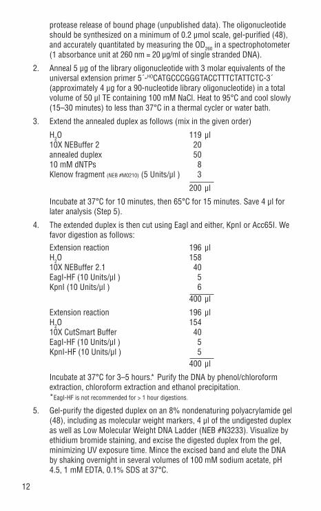
protease release of bound phage (unpublished data). The oligonucleotide should be synthesized on a minimum of 0.2 µmol scale, gel-purified (48), and accurately quantitated by measuring the OD260 in a spectrophotometer (1 absorbance unit at 260 nm = 20 µg/ml of single stranded DNA).
2. Anneal 5 µg of the library oligonucleotide with 3 molar equivalents of the universal extension primer 5´-HOCATGCCCGGGTACCTTTCTATTCTC-3´ (approximately 4 µg for a 90-nucleotide library oligonucleotide) in a total volume of 50 µl TE containing 100 mM NaCl. Heat to 95°C and cool slowly (15–30 minutes) to less than 37°C in a thermal cycler or water bath.
3. Extend the annealed duplex as follows (mix in the given order)
H2O 119 µl 10X NEBuffer 2 20 annealed duplex 50 10 mM dNTPs 8 Klenow fragment (NEB #M0210) (5 Units/µl ) 3
200 µl
Incubate at 37°C for 10 minutes, then 65°C for 15 minutes. Save 4 µl for later analysis (Step 5).
4. The extended duplex is then cut using EagI and either, KpnI or Acc65I. We favor digestion as follows:
Extension reaction 196 µl H2O 158 10X NEBuffer 2.1 40 EagI-HF (10 Units/µl ) 5 KpnI (10 Units/µl ) 6 400 µl
Extension reaction 196 µl H2O 154 10X CutSmart Buffer 40 EagI-HF (10 Units/µl ) 5 KpnI-HF (10 Units/µl ) 5 400 µl
Incubate at 37°C for 3–5 hours.* Purify the DNA by phenol/chloroform extraction, chloroform extraction and ethanol precipitation.
*EagI-HF is not recommended for > 1 hour digestions.
5. Gel-purify the digested duplex on an 8% nondenaturing polyacrylamide gel (48), including as molecular weight markers, 4 µl of the undigested duplex as well as Low Molecular Weight DNA Ladder (NEB #N3233). Visualize by ethidium bromide staining, and excise the digested duplex from the gel, minimizing UV exposure time. Mince the excised band and elute the DNA by shaking overnight in several volumes of 100 mM sodium acetate, pH 4.5, 1 mM EDTA, 0.1% SDS at 37°C.

13
6. Briefly microfuge to separate the gel fragments from the elution buffer, and transfer the supernatant to a clean tube. Repeat wash to improve yield, if desired. Purify the DNA duplex from the supernatant by phenol/chloroform extraction, chloroform extraction and ethanol precipitation (48). Resuspend the pellet in 50 µl of TE and quantitate a small amount by PAGE or spectrophotometrically. One µg of purified insert is more than sufficient for a library of complexity 109.
7. For a high-complexity library, digest 10–20 µg of M13KE Vector with the same enzymes used to prepare insert in step 4, for example, using 10 units each of EagI-HF and KpnI per µg DNA (total volume = 400–800 µl). Digest 3–5 hours at 37°C. Use KpnI in place of Acc65I if you used KpnI for insert digestion. Gel purify using standard methods (we recommend the Monarch DNA Gel Extraction Kit, NEB #T1020 or β-Agarase, NEB #M0392). Quantitate a small amount of purified cut vector on an agarose gel or spectrophotometrically.
8. Optimize the ligation conditions. Suggested starting parameters per 20 µl ligation: 40 and 100 ng of cut vector; 3:1, 5:1 and 10:1 molar excess of cut duplex; 2 µl of 10X ligase buffer; and 200 units (= 3 Weiss units) of T4 DNA Ligase (NEB #M0202). Incubate overnight at 16°C.
9. Heat-kill the test ligations at 65°C for 15 minutes, then electroporate 1 µl of each into 100 µl of electrocompetent ER2738 or other F+ strain (for suggested electroporation parameters see step 8 in the previous section). Outgrowths are carried out in 1 ml of SOC medium for 30–45 minutes at 37°C with shaking.
10. Prepare 10, 100, and 1000-fold dilutions of the outgrowth in LB. Transfer 10 µl of each dilution to a test tube containing 3 ml of top agar + 200 µl of a mid-log culture of ER2738, equilibrated at 45°C. Vortex briefly and spread on LB/IPTG/Xgal plates. Incubate overnight at 37°C and count blue plaques the next day.
11. Scale up the protocol using the highest plaque/µg ratio to desired library complexity. For example, a library with a compexity of 1 x 10⁹ clones would require a 5 µg ligation if the test ligations yield a ratio of 2 x 10⁸ plaques/µg of vector. Use no more than 500 µl per individual ligation reac-tion; use multiple tubes if necessary.
12. Purify the large-scale ligation by phenol/chloroform extraction, chloroform extraction and ethanol precipitation. Wash with 70% ethanol to desalt. Re-suspend the DNA in low salt buffer and electroporate as described above. To reduce the likelihood of cells picking up more than one DNA sequence, the ligation should be divided and electroporated using as many cuvettes as convenient. For a 10–20 µg scale ligation we typically carry out 100 electroporations, using 3 µl of resuspended ligated DNA per 100 µl of electrocompetent cells.
13. Add 1 ml of SOC to each cuvette immediately after electroporation. For high-complexity libraries it may be convenient to pool the SOC outgrowths

14
in groups of 5. Each outgrowth (or pool of 5) should be incubated for 30–45 minutes (no longer) before amplification. Titer several outgrowths or pools (as in Step 10) prior to amplification in order to obtain library complexity.
14. Amplify the electroporated cells by adding 20 ml of pooled SOC outgrowths to 1 liter of early-log cells (OD600 0.01–0.05) in LB medium. Incubate with vigorous aeration (250 rpm) at 37°C for 4.5 to 5 hours. Centrifuge at 5000 g for 20 minutes at 4°C. Transfer the supernatant to a clean bottle and discard the cells.
15. Recover the phage from the supernatant by adding 1/6 volume of 20% PEG/2.5 M NaCl and incubating overnight at 4°C. Pellet the phage by cen-trifugation at 5000 g for 45 minutes at 4°C. Discard the supernatant.
16. Thoroughly resuspend the phage pellet in 100 ml of TBS by gently rocking over ~1–3 hour or overnight at 4°C. Remove residual cells by centrifugation at 5000 g for 10 minutes at 4°C.
17. Transfer the supernatant to a new tube and discard the pellet. Reprecipitate the phage by adding 1/6 volume of 20% PEG/2.5 M NaCl and incubating for 1 hour at 4°C. Centrifuge at 5000 g for 20 minutes and discard the superna-tant.
18. Resuspend the final library in 10–40 ml of TBS by gentle rocking for 24–48 hours at 4°C. For long-term storage, add an equal volume of sterile glycerol, mix thoroughly and store at –20°C. The titer of the library should remain constant for several years at this temperature. Further amplification of the library is not recommended, as sequence biases may occur upon reamplification.
Surface Panning Procedure (Direct Target Coating)The most straightforward method of affinity partitioning (panning) involves directly coating a plastic surface with the target of interest (by nonspecific hydrophobic and electrostatic interaction), washing away the excess, and passing the pool of phage over the target-coated surface (Figure 1). Because of its relative simplicity, we recommend trying the direct coat-ing method described below first. If no clear consensus binding sequence emerges, then proceed with either of the solution binding procedures.
Day OneDepending on the available quantity of target molecule and the number of different targets being panned against simultaneously, panning can be carried out in individual sterile poly-styrene petri dishes, 12- or 24-well plates, or 96-well microtiter plates. Coat a minimum of 1 plate (or individual well) per target. It is not productive to do a separate negative control panning experiment without target. Volumes given in the following procedure are for 60 x 15 mm petri dishes, with volumes for microtiter wells given in parentheses. For wells of intermediate size adjust volumes accordingly, but in all cases the number of input phage should remain the same (1011 virions).
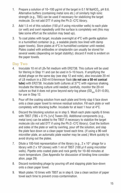
15
1. Prepare a solution of 10–100 µg/ml of the target in 0.1 M NaHCO3, pH 8.6. Alternative buffers (containing metal ions etc.) of similarly high ionic strength (e.g., TBS) can be used if necessary for stabilizing the target molecule. Do not add DTT if using the Ph.D.-C7C library.
2. Add 1.5 ml of this solution (150 µl if using microtiter wells) to each plate (or well) and swirl repeatedly until the surface is completely wet (this may take some effort as the solution may bead up).
3. To coat plates with target, incubate overnight at 4°C with gentle agitation in a humidified container (e.g., a sealable plastic box lined with damp paper towels). Store plates at 4°C in humidified container until needed. Plates coated with antibodies or streptavidin can usually be stored for several weeks (depending on target stability); discard if mold is evident on the paper towels.
Day Two4. Inoculate 10 ml of LB+Tet medium with ER2738. This culture will be used
for titering in Step 11 and can be used in 5–10 hours. If amplifying the eluted phage on the same day (see step 12 and note), also inoculate 20 ml of LB medium in a 250-ml Erlenmeyer flask (do not use a 50-ml conical tube) with ER2738. Incubate both cultures at 37°C with vigorous shaking. Incubate the titering culture until needed; carefully, monitor the 20-ml culture so that it does not grow beyond early-log phase (OD600 0.01–0.05), for use in Step 12.
5. Pour off the coating solution from each plate and firmly slap it face down onto a clean paper towel to remove residual solution. Fill each plate or well completely with blocking buffer. Incubate for at least 1 hour at 4°C.
6. Discard the blocking solution as in step 5. Wash each plate rapidly 6 times with TBST (TBS + 0.1% [v/v] Tween-20). Additional components (e.g., metal ions) can be added to the TBST if necessary to stabilize the target molecule (do not add DTT if using the Ph.D.-C7C library). Coat the bottom and sides of the plate or well by swirling, pour off the solution, and slap the plate face down on a clean paper towel each time. (If using a 96-well microtiter plate, an automatic plate washer may be used.) Work quickly to avoid drying out the plates.
7. Dilute a 100-fold representation of the library (e.g., 2 x 10¹¹ phage for a library with 2 x 10⁹ clones) with 1 ml of TBST (100 µl if using microtiter wells). Pipette onto coated plate and rock gently for 10–60 minutes at room temperature. (See Appendix for discussion of binding time consider-ation, page 29)
8. Discard nonbinding phage by pouring off and slapping plate face-down onto a clean paper towel.
9. Wash plates 10 times with TBST as in step 6. Use a clean section of paper towel each time to prevent cross-contamination.

16
10. Elute bound phage with 1 ml (100 µl if using microtiter wells) of an appropriate elution buffer for the interaction being studied. Typically this will be a solution of a known ligand for the target (0.1–1 mM) in TBS, or a solution of the free target (~100 µg/ml in TBS) to compete the bound phage away from the immobilized target on the plate. Rock the elution mixture gently for 10–60 minutes at room temperature. Pipette eluate into a microcentrifuge tube. Alternatively, a general buffer for nonspecific disruption of binding interactions is 0.2 M Glycine-HCl (pH 2.2), 1 mg/ml BSA. If using this buffer, rock gently for no more than 10–20 minutes, pipette eluate into a microcentrifuge tube, and neutralize with 150 µl (15 µl for microtiter wells) of 1 M Tris-HCl, pH 9.1.
11. Titer a small amount (~1 µl) of the eluate as described in General M13 Methods (page 8). Plaques from the first or second round eluate titering can be sequenced if desired (page 22).
12. Amplify the rest of the eluate by adding the eluate to the 20-ml ER2738 culture from Step 4 (should be early-log at this point) and incubating with vigorous shaking for 4.5 hours at 37°C.
Note: The remaining eluate can be stored overnight at 4°C at this point, if preferred, and amplified the next day. In this case, inoculate 10 ml of LB+Tet with ER2738 and incubate with shaking overnight at 37°C. The next day, dilute the overnight culture 1:100 in 20 ml of LB in a 250-ml Erlenmeyer flask (do not use a 50 ml conical tube) and add the unamplified eluate. Incubate with vigorous shaking for 4.5 hours at 37°C and proceed to Step 13.
13. Transfer the culture to a centrifuge tube and spin for 10 minutes at 12,000 g at 4°C. Transfer the supernatant to a fresh tube and re-spin (discard the pellet).
14. Transfer the upper 80% of the supernatant to a fresh tube and add to it 1/6 volume of 20% PEG/2.5 M NaCl. Allow the phage to precipitate at 4°C for at least 2 hours, preferably overnight.
Day Three15. Spin the PEG precipitation at 12,000 g for 15 minutes at 4°C. Decant and
discard the supernatant, re-spin the tube briefly, and remove residual supernatant with a pipette. The phage pellet should be a white finger print sized smear on the side of the tube.
16. Suspend the pellet in 1 ml of TBS. Transfer the suspension to a micro-centrifuge tube and spin at maximum (14,000 rpm) for 5 minutes at 4°C to pellet residual cells.
17. Transfer the supernatant to a fresh microcentrifuge tube and reprecipitate by adding 1/6 volume of 20% PEG/2.5 M NaCl. Incubate on ice for 15–60 minutes. Microcentrifuge at 14,000 rpm for 10 minutes at 4°C, discard the supernatant, re-spin briefly, and remove residual supernatant with a micropipet.
17
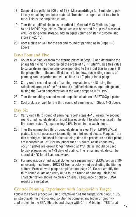
18. Suspend the pellet in 200 µl of TBS. Microcentrifuge for 1 minute to pel-let any remaining insoluble material. Transfer the supernatant to a fresh tube. This is the amplified eluate.
19. Titer the amplified eluate as described in General M13 Methods (page 8) on LB/IPTG/Xgal plates. The eluate can be stored for up to 3 weeks at 4°C. For long-term storage, add an equal volume of sterile glycerol and store at –20° C.
20. Coat a plate or well for the second round of panning as in Steps 1–3 above
Days Four and Five21. Count blue plaques from the titering plates in Step 19 and determine the
phage titer, which should be on the order of 1013–14 pfu/ml. Use this value to calculate an input volume corresponding to the input titer in Step 7. If the phage titer of the amplified eluate is too low, succeeding rounds of panning can be carried out with as little as 109 pfu of input phage.
22. Carry out a second round of panning: repeat steps 4–18 using the calculated amount of the first round amplified eluate as input phage, and raising the Tween concentration in the wash steps to 0.5% (v/v).
23. Titer the resulting second round amplified eluate on LB/IPTG/Xgal plates.
24. Coat a plate or well for the third round of panning as in Steps 1–3 above.
Day Six25. Carry out a third round of panning: repeat steps 4–10, using the second
round amplified eluate at an input titer equivalent to what was used in the first round (step 7), again using 0.5% Tween in the wash steps.
26. Titer the unamplified third round eluate as in step 11 on LB/IPTG/Xgal plates. It is not necessary to amplify the third round eluate. Plaques from this titering can be used for sequencing: time the procedure so that plates are incubated at 37°C for no longer than 18 hours, as deletions may occur if plates are grown longer. Stored at 4°C, plates should be used to pick plaques within 1–3 days of plating. The remaining eluate can be stored at 4°C for at least one week.
27. For preparation of individual clones for sequencing or ELISA, set up a 10-ml overnight culture of ER2738 from a colony, not by diluting the titering culture. Proceed with plaque amplification, page 22. Do not amplify the third round eluate and carry out a fourth round of panning unless the characterization shows no clear consensus sequence or phage ELISA results are negative.
Control Panning Experiment with Streptavidin TargetFollow the above procedure using streptavidin as the target, including 0.1 µg/ml streptavidin in the blocking solution to complex any biotin or biotinyl-ated protein in the BSA. Elute bound phage with 0.1 mM biotin in TBS for at

18
least 30 minutes. After 3 rounds of enrichment/amplification, the consensus sequence for streptavidin-binding peptides should include the motif His-Pro-Gln (HPQ) (7).
Solution-phase Panning with a Biotinylated Target and Streptavidin Plate CaptureAs an alternative to directly coating a plate with the target molecule, the target can be reacted with the phage in solution, followed by affinity capture of the phage-target complexes (6). Depending on the target, direct coating can result in an inaccessible ligand binding site, either due to steric blocking or partial denaturation of the target along the surface. Affinity capture requires some sort of affinity tag on the target; one way this can be accomplished is by biotinylating the target and capturing the complexes with a streptavidin-coated polystyrene plate. Alternatively, the biotinylated target–phage com-plexes can be captured with streptavidin-agarose beads, using the general bead selection procedure described on page 19. Capture of the target can be accomplished either before or after addition of phage.
Biotinylation of Target1. Dissolve or dilute 2 mg of target protein in 1 ml of 50 mM NaHCO3,
pH 8.5. Other buffers may be used if necessary to maintain stability of target, but do NOT use buffers with free primary or secondary amine groups (e.g., Tris, ethanolamine).
2. Immediately prior to use, dissolve ~1 mg of sulfo-NHS-LC-Biotin (avail-able from Pierce, cat. #21335) in 1 ml of H2O. Vortex vigorously. Add 74 µl of this (18 mM) solution to the target solution. This reagent is a water-soluble activated ester of biotin that specifically targets the ε-amine of solvent-accessible (surface) lysine residues. The remaining solution should be discarded, as the ester hydrolyzes rapidly upon storage.
3. Incubate the biotin–target mixture for 2 hours on ice.
4. Remove unreacted biotin by dialysis against a minimum of 2 one-liter changes of TBS, gel-filtration, or ultrafiltration. Alternate buffers of simi-lar ionic strength can be used at this point.
5. Quantitate the biotinylated protein by Bradford or Lowry assay. Bioti-nylation can be confirmed by Western blot detection or ELISA using commercially available anti-biotin antibodies. The degree of biotinylation can be quantitated by a colorimetric assay based on displacement of the dye HABA (2-[4´-Hydroxyazobenzene]-benzoic acid), also available from Pierce (cat. #28010). Based on results obtained at NEB, the described reaction conditions result in an average of 2 biotinylated lysines per ap-proximately 25-kDa protein molecule.

19
Panning with Surface Streptavidin CaptureFollow the panning procedure described above for the surface panning, direct target coating method (page 14), but coat the plates with streptavidin (100 µg/ml of streptavidin in 0.1 M NaHCO3, pH 8.6) instead of target. The blocking buffer should contain 0.1 µg/ml streptavidin in order to complex any biotin or biotinylated protein in the BSA. Replace Step 7 in the direct target coating method (page 15) with the following:7a. While plates are blocking (step 5), pre-complex the phage with the
biotinylated target: Combine in a microfuge tube 0.1 µg of biotinylated target (~10 nM final for a 25 kDa protein) and a 100-fold representation of the library (e.g., 2 x 10¹¹ pfu for a library with complexity 2 x 10⁹) in 400 µl TBST. In order to isolate low-affinity binders it may be necessary to increase the target concentration as high as 1–2 µM (33). Alternate buffers (containing metal ions etc.) of similarly high ionic strength can be used if necessary for stabilizing target molecule. Buffers should NOT contain DTT or other reducing reagents if using the Ph.D.-C7C library. Incubate for 10–60 minutes at room temperature.
7b. Add the phage–target solution to the washed, blocked plate. Incubate for 10 minutes at room temperature.
7c. Add biotin to a final concentration of 0.1 mM and incubate an additional 5 minutes. This will displace any streptavidin-binding phage (displaying the HPQ sequence) from the plate. The sufficiently slow off rate for the biotinylated target will ensure that the target will not be displaced by the biotin. Continue from Step 8 in the direct target coating protocol, above.
Solution-phase Panning with Affinity Bead CaptureAs a general alternative to panning against a target that has been immobilized on a surface, the library can be reacted with the target in solution, followed by affinity capture of the target–phage complexes onto an affinity matrix (bead) specific for the target protein. For example, if the target protein has a GST, MBP or polyhistidine affinity tag, the target-phage complexes can be captured on glutathione, amylose or chelated nickel beads, respectively. If the target is an antibody, Protein A and/or Protein G beads can be used for capture. In addition to requiring substantially less target per experiment than surface panning, solution panning can result in improved accessibility of the putative ligand binding site to phage-displayed peptides, as well as avoiding partial denaturation of the target on a plastic surface. Fortuitous selection of peptide sequences that specifically bind the bead can be avoided by employing a negative selection beginning with Round 2, in which the amplified phage is pre-incubated with the bead in the absence of target. The super-natant from this step is then reacted with the target in a positive selection. For antibodies or other target proteins that bind to more than one matrix type, bead-specific peptides can be avoided by alternating rounds between the matrix types. For example, for antibody targets, peptides specific for Protein A or Protein G are avoided by alternating rounds of panning between Protein A- and Protein G-agarose (magnetic beads can also be used). For antibodies that do not bind well to Protein A (sheep, goat, chicken and rat polyclonals, as well as some human IgG3 and mouse IgG1 monoclonal antibodies), Protein G-agarose can be used in all rounds, employing a negative selection strategy as described above.
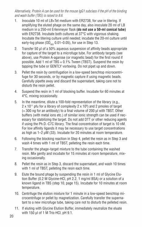
20
Alternatively, Protein A can be used for the mouse IgG1 subclass if the pH of the binding and wash buffer (TBS) is raised to 8.6.1. Inoculate 10 ml of LB+Tet medium with ER2738, for use in titering. If
amplifying the eluted phage on the same day, also inoculate 20 ml of LB medium in a 250-ml Erlenmeyer flask (do not use a 50-ml conical tube) with ER2738. Incubate both cultures at 37°C with vigorous shaking. Incubate the titering culture until needed; incubate the 20-ml culture until early-log phase (OD600 0.01–0.05), for use in Step 13.
2. Transfer 50 µl of a 50% aqueous suspension of affinity beads appropriate for capture of the target to a microfuge tube. For antibody targets (see above), use Protein A-agarose (or magnetic bead) for the first round if possible. Add 1 ml of TBS + 0.1% Tween (TBST). Suspend the resin by tapping the tube or GENTLY vortexing. Do not pipet up and down.
3. Pellet the resin by centrifugation in a low-speed benchtop microcentri-fuge for 30 seconds, or by magnetic capture if using magnetic beads. Carefully pipette away and discard the supernatant, taking care not to disturb the resin pellet.
4. Suspend the resin in 1 ml of blocking buffer. Incubate for 60 minutes at 4°C, mixing occasionally.
5. In the meantime, dilute a 100-fold representation of the library (e.g., 2 x 10¹¹ pfu for a library of complexity 2 x 10⁹) and 2 pmoles of target (= 300 ng for an antibody) to a final volume of 200 µl with TBST. Other buffers (with metal ions etc.) of similar ionic strength can be used if nec-essary for stabilizing the target. Do not add DTT or other reducing agents if using the Ph.D.-C7C library. The final concentration of target is 10 nM. For low affinity ligands it may be necessary to use target concentrations as high as 1–2 µM (33). Incubate for 20 minutes at room temperature.
6. Following the blocking reaction in Step 4, pellet the resin as in Step 3 and wash 4 times with 1 ml of TBST, pelleting the resin each time.
7. Transfer the phage–target mixture to the tube containing the washed resin. Mix gently and incubate for 15 minutes at room temperature, mix-ing occasionally.
8. Pellet the resin as in Step 3, discard the supernatant, and wash 10 times with 1 ml of TBST, pelleting the resin each time.
9. Elute the bound phage by suspending the resin in 1 ml of Glycine Elu-tion Buffer (0.2 M Glycine-HCl, pH 2.2, 1 mg/ml BSA) or a solution of a known ligand in TBS (step 10, page 15). Incubate for 10 minutes at room temperature.
10. Centrifuge the elution mixture for 1 minute in a low-speed benchtop mi-crocentrifuge or pellet by magnetization. Carefully transfer the superna-tant to a new microfuge tube, taking care not to disturb the pelleted resin.
11. If eluting with Glycine Elution Buffer, immediately neutralize the eluate with 150 µl of 1 M Tris-HCl, pH 9.1.

21
12. Titer a small aliquot of the eluate on LB/IPTG/Xgal plates as described in General M13 Methods (page 8).
13. Amplify the remaining eluate by adding it to the 20 ml ER2738 culture from Step 1 (must be early-log; no later) and incubating at 37°C with vigorous shaking for 4.5 hours.
Alternatively, the eluate can be stored overnight at 4°C and amplified the next day, if preferred. In this case, inoculate 10 ml of LB+Tet with ER2738 and incubate overnight at 37°C with shaking. The next day, dilute the overnight culture 1:100 in 20 ml of LB in a 250-ml Erlenmeyer flask (do not use a 50-ml conical tube) and add the unamplified eluate. Incubate at 37°C with vigorous shaking for 4.5 hours.
14. Transfer the culture to a centrifuge tube and spin for 10 minutes at 12,000 g at 4°C. Transfer the supernatant to a fresh tube and re-spin (discard the pellet).
15. Pipette the upper 80% of the supernatant to a fresh tube and add to it 1/6 volume of 20% PEG/2.5 M NaCl. Allow the phage to precipitate at 4°C for 2 hours or overnight.
16. Spin the PEG precipitation at 12,000 g rpm for 15 minutes at 4°C. Decant and discard the supernatant, re-spin briefly, and remove the residual supernatant with a pipette.
17. Suspend the pellet in 1 ml of TBS. Transfer the suspension to a micro-centrifuge tube and spin for 5 minutes at 4°C to pellet residual cells.
18. Transfer the supernatant to a fresh microcentrifuge tube and reprecipitate with 1/6 volume of 20% PEG/2.5 M NaCl. Incubate for 15–60 minutes on ice. Microcentrifuge at 14,000 rpm for 10 minutes at 4°C. Discard the supernatant, re-spin briefly, and remove residual supernatant with a micropipet.
19. Suspend the pellet in 200 µl of TBS. Microcentrifuge at 14,000 rpm for 1 minute to pellet any remaining insoluble matter. Transfer the supernatant to a fresh tube. This is the amplified eluate.
20. Titer the amplified eluate on LB/IPTG/Xgal plates as described in General M13 Methods, page 8. The eluate can be stored for several weeks at 4°C. For long-term storage, add an equal volume of sterile glycerol and store at –20°C.
21. The next day, count blue plaques and determine phage titer (see page 8). Use this value to calculate an input volume corresponding to the input titer used in Step 5. If the titer is too low, succeeding rounds of panning can be carried out with as little as 109 pfu of input phage.
22. Perform a second round of panning: repeat Steps 1–21 using the cal-culated amount of the first round amplified eluate as input phage. Raise the Tween concentration in the binding and wash steps to 0.5% (v/v). For antibody targets, use Protein G-agarose (or magnetic bead) for this round. If using the same resin for all three rounds, a negative selection

22
can be added at this stage if desired (do not carry out a negative selection in the first round):
Prepare an additional 50 µl of washed, blocked resin as in Steps 2–4. Pellet the resin and wash 4 times with TBST. Dilute the library in 200 µl of TBST as in Step 5, but leave out the target. Add the diluted phage to the washed, blocked resin and incubate for 15 minutes at room temperature with occasional mixing. Spin out the resin (or capture magnetically) and transfer the supernatant to a fresh microfuge tube. Add 2 pmoles of target to the supernatant, incubate for 20 minutes at room temperature, and continue with Step 6, using another 50 µl of washed, blocked resin.
23. Perform a third round of panning: repeat Steps 1–12 using the calculated amount of the second round amplified eluate as input phage. Keep the Tween concentration at 0.5% (v/v) in the binding and wash steps. For antibody tar-gets, use protein A-agarose (or magnetic bead) if you used protein A for the first round. If using the same resin in all three rounds, a negative selection can be performed as described in Step 22. The eluate can be stored for up to a week at 4°C.
24. For preparation of individual clones for sequencing or ELISA, inoculate a 10 ml overnight culture of ER2738 from a colony. Time the titering step so the plates are incubated no longer than 18 hours, as deletions may occur if the plates are incubated longer. Proceed with plaque amplification, page 22. Do not amplify the third round eluate and carry out a fourth round of panning unless the characterization shows no clear consensus sequence or ELISA signal.
Plaque Amplification for ELISA or SequencingSelected phage clones can be identified by DNA sequencing, and target specificity can be confirmed by phage ELISA. In both cases it is necessary to amplify phage, either from individual plaques or from the eluted pool, to obtain sufficient quantities to work with.
1. Dilute an overnight culture of ER2738 1:100 in LB. Dispense 1 ml of diluted culture into culture tubes, one for each clone to be characterized. 10–20 clones from the third round are usually sufficient to detect a consensus binding sequence.
2. Use a sterile wooden stick or pipette tip to stab a blue plaque from a titering plate (important: plates should be <1–3 days old, stored at 4°C and have <100 plaques) and transfer to a tube containing the diluted culture. Pick well-separated plaques. This will ensure that each plaque contains a single DNA sequence.
(Optional) For ELISA characterization, the entire pool of selected phage can be examined for binding activity, rather than individual clones. In order to detect signal, it is generally necessary to amplify the third round of eluted phage, as unamplified titers are typically no more than 107 pfu/ml. Add 10 µl of the unamplified eluate to 1 ml of diluted overnight culture.
23
3. Incubate the tubes at 37°C with shaking for 4.5–5 hours (no longer).
4. Transfer the cultures to microcentrifuge tubes, and microfuge at 14,000 rpm for 30 seconds (see next section to purify sequencing template). Transfer the supernatant to a fresh tube and re-spin. Using a pipette, transfer the upper 80% of the supernatant to a fresh tube. This is the amplified phage stock and can be stored at 4°C for several weeks with little loss of titer. For long-term storage (up to several years), dilute 1:1 with sterile glycerol and store at –20°C.
Sequencing of Phage DNARapid Purification of Sequencing TemplatesThis extremely rapid procedure (54) produces template of sufficient purity for manual or au-tomated dideoxy sequencing, without the use of phenol or chromatography. See Appendix (page 34) for notes and alternatives.1. Carry out the plaque amplification procedure described above. After the first
centrifugation in Step 4, transfer 500 µl of the phage-containing supernatant to a fresh microfuge tube.
2. Add 200 µl of 20% PEG/2.5 M NaCl. Invert several times to mix, and let stand for 10–20 minutes at room temperature.
3. Microfuge at 14,000 rpm for 10 minutes at 4°C and discard the supernatant. Phage pellet may not be visible.
4. Re-spin briefly. Carefully pipet away and discard any remaining supernatant.
5. Suspend the pellet thoroughly in 100 µl of Iodide Buffer by vigorously tap-ping the tube. Add 250 µl of ethanol and incubate 10–20 minutes at room temperature. Short incubation at room temperature will preferentially pre-cipitate single-stranded phage DNA, leaving most phage protein in solution.
6. Spin in a microfuge at 14,000 rpm for 10 minutes at 4°C, and discard the supernatant. Wash the pellet with 0.5 ml of 70% ethanol (stored at –20°C), re-spin, discard the supernatant, and briefly dry the pellet under vacuum.
7. Suspend the pellet in 30 µl of TE buffer. The template can be suspended in H2O instead of TE if desired, but this is not recommended for long-term storage. In TE buffer, the phage DNA should be stable indefinitely at –20°C.
8. Quantitate product by agarose gel electrophoresis. 5 µl of resuspended template should give a band of comparable intensity to 0.5 µg of purified single-stranded M13mp18 DNA (NEB #N4040).
Sequencing Guidelines1. The sequence being read corresponds to the anticodon strand of the tem-
plate. Write out the complementary strand and check against the top strand of the insert sequence shown in Figure 5 (the primers hybridize downstream of the insert). Check that the third nucleotide of each codon in the random-ized region is G or T.

24
2. TAG stop codons are suppressed by glutamine in ER2738 (glnV). If the library was amplified in this strain or any glnV (also known as supE) strain, TAG should be considered a glutamine codon when translating.
3. Libraries often contain a small percentage (< 1%) of clones containing multiple inserts of the randomized region. Preferential selection and amplification of these clones may occur when panning against targets whose ligand specificity spans a length greater than that specified by the insert. When interpreting sequence data, be sure the sequence outside the restriction sites used for inserting the randomized sequence (Acc65I and EagI) matches the sequence shown in Figure 5. Once the flanking regions of the library insert have been identified, the insert size is easily determined.
Sequence of random peptide library-gIII fusions. Each library is expressed with an N-terminal leader sequence that is removed upon secretion at the position indicated by the arrow, resulting in the randomized peptide positioned directly at the N-terminus of the mature protein. The hybridization positions of the -28 and -96 sequencing primers are indicated.
pIII leader sequence Kpn I/Acc65 I5´-...TTA TTC GCA ATT CCT TTA GTG GTA CCT TTC TAT TCT CAC TCT 3´-...AAT AAG CGT TAA GGA AAT CAC CAT GGA AAG ATA AGA GTG AGA ...Leu Phe Ala Ile Pro Leu Val Val Pro Phe Tyr Ser His Ser
Start of mature peptide-gIII fusion ↓ Eag I Library Insert Sequence TCG GCC GAA ACT GTT GAA See below AGC CGG CTT TGA CAA CTT Ser Ala Glu Thr Val Glu
AGT TGT TTA GCA AAA TCC CAT ACA GAA AAT TCA TTT ACT AAC GTC TGGTCA ACA AAT CGT TTT AGG GTA TGT CTT TTA AGT AAA TGA TTG CAG ACCSer Cys Leu Ala Lys Ser His Thr Glu Asn Ser Phe Thr Asn Val Trp ← 28 sequencing primer
AAA GAC GAC AAA ACT TTA GAT CGT TAC GCT AAC TAT GAG GGC...-3´TTT CTG CTG TTT TGA AAT CTA GCA ATG CGA TTG ATA CTC CCG...-5´Lys Asp Asp Lys Thr Leu Asp Arg Tyr Ala Asn Tyr Glu Gly... ← 96 sequencing primer
Library Insert SequencesPh.D.-7: (NNK)7 GGT GGA GGT (NNM)7 CCA CCT CCA Xxx7 Gly Gly Gly Ph.D.-12: (NNK)12 GGT GGA GGT (NNM)12 CCA CCT CCA Xxx12 Gly Gly Gly Ph.D.-C7C: GCT TGT (NNK)7 TGC GGT GGA GGT CGA ACA (NNM)7 ACG CCA CCT CCA Ala Cys Xxx7 Cys Gly Gly Gly
K = G or T; M = A or C
Figure 5:
25
Phage ELISA Binding Assay with Direct Target CoatingIt is useful to include a phage ELISA in any panning experiment since artifacts of the panning process cannot always be anticipated or prevented. The following ELISA protocol is sufficient for rapidly determining whether a selected phage clone binds the target, without the need for an antibody specific for the target. In this procedure a microtiter plate is coated with the target at high density, and each purified phage clone is applied to the plate at various dilutions. Bound phage is then detected with an anti-M13 antibody (see Invitrogen M13 antibodies from Thermo Fisher Scientific, PA1-26758 or MA1-34468). The amount of target coated on the plate is not quantifiable, and is present at sufficiently high density to allow multivalent binding to the phage, this method will not determine whether the selected phage binds with high or low affinity. The method is useful for qualitative determination of relative binding affinities for a number of selected clones in parallel, and will distinguish true target binding from binding to the plastic support. The latter is particularly useful if the direct coating method of panning (page 14) is used. In the Appendix (page 36) another approach is described, “sandwich” ELISA.
1. Carry out the plaque amplification procedure described above (page 22). After the first centrifugation in Step 5, save the phage-containing superna-tants at 4°C.
2. In addition to individual clones, the amplified first, second, and third round eluted pools may all be assayed. For each clone or pool to be characterized, inoculate 20 ml of LB medium with a single colony of ER2738 and incubate at 37°C until OD600 0.01–0.05. Alternatively, dilute an overnight culture of ER2738 1:100 in 20 ml of LB.
3. Add 5 µl of phage stock from Step 1(or a single phage plaque) to a 20 ml culture for each clone or pool to be characterized to each culture and incubate with vigorous aeration for 4.5–5 hours at 37°C.
4. Transfer the culture to a centrifuge tube and spin at 12,000 g for 10 min-utes at 4°C. Transfer the supernatant to a fresh tube and re-spin (discard the pellet).
5. Pipette the upper 80% of the supernatant to a fresh tube and add 1/6 volume of 20% PEG/2.5 M NaCl. Allow the phage to precipitate at 4°C for at least 2 hours or overnight.
6. Spin the PEG precipitation at 12,000 g for 15 minutes at 4°C. Decant and discard the supernatant, re-spin briefly, and remove residual supernatant with a pipette.
7. Suspend the pellet in 1 ml of TBS. Transfer the suspension to a microcen-trifuge tube and spin at 14,000 rpm for 5 minutes at 4°C to pellet residual cells.
8. Transfer the supernatant to a fresh microcentrifuge tube and re-precipitate with 1/6 volume of 20% PEG/2.5 M NaCl. Incubate 15–60 minutes on ice. Microcentrifuge at 14,000 rpm for 10 minutes at 4°C. Discard the super-natant, re-spin briefly, and remove residual supernatant with a micropipet.
26
9. Suspend the pellet in 50 µl of TBS. Titer as described in General M13 Methods, page 8. The titer should be approximately 1014 pfu/ml. The phage stock can be stored for several weeks at 4°C. For long-term stor-age, add an equal volume of sterile glycerol and store at –20°C.
10. Coat one row of ELISA plate wells for each clone or pool to be character-ized with 100–200 µl of 100 µg/ml of target in 0.1 M NaHCO3, pH 8.6. Incubate overnight at 4°C in an air-tight humidified box (e.g., a sealable plastic box lined with damp paper towels).
11. Shake out excess target solution and slap plate face-down onto a paper towel. Fill each well completely with blocking buffer. Additionally, one row of uncoated wells per clone to be characterized should also be blocked to test for binding of each selected sequence to BSA-coated plastic (this test for background signal is extremely important, especially if panning was carried out on a polystyrene surface). A second, fully uncoated microtiter plate should be blocked for use in serial dilutions of phage (step 13) before addition to the target-coated plate. Dilutions are done in a separate blocked plate to ensure that phage are not absorbed onto the target dur-ing the course of performing dilutions, which would result in a sudden “falling-off” of signal as the phage is diluted. Incubate the plates filled with blocking buffer for 1–2 hours at 4°C.
12. Shake out the blocking buffer and wash each plate 6 times with TBST, slapping the plate face-down onto a clean section of paper towel each time. The percentage of Tween should be the same as the concentration used in the panning wash steps.
13. In the separate blocked plate, carry out fourfold serial dilutions of the phage in 200 µl of TBS/Tween per well, starting with 1012 virions in the first well of a row and ending with 2 x 10⁵ virions in the 12th well.
14. Using a multichannel pipettor, transfer 100 µl from each row of diluted phage to a row of target-coated wells, and transfer 100 µl to a row with-out target. Incubate at room temperature for 1–2 hours with agitation.
15. Wash plate 6 times with TBST as in Step 12.
16. Follow manufacturer instructions for antibody application and develop-ment.

27
Use of Synthetic Peptides in Specificity AnalysisFor more detailed binding or inhibition studies, it may be necessary to synthe-size selected sequences as free, soluble peptides. This allows precise control of peptide concentration, without the avidity artifacts associated with pentava-lent display on phage. Additionally, without the phage attached, the peptide can be used at much higher concentrations and can be used in vivo. When designing a synthetic peptide corresponding to a selected binding sequence, it is important to realize that, while the N-terminus of the displayed sequence was free during panning, the C-terminus was fused to the phage. Furthermore, the C-terminal residue of the selected sequence did NOT have a free negatively charged carboxylate during panning, so a simple synthetic peptide with a free carboxy terminus will introduce a negatively charged group at a position occu-pied by a neutral peptide bond during panning. Depending on the nature of the target-ligand interaction, this negative charge can completely abolish binding. It is, therefore, recommended that the C-terminal carboxylate of the synthetic peptide be amidated to block the negative charge. Additionally, if the library insert was designed to include a peptide spacer between the random sequence and pIII (as are the commercially available Ph.D. libraries, which include a Gly-Gly-Gly-Ser spacer), this spacer sequence should be added to the C terminus of the peptide. For chemical conjugation of the peptide to a reporter enzyme or a solid support, the peptide can be designed with a C-terminal cysteine (if there are no other cysteines present in the sequence). The resulting peptide thiol can be easily coupled to maleimide-activated HRP, alkaline phosphatase or agarose beads.

28
AppendixOptimizing Peptide Binding InteractionsThere are several variables affecting the stringency of selection during panning. Depend-ing on the interaction being studied, adjustment of the stringency of selection or elution may be necessary to obtain a consensus binding sequence.
1. Detergent: The presence of detergent (typically Tween 20) in the binding and wash buffers reduces non-specific hydrophobic interactions between the phage and the target and/or blocking agent (BSA), which lead to higher levels of background binding. Lower Tween concentrations in early rounds will result in higher eluate titers, and the stringency can be gradually increased with each round by raising the Tween concentration stepwise to a maximum of 0.5%. In side-by-side experiments, however, we have obtained identical consensus sequences when Tween concentra-tions were held constant (0.5%) or increased stepwise (0.1, 0.3, 0.5%) in 3 rounds of panning. The use of lower Tween concentrations in earlier rounds is recommended when the interaction under study is so specific that the eluate titer (i.e., the number of bound sequences) in early rounds is expected to be very low.
2. Salt: Hydrophobic interactions are favored at high ionic strength, while ionic interactions are favored at low ionic strength. Nonspecific ionic interactions between surface charged groups on the target and phage, which would lead to high background binding, are thus avoided by the use of a high salt buffer such as TBS in the binding steps. Since it is impossible to predict in advance, however, whether the peptide-target interaction will be largely ionic or hydrophobic in nature, it may be neces-sary to test for the optimum salt concentration of the binding buffer.
3. Target Concentration: If panning against the target in solution, the stringency can be increased by lowering the concentration of target (59). An initial target concentration of 10 nM is recommended; this can be lowered to 1 nM in later rounds for selection of ligands with high binding affinity. If no consensus sequence is evident, it is possible that the best sequences in the library bind with very low affinity. Even with the avidity effects associated with multivalent display, target concentrations in the nanomolar range may simply be too low to see any binding. In this case it may be necessary to raise the target concentration to 1–2 µM in all rounds of panning (55). Also, low affinity binders present in low numbers in the initial unpanned library might be lost in the first round of solution-phase panning, regardless of target concentration. In this case it may be helpful to carry out the first round by panning against target which has been immobilized by direct coating (page 14), and then carrying out subsequent rounds in the solution phase.

29
4. Number of Rounds: With each round of panning and amplification, the pool of phage becomes enriched in favor of sequences that bind to the target. Maintaining a constant input phage concentration in each round results in a stepwise increase in the number of particles displaying a given sequence until a point is reached where most or all of the eluted particles display a consensus binding sequence. Depending on the interaction being studied and the applied stringency, this usually happens after 2 or 3 rounds. If no clear consensus sequence emerges after 3 rounds, the 3rd round eluate can be amplified and a fourth round of panning carried out. Reports in the literature of 5 or more rounds being carried out (60) generally refer to an iterative selection technique where a low affinity ligand is selected after 3 rounds and then subjected to further mutagenesis followed by additional rounds of selection.
Choice of LibraryThe choice of library to be used in a particular experiment is dependent upon many factors. Unfortunately, in the absence of detailed structural information about the target-ligand interaction, it is impossible to predict in advance which type of library will yield the most productive ligands (see, for example, 61). The Ph.D.-7 library consists of randomized linear 7-mer peptides, and may be most useful for targets requiring binding elements concentrated in a short stretch of amino acids. The Ph.D.-12 library consists of randomized linear 12-mer peptides, but with a diversity equivalent to the Ph.D.-7 library. It may be useful for targets requiring 7 or fewer defined residues for binding, but which cannot be contained within the 7-residue “window” of the Ph.D.-7 library. For example, the motif ASDXXXTXPY has only six defined positions, but cannot be present in the Ph.D.-7 library. Additionally, 12-mers are long enough to fold into short structural elements, which may be useful when panning against targets that re-quire structured ligands. A caveat is that the increased length of the randomized segment may allow your target to select sequences with multiple weak binding contacts, instead of a few strong contacts.
If, following three or four rounds of panning with a linear peptide library, there is no clear consensus ligand sequence (or all of the plaques are white, see page 32), it is possible that the library simply does not contain any clones that bind tightly to the target, i.e., the most “fit” peptide in the library does not bind with high enough affinity to be selected. In this case a structurally constrained library such as the Ph.D.-C7C library might work better. Such libraries are especially useful for targets whose native ligands are in the context of a surface loop, such as antibodies with structural epitopes. Additionally, imposing structural con-straints on the unbound ligand may result in a less unfavorable binding entropy, improving the overall free energy of binding compared to unconstrained ligands (62). A major disadvantage of structurally constrained libraries is that the con-straint may “freeze out” a conformation required for target binding, preventing binding outright rather than improving affinity (63).

30
Selection of Cell-specific PeptidesA particularly notable application of phage displayed peptide libraries is the identification of peptides that recognize specific cell-surface receptors and cell types (reviewed in reference 64). Such peptides can be bioactive (e.g. receptor agonists/antagonists), or can be used in diagnostic applications as well as delivery of genes or cytotoxic agents to specific cell types. Such peptides can be identified by panning against isolated cell-surface receptors (60,62,65, 69), if the receptor can be purified in stable, soluble form. If the receptor is not amenable to isolation (e.g., a 7-transmembrane receptor), whole cells or membrane fractions can be panned in vitro (66-69) or in vivo (70-75). Since a given cell type will have hundreds or thousands of different receptors, each capable in theory of pulling a specific ligand out of the library, the likelihood of pulling out a peptide specific for a single receptor is dependant both on the cleverness and stringency of the panning procedure. Simple panning against intact cells may yield a complex mixture of peptides with no clear consensus. To target the library to the receptor of interest, bound phage can be eluted with a known ligand for that receptor (66). Panning can be carried out in the presence of peptides known to bind to undesirable receptor targets (76). Al-ternatively, rounds may be switched between different cell lines, each of which overexpresses the desired target receptor, resulting in an overall counterselec-tion against ligands specific for the other receptors (67). However, peptide ligands for specific cell types have been successfully identified by panning against a single cell line without any counterselection (36,68).
An exciting development is the use of phage display to select organ-specific peptides in vivo. Following injection of a peptide library into an animal, the organs of interest are harvested, washed, and the eluted phage is used in subsequent rounds of injection and selection. Peptides selected in this manner have been successfully used to specifically deliver drugs to tumor cells (77). The C7C library was used to find a peptide that mediates protein delivery through skin (78).

31
Troubleshooting:I have used up almost all of my M13KE RF DNA. How can I make more?RF M13KE DNA may be purified from infected cells. Follow amplification steps outlined in the panning protocol (page 14) to amplify a single plaque or 5 µl of phage stock in a 20 ml culture. Alternatively, see protocol with M13KE phage (NEB #N0316) at neb.com. DNA to be used to make a phage displayed peptide library should be of the highest possible purity; a cesium chloride gradient is often the best method (48). Trace contaminants will result in poor ligation efficiencies that cannot be overcome.
I do not see any plaques after electroporation of my ligation reac-tion mixtures.Review M13 general methods and troubleshooting comments (below), then if necessary, troubleshoot the cloning steps from digestion to ligation and electroporation.
No plaques are visible when titering.Unlike lambda, M13 is a non-lytic phage and does not produce clear plaques. M13 plaques are areas of diminished cell growth, not lysis, and consequently, can be difficult to see. Try holding the plate up to a light. Also, since the vector used to prepare the library carries the lacZα gene, plaques will be blue, and easier to see, when using an alpha-complementing strain such as the supplied strain ER2738 and plating on Xgal/IPTG plates.
Also, be sure the dilution range is appropriate for the phage you are titering. For amplified phage, plate 10 µl of 1:109 - 1:1011 dilutions; for unamplified panning eluates, try 1:10–1:104 dilutions for early rounds, 1:104-1:107 for later rounds. If the phage is not sufficiently dilute, the plaques will be confluent on the plate and it will look like there are no plaques at all (or a bluish tinge when using Xgal plates). As a negative control, cells may be plated with top agar without added phage. Occasionally after PEG precipitation, the phage will clump and not dilute properly. As a result, you might have a plate containing too many plaques merged together. Make sure to give the phage ample time to resuspend after precipitation (>1 hour) and vortex each dilution tube well (~10 seconds).
All or most of the eluted phage plaques are white (colorless) on Xgal/IPTG platesIf all of your plaques are white (colorless), it is possible that the Xgal/IPTG plates were incorrectly prepared. Test your plates by titering the naïve unpanned library, using 109 and 1010 dilutions. If both the unpanned library and your selected phage produce white plaques, then the plates are defective and should be re-prepared. Also, the bacterial strain used for plating must be capable of α-complementation (lacZΔM15 or equivalent), such as the supplied strain ER2738, in order for plaques to appear blue.

32
The most likely explanation for white plaques is that the pool of phage became contaminated with an environmental M13-like phage during panning and amplification. Display of foreign peptides as N-terminal fusions to the infectivity protein pIII, as in the Ph.D. libraries, slightly attenuates infectivity of the library phage relative to wild-type M13. As a result, there is an in vivo selection for the contaminating phage during the amplification steps between rounds of panning. In the absence of a correspondingly strong in vitro binding selection during panning, even vanishingly small levels of contamination can result in a majority of the phage pool being wild-type phage after 3 (or especially 4) rounds of pan-ning. The Ph.D.-C7C library is particularly susceptible to contamination, since phage infectivity is further attenuated by displayed cysteine-containing peptides.
Contamination is an extremely common problem with any phage display sys-tem, but fortunately there are a few things you can do to minimize this problem:
a) Use Xgal/IPTG plates for all titering steps, and if white plaques are evident, pick only blue plaques for sequencing.
b) Use aerosol-resistant pipet tips and cotton-plugged pipets for all protocols described in the manual.
c) If contamination problems persist, all of the solutions used for panning should be autoclaved, with the exception of BSA-containing solutions which should be filter sterilized. Solutions used for phage display should not be used for anything else. Pipettors should be disassembled, the bar-rel autoclaved, and the internal plunger machinery soaked overnight in a detergent solution such as Count-Off™.
d) Since wild-type phage are preferentially amplified during the amplification steps, pick plaques for sequencing directly after the 3rd round elution step. Do not amplify the 3rd round eluate and carry out a 4th round unless the third round sequences show no clear consensus.
e) If all or most of the plaques are white (colorless) after 3 rounds of pan-ning, it is possible that the library simply does not contain any clones that bind tightly to the target. The ideal ligand sequence may not be statistically represented in the library, or the target simply is not capable of binding to a short peptide sequence.(see also, Choice of Library, page 29).
The amplified phage titer is low.In order for M13 phage to be efficiently amplified, it is critical that cultures be well aerated, and infected early in their growth phase. We recommend amplifica-tion in 20 ml cultures in 250 ml Erlenmeyer flasks, in a shaker set to 250 rpm. Amplification in smaller vessels, such as 50 ml conical tubes, will result in much lower yields of amplified phage. M13 phage should either be added to an early-log culture, OD600 < 0.05, or to a 1:100 dilution of an overnight culture. Yield of amplified phage is maximal after 4.5–5 hours at 37°; longer incubation may result in deletions and is not recommended. If carrying out nonspecific elution with pH 2.2 glycine buffer, the eluted phage must be neutralized as described in the manual prior to amplification.

33
The phage DNA templates do not yield readable sequence.The sequencing template purification protocol on page 23 should provide single-stranded template of sufficient purity for dideoxy sequencing with Sequenase™ (GE Healthcare), or automated cycle sequencing with dye-labeled terminators (ABI). The procedure should be followed exactly as described in the manual: prolonged ethanol precipitation, precipitation at –20°C or centrifugation longer than 10 minutes will result in co-precipitation of salt and phage proteins, which will inhibit sequencing. Additionally, it is crucial that the phage pellet is thoroughly suspended in the iodide buffer prior to adding ethanol. If problems persist, or if another sequencing method is used, a phenol:chloroform extrac-tion step can be added: Following suspension in Iodide Buffer, add 2 volumes of TE, extract once with phenol:chloroform (1:1) and once with chloroform, and ethanol precipitate. 5 µl (one sixth) of suspended template (approximately 0.5 µg) should be sufficient for sequencing; quantitation should be confirmed by agarose gel electrophoresis using 0.5 µg single stranded M13 DNA (NEB #N4040) as a standard. Alternatively, replicative form (RF, i.e. double stranded) DNA may be purified from cell pellets by standard miniprep protocols.
The sequencing templates do not run where they should on a gel.The sequencing templates prepared by the method on page 23 are single-stranded (approx. 7250 nucleotides), and as a result will not line up with double-stranded markers of the same length. The apparent size will vary depending on the applied voltage, ethidium and agarose concentration in the gel, and whether TBE or TAE is used as running buffer. We strongly recommend using single-stranded M13 DNA as a marker (see above). Different folding patterns of the single-stranded product may also lead to multiple banding on an agarose gel.
The sequence does not have the cloning sites or insert as shown in Figure 5 (page 24).If the sequence matches the sequence in Figure 5 from the primer back to the 3´ G of the EagI site, but then deviates upstream from this position, you may have sequenced a wild-type M13 contaminant (see “white plaques”):
Ph.D. Library Sequence: EagI....GGTGGAGGTTCGGCCGAAACTGTTGAA...(← primer)
wt M13 sequence: ....TATTCTCACTCCGCTGAAACTGTTGAA...(← primer)
Since the library phage are derived from the common cloning vector M13mp19, which carries the lacZα gene, phage plaques appear blue when plated on media containing Xgal and IPTG, provided an α-complementing strain such as ER2738 is used for plating. Environmental M13-like phage will typically yield white plaques when plated on the same media. These plaques are also slightly larger and “fuzzier” than the library phage plaques. We therefore recommend

34
plating on LB/Xgal/IPTG plates for all titering steps and, if white plaques are evident, picking ONLY blue plaques for sequencing.
The sequence is fine back to the KpnI/Acc65I site, but then differs from Figure 5 (page 24), or the KpnI site is missing altogether.If the sequence matches the sequence in Figure 5 starting from the primer back to the KpnI site, but then deviates upstream from the KpnI site (or the KpnI site is missing), the clone likely contains multiple inserts. All of the Ph.D. libraries were constructed by directional cloning of a synthetic randomized duplex into KpnI/Acc65I and EagI sites that had previously been engineered into the M13 genome. A small percentage (< 1%) of clones in each library picked up more than one insert during ligation. Typically such clones contain 2–5 randomized inserts, with one or more inverted relative to the others. Preferential selection and amplification of these clones may occur when panning against targets which prefer longer ligands; consequently, selection of clones with multiple inserts is more likely to occur when using the Ph.D.-7 library.
To properly characterize these clones, it is necessary to read the sequence back to the occurrence of a KpnI site preceded by the upstream vector se-quence TTAGT, as shown in Figure 4 (page 11). Starting from this KpnI site, the translated sequence Val-Pro-Phe-Tyr-Ser-His-Ser is the C-terminal end of the pIII leader sequence. Everything downstream from this sequence is displayed on the phage, and must be considered when identifying consensus binding elements. In experiments carried out at NEB, however, we have failed to identify meaningful consensus binding motifs from multiple insert clones, and typically ignore these clones when interpreting data. If all or most of your selected clones contain multiple inserts, we recommend repeating your panning with one of our other libraries.
Extensive white plaques after 4 or more roundsIn a typical round of panning, approximately 1011 input phage are reacted with the target, and between 103 and 107 total phage are eluted off following washing. This corresponds to an enrichment of 104- to 108-fold per round. Since a typical high-complexity library contains approximately 109 different clones, the eluted pool of phage should in theory be fully enriched in favor of binding sequences after only 2 or 3 rounds. Once this point is reached, further rounds of ampli-fication and panning will result only in selection of phage that have a growth advantage over the library phage (9). For example, vanishingly small levels of contaminating environmental wild-type phage (less than one part per billion) will completely overtake the pool if too many rounds of amplification are carried out, regardless of the strength of the in vitro selection.
35
The streptavidin control experiment did not yield the HPQ consensus sequence.If you used low pH glycine rather than biotin to elute your phage, you will likely not get an HPQ consensus sequence. Due to the relatively low affinity of the peptide-streptavidin interaction, nonspecific elution is incapable of selec-tively enriching for HPQ-containing peptides. HPQ-containing peptides can be competitively eluted using the natural ligand biotin.
If you used biotin to elute and still did not get a consensus sequence, the most likely explanation is that you did not carry out sufficiently rigorous washes. When you wash, pour the wash buffer in the plate from a bottle (don’t gently pipet it in) and swirl it for 10–60 seconds each time. The number of phage that you elute after the first round of biopanning should be in the range of 103–107 (closer to 103 for an ELISA well and closer to 107 for larger wells). If you are eluting more phage, you are not washing well enough and as a result, not get-ting sufficient enrichment. It also may help to add 0.1 µg/ml streptavidin to the blocking buffer to complex any contaminating biotin in your BSA, which could otherwise complex the streptavidin on the plate during the blocking step.
ELISA indicates that background binding to the plate is as high as binding to the targetIf panning against a polystyrene plate coated with the target (Direct target coating, page 14), it is possible to inadvertently select peptides that specifi-cally bind the polystyrene surface (79). These peptides will yield identical ELISA signals in the presence and absence of target, since the ELISA plate is also made of polystyrene. Such “plastic binders” are typically rich in aromatic residues (Phe, Tyr, Trp, His), which often alternate (the sequence FHWTWYW is a plastic binder discovered and characterized at NEB). Selection of plastic binders often occurs in the absence of a strong target preference for pep-tide sequences present in the library: other libraries may yield the desired target-specific sequences. Selection of polystyrene-specific peptides can be avoided by using the bead capture protocol described on page 19. The phage are reacted with the target in solution, and the phage-target complexes are then captured onto beads that specifically bind the target (protein A-agarose for antibody targets, glutathione-agarose for GST fusions, etc.). Unbound phage are removed by extensively washing the beads in a microfuge tube. Unlike polystyrene, neither the beads (typically crosslinked agarose) nor the microfuge tubes (polypropylene) are likely to select specific peptide sequences from the library, although the species conjugated to the beads (protein A, glutathione, etc.) might do so. To avoid selection of bead-specific ligands, we suggest either alternating rounds between different beads specific for the target (e.g., for antibody targets, use protein A beads for rounds 1 and 3, and protein G beads for round 2), or adding a subtractive panning step, beginning with round 2, in which the phage pool is first reacted with the beads alone (no target), the beads discarded, and the supernatant from this step reacted with the target.

36
Panning yielded a consensus sequence, but no ELISA signal.When characterizing phage clones by the ELISA protocol in the manual, it is difficult to add more than 1012 virions per 100 µl well. This corresponds to a phage concentration of only 16 nM. At this concentration, an unambiguously positive ELISA signal can only be observed if the binding affinity is in the micromolar range or better. The iterative nature of phage selection permits identification of ligands with a broad range of affinities, from sub-nM to 1 mM, so lower affinity ligands will not show a positive ELISA signal. In this case it is necessary to increase the concentration of the selected ligand, perhaps by synthesizing a peptide corresponding to the selected sequence, page 27.
Alternatively, a sandwich ELISA can be carried out in which the selected phage is immobilized and an excess of target applied in the liquid phase (55). This procedure requires an antibody against the target protein, or some other means of detecting bound target protein. Coat the wells overnight with anti-M13 antibody (no HRP), wash, and add serial dilutions of each phage clone (one clone per row). After 1 hour, wash away unbound phage and add an excess of target protein (0.1–1 µM) in TBST. Incubate 1–2 hours at RT, wash away unbound target, and detect bound target with an enzyme-linked antibody.
What is the size of pIII with/without leader?The molecular weight of the unprocessed coat protein pIII (containing a leader sequence but no displayed peptide) is 44651 daltons. Without the leader se-quence, the molecular weight of mature pIII is 42579. By SDS-PAGE, however, pIII usually runs with an apparent molecular weight of 60–65 kDa, possibly as a result of the unusual glycine-rich spacer regions between the domains of the protein (80). The amino acid sequence of the leader peptide is MKKLLFAIPLV-VPFYSHS (note that the initiator Met is encoded by a GTG codon)(81).
Can the library be amplified for additional panning experiments?We strongly recommend against amplification of the supplied libraries, as sequence biases in vivo will likely result in certain sequences being under-represented in the resulting library, or absent altogether. Displayed peptides in the Ph.D. libraries are expressed as fusions to the coat protein pIII, which modulates infectivity by binding to the F-pilus of the recipient cell. As a result, there is a biological selection against certain displayed sequences during in vivo amplification, particularly sequences with multiple positive charges (which inhibit secretion) and unpaired cysteines. The supplied libraries have each been amplified only once following ligation, and all characterization (representative sequencing, panning etc.) carried out on this amplified selling stock. We cannot guarantee that the amino acid distribution data we report for each library as supplied will hold upon reamplification.

37
References:1. Sidhu, S.S. (2000) Curr. Opin. Biotechnol. 11, 610–616.
2. Rodi, D.J. and Makowski, L. (1999) Curr. Opin. Biotechnol. 10, 87–93.
3. Wilson, D.R. and Finlay, B.B. (1998) Can. J. Microbiol. 44, 313–229.
4. Parmley, S.F. and Smith, G.P. (1988) Gene 73, 305–318.
5. Scott, J.K. and Smith, G.P. (1990) Science 249, 386–390.
6. Cwirla, S.E., Peters, E.A., Barrett, R.W. and Dower, W.J. (1990) Proc. Natl. Acad. Sci. USA 87, 6378–6382.
7. Devlin, J.J., Panganiban, L.C. and Devlin, P.E. (1990) Science 249, 404–406.
8. Russel, M., Linderoth, N.A. and Sali, A. (1997) Gene 192, 23–32.
9. Rodi, D.J., Janes, R.W., Sanganee, H.J., Holton, R.A., Wallace, B.A. and Makowski, L. (1999) J. Mol. Biol. 285, 197–203.
10. Peters, E.A., Schatz, P.J., Johnson, S.S. and Dower, W.J. (1994) J. Bacteriol. 176, 4296–4305.
11. Birkenmeier, G., Osman, A.A., Kopperschlager, G. and Mothes, T. (1997) FEBS Lett. 416, 193–196.
12. Dore, J.M., Morard, F., Vita, N. and Wijdenes, J. (1998) FEBS Lett. 426, 67–70.
13. Rowley, M.J., Scealy, M., Whisstock, J.C., Jois, J.A., Wijeyewickrema, L.C. and Mackay, I.R. (2000) J. Immunol. 164, 3413–3419.
14. Youn, J.H., Myung, H.J. Liav, A., Chatterjee, D., Brennan, P.J., Choi, I.H., Cho, S.N., Shin, J.S. (2004) FEMS Immunol. Med. Microbiol. 41, 51–57.
15. Dieltjens, T. et al. (2010) J. Virol. Meth. 169, 95–102.
16. Ferrer, M. and Harrison, S.C. (1999) J. Virol. 73, 5795–5802.
17. Lavilla, M. et al. (2009) J. Microb. Meth. 69, 55–66.
18. Rajik, M. et al. (2009) J. Virol. 6, 74–86.
19. Tiwari, V. et al. (2011) J. Biol. Chem. 286, 25406-25415.
20. Dharmasena, M.N., Jewell, D.A., and Taylor, R.K. (2007) J. Biol. Chem. 282, 33805–33816.
21. Lavi, T., Siman-Tov, R., and Ankri, S. (2008) Mol. Microbiol. 69, 55–66.
22. Welch, B.D., VanDemark, A.P., Heroux, A., Hill, C.P., and Kay, M.S. (2007) Proc. Natl. Acad. Sci. USA, 104, 16828–16833.
23. Whaley, S.R., English, D.S., Hu, E.L., Barbara, P.F. and Belcher, A.M. (2000) Nature 405, 665–668.
24. Ahmad, G., Dickerson, M.B., Cai, Y., Jones, S.E., Ernst, E.M., Vernon, J.P., Haluska, M.S., Fang, Y., Wang, J., Subramanyam, G., Naik, R.R. and Sandhage, K.H. (2008) J. Am. Chem. Soc. 130, 4–5.
25. Sawada, T. et al. (2009) J. Amer. Chem. Soc. 131, 14434–14441
26. Schwemmer, T. et al. (2012) J. Amer. Chem. Soc. 134, 2385–2391
27. Rozinov, M.N. and Nolan, G.P. (1998) Chem. Biol. 5, 713–728.
28. Miura, Y., Sasao, Y., Kamihira, M., Sakaki, A., Iijima, S., Kobayashi, K. (2004) Biochim. Biophys. Acta. 1673(3), 131–138.
29. Zhao, S. W. (2005) Environ. Int. 31, 535–541.
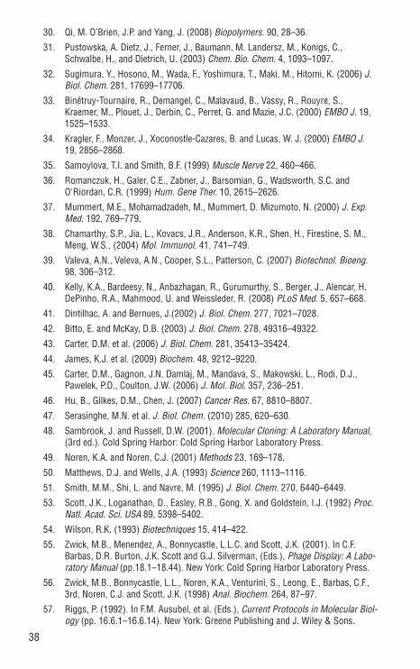
38
30. Qi, M. O’Brien, J.P. and Yang, J. (2008) Biopolymers. 90, 28–36.
31. Pustowska, A. Dietz, J., Ferner, J., Baumann, M. Landersz, M., Konigs, C., Schwalbe, H., and Dietrich, U. (2003) Chem. Bio. Chem. 4, 1093–1097.
32. Sugimura, Y., Hosono, M., Wada, F., Yoshimura, T., Maki, M., Hitomi, K. (2006) J. Biol. Chem. 281, 17699–17706.
33. Binétruy-Tournaire, R., Demangel, C., Malavaud, B., Vassy, R., Rouyre, S., Kraemer, M., Plouet, J., Derbin, C., Perret, G. and Mazie, J.C. (2000) EMBO J. 19, 1525–1533.
34. Kragler, F., Monzer, J., Xoconostle-Cazares, B. and Lucas, W. J. (2000) EMBO J. 19, 2856–2868.
35. Samoylova, T.I. and Smith, B.F. (1999) Muscle Nerve 22, 460–466.
36. Romanczuk, H., Galer, C.E., Zabner, J., Barsomian, G., Wadsworth, S.C. and O'Riordan, C.R. (1999) Hum. Gene Ther. 10, 2615–2626.
37. Mummert, M.E., Mohamadzadeh, M., Mummert, D. Mizumoto, N. (2000) J. Exp. Med. 192, 769–779.
38. Chamarthy, S.P., Jia, L., Kovacs, J.R., Anderson, K.R., Shen, H., Firestine, S. M., Meng, W.S., (2004) Mol. Immunol. 41, 741–749.
39. Valeva, A.N., Veleva, A.N., Cooper, S.L., Patterson, C. (2007) Biotechnol. Bioeng. 98, 306–312.
40. Kelly, K.A., Bardeesy, N., Anbazhagan, R., Gurumurthy, S., Berger, J., Alencar, H. DePinho, R.A., Mahmood, U. and Weissleder, R. (2008) PLoS Med. 5, 657–668.
41. Dintilhac, A. and Bernues, J.(2002) J. Biol. Chem. 277, 7021–7028.
42. Bitto, E. and McKay, D.B. (2003) J. Biol. Chem. 278, 49316–49322.
43. Carter, D.M. et al. (2006) J. Biol. Chem. 281, 35413–35424.
44. James, K.J. et al. (2009) Biochem. 48, 9212–9220.
45. Carter, D.M., Gagnon, J.N. Damlaj, M., Mandava, S., Makowski, L., Rodi, D.J., Pawelek, P.D., Coulton, J.W. (2006) J. Mol. Biol. 357, 236–251.
46. Hu, B., Gilkes, D.M., Chen, J. (2007) Cancer Res. 67, 8810–8807.
47. Serasinghe, M.N. et al. J. Biol. Chem. (2010) 285, 620–630.
48. Sambrook, J. and Russell, D.W. (2001). Molecular Cloning: A Laboratory Manual, (3rd ed.). Cold Spring Harbor: Cold Spring Harbor Laboratory Press.
49. Noren, K.A. and Noren, C.J. (2001) Methods 23, 169–178.
50. Matthews, D.J. and Wells, J.A. (1993) Science 260, 1113–1116.
51. Smith, M.M., Shi, L. and Navre, M. (1995) J. Biol. Chem. 270, 6440–6449.
53. Scott, J.K., Loganathan, D., Easley, R.B., Gong, X. and Goldstein, I.J. (1992) Proc. Natl. Acad. Sci. USA 89, 5398–5402.
54. Wilson, R.K. (1993) Biotechniques 15, 414–422.
55. Zwick, M.B., Menendez, A., Bonnycastle, L.L.C. and Scott, J.K. (2001). In C.F. Barbas, D.R. Burton, J.K. Scott and G.J. Silverman, (Eds.), Phage Display: A Labo-ratory Manual (pp.18.1–18.44). New York: Cold Spring Harbor Laboratory Press.
56. Zwick, M.B., Bonnycastle, L.L., Noren, K.A., Venturini, S., Leong, E., Barbas, C.F., 3rd, Noren, C.J. and Scott, J.K. (1998) Anal. Biochem. 264, 87–97.
57. Riggs, P. (1992). In F.M. Ausubel, et al. (Eds.), Current Protocols in Molecular Biol-ogy (pp. 16.6.1–16.6.14). New York: Greene Publishing and J. Wiley & Sons.
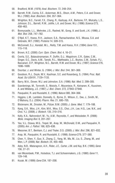
39
58. Bradford, M.M. (1976) Anal. Biochem. 72, 248–254.
59. Barrett, R.W., Cwirla, S.E., Ackerman, M.S., Olson, A.M., Peters, E.A. and Dower, W.J. (1992) Anal. Biochem. 204, 357–364.
60. Wrighton, N.C., Farrell, F.X., Chang, R., Kashyap, A.K., Barbone, F.P., Mulcahy, L.S., Johnson, D.L., Barrett, R.W., Jollife, L.K. and Dower, W.J. (1996) Science 273, 458–463.
61. Bonnycastle, L.L., Mehroke, J.S., Rashed, M., Gong, X. and Scott, J.K. (1996) J. Mol. Biol. 258, 747–762.
62. O'Neil, K.T., Hoess, R.H., Jackson, S.A., Ramachandran, N.S., Mousa, S.A. and DeGrado, W.F. (1992) Proteins 14, 509–515.
63. McConnell, S.J., Kendall, M.L., Reilly, T.M. and Hoess, R.H. (1994) Gene 151, 115–118.
64. Brown, K.C. (2000) Curr. Opin. Chem. Biol. 4, 16–21.
65. Cwirla, S.E., Balasubramanian, P., Duffin, D.J., Wagstrom, C.R., Gates, C.M., Singer, S.C., Davis, A.M., Tansik, R.L., Mattheakis, L.C., Boytos, C.M., Schatz, P.J., Baccanari, D.P., Wrighton, N.C., Barrett, R.W. and Dower, W.J. (1997) Science 276, 1696–1699.
66. Doorbar, J. and Winter, G. (1994) J. Mol. Biol. 244, 361–369.
67. Goodson, R.J., Doyle, M.V., Kaufman, S.E. and Rosenberg, S. (1994) Proc. Natl. Acad. Sci USA 91, 7129–7133.
68. Barry, M.A., Dower, W.J. and Johnston, S.A. (1996) Nat. Med. 2, 299–305.
69. Szardenings, M., Tornroth, S., Mutulis, F., Muceniece, R., Keinanen, K., Kuusinen, A. and Wikberg, J.E. (1997) J. Biol. Chem. 272, 27943–27948.
70. Pasqualini, R. and Ruoslahti, E. (1996) Nature 380, 364–366.
71. Higgins, L.M., Lambkin, Donnelly, G., Byrne, D., Wilson, C., Dee, J., Smith, M., O'Mahony, D.J. (2004) Pharm. Res. 21, 695–705.
72. Böckmann, M., Drosten, M., Pützer, B.M. (2005) J. Gene. Med. 7, 179–188.
73. Kang, S.K., Woo, J.H., Kim, M.K., Woo, S.S., Choi, J.H., Lee, H.G., Lee, N.K., and Choi, Y.J. (2008) J. Biotech. 135, 210–216.
74. Kelly, K.A., Nahrendorf, M., Yu, A.M., Reynolds, F., and Weissleder, R. (2006) Mole. Imaging Biol. 8, 201–207.
75. Yao, V.J., Ozawa, M.G., Trepel, M., Arap, W., McDonald, D.M., and Pasqualini, R. (2005) Am. J. Pathol. 166, 625–636.
76. Messmer, B.T., Benham, C.J. and Thaler, D.S. (2000) J. Mol. Biol. 296, 821–832.
77. Arap, W., Pasqualini, R. and Ruoslahti, E. (1998) Science 279, 377–380.
78. Chen, Y., Shen, Y., Guo, X., Zhang, C., Yang, W., Ma, M., Liu, S., Zhang, M., and Wen, L.P. (2006) Nat. Biotech. 24, 455–460.
79. Adey, N.B., Mataragnon, A.H., Rider, J.E., Carter, J.M. and Kay, B.K. (1995) Gene 156, 27–31.
80. van Wezenbeek, P.M., Hulsebos, T.J. and Schoenmakers, J.G. (1980) Gene 11, 129–148.
81. Kozak, M. (1999) Gene 234, 187–208.

40
PRODUCT NEB # SIZE
Ph.D.-7 Peptide Library Kit E8100S 10 panning exps.
Ph.D.-12 Peptide Library Kit E8110S 10 panning exps.
Ph.D.-C7C Peptide Library Kit E8120S 10 panning exps.
Ph.D. Peptide Display Cloning System E8101S 20 µg
KIT COMPONENTS SOLD SEPARATELY
Ph.D.-12 Peptide Library E8111L 50 panning exps.
Ordering Information

DNA CLONING
DNA AMPLIFICATION & PCR
EPIGENETICS
RNA ANALYSIS
LIBRARY PREP FOR NEXT GEN SEQUENCING
PROTEIN EXPRESSION & ANALYSIS
CELLULAR ANALYSIS
USANew England Biolabs, Inc.240 County RoadIpswich, MA 01938-2723Telephone: (978) 927-5054Toll Free: (USA Orders) 1-800-632-5227Toll Free: (USA Tech) 1-800-632-7799Fax: (978) 921-1350e-mail: [email protected] www.neb.com
CANADANew England Biolabs, Ltd.Telephone: (905) 665-4632Toll Free: 1-800-387-1095Fax: (905) 665-4635Fax Toll Free: 1-800-563-3789e-mail: [email protected]
CHINANew England Biolabs (Beijing), Ltd.Telephone: 010-82378265/82378266Fax: 010-82378262e-mail: [email protected]
FRANCENew England Biolabs FranceFree Call: 0800-100-632Free Fax: 0800-100-610e-mail: [email protected]
GERMANY & AUSTRIANew England Biolabs GmbHTelephone: +49/(0)69/305 23140Free Call: 0800/246 5227 (Germany) Free Call: 00800/246 52277 (Austria)Fax: +49/(0)69/305 23149Free Fax: 0800/246 5229 (Germany) e-mail: [email protected]
JAPANNew England Biolabs Japan, Inc.Telephone: +81 (0)3 5669 6191Fax: +81 (0)3 5669 6192e-mail: [email protected]
SINGAPORENew England Biolabs Pte. Ltd.Telephone: +65 638 59623Fax: +65 638 59617e-mail: [email protected]
UNITED KINGDOMNew England Biolabs (UK) Ltd.Telephone: (01462) 420616Call Free: 0800 318486Fax: (01462) 421057Fax Free: 0800 435682e-mail: [email protected]