manjula et al. - 2004 - pegylated hemoglobin role of surface configuration of peg for the modulation...
DESCRIPTION
hbocTRANSCRIPT
Vasoinactive PEGylated hemoglobin
1
PEGylated Hemoglobin: Role of surface configuration of PEG for the modulation of hemoglobin vasoactivity
Belur N. Manjula1, Amy G. Tsai2, Marcos Intaglietta2, Ching-Hsuan Tsai3, Chien Ho3, Ashok Malavalli4,5,
Kim D. Vandegriff4, Robert M. Winslow2,4, Paul K. Smith6, Krishnaveni Perumalsamy5, Nirmala Devi Kanika5, Joel M. Friedman1 and Seetharama A. Acharya1,5.
Departments of 1Physiology and Biophysics, and of 5Medicine, Albert Einstein College of Medicine, Bronx, NY; 2Department of Bioengineering, University of California-San Diego, La Jolla, CA;
3Department of Biological Sciences, Carnegie Mellon University, Pittsburgh, PA; 4Sangart Inc., San Diego, CA and 6BioAffinity Systems, Rockford, IL.
Running Title: Vasoinactive PEGylated hemoglobin Corresponding author: Belur N. Manjula, Ph.D. Department of Physiology and Biophysics, Albert Einstein College of Medicine, 1300 Morris Park Avenue, Bronx, NY. 10461. Phone: 718-430-2062 Fax: 718-430-8819 E-mail: manjula @aecom.yu.edu.
JBC Papers in Press. Published on June 12, 2004 as Manuscript M403468200
Copyright 2004 by The American Society for Biochemistry and Molecular Biology, Inc.
at Yale U
niversity on April 26, 2007
ww
w.jbc.org
Dow
nloaded from
Vasoinactive PEGylated hemoglobin
2
SUMMARY
PEGylation induced changes in the colligative properties of HbA have been implicated as potential
modulators of its vasoactivity. However, our recent studies have indicated that this modulation is not a direct
correlate of the colligative properties. With a view to delineate the role of surface charge and/or the pattern of
surface decoration with PEG for the vasoinactivation of hemoglobin, HbA has now been modified by thiolation
mediated PEGylation that does not alter its surface charge. Incubation of HbA with PEG5K maleimide in the
presence of iminothiolane generated a PEGylated Hb, namely (SP-PEG5K)6-Hb, that carried an average of ~six
PEG5K chains/Hb. The total PEG mass in (SP-PEG5K)6-Hb, its enhanced molecular volume, molecular
radius, viscosity, oncotic pressure and O2 affinity are comparable to that of another PEGylated Hb, namely (SP-
PEG20K)2-Hb, that carries two PEG20K chains/Hb, one on each of its two Cys-93(β). However, (SP-
PEG5K)6-Hb exhibited significantly reduced vasoconstriction mediated response relative to that of (SP-
PEG20K)2-Hb. These results demonstrate that surface decoration of Hb by conservative PEGylation with an
average of six PEG5K chains neutralizes its vasoactivity. More significantly, this study demonstrates that, in
addition to the colligative properties, a surface configuration accomplished by conjugation of multiple copies
of small PEG chains is more effective for decreasing the vasoactivity of Hb than a corresponding increase in
total PEG mass achieved through conjugation of a small number of large PEG chains. The simplicity in the
generation of this new vasoinactive PEG-Hb conjugate makes it a potential candidate for a Hb based oxygen
carrier.
at Yale U
niversity on April 26, 2007
ww
w.jbc.org
Dow
nloaded from

Vasoinactive PEGylated hemoglobin
3
INTRODUCTION
Vasoactivity of acellular Hb has been a major impediment in the development of Hb-based oxygen
carriers (1-6). This has been attributed, at least in part, to the NO scavenging effect of Hb (5-12). Reduction of
NO binding through site-directed mutagenesis is one of the approaches being evaluated for the reduction of the
vasoactivity of acellular Hb (10,13). The other approach involves enhancement of the molecular size of Hb to
reduce or possibly prevent the extravasation of Hb, based on the concept that the vasoactivity of acellular Hb is
due to its extravasation into the interstitial spaces and trapping of NO (14). However, the recent studies of
Winslow and co-workers (15-17) utilizing Hb preparations varying in molecular size namely, oligomerized Hb,
polymerized Hb and PEGylated Hb have suggested a lack of correlation between their NO binding activity and
pressor effect. These studies also suggested that PEGylation modulates the Hb induced hypertension and thus
could be an efficient approach to reduce the vasoactivity of Hb. The high viscosity and oncotic pressure of the
PEGylated Hb were implicated as the potential modulators of the vasoactivity of Hb.
In the PEGylated Hbs described in the above study, the PEG chains are conjugated to the surface
amino groups of the protein through isopeptidyl linkages (18). Thus, this PEGylation is accompanied by a loss
of the net positive charge of the protein (non-conservative PEGylation). In an attempt to delineate the role of
PEGylation induced changes in solution properties, surface charge and the pattern of surface decoration of
PEG on Hb for the modulation of its vasoactivity, we have been exploring new chemical approaches for the
PEGylation of Hb without altering its surface charge i.e., conservative PEGylation (19-21). We have recently
reported the preparation of three site-specifically PEGylated Hbs, namely (SP-PEG5K)2-Hb, (SP-PEG10K)2-
Hb, and (SP-PEG20K)2-Hb (19). Each of these PEGylated Hbs carry two copies of PEG chains/Hb,
conjugated at its two Cys-93(β) residues, but differ in the size of the PEG chains i.e., PEG5000, PEG10000
and PEG20000, respectively. The hydrodynamic volume, molecular radius, viscosity and oncotic pressure of
Hb increased with PEGylation and exhibited a correlation with the mass of the PEG conjugated (i.e., chain
length). However, the vasoactivity of the preparations was not a direct correlate of the PEG mass. Thus, the
neutralization of the vasoactivity was not a direct correlate of the colligative properties of the PEGylated Hb.
There appeared to be a threshold for the PEG chain length beyond which the ability to modulate the
at Yale U
niversity on April 26, 2007
ww
w.jbc.org
Dow
nloaded from
Vasoinactive PEGylated hemoglobin
4
vasoactivity was decreased, despite an increase in the colligative properties. Furthermore, the surface coverage
of Hb with the PEG chain was also not directly proportional to the length of the PEG chain and suggested a
potential relation between the surface coverage by the conjugated PEG and the vasoactivity.
In the present study, the maleimide chemistry based PEGylation protocol has been used to conjugate
multiple copies of PEG500 chains to Hb to accomplish a more uniform surface coverage of the protein without
altering its surface charge. Since there are only two reactive -SH groups in oxy Hb (the two Cys-93(β)
residues), we used the propensity of iminothiolane to react with protein amino groups to generate new thiols
(22, 23) to introduce additional PEG-maleimide reactive sites on Hb (Fig. 1). Thus, PEGylation of oxy Hb in
the presence of iminothiolane is targeted to the two intrinsic thiols of Cys-93(β) and the extrinsic thiols
generated on the ε-amino groups by the iminothiolane. Employing this protocol and using Mal-Phe-PEG5000
as the PEGylating reagent, a new PEGylated Hb carrying an average of ~six PEG5000 chains on the Hb,
namely (SP-PEG5K)6-Hb, has been generated. This new PEGylated Hb exhibits a size enhancement,
colligative properties and O2 affinity comparable to that of (SP-PEG20K)2-Hb, but has a significantly reduced
vasoactivity relative to the latter. These results establish that the PEGylation induced colligative properties,
without an accompanying loss of surface positive charge, can neutralize the vasoactivity of Hb and demonstrate
that the PEGylation mediated modulation of the vasoactivity of Hb is a function of the colligative properties of
the PEGylated Hb in conjunction with the configuration of PEG on the surface of Hb.
EXPERIMENTAL PROCEDURES
Materials: HbA was purified from the human erythrocyte lysate by DE-52 chromatography (24). 2-
Iminothiolane was a product of BioAffinity Systems, Rockford, IL. 4,4'-dithiopyridine (4-PDS) was purchased
from Aldrich Chemical Co. Polymerized bovine Hb solution (Oxyglobin, average MW 180,000) was a product
of Biopure Corp., Boston, MA. Synthesis of the mono functional maleimido-phenyl (Mal-Phe) derivatives of
PEG5000, PEG10000 and PEG20000, and preparation of the site-specifically PEGylated Hbs, namely (SP-
PEG5K)2-Hb, (SP-PEG10K)2-Hb, and (SP-PEG20K)2-Hb were carried out as described earlier (19).
Iminothiolane dependent thiolation mediated maleimide chemistry based PEGylation of HbA: This was
carried out either as a one-step or as a two-step reaction. In the one step reaction, HbA (0.5 mM tetramer) in
at Yale U
niversity on April 26, 2007
ww
w.jbc.org
Dow
nloaded from
Vasoinactive PEGylated hemoglobin
5
PBS, pH 7.4 was incubated with Mal-Phe-PEG5000 in the presence of iminothiolane, at the concentrations
indicated in the text, either at 4oC or at room temperature. Generally, 4 to 6 h incubation was needed for
completion of the reaction at room temperature. Reactions at 4oC were routinely carried out for ~16 h. The
reaction was generally carried out at a HbA concentration of 0.5 mM, but it could also be carried out at HbA
concentrations of either 0.25 mM or 1 mM. In the two-step reaction, HbA (1.0 mM) in PBS pH 7.4 was first
incubated with 10 mM iminothiolane overnight at 4oC. The reaction mixture was then diluted with an equal
volume of 20 mM Mal-Phe-PEG5000 in PBS, pH 7.4, and the PEGylation was carried out for 6 h of 4oC. In
both the one-step and the two-step reactions, after the desired incubation period, the reaction mixtures were
dialyzed extensively against PBS, pH 7.4 prior to analysis.
Identification of the sites of PEGylation: This was determined by tryptic peptide map analysis, as previously
described (25). Briefly, the globin chains of Hb prepared by acid-acetone precipitation were dissolved in 100
mM ammonium bicarbonate to a final concentration of 1 mg/ml, and digested for 3 h at 37oC with TPCK-
trypsin (Sigma Chemical Co., St. Louis, MO) at a enzyme to substrate ratio of 1:100 (w/w). The resulting
tryptic peptides were analyzed by RPHPLC on a Vydac C18 column (10× 250 mm) using a linear gradient of
5-50% acetonitrile containing 0.1% TFA in 160 min, followed by a linear gradient of 50-70% acetonitrile
containing 0.1% TFA in 20 min at a flow rate of 2 ml/min. The elution of the peptides was monitored at 210
nm. The recovery of peptide α-T4 (a peptide derived by arginyl peptide bond cleavage), was used as an
internal standard, and the ratio of the peak area of each peptide to the α-T4 peptide was used to elucidate
differences between the HbA control and the PEGylated Hb.
Vasoactivity of PEGylated Hbs: Analysis of the vasoactivity and microvascular hemodynamics of the
PEGylated Hb were carried out in a hamster skin fold window microcirculation model, essentially according to
the procedures previously described (26-30).
Analytical Methods: The thiolation of Hb by 2-iminothiolane was followed by estimating the number of thiol
groups formed as a function of time using 4,4'-dithiopyridine (4-PDS), as described by Ampulski et al (31).
The number of PEG chains conjugated per Hb tetramer was determined by proton NMR spectroscopy, as
described by Jackson et al (32). Analyses of the size enhancement of Hb as a function of the various thiolation
at Yale U
niversity on April 26, 2007
ww
w.jbc.org
Dow
nloaded from

Vasoinactive PEGylated hemoglobin
6
mediated PEG-maleimide based PEGylation conditions, determination of the molecular radius of the
PEGylated Hbs, their O2 equilibrium curves, and measurements of their viscosity and colloidal osmotic
pressure were carried out as previously described (19). Structural characterization of the PEGylated Hbs by
proton NMR spectroscopy was carried out as previously described (33).
RESULTS
Iminothiolane dependent thiolation mediated maleimide chemistry based PEGylation of Hb:
A. One step protocol: In situ thiolation mediated PEGylation of Hb: In this protocol, thiolation of
HbA with iminothiolane is carried out in the presence of the PEG-maleimide. 2-Iminothiolane, by itself, does
not carry any free thiol group and the thiol group is generated only after it reacts with the ε-amino groups of the
protein. Thus, HbA can be incubated with iminothiolane in the presence of the PEG-maleimide without the
concern of the thiolating reagent itself consuming the PEGylating reagent. Thus, as new thiol groups are
generated in situ they are trapped by the PEG maleimide. Hence, HbA with multiple thiol groups is not
accumulated as an intermediate in the reaction mixture.
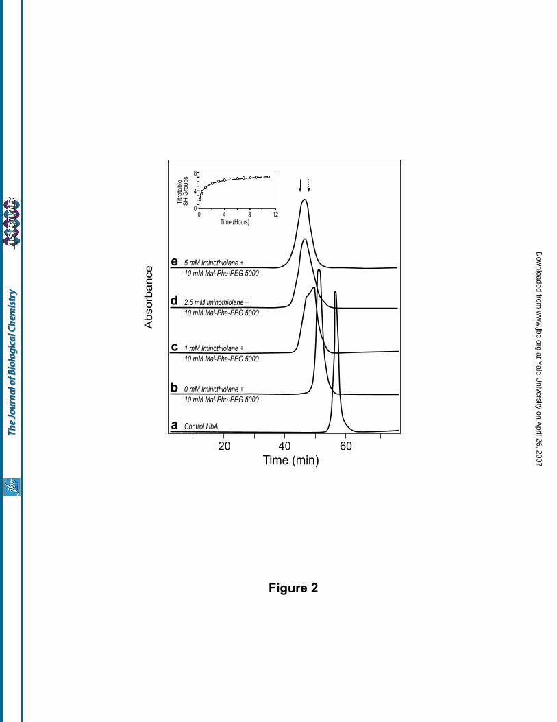
The size exclusion chromatographic pattern of HbA (0.5 mM) in PBS, pH 7.4, incubated with a 20
fold molar excess of Mal-Phe-PEG5000 (10 mM) for 4.5 h at room temperature, in the absence and in the
presence of varying concentrations of iminothiolane is shown in Fig 2. The retention time of HbA reacted with
Mal-Phe-PEG5000 in the absence of iminothiolane (curve b) corresponds to that of (SP-PEG5K)2-Hb, a
product that has been previously identified as HbA conjugated with two PEG5000 chains, one each at its two
Cys-93(β) (19). On inclusion of iminothiolane in the reaction mixture of HbA and Mal-Phe-PEG5000, the
modified HbA eluted earlier than the (SP-PEG5K)2-Hb from the size exclusion chromatographic column (Fig.
2, curves c, d and e). Furthermore, the retention time of the PEGylated Hb exhibited an inverse relation with
the iminothiolane concentration, suggesting that an increased level of thiolation of HbA is responsible for the
increased apparent molecular size of Hb. The PEGylated Hb generated in the presence of 2.5 mM
iminothiolane eluted at a position close to that of (SP-PEG10K)2-Hb (Fig. 2, curve d), and that generated in the
presence of 5 mM iminothiolane eluted in between the elution positions of (SP-PEG10K)2-Hb and (SP-
PEG20K)2-Hb (Fig. 2, curve e).
at Yale U
niversity on April 26, 2007
ww
w.jbc.org
Dow
nloaded from
Vasoinactive PEGylated hemoglobin
7
The retention time did not change significantly on increasing the iminothiolane concentration further to
7.5 mM and 10 mM; however, the elution pattern of the PEGylated Hb peak became slightly broader, and
revealed a small shoulder on the ascending side of the peak (data not shown). These results suggested that
under the conditions described above, optimal PEGylation of HbA is achieved in the presence of a 10 fold
molar excess of iminothiolane.
The number of –SH groups introduced on to HbA (0.5 mM) by a 10 fold molar excess of
iminothiolane (5 mM), was determined independently as a function of time by titration with 4-PDS, and the
results are shown in Fig. 2, inset. The two thiol groups at zero time represent the two reactive thiols of Cys-
93(β). As can be seen from the figure, the thiolation of Hb by iminothiolane exhibits an initial fast phase
wherein about 4 new thiols are introduced in the first two hours and a subsequent slow phase wherein only
approximately one additional thiol group is introduced. After 11 h of incubation, the thiolated HbA carried a
total of ~7 reactive -SH groups per tetramer. Thus, a quantitative PEGylation of such a thiolated HbA will
generate a molecule carrying an average of seven PEG5000 chains. A 4-PDS titration of the PEGylated HbA
generated by reaction of HbA (0.5 mM) with 10 mM Mal-Phe-PEG5000 (20 fold excess over Hb) in the
presence of 5 mM iminothiolane (10 fold excess over Hb) revealed the presence of about 0.5 moles of reactive
thiols per tetramer. In conjunction with the results of the kinetics of thiolation, this result suggests that an
average of ~ 6.5 copies of PEG5000 chains are introduced on to HbA in generating this PEGylated Hb.
Titration with 4-PDS also revealed that increasing the concentration of iminothiolane from 10 fold
molar excess to 30 fold molar excess nearly doubles the total number of thiols on the HbA. However, the size
enhancement of Hb on PEGylation in the presence of this iminothiolane concentration was only marginal (data
not shown). These results are suggestive of a crowding effect induced by the ~six PEG5000 chains
incorporated on the molecular surface of Hb, and hence resistance to further PEGylation.
The rate of thiolation of HbA was not significantly influenced when the temperature was lowered from
room temperature to 4oC. However, the rate of PEGylation appeared to slow down. A 9 h incubation ensured
the completion of the reaction; however, routinely the reaction was carried out overnight. The elution
characteristics of the PEGylated HbA obtained by reaction at 4oC were quite comparable to that obtained at
at Yale U
niversity on April 26, 2007
ww
w.jbc.org
Dow
nloaded from
Vasoinactive PEGylated hemoglobin
8
room temperature.
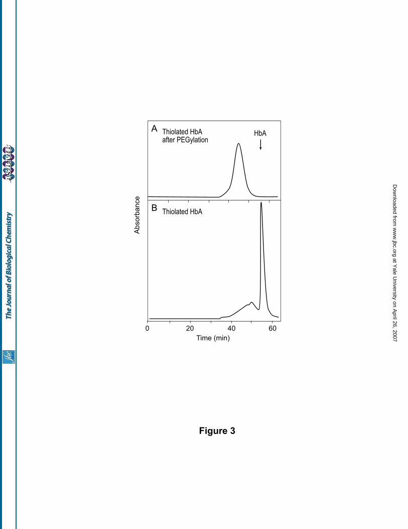
B. Two-step Reaction: Thiolation of Hb followed by PEGylation: In the two-step protocol, HbA
was first reacted with iminothiolane to achieve the desired level of thiolation, and the resulting thiolated HbA
was subjected to PEGylation. Thus, unlike the one-step protocol described above, thiolated HbA is generated
as a product of the first step in this protocol. Size exclusion chromatographic profile of the PEGylated Hb
prepared by the two-step protocol, as described under ‘Methods’, is shown in Fig. 3A. As can be seen, this
PEGylated Hb elutes as a slightly broader peak compared to the product generated by the in situ thiolation
mediated PEGylation protocol. Besides, analysis of the thiolated HbA intermediate (i.e., the product of the first
step, obtained prior to the addition of the PEG maleimide), indicated that about 10 to 15% of the protein eluted
at the position corresponding to the octameric and dodecameric forms of HbA, suggesting air oxidation of the
new thiols introduced on to Hb (Fig. 3B). However, the generation of the oligomerized products was
completely inhibited when the thiolation was carried out in the presence of 20 mM N-ethylmaleimide. The
susceptibility of the thiols generated by reaction with iminothiolane for side reactions has also been observed in
other studies on the generation of bioconjugates (34,35). Hence, the one step in situ thiolation mediated
protocol was selected for the preparation of the PEGylated HbA for all subsequent studies.
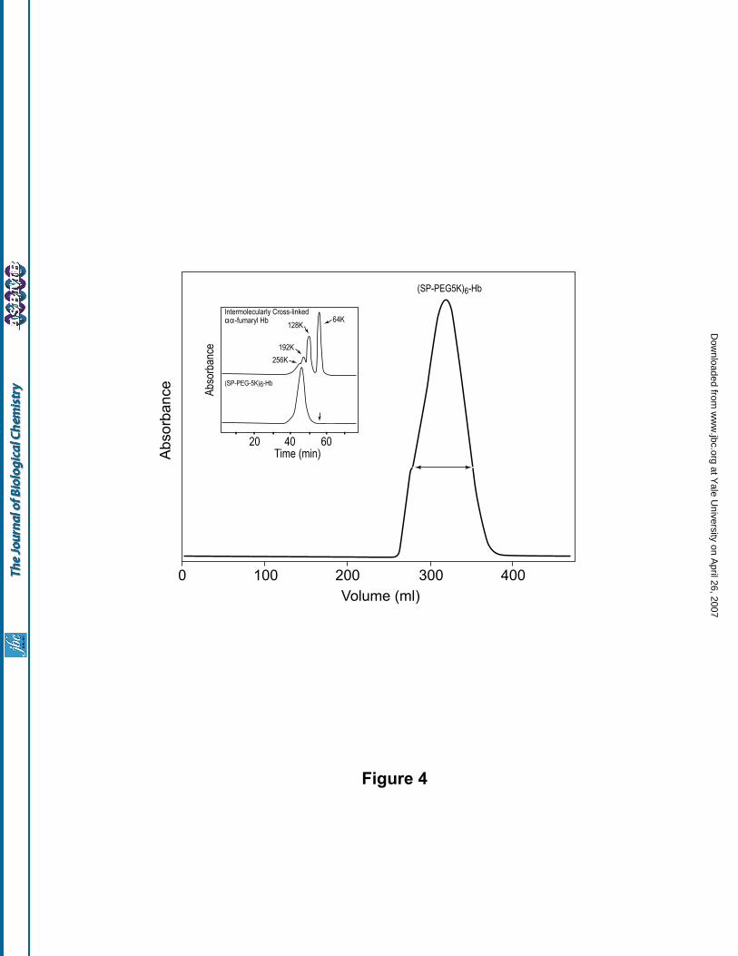
Purification of PEGylated-Hb: HbA (0.5 mM in PBS, pH 7.4) PEGylated using a 10 fold molar excess of
iminothiolane in the presence of a 20 fold molar excess of Mal-Phe-PEG5000 (2 fold molar excess over
iminothiolane) overnight at 4oC was subjected to size exclusion chromatography on a Prep grade Superose 12
column. A typical chromatographic profile of a preparation of PEGylated Hb is shown in Fig 4. As can be
seen, but for a small shoulder on the ascending side, the PEGylated Hb eluted as a single, fairly symmetrical
peak. The PEGylated Hb peak was pooled as indicated and the protein concentrated to about 6 g/dl. The purity
of the PEGylated-Hb thus isolated was further confirmed by analytical SEC analysis (Fig. 4, inset, lower
panel). The PEG content of the purified PEGylated-Hb as determined by NMR analysis (32, 36), and by –SH
titration is presented in Table I, along with those of two site specifically PEGylated Hbs, namely (SP-
PEG5K)2-Hb and (SP-PEG10K)2-Hb (i.e., Hb PEGylated at Cys-93(β). The values determined for (SP-
PEG5K)2-Hb and (SP-PEG10K)2-Hb are in agreement with the expected values of two copies each of PEG5K
at Yale U
niversity on April 26, 2007
ww
w.jbc.org
Dow
nloaded from
Vasoinactive PEGylated hemoglobin
9
and PEG10K, respectively. The purified PEGylated-Hb was found to carry an average of 6.7 copies of PEG-
5000 chains per tetramer by NMR analysis. This product will hereafter be referred to as (SP-PEG5K)6-Hb.
The apparent molecular size of (SP-PEG5K)6-Hb was estimated by comparison with the SEC profile of
oligomeric forms of Hb (generated by inter-tetrameric crosslinking of αα -fumaryl Hb using Bis Mal-Phe-
PEG600) (Fig. 4, inset, upper panel). As can be seen, the hydrodynamic volume of (SP-PEG5K)6-Hb
corresponds to that of a Hb oligomer of a molecular mass of about 256,000 daltons (i.e., a tetrameric form of
Hb). No detectable autooxidation of the PEGylated Hb to generate met-Hb type of products was observed
either during the thiolation mediated maleimide chemistry based PEGylation reaction or during the subsequent
purification steps. The PEGylated-Hb thus isolated could be stored at -80oC without any significant
autooxidation for periods of at least up to one year.
The sites of conjugation of PEG-chains in the (SP-PEG5K)6-Hb were determined by a comparison of
the tryptic peptide map of its globin chains with that of the unmodified HbA. The results are presented in Table
II. The data revealed complete modification of Cys-93(β), and predominant modification of four lysine
residues, namely Lys-60(α), Lys-120(β), Lys-11(α), and Lys-8(β); only minor modification of the α-amino
groups was observed. Together, this accounted for an average of 6.2 residues modified per Hb, a value that is
close to the number of PEG chains per Hb estimated by NMR analysis and by thiol titration. Thus, two of the
PEG chains in (SP-PEG5K)6-Hb are on the two Cys-93(β)s and the remainder are distributed for the most part
on a limited number of lysines and to a lesser degree on the α-amino groups. The tryptic peptide map of the
(SP-PEG5K)6-Hb globins was reproducible from batch to batch of the preparation, indicating that the
PEGylation of the amino groups of Hb by the present thiolation mediated maleimide chemistry based protocol
is not random, but exhibits a high degree of site selectivity.
Molecular, colligative and functional characterization of (SP-PEG5K)6-Hb
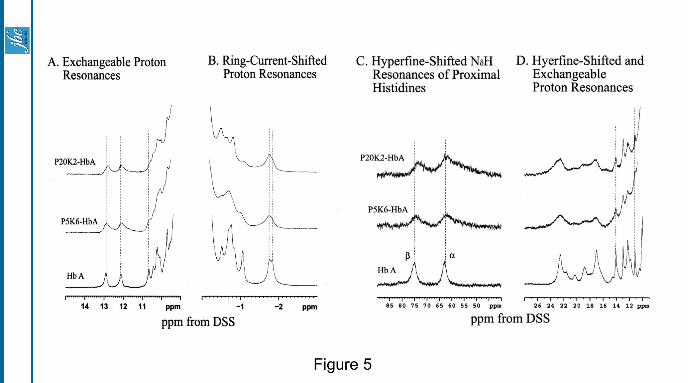
1H-NMR spectroscopy of (SP-PEG5K)6 -Hb: A comparison of the proton NMR spectra of (SP-PEG20K)2-
Hb and (SP-PEG5K)6-Hb with that of HbA in 0.1 M phosphate buffer at pH 7.0 and 29oC in both the
carbonmonoxy and deoxy forms is presented in Fig. 5A. These two samples carry a comparable amount of
total PEG-mass/Hb [40,000 Da in (SP-PEG20K)2-Hb vs 30,000 Da in (SP-PEG5K)6-Hb]. With the exception
at Yale U
niversity on April 26, 2007
ww
w.jbc.org
Dow
nloaded from

Vasoinactive PEGylated hemoglobin
10
of broader resonances observed with the PEGylated samples due to an increase in the molecular size as a result
of PEGylation, there is no significant difference in the chemical shift over the spectral region of 10 to 14 ppm
indicating no alterations in the α1β1 interface of Hb as a result of PEGylation either only at Cys-93(β) with
PEG-20000 or at Cys-93(β) and at least four of its ε-amino groups with PEG-5000. Fig 5B compares the ring-
current-shifted proton resonances of the two PEGylated Hbs with that of HbA in the carbonmonoxy form.
There are some alterations in the ring-current shifted proton resonances reflecting some perturbation in the
micro-environment of the heme of the PEGylated Hb-samples. Fig 5C shows the hyperfine shifted NδH 1H-
resonances of proximal histidine residues of the α- and the β-chains of PEGylated Hbs in the deoxy form. The
chemical shift at -75 ppm assigned to NδH of the proximal histidine of the β-chain is shifted upfield by -2 to -3
ppm reflecting the perturbation of the β-heme environment in the PEGylated samples. This upfield shift is
somewhat more pronounced in (SP-PEG20K)2-HbA than in (SP-PEG5K)6-Hb. Fig. 5D compares the hyperfine
shifted and exchangeable proton resonances of the two PEGylated samples of HbA with that of HbA in the
deoxy form. The hyperfine-shifted resonances are broader than that of HbA. Besides, there are some changes in
the resonances in the spectral region from 16 to 24 ppm, reflecting changes in the microenvironment of the β-
heme of Hb as a result of PEGylation of the molecule. The resonance at 14 ppm, assigned to an important H-
bond between α-Tyr(42) and β-Asp(99) in the α1β2 subunit interface (37) is unchanged in the PEGylated
samples. Thus, there are no significant changes in the α1β2 subunit interface of the PEGylated Hb.
Functional properties of (SP-PEG5K)6-Hb: The O2 affinity of (SP-PEG5K)6-Hb in 50 mM Bis-Tris/50 mM
Tris acetate buffer, pH 7.4 and 37oC and its modulation in the presence of allosteric effectors is shown in Table
III. The P50 of Hb is lowered (i.e., the O2 affinity is increased) on PEGylation, from the control value of 8.0
mmHg to 6.5 mmHg. The presence of a five fold molar excess of DPG, an effector that lowers the O2 affinity
of HbA by binding at the ββ-cleft, had no significant influence on the O2 affinity of (SP-PEG5K)6-Hb. On the
other hand, the presence 1 M sodium chloride lowered the O2 affinity of the PEGylated Hb, but only slightly
compared to unmodified HbA. L35, an allosteric effector that reduces the O2 affinity of Hb by binding at the
αα -end of the molecule, also reduced the O2 affinity of (SP-PEG5K)6-Hb. However, as with NaCl, this effect
at Yale U
niversity on April 26, 2007
ww
w.jbc.org
Dow
nloaded from
Vasoinactive PEGylated hemoglobin
11
was markedly reduced compared to that observed with unmodified HbA. Thus, the PEGylation of HbA
significantly reduces the propensity of HbA to respond to the presence of the allosteric effectors DPG, chloride
and L35.
In addition to the measurements in the BisTris-Tris buffer, pH 7.4, the O2 affinity of (SP-PEG5K)6-Hb
was also determined in PBS, pH 7.4. Even in PBS, (SP-PEG5K)6-Hb exhibited a higher O2 affinity than
control HbA (a P50 of 8.5 mmHg vs 15.3 mmHg for HbA). Furthermore, the increase in O2 affinity observed in
PBS was more significant than that observed in the BisTris-Tris buffer.
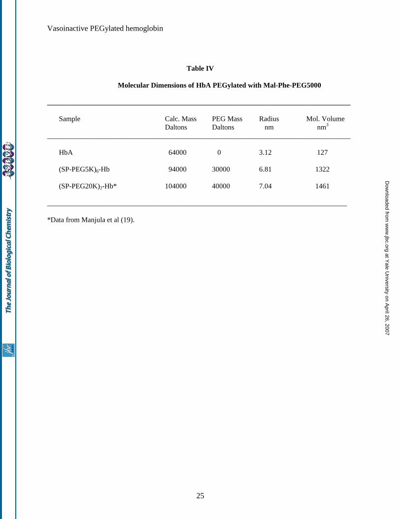
Molecular radius of (SP-PEG5K)6-Hb: The molecular radius of (SP-PEG5K)6-Hb, as determined by
dynamic light scattering measurements, along with that of (SP-PEG20K)2-Hb is presented in Table IV. As
can be seen, the molecular radius of (SP-PEG5K)6-Hb is slightly smaller than the radius of (SP-PEG20K)2-Hb.
It may also be seen from the data presented in Table IV, that the molecular volume of HbA is increased >10
fold when it is surface decorated either with about six copies of PEG5000 chains or with 2 copies of
PEG20000.
Colligative properties of (SP-PEG5K)6-Hb: A comparison of the viscosity of (SP-PEG5K)2-Hb and (SP-
PEG5K)6-Hb as a function of Hb concentration is presented in Fig. 6A. (SP-PEG5K)2-Hb showed only a small
increase in viscosity with the increase in protein concentration, the viscosity being directly proportional to the
concentration of the protein. On the other hand, even though the viscosity of (SP-PEG5K)6-Hb is comparable
to that of (SP-PEG5K)2-Hb in dilute solutions, the viscosity of the former increased exponentially with the
concentration of the protein.
The colloidal osmotic pressure of the PEGylated Hbs as a function of the protein concentration is
shown in Fig 6B. As can be seen, the oncotic pressure of the PEGylated Hbs increased as a function the
protein concentration. Like the viscosity, the increase in COP is small with (SP-PEG5K)2-Hb, and appears to
increase linearly with the protein concentration. On the other hand, the oncotic pressure of (SP-PEG5K)6-Hb
increased exponentially with the increase in protein concentration. These results suggest that the oncotic
pressure of PEGylated Hb is a direct correlate of its viscosity. Similar results were observed with PEGylated
Hbs carrying 2 PEG chains/tetramer (19).
at Yale U
niversity on April 26, 2007
ww
w.jbc.org
Dow
nloaded from
Vasoinactive PEGylated hemoglobin
12
Vasoactivity of (SP-PEG5K)6-Hb:
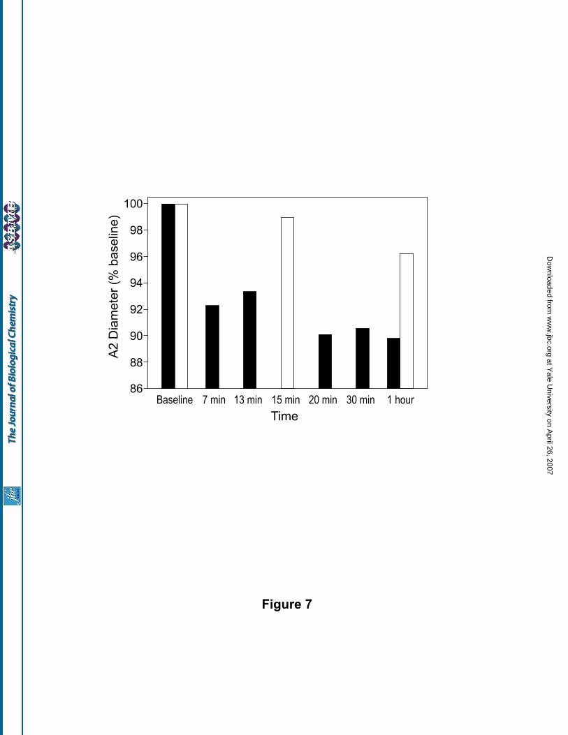
(i) Comparison of the systemic and microvascular responses to PEGylated-Hb: Changes in the A2
arteriolar diameter (expressed as percent of baseline values) after various periods of 10% top load infusions of
(SP-PEG5K)2-Hb and (SP-PEG5K)6-Hb in the hamster skin fold-window model are shown in Fig. 7. As can be
seen, (SP-PEG5K)6-Hb conserves the arteriolar diameter more closer to the starting value than that observed
with (SP-PEG5K)2-Hb. These results demonstrate that the PEGylation of acellular Hb, to a level of ~six copies
of PEG5000 chains reduces its intrinsic vasoactivity significantly relative to that observed with two PEG5000
chains. This is consistent with the notion that the increased PEG mass mediated colligative properties of the
PEGylated Hb play a role for the modulation of its vasoactivity.
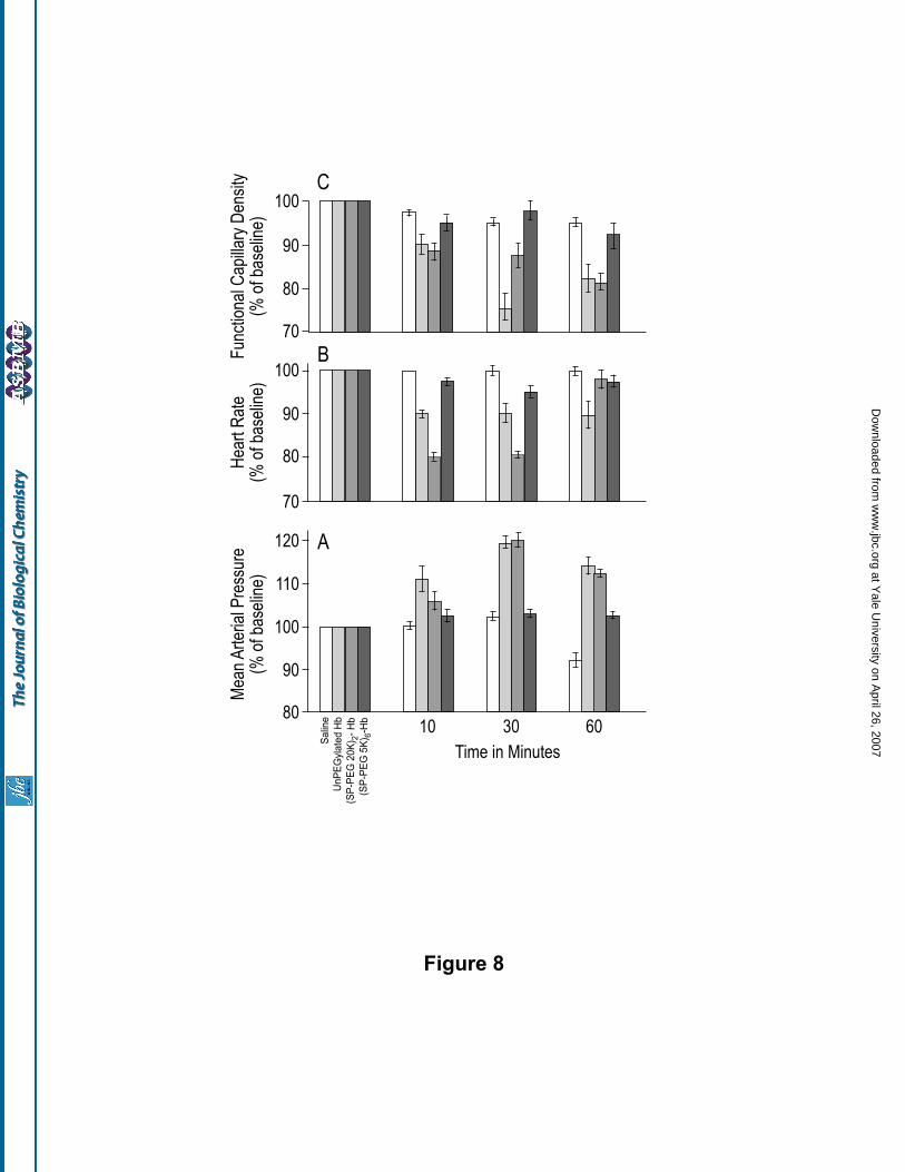
The relative merits of increasing the PEG mass to an equivalent level by conjugation with ~six copies
of PEG5000 chains vs two copies of PEG20000 chains per Hb tetramer for the microcirculation were also
evaluated in the hamster skin fold window model by measuring the acute systemic and microvascular response
to a 10% hypervolemic infusion of the two PEGylated Hbs. The results are presented in Fig. 8. Saline was used
as the control; the results obtained with unPEGylated Hb, (SP-PEG20K)2-Hb and (SP-PEG5K)6-Hb are
compared. MAP increased and the heart rate decreased after infusion of (SP-PEG20K)2-Hb. The increase in
MAP was more close to unmodified Hb but the decrease in heart rate was significantly lower than that
observed with unPEGylated Hb. The FCD was also decreased, much more than observed with saline, and
remained closer to that of unPEGylated Hb. In this case, large arterioles tended to vasoconstrict, whereas the
venules were relatively unchanged (data not shown). Interestingly, MAP and heart rate were not statistically
changed after infusion with (SP-PEG5K)6-Hb and remained close to the baseline values. Although there was a
small decrease in FCD with (SP-PEG5K)6-Hb, the values are much closer to that of the saline control than to
that of unPEGylated Hb. Some arteriolar and venular constrictions were observed, but significantly less than
that observed with (SP-PEG20K)2-Hb. These results demonstrate that the configuration of surface decoration
of Hb with PEG has a significant influence on the pressor effect and the vasoconstrictive activity of acellular
Hb. Surface decoration of HbA with six copies of PEG5000 significantly reduces its acute systemic response,
whereas decoration with two copies of PEG20000 (conjugation of comparable PEG mass) is not effective for
at Yale U
niversity on April 26, 2007
ww
w.jbc.org
Dow
nloaded from

Vasoinactive PEGylated hemoglobin
13
achieving the same.
(ii) Effect of extreme hemodilution with (SP-PEG5K)6-Hb: Previous studies by Tsai et al (30) have
demonstrated that functional capillary density is an accurate predictor of survival during acute blood loss. To
determine if the (SP-PEG5K)6-Hb is effective in the treatment of hemorrhagic shock i.e., to evaluate the ability
of (SP-PEG5K)6-Hb to keep the capillaries open to facilitate the circulation of the O2-carrier under conditions
of severe blood loss, the (SP-PEG5K)6-Hb solution was analyzed in an extreme hemodilution protocol. To
achieve a scenario closer to actual practice, the hemodilution was accomplished in three steps. Each volume
exchange was a percent of the animal's total blood volume, estimated at 7% of the bodyweight. The animal's
blood was first hemodiluted in two steps with a plasma expander to a Hb concentration of 7 g/dl, a
recommended red cell transfusion threshold. The third step of the hemodilution was with the test solution to
achieve a final systemic hematocrit equivalent to 25% of baseline.
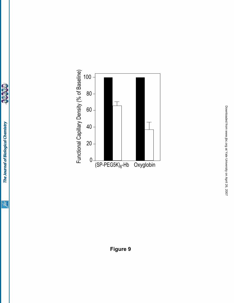
The results obtained with (SP-PEG5K)6-Hb are presented in Fig. 9, and are compared with that
obtained with the Biopure product Oxyglobin (glutaraldehyde polymerized bovine Hb), a commercially
available hemoglobin based oxygen carrier. The first two steps of the hemodilution protocol were performed
with a 6% solution of Dextran-70 to reduce the hematocrit to 40% of baseline (60% red blood cell exchange)
and the third step was performed with the test solutions - 5 g Hb/dl of (SP-PEG5K)6-Hb and 14 g Hb/dl of
Oxyglobin. As can be seen, (SP-PEG5K)6-Hb maintained the functional capillary density at 65.5% + 5% of the
base-line whereas the Oxyglobin maintained the functional capillary density only at 37 + 9% of the baseline.
DISCUSSION
A simple and versatile procedure for enhancing the hydrodynamic volume of HbA by conjugation of
multiple PEG chains without altering the surface charge of the protein (i.e., conservative PEGylation) and
generation of a nonhypertensive PEGylated Hb, namely (SP-PEG5K)6-Hb, by this protocol is described in the
present study. Although the present studies were carried out using maleimide PEG with an aryl linker between
the PEG chain and the maleimide moiety, other maleimide PEG reagents that carry an alkyl or an alkylamide
linker between the PEG and the maleimide are equally efficient in the maleimide chemistry based PEGylation
reactions of Hb (20, 38-40). In view of its proximity to the protein surface, the succinimidophenyl moiety of
at Yale U
niversity on April 26, 2007
ww
w.jbc.org
Dow
nloaded from

Vasoinactive PEGylated hemoglobin
14
the PEG-Hb conjugate is anticipated to be shielded from the macro-environment by the hydration shell of the
PEG chains around the protein and hence unlikely to be immunogenic. However, if the succinimidophenyl
linkage should turn out to be immunogenic, PEG reagents with alkyl linkages could be used to generate the
desired material.
(SP-PEG5K)6-Hb, has many of the attributes that have been advanced as needed for minimizing the
vasoactivity of acellular Hb (15, 41): (i) Increased O2 affinity to limit the O2 off-loading by acellular Hb in
arterioles, thus minimizing the potential for vasoconstriction through autoregulatory mechanisms, (ii) Retention
of the cooperative binding to insure off-loading of O2 in the capillary beds, (iii) An enhanced molecular size
(hydrodynamic volume) to reduce extravasation, (iv) an increase in the viscosity of Hb solution both to create
appropriate shear stress on the arteriolar walls to maintain vascular tone and to lower the diffusion constants for
oxy Hb to limit the O2 off-loading to vessel walls, (v) A colloidal osmotic pressure greater than that of the
conventionally modified Hbs to increase the effectiveness of the blood substitute as a plasma expander.
Studies with (SP-PEG5K)6-Hb in hamsters, at 10% top load infusion, suggest that conjugation of an
average of six PEG5000 chains on to HbA, without alteration of its surface charge, significantly reduces the
Hb induced vasoactivity. The Enzon PEG5K-bovine Hb that was observed previously to be nonhypertensive
(15-17) was generated by the active ester chemistry (18) which results in the loss of the net positive charge of
the protein, and has been suggested to carry an average of ten PEG-5K chains/Hb. The results of the present
study suggest that the conservation of the net surface charge of the parent Hb is not crucial to generate a non-
hypertensive Hb and that the nonhypertensive property can be endowed to Hb with less than ten PEG5K
chains. In addition, the absence of a deoxygenation step in the present protocol, unlike that used for the
generation of the Enzon PEG-bovine Hb, makes the scale-up of this PEGylation process simple and cost
effective. Furthermore, the results of the present study also demonstrate that under conditions of extreme
hemodilution, (SP-PEG5K)6-Hb, a size enhanced PEGylated Hb, is far better than the polymerized bovine Hb
(Biopure product Oxyglobin) in maintaining the functional capillary density, a property shown to be an
accurate predictor of survival during acute blood loss (30).
More strikingly, the results of the present study suggest an important role for the surface configuration
at Yale U
niversity on April 26, 2007
ww
w.jbc.org
Dow
nloaded from
Vasoinactive PEGylated hemoglobin
15
of the PEG chains on the Hb molecule in neutralizing its vasoactivity. Although the molecular and colligative
properties, and the O2 affinity of (SP-PEG5K)6-Hb and (SP-PEG20K)2-Hb are comparable, the two PEG-Hb
conjugates differ significantly in their vasoactive properties. Apparently, modulation of the vasoactivity of Hb
is not simply a direct translation of its PEGylation induced colligative properties. Thus, in conjunction with
enhanced molecular size and colligative properties, and high O2 affinity, the architecture/surface configuration
of the PEG on the Hb molecule, i.e., the number and size of the PEG-chains, and possibly the site of
PEGylation appears to play a key role in the modulation of the vasoactivity of the Hb molecule. A plausible
explanation for the difference in the vasoactive properties of (SP-PEG5K)6-Hb and (SP-PEG20K)2-Hb could
be that a better shielding of the molecular surface of Hb is afforded by multiple copies of PEG5000 chains on
Hb relative to that afforded by two copies of PEG20000, thus camouflaging the acellular Hb from interactions
with the vasculature. This raises an important question as to whether the location of the PEG-5000 chains on
the molecular surface of Hb, along with its number and size, plays any role in achieving the shielding of the
molecular surface of Hb. Studies with site-specifically PEGylated Hbs with well defined number of copies of
PEG chains of varying size are needed to gain further insights into this molecular aspect of the PEGylation
mediated modulation of Hb induced vasoactivity. The lysines identified as the PEGylated sites in the present
study could serve as potential target sites to engineer cysteine residues by site directed mutagenesis to
incorporate the desired number of PEG chains on to Hb through maleimide chemistry, which in turn would
enable the generation of well-defined, site-specifically PEGylated Hbs that are much needed for such studies.
In conclusion, it is apparent from the results of the present study that, in addition to the increase in
colligative properties, a surface coverage of Hb accomplished by conjugation of multiple copies of small PEG
chains vs conjugation of two long PEG chains to obtain comparable PEG mass is desirable due to its potential
for reducing the vasoactivity of the molecule. In contrast, in the PEGylation of other therapeutic proteins, an
increase in the PEG chain length is preferred over an increase in PEG mass by increasing the number of small
PEG chains to achieve longer in vivo half life, decreased clearance and also to retain potency without the
possible loss of the bioactivity of the molecule, for example - masking of receptor binding activity due to
substitution at multiple sites (42-45). Thus it appears that the selection of a PEGylation strategy for a particular
at Yale U
niversity on April 26, 2007
ww
w.jbc.org
Dow
nloaded from
Vasoinactive PEGylated hemoglobin
16
protein will be dependent on the specific application under consideration.
Acknowledgements: This research was supported by a grant-in-aid from the American Heart
Association Heritage Affiliate, the National Institutes of Health grants HL58247, HL71064 and USPHS NIH
Bioengineering Partnership grant 1R24 HL 64395, and the US Army grant PR023085.
REFERENCES
1. Amberson, W., Jennigs, J., and Rhodes, C. (1949) J. Appl. Physiol. 1, 469-489
2. Savitsky, J., Doczi, J., and Black, J. (1978) Clin. Pharmacol. Therap. 23, 73-80
3. Sloan, E. P., Koenigsberg, M., and Gens, D. (1999) J. Amer. Med. Assoc. 282,1857-1864
4. Saxena, R., Wijnhoud, A.D., and Carton, H. (1999) Stroke 30, 993-996
5. Hess, J. R., Macdonald, V. W., and Brinkley, W. W. (1993) J. Appl. Physiol. 74, 1769-1778
6. Thomson, A.., McGarry, A. E., Valeri, C. R., and Lieberthal, W. (1994) J. Appl. Physiol. 77, 2348-2354
7. Muldoon, S. M., Ledvina, M. A., Hart, J. L., and Macdonald, V. W. (1996) J. Lab. Clin. Med. 128, 579-
584
8. Motterlini, R., Vandegriff, K. D., and. Winslow, R. M. (1996) Transfusion Medicine Rev. 10, 77-84
9. Doherty, D. H., Doyle, M. .P., and Curry, S. R. (1998) Nature Biotechnol. 16, 672-676
10. Dou, Y., Maillett, D. H., Eich, R. F., and Olson, J. S. (2002). Biophys. Chem. 98,127-148
11. Furchgott, R. (1984) Ann. Rev. Pharmacol. 24,175-197
12. Kilbourn, R., Ghislaine, J., Cashon, B., DeAngelo, J., and Bonaventura, J. (1994) Biochem. Biophys.
Res. Commun. 199, 155-162
13. Eich R.F., Li T., Lemon, D. D., Doherty, D. H., Curry, S. R., Aitken, J. F., Mathews, A. J., Johnson, K.
A., Smith, R. D., Phillips, G. N. Jr., Olson, J. S., and Lemon, D. D. (1996) Biochemistry 35, 6976-6983
14. Macdonald, V.W., and Motterlini, R. (1994) Artificial Cells, Blood Substitutes and Immobilization
Biotechnology 22, 565-575
15. Rohlfs, R.J., Bruner, E., Chiu, A.., Gonzales, A.,. Gonzales, M. L., Magde, M. .D., Vandegriff, K. D.,
and Winslow, R. M. (1998). J. Biol. Chem. 273, 12128-12134
16. Winslow, R.M., Gonzales, A., Gonzales, M. L., Magde, M. D., McCarthy, M., Rohlfs, R. J., and
at Yale U
niversity on April 26, 2007
ww
w.jbc.org
Dow
nloaded from

Vasoinactive PEGylated hemoglobin
17
Vandegriff, K. D. (1998). J. Appl. Physiol. 85, 993-1003
17. Vandegriff, K.D., McCarthy, M., Rohlfs, R. J., and Winslow, R. M. (1997) Biophys. Chem. 69, 23-30
18. Nho, K., Linberg, R., Johnson, M., Gilbert, C., Shorr, R. (1994) Artificial Cells, Blood Substitutes and,
Immobilization Biotechnol. 22, 795-803
19. Manjula, B.N., Tsai, A., Upadhya, R., Perumalsamy, K., Smith, P.K., Malavalli, A, Vandegriff, K.D.,
Winslow, R.M., Intaglietta, M., Prabhakaran, M., Friedman, J.M., and Acharya, A.S. (2003)
Bioconjugate Chem. 14, 464-472.
20. Acharya, A.. S., Manjula, B. N., and Smith, P. K. Hemoglobin crosslinkers. (1996) US Patent
5,585,484.
21. Manjula, B. N., Malavalli, A., Smith, P.K., Chan, N.-L., Arnone, A.,. Friedman, J. M., and Acharya, A.
S. (2000) J. Biol. Chem. 275, 5527-5534
22. Traut, R. R., Bollen, A., Sun, T. T., Hershey, J. W. B., Sundberg, J., and Pierce, L. R. (1973)
Biochemistry 12, 3266-3273
23. Jue, R., Lambert, J. M., Pierce, L. R., and Traut, R. R. (1978) Biochemistry 17, 5399-5406
24. Manjula, B. N., and Acharya, A. S. (2003) Methods in Molecular Medicine: Hemoglobin Disorders:
Molecular Methods and Protocols. Ed. Nagel R.L. Humana Press, Totowa, NJ. 82, p31-47
25. Rao, M.J., Schneider, K., Chait, B.C., Chao, T.L., Keller, H.L., Anderson, S.M., Manjula, B.N., Kumar,
R.A. and Acharya, A.S. (1994) Artificial Cells, Blood Substitutes and Immobilization Biotechnology 22,
695-700
26. Mirhashemi, S., Breit, G.A., Chavez, R. H., and Intaglietta, M. (1988) Am. J. Physiol. (Heart Circ.
Physiol. 23) 254, H411-H416
27. Tsai, A., Kerger, H., and Intaglietta, M. (1996) Blood Substitutes. New Challenges. R.M. Winslow, K.D.
Vandegriff, and M. Intaglietta, editors. Birkhauser, Boston. Pp. 124-131.
28. Tsai, A.G., Friesenecker, B., McCarthy, M., Sakai, H., and Intaglietta, M. (1998) Am. J. Physiol. 275,
H2170-H2180
29. Kerger, H., Saltzman, D. J., Menger, M. D., Messmer, K., and Intaglietta, M. (1996) Am. J. Physiol. 270,
at Yale U
niversity on April 26, 2007
ww
w.jbc.org
Dow
nloaded from
Vasoinactive PEGylated hemoglobin
18
H827-H836
30. Tsai, A.G., Friesenecker, B., and Intaglietta, M. (1995) Capillary flow impairment and functional
capillary density. Int. J. Microcirc. Clin. Exp. 15, 238-243
31. Ampulski, R.S., Ayers, V.E., and Morell, S.A. (1969) Anal. Biochem 32, 163-169
32. Jackson, C-J, Charlton, J. L., Kuzminski, K., Lang, G. M., and Sehon, A. H. (1987) Anal. Biochem. 165,
114-127
33. Plateau, P., and Gueron, M. (1982) J. Am. Chem. Soc. 104, 7310-7311
34. McCall, M. J., Diril, H., and Meares, C. F. (1990) Bioconjugate Chem. 1, 222-226
35. Singh, R., Kats, L., Blattler, W. A., and Lambert, J. M. (1996) Anal.Biochem 236, 114-125
36. Chamow, S. M, Kogan, T. P., Venuti, M., Gadek, T., Harris, R. J., Peers, D. H., Mordenti, J., Shak, S.,
and Ashkenazi, A. (1994) Bioconj. Chem. 5, 133-140
37. Fung, L. W. M., and Ho, C. (1975) Proton nuclear magnetic resonance study of the quaternary structure
of human hemoglobins in water. Biochemistry 14, 2526-2535
38. Khan, I, Dansker, D., Samuni, U., Friedman, A. J., Bonaventura, C., Manjula, B. N., Acharya, A. S., and
Friedman, J. M. (2001) Biochemistry 40, 7581-7592
39. Juszczak, L. J., Manjula, B. N., Bonaventura, C., Acharya, A. S., and Friedman J. M. (2002)
Biochemistry 41, 376-385
40. Vandegriff K.D., Malavalli, A., Wooldridge, J., Lohman, J., and Winslow, R. M. (2003) Transfusion 43,
509-516
41. Winslow, R.M. (1999) New Transfusion Strategies: Red Cell Substitutes. Ann. Rev. Med. 50, 337-353
42. Bailon, P., and Berthold, W. (1998) Pharm. Sci. Technol. Today. 1, 352-356
43. Satake-Ishikawa, R., Ishikawa, M., Okada, Y., Kakitani, M., Kawagishi, M., Matsuki, S., and Asano, K.
(1992) Cell Struc. Funct. 17, 157-160
44. Lee, L.S., Conover, C., Shi, C., Whitlow, M. and Filpula, D. (1999) Bioconj. Chem. 10, 973-981.
45. Bailon, P., Palleroni, A., Schaffer, C. A., Spence, C. L., Fung, W-J., Porter, J. E., Ehrlich, G. K., Pan,
W., Xu, Z-X., Modi, M. W., Farid A., and Berthold, W. (2001) Bioconj. Chem. 12, 195-202
at Yale U
niversity on April 26, 2007
ww
w.jbc.org
Dow
nloaded from

Vasoinactive PEGylated hemoglobin
19
LEGENDS TO FIGURES
Figure 1. Schematic representation of the iminothiolane dependent thiolation mediated maleimide chemistry
based PEGylation of Hb.
Figure 2. In situ thiolation mediated maleimide chemistry based PEGylation of Hb: Influence of iminothiolane
concentration on the size enhancement of Hb with maleidophenyl PEG5000. HbA (0.5 mM in tetramer) in
PBS, pH 7.4 was incubated with 10 mM Mal-Phe-PEG5000 for 4.5 h at room temperature either in the absence
or in the presence of a known concentration of iminothiolane. The products were analyzed by size exclusion
chromatography using two analytical Superose 12 columns (HR 10/30, Amersham Biosciences) connected in
series. The column was eluted with PBS, pH 7.4, at a flow rate of 0.5 ml/min, and the effluent was monitored
at 540 nm. Curve a, Control HbA; curve b, HbA incubated with 10 mM Mal-Phe-PEG-5000 in the absence of
iminothiolane; Curves c, d, and e represent PEGylation in the presence of 1, 2.5 and 5 mM, respectively of
iminothiolane (2, 5 and 10 fold molar excess over Hb). The elution positions of (SP-PEG10K)2-Hb and (SP-
PEG20K)2-Hb are indicated by dashed down arrow and solid down arrow, respectively. Inset shows the
kinetics of thiolation of HbA (0.5 mM) in the presence of a 10 fold molar excess of iminothiolane (5 mM).
Figure 3. Size exclusion chromatographic analysis of PEGylated Hb generated by the two-step thiolation
mediated PEGylation protocol. The chromatographic conditions are the same as in Fig. 2. A: HbA thiolated
first and then PEGylated. HbA (1 mM) in PBS, pH 7.4 was incubated with 10 mM iminothiolane overnight at
4oC, followed by dilution with an equal volume of 20 mM Mal-Phe-PEG5000 and further incubated at 4oC for
6 h. B: Thiolated HbA obtained by incubation of HbA (1 mM) in PBS, pH 7.4 with 10 mM iminothiolane
overnight at 4oC (i.e., the product of the first step of the two-step protocol).
Figure 4. Purification of (SP-PEG5K)6-Hb by size exclusion chromatography on a Superose 12 Prep grade
column (2.6 cm x 130 cm) using an AKTA Explorer 10 Protein Purification System (Amersham Biosciences).
Protein load: 180 mg. The column was eluted with PBS, pH 7.4 at a flow rate of 1 ml/min, and the effluent was
monitored at 540 nm. The inset compares the molecular size of the purified (SP-PEG5K)6-Hb with that of
oligomeric αα -fumaryl Hb (i.e., intra-molecularly crosslinked Hb oligomerized by inter tetrameric crosslinking
using Bis Mal-Phe-PEG600). The SEC profile of oligomeric αα -fumaryl Hb helps to mark the position of
at Yale U
niversity on April 26, 2007
ww
w.jbc.org
Dow
nloaded from
Vasoinactive PEGylated hemoglobin
20
tetrameric, octameric, dodecameric and hexadecameric forms of αα -fumaryl Hb.
Figure 5. NMR Spectra of (SP-PEG5K)6-Hb. 300 MHz 1H-NMR spectra of 5 g% solutions of PEGylated Hbs.
Panel A shows the exchangeable proton resonances of CO-forms of the PEGylated Hbs and Panel B shows
ring current shifted proton resonances of the same. Panel C shows the hyperfine shifted NδH resonances of the
proximal histidine in the deoxy state whereas Panel D shows the hyperfine shifted and exchangeable proton
resonances in the deoxy state. P20K2-Hb and P5K6Hb refer to (SP-PEG20K)2-Hb and (SP-PEG5K)6-Hb,
respectively.
Figure 6. A: Viscosity of PEGylated Hb as function of protein concentration. Open triangles represent (SP-
PEG5K)2-Hb; open circles represent (SP-PEG5K)6-Hb. B: Colloidal osmotic pressure of (SP-PEG5K)6-Hb as a
function of protein concentration. Open triangles represent (SP-PEG5K)2-Hb, and open circles represent (SP-
PEG5K)6-Hb.
Figure 7. Changes in the A2 arteriolar diameter (% of base line) in response to a 10% top load (hypervolemic)
infusion of (SP-PEG5K)2-Hb (solid bar) and of (SP-PEG5K)6-Hb (open bar) as a function of time.
Figure 8. Changes in mean arterial pressure, heart rate and functional capillary density in response to a 10%
hypervolemic infusion with (SP-PEG20K)2-Hb and (SP-PEG5K)6-Hb as compared to saline and unPEGylated
Hb controls.
Figure 9. Functional capillary densities on extreme hemodilution with (SP-PEG5K)6-Hb (5 g Hb/dL) and its
comparison with the Biopure product Oxyglobin (Polymerized bovine hemoglobin, 14 g/dL).
at Yale U
niversity on April 26, 2007
ww
w.jbc.org
Dow
nloaded from
Vasoinactive PEGylated hemoglobin
21
Abbreviations:
Hb, hemoglobin; PEG, poly(ethylene glycol); 4-PDS, 4,4'-dithiopyridine; SP, succinimidophenyl; PBS,
phosphate buffered saline; Tris, tris(hydroxymethyl) amino methane; SEC, size exclusion chromatography;
COP, colloidal osmotic pressure; FCD, functional capillary density; MAP, mean arterial pressure; Oxyglobin,
glutaraldehyde polymerized bovine hemoglobin, a product of Biopure Corp. Boston, MA; hypervolemic
infusion, infusion of a known volume of the test solution without removal of an equal volume of blood;
isovolemic hemodilution, progressive infusion of the test solution with a simultaneous withdrawal of an equal
volume of blood at the same rate, to maintain the total blood volume.
at Yale U
niversity on April 26, 2007
ww
w.jbc.org
Dow
nloaded from

Vasoinactive PEGylated hemoglobin
22
Table I
Quantitation of PEGylation in PEG-Hb conjugates
_________________________________________________________________________
Number of PEG chains per Hb (moles/mole) Hb Sample ____________________________________
By -SH titration* By NMR**
________________________________________________________________________
HbA 0 0
(SP-PEG5K)2-Hb 2.1 1.93
(SP-PEG10K)2-Hb 2.1 2.22
(SP-PEG5K)6-Hb 6.5 6.69 ________________________________________________________________________
*Number of PEG groups estimated by indirect method. The number of PEG groups attached was estimated by titration of the thiol groups before and after PEGylation. ** Number of PEG groups estimated by direct method. The mass of PEG in a given PEGylated Hb sample was estimated by NMR analysis as described by Jackson et. al. (32). at Y
ale University on A
pril 26, 2007 w
ww
.jbc.orgD
ownloaded from
Vasoinactive PEGylated hemoglobin
23
Table II
Identification of sites of PEGylation in (SP-PEG5K)6-Hb ____________________________________
Residue modified % Modification ____________________________________
Cys-93(β) 100
Lys-60(α) 65
Lys-120(β) 62
Lys-11(α) 36
Lys-8(β) 29
Val-1(β) 12
Val-1(α) 8 ____________________________________
The sites of PEGylation in (SP-PEG5K)6-Hb were identified by a comparison of the tryptic peptide map of its globin chains with that of unmodified HbA, as described under ‘Methods’. The number of groups PEGylated is calculated to be 3.12 groups per αβ dimer, and hence 6.24 groups per Hb molecule, since Hb is a tetramer consisting of two αβ dimers.
at Yale U
niversity on April 26, 2007
ww
w.jbc.org
Dow
nloaded from
Vasoinactive PEGylated hemoglobin
24
Table III
Oxygen affinity of (SP-PEG5K)6-Hb and its Modulation by Allosteric Effectors
_____________________________________________________
Effector P50, mmHg (n)*
_________________________________________
HbA (SP-PEG5K)6-Hb _____________________________________________________
None 8.0 (2.5) 6.5 (2.2)
DPG 22.5 (2.3) 8.5 (2.0)
NaCl 24.0 (2.4) 8.2 (1.9)
L35 57.0 (1.7) 12.0 (1.5)
______________________________________________________
The O2 affinity measurements were carried out in 50 mM Bis-tris/50 mM tris acetate, pH 7.4 at 37oC using Hem-O-Scan (Aminco). The protein concentration was 0.6 mM. The samples analyzed contained less than 2% met Hb. *P50, partial pressure of O2 at half saturation; n, Hill coefficient
at Yale U
niversity on April 26, 2007
ww
w.jbc.org
Dow
nloaded from
Vasoinactive PEGylated hemoglobin
25
Table IV
Molecular Dimensions of HbA PEGylated with Mal-Phe-PEG5000
____________________________________________________________________________________
Sample Calc. Mass PEG Mass Radius Mol. Volume Daltons Daltons nm nm3
____________________________________________________________________________________
HbA 64000 0 3.12 127
(SP-PEG5K)6-Hb 94000 30000 6.81 1322
(SP-PEG20K)2-Hb* 104000 40000 7.04 1461
___________________________________________________________________________________ *Data from Manjula et al (19).
at Yale U
niversity on April 26, 2007
ww
w.jbc.org
Dow
nloaded from

Thiolated Hb(Transient Intermediate species)
O
O
=
=NH–C–O–PEG
O
Maleimidophenyl Carbamate
of PEG 5000
2-Iminothiolane
NH2
+N
SHb –CH2–CH2–CH2–CH2–NH2
Hb –CH2–CH2–CH2–CH2– NH–C–CH2–CH2–CH2–SH
Hb –(CH2–CH2–CH2–CH2–NH–C–CH2–CH2–CH2–S––NH–C–O–PEG)n
– – – –
– –
– –
NH2 O
O
O
(SP-PEG5K)n-Hb
Linker Group
Conjugating Group
Extension Arm
Extension Arm
+
N–
– –
NH2
+
+ +
Figure 1
at Yale U
niversity on April 26, 2007
ww
w.jbc.org
Dow
nloaded from
20 40 60
Time (min)
5 mM Iminothiolane +
10 mM Mal-Phe-PEG 5000
2.5 mM Iminothiolane +
10 mM Mal-Phe-PEG 5000
1 mM Iminothiolane +
10 mM Mal-Phe-PEG 5000
0 mM Iminothiolane +
10 mM Mal-Phe-PEG 5000
Ab
so
rba
nce
00
4
4
8
8
12Time (Hours)
Titra
tab
le-S
H G
rou
ps
Control HbAa
b
c
d
e
Figure 2
at Yale U
niversity on April 26, 2007
ww
w.jbc.org
Dow
nloaded from
0 20 40 60
Time (min)
Ab
so
rba
nce
A
B Thiolated HbA
Thiolated HbAafter PEGylation
HbA
Figure 3
at Yale U
niversity on April 26, 2007
ww
w.jbc.org
Dow
nloaded from
0 100 200 300 400
Volume (ml)
Ab
so
rba
nce
Intermolecularly Cross-linkedαα -fumaryl Hb
256K
192K
128K64K
(SP-PEG-5K)6-Hb
Abs
orba
nce
20 40 60Time (min)
(SP-PEG5K)6-Hb
Figure 4
at Yale U
niversity on April 26, 2007
ww
w.jbc.org
Dow
nloaded from
16
12
8
4
00 2 4 6 8 10
Vis
co
sity (
cp
)
0 2 4 60
40
80
120
160
[Hb] (g/dl)
CO
P (
mm
Hg
)
A
B
Figure 6
at Yale U
niversity on April 26, 2007
ww
w.jbc.org
Dow
nloaded from
Baseline 7 min 13 min 15 min 20 min 30 min 1 hour
100
98
96
94
92
90
88
86
A2
Dia
me
ter
(% b
ase
line
)
Time
Figure 7
at Yale U
niversity on April 26, 2007
ww
w.jbc.org
Dow
nloaded from
(SP
-PE
G 5
K) 6
-Hb
(SP
-PE
G 2
0K
) 2-
Hb
Un
PE
Gyl
ate
d H
b
100
100
80
90
90
80
70
120
110
100
90
8010 30 60
70
Time in Minutes
Mea
n A
rter
ial P
ress
ure
(% o
f bas
elin
e)H
eart
Rat
e(%
of b
asel
ine)
Fun
ctio
nal C
apill
ary
Den
sity
(% o
f bas
elin
e)
A
B
C
Sa
line
Figure 8
at Yale U
niversity on April 26, 2007
ww
w.jbc.org
Dow
nloaded from
Oxyglobin(SP-PEG5K)6-Hb0
20
40
60
80
100F
unct
iona
l Cap
illar
y D
ensi
ty (
% o
f Bas
elin
e)
Figure 9
at Yale U
niversity on April 26, 2007
ww
w.jbc.org
Dow
nloaded from