mandibular symphysis of large-bodied hominoids · mandibular symphysis is a focal point for the...
TRANSCRIPT

Mandibular Symphysis of Large-Bodied Hominoids
RICHARD J. SHERWOOD,1 LESLEA J. HLUSKO,2 DANA L. DUREN,1 VICTORIA C. EMCH,3
AND ALAN WALKER4
Abstract The hominoid mandibular symphysis has received a great dealof attention from anatomists, human biologists, and paleontologists. Much ofthis research has focused on functional interpretations of symphyseal shapevariation. Here, we examine the two-dimensional cross-sectional shape ofthe adult mandibular symphysis for 45 humans, 42 chimpanzees, 37 gorillas,and 51 orangutans using eigenshape analysis, an outline-based morphomet-ric approach. Our results demonstrate that a large proportion of the variationdescribed by the first eigenshape correlates with proposed functional adapta-tions to counteract stresses at the mandibular midline during mastication.Subsequent eigenshapes describe subtle aspects of shape variation in themandibular symphysis. The morphology associated with these eigenshapesdoes not conform with functional predictions, nor does it show a relationshipwith sexual dimorphism. However, eigenshapes provide for considerabletaxonomic discrimination between the four taxa studied and may conse-quently prove useful in the analysis of fossil material. Comparison with ellip-tical Fourier analysis of the mandibular symphysis identifies eigenshapeanalysis as providing superior taxonomic discrimination. The results pre-sented here demonstrate that the cross-sectional shape of the mandibularsymphysis results from a complex interplay of functional and nonfunctionalinfluences and for the first time identifies and quantifies the specific aspectsof variation attributable to these factors.
The mammalian mandibular symphysis has received a great deal of attentionfrom anatomists, human biologists, and paleontologists. This is due, in part, tointerest in the complex biomechanical stresses placed on the mandible duringmastication (Beecher 1979; Hylander 1984; Hylander et al. 1987; Hylander andJohnson 1994; Daegling and Hylander 1997, 2000; Vinyard and Ravosa 1998;Daegling 2001; Taylor 2002), the wide diversity of symphyseal forms present(Beecher 1977; McCrossin and Benefit 1993; Lieberman and Crompton 2000;
1Lifespan Health Research Center, Wright State University, Kettering, OH 45420.2Department of Integrative Life Sciences, University of California, Berkeley, CA 94720.35366 Ash Road, Doylestown, PA 18901.4Department of Anthropology, Pennsylvania State University, State College, PA 16802.
Human Biology, December 2005, v. 77, no. 6, pp. 735–759.Copyright � 2005 Wayne State University Press, Detroit, Michigan 48201-1309
KEY WORDS: MANDIBULAR SYMPHYSIS, EIGENSHAPE ANALYSIS, MANDIBLE, PRIMATES,CHIMPANZEE, GORILLA, ORANGUTAN, HOMINOIDS.
PAGE 735................. 15768$ $CH2 02-21-06 11:49:31 PS

736 / sherwood et al.
Nicolay and Sherwood 2000), the frequent preservation of mandibular specimensin the mammalian fossil record (White and Johanson 1982; Beecher 1983; Dae-gling and Grine 1991; Tobias 1991; Ward 1991; Wood 1991; Ravosa and Simons1994; Brown 1997; Ravosa 1996, 2000; Takai et al. 2000), and, finally, an anthro-pocentric interest in the unique morphology of the human chin (Lam et al. 1996;Schwartz and Tattersall 2000; Dobson and Trinkaus 2002). Here, we study thetwo-dimensional symphyseal morphology of extant large-bodied hominoids inan attempt to identify factors that influence variation in mandibular shape.
Background
There is a extensive literature detailing the functional environment of themandibular symphysis, which demonstrates an adaptive morphological responseto the biomechanical loads experienced at various points in the masticatory cyclein primates (Beecher 1977, 1979, 1983; Hylander 1985; Daegling 1989, 2001;Daegling and Grine 1991; Brown 1997; Daegling and Hylander 1998, 2000; Vin-yard and Ravosa 1998; Ravosa 1999, 2000; Daegling and Jungers 2000; Taylor2002; Taylor and Groves 2003). For cercopithecoids these studies have demon-strated that variation in symphyseal cross-sectional shape correlates with the vari-ation in the stresses experienced.
This correlation between form and function is not surprising, given that themandibular symphysis is a focal point for the unique set of bending and shearforces to which the mandible is subjected during behaviors such as gape andmastication (Hylander 1975, 1979, 1981, 1985, 1986, 1992; Beecher 1977, 1979;Hiiemae 1978; Demes et al. 1984; Wolff 1984; Hylander and Crompton 1986;Hylander et al. 1987; Daegling 1989, 1993; Daegling and Grine 1991; Daeglingand Hylander 1998; Daegling and Jungers 2000; Taylor 2002). During mouthopening, the bilateral contraction of the lateral pterygoid muscles produces me-dial transverse bending, resulting in ‘‘tensile stress along the labial aspect of thesymphysis and compressive stress along its most lingual aspect’’ [Hylander 1985,p. 317; see also Hylander (1981)]. During mastication, stress patterns are morecomplicated, with dorsoventral shear and lateral transverse bending of the cor-pora showing the greatest magnitude. Lateral transverse bending, generally re-ferred to as wishboning, presents symphyseal stress patterns opposite to that seenduring opening, with compressive stress along the labial aspect of the symphysisand tensile stresses on the lingual aspect (Hylander 1979, 1984, 1985; Ravosa1999; Daegling 2001). These two examples clearly demonstrate that stresses andstrains imposed on the mandibular symphysis are varied and complex. In addi-tion, in vivo strain analysis in macaques has demonstrated that maximum stressesare placed on the symphysis during wishboning (Hylander 1984, 1985). It isreasonable to assume that strong selective pressure would operate on the shapeof the mandibular symphysis because bite forces exceeding the strength of amandible during wishboning would be detrimental to the individual.
PAGE 736................. 15768$ $CH2 02-21-06 11:49:31 PS

Mandibular Symphysis of Large Hominoids / 737
Daegling (2001) described three possible structural options for minimizingthe regional stresses in the symphysis: (1) increase the relative size of the sym-physeal section; (2) alter symphyseal shape, primarily by increasing the dimen-sion in the dorsoventral axis; and (3) rotate the major axis of the mandibularsymphysis. Although the first option would successfully strengthen the symphy-sis, Daegling argues that this is an excessive or inefficient use of bone tissueand therefore an unlikely adaptive solution. The remaining two options, eitherindependently or in combination, would increase the second moment of areaabout the vertical axis, thus producing a stronger symphysis while minimizingmaterial usage (Hylander 1984, 1985; Daegling 2001). Allometric analyses (Hy-lander 1985; Vinyard and Ravosa 1998) have demonstrated a consistent patternof scaling in symphyseal dimensions in cercopithecoids, indicating that alterationof symphyseal shape, seen as the elaboration of the superior transverse torus,adequately counters stresses resulting from wishboning during mastication in thisdiverse group. Great apes, however, do not fit the cercopithecoid pattern, andDaegling (2001), noting that the ‘‘superior transverse torus is inconstant among’’hominoids (p. 20), suggests that changes in the inclination of the symphysis mayserve as the primary, or at least an additional, mechanism to counter wishboningstresses in this group.
A purely mechanical model may not be sufficient, however, to fully explainthe observed morphological variation of the mandibular symphyseal cross-sec-tion. For example, Ward (1991), in discussing the morphology of Paranthropusmandibles, suggested that they are ‘‘overdesigned’’ relative to the forces placedon them [see Daegling and Hylander (1997) for a response to this suggestion].In addition, differences that have been reported in the pattern of scaling of themandibular symphysis of hominoids relative to cercopithecoids (Bouvier 1986;Ravosa 2000; Daegling 2001) argue against a strict biomechanical control ofsymphyseal shape. In general, symphysis width and length exhibit positive al-lometry relative to body mass in cercopithecines (Hylander 1985; Vinyard andRavosa 1998; Daegling 2001). Hominoids diverge from this pattern by havingrelatively shallow superior tori and elongated symphyses in larger-bodied indi-viduals (Daegling 2001). Furthermore platyrrhine symphyseal width scales iso-metrically with mandibular length (Bouvier 1986), whereas cercopithecines showpositive allometry for the same measures.
Mandibular shape may also be constrained to some degree by phylogeneticinertia. In situations where selection is reduced or absent, evolutionary changein a trait may be minimized. Such traits would be expected to be similar amongclosely related taxa and therefore would be useful in systematic analyses. Theutility of the mandibular symphysis in systematic inquiry has been suggested, orapplied, by several researchers for a wide variety of hominoids (Kelley and Pil-beam 1986; Leakey et al. 1995; Daegling and Jungers 2000; Dunsworth andWalker 2002; Ward and Duren 2002; Taylor and Groves 2003; Kimbel et al.2004).
PAGE 737................. 15768$ $CH2 02-21-06 11:49:32 PS

738 / sherwood et al.
In addition to biomechanical and phylogenetic constraints, other epigeneticinfluences may also play a role in determining symphyseal morphology. As withany complex phenotype, symphyseal shape results from the interplay of differentgenetic and nongenetic, as well as adaptive and nonadaptive, factors (Gould1997, 2002). Gould and Lewontin (1979) used the term epiphenomenal to de-scribe the nonadaptive forces and the architectural term spandrel to ‘‘designatethe class of forms and spaces that arise as necessary byproducts of another deci-sion in design, and not as adaptations for direct utility in themselves’’ (Gould1997, p. 10,750). Gould (1997, 2002) later justified the retention of spandrel,originally used metaphorically, within biology, claiming ‘‘evolutionary biologyneeds such an explicit term for features arising as byproducts, rather than adapta-tion, whatever their subsequent exaptive utility’’ (Gould 1997, p. 10,750).
Considering the variety of influences on morphology, Lovejoy et al. (1999)proposed a classification system for mammalian postcranial traits derived fromcurrent knowledge of limb development. Their five categories help to classifytraits that are functionally and phylogenetically relevant versus those that are lessso. In this system Type 1 traits result from changes in the genetic patterningmechanism with a real or direct effect on fitness. These traits experience strongdirectional or stabilizing selection. Type 2 traits are pleiotropically related toType 1 traits but do not directly interact with selection. Type 3 traits result frommodifications of overall systemic factors, such as allometric effects related tobody size. Type 4 traits result from the influence of the environment on systemicassembly mechanisms; that is, they do not result from changes in genetic pattern-ing mechanisms but from habitual behaviors during development. These traitsare particularly useful in the study of functional morphology but have little to nophyletic value. Finally, similar to Type 4 traits, Type 5 traits result from environ-mental stimuli but, because of greater variability in expression, are not indicativeof a significant behavior and are uninformative to both phylogeny and behavior.All traits are considered to lie along a continuum ranging from traits whose ex-pression and variance are defined by strong selective factors, to traits whose tra-jectories are largely defined by covariance and codependence with other traits(equivalent to Gould’s spandrels), to traits whose variable expression is main-tained through phylogenetic inertia (low trait variation) or subjected to stochasticprocesses (high trait variation).
Although the focus of many studies is to examine the direct response ofbone to biomechanical stimuli, a variety of mechanisms for the covariance ofphenotypes has been posited, including genetic covariance (pleiotropy), epige-netic effects, and functional and developmental constraints (Olson and Miller1958; Cheverud et al. 1979; Cheverud and Buikstra 1981; McGrath et al. 1984;McCollum 1999; McCollum and Sharpe 2001). Aspects of dental morphologyare commonly considered as influencing symphyseal morphology in large-bodiedprimates. For example, in considering the cause of the hominoid deviation fromthe cercopithecoid pattern, Daegling (2001) suggested that the symphysis inlarge-bodied hominoids ‘‘may reflect a structural requirement of accommodating
PAGE 738................. 15768$ $CH2 02-21-06 11:49:33 PS

Mandibular Symphysis of Large Hominoids / 739
relatively large canine roots, especially in males of dimorphic species’’ (p. 20).Similarly, it has also been posited that the length of the canine root may influencemandibular corpus proportions (Wood 1978; Chamberlain and Wood 1985; Kim-bel and White 1988). Vinter et al. (1996) demonstrated the influence of the denti-tion on mandibular variables, such as the angle between the corpus and ramus. Ifthis relationship is further demonstrated, mandibular morphology may be inter-preted as an epigenetic response to tooth root morphology, development, andfunction. More research on the genetic and developmental mechanisms determin-ing mandibular symphyseal shape is needed to clarify these relationships.
Eigenshape Analysis
Variation in mandibular symphyseal shape has broad implications acrossall of primate biology. Given that we do not have a clear understanding of theinterplay between genetic and nongenetic influences on symphyseal cross-section, further analyses that elucidate this relationship are critical. The studyconducted here attempts to elucidate the influences of cross-sectional shape ofthe mandibular symphysis on variation.
The mandibular symphysis has a complex curvilinear shape and lacksclearly identifiable landmarks, making it a challenging anatomical region to study(Scott 1980). Consequently, linear metric analyses tend to oversimplify theshape. However, two broad approaches have been developed for characterizingmore complicated anatomical shapes: landmark-based and outline-based analyti-cal platforms. Landmark-based approaches (Bookstein et al. 1985; Bookstein1991; Zelditch et al. 1995; MacLeod 2002a) identify a discrete set of points onthe organism or structure and use these points as the basis for comparison. Land-marks are defined as ‘‘points whose comparisons are consistent with the rules ofhomology and that have reliable anatomical definitions’’ (Bookstein et al. 1985,p. 6). However, not all structures of interest present a sufficient number of suchhomologous points to be useful in a landmark-based analysis. The second ap-proach, outline-based analysis, may be more appropriate in these cases (Scott1980). Two common outline approaches are elliptical Fourier analysis (Ferson etal. 1985; Rohlf 1986; Daegling and Jungers 2000) and eigenshape analysis (Loh-mann 1983; Lohmann and Schweitzer 1990; MacLeod and Rose 1993; MacLeod1999; 2002a, 2002b).
The mandibular symphysis of primates provides a good candidate for anoutline approach because it presents a continuously curved surface that lacksclearly definable landmarks. Following MacLeod and Rose (1993), we use a stan-dard eigenshape analysis (see also Macleod 1999, 2002a).
An extensive comparison of eigenshape and Fourier analyses is beyond thescope of this paper and has been discussed thoroughly elsewhere (Lohmann1983; MacLeod and Rose 1993; Macleod 1999). To summarize briefly, Macleod(2002a) advocates the use of eigenshape analyses because (1) �* functions (the
PAGE 739................. 15768$ $CH2 02-21-06 11:49:34 PS

740 / sherwood et al.
shape function used in eigenshape analyses) always remain single valued, (2)they represent any outline as a mathematical function, and (3) they possess theability to ‘‘move freely between the ordinative-analytic and geometric-representa-tional domains’’ (MacLeod 2002a, p. 30). MacLeod and Rose (1993) claim thatthe ‘‘shape summaries possess a number of desirable analytic properties, includ-ing mutual independence of the various shape indices (the eigenshapes) and sup-port of direct graphic portrayal of individual modes of shape variation in theform of empirically-determined shape models’’ (p. 302). Lohmann (1983) addsthat with eigenshape analysis, ‘‘unlike . . . Fourier shape analysis, the originalshape can always be reconstructed precisely from its eigenshape representation’’(p. 669). It must be noted, however, that Rohlf (1986) claims that ‘‘when alleigenvectors and all harmonics are retained both [eigenshape and Fourier] ap-proaches represent orthogonal rotations of the same points’’ (p. 845). At the timeof Rohlf’s comment, the mid 1980s, the described benefit of Fourier analysis wasa reduction in computer time and hence computational cost; this is clearly nolonger the issue it once was. We believe, based on this discussion, that there isan a priori reason to consider that eigenshape analysis is a superior approach fordescribing and analyzing the shape of the mandibular symphysis. However, be-cause a Fourier analysis of the mandibular symphysis has already been conducted(Daegling and Jungers 2000), we are able to test this assumption empirically bycomparing that study with our own.
Our investigation of mandibular symphyseal cross-sectional shape firsttests the hypothesis that all variation between and within taxa can be attributedto functional differences. Following the functional analysis of Daegling (2001),we predict that cross-sectional symphyseal variation will be either an increase inthe dimensions along the dorsoventral axis or rotation of the major axis in re-sponse to the stresses experienced during mastication. Although this hypothesisis clearly overly simplistic, it does provide a foundation on which to define thoseregions of the mandibular symphysis that are in accord with functional predic-tions of dorsoventral expansion or rotation of the symphyseal major axis, andthose that are not. We then test whether or not the variation that violates thisassumption can be explained by sexual dimorphism and thereby may be indica-tive of an epigenetic influence of other dentognathic features, such as canine size.
Materials and Methods
Sample. We examined mandibles of humans (Homo sapiens), chimpanzees(Pan troglodytes), gorillas (Gorilla gorilla), and orangutans (Pongo pygmaeus)from the Hamann-Todd collection, Cleveland Museum of Natural History, as wellas a collection of orangutan (P. pygmaeus) mandibles from the National Museumof Natural History (Table 1). All specimens were judged to be adult on the basisof full eruption and occlusion of the permanent dentition. Using digital calipers,we took two linear metric measurements frequently used in descriptions of the
PAGE 740................. 15768$ $CH2 02-21-06 11:49:35 PS

Mandibular Symphysis of Large Hominoids / 741
Table 1. Sample Size by Taxon and Sex
Genus Male Female
Homo 28 17Pan 21 21Gorilla 17 20Pongo 26 25
mandibular symphysis (symphyseal width and length; Figure 1) for all specimens(all measurements were to the nearest 0.01 mm). Symphyseal width was definedas the maximum dimension of the symphysis in the sagittal plane taken parallelto the occlusal plane. Symphyseal length was defined as the maximum distancefrom the midline crest of the incisor alveolus and the most inferior point of thesymphysis.
Symphyseal Outline. Midline symphyseal outlines were obtained by moldingthe symphysis with a quick-setting molding putty (Coltene President soft putty).A wooden stick was placed at the alveolar margin of the symphysis and directedlingually to rest on a second stick spanning the posterior margin of the tooth row.This was done in order to later align the specimen for eigenshape analysis. Moldswere separated from the mandibles and sectioned to provide a midline outline ofthe symphysis.
Figure 1. Mandibular symphyseal measures. Symphyseal width (A) is measured as the maximumdimension parallel to the occlusal plane. Symphyseal length (B) is measured as themaximum distance from the midline crest of the incisor alveolus to the most inferiorpoint of the symphysis. The dot indicates the starting point for outline digitization usedin the eigenshape analysis.
PAGE 741................. 15768$ $CH2 02-21-06 11:49:43 PS

742 / sherwood et al.
The perimeter of the symphyseal outline was digitized using an image anal-ysis system, including a CCD camera (Panasonic WV-CD50) connected to anImage Technology PC Vision Plus frame grabber. Optimas software (Media Cy-bernetics) was used to digitize the images, starting at the infradentale and contin-uing around the symphysis, capturing first the labial and then the lingual surfaces(see Figure 1); teeth were excluded from the outline. The digitized outline wasconverted to 200 evenly spaced x-y coordinates by Optimas. This set of coordi-nates was then converted into 100 �* coordinates. As defined by Zahn andRoskies (1972), � coordinates characterize a shape by noting the net angulardeviation from a line at each step; �* coordinates differ from � coordinates bydescribing the net angular deviation from a circle. Because � and �* coordinatesare angular measures, they provide a useful method for analyzing shape indepen-dent of the effects of scale. Once converted, the �* (or �) coordinates are thensubjected to a singular value decomposition (Lohmann 1983; Rohlf 1986; Mac-leod and Rose 1993; MacLeod 1999; 2002a). (Programs for coordinates conver-sion, singular value decomposition, and shape model construction were madeavailable by Norman MacLeod and are currently available at http://www.nhm.ac.uk/hosted_sites/paleonet/ftp/ftp.html.) Following the procedure of MacLeodand Rose [1993; see also MacLeod (1999)], the �* functions were not standard-ized and the covariance matrix was used for the singular value decomposition.
To compare our results with the results of Daegling and Jungers (2000), wesubjected the eigenshape scores to a discriminant function analysis to test theability to distinguish between taxa and sexes. This was done using SPSS (version11.0.1), with prior probabilities computed from group sizes, within-group covari-ance used, and independent variables entered together.
Results
African Apes. To illustrate the use of eigenshape analysis, we begin by re-stricting the comparison to the mandibular symphyses of the chimpanzee andgorilla. Figure 2 shows bivariate plots of the first four eigenshape values. Shapemodels generated from the analysis are displayed along the axes (Lohmann andSchweitzer 1990; Macleod and Rose 1993; MacLeod 1999, 2002a). These shapemodels provide a more meaningful description of features described by eachcomponent and how that shape varies along the axis. As noted, �* coordinatesare calculated as the net deviation from a circle. This relationship is demonstratedby the first eigenshape scores (eigenshape 1; � � 81.06%) [following Macleodand Rose (1993), � refers to the percentage of variation explained by the giveneigenshape), as seen in Figure 2A; low values along the eigenshape 1 axis arerepresented by fairly circular shapes, whereas high values are much more ellip-tical. Note also that the depth of the genial fossa increases with higher values.
Variation along the subsequent eigenshape axes is subtle because of thestrong similarity in form of the African ape symphysis. For all eigenshapes little
PAGE 742................. 15768$ $CH2 02-21-06 11:49:44 PS

Mandibular Symphysis of Large Hominoids / 743
change is seen along the labial surface of the symphysis. Changes along eigen-shape 2 (� � 6.31%) are primarily seen in the depth of the superior torus; lowvalues are characterized by a deep torus, and high values are characterized by arelatively shallow torus. Minor changes are also seen in the depth of the inferiortorus. Changes along the eigenshape 3 axis (Figure 2B; � � 2.78%) are primar-ily seen in superior aspects of the symphysis, particularly notable toward thealveolar margin. Low values are represented by a deep, somewhat curved profilefrom superior torus to alveolar margin. High values show the alveolar margin asless curved and thinner and as forming a true planum alveolare. The inferiortorus and genial fossa show little change along this axis. Eigenshape 4 (Figure2C; � � 1.40%), like eigenshape 3, primarily shows changes along the superiortorus from deep to shallow with concomitant changes in the depth of the genialfossa.
Figure 3 illustrates the shape variation within the space defined by the firstthree eigenshapes. As noted by MacLeod and Rose (1993), it is important torecognize that these models represent mathematical abstractions of the shapesand, although a sizable proportion of the variation is explained by these threeaxes (90.15%), there remains residual variation that may contribute to an individ-ual shape in interesting ways. Having described the shape changes seen along theaxes, we can now examine the distribution of eigenshape values for individualspecimens. Results show that gorillas tend to score lower on eigenshape 1 thanchimpanzees. As noted, this component indicates relative circularity, or in otherwords, if length is held constant, eigenshape 1 essentially becomes an indicatorof symphyseal width. Gorillas, therefore, are seen as having a relatively widersymphysis than chimpanzees; this is supported by a plot of symphyseal width tolength (Figure 4) [see also Ravosa (2000) and Taylor (2002)].
Although eigenshape 1 may be interpreted as analogous to the width andlength metrics described, the remaining eigenshapes do not have simple metricanalogues. It is interesting to note, therefore, that eigenshape scores for the vari-ables presented visually discriminate between the two taxa very well. The scoresdo not, however, discriminate between sexes within taxa, despite large levels ofbody size dimorphism in gorillas.
Eigenshape of Large-Bodied Hominoids. The next step was to subject theentire sample to eigenshape analysis. Plots of the eigenshape functions are seenin Figure 5. As with the African ape example, shape models become progres-sively more circular with lower eigenshape 1 scores (� � 76.11%) and moreelliptical with higher scores. Gorillas and chimpanzees retain the same relativeposition as before, namely, with gorillas showing slightly more circular outlines.Despite a size difference, orangutans cluster with chimpanzees in having moreelliptical outlines. Humans show a wide range of eigenshape 1 scores, with valuesoverlapping much of the ape distribution. This is not surprising because humanand ape symphyses are both strongly elliptical, albeit in different ways.
PAGE 743................. 15768$ $CH2 02-21-06 11:49:45 PS

744 / sherwood et al.
Figure 2. Scatterplots of the first four eigenshape functions for African apes. Axis values havebeen enhanced with shape models to better indicate the shape variation described byeach eigenshape. Gray triangles, chimpanzee females; black triangles, chimpanzeemales; light-gray circles, gorilla females; dark-gray circles, gorilla males.
PAGE 744................. 15768$ $CH2 02-21-06 11:50:28 PS

Mandibular Symphysis of Large Hominoids / 745
Figure 2. Continued
Variation along the eigenshape 2 axis (� � 8.38%) can simply be describedas the ‘‘chin to no-chin’’ axis. In the chimpanzee and gorilla comparison littlevariation was seen in the labial surface of the symphysis for any of the eigen-shapes. Shape models corresponding to low values along the eigenshape 2 axisshow a very human morphology with an obvious chin. As values increase alongthe axis, shapes give way to a rather indeterminate shape and then an easilyrecognizable ape morphology identifiable by a pronounced simian shelf.
Predominant changes along the eigenshape 3 axis (� � 4.55%) are seen inthe depth of the genial fossa, progressing from deep to shallow. As in the Africanape example, change along the superior torus is predominantly seen as a reduc-tion in thickness at the alveolar margin. There is also an obvious relationshipbetween the depth of the genial fossa and the inferior torus. As the former losesdepth, the latter gains it. Eigenshape 4 (� � 1.49%) primarily reflects changesalong the lingual margin of the superior half of symphysis. The acquisition of awell-developed superior torus is seen as values progress from low to high.
Effects of Dimorphism. In order to investigate possible sex differences insymphyseal shape, we subjected sex-specific groups for each taxon to eigenshapeanalysis. Models for the average shape for each sex are seen in Figure 6. Notethat there is little difference between the sexes, with the exception of the gorilla.
PAGE 745................. 15768$ $CH2 02-21-06 11:50:52 PS

746 / sherwood et al.
Figure 3. Two-dimensional shape models for the mandibular symphysis of African apes.
PAGE 746................. 15768$ $CH2 02-21-06 11:51:30 PS

Mandibular Symphysis of Large Hominoids / 747
Figure 4. Bivariate plot of symphyseal measures for all taxa. Lines are species-specific regressionlines: human, dash/double-dotted line [SL � 0.18(SW) � 7.89; r � 0.38]; chimpanzee,solid line [SL � 0.19(SW) � 7.92; r � 0.50]; gorilla, dash/single-dotted line[SL � 0.22(SW) � 10.92; r � 0.71]; orangutan, dashed line [SL � 0.25(SW) � 4.57;r � 0.64]. Diamonds, humans; triangles, chimpanzees; squares, gorillas; circles, orang-utans.
In this group females show a slightly more prominent superior torus than themales.
The ability to discriminate sexes was also tested both within individualtaxa and for the data sets as a whole (Tables 2 and 3). Overall, discrimination ofsexes is poor, with error rates averaging about 40%. The one exception is foundin the gorilla, where the error rate was marginally better at 27%. A MANOVAtest of within-species sex differences using eigenshape scores (Table 4) found nosignificant sex difference in shape for any taxon.
Systematic Classification. An analysis for systematic classification was alsodone using eigenshape scores. We found that the overall error rate is 16.6% whendiscriminating among all four taxa and 18.5% for just the great apes (Table 5).By using eigenshape scores with all four taxa, the discriminant analysis did notmisclassify any chimpanzees as gorillas or vice versa. By restricting the discrimi-nant analysis based on eigenshape scores to just the great apes, a single gorillawas misclassified as a chimpanzee (3% of the gorilla sample).
PAGE 747................. 15768$ $CH2 02-21-06 11:52:04 PS

748 / sherwood et al.
Figure 5. Scatterplots of the first four eigenshape functions for all taxa. Axis values have beenenhanced with shape models to better indicate the shape variation described by eacheigenshape. Circles, orangutans; triangles, chimpanzees; diamonds, humans; squares,gorillas.
PAGE 748................. 15768$ $CH2 02-21-06 11:52:21 PS

Mandibular Symphysis of Large Hominoids / 749
Figure 5. Continued
Discussion
Our results show that eigenshape is well suited for the analysis of the two-dimensional cross-sectional shape of the mandibular symphysis. We were able toidentify shape variation within and between taxa with a high level of specificity.The results from this refined approach identify those aspects of mandibular sym-physeal shape that appear to conform to functional predictions and those aspectsthat do not. In addition, our results have implications for taxonomic discrimina-tion and sexual dimorphism.
As noted, the first eigenshape largely describes deviation from a circle. Interms of the mandibular symphysis, this first eigenshape value essentially de-scribes the variation of dorsoventral width of the symphysis relative to a constantlength. Dorsoventral width has been identified as a possible structural option forminimizing the regional stresses in the symphysis during wishboning (Daegling2001). The first eigenshape, which accounts for a significant proportion (76–80%) of the variation in symphyseal cross-sectional shape, is in accord withpredictions from functional studies. Further analyses are needed to determinewhether the dorsoventral width of the mandible results from environmental in-fluences on a genetic patterning mechanism (Type 1 trait) or a systemic assemblymechanism for bone growth (Type 4 trait).
The second and third eigenshapes, in contrast to the first eigenshape, donot describe variation that can be attributed to Daegling’s (2001) functional
PAGE 749................. 15768$ $CH2 02-21-06 11:52:29 PS

750 / sherwood et al.
Figure 6. Mean shape models for the total sample and for sex-specific samples of each taxon.
predictions. Because both eigenshapes 2 and 3 are, by definition, independent ofeigenshape 1, they can be used to investigate morphology unrelated to biome-chanical stresses (Lovejoy et al. 1999). As noted, several researchers have sug-gested that canine root growth and size influences mandibular dimensions (Wood1978; Chamberlain and Wood 1985; Kimbel and White 1988; Daegling 2001). Ifsymphyseal dimensions are in direct response to the size of the dentition, theywould be classified as a Type 2 trait or as a spandrel in the parlance of Gouldand Lewontin (1979). If this were true, one would expect a significant sex differ-ence in taxa with highly dimorphic canines. Again, the inability of the first five
Table 2. Results of Discriminant Function Analysis Using Eigenshape Scores (Eigen-shapes 1–5)
Predicted Sex
Taxon Actual Sex Male Female
All taxa (57.7% accuracy) Male 61 31Female 43 40
Great apes only (65.4% accuracy) Male 43 21Female 24 42
PAGE 750................. 15768$ $CH2 02-21-06 11:52:54 PS
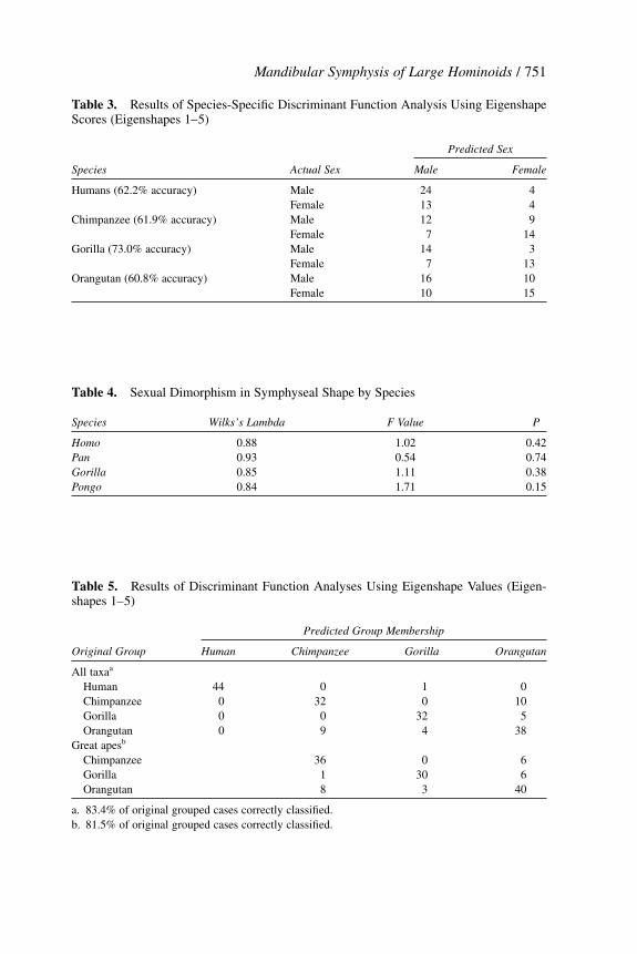
Mandibular Symphysis of Large Hominoids / 751
Table 3. Results of Species-Specific Discriminant Function Analysis Using EigenshapeScores (Eigenshapes 1–5)
Predicted Sex
Species Actual Sex Male Female
Humans (62.2% accuracy) Male 24 4Female 13 4
Chimpanzee (61.9% accuracy) Male 12 9Female 7 14
Gorilla (73.0% accuracy) Male 14 3Female 7 13
Orangutan (60.8% accuracy) Male 16 10Female 10 15
Table 4. Sexual Dimorphism in Symphyseal Shape by Species
Species Wilks’s Lambda F Value P
Homo 0.88 1.02 0.42Pan 0.93 0.54 0.74Gorilla 0.85 1.11 0.38Pongo 0.84 1.71 0.15
Table 5. Results of Discriminant Function Analyses Using Eigenshape Values (Eigen-shapes 1–5)
Predicted Group Membership
Original Group Human Chimpanzee Gorilla Orangutan
All taxaa
Human 44 0 1 0Chimpanzee 0 32 0 10Gorilla 0 0 32 5Orangutan 0 9 4 38
Great apesb
Chimpanzee 36 0 6Gorilla 1 30 6Orangutan 8 3 40
a. 83.4% of original grouped cases correctly classified.b. 81.5% of original grouped cases correctly classified.
PAGE 751................. 15768$ $CH2 02-21-06 11:52:54 PS
752 / sherwood et al.
eigenshapes to discriminate males and females consistently for any of the sam-ples included in this analysis, along with the MANOVA results indicating nowithin-species sex differences, suggests that root size and morphology do notplay a role in determining symphyseal morphology. Therefore we interpret thevariation seen in eigenshapes 2 and 3 as not indicative of a Type 2 trait or span-drel. Lovejoy’s Type 3 traits are primarily a result of allometric factors. Again,the inability to accurately discriminate the sexes in highly dimorphic species(gorilla and orangutan) indicates that overall allometric effects may not play asignificant role in these eigenshapes.
Daegling and Jungers (2000) suggested that the primary utility of shapeanalysis of the mandibular symphysis may be for taxonomic identification offossil specimens. The potential for such analyses is portrayed in Figure 5 bydistributions of eigenshape scores for three taxa (chimpanzee, gorilla, andhuman). These distributions are well defined, virtually nonoverlapping, and shownear perfect discrimination. This utility, however, may be called into questionwhen examining the broad distribution of scores for orangutans.
In our analysis the variation in eigenshape scores for orangutans is roughlyequivalent to that of the African apes combined. In her study of mandibularvariation, Brown (1997) similarly found a higher level of variation in orangutanmorphology and identified three possible reasons: (1) intersubspecies variationbetween Bornean (P. pygmaeus pygmaeus) and Sumatran (P. p. abelli) speci-mens; (2) lack of the anterior belly of the digastric muscle; or (3) presence ofmultiple male morphologies. More specifically, Brown (1997) found thatBornean orangutans possess ‘‘symphyseal sections that are generally larger,thicker, and more bulbous than those from Sumatra,’’ whereas sections of Suma-tran orangutans ‘‘are long and narrow with extensive inferior transverse tori’’(Brown 1997, p. 166). Not all specimens used in the current analysis have aknown subspecies designation; among specimens for which the subspecies isknown, P. p. pygmaeus is the most common (P. p. pygmaeus, n � 38; P. p. abelli,n � 9; unknown, n � 4). In contrast to Brown’s (1997) results, comparison ofeigenshape scores between subspecies samples of known individuals reveals noclear differences. This may be due to our small sample of P. p. abelli. However,until a larger sample can be obtained, we conclude that subspecies variation alonecannot account for this large range of variation.
A second cause of variation within this genus may be the differing muscu-lar anatomy of orangutans compared to other large-bodied hominoids. Unlike theAfrican great apes and humans, orangutans lack an anterior belly of the digastricmuscle extending from the hyoid and attaching to the digastric fossae on theinferior surface of the mandibular symphyseal region. The functions of the digas-tric muscles are to assist in retrusion and opening of the mandible (Hylander1992). It is possible that the lack of a digastric muscle and the concomitantstrains it produces may alter the morphology in the inferior torus of orangutans.Examination of other animals with similar morphology (e.g., Alouatta) is neces-sary before this variation can be fully understood.
PAGE 752................. 15768$ $CH2 02-21-06 11:52:55 PS
Mandibular Symphysis of Large Hominoids / 753
A third consideration concerns hormonal differences in orangutan males.There is a growing body of evidence indicating that testosterone suppression insubordinate orangutan males may result in relatively small bodied individuals(Rodman 1988; Brown 1997). Although canine dimorphism has been shown notto affect the symphysis shape in normal males, the combination of a male caninemorphology and reduced corpus dimensions in these individuals may result in aunique anatomy, and hence increased variation. This would have had virtually noeffect on our study, given that none of the individuals included in these analyseswere unusually small or unusually proportioned males.
The apparent discrepancy between the distributions of eigenshape scoresbetween orangutans and the other taxa is not of minor concern and raises severalinteresting questions when considering applications to the fossil record. Asnoted, without orangutans, eigenshape scores almost perfectly discriminate be-tween the three remaining taxa. If the analysis were to end there, one couldassume, as did Daegling and Jungers (2000), that the mandibular symphysiswould be an effective tool in systematic assignment of unknown specimens. Theaddition of the more variable orangutan results in error rates that may not beacceptable when attempting to assign fossil specimens.
Of course, this raises the question of what should be considered a ‘‘typical’’primate distribution for symphyseal shape descriptors. We have made the implicitassumption that the orangutan distribution is atypical for hominoids and havesought additional explanations for the perceived pattern. Given the small numberof taxa considered in this study, it is also possible that orangutans typify primatesin terms of symphyseal variation and that chimpanzees, gorillas, and humanshave relatively low levels of variation. The possibility that the African great apesand humans demonstrate a reduced level of mandibular symphyseal morphologi-cal diversity should be pursued through further analyses.
Comparison of Analytical Approaches. Complex analyses of shape requirea considerably more involved data-collection protocol than is frequently feasible.Therefore linear metric methods are often implemented. However, as noted pre-viously, such approaches tend to oversimplify the morphology and therefore mayresult in insufficient resolution.
Ravosa (2000) suggested that linear metric data are often sufficient whentrying to reconstruct habits of extinct species. Similarly, our results suggest thatlinear metrics may provide a good proxy measure for more complicated descrip-tions of the two-dimensional cross-section of the mandibular symphysis, depend-ing on the research question. The first eigenshape in our analysis serves as ananalogue of the relative dorsoventral width of the symphysis if length is heldconstant. Because this variation is in accord with the functional predictions ofthe mandibular symphysis, we suggest that this metric measurement may providean ideal alternative to more complicated shape data when the primary questionconcerns adaptations to the stresses encountered during mastication.
PAGE 753................. 15768$ $CH2 02-21-06 11:52:55 PS

754 / sherwood et al.
However, we suggest that when the research question is concerned withshape differences, linear metrics can be problematic. Metric data are often framedas ratio data in an effort to control for size. Such ratios provide a unitless measureoften referred to as ‘‘shape’’ (Bookstein 1991; MacLeod and Rose 1993). Mac-Leod and Rose (1993) discussed the problems with ratio data, including the simi-larity of ratio values from differing shapes. This is demonstrable in our data setby calculating a ratio of symphyseal width to symphyseal length. With this sim-ple ratio, gorillas (x � 0.41 � 0.01) and humans (x � 0.43 � 0.02) would be in-distinguishable, despite the obvious shape differences.
Scott (1980, p. 766) declared that ‘‘outlines are rich in information for thetaxonomist.’’ If this is true, the question is then, Which outline approach is best?MacLeod and Rose (1993) and Lohman (1983) expressed a preference for eigen-shape analysis on theoretical grounds, whereas Rohlf (1986) considered Fourierand eigenshape analyses to be roughly equivalent. Comparison of eigenshapedata with the Fourier approach of Daegling and Jungers (2000) shows manysimilarities. In both cases overall variation in gorilla symphyseal shape is some-what less than that in chimpanzees, whereas orangutan variation is greater thanboth. The reduced gorilla variation and the extreme variation in orangutan sym-physes is more pronounced in the Fourier analysis, but not to the extent that theinterpretations presented here would change.
Discriminant function analyses based on both Fourier and eigenshape anal-yses demonstrate an ability to distinguish between taxa with minimal error. How-ever, discrimination based on eigenshape scores shows an overall reduction inclassification error, with virtual elimination of some types of misclassifications(as noted, gorillas and chimpanzees showed near perfect separation). In theirFourier discriminant function analysis, Daegling and Jungers (2000) reported anoverall error rate of 22–33% in assigning specimens correctly to taxa when ex-amining just the great apes. When a similar analysis is done with eigenshapescores, the overall error rate is 16.6% when discriminating among all four taxaand 18.5% for just the great apes (see Table 2). Although these error rates areonly slightly lower than those in Daegling and Jungers’s analysis, the type oferror may hold some interest. In the Fourier analysis 10% of gorillas were incor-rectly classified as chimpanzees and 15% of chimpanzees were incorrectly classi-fied as gorillas. Eigenshape analysis showed no errors in assigning gorillas tochimpanzee or vice versa when all taxa were examined, and only one gorilla(3%) was incorrectly classified as a chimpanzee when the analysis was restrictedto great apes. In both the eigenshape and Fourier analyses errors were made inclassifying chimpanzee or gorilla as orangutan, and orangutan as either of thosetaxa. These errors are not surprising, given the large variation seen in the orang-utan sample. In addition, in the eigenshape analysis one error was made in classi-fying a human (a 30-year-old white female) as a gorilla; no apes weremisclassified as human. It is difficult to assess, however, whether the reductionin error seen in these eigenshape analyses is solely due to differences in method-ology or whether it may be related to differences in the samples used.
PAGE 754................. 15768$ $CH2 02-21-06 11:52:56 PS

Mandibular Symphysis of Large Hominoids / 755
Shape and Size. Finally, we highlight a potential caveat to shape-based analy-ses that attempt to remove size (i.e., scale) from the analysis. Because �* coordi-nates are angular in nature, they do effectively remove size, in the strictmathematical sense, before analysis. However, size may still play a role in deter-mination of the morphology of the symphysis because of allometry. Althoughwe did not focus on allometric effects in this analysis, the major feature of sym-physeal shape noted in this study clearly revolves around the shape of the supe-rior transverse torus. The eigenshape axes shown in Figures 2 and 5 show thatthe superior torus clearly influences other aspects of shape, such as depth of thegenial fossa and thickness of the inferior torus and alveolar margin. Given thatthe size of the superior torus is highly correlated with overall size of the mandi-ble, it should not be surprising that shape of the superior torus (and related partsof the symphysis) is, in some way, also related to size differences between taxa(Daegling 2001).
This criticism that shape and size are not independent is not novel (Gould1966), but we raise it in order to argue that it should not be used to discount thevalue of shape analyses. There are clear cases where metrics will fall short oftechniques, such as eigenshape analysis, in providing useful descriptions of mor-phology [see Scott (1980), MacLeod and Rose (1993), and Macleod (2002a)].For example, it would be difficult to capture the subtle shape differences in themandibular symphysis described in this study with a small set of scalar metrics.
Conclusion
We used an outline approach, eigenshape analysis, to examine the two-dimensional cross-sectional shape of the mandibular symphysis of extant large-bodied hominoids. To some degree, variation along the first eigenshape mimicsthe variation seen in metric analyses by describing variation in relative width ofthe symphysis. This variation may be indicative of an adaptive response to thestresses of normal activities and accounts for 76–80% of the variation betweenthese taxa. Subsequent eigenshapes identify subtle aspects of morphological vari-ation in the superior transverse torus that do not appear to be functionally influ-enced and are not sexually dimorphic. Because there was no apparent effect ofcanine size on symphyseal morphology in sexually dimorphic species, contraryto previous suggestions that dimorphic canines may have an effect on mandibularmorphology, additional factors that may contribute to these second, third, andfourth eigenshapes need to be considered.
Using the first four eigenshapes, strong discrimination among the Africanape and human symphyses suggests that eigenshape analyses may prove usefulin systematic studies of fossil specimens. The large variation seen in orangutansymphyses, however, suggests that patterns of variation may differ in some taxaand that caution should be exercised.
PAGE 755................. 15768$ $CH2 02-21-06 11:52:56 PS

756 / sherwood et al.
It is clear that Scott’s optimistic assertion that ‘‘outlines are rich in informa-tion’’ (Scott 1980, p. 766) is true for the mandibular symphysis of hominoids andthat eigenshape analysis is an effective means to extract that information. Furtherapplication of this method to other taxa, such as the ‘‘anterior digastric-less’’Alouatta or the strongly dimorphic Papio, will continue to refine the role ofbiomechanical, phylogenetic, and pleiotropic influences on mandibular symphy-seal cross-sectional morphology.
Acknowledgments We wish to thank the individuals and institutions that made speci-mens available for study: Bruce Latimer and Lyman Jellema of the Cleveland Museum ofNatural History; and Richard Thorington and Linda Gordon of the National Museum ofNatural History. In the course of the eigenshape work, we experimented with severalsoftware packages for various aspects of the analysis. G. Lohmann, H. Harpending, N.Macleod, and K. Weiss played various roles in this process, and we appreciate their help.As noted in the text, the programs produced by N. Macleod were ultimately used in thefinal analysis, and we are particularly grateful for his copious assistance. Wendy Dirks,Suzanne Hagell, and Ariana Ridgely provided comments along the way, and we appreciatetheir assistance. We also thank Terry Harrison, Bill Kimbel, and several anonymous re-viewers for their comments.
Received 15 August 2005.
Literature Cited
Beecher, R. M. 1977. Function and fusion at the mandibular symphysis. Am. J. Phys. Anthropol.47:325–335.
Beecher, R. M. 1979. Functional significance of the mandibular symphysis. J. Morphol. 159:117–130.
Beecher, R. M. 1983. Evolution of the mandibular symphysis in Notharctinae (Adapidae, Primates).Int. J. Primatol. 4:99–112.
Bookstein, F. L. 1991. Morphometric Tools for Landmark Data: Geometry and Biology. Cambridge,England: Cambridge University Press.
Bookstein, F. L., B. Chernoff, R. L. Elder et al. 1985. Morphometrics in Evolutionary Biology.Philadelphia: Academy of Natural Sciences.
Bouvier, M. 1986. Biomechanical scaling of mandibular dimensions in New World monkeys. Int. J.Primatol. 7:551–567.
Brown, B. 1997. Miocene hominoid mandibles: Functional and phylogenetic perspectives. In Func-tion, Phylogeny, and Fossils. D. R. Begun, C. V. Ward, and M. D. Rose, eds. New York:Plenum Press, 153–172.
Chamberlain, A. T., and B. A. Wood. 1985. A reappraisal of variation in hominid mandibular corpusdimensions. Am. J. Phys. Anthropol. 66:399–406.
Cheverud, J. M., and J. E. Buikstra. 1981. Quantitative genetics of skeletal nonmetric traits in therhesus macaques on Cayo Santiago. II. Phenotypic, genetic, and environmental correlationsbetween traits. Am. J. Phys. Anthropol. 54:51–58.
PAGE 756................. 15768$ $CH2 02-21-06 11:52:57 PS

Mandibular Symphysis of Large Hominoids / 757
Cheverud, J. M., J. E. Buikstra, and E. Twichell. 1979. Relationships between nonmetric skeletaltraits and cranial size and shape. Am. J. Phys. Anthropol. 50:191–198.
Daegling, D. J. 1989. Biomechanics of cross-sectional size and shape in the hominoid mandibularcorpus. Am. J. Phys. Anthropol. 80:91–106.
Daegling, D. J. 1993. Shape variation in the mandibular symphysis of apes: An application of amedian axis method. Am. J. Phys. Anthropol. 91:505–516.
Daegling, D. J. 2001. Biomechanical scaling of the hominoid mandibular symphysis. J. Morphol.250:12–23.
Daegling, D. J., and F. E. Grine. 1991. Compact bone distribution and biomechanics of early hominidmandibles. Am. J. Phys. Anthropol. 86:321–339.
Daegling, D. J., and W. L. Hylander. 1997. Occlusal forces and mandibular bone strain: Is the primatejaw ‘‘overdesigned’’? J. Hum. Evol. 33:705–717.
Daegling, D. J., and W. L. Hylander. 1998. Biomechanics of torsion in the human mandible. Am. J.Phys. Anthropol. 105:73–87.
Daegling, D. J., and W. L. Hylander. 2000. Experimental observation, theoretical models, and biome-chanical inference in the study of mandibular form. Am. J. Phys. Anthropol. 112:541–551.
Daegling, D. J., and W. L. Jungers. 2000. Elliptical Fourier analysis of symphyseal shape in great apemandibles. J. Hum. Evol. 39:107–122.
Demes, B., H. Preuschoft, and J. E. A. Wolff. 1984. Stress-strength relationships in the mandibles ofhominoids. In Food Acquisition and Processing in Primates, D. J. Chivers, B. A. Wood, andA. Bilsborough, eds. New York: Plenum Press, 369–390.
Dobson, S. D., and E. Trinkaus. 2002. Cross-sectional geometry and morphology of the mandibularsymphysis in middle and late Pleistocene Homo. J. Hum. Evol. 43:67–87.
Dunsworth, H., and A. Walker. 2002. Early genus Homo. In The Primate Fossil Record, W. C.Hartwig, ed. Cambridge, England: Cambridge University Press, 419–435.
Ferson, S., F. J. Rohlf, and R. K. Koehn. 1985. Measuring shape variation of two-dimensional out-lines. Syst. Zool. 34:59–68.
Gould, S. J. 1966. Allometry and size in ontogeny and phylogeny. Biol. Rev. Camb. Philos. Soc.41:587–640.
Gould, S. J. 1997. The exaptive excellence of spandrels as a term and prototype. Proc. Natl. Acad.Sci. USA 94:10,750–10,755.
Gould, S. J. 2002. The Structure of Evolutionary Theory. Cambridge, England: Belknap Press.Gould, S. J., and R. J. Lewontin. 1979. The spandrels of San Marco and the Panglossian paradigm:
A critique of the adaptationist programme. Proc. R. Soc. Lond. 205:581–598.Hiiemae, K. M. 1978. Mammalian mastication: A review of the activity of the jaw muscles and the
movements they produce in chewing. In Development, Function, and Evolution of Teeth, P. M.Butler and K. A. Joysey, eds. London: Academic Press, 359–398.
Hylander, W. L. 1975. The human mandible: Lever or link? Am. J. Phys. Anthropol. 43:227–242.Hylander, W. L. 1979. The functional significance of primate mandibular form. J. Morphol. 160:223–
240.Hylander, W. L. 1981. Patterns of stress and strain in the macaque mandible. In Craniofacial Biology,
D. S. Carlson, ed. Craniofacial Growth Series, Monograph 10. Ann Arbor, MI: Center forHuman Growth and Development, University of Michigan, 1–37.
Hylander, W. L. 1984. Stress and strain in the mandibular symphysis of primates: A test of competinghypotheses. Am. J. Phys. Anthropol. 64:1–46.
Hylander, W. L. 1985. Mandibular function and biomechanical stress and scaling. Am. Zool. 25:315–330.
Hylander, W. L. 1986. In-vivo bone strain as an indicator of masticatory bite force in Macaca fascicu-laris. Arch. Oral Biol. 31:149–157.
Hylander, W. L. 1992. Functional anatomy. In The Temporomandibular Joint: A Biological Basis forClinical Practice, B. G. Sarnat and B. G. Laskin, eds. Philadelphia: W. B. Saunders, 60–92.
Hylander, W. L., and A. W. Crompton. 1986. Jaw movements and patterns of mandibular bone strainduring mastication in the monkey Macaca fascicularis. Arch. Oral Biol. 31:841–848.
PAGE 757................. 15768$ $CH2 02-21-06 11:52:57 PS
758 / sherwood et al.
Hylander, W. L., and K. R. Johnson. 1994. Jaw muscle function and wishboning of the mandibleduring mastication in macaques and baboons. Am. J. Phys. Anthropol. 94:523–547.
Hylander, W. L., K. R. Johnson, and A. W. Crompton. 1987. Loading patterns and jaw movementsduring mastication in Macaca fascicularis: A bone-strain, electromyographic, and cineradio-graphic analysis. Am. J. Phys. Anthropol. 72:287–314.
Kelley, J., and D. Pilbeam. 1986. The Dryopithecines: Taxonomy, comparative anatomy, and phylog-eny of Miocene large hominoids. In Comparative Primate Biology, v. 1, Systematics, Evolu-tion, and Anatomy, D. R. Swindler and J. Erwin, eds. New York: Alan R. Liss, 361–411.
Kimbel, W. H., Y. Rak, and D. C. Johanson. 2004. The Skull of Australopithecus afarensis. Oxford:Oxford University Press.
Kimbel, W. H., and T. D. White. 1988. Variation, sexual dimorphism, and the taxonomy of Australo-pithecus. In Evolutionary History of the ‘‘Robust’’ Australopithecines, F. E. Grine, ed. NewYork: Aldine de Gruyter, 175–191.
Lam, Y. M., O. M. Pearson, and C. M. Smith. 1996. Chin morphology and sexual dimorphism in thefossil hominid mandible sample from Klasies River Mouth. Am. J. Phys. Anthropol. 100:545–557.
Leakey, M. G., C. S. Feibel, I. McDougall et al. 1995. New four-million-year-old hominid speciesfrom Kanapoi and Allia Bay, Kenya. Nature 376:565–571.
Lieberman, D. E., and A. W. Crompton. 2000. Why fuse the mandibular symphysis? A comparativeanalysis. Am. J. Phys. Anthropol. 112:517–540.
Lohmann, G. P. 1983. Eigenshape analysis of microfossils: A general morphometric procedure fordescribing changes in shape. Math. Geol. 15:659–672.
Lohmann, G. P., and P. N. Schweitzer. 1990. On eigenshape analysis. In Proceedings of the MichiganMorphometrics Workshop, F. J. Rohlf and F. L. Bookstein, eds. Ann Arbor, MI: University ofMichigan Museum of Zoology, 145–166.
Lovejoy, C. O., M. J. Cohn, and T. D. White. 1999. Morphological analysis of the mammalianpostcranium: A developmental perspective. Proc. Natl. Acad. Sci. USA 96:13,247–13,252.
Macleod, N. 1999. Generalizing and extending the eigenshape method of shape space visualizationand analysis. Paleobiology 25:107–138.
Macleod, N. 2002a. Geometric morphometrics and geological shape-classification systems. EarthSci. Rev. 59:27–47.
Macleod, N. 2002b. Phylogenetic signals in morphometric data. In Morphology, Shape, and Phylog-eny, N. Macleod and P. L. Forey, eds. London: Taylor and Francis, 100–138.
Macleod, N., and K. D. Rose. 1993. Inferring locomotor behavior in Paleogene mammals via eigen-shape analysis. Am. J. Sci. 293A:300–355.
McCollum, M. A. 1999. The robust australopithecine face: A morphogenetic perspective. Science284:301–305.
McCollum, M. A., and P. T. Sharpe. 2001. Developmental genetics and early hominid craniodentalevolution. BioEssays 23:481–493.
McCrossin, M. L., and B. R. Benefit. 1993. Recently recovered Kenyapithecus mandible and itsimplications for great ape and human origins. Proc. Natl. Acad. Sci. USA 90:1962–1966.
McGrath, J. W., J. M. Cheverud, and J. E. Buikstra. 1984. Genetic correlations between sides andheritability of asymmetry for nonmetric traits in rhesus macaques on Cayo Santiago. Am. J.Phys. Anthropol. 64:401–411.
Nicolay, C. W., and R. J. Sherwood. 2000. Eigenshape and biomechanical analysis of the phyllo-stomid mandibular symphysis. Am. Zool. 40:1148.
Olson, E., and R. Miller. 1958. Morphological Integration. Chicago: University of Chicago Press.Ravosa, M. J. 1996. Mandibular form and function in North American and European Adapidae and
Omomyidae. J. Morphol. 229:171–190.Ravosa, M. J. 1999. Anthropoid origins and the modern symphysis. Fol. Primatol. 70:65–78.Ravosa, M. J. 2000. Size and scaling in the mandible of living and extinct apes. Fol. Primatol.
71:305–322.
PAGE 758................. 15768$ $CH2 02-21-06 11:52:58 PS

Mandibular Symphysis of Large Hominoids / 759
Ravosa, M. J., and E. L. Simons. 1994. Mandibular growth and function in Archaeolemur. Am. J.Phys. Anthropol. 95:63–76.
Rodman, P. S. 1988. Diversity and consistency in ecology and behavior. In Orangutan Biology, J. H.Schwartz, ed. New York: Oxford University Press, 31–51.
Rohlf, F. J. 1986. Relationships among eigenshape analysis, Fourier analysis, and analysis of coordi-nates. Math. Geol. 18:845–857.
Schwartz, J. H., and I. Tattersall. 2000. The human chin revisited: What is it and who has it? J. Hum.Evol. 38:367–409.
Scott, G. H. 1980. The value of outline processing in the biometry and systematics of fossils. Palaeon-tology 23:757–768.
Takai, M., F. Anaya, N. Shigehara et al. 2000. New fossil materials of the earliest new world monkey,Branisella boliviana, and the problem of platyrrhine origins. Am. J. Phys. Anthropol.111:263–281.
Taylor, A. B. 2002. Masticatory form and function in the African apes. Am. J. Phys. Anthropol.117:133–156.
Taylor, A. B., and C. P. Groves. 2003. Patterns of mandibular variation in Pan and Gorilla andimplications for African ape taxonomy. J. Hum. Evol. 44:529–563.
Tobias, P. V. 1991. Olduvai Gorge, v. 4A and 4B, Homo habilis: Skulls, Endocasts and Teeth. Cam-bridge, England: Cambridge University Press.
Vinter, I., J. Krmpotic-Nemanic, D. Ivankovic et al. 1996. The influence of the dentition on the shapeof the mandible. Colleg. Antropol. 20:555–560.
Vinyard, C. J., and M. J. Ravosa. 1998. Ontogeny, function, and scaling of the mandibular symphysisin papionin primates. J. Morphol. 235:157–175.
Ward, S. C. 1991. Taxonomy, paleobiology, and adaptations of the ‘‘robust’’ australopithecines. J.Hum. Evol. 21:469–483.
Ward, S. C., and D. L. Duren. 2002. Middle and late Miocene African hominoids. In The PrimateFossil Record, W. C. Hartwig, ed. Cambridge, England: Cambridge University Press, 385–397.
White, T. D., and D. C. Johanson. 1982. Pliocene hominid mandibles from the Hadar formation,Ethiopia: 1974–1977 collections. Am. J. Phys. Anthropol. 57:501–544.
Wolff, J. E. A. 1984. A theoretical approach to solve the chin problem. In Food Acquisition Process-ing in Primates, D. J. Chivers, B. A. Wood, and A. Bilsborough, eds. New York: PlenumPress, 391–405.
Wood, B. A. 1978. Allometry and hominid studies. In Geological Background to Fossil Man, W. W.Bishop, ed. Edinburgh: Scottish Academic Press, 125–128.
Wood, B. 1991. Koobi Fora Research Project, v. 4, Hominid Cranial Remains. Oxford: ClarendonPress.
Zahn, C. T., and R. Z. Roskies. 1972. Fourier descriptors for plane closed curves. IEEE Trans. Comp.C21:269–281.
Zelditch, M. L., W. L. Fink, and D. L. Swiderski. 1995. Morphometrics, homology, and phylogenet-ics: Quantified characters as synapomorphies. Syst. Biol. 44:179–189.
PAGE 759................. 15768$ $CH2 02-21-06 11:52:59 PS