mandibular movements -...
TRANSCRIPT
DEVELOPMENT OF THE TOTAL DIAGNOSTIC SYSTEM FOR MASTICATORY FUNCTIONS
M. Koseki+, N. Inou+ and K. Maki*
+ Department of Mechanical and Control Engineering, Graduate School of Science and Engineering, Tokyo Institute of Technology, JAPAN
* Department of Orthodontics, Showa University, JAPAN
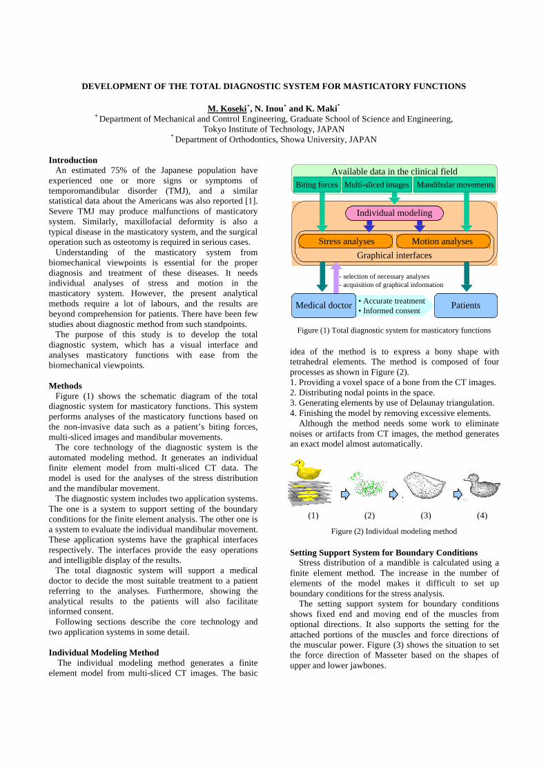
Introduction An estimated 75% of the Japanese population have experienced one or more signs or symptoms of temporomandibular disorder (TMJ), and a similar statistical data about the Americans was also reported [1]. Severe TMJ may produce malfunctions of masticatory system. Similarly, maxillofacial deformity is also a typical disease in the masticatory system, and the surgical operation such as osteotomy is required in serious cases. Understanding of the masticatory system from biomechanical viewpoints is essential for the proper diagnosis and treatment of these diseases. It needs individual analyses of stress and motion in the masticatory system. However, the present analytical methods require a lot of labours, and the results are beyond comprehension for patients. There have been few studies about diagnostic method from such standpoints. The purpose of this study is to develop the total diagnostic system, which has a visual interface and analyses masticatory functions with ease from the biomechanical viewpoints. Methods Figure (1) shows the schematic diagram of the total diagnostic system for masticatory functions. This system performs analyses of the masticatory functions based on the non-invasive data such as a patient’s biting forces, multi-sliced images and mandibular movements. The core technology of the diagnostic system is the automated modeling method. It generates an individual finite element model from multi-sliced CT data. The model is used for the analyses of the stress distribution and the mandibular movement. The diagnostic system includes two application systems. The one is a system to support setting of the boundary conditions for the finite element analysis. The other one is a system to evaluate the individual mandibular movement. These application systems have the graphical interfaces respectively. The interfaces provide the easy operations and intelligible display of the results. The total diagnostic system will support a medical doctor to decide the most suitable treatment to a patient referring to the analyses. Furthermore, showing the analytical results to the patients will also facilitate informed consent. Following sections describe the core technology and two application systems in some detail. Individual Modeling Method
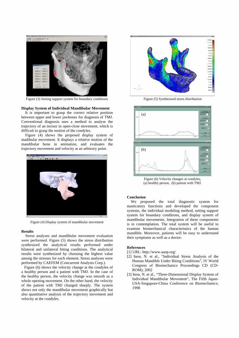
The individual modeling method generates a finite element model from multi-sliced CT images. The basic
idea of the method is to express a bony shape with tetrahedral elements. The method is composed of four processes as shown in Figure (2). 1. Providing a voxel space of a bone from the CT images. 2. Distributing nodal points in the space. 3. Generating elements by use of Delaunay triangulation. 4. Finishing the model by removing excessive elements.
Although the method needs some work to eliminate noises or artifacts from CT images, the method generates an exact model almost automatically.
Setting Support System for Boundary Conditions Stress distribution of a mandible is calculated using a
finite element method. The increase in the number of elements of the model makes it difficult to set up boundary conditions for the stress analysis.
The setting support system for boundary conditions shows fixed end and moving end of the muscles from optional directions. It also supports the setting for the attached portions of the muscles and force directions of the muscular power. Figure (3) shows the situation to set the force direction of Masseter based on the shapes of upper and lower jawbones.
Figure (1) Total diagnostic system for masticatory functions
Multi-sliced imagesBiting forces
Available data in the clinical fieldMandibular movements
Patients
Individual modeling
Medical doctor • Accurate treatment• Informed consent
Motion analysesGraphical interfaces
Stress analyses
- selection of necessary analyses- acquisition of graphical information
1) 2) 3) 4) Figure (2) Individual modeling method
(1) (2) (3) (4)
Display System of Individual Mandibular Movement It is important to grasp the correct relative position
between upper and lower jawbones for diagnosis of TMJ. Conventional diagnosis uses a method to analyse the trajectory of an incisor in open-close movement, which is difficult to grasp the motion of the condyles.
Figure (4) shows the proposed display system of madibular movement. It displays a relative motion of the mandibular bone in animation, and evaluates the trajectory movement and velocity at an arbitrary point.
Results Stress analyses and mandibular movement evaluation
were performed. Figure (5) shows the stress distribution synthesized the analytical results performed under bilateral and unilateral biting conditions. The analytical results were synthesized by choosing the highest value among the stresses for each element. Stress analyses were performed by CAEFEM (Concurrent Analysis Corp.). Figure (6) shows the velocity change at the condyles of a healthy person and a patient with TMJ. In the case of the healthy person, the velocity change was smooth as a whole opening movement. On the other hand, the velocity of the patient with TMJ changed sharply. The system shows not only the mandibular movement graphically but also quantitative analysis of the trajectory movement and velocity at the condyles.
Conclusion
We proposed the total diagnostic system for masticatory functions and developed the component systems, the individual modeling method, setting support system for boundary conditions, and display system of mandibular movements. Integration of these components is in contemplation. The total system will be useful to examine biomechanical characteristics of the human mandible. Moreover, patients will be easy to understand their symptoms as well as a doctor. References [1] URL: http://www.aaop.org/ [2] Inou, N. et al., “Individual Stress Analysis of the
Human Mandible Under Biting Conditions”, IV World Congress of Biomechanics Proceedings CD (CD-ROM); 2002
[3] Inou, N. et al., “Three-Dimensional Display System of Individual Mandibular Movement”, The Fifth Japan-USA-Singapore-China Conference on Biomechanics; 1998
Figure (3) Setting support system for boundary conditions Figure (5) Synthesized stress distribution
Figure (4) Display system of mandibular movement
Figure (6) Velocity changes at condyles, (a) healthy person, (b) patient with TMJ.
(a)
(b)