mandibular molar protraction
TRANSCRIPT

Mandibular Molar Protraction with Temporary Anchorage
Devices
Faculty of dentistry-Faculty of dentistry-Mansoura university - EgyptMansoura university - Egypt

Protraction of mandibular molars is challenging because of the high density of mandibular bone. Anterior dental anchorage is often inadequate to protract even a single first molar without reciprocal retraction of the incisors or movement of the dental midline. Furthermore, if the buccal and lingual cortical plates in the edentulous region hav collapsed, safe and effective protraction may be impossible.

Orthodontic temporary anchorage devices(TADs) can provide skeletal anchorage for mandibular molar protraction, avoiding the problems often encountered with the use of dental anchorage. This article presents various strategies for molar protraction with miniscrews and reviews the periodontal classifications for atrophic edentulous regions.

Lingual Elastic Tied to the Archwire Direct protraction from a miniscrew placed lateral and inferior to the archwire can create posterior crossbite and open bite. To counteractthese effects, the following steps should beconsidered:-

Protraction without balancing lingual forcecan quickly swing posterior dentition into
unilateral crossbite.

1. Protraction with a balancing lingual force, suchas an elastic thread tied from the lingual cleat ofthe molar to the archwire. When tying thelingual elastic to the archwire, the incisors andcanines must be ligated to prevent rotation of theanterior teeth.

A Lingual elastic thread tied to archwire to provide balancing lingual force without sacrificing anterior dental anchorage. First and second
molars must be ligated to prevent rotation of anterior teeth.B. Protraction through atrophic edentulous ridge (moderate Seibert
Class I) with lingual elastic thread tied to archwire, producing complete space closure in eight months without loss of pulp vitality. (Case
treated by Drs. Clara Chow and Budi Kusnoto; photographs courtesy of University of Illinois-Chicago.)

2. Incorporating the second molar into the archwire to minimize arch expansion.3. Using a rectangular archwire to prevent the molar from rolling out buccally.4. Placing an occlusal gable bend (upward V-bend) in the archwire mesial to the edentulous space to counteract molar intrusion. Alternatively, if an auxiliary slot is used, a buccal hook can be fabricated from a wire segment to protract the tooth at its center of resistance.
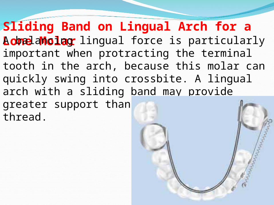
Sliding Band on Lingual Arch for a Lone MolarA balancing lingual force is particularly important when protracting the terminal tooth in the arch, because this molar can quickly swing into crossbite. A lingual arch with a sliding band may provide greater support than lingual elastic thread.

Sliding band with lingual arch for protraction of lone molar.

The lingual arch consists of an .040" wire soldered to the molar band on the side opposite the lone molar. A sliding band with a headgear tube soldered to its lingual surface is cemented to the lone molar at the same appointment. The lingual arch extends through this tube, acting as a guide rail during protraction. After protraction is complete, the clinician can cut the lingual arch from the soldered band.


The “Push-Pull” TechniqueConventionally, a miniscrew is placed mesial to the edentulous space to avoid impeding the molar protraction. As an alternative, the clinician may insert the TAD within the edentulous space and protract from the second tooth back, using an open-coil spring to push the tooth in front of it. The open-coil spring tips the crown enough to provide complete space closure.

A. “Push-pull” technique. Miniscrew is placed in edentulous space and used to pull first molar. Open-coil spring pushes second premolar mesially. Buccal
hook for molar band is fabricated at chairside. B. “Push-pull” technique using sliding band.
A B

Fig. 5 A. Improper protraction from terminal tooth.Lower buccal segment has quickly rolled outwardinto buccal crossbite, canting lower incisors.B. Proper protraction with “push-pull” techniqueusing sliding band. First molar is protracted at itscenter of resistance, using open-coil springbetween first molar and second premolar; noteincisal gable bend mesial to edentulous space.C. Model of sliding band setup. Lingual guide railwas shortened for comfort before insertion.(Photographs courtesy of University of Illinois-Chicago.)
A B
C


The “push-pull” technique has the following advantages over other protraction methods:• Simplifies miniscrew insertion.• Minimizes the risk of root perforation.• Obviates surgical stent fabrication and periapical radiography.• Ensures adequate bone stock.• Prevents the auxiliary from crossing the canine eminence.• Applies two active forces (a nickel titanium coilspring and the open-coil spring) for efficient multitooth protraction.
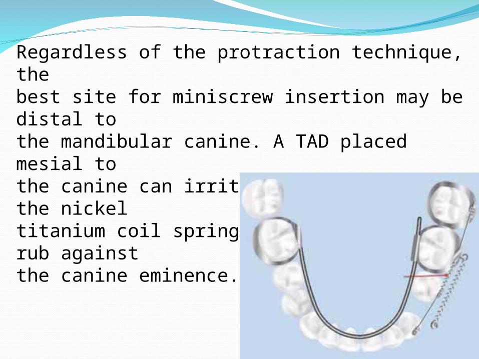
Regardless of the protraction technique, thebest site for miniscrew insertion may be distal tothe mandibular canine. A TAD placed mesial tothe canine can irritate the lip or cause the nickeltitanium coil spring to overextend and rub againstthe canine eminence.

DiscussionMany orthodontic patients have posterior spacing due to missing mandibular teeth. Excluding the third molars, the mandibular second premolar is the most common congenitally absent tooth.The mandibular first molar is the most frequently lost tooth in adults.Molar protraction can be an alternative to restoration with posterior dentalimplants or fixed partial dentures. Avoiding anchorage loss is considerably more challenging in the mandible than in the maxilla, in part because of the structural differences between the two jaws. The posterior maxilla is composed of uniformly thin cortices interconnectedby a network of spacious trabeculae,
while the posterior mandible consists of thicker cortical bone with dense, radially oriented trabeculae. In the molar region, the maxilla has an average buccal cortical thickness of 1.5mm, compared with 2mm in the mandible. The rate of molar protraction is inversely related to the radiographic density or cortical thickness of the resisting alveolar bone. Because of the increased thickness of mandibular cortical bone, the rate of mandibular molar translation with skeletal anchorage is nearly half that of maxillary molar translation approximately 34-.60mm per month.

To further complicate matters, the failure rateof TADs is greater in the mandible than in themaxilla.The primary biological factors thatdetermine miniscrew stability are bone density (orbone quality), peri-implant soft-tissue health,adequacy of peri-implant bone stock, and operatortechnique. The greater failure rate of mandibularminiscrews, despite the thicker mandibular corticalbone, is probably due to root proximity (or inadequate peri-implant bone stock) and greater buccal tissue mobility.

Many adult orthodontic patients with posterior edentulous spacing have been missing teeth for years and therefore exhibit alveolar ridge resorption. The rate of resorption is greatest during the first several months to two years after extraction,but decreases thereafter. The amount of post-extraction resorption is significantly greater on the buccal than on the lingual side in botharches. During the first year after tooth extraction,the amount of resorption in the mandible is twice that in the maxilla—a ratio that increases to 4:1 after seven years.13

The simplest way to diagnose edentulous ridge resorption is with the Seibert classification. Seibert Class I is defined as buccolingual loss of hard- and soft-tissue contour with normalapicocoronal height. Seibert Class II is an apicocoronal loss of hard- and soft-tissue contour with normal buccolingual width. Seibert Class III is a combination of Class I and II, with both buccolingual and apicocoronal loss of hard and soft tissue.
Fig. 6 A. Seibert Class I: Buccolingual loss of hard- and soft-tissue contour with normal apicocoronal height. B. Seibert Class II: Apicocoronalloss of hard- and soft-tissue contour with normal buccolingual width.

Allen and colleagues modified and expanded on Seibert’s original classification. Allen Type A is an apicocoronal loss of ridge height. Type B is a buccolingual loss of ridge width. Type C is a combination of buccolingual and apicocoronalloss. The ridge is further assessed in terms of the amount of tissue loss: mild, less than 3mm; moderate, 3-6mm; and severe, more than 6mm. Therefore, an edentulous site with a 3-6mm loss of hard and soft tissue in the buccolingual direction may be classified as a moderate Seibert ClassI or Allen Type B defect.

Potential risks of molar protraction through an atrophic ridge include loss of attachment (particularly in the presence of plaque),dehiscence, mobility, ankylosis, root resorption, devitalization, and tooth morbidity. Although successful molar protraction through atrophic ridges has been reported, no clinical study to date has evaluated the correlation between an atrophic ridge and periodontal response during bodily tooth movement.
NEAL D. KRAVITZ, DMD, MS TYLER JOLLEY, DMD
JCO/JUNE 2008
