manag{m{nt o osnoarthrltls - aulakinesica.com.ar cap 28 ar.pdf · tud ligamentosa; la rigidez...
TRANSCRIPT

MANAG{M{NT O� OSnOARTHRlTlS
AND RH{UMATOID ARTHRITIS Maura Daly Iversen and Linda Steiner
Introduction
The term arthritis comprises a complex of diseases that affect more than 70 million people in the United States.l Arthritis can manifest in more than 1 00 forms. This chapter focuses on rheumatoid arthritis and osteoarthritis and provides an analysis and discussion of the rehabilitation management of these two conditions.
Osteoarthritis
Epidemiology and Pathophysiology
Osteoarthritis (OA) involves the entire joint but is primarily a disease of the cartilage (Figure 28-1). OA affects nearly 40 million people in the United States and is the second leading cause of disability. The exact pathogenesis is unknown. The most commonly affected and symptomatic joints are the apophyseal joints of the spine, the distal and proximal interphalangeal joints, the carpometacarpal joints, the first metatarsophalangeal joint, and the knee, hip, and patellofemoral joints? The clinical features of OA are presented in Table 28- 1 .
Joints Commonly Affected by Osteoarthritis
• Spinal apophyseal joints
• Proximal interphalangeal (PIP) joints
• Distal interphalangeal (DIP) joint
• Carpometacarpal (CMC) joints
• First metatarsophalangeal (MTP) joint
• Hip
• Knee
• Patellofemoral joints
lZisk factors for OA can be classified into two major categories: intrinsic risk factors and extrinsic risk factors. Intrinsic f:lctors include knee alignment (a determinant of joint load), muscle strength, obesity, ligament laxity, and proprioception. Extrinsic factors include repetitive physical activity and injury.2 Osteoarthritis is commonly referred to as osteoarthrosis. Although OA used to be considered a degenerative joint disease, this designation is no longer appropriate. Scientists now recognize that OA is a slowly progressing, dynamic disease that involves biomechanical, environmental, genetic, and biochemical factors (e.g., cytokines). 3
Categories of Osteoarthritis
• Primary osteoarthritis: Results in articular changes, although the
etiological basis for the disease is unknown
• Secondary osteoarthritis: Caused by underlying factors that
accelerate age-related degeneration of cartilage
• These factors include inflammatory arthritis (e.g., rheumatoid
arthritis or spondyloarthritis), hypermobility syndromes, metaboliC
diseases (e.g., diabetes), and congenital and acquired joint
surface incongruities that accelerate damage to the cartilage, as
well as trauma or sports-related injuries
Our understanding of OA has increased considerably over the past 1 0 years, partly as a result of advances in cartilage imaging techniques. Early OA is a focal disease that presents as a distinct lesion of tile cartilage.4,5 Patients with early OA have joint stiffness and progressive cartilage destruction with pain on loading of tile affected joint. Over time, OA involves the entire joint, including the subchondral bone. Lesions progress with repeated biomecharucal loading, synovial membrane inflammation, and release of
859

860 CHAPTER 28 • Management of Osteoarthritis and Rheumatoid Arthritis
Normal knee
Table 28-1
Late stages of osteoarthritis
Pathology and Clinical Features of Rheumatoid Arthritis and Osteoarthritis
Disease
Rheumatoid arthritis (RA)
Osteoarthritis (OA)
Tissue Predominantly Involved Clinical Features
Synovium Symmetrical and bilateral joint involvement (inflammation) Joint pain, swelling, stiffness, and contracture
Muscle weakness and fatigue Acute: Red, hot, swollen, painnd joints; boggy
feel, fatigue, with or without fever; ligamentous laxity; morning stiffness up to a few hours
Subacute: Effusion, reduced redness and pain Stable: Generally no effusion, minimal stiffness
Cartilage Affects hips, knees, spine, ankles, distal (degradation ) interphalangeal ( D I P ) joints, proximal
interphalangeal (PIP) joints, and metacarpophalangeal (MCP) joints
Joint pain, malalignment Decreased proprioception Muscle weakness Early: Focal cartilaginous lesions End stage: Loss of cartilage, bone on bone
Figure 28-1 Osteoarthritis of the knee.
Radiographic Features
Periarticular swelling, joint effusion, regional osteoporosis, subchondral osteolytic erosions, joint subluxation
Osteophytes at joint margins, joint space narrowing, subchondral sclerosis and cysts
Modified from Iversen M D , Liang M H , Bae SC: Exercise therapy in selected arthritides: rheumatoid arthritis, osteoarthritis, systemic lupus
erythematosus, systemic sclerosis and polymyositis/dermatomyositis. In Frontera WR, Dawson DM, Siovik OM, editors: Exercise in rehabilitation
medicine, Champaign, IL, 1999, H u man Kinetics.
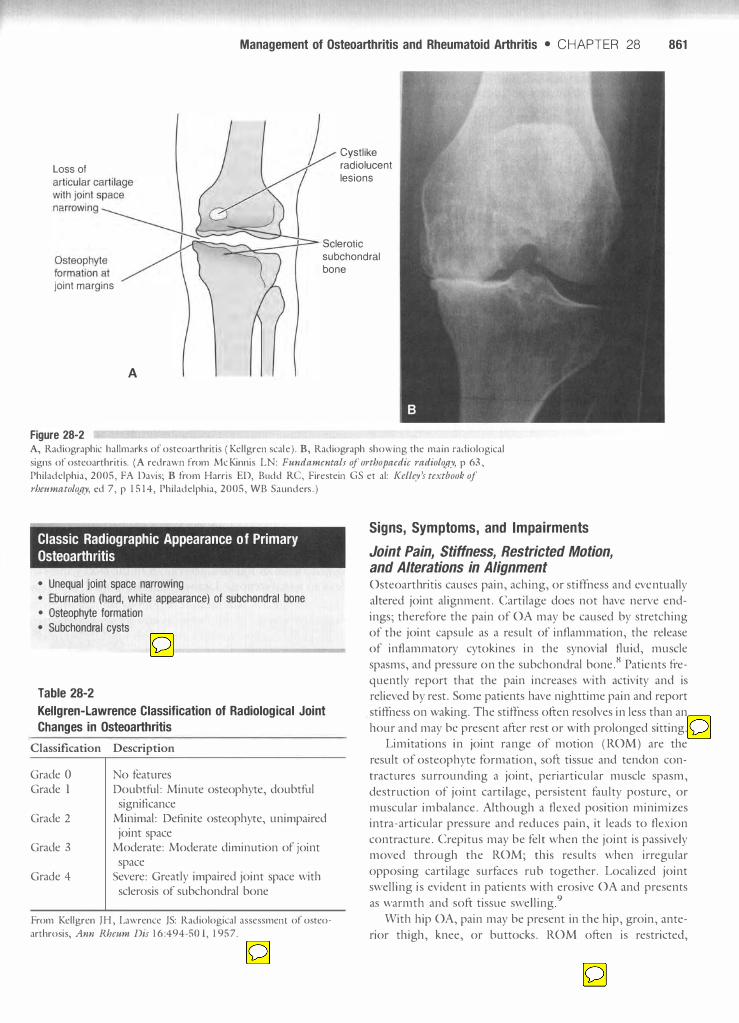
cytokines; this stimulates tlle production of matrix metalloproteinases, which cause cartilage degradation and loss. Eventually the joint surface is destroyed. The c lassic appearance of primary OA o n radiographs is localized disease with evidence of unequal joint space narrowing, eburnation (a hard, white appearance) of the subchondral bone, osteophytes, and subchondral cysts (Figure 28-2) . These findings increase in frequency after age 50. O nly 40% of patients with severe radiographic features present witl1 pain .6,7
The radiographic features ofOA are graded according to specific criteria. The Kel lgren scale is commonly used for this purpose (Table 28-2 ) . On radiographs, secondary OA may appear as difnlse or localized disease, depending on the etiology (e .g . , rheumatoid arthritis [RAJ, diabetes, or trauma). Secondary contractu res occur around the involved joint and contiguous joints . These contractures alter joint al ignment and can increase the biomechanical forces on the joint, accelerating cartilage loss and increas ing energy requirements for activities .
Management of Osteoarthritis and Rheumatoid Arthritis • CHAPTER 28 861
Loss of
Cystlike radiolucent lesions
Sclerotic subchondral bone
jOint margins
A
Figure 28-2 A, Radiographic hallmarks of osteoarthritis (Kellgren scale) . B, Radiograph showing the main radiological
signs of osteoarthritis. (A redrawn from McKinnis LN: Fundamentals of orthopaedic radiology, p 63,
Philadelphia, 2005, FA Davis; B from Harris ED, Budd Re, Firestein GS et al: Kelley's textbook of rhmmatology, cd 7, p 1 5 1 4, Philadelphia, 2005, WE Saunders . )
Classic Radiographic Appearance of Primary Osteoarthritis
• Unequal jOint space narrowing
• Eburnation (hard, white appearance) of subchondral bone
• Osteophyte formation
• Subchondral cysts
Table 28-2 Kellgren-lawrence Classification of Radiological Joint
Changes in Osteoarthritis
Classification Description
Grade 0 Grade 1
Grade 2
Grade 3
Grade 4
No features Doubtful: Minute osteophyte, doubtful
significance Minimal: Definite osteophyte, unimpaired
joint space Moderate: Moderate diminution of joint
space Severe: Greatly impaired joint space with
sclerosis of subchondral bone
From Kellgren JH, Lawrence JS: Radiological assessmenr of osteo
arthrosis, Ann Rheum Dis 1 6 :494-501, 1 95 7.
Signs, Symptoms, and Impairments
Joint Pain, Stiffness, Restricted Motion, and Alterations in Alignment Osteoarthritis causes pain, aching, or stiffness and eventually altered joint alignment. Cartilage does not have nerve endings; therefore the pain of OA may be caused by stretching of the joint capsule as a result of inflammation, the release of inflammatory cytokines in the synovial fluid, muscle spasms, and pressure on the subchondral bone.8 Patients fi-equently report tlut the pain increases with activity and is relieved by rest. Some patients have nighttime pain and report stiffness 011 waking. The stiffness often resolves in less than an hour and may be present after rest or with prolonged sitting.
Limitations in joint range of motion (ROM) are the result of osteophyte formation, soft tissue and tendon contractures surrounding a joint, periarticular muscle spasm, destruction of joint cartilage, persistent faulty posture, or muscular imbalance. Although a flexed position minimizes intra-articular pressure and reduces pain, it leads to flexion contracture. Crepitus may be felt when the joint is passively moved through the ROM; this results when irregular opposing cartilage surfaces rub together. Localized joint swelling is evident in patients witll erosive OA and presents as warmtll and soft tissue swelling9
Witl1 hip OA, pain may be present in tile hip, groin, anterior thigh, knee, or buttocks. ROM often is restricted,

862 CHAPTER 28 • Management of Osteoarthritis and Rheumatoid Arthritis
particularly internal rotation, and may be accompanied by crepitus. Patients may experience difficulties with mobility and personal hygiene, as well as increased energy expenditure with activities. Loss of hip range adversely affects the spine and other joints, including the knee and ankle. Preventive stretching is important and should be initiated early and often. Functionally based exercises, such as repetitive sit to stand, help maintain strength in hip and knee extensors. For patients unable to tolerate full gravity exercise, water (aquatic therapy) can be used to allow exercise with red�ced load.s
Signs and Symptoms of Osteoarthritis
• Pain or aching with activity
• Morning stiffness
• Altered jOint alignment
• Limited range of motion
• Joint contracture
• Crepitus
• Joint swelling
The knee is the most commonly affected weight-bearing joint. Malalignment of the knee, either varus or valgus, may be evident with severe disease (Figure 28-3) . Knee varus is the more common presentation in knee osteoarthritis. The
Figure 28-3 Val gus deformity of the k nees. This is a common k n ee deformity that
develops in patients with osteoarthritis of the lateral compartment.
( From Polley H F , H under G G : Rhettmatologic intervie1Ving and
physical examination of the joints, cd 2 , P 214, Philadelphia, 1978,
WE Saunders . )
presence of varus or valgus is associated with a threefold to fourfold increase in the odds of OA progression in the medial compartment of the knee.2 Malalignment and abnormal tracking of the patella may produce retropatellar pain and chondromalacia patellae. Retropatellar pain often is experienced when the individual walks upstairs or on inclines or sits for prolonged periods. Knee pain can lead to disuse atrophy and deconditioning.2,]O However, one research study suggested that muscle weakness may be a cause rather than a result of knee osteoarthritis.]] Muscle weakness leads to changes in joint biomechanics and unequal forces across the joint surface. Joint laxity from muscle inhibition and joint space narrowing distributes the forces across the cartilage surface unequally and can accelerate the process of cartilage degeneration.s
Common features of hand OA are Heberden's and/or Bouchard's nodes, which are the result of bony overgrowths (Figure 28-4) . Heberden's nodes appear at the medial and dorsolateral aspects of the distal interphalangeal (DIP) joints, and Bouchard's nodes appear at the proximal interphalangeal (PIP) joints. The finger joints appear bony and become less stable and less functional. Effusions may be present and over time lead to joint ankylosis. As a result of these deformities, patients lose their ability to grip small objects or to make a tight fist.
OA of the spine, also known as spondylosis, may affect the cervical, thoracic, and/or lumbar regions. Common changes evident on radiographs include osteophytes, which may form adjacent to the end plates and reduce blood supply to the vertebrae; stiffening and sclerosis (thickening or hardening) of the bone; facet joint degeneration; and degeneration of the disc. Severe OA of the facet joints can lead to spinal stenosis. Limited range of motion, especially
Figure 28-4 Osteoarthritic joint changes of the hand, including Heberden's nodes
at the distal interphalangeal joints and Bouchard's nodes at the
proxi mal interphalangeal joints. ( From Polley H F, Hunder GG:
Rhettmatologic interviewing and physical examination of the joints,
cd 2, p 1 2 0, Philadelphia, 1978, WE Saunders . )

Management of Osteoarthritis and Rheumatoid Arthritis • CHAPTER 28 863
rotation of the neck, is evident with cervical OA, along with pain and stiffi1ess with movement3
Muscle Weakness Periarticular muscular weakness adds to the progression of disease through functional instability and diminished neuromuscular protective mechanisms9 Disuse atrophy probably results from ligament stretching, reflex inhibition from pain, capsular contraction, and joint irritation caused by pain and effusion.8 Atrophy of the muscles because of reflex inhibition starts a vicious circle (Figure 28-5 ), increasing force across the damaged cartilage and altering the mechanics of the joint.
Proprioceptive Deficits Proprioceptive changes occur with age,1 2 with sedentary lifestyles, and with joint instability. These factors may contribute to or result from osteoarthritis. Proprioception is necessary for appropriate spatial and temporal coordination of the limb during movement. This increased coordination enhances stability and leads to more normal load distribution, reducing the risk of injury. Changes in proprioception may result from destructive alterations in the ligaments, cartilage, capsule, or muscle and tendon that alter joint alignment.8 On examination, reduced joint proprioception may be present along with joint hypermobility. Barrett et al. 13 demonstrated that individuals with knee osteoarthritis have poorer knee proprioception than their healthy older counterparts. In another sUldy, 28 adults with unilateral knee osteoarthritis (Kellgren scale grade 2 or higher) and 29 adults without knee osteoarthritis were recruited to allow examination of the impact of OA on joint proprioception.1 4 A computer device and a stepper motor provided angular motion at 0.3/,sec and recorded angular displacement as patients reported the point at which they detected knee joint deflection. Patients with unilateral knee osteoarthritis demonstrated worse proprioception than their healthy counterparts.
Joint damage
Reflex inhibition
Muscle wasting
Figure 28-5 Vicious circle of in jury. ( Redrawn from Stokes M, Young A: The
contribution of reflex inhibition to arthrogenous muscle weakness,
Clitl Sci 67:7- 1 4, 1 984 . )
Basic Principles of Rehabilitation and Studies on the Effects of Exercise in Patients with Osteoarthritis
Goals of Rehabilitation and Studies on the Effects of Exercise The goals of rehabilitation in OA are to maximize function and muscle force production and to reduce the deconditioning associated with OA of the weight-bearing joints. Evaluations of regimens to correct or prevent contractu res are almost nonexistent. Heat, followed by passive ROM exercises and joint mobilization, is Llsed clinically to reduce contractures. Proper posture and positioning during extended inactivity or sleep, along with active ROM exercises, are used to maximize functional range and strength. In difficult cases, serial casting or splinting Gill reduce contracture when followed by maintenance exercises.
Goals of Osteoarthritis Rehabilitation
• Maximize function
• Maximize strength
• Reduce deconditioning
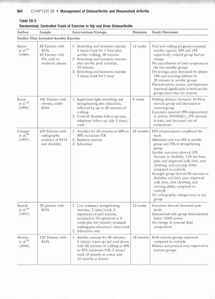
In a systematic review of exercise therapy tor knee and hip osteoarthritis, van Baar et al. 15 found that exercise interventions of varying modes yielded small reductions in disability, small to moderate effects on pain, and moderate effects of exercise on self-reported global assessments. Although studies have examined the impact of exercise on function, few investigators have examined the impact of strengthening programs on cartilage. Table 28-3 presents a summary of the various exercise sUldies designed for individuals with knee OA.
Few studies specifically measure changes in joint proprioception or the impact of exercise programs on joint proprioception. However, some studies emphasize the role of progressive balance activities (e.g., double-limb stance activities to single-limb stance) as methods of improving joint proprioception and strength, based on the assumption that proprioception does not spontaneously return when
. d·
. . I 1617 A
. f .
I d· palll Imlllis 1es. ' vanety 0 exerCises, suc 1 as stan IIlg on unsteady surfaces, using biomechanical ankle platform system (BAPS) boards, and tilt/rocker boards, are used for balance and proprioception training. Although the way that exercise influences proprioception is unclear, clinicians agree that addressing proprioception is important in the rehabilitation program.
In one small trial, patients with hand OA were instructed in yoga and relaxation techniques for 10 weeks. At the end of the trial, patients reported decreased pain and tenderness and improved motion of finger joints. 18
Most studies on the effects of exercise focus on strengthening exercises for patients with mild to moderate �A.
864 CHAPTER 28 • Management of Osteoarthritis and Rheumatoid Arthritis
Table 28-3
Randomized, Controlled Trials of Exercise in Hip and Knee Osteoarthritis
Author Sample Intervention/Groups Duration Study Outcomes
Studies That Included Aerobic Exercise
Minor 80 Patients with l. Stretching and isometric exercise 1 2 weeks Pool and walking programs increased et al 24 KOA 3 times/week for 1 hour plus aerobic capacity 20% and 19% ( 1 989) 40 Patients with aerobic walking, 30 minutes respectively; control group had no
RA, mild to 2 . Stretching and isometric exercise change moderate djsease plus aerobic pool activities, No exacerbation of joint symptoms in
30 minutes the two aerobic groups 3 . Stretching and isometric exercise On average, pain decreased by almost
3 times/week for 1 hour 1 0% and morning stiffness by 20 minutes in aerobic groups
Physical activity, anxiety, and depression improved significantly in both aerobic groups more than for controls
Kovar 1 02 Patients with l. Supervised light stretching and 8 weeks Walking rustance increased 1 8 .4% in et al 25 chronic, stable strengthening plus education, exercise group and decreased in ( 1 992) KOA followed by up to 30 minutes of control group
walking Exercisers reported 39% improvement 2 . Control : Routine follow-up care, in activity (WOMAC), 27% decrease
telephone follow-up calls 3 times/ in pajn, and decreased use of week merucations
Ettinger 439 Patients with l. Aerobics for 60 minutes at 60% to 1 8 months 83% of participants completed the et al 26 radiographic 80% marimum H R study ( 1 997) evidence of KOA 2 . Resistive exercise AdJlerence rate was 68% in aerobic
and disability 3 . Education group and 70% in strengthening group
Aerobic exercisers showed 1 0% decrease in disability, 1 2% less knee pajn, and improved walk time, stair climbing, and carrying ability compared to controls
Strength group showed 8% decrease in disability and knee pain, improved walk time, stajr climbing, and carrying ability compared to conu'ols
No radiographic changes seen in any group
Bautch 30 patients with l. Low resistance strengthelung 1 2 weeks Exercisers showed decreased pajn et al 2 9 KOA exercises, 3 times/week, 3 levels ( 1 997) repetitions of each exercise, Educational only group demonstrated
increased to 1 0 repetitions at 4 better AlMS scores weeks plus low intensity tread nUll No change in synovial fluid walking plus education 1 time/week composition
2 . Education only
Messier 1 03 Patients with l. Aerobic exercise for 50 minutes, 1 8 months Both exercise groups improved et al 28 KOA 5-minute warm-up and cool-down, compared to conu'ols (2000) with 40 minutes of walking at 50% Balance and postural sway improved in
to 85% maximum H R, 3 times/ exercise groups week (3 months at center and 1 5 months at home)
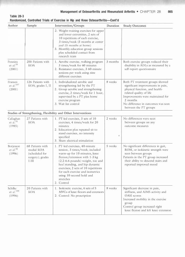
Table 28-3
Management of Osteoarthritis and Rheumatoid Arthritis • CHAPTER 28 865
Randomized, Controlled Trials of Exercise in Hip and Knee Osteoarthritis-Cont'd
Author Sample Intervention/Groups
2 . Weight-training exercises for upper and lower extremities, 2 sets of 1 0 repetitions of each exercise, 3 times/week (3 months at center and 15 months at home)
3. Monthly education group sessions plus scheduled contact from research team
Penninx 250 Patients with 1 . Aerobic exercise, walking program et al. 30 KOA 3 times/week for 60 minutes (200 1) 2 . Resistance exercises, 3 60-minute
sessions per week using nine different exercises
Fransen 1 26 Patients with 1. I ndividualized aerobic and et al . I07 KOA; grades I, II strengthening led by the PT (2001) 2. Group aerobic and strengthening
exercise, 2 times/week for 1 hour, supervised by a PT plus home exercise program
3 . Wait list control
Studies of Strengthening, Flexibility and Other Interventions
Callaghan et al 3 1 ( 1 985)
Borjesson et al .32 ( 1 996)
Schilke et al. 108
( 1 996)
27 Patients with 1 . KOA
2 .
3 .
68 Patients with 1 . medial KOA (scheduled for surgery); grades I - I I I
2 .
2 0 Patients with 1 . KOA
2.
PT-Ied exercises, 3 sets of 1 0 exercises, 4 times/week for 20 minutes Education plus repeated sit to stand exercises, no intensity specified Sham electrical stimulation
PT-Ied exercises, 40-minute session, 3 times/week; included warm-up for 1 0 minutes, knee flexion/extension with 1 -3 kg (2 . 2 -6.6 pounds) weight, toe and heel standing, and hip dynamic exercises; 2 sets of 1 0 repetitions for c;ach exercise and isometrics using 1 0-second hold and stretches Control
Isoki netic exercise, 6 sets of 5 MVCs of knee flexors and extensors Control: No prescription
Duration Study Outcomes
3 months Both exercise groups reduced their disability in ADLs as measured by a self-report questionnaire
8 weeks Both PT treatment groups showed significant improvements in pain, physical function, and health-related quality of life
Improvements were maintained for 2 months
No difference in outcomes was seen between the PT groups
2 weeks No differences were seen between groups on any outcome measures
•
5 weeks No significant differences in gait, ROM, or isokinetic strength were seen between groups
Patients in the PT group increased their ability to descend stairs and reported improved mood
8 weeks Significant decrease in pain, stiffness, and AIMS activity and OASI scores
I ncreased mobility in the exercise group
Control group increased right knee flexion and left knee extension

866 CHAPTER 28 • Management of Osteoarthritis and Rheumatoid Arthritis
Table 28-3 Randomized, Controlled Trials of Exercise in Hip and Knee Osteoarthritis-Cont'd
Author Sample Intervention/Groups Duration Study Outcomes
Van Baar et al.33 ( 1 998)
Rogind et al .22 1998
O'Reilly ct al. 109
( 1 999)
Dcyle ct al . I IO ( 2000)
Hopman-Rock and Westhoff21 (2000)
Petrella and Bartha 19
( 2000)
Baker et al . 1 I I
(200 1 )
Quilty et al. I 12 ( 200 1 )
2 0 1 Patients with 1 . HOA o r KOA
2 .
25 Patients with 1 . KOA ( Kellgren grade 3 on radiograph )
2 .
19 1 Patients with 1 . KOA symptomatic
2 .
83 Patients with l.
KOA, grades I, III
2.
1 03 Patients with 1 . HOA or KOA
2 .
1 79 Patients with 1 . KOA, mild to moderate ( medial compartment) 2 .
4 6 Patients with 1 . KOA
87 Patients l.
2 .
Individual exercises 1 -3 12 weeks Exercisers showed 1 7 -point decrease times/week for 30 minutes in pain and 1 9-point reduction in Education disability ( effect sizes were medium
to small, respectively)
PT-Ied outpatient exercise, 12 weeks 78% Adherence including general fitness, balance, Exercise group increased quadriceps coordination, flexibility, and strength by 20% strengthening exercises, At 1 year, walking speed improved by 2 times/week 1 3%, decreased 3 .8 points, pain Control reduced by 2 points
I ncrease in palpable effusions
Home exercises, including 6 months Decrease in WOMAC pain score by isometric knee exercise ( 5 -second 22 .5% and by 6.2% in controls hold ), dynamic knee and stepping Significant improvement in VAS pain exercises performed daily and 17.4% change in function among Control exercIsers
Manual therapy to the knee, spine, 4 weeks At 4 and 8 weeks, significant hip, and ankle plus dynamic exercises improvements in exercisers for for the hip and knee, as weB as 6MWT and WOMAC scores stretching exercise, 2 times/week At 8 weeks, 6MWT improved 1 3 . 1% for 30 minutes, plus a home exercise and WOMAC improved 55 .8% program compared to controls Subtherapeutic ultrasound Gains still evident at 1 year
Peer-led educational program 6 weeks Significant improvements in pain, lasting 1 hour, plus 1 hour of quadriceps strength, knowledge, PT-led dynamic and static and self-efficacy at postassessment exercises for the hip and knee, Benefits still evident at 6 months in 1 time/week exercise group Control
Progressive home-based exercise. 8 weeks Improvement seen in activity Simple ROM and resistance ( self-paced walking, ste.pping) and exercises plus oxaprozin activity-related pain in both exercise Oxaprozin alone groups
Progressive strength training 16 weeks Improvements seen in strength, program ( squats, step-ups, pain, physical function, and qualIty isotonic exercises of the lower of life l imbs)
Nine 20-minute PT sessions, 10 weeks At 5 months, treatment group including quadriceps exercises, showed small decrease in pain and postural exercises and education, significant improvement in functional exercises, footwear quadriceps strength recommendations, and patellar No difference at 1 year taping. Exercises performed at home, 1 0 times/day Usual care

Table 28-3
Management of Osteoarthritis and Rheumatoid Arthritis • CHAPTER 28 867
Randomized, Controlled Trials of Exercise in Hip and Knee Osteoarthritis-Cont'd
Author Sample Intervention/Groups Duration Study Outcomes
Foley et al .2O 1 05 Patients over l. Hydrotherapy, 3 times/week 6 weeks Both exercise programs produced (2003) age 50 with hip 2 . Gym exercise, 3 times/week nll1ctional gains and improved
OA or KOA 3 . Control quadriceps strength compared with the control group
Compliance was similar for the two exercise groups
Compliance was similar for the two exercise groups
Hydrotherapy group had increased their distance walked compared to
control group Gym exercise group showed increased
walking speed and self-efficacy compared to control group
Gur et al .34 2 3 Patients with l. 1 2 Concentric knee exercises 8 weeks Both exercise groups improved (2002 ) bilateral KOA, performed 3 times/week functional capacity, peak torque of
grade I I or I I I 2 . 6 Repetitions of concentric and 6 knee, and pain relief repetitions of eccentric knee Concentric-eccentric group performed exercises, at angular velocities of better on the stair climbing/ 30o- 1 80/'sec descending than the concentric
3 . Control : Usual activity group, and the concentric-eccentric group showed more reduction in pain
Usual activity group did not show any improvements
Topp et al.3S l . Dynamic resistance Thera-Band 1 6 weeks Isometric group decreased their time (2002) exercises for 40 minutes ( 1 0- to perform functional tasks by 1 6%
minute warm-up and cool-down), to 23% 3 times/week A 1 3% to 1 7% decrease in time to
2 . Isometric exercises, 3 times/week ascend/descend stairs was seen in the dynamic group
Pain decreased in both groups No change was seen in controls
Hoeksma 1 09 Patients with 1 . Manual therapy manipulations of 5 weeks An 8 1 % improvement was seen in the et al.23 hip OA the hip by manual therapist manual therapy group compared to
(2004) 2 . Active exercises for the hip ( nine a 50% improvement in the sessions) plus home program with exercisers weights, endurance training, ROM, and stretching
KOA, Knee osteoarthritis; HOA, hip osteoarthritis; RA, rheumatoid arthritis; WOMAC, Western Ontario and McMaster U niversities
Osteoarthritis Index; AIMS, Arthritis Impact Measurement Scale; HR, heart rate; MVC, maximum volwHary contraction; ADLs, activities of daily
living; PT, physical therapist; OASI, Osteoarthritis Screening I ndex; ROM, range of motion; VAS, Visual Analogue Scale; 6MWr, 6-Min utc Walk
Test.
I n Table 28-3 , randomized, controlled trials of exercise for hip and knee osteoarthritis are described. In an innovative study by Petrella and Bartha, 1 9 patients with knee OA were randomized to either a progressive resistive exercise program using common household items and nonsteroidal antiinflammatory drugs (NSAIDs), or a progressive resistive exercise progranl using common household items combined with simple ROM exercises and NSAIDs, or NSAIDs alone. At the end of 8 weeks, patients in both exercise groups
reported improvements in pain and fWlCtional performance compared to the group that received NSAIDs alone.
Foley et al.20 randomly assigned 1 05 community dwelling patients with OA of the hip or knee to a gym-based or hydrotherapy exercise program or to a control group. The exercise groups attended exercise class 3 times a week for 6 weeks. Both the gym-based and hydrotherapy-based exercise groups showed increased quadriceps muscle su"ength and function compared to the control group. However, the hydrotherapy group tended to

868 CHAPTER 28 • Management of Osteoarthritis and Rheumatoid Arthritis
show greater gains in aerobic qualities of fimction, such as distance walked, whereas the gym-based group showed greater self-efficacy (i .e . , confidence in self-management) .
Hopman-Rock and Westhoff2 1 randomized 1 03 patients with hip and knee OA to a program of health education and exercise. The program lasted 6 weeks and consisted of weekly sessions lasting 2 hours. The sessions included 1 hour of education and self-management led by a peer and 1 hour of exercise led by a physical therapist. The exercise program included a 1 5-minute discussion of the pros and cons of exercise and the importance of rest and alternating activities; this was followed by a warm-up, static and dynamic strengthening exercises for the hip and knee, and a cool-down. Patients not allocated to the education and exercise group received usual care. Significant improvements were fOLmd for pain, quality of life, quadriceps strength, knowledge, self-efficacy, and a physically active lifestyle in the intervention group. The effects were present, although to a lesser degree, at the 6-month assessment.
Rogind et a1.22 found positive outcomes in patients with severe knee OA who combined strengthening exercises and balance and coordination activities .22 In this trial, 25 subjects were allocated to a 3 -month training program that consisted of general fitness exercises, balance and coordination activities, stretching, and lower extremity strengthening plus a home program at an outpatient clinic, or control. Subjects exercised in groups twice a week. At the 3-month assessment, quadriceps strength had increased 20%, as measured by isokinetic testing. At 1 year, improvements were found in pain (a reduction of two points) and in functional index scores (a 3 .S reduction) compared to controls.
Few randomized, controlled studies have examined the impact of manual mobilization techniques combined with exercise in patients with OA. Hoeksma et al 23 allocated 1 09 patients with hip OA to 5 weeks of either manual therapy and manipulations or active resistive hip exercises plus endurance training, ROM, and stretching. At the end of the trial, the manual therapy group demonstrated an S I % improvement compared with a 50% improvement in the exercise and stretching group.
Aerobic walking at moderate intensity appears to improve aerobic capacity by up to 20% without exacerbation of symptoms. In studies by Minor et a1 .24 and Kovar et a1 . ,25 patients with mild to moderate osteoarthritis of the hips and knees not only improved aerobic capacity but demonstrated improvements in mood when aerobic walking was coupled with supervised stretching and strengthening exercises.
Ettinger et al.26 sUldied the effects of exercise in a less controlled setting. In this study, 439 community dwelling subjects age 60 or older who had radiographic hallmarks of knee OA, pain, and physical disability were randomly allocated to an aerobic exercise program at 60% to SO% maximum heart rate, a resistance exercise program, or a health education program. At IS months, patients in the aerobic and resistance exercise groups demonstrated modest improvements in pain ( 1 2%) and disability ( 1 0%), and better
scores on timed walk, stair climbing, and carrying ability than those in the health education group . The authors noted that education alone produced small improvements in outcomes, and this change may have led to the small differences between groups. Previous exercise behavior was the strongest predictor of exercise compliance in this sample?7
In a sinli1arly designed study, Messier et al.28 showed that aerobic exercise and strengthening programs improved balance and postural control . They enrolled 1 03 patients with knee osteoarthritis in an I S-month trial and allocated them to one of three groups: ( 1 ) supervised aerobic exercise at 50% to S5% of the maximum heart rate for 50 minutes, 3 times a week, for 3 months, followed by a home prescription; (2 ) supervised upper and lower extremity weighttraining, 2 sets of l O repetitions, 3 times a week, for 3 months, followed by a home exercise prescription; or ( 3 ) monthly educational sessions and scheduled contacts from the research team . At the end of the trial, patients in the exercise groups showed improved balance and postural stability.
Conclusions about Exercise and Osteoarthritis The evidence from the preceding studies and others demonstrates that patients with mild to moderate knee and hip osteoarthritis can safely engage in effective aerobic and strengthening exercise without exacerbating joint symptoms. Improvements in mood, strength, and aerobic capacity are found, ranging from 1 5% to 40%, depending on the intensity of the prescribed exercise.8 Gains in fill1ctional performance and reductions in limitations are greatest when the progranl lasts at least S weeks and exercises are performed at least 3 times a week.20,24,25,28-35 As with other populations, compliance with an exercise program is critical to its effectiveness for the arthritic patient. Efficacy can be improved when home exercise is supplemented with a supervised program.
Clinical Point
Few studies have addressed the impact of resistance or aerobic
exercise on patients with severe osteoarthritis, and the long-term
effects of exercise on joint integrity and disability are unknown.
Aerobic exercise improves endurance, reduces fatigue, and has modest effects on muscle strength. With structured exercise (3 times a week over 4 months), individuals can improve strength and endurance, leading to decreased dependency and pain and increased functional activity. Some of these benefits continue for up to S months after an intense program.36 The role of aerobic exercise in the management of patients with OA, particularly of the hips and knees, has received greater attention over the past 1 0 years. The concept that inactivity is a risk factor for osteoarthritis, rather than just an outcome of the disease,

Management of Osteoarthritis and Rheumatoid Arthritis • CHAPTER 28 869
emphasizes the importance of aerobic exercise in disease management.
Achieving a Therapeutic Effect with Exercise. Some evidence indicates that exercises that focus on proprioception and balance may reduce disability and improve strength. However, more research is needed i n this area. Despite the evidence that symptoms of OA of the hip and knee can be improved with exercise and that exercise is recom mended in practice guidelines, most patients with OA have not had a prescription for exercise. Even when physicians prescribe exercise, only a small percentage of patients exercise in a manner that can achieve a therapeutic effect.37 Certain patients with osteoar thritis of the hip can exercise at home as effectively as with outpatient hydrotherapy to improve joint mobility and increase m uscle strength.38 Including m an ual therapy with the exercise progran1s appears to provide added benefits. Caution needs to be applied in patients with lax or malaligned knees, especially with tibiofemoral OA, in which q uadriceps strengthening may exacerbate symptoms. Table 28-4 summarizes the exercise recom mendations for OA based on pain level and pathology.
Table 28-4
Summary of Rehabilitation Interventions for Osteoarthritis
Type of Osteoarthritis (OA) Recommendations
Gait Problems and Use of Assistive Devices A flexion contracture across the acetabulum toward the lateral margin39 increases valgus forces at the knee and ankle and causes inefficient gait patterns and increased energy expenditure. Patients describe difficulty and pain when walking or climbing stairs and reduced functional independence. Decreased m uscle strength and reduced joint proprioception also are associated with an increased incidence of falls. I 7
I n one study, an exercise program consist ing of individ ualized progressive training, i ncluding isometric and dynamic exercises for patients with knee osteoarthritis, sign ificantly improved muscle function, functional capacity, and walkin g time ( as much as 2 1 %) and reduced selfreported difficulty with walking and pain. 36 Stationary cycling also has demonstrated improvements in walking speed, aerobic capacity, and pain .40
In a study by Leivseth et a 1 . ,4 1 six patients with severe OA of the hip who were awaitin g total hip arthroplasty (THA ) increased hip adduction by 8 .30 , increased the type I and I I fiber cross- sectional area, a n d i ncreased glycogen levels after passive muscle stretching perpendicular to the direction of
OA of the hip and knee Mild pain AROM exercises ( 1 0 repetitions), 3 to 5 repetitions of flexibility and static exercises (8 to 1 0
Moderate pain
Severe pain
Bone on bone
OA of the hand
repetitions of 6 seconds' duration ) Dynamic exercises, especially of the quadriceps and hamstrings (8 to 1 0 repetitions ) Low impact aerobic activities (pool, bicycling) 20 minutes, 3 times/week Balance activities ( BAPS and tilt boards), single-limb stance
Static and dynamic exercises-reduce to 5 repetitions. Flexibility exercises, 3 to 5 repetitions Low impact aerobic exercises (pool, bicycling) for 20 minutes, 3 times/week Balance and proprioception activities-bilateral Use of cane or lateral heel wedge foot orthosis, neoprene knee sleeve
Static and dynamic exercises ( no resistance ), 3 to 5 repetitions except with internal joint derangement Low to no impact aerobic exercises (pool) Note: Advise functional activities to keep moving
Same as for severe form but few or no repetitions of dynamic exercises; patient education is very important
Note: Caution should be used in prescribing quadriceps strengthening exercises for patients with ligamentous laxity and malalignment
Orthosis: Varus unloader-type knee orthosis; may need crutches or walker
Active movements, few repetitions, low resistance Teach home exercises, which the patient should repeat daily Aim to maintain full range of motion of MCP, P I P, and D I P joints
Modified from Iversen MD, Liang MH, Bae SC: Exercise therapy i n selected arthritides: rheumatoid arthritis, osteoarthritis, systemic lupus
erythematosus, systemic sclerosis and polymyositis/dermatomyositis. I n Frontera WR, Dawson DM, Siovik DM, editors: Exercise iu rehabilitation
medici1le, Champaign, I L, 1 999, Human Kinetics.
AR OM, Active range of motion; BAPS, biomechanical ankle platform system; MCp, metacarpophalangeal; PI1� proximal interphalangeal; DJJ�
distal interphalangeal.

870 CHAPTER 28 • Management of Osteoarthritis and Rheumatoid Arthritis
the adductor muscle (without hip movement) . The subjects stretched with a force of 20 to 30 kg (44 . 1 to 66 . 1 Ibs) applied manually for 30 seconds, rested for 10 seconds, and repeated; the sessions lasted 25 minutes and were performed 5 days a week for 4 weeks.4 1 Recent studies have also demonstrated improved rates of recovery in patients with end stage hip arthritis after preoperative and perioperative exercise programs for THA.42,43
Gait problems are common in patients with OA of any of the weight-bearing joints and are a clue to underlying pathology of the soft tissues or the joints themselves. Left untreated, they can cause problems. Clinicians should be attentive this diagnosis and design treatments that maximize joint function.
Orthoses for Osteoarthritis Much of the research into bracing for OA has concentrated on devices to relieve knee pain and disability. In OA, the knee is subjected to increased medial compartment pressures secondary to varus loading during gait. Two main methods have been advanced to reduce these excessive forces: unloader-type knee orthoses ( KO ) or lateral wedge insoles at the foot. An
unloader KO for varus gonarthrosis, or knee varus, can be either a thrust-type (Figure 28-6) or prestressed brace. The thrust-type, hinged KO has a single upright designed to create a valgus correcting force at the knee to unload the medial compartment.44 Kirkley et al.45 compared use of a varus unloader KO to a neoprene sleeve or to medical treatment alone in a group of 1 1 9 subjects with OA. After 6 months, the subjects who wore the unloader KO showed a significant difference in pain relief with fi.ll1ctional tasks (e .g., a 6-minute walk and a 3-minute stair climb) compared to the group who
Figure 28-6 Varus unloader knee brace.
wore the neoprene sleeve. Both groups performed better than the non brace group.
Lindenfeld et al.46 assessed the biomechanical properties of the brace to actually reduce the varus forces as well pain. They compared 1 1 subjects with OA before and after 4 weeks of brace wear and then compared them to 1 1 healthy controls. As measured on the Cincinnati Knee Rating System, pain, function, and biomechanical alignment all improved in the brace group. Alignment levels were reported to approach that of the normal control group.
Lateral wedge insoles or a variant, subtalar strapped insoles, are prescribed to make use of the closed chain properties of a foot orthosis (FO) on reducing compressive forces on the medial tibiofemoral joint compartment. Brouwer et aJ .47 concluded that some limited evidence indicated that lateral wedges reduced the use of pain medication by OA patients when compared those who wore a neutral insole. However, function, as measured by the Western Ontario and McMaster Universities Osteoarthritis Index (WOMAC) functional scale, was not improved over the group that used neutral insoles after 6 months of wear. These researchers also reported that a lateral wedge insole with strapping for the subtalar joint demonstrated a biomechanical realignment effect, as measured by the femorotibial angle (FTA), but that those wearing these insoles reported more low back and foot pain with use. As yet, no studies have directly compared tile use of an unloader KO with a lateral wedge insole or strapped subtalar lateral wedge insole. The clinical decision for use of tllese devices should be based on tile patient's degree of disability and potential for compliance with an ortllosis and tile cost of the device.
Case Study in Osteoarthritis
John is a 77-year-old male who presents with a diagnosis of right hip OA. His radiographic findings suggest decreased joint space with small acetabular osteophytes off the anterior aspect of We joint. He has opted to delay surgical intervention and wants to use physical therapy to enhance his overall physical status. He reports minimal difficulty with activities of daily living (ADLs) and instrumental activities of daily living ( IADLs) ( see volume 1 of tllis series, Ortho
pedic Physical Assessment, Chapter 1 ) . However, he has been increasingly sedentary since retiring from the post office 1 0 years ago. Currently, he reports late afternoon soreness on tile anterior aspect of the leg and along tile groin to his medial tlligh, especially after doing a lot of walking.
On examination, John walks with a slow, deliberate gait pattern. He demonstrates decreased stance time on tile right leg, and his right hip is maintained in flexion throughout the gait cycle. Decreased step length is noted on the left, and a positive Trendelenburg's sign on tile right. Right hip ROM is 20° to 1 00° with a firm, unyielding end feel; internal rotation and abduction is 0° to 20° Witll a firm end feel; and external rotation is 0° to 35°. Left hip ROM

Management of Osteoarthritis and Rheumatoid Arthritis • CHAPTER 28 871
is within normal limits. Right hip muscle strength is in the fair range. Lower abdominal strength is poor. Knee strength is good, and ankle strength is within normal limits (WNL). The patient has a positive Thomas test (200 ) on the right. He demonstrates an inability to squat without visible muscle sluking and altered posture, a positive Trendelenburg's sign with unilateral squat, and increased trunk flexion with bilateral squat. Sensation is intact to light touch, but right hip kinesthesia and proprioception are reduced. He is able to maintain single-limb stance for approximately 5 seconds with eyes open. His past medical history is significant for coronary artery disease (no history of myocardial infarction), hypercholesteremia, gastroesophageal reflux disease, and a weight gain of 1 3 .6 kg (30 Ibs) over the past 1 0 years. His body mass index (BMI) is 27. He currently is taking the following medications: Zesteril, Lipitor, and Prilosec.
Questions: What rehabilitation program would you advise? What education about restrictions to his activities would you recommend? What type of exercise would you recommend? Do you th.ink an assistive device would be usefi.t1?
Case Discussion The patient's impairments of decreased hip muscle flexibility, poor muscle performance of the hip and trunk, poor motor control, pain, joint mobility, and diminished proprioception contribute to his altered gait and should be addressed in the rehabilitation program. Flexibility exercises and joint mobilization will help restore mobility and unload the hip joint while allowing a more normal gait pattern. Depending on the degree of pain, short-term use of a straight cane can fi.lrther unload the joint for long distance walking. He will benefit from an exercise program that emphasizes strengthening of tile proximal musculature of tlle hip, especially the weak gluteus medius. An exercise program should be started that incorporates stability exercises in weight bearing, such as rhytllmic stabilization in standing, and progresses to unilateral static and dynamic control. The progression should take place as pain diminishes and proximal motor control improves. These activities not only improve motor coordination and strength, but also enhance proprioception. Aerobic exercise, such as aquatic classes a.nd/or elliptical training, performed at 60% to 80% of his ma,ximum heart rate, 3 times per week, should be incorporated into his program. The patient needs to learn how to modifY his exercise regimen to adjust for his level of pain and to progress his exercises over time.
Rheumatoid Arthritis
Epidemiology and Pathophysiology
Classic rheumatoid arthritis is a chronic inflammatory disorder that not only affects joints, but also has multiple systemic manifestations. It affects approximately 1 % to 2% of
the U.S. population,48 and it shortens life expectancy by about 1 0 years.49 RA begins in early to middle life, affects women more often tlun men and the incidence increases with age.
The predominant patllology in rheumatoid disease is an inflammation of tlle synovium of diartlu·odial joints, leading to a state of chronic synovitis. Synovitis tllat goes uncontrolled can lead to joint destruction (see Table 28- 1 ) . RA is characterized by periods of exacerbation and remission. The chronic fluctuati.ng course of RA can follow various patterns. Patients may experience a continuous, low grade exacerbation or have periods of remission followed by exacerbations of various intensities. Although exacerbations are a feature of the disease, they can be triggered by infection or trauma or can occur after medications have been stopped. Fewer than 1 0% of patients witll RA go into prolonged remissions. 50
The pattern of joint involvement generally is symmetrical and polyarticular. Nearly 1 0% of those affected develop some joint deformities within 2 years of diagnosis. Radiological changes often are seen earliest in tlle feet and hands. The Sharp score, as modified by van der Heijde,5 1 is a method of documenting the progression of the disease. Radiologists score the number of bony erosions and areas of joint space narrowing in 30 to 32 joints of the hand and 1 2 joints in tlle feet. The total score is recorded (maximum involvement equals 448 points) and monitored on successive radiographs to track progression.
AltllOugh RA can affect any area, the most commonly affected joints are the wrists, metacarpophalangeal (Mep) joints, and PIP joints in the upper extremity (Figure 28-7) and tl1e ankle and foot ( Figure 28-8) in the lower extremity. In tl1e spine, RA is more common in the cervical area but can involve all segments. Because tllis is a systemic disease, extra-articular clinical symptoms may involve the cardiovascular system, pulmonary system, integument, and nervous system. Pleurisy, often presenting as shortness of breath or dyspnea on exertion, has been found in up to 70% of patients witl1 RA. Pericarditis, myocarditis, and vasculitis, which may affect about 30% of patients, as well as renal ef fects secondary to vasculitis or to the toxic effects of long-term drugs used to control the disease, are all conditions tl1at af fect the physical fi.l11ctioning and exercise tolerance of patients witll RA.49,50, 52
Joints Commonly Affected by Rheumatoid Arthritis
• Wrist
• Metacarpophalangeal (MCP) joints
• Proximal interphalangeal (PIP) jOints
• Foot
• Ankle
• Knee

872 CHAPTER 28 • Management of Osteoarthritis and Rheumatoid Arthritis
Figure 28-7 A, Ulnar deviation at the metacarpophalangeal ( MC P ) joints. This is a common deformity in patients
with inflam matory arthritis, such as this patient with rheumatoid arthritis ( RA) . B, Ulnar deviation i n rheumatoid arthritis. Severe ulnar deviation c a n b e seen a t t h e MCl' joi nts with extensive erosions. l'ancompartmental bon)' ankylosis and erosion are also seen at the wrist. C, Rheumatoid arthritis
of the hand, showing advanced changes with ulnar drift and palmar subl uxation at the MCl' joints and
swan neck and boutonniere deformities in the fingers. (A and B from Harris ED, Budd RC, Firestein
GS ct al: Kelley's textbook of rheumatology, ed 7, pp 49 1 , 745 , Philadelphia, 2 0 0 5 , WB Saunders;
C from Swanson AB: l'athomechanics of deformities i n the hand and wrist. In H unter ], Schneider
LH, Mackin E], Callahan AD, editors: Rehabilitation of the hand: sttrgery and therapy, p 895,
St Louis, 1 990, Mosby.)
Management of Osteoarthritis and Rheumatoid Arthritis • C HAPTER 28 873
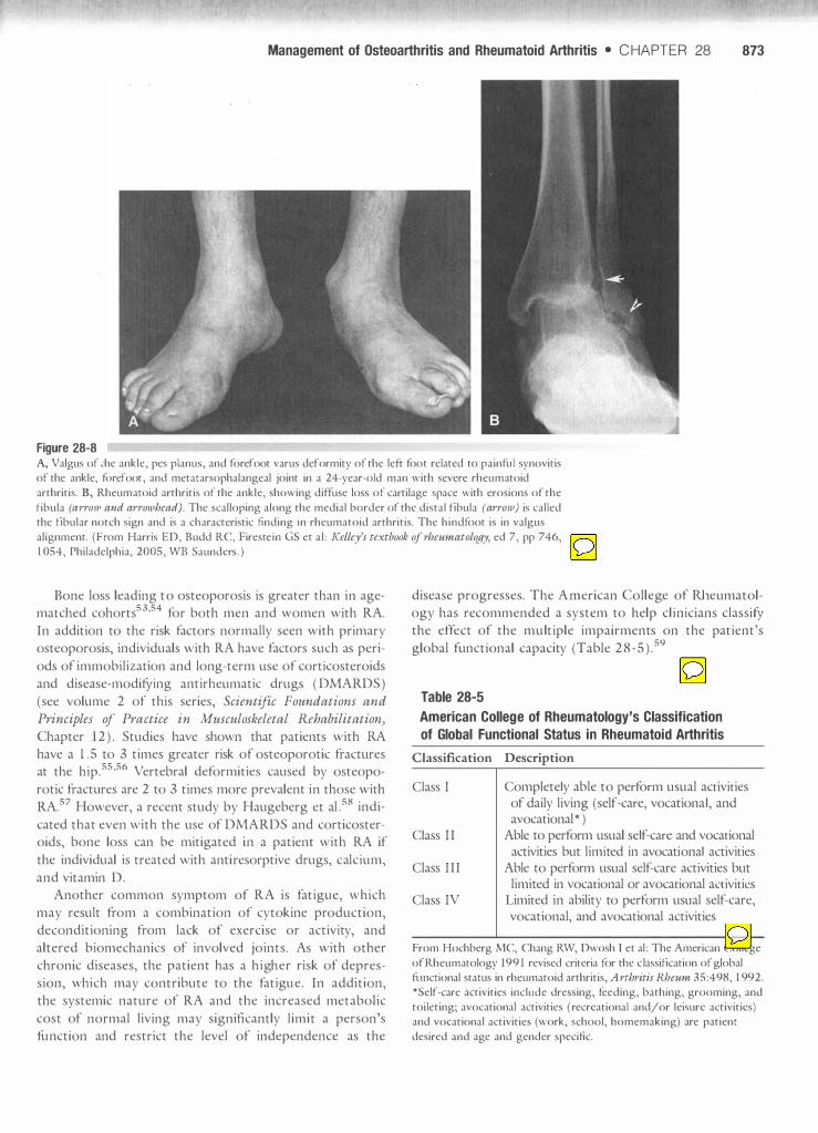
Figure 28-8 A, Valgus of �he ankle, pes planus, and forefoot varus deformity of the left: foot related to painnIi synovitis
of the ankle, forefoot, and metatarsophalangeal joint in a 24-year-old man with severe rheumatoid
arthritis. B, Rheumatoid arthritis of the ankle, showing diffuse loss of cartilage space with erosions of the
fibula (arroJII and arrowhead). The scalloping along the medial border of the distal fibula (arr01v) is called the fibular notch sign and is a characteristic finding in rheumatoid arthritis. The hindfoot is in valgus
alignment. (From Harris ED, Budd RC, Firestein GS et al : Kelley's textbook of rheumatology, ed 7, pp 746,
1 054, Philadelphia, 2005, WE Saunders . )
Bone loss leading to osteoporosis is greater than in agematched cohorts53,54 for both men and women with RA. In addition to the risk factors normally seen with primary osteoporosis, individuals with RA have factors such as periods of immobilization and long-term use of corticosteroids and disease-modifYing antirheumatic drugs ( DMARDS) (see volume 2 of this series, Scientific Foundations and
Principles of Practice in Musculoskeletal Rehabilitation,
Chapter 1 2 ) . Studies have shown that patients with RA have a 1 .5 to 3 times greater risk of osteoporotic fractures at the hip.55,56 Vertebral deformities caused by osteoporotic fractures are 2 to 3 times more prevalent in those with RA.57 However, a recent study by Haugeberg et al .58 indicated that even with the use of DMARDS and corticosteroids, bone loss can be mitigated in a patient with RA if the individual is treated with anti resorptive drugs, calcium, and vitamin D.
Another common symptom of RA is fatigue, which may result from a combination of cytokine production, deconditioning from lack of exercise or activity, and altered biomechanics of involved joints. As with other chronic diseases, the patient has a higher risk of depression, which may contribute to the fatigue . In addition, the systemic nature of RA and the increased metabolic cost of normal living may significantly limit a person's function and restrict the level of independence as the
disease progresses. The American College of Rheumatology has recommended a system to help clinicians classifY the effect of the multiple impairments on the patient's global functional capacity (Table 2 8 _ 5 ) . 59
Table 28-5
American College of Rheumatology's Classification
of Global Functional Status in Rheumatoid Arthritis
Classification Description
Class I
Class I I
Class I I I
Class IV
Completely able to perform usual activities of daily living (self-care, vocational, and avocational * )
Able to perform usual self-care and vocational activities but limited in avocational activities
Able to perform usual self-care activities but limited in vocational or avocational activities
Limited in ability to perform usual self-care, vocational, and avocational activities
From Hochberg MC, Chang RW, Dwosh I et al : The American College
of Rheumatology 199 1 revised criteria for the classification of global
nmctional status in rheumatoid arthritis, Arthritis Rheum 3 5 :498, 1 992.
*Self-care activities include dressing, feeding, bathing, grooming, and
toileting; avocational activities ( recreational and/or leisure activities)
and vocational activities (work, school, homemaking) are patient
desired and age and gender specific.
874 CHAPTER 28 • Management of Osteoarthritis and Rheumatoid Arthritis
RA may also affect children. Juvenile rheumatoid arthritis ORA) ( referred to as juvenile idiopathic arthritis [JIA] by the European League Against Rheumatism) is diagnosed when the onset of symptoms occurs before age 1 6 and the symptoms persist for at least 6 consecutive weeks. No specific diagnostic tests can be used to confirm JRA; rather, several tests can be performed to support the clinical impression of the disease. The three primary subtypes of J RA established by the American College of Rheumatology are pauciarticular JRA, polyarticular JRA, and systemic JRA. These subtypes are based on the number of joints involved, the presence or absence of systemic features and, in some cases, the age of onset.60 Because these classifications are based on the characteristics of the disease at onset, they may not indicate its course over time 61
Pauciarticular JRA is defmed as synovitis in four or fewer joints, usually the elbows, knees, and ankles. The arthritis, which most often is asymmetrical, presents in the absence of systemic features. In pauciarticular JRA, the rheumatoid factor usually is negative. Pauciarticular JRA has two subtypes, based on the onset of disease and the clinical presen tation. Early onset pauciarticular disease often occurs in girls before the age of 5 . Late onset pauciarticular disease occurs around the ages of 1 0 to 1 2 years and most often affects the large weight-bearing joints (h ips and knees) and en theses (muscle insertions ) . Stiffness and pain are the predominant complaints . 6 1
Classification of Rheumatoid Arthritis in Children
• Pauciarticular (four or fewer joints)
o Early onset
o Late onset
• Polyarticular (five or more jOints)
• SystemiC
Polyarticular JRA affects girls more often than boys. It is characterized by synovitis in five or more joints, and patients may present with systemic features of the disease. The synovitis often is symmetrical . Although polyarticular J RA may involve any joint, the cervical spine frequently is affected. It is important to note that polyarticular JRA affects normal growth and development.61
Systemic JRA can occur at any age and is gender neutral. It is characterized by high, spiking fevers, a classic rash (pink to salmon colored) that often occurs on the trunk and proximal extremities, and synovitis in one or more joints. The fever generally is the first symptom and may precede other symptoms by up to months. Malaise, anemia, pericarditis, thrombocytopenia, lymphadenopathy, hepatomegaly, and other systemic features evident with adult RA
may be present. These children are so ill that they often present with severe functional limitations and growth retardation ( below the 5th percentile ) 6 1
Signs, Symptoms, and Impairments
Pain, Stiffness, and Swelling RA can cause adaptive shortening of the soft tissues, tendons, and joint capsules. Bone erosion and cartilage loss reduce joint space and may contribute to sLlbluxation-limiting joint motion. Swelling associated with joint effusion and extra-articular edema further reduces mobility. These changes result in significant mobility impairments and pain, especially during exacerbations. Analgesic and antiinflanmlatory medications are the mainstays of medical management for these patients (see volume 2 of this series, Scientific Foundations and Principles of Practice in Mus
culoskeletal Rehabilitation, Chapter 1 2 ) . However, physical modalities and exercise play an important role in reducing functional loss.
Signs and Symptoms of Rheumatoid Arthritis
• Chronic inflammation
• Exacerbations and remissions
• Symmetrical, polyarticular joint involvement
• Osteoporosis
• Fatigue
• Pain
• Deformity (subluxations)
• Mobility impairment
• Muscle weakness
• Depression
Studies that Address Pain, Stiffness, and Swelling. Cold helps reduce inflammation and pain when the joints are acutely inflamed. Heat should be avoided at this time, because it may exacerbate the inflammatory process.62 When the inflammation resolves, either heat or cold can be used to relieve pain.63 Local heat may be preferred before gentle exercise to reduce join stiffiless. In general, few studies examine the effectiveness of modalities in the management of RA. A recent Ottawa Panel64 concluded that some (but limited ) evidence supports the use of ultrasound, paraffin, transcutaneous electrical nerve stimulation (TENS ), and low level laser therapy in the management of pain and flexibility. The panel's guideline further stated that evidence for including or excluding electrotherapy modalities is inconclusive at tllis time .
Exercise may be used to reduce joint pain and stiffiless through the stimulation of endogenous opiates. Ekdalll et al 65 studied endorphin levels in patients with RA during exercise. They showed tllat a rise in serum levels of these internal opiates is directly related to tlle intensity and frequency of active exercise. Numerous research studies have found tllat ROM exercises safely and efficiently maximize joint motion in patients with RA even when performed for long periods 66-7o Patients with early or stage I disease

Management of Osteoarthritis and Rheumatoid Arthritis • CHAPTER 28 875
can safely perform daily exercise, including active flexibility and ROM , doing a few repetitions at each joint. Passive stretching should be deferred unti l the joints are not in an active exacerbation.8
Muscle Weakness M uscle weakness is common with RA. Compared to agematched healthy individuals, individuals with RA are as much as 33% to 55% weaker in knee flexion and extension.62,7 1 The weakness is attributable to a n umber of causes, such as myositis or muscle fiber atrophy. Myositis, characterized by inflammation within the muscle itself, is not always apparent but can be assessed through m uscle enzyme testing. If myositis is not present or is minimal, muscles can be exercised sufficiently so that training effects for muscle strengthening can be achieved .72 Atrophy has been shown to occur in both type I and type II m uscle
Table 28-6
fibers and may stem either from disuse or from changes to the muscle tissue itself.73
Studies of Strength Training. Studies of strength training for patients with RA have focused on both effectiveness and safety.74-76 Early studies of strength training focused on the impact of low intensity training, such as active ROM , isometric, or low load resistance exercise programs.74 These studies incorporated exercise of varying intensity, frequency, and duration to avoid exacerbation of symptoms. More recent investigations sUldied moderate to high intensity strength training programs, defined as 50% or greater of maxim u m voluntary contraction ( MVC ) . Considerable emphasis was placed on determining whether patients with RA could tolerate higher loads without sacrificing joint integrity. The randomized, control led trials of exercise in patients with RA are reviewed in Table 28 _6.75-82
Randomized, Controlled Trials of Exercise in Rheumatoid Arthritis
Author Sample
SUldies That Included Aerobic Exercise
Ekblom et al. 1 1 3
( 1 975)
Ekblom et al.67
( 1 975 )
Harkcom et al. 84 ( 1 985)
Minor et al.24 ( 1989)
34 Patients with nonactive disease, class I I , 1 1 1
30 Patients with nonactive disease, class I I , I I I
20 Patients with nonactive disease, class I I
4 0 Patients with variable disease activity
Intervention/Groups
1 . General rehabilitation (3 times/day) and ROM
2 . Strength training and aerobic bicycle ergometry, 5 days/ week, 20 to 40 minutes/day
1 . 6-Month follow-up on patients enrolled in study by Ekblom et al. above
1 . Low intensity biking, 3 times/ week at 50 rpm for 15 minutes
2. Low intensity biking for 25 minutes
3 . Low intensity biking for 35 minutes
4. Control
1 . Aerobic walking 2 . Aqua aerobics 3. ROM exercise al l groups;
weekly aerobic exercise at 60% to 80% max H R, 3 times/week for 1 hour
Duration Study Outcomes
6 Weeks Trained group showed significant improvement in muscle strength and aerobic capacity measures, such as V02 max, decreased perceived exertion, and stair climbing
Control group showed no changes
Joint status was unchanged with exercise
6 Weeks 6 Patients who exercised 4 times/week or more demonstrated sustained benefits
1 2 Weeks Al l exercisers improved aerobic capacity (V02 max ) and exercise duration
Only the group that exercised for 35 minutes showed differences in aerobic capacity
Exercise tolerated by subjects in all exercise groups
1 2 Weeks Significant improvement was seen in aerobic capacity, 1 5 .2 m (50 feet) walk time, and daily physical activity in aerobic groups versus ROM group
No difference was seen between groups with regard to morning stiffness, flexibility, clinically active joints, or grip strength

876 CHAPTER 28 • Management of Osteoarthritis and Rheumatoid Arthritis
Table 28-6 Randomized, Controlled Trials of Exercise in Rheumatoid Arthritis-Cont'd
Author
Baslund et al . 1 1 4 ( 1 993)
Hansen et al . I 1 5
( 1 993 )
Van den Ende et a l .70 ( 1 996)
Hall et al 86 ( 1 996)
van den Ende et a1. 85 ( 2000)
Westby et al . 1 1 6 (2000)
Sample
1 8 Patients with moderate disease activity
75 Patients with mixed disease activity, class I, I I
1 00 Patients with stable disease
64 Patients with active RA, no TKR, mean disease of 8 years, in hospital for care
30 Women with c lass I , I I disease; low dose steroids
Intervention/Groups Duration
l. Progressive biking, 4 to 5 8 Weeks times/week at 80% V02 max for 35 minutes
l. Individ ualized general exercise, 2 Years 1 5 minutes, plus aerobic exercise, 2 to 3 t imes/week for 30 minutes
2 . Group exercise SE plus weekly group
3 . PT exercisers 4. No exercise
1 . High intensity group: 1 2 Weeks Walking/biking at 70% to 85% maximum HR plus strengthening exercise, 3 times/week for 1 hour
2 . Low intensity group: ROM and isometrics, 2 times/week for 1 hour
3 . Low intensity individual program: 2 times/week
4 . Low intensity home program: 2 times/week
l . Hydrotherapy, 2 times/week 4 Weeks for 30 minutes
2 . Land exercises, 2 times/week for 30 minutes
3 . Relaxation train ing
1 . Intensive exercise biking, 1 Month 3 times/week, resistance exercise at 70% MVC
2 . Conservative exercises, ROM and isometric exercise
l . Weight-bearing aerobic dance 1 2 Months and strength exercise 3 times/ week for 45 to 60 minutes
2 . Usual activity with written information about exercise
Study Outcomes
V02 max uptake increased 1 8% in the trained versus the control groups
HR at stage 2 and BORG decreased
No change seen in immune response
No effect was seen on stiffness, pain scores, number of swoLlen joints, or functional scores; muscle strength improved in all groups, but no change was seen in aerobic fitness
50% Attendance in program was achieved by half of participants
25% Reported wanting to do more intense program
High intensity group improved the most compared to other exercisers in aerobic capacity, muscle strength, and joint mobil ity significantly
No differences were seen in deterioration of disease activity
Follow-up at 1 2 weeks after program ended showed loss of aerobic capacity gains in high intensity group with no exercise
All groups reported improved mood
Hydrotherapy group had 27% less joint tenderness than other groups and increased knee ROM by 6.60
Changes in mood for hydrotherapy group remained at follow-up
Significant increases seen in muscle strength, as well as improvement in disease activity (swollen joints decreased at 6-month follow-up for intensive exercise group)
No differences seen in pain or joint mobil ity between groups
8% Decrease in active joints among the exercise group compared to a 2% decrease in control group

Management of Osteoarthritis and Rheumatoid Arthritis • CHAPTER 28 877
Table 28-6 Randomized, Controlled Trials of Exercise in Rheumatoid Arthritis-Cont'd
Author Sample Intervention/Groups Duration Study Outcomes
Exercisers had a 2 1 % reduction in ESR values versus a 1 3% increase in controls.
Significant changes were seen in HAQ and fitness level
No change was seen in bone density
Bilberg et a 1 .8? 46 Patients with class l. Group pool exercise, moderate 1 2 Weeks Moderate pool exercise resulted (2005 ) I, I I I disease; stable intensity aerobic and strength in significant improvement in
medications exercise muscle endurance and vitality 2 . Home exercise score on SF-36
No significant difference was seen in aerobic capacity measured by submaximum cycle ergometer
Studies of Strengthening Exercises, Flexibility, and Other Interventions
Wessel and 32 Patients with l . Isometric knee exercise, 7 Weeks Pain ( both VAS and modified Quinney L I ? stable disease, 3 times/week BORG scale) was significantly ( 1 984) class 1 1 2 . Isokinetic knee exercise, higher at all tests for isometric
3 times/week exercises tllan for isoki netic 3 . Control exercises at 1 80o/sec
Swelling was not detected in any group
Ekdah I et a1. 65 67 Patients Witll low Home exercises supervised PT 6 Weeks Dynamic exercisers had ( 1 990) to moderate disease visits four groups: significantly greater increases in
activity, class I I 1 . Dynamic plus 1 2 PT visits strength, aerobic capacity, 2 . Dynamic plus 4 PT visits endurance, and functional 3. Static exercise plus 1 2 PT ability tllaIl static exercisers; 4. Static plus 4 PT visits; HEP effects persisted a t 3 months
for additional 3 montlls No joint flares occurred in either group
Brighton et al .66 44 Patients with class 1 . Hand exercises daily 48 Months Significant i ncreases were seen in ( 1993) I disease 2 . N o exercise o r active disease strengtll aIld pinch grip in the
exercisers Deterioration was noted in controls
Hoenig et al. I 1 8 57 Patients with class All patients performed exercises 3 Months ROM improved hand joint ( 1993) II, III disease 2 times/daily for 1 0 to 20 count
minutes Resistance and dexterity improved l . Hand ROM exercises 2 . Hand resistance exercise 3 . Hand ROM plus resistive
exercises 4 . Control
Hakkinen 43 Patients 1 . Progressive dynamic strength 1 0 Montlls Exercise group significantly et a1 .77 ( 1994) training, 2 to 3 times/week at improved bilateral dynamic
40% to 60% max RM strength ( 32%) and unilateral 2 . Habitual physical activity strength; ESR levels and
function i ncreased Only slight changes were seen in joints

878 CHAPTER 28 • Management of Osteoarthritis and Rheumatoid Arthritis
Table 28-6 Randomized, Controlled Trials of Exercise in Rheumatoid Arthritis-Cont'd
Author
Stenstrom et al . I 1 9 ( 1 975)
Rail et al.78 ( 1 996)
Komatireddyl 2o ( 1997)
Stenstrom et al . 1 2 l ( 1 997)
Bostrom et al . 1 22 ( 1998)
McMeeken et a1.83 ( 1 999)
Sample
54 Patients with class I, II disease
30 Patients with controlled disease
49 Patients with class I I , II I disease
54 Patients with class I, II disease
45 Patients with mild RA
36 Patients with nonacute RA
Intervention/Groups Duration
Two groups: 3 Months 1 . Dynamic exercise, 3 times/
week for 30 minutes 2 . Relaxation training, 3 times/
week for 30 minutes
Four groups: 1 2 Weeks 1 . 8 Healthy elderly adults 2 . 8 Patients with RA
3 . 8 Young, healthy patients who performed resistance exercises 2 to 3 times/week at 80% IRM
4. 6 Elderly adults who did warm-up swimming exercise
1 . Progressive resistive circuit 1 2 Weeks exercises at 30% to 40% of maximum load, more than 3 times/week for 20 to 30 minutes, plus video
2 . N o exercise
Two groups: 1 2 Months 1 . Dynamic train ing 2 . Relaxation exercises for 30
minutes 5 times/week for 3 months then 2 to 3 times/ week for 9 months ( total time is 1 year)
1 . Progressive dynamic shoulder 1 0 Weeks exercises at 30% maximum load, 3 times/week for 40 to 60 minutes
2 . Static shoulder exercises
1 . Knee extensor and flexor 6 Weeks strengthening at 70% max RM,
2 to 3 times/week for 45 minutes
2 . Attention control
Study Outcomes
Dynamic group showed less perceived exertion with activity and improved scores on walking tasks
Relaxation group improved in health-related perception, had less joint tenderness, and better L E muscle function
Exercisers increased muscle strength compared to controls: RA, 57%; young exercisers, 44%; elderly exercisers, 35%; no change was seen in joint symptoms
No change was seen in joint symptoms
RA patients reported improvements in pain and fatigue
Exercisers had significant improvements in self-reported joint counts and number of painfi.ll joints, knee extension HAW scores, and sit to stand time
No significant changes were seen in V02 max or treadmill time
I mproved physical and work impact was seen among dynamic exercisers, as measured by AlMS2
Relaxation group reported less pain, emotion reaction
No significant differences were seen in health status or joint tenderness between the groups
Compliance was about 65% for both groups over 1 year
Both groups reported fewer UE swollen joints and less shoulder-arm pain
Dynamic exercisers showed improvements in physical and overall dimensions in the Sickness Impact Profile
Exercisers reported significantly less pain and had improved knee extension/flexion strength and improved function as measured by HAQ and increased TUG time

Table 28-6
Management of Osteoarthritis and Rheumatoid Arthritis • CHAPTER 28 879
Randomized, Controlled Trials of Exercise in Rheumatoid Arthritis-Cont'd
Author
Hakkinen et aL 123 ( 1999)
Hakkinen et a! . 79 (200 1 )
Dejong et al .75
(2003 )
Hakkinen et al 8 1 (2004)
Dejong et al .88 (2004)
Sample
67 Patients with recent onset disease « 2 years)
70 Patients with recent onset disease « 2 years)
309 Patients with class I - I I I disease
70 Patients with recent onset disease « 2 years)
309 Patients with stable disease, class I - I I I
Intervention/Groups
l . Dynamic whole body exercises at 50% to 60% maximum load, 2 times/week for 45 minutes
2 . ROM exercises 2 times/week and normal recreational activities
1 . Dynamic whole body strengthening at 50% to 70% max RM, 2 times/week for 45 minutes
2 . 2 times/week for 24 months; all patients were encouraged to participate in recreational activities 2 times/week
1 . H igh intensity bicycle, circuit exercises, sports
2 . Usual care
5-Year follow-up study of above groups
l . High intensity weight-bearing exerCIse
2 . Usual care
Duration Study Outcomes
1 2 Months Significant strength increases (22% to 35%) were seen in the dynamic group versus 3% to
24% in the ROM group No significant changes were seen
in BMD
24 Months 62 Patients completed the study. Muscle strength disease activity
( HAQ) and function improved in both groups, but greater improvements were seen in those who did dynamic exercises ( 1 9% to 59% increase in muscle strength)
ROM group showed decreased BMD, whereas dynamic exercisers showed slight improvements in BMD femoral neck (0. 5 1 ± 1 .64%) and spine ( 1 . 1 7 ± 5 . 34%)
2 Years Significant improvements were seen in aerobic fitness, muscle endurance, and self-reported improved function (MACTAR); no significant effect was seen on the small joints of the hands and feet
No mean radiological changes were seen in the large joints except in those with baseline moderate changes
24 Months, 59/62 Subjects were re-monitored evaluated at 5 years by Gains in muscle strength in researchers; exercisers were maintained with
36 months, self- monitored exercise self- BMD values remained unchanged monitored from 2-year status
Few joint changes were seen
2 Years Exercise groups showed significant improvement in aerobic fitness and a decrease in radiological joint changes in the feet
Increase in BMD of the hips was seen in the weight-bearing group
IRM, Illness Representation Model; AIMS2, Arthritis I mpact Measurement Scale 2; BMD, bone mineral density; BORG, Borg scale of perceived
exertion; ESR, erythrocyte sedimentation rate; HAQ, Health Assessment Questionnaire; HEP, home exercise program; HR, heart rate; LE, lower
extremity; MA CTAR, McMaster Toronto Arthritis Patient Preference Questionnaire; max R M, repetition maximum; MVC, maximum voluntary
contraction; PT, physical therapist; RA, rheumatoid arthritis; rpm, revol utions/minute; SE, standard exercise; SF-36, McGill Pain Questionnaire
and Short Form SF-36; TICR, total knee replacement; TUG, timed up and go test; UE, upper extremity; VAS, Visual Analogue Scale; V02 max,
maximum amount of oxygen in milliliters.
880 CHAPTER 28 • Management of Osteoarthritis and Rheumatoid Arthritis
In the aggregate, stuclies show that both static and dynamic strengthening programs improve strength and function in patients with RA in the range of 1 5% to 50%. Most of the studies included exercise at a frequency of 2 to 3 times a week. Studies of low to moderate intensity ( 25% to 50% MVC) demonstrated improvements i n strength of approximately 1 5% to 30%; those of higher intensity showed greater gains. For example, McMeekan et al 83 assigned 36 patients with nonactive RA either to a control group or to a group that performed knee flexor/extensor strengthening exercises at 70% MVC for 45 minutes, 2 to 3 times a week, for 6 weeks. At the 6-week follow-up, patients in the exercise group were able to generate significantly greater isokinetic torque than tlle control group. In addition, the exercise group reported less pain and greater function and mobility on a Health Assessment Questionnaire ( HAQ) .
Hakkinen et al.80 compared the effect of a home strength training program performed at 50% to 70% maximal repetitions for upper extremity ( DE ) and lower extremity ( LE ) muscle groups with a home program of ROM exercises; both groups exercised twice weekly. BOtll groups also performed an aerobic exercise program 3 times a week. Variables studied included knee extension and hand grip strengtll, bone mineral density ( BMD) of the femoral neck and lumbar spine, and radiographic joint changes. At the 2-year point, 62 patients completed me program. The exercise group showed a significant difference in maximum strength on a dynamometer and little joint damage. BMD showed little or no change . The subjects continued the exercise program on a self-monitored basis for 3 years. At the end of 3 years (5 years total of exercise ) , 59 of the original 62 patients were evaluated again. Strength gains were maintained for the resistance-trained groups through the subsequent 3-year self-monitored phase . BMD was unchanged, and radiographic evidence of joint damage continued to be low.
Fatigue and Endurance Studies demonstrate that patients with RA have decreased aerobic capacity compared to healthy subjects. This is the result of deconditioning secondary to inactivity and pain, as well as the d irect effects of the disease on the cardiovascular or pulmonary systems. However, with short-term conditioning programs, physical function can increase and cardiovascular fitness can be improved by 20%, as measured by V02 max.67•70 The greatest improvements in aerobic capacity are gained with exercise regimens at 50% to 80% of V02 max.8
Aerobic exercise programs for patients with RA should be designed to accommodate the patient's functional l imitations and degree of joint involvement. Non-impactloading forms of land exercise, such as walking or cycling, should be considered to reduce stress to joints. Swimming and other aquatic exercise regimens have the advantage of using the buoyancy of water to reduce load. The
recommended water temperature for patients with arthritis is 37° to 40°C (98 .6° to 1 04°F) , warm enough to help control pain and reduce muscle tension and joint stiffness. 72
Studies of Aerobic Exercise. Initial studies of aerobic exercise enrolled patients witll nonactive disease and used bicycle ergometry as the mode of exercise. In the earlier trials, exercise prescriptions were maintained at a relatively low intensity. Harkcom et al 84 examined the impact of short burst (e .g . , 1 5 , 25, or 35 minutes) , low intensity aerobic conditioning over 1 2 weeks. Only subjects assigned to the 35-minute low-intensity conditioning program demonstrated statistically significant improvements in aerobic capacity (6.9 mL/kg/minute, on average) . Improvements among exercisers in the 25 - and I S -minute programs were not statistically significant. All exercisers reported significant decreases in joint pain and swelling compared to nonexercising controls (p <0.0 1 ) .
Van Ende et al .85 studied 1 00 patients with RA in a 1 2-week program of high intensity exercise versus ROM and isometric exercises. The high intensity program included weight-bearing exercises and bicycling at 70% to 85% of the patient'S age-predicted maximum heart rate. The low intensity groups did ROM and static strengthening exercises for the trunk and lower extremity; or an individual low intensity ROM and static exercise program, with insu'uction from a physical therapist; or a written home program of ROM and static exercises. At the end of 1 2 weeks, those exercising at the higher intensities showed significant improvement in aerobic capacity and knee muscle strength and mobility. An important find was that no exacerbation of joint symptoms was seen in any group, and joint symptoms actually decreased among the high intensity exercisers. Muscle su"ength gains were maintained, but aerobic gains were lost at a follow-up 12 weeks after tile program ended .
Research on alternative forms of aerobic exercise, such as aquatic programs, has shown mixed results. Minor et al .24 conducted a study of aerobic walking and aquatic programs that demonstrated improvements in both aerobic capacity and emotional satisfaction. Hall et al 86 evaluated the effect of 4 weeks of seated immersion hydrotherapy versus land exercises or relaxation training. Each group met twice a week for 30 minutes. Subjects in the hydrotherapy group reported 2 7% less joint tenderness and an improvement in total combined knee motion.
In a more recent study, B ilberg et al .87 assigned 46 subjects with class I to class III RA to either a treatment group, which participated in supervised aquatic therapy 2 times a week for 1 2 weeks, or to a control group, which continued with their regular activities. These authors found no significant aerobic effect from water exercise, as measured by cycle ergometry. However, they did report improved muscle endurance for both DE and LE muscle groups, as measured by isometric grip strength and a functional chair rise test.

Management of Osteoarthritis and Rheumatoid Arthritis • CHAPTER 28 881
Articular Changes The effect of moderate to high intensity exercise on joint integrity has been a major focus of recent research. De Jong et a1.75,76,82,88 reported on a series of recent studies in which patients with stable class I to class I I I RA who participated in these higher levels of exercise were monitored over 2 years. In one study, 309 patients with stable RA who were enrolled in the Rheumatoid Arthritis Patients in Training ( I�IT) program were randomly assigned either to a usual care group or to an intensive supervised exercise group. The exercise group met for 75 minutes, 2 times a week, for 2 years. The exercise program was threefold : bicycle aerobic training for 20 minutes, a 20-minute exercise circuit consisting of 8 to 1 0 exercises, and a sport or game for 20 minutes. The exercise session also included warm-up and cool-down periods. Impact loading for joints included jumping during the warm-up and the selection of sports such as basketbal l , volleyball , indoor soccer, and badminton; 2 8 1 subjects finished the study.
The researchers assessed the effect of high intensity impact-generating exercise on the small joints of the feet and hands of l 36 participants; 1 45 subjects had usual care. After 2 years, these researchers found that, not only did the rate of joint changes for tlle small joints not increase, but tllat tlle exercisers' feet showed less progression. In the larger weight-bearing joints ( i .e . , the knee and hip), the mean radiographic change was not significant for tlle whole group. However, participants who began the sUldy with more radiological evidence of changes in the larger weight-bearing joints did show a progression of changes 75,82 The researchers also reported improved aerobic capacity for tlle exercise group.
These studies suggest caution in prescribing exercise to patients who already have significant joint damage, especially in tlle large weight-bearing joints; however, they also support tlle use of such exercise in the small joints of tlle hands and feet?6
Basic PrinCiples of Rehabilitation and Studies on the Effects of Exercise in Patients with Rheumatoid Arthritis
The goals of rehabilitation in the management of RA are to maximize strengtll , flexibil ity, endurance, and mobility and to promote independence while minimizing tlle potential for fur tll er joint destruction and deformity. A well-designed intervention program incorporates information about the extent of the joint impairments, as well as the patient's stage of disease, motivation, and adherence to tllerapy 8 It is important to consider the patient's psychological state and motivation for rehabil itation. Depression and reduced self-efficacy are common in a chronic disease such as RA.89,90 Interventions used in the management of RA include various forms of exercise, orthoses, adapted ambulatory aids, modalities, and patient education in energy conservation teclmiques.
Physical activity levels are low in patients with RA. A recent study on physical fitness and health perceptions of individuals
with RA indicated that 47% of298 participants reported low to fair levels of activity on a regular basis 9 1 Patients witll a chronic disease such as RA have to incorporate exercise and rehabilitation techniques into tlleir daily lives; tllis makes adherence more difficult. In a recent trial by Munneke et al.92 that examined exercise adherence in 1 46 patients with RA, 8 1 % of tlle participants were sti ll engaged in the supervised exercise program after 2 years. Patients with high disease activity and low fwlCtional ability at baseline were sLightly more likely to drop out of tlle trial. However, strategies can be used to enhance adherence. Iversen et al.93 found that a positive social support system can improve tlle adherence rate by 300%. It also is inlportant to recognize the impact of tlle clinicians' own beliefs and expectations on tlle patient's adherence to therapy. Provider expectations and beliefs about various modes of exercise have been shown to affect patient adherence to exercise prescriptionsY4,95
Rehabilitation Goals for Rheumatoid Arthritis
• Maximize strength
• Maximize flexibility and mobility
• Maximize endurance
• Promote independence
• Minimize deformity and joint destruction
Summary of Exercise Recommendations for the Patient with Rheumatoid Arthritis Exercise should be considered an important, lifelong component of tlle management of rheumatoid arthritis. It has been shown to improve mobility, strength, endurance, function for daily tasks, and mood in patients with RA. Table 28-7 summarizes tlle exercise recommendations at various levels of disease presentation for RA. Daily ROM exercises should be performed on affected joints to maintain mobility and prevent contractures. During an exacerbation, 2 to 3 repetitions of active range of motion (MOM) exercises can be performed without unduly stressing the joints. Byers96 recommends that patients exercise in the evening to help reduce the morning stiffness associated Witll RA. When the disease is in remission, ROM exercises can be increased to 8 to 1 0 repetitions a day for each involved joint.
Strengthening programs can incorporate isometric or active exercises with resistance when tlle patient is not experiencing an active flare-up ( also called a flare) , or exacerbation. Isometric exercise generally results in less inflammation, less of an increase in intra-articular pressure, and less shear.62,97 The clinician must keep in mind, however, that sustained exercises of large muscle groups may put an increased load on tl1C cardiovascular system and may be contraindicated in patients with RA who have cardiac problems.
Clearly, aerobic exercise programs, including those with weight-bearing components, appear to be better tolerated

882 CHAPTER 28 • Management of Osteoarthritis and Rheumatoid Arthritis
Table 28-7
Summary of Rehabilitation Interventions for Rheumatoid Arthritis
Form of Rheumatoid Arthritis Recommendations
Acute flare (hot joint) AROM exercises for involved joints, 2 repetitions/joint/day Resting orthoses and assistive devices with built-up handles or platform attachments
Subacute AROM exercises, 8 to 1 0 repetitions/joint/day Static exercises, 4 to 6 contractions lasting 6 seconds each Isotonic exercises with light resistance (avoid if joints are unstable or with tense popliteal cysts or
internal joint derangement) Aerobic training, 1 5 to 20 minutes, 3 times/week. Cardiac evaluation is recommended for men over
age 35 and women over age 45 . Establish heart rate parameters and use perceived rating of exertion scale (e.g., Borg scale of perceived exertion)
Stable or inactive AROM and flexibility exercises Static and dynamic strength training (avoid dynamic exercises if joints are unstable or with tense
popliteal cysts) Aerobic training, 1 5 to 20 minutes, 3 times/week. Cardiac evaluation is recommended for men over
age 35 and women over age 45 . Establish heart rate parameters and use perceived rating of exertion scale
Orthoses: Lower extremity--accommodative or custom foot orthoses in extra depth shoes or moldable shoes; upper extremity-consider working wrist/hand support
Modified from Iversen M D , Liang M H , Bae SC: Exercise therapy in selected arthritides: rheumatoid arthritis, osteoarthritis, systemic lupus
erythematosus, systemic sclerosis and polymyositis/dermatomyositis. I n Frontera WR, Dawson OM, Siovik OM, editors: Exercise in l'ehabilitation
medicine, Champaign, I L, 1 999, Human Kinetics. AROM, Active range of motion.
than previously thought, at least in patients with stable disease ( i . e . , disease that is not in a period of active flare-up ) . The effect of more intensive exercise on patients with a more aggressive disease p rocess is not yet clear, but the most recent studies recommend caution with high intensity or impact loading on the larger weight-bearing joints if the patient shows significant radiological joint changes. In addition to other signs ofinflammation, pain should be a guide to determining the appropriate intensity of the program . Acute pain dUling exercise indicates a need to modifY the program; vague, diffuse pain that resolves in less than 2 hours does not indicate a need for program modification.63 Patients m ust recognize the signs of an acute flare ( i . e . , redness, inflammation, pain, and stiffness) and reduce the frequency and intensity of exercise. Exercise prescriptions should include the intensity, frequency, and duration at which each exercise should be performed and take into acCOUllt the current stage of the disease.
Orthoses for Rheumatoid Arthritis Orthoses, splints, and special shoes often are recommended, with the stated goals of relieving pain, reducing edema, providing increased joint stabi l ity, and thereby i mproving overall function . The most commonly prescribed devices are wrist and hand splints, extra depth shoes, and custom or prefabricated insole or foot orthoses.
Wrist and hand splints are categorized as resting splints and working splints ( those designed to be worn for functional activities ) . Such splints can be custom-made for the individual or purchased over the counter. J anssen et al 98
evaluated the effect of using a resting splint on pain, grip strength, and swollen joints and found no difference between use and nonuse. Similarly, Kjeken et al .99 found no differences in pain, stiffiless, grip strength or self-reported quality of l ife between patients who used a working splint and those who did not. However, they did find evidence of some loss of motion while the splint was worn for work tasks. Stern et a l . , 1 00 on the other hand, evaluated three common commercial elastic working splints and found no difference in dexterity with or without the splints in any of the ones used. Egan et a1 . , 1 0 1 in a recent Cochrane review, concluded that, since working wrist splints do not appear to be detrimental in terms of motion, they might be useful for a degree of pain relief in some patients. They fur ther concluded that the evidence currently is insufficient to support tlle use of resting splints, although tlley acknowledged tllat some patients report a preference for wearing them for comfort reasons during acute exacerbations.
In me foot, a loss of plantar fat and muscle atrophy of interossei often are seen. Along witll subluxation of tendons, these can led to deformities, especially of the forefoot, such as hanlmer toes and metatarsal subluxation. 1 02 Loss of support for tlle medial arch from overstretching of repetitively inflamed soft tissues may lead to significant overpronation . Extra deptll shoes and shoes made of heat-moldable material are commonly prescribed footwear. 1 03 The extra depth shoe has an additional 0.64 to 0.95 cm ( 0.25 to 0.38 inch) of volume to accommodate any foot deformities and to allow use of an insert or foot orthosis ( Figure 28-9) . Inserts in the
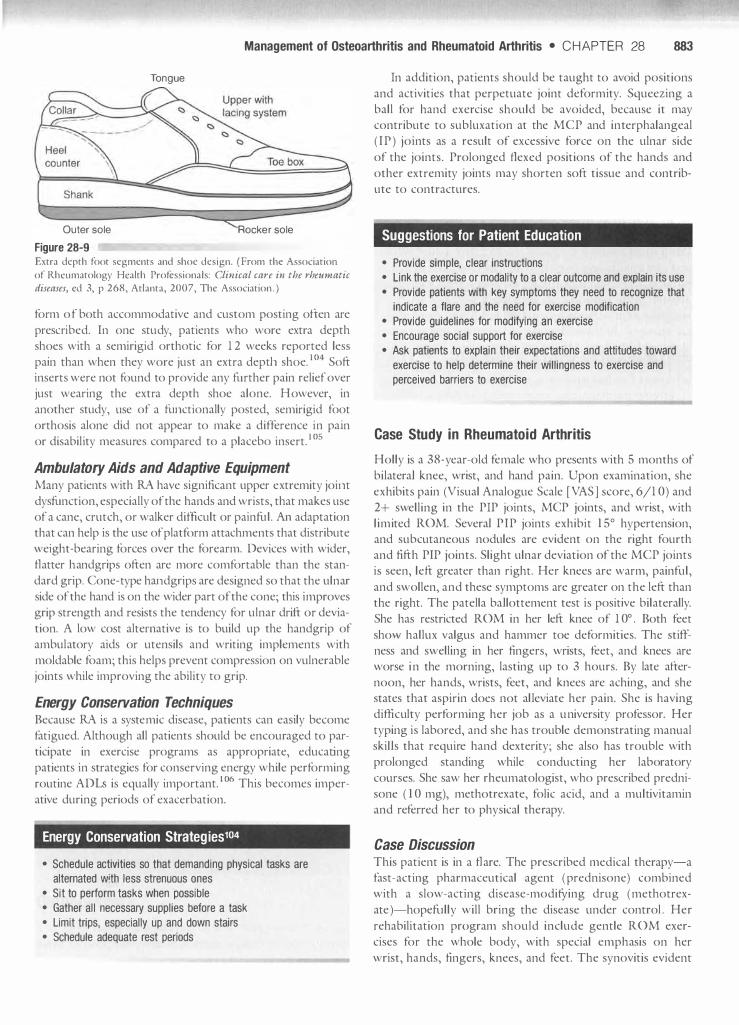
Management of Osteoarthritis and Rheumatoid Arthritis • CHAPTER 28 883
Tongue
Outer sole Rocker sole
Figure 28-9 Extra depth foot segments and shoe design. ( From the Association
of Rhcumatology Hcalth Professionals: Clinical care in the rhettmatic diseases, ed 3, p 268, Atlanta, 2007, The Association . )
form of both accommodative and custom posting often are presClibed. In one study, patients who wore extra depth shoes with a semirigid orthotic for 1 2 weeks reported less pain than when they wore just an extra depth shoe. 1 04 Soft inserts were not found to provide any ftuther pain relief over just wearing the extra depth shoe alone. However, in another study, use of a ftLllctionally posted, semirigid foot orthosis alone did not appear to make a difference in pain or disability measures compared to a placebo insert. I OS
Ambulatory Aids and Adaptive Equipment Many patients with RA have significant upper extremity joirlt dysfunction, especially of the hands and wrists, that makes use of a cane, crutch, or walker difficult or painftil . An adaptation that can help is the use of platform attachments that distribute weight-bearing forces over the forearm. Devices with wider, flatter handgrips often are more comfortable than the standard grip. Cone-type handglips are designed so that the ulnar side of the hand is on the wider part of the cone; this improves grip strength and resists the tendency for ulnar drift or deviation. A low cost alternative is to build up the handgrip of ambulatory aids or utensils and writing implements with moldable foam; this helps prevent compression on vulnerable joints while improving the ability to grip.
Energy Conservation Techniques Because RA is a systemic disease, patients can easily become fatigued. Although all patients should be encouraged to participate in exercise programs as appropriate, educating patients in strategies for conserving energy while performing routine ADLs is equally important. 1 06 This becomes imperative during periods of exacerbation.
Energy Conservation Strategies104
• Schedule activities so that demanding physical tasks are
alternated w!th less strenuous ones
• Sit to perform tasks when possible
• Gather all necessary supplies before a task
• Limit trips, espeCially up and down stairs
• Schedule adequate rest periods
In addition, patients should be taught to avoid positions and activities that perpetuate joint deformity. Squeezing a ball for hand exercise should be avoided, because it may contribute to subluxation at the MCP and interphalangeal ( IP ) joints as a result of excessive force on the ulnar side of the joints. Prolonged flexed positions of the hands and other extremity joints may shorten soft tissue and contribute to contractures.
Suggestions for Patient Education
• Provide simple, clear instructions
• Link the exercise or modality to a clear outcome and explain its use
• Provide patients with key symptoms they need to recognize that
indicate a flare and the need for exercise modification
• Provide guidelines for modifying an exercise
• Encourage social support for exercise
• Ask patients to explain their expectations and attitudes toward
exercise to help determine their willingness to exercise and
perceived barriers to exercise
Case Study in Rheumatoid Arthritis
Holly is a 38-year-old female who presents with 5 months of bilateral knee, wrist, and hand pain. Upon examination, she exhibits pain (Visual Analogue Scale [VAS ] score, 6/1 0) and 2+ swelling in the PIP joints, MCP joints, and wrist, with limited ROM. Several PIP joints exhibit 1 5° hypertension, and subcutaneous nodules are evident on the right fourth and fifth PIP joints. Slight ulnar deviation of the MCP joints is seen, left greater than right. Her knees are warm, painful, and swollen, and these symptoms are greater on the left than the right. The patella ballottement test is positive bilaterally. She has restricted ROM in her left knee of 1 0° . Both feet show hallux valgus and hammer toe deformities. The stiffness and swelling in her fingers, wrists, feet, and knees are worse in the morning, lasting up to 3 hours. By late afternoon, her hands, wrists, feet, and knees are aching, and she states that aspirin does not al leviate her pain. She is having difficulty performing her job as a university professor. Her typing is labored, and she has trouble demonstrating manual skills that require hand dexterity; she also has trouble with prolonged standing while conducting her laboratory courses. She saw her rheumatologist, who prescribed prednisone ( 1 0 mg), methotrexate, folic acid, and a multivitamin and referred her to physical therapy.
Case Discussion This patient is in a flare. The prescribed medical therapy-a fast-acting pharmaceutical agent (prednisone) combined with a slow-acting disease-modifYing drug (methotrexate )-hopeftllly will bring the disease under control . Her rehabilitation program should include gentle ROM exercises for the whole body, with special emphasis on her wrist, hands, fingers, knees, and feet. The synovitis evident

884 CHAPTER 28 • Management of Osteoarthritis and Rheumatoid Arthritis
in her hands and knees can lead to stretching of the joint capsule and ligaments. With synovitis, biomechanical forces across the joint are increased, and ligaments and tendons can become involved, leading to joint instability. Common changes evident in the hands ( the joints commonly involved first) are radial deviation of the wrist, subluxation of the proximal carpal row, and radial rotation of the distal carpal bones. Therefore it is important to check for volar drop of the MCP joints by palpating the heads of these joints in relation to the extended first phalanx . Although flexed posturing generally is more comfortable, flexion can lead to intrinsic tightness. Resting splints for evening and dynanlic splints may help maintain alignment during physical activities. Resistive dynamic exercises should be avoided with synovitis. In the hand, forceful squeezing results in joint deformities and may result in pathomechanical changes. ROM and isometric exercises are preferred, along with the use of ice to reduce swelling. A can of food or a cylinder can be used to perform isometric grasping and is preferred to ball squeezing, which can lead to ulnar drift .
Proper footwear that provides adequate support and allows accommodation of her foot deformities should be recommended. An extra depth shoe allows for use of a soft insole with enough room to accommodate the foot deformities . An ergonomic evaluation of her work space and home environment should be done so that recommendations can be made for individualized energy conserving techniques. She may benefit from an ergonomically designed computer keyboard and/or mouse that minimizes
ulnar drift and allows an easier grasp. Because prolonged standing is fatiguing, an appropriate supportive chair should be sought. Frequent monitoring of blood pressure is required with the use of steroids, because these drugs can lead to weight gain and subsequent hypertension.
Summary
This chapter discussed the roles of exercise therapy, ambulatory aids, modalities, and orthoses in addressing osteoarthritis and rheumatoid arthritis. The evidence supporting the use of various types of exercise, the strengths and limitations of these studies, and new recommendations were presented . The importance of prescribing the correct frequency, duration, intensity, and type of exercise for the various stages of disease was highlighted, as was the current data on the use of orthoses and appropriately adapted aids and assistive devices. Suggestions were presented for important patient educational material that can be included in the intervention plan .
References
To enhance this text and add value for the reader, all references have been incorporated into a CD-ROM that is provided with this text. The reader can view the reference source and access it online whenever possible. There are a total of 123 references for this chapter.