managing cornealectasia: scleral lenses and … cornealectasia: scleral lenses and corneal ......
TRANSCRIPT

Managing CornealEctasia: Scleral Lenses and Corneal
Collagen Cross Linking Yin-Yin Aung OD FAAO
Attending Optometrist, Goodman Eye Center Clinical Faculty, UC Berkeley School of Optometry

Disclosures
• Full time attending optometrist at Goodman Eye Center, a clinical trial site for CXL USA
• No other financial disclosures

Keratoconus
• Bilateral, asymmetric, non-inflammatory condition
• Progression is unpredictable corneal thinning and protrusion inducing highly irregular astigmatism


Treatment Options
1. Spectacles
2. Specialty Contact Lenses
3. Corneal Collagen Cross-Linking
4. Corneal Surgery
– PKP, DALK, KPro

Scleral Lenses

Indications
• Best option for very steep/large ectasia
– Pellucid Marginal Degeneration
– Keratoglobus
– Keratoconus with very steep topography
• Cons: difficult to fit, more expensive, more difficult I/R

• Photos:

Scleral Options
• Fenestrated or Non-fenestrated – Fenestrations will increase oxygen flow and create a
bubble; more difficult to fit
• Toric Periphery • Front surface torics (residual/lenticular astigmatism) • Diameter ranges from 14.3 (mini-sclerals) to 20.2 full
sclerals (24 PROSE) • Materials: Dk as high as 141 with Boston XO2
• Custom Sclerals: – Boston Foundation for Sight (PROSE) – EyePrint PRO: 3-D designed lens from custom mold of
patient’s eye

Fitting Goals
• Complete Corneal Vault
• Scleral Alignment
• No impression ring

Curves of a Scleral Lens

Limbal Zone Figure (left): Excess
central clearance with a
bubble. Limbal bearing
360.
*Need to vault limbus to
protect corneal stem cells.
Figure (right): Completely
clearing cornea, including
the limbus, with 75 µ of
vault.

Peripheral Fitting

Edge Lift
Excess edge lift: •Pt sx: Awareness of edge
in that region
•Bubbles in
corresponding area
•Be sure to check for
bubbles upon insertion
•Area for debris to enter
reservoir “foggy” vision
•Figure (left): Bubbles
were not present on
insertion

Conjunctival Blanching • Indicating a tight periphery (scleral landing zone)
• Lens will have strong suction/difficulty removing the lens
If blanching causing a tight fit only in certain area, consider quadrant specific
periphery or a toric periphery. If blanching is present 360, flatten the peripheral
curves uniformly.

Pinguecula
•Vault lens periphery over pinguecula, or avoid it by
keeping the diameter small enough not to impinge
upon pinguecula
•You can also notch a segment of the periphery to
avoid a pinguecula
*Note: figure on right shows tight periphery which will
need to be flattened

Complications S/P Penetrating Keratoplasty
•Consider High Oxygen material eg Boston XO2
• Optimal peripheral fit to decrease suction
• fenestration to increase Oxygen delivery
• endothelial cell count prior to fitting
•<700 cells/mm2 preliminary corneal consult
•Average post graft cell count: 1000-2000

Mucous Debris
•Can be on surface of lens (left) or in reservoir (right).
•Greater degree of ocular surface disease more
problems with debris
Debris on surface:
•cotton tip applicator with saline to wipe
off
In Reservoir:
•Remove, clean and re-insert
•Try small amount of PF celluvisc in
reservoir
•Make sure vault is not excessive
•Fix any areas of excess edge lift

Troubleshooting
• Reverse Geometry
– PMD, post graft, oblate corneas (s/p refractive surgery)

• Diameter considerations • Weight (toricity in periphery
• Thickness
– Residual astigmatism due to flexure

Toric Peripheral Curve Capability

Follow Up Schedule
• 6 hour fit check: Make sure that the fit is still appropriate towards the end of the day.
• Check for adequate vault
• Rule out compression in periphery
• Check for staining (of conjunctiva and cornea) after lens removal

Starting a Keratoconus Fit

Topography -Cone Size
Large diffuse cones will usually require larger diameter KC lenses; ie Rose K IC
Small central nipple cones work well with small diameter lenses including Rose K 8.7 DIA

S/P Radial Keratotomy

Asymmetric PMD

EyePrintPROTM

Technology
• Custom imprint (mold) of patient’s eye with ocular safe polymer
• Mold provides basis for computer generated custom scleral shell

Contraindications
• Endothelial cells counts under 800
• Excessively elevated blebs
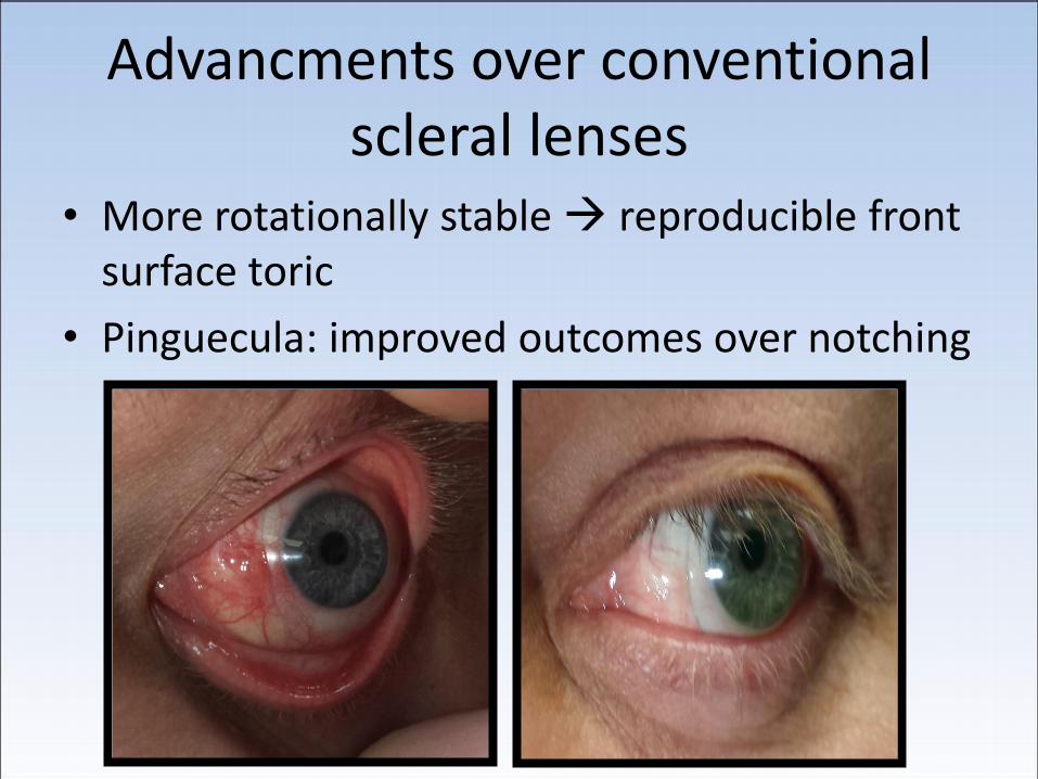
Advancments over conventional scleral lenses
• More rotationally stable reproducible front surface toric
• Pinguecula: improved outcomes over notching

Optics
Uncorrected Standard scleral lens
Front surface toric EyePrint Pro

Procedure
• D/C lens wear 2 days prior to imprint process
• In office imprint
• Lab manufacturing/shipping (2-3 weeks)
• Lathe manufacturing time 24 hours vs 10 min with conventional scleral
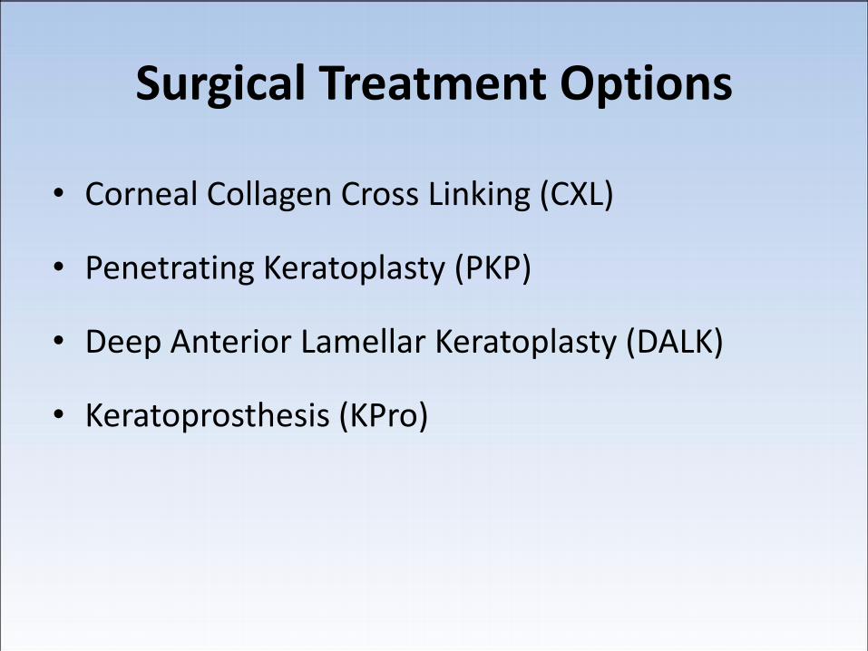
Surgical Treatment Options
• Corneal Collagen Cross Linking (CXL)
• Penetrating Keratoplasty (PKP)
• Deep Anterior Lamellar Keratoplasty (DALK)
• Keratoprosthesis (KPro)

CXL - History
Basic research: 1993-97
by Theo Seiler (IROC, Zurich)
First patients: 1998
Dresden protocol:
10-12mm epithelial
debridement

Dresden Protocol
1. Complete epithelial debridement
2. Riboflavin 0.1% every 1-5 min for 15-30 minutes (or until Riboflavin present in AC)
3. Minimum corneal thickness 400 μ at start of UV light
4. UV light (370 nm) for 30 minutes
5. BCL until full epithelialization happens
6. Topical Ab QID until epithelializaion

Cross-Linking Sites
Over 500 centers worldwide
Standard of care for KCN
Patients as young as 9
Investigational in the US

CXL – Basic Science
• “Natural collagen cross-linking”:
– In vivo LYSYL OXIDASE catalyzes this process • Activates Oxygen from singlet to active triplet state
• Causes oxidative deamination forms aldehyde group cross links with other aldehydes or amine groups (in Collagen molecules)
Oxidative deamination via Lysyl Oxidase +O2

CXL – Basic Science
Photochemical Rxn:
Riboflavin + UVA light
• Activates Oxygen to triplet state that causes same oxidative deamination rxn cross linking of aldehydes and amines in collagen molecules
– Riboflavin is the “photosensitizer”

CXL – Basic Science Summary
• Corneal collagen fibers “cross-link” photochemically via the lysyl oxidative pathway or riboflavin/UVA application
• Increased inter- and intra-fibrillar covalent bonds
• Change in the quaternary structure of the fibrils • Fibrils shorten, thicken
• Fibrils move closer together
• Change in corneal elasticity
• Secondary change in GAG’s between fibrils

Animal Studies
UV + Riboflavin:
– New high-molecular-weight collagen polymer
– increased fiber diameter after cross-linking treatment
– Corneal rigidity increased by 329%
– Chemical stability of polymer indicates likely long-term corneal stability after treatment

3.00 mW/cm²
1.49 mW/cm²
0.74 mW/cm²
0.36 mW/cm²
0.18 mW/cm²
0.09mW/cm²
0.06 mW/cm²
0μm
100μm
200μm
300μm
400μm
500μm
600μm
100%
50%
25%
12%
6%
3%
2%
Endothelium
Damage threshold 3.00 mW/cm²
Safety of Cross-Linking
With Riboflavin loading

CXLUSA Prospective Non-randomized Multicenter study evaluating Trans-epithelial (“Epi-On”) CXL
• Physician-sponsored , IRB-approved clinical trial
• Proprietary UV light Source • Enrolling patients since October 2009 • Initially started as Epithelial-Off then
changed to Transepithelial in 2010.

Study Criteria Indications:
• Keratoconus • FFKC • Pellucid Marginal Degeneration • Post-LASIK ectasia • RK with diurnal visual fluctuations
Inclusion Criteria • Age 12 or older • Corneal thickness >350 microns Exclusion Criteria • Visually significant apical scarring • Pregnant/Breast Feeding • History of Ocular Herpes Simplex Infection

CXLUSA US Study Centers
Roy Rubinfeld, MD Bethesda, MD
William Trattler, MD Miami, FL
Dan Goodman, M D San Francisco, CA
Audrey Talley, MD Seattle, WA
Jodi Luchs, MD: Long Island, NY
Richard Lindstrom, MD Minneapolis, MN
Parag Majmudar, MD Chicago, IL
Kathy Hatch, MD Boston, MA
Lance Forstot, MD Littleton, CO
Sandy Feldman, MD San Diego, CA
Jay Schwartz, MD Phoenix, AZ
Ty McCall, MD Dallas, TX
Ranjan Malahotra, MD St. Louis, MO
Gregg Berdy, MD St. Louis, MO
David Wallace, MD Los Angeles, CA
Doyle Stulting, MD Atlanta, GA
ClinicalTrials.gov or CXLUSA.com
16 centers
37 Investigators

CXL-USA International Sites
• Zurich, Switzerland Theo Seiler M.D. IROC - Institute for Refractive and Ophthalmic Surgery
• Brescia Italy
Roberto Pinelli, M.D. Istituto Laser Microchirurgia Oculare
• Dublin, Ireland Arthur Cummings M.D. Wellington Eye Clinic

Study Information
• ClinicalTrials.gov ID NCT01189864
• Currently enrolling patients
Local Study Sites
(National Keratoconus Foundation)
• Stanford, Sacramento, San Leandro
http://www.nkcf.org/cxl-sites-in-usa-2013/#ca

Topography-Selecting Candidates
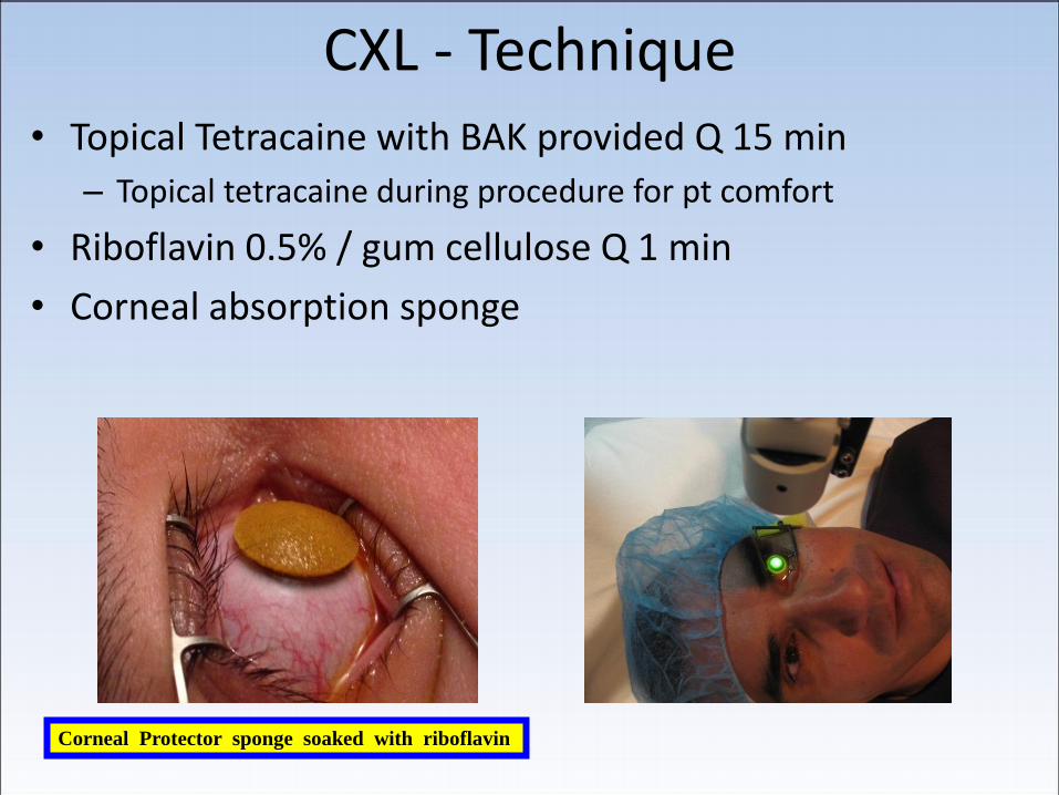
CXL - Technique • Topical Tetracaine with BAK provided Q 15 min
– Topical tetracaine during procedure for pt comfort
• Riboflavin 0.5% / gum cellulose Q 1 min
• Corneal absorption sponge
Corneal Protector sponge soaked with riboflavin
CXL - technique
• 15 - 90 minutes of Riboflavin drops required
– Average ~ 50 minutes of Riboflavin instillation
• Homogeneous and complete stromal saturation with riboflavin
• Confirm saturation with slit lamp examination
• Check Pachymetry (confirm ≥ 400 μ)
• UVA applied x 30 minutes 3.0mW/cm2
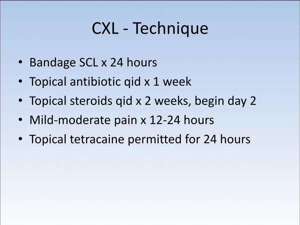
CXL - Technique
• Bandage SCL x 24 hours
• Topical antibiotic qid x 1 week
• Topical steroids qid x 2 weeks, begin day 2
• Mild-moderate pain x 12-24 hours
• Topical tetracaine permitted for 24 hours
Homogeneity

Protocol Specifies
Homogeneous Loading
This is Not

Concentration

Before Riboflavin Loading
Grade 0/V

Grade I/V

Grade III/V

Grade IV/V
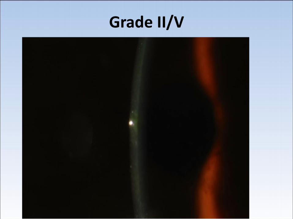
Grade II/V

Grade V/V

Epi-on “flare “

Epi-On CXL POD 1

Epithelial-Off CXL
Long Term Results
• 5 year study, 48 eyes (60 pts treated)
–No patient had progression of ectasia • Mean improvement 2.87 D (average K)
• Mean Improvement in BSCV by 1.4 lines
• Improvement in BSCV 62%
• Improvement in UCVA 74%
Wollensak G. Crosslinking treatment of progressive keratoconus: New hope.
Curr Opin Ophthalmol. 2006 Aug;17:356-60

Cross-Linking and Keratoconus Keratometry Over Time
Wollensak, Spoerl, and Seiler, AJO 135:620, 2003.

Long-term Results
• 241 eyes
• Follow-up 6 months to 6 years
• Flattening: 2.68 D at 1 year; 4.84D at 3 years
• BSCVA improvement (> 1 line): 53% at 1 year
• No loss of BCVA
• No progression of KC 99%
• 2 patients had KCN progression repeat CXL
Raiskup-Wolf, Hoyer, Spoerl.
AJO April 2010

Cross-linking Results: 6 year Results (Carus University Hospital, Dresden, Germany)
Frederik Raiskup-Wolf, MD et al: Collagen cross-linking with riboflavin & ultraviolet-A light in keratoconus: Long-term results; JCRS; May 2010

Complications with Epithelial-Off Cross Linking

Delayed Epithelial Healing and Infiltrate after
Epi-Off CXL
Postop Day 2 Courtesy of Wm. Trattler, MD

Postop Day 5

Haze after Epi-Off CXL
Courtesy of Roy Rubinfeld MD

alpha Strep Keratitis post Epi-Off CXL
Courtesy of Robert Fintelman, MD (Phoenix, Arizona)
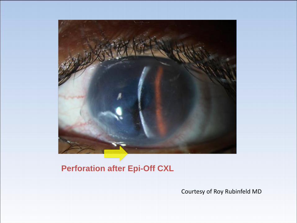
Perforation after Epi-Off CXL
Courtesy of Roy Rubinfeld MD

Epi-off CXL-adverse events
• Corneal melting in both eyes after simultaneous corneal cross-linking in a patient with keratoconus and Down syndrome. Ophthalmologe. 2010 Oct; 107(10):951-5
• Corneal melting corneal collagen cross-linking for keratoconus: A case report. Labiris. Journal of Medical Case Reports 5:15 2012
• Ocular surface complications of CXL. Frucht-Perry. AAO annual meeting poster (P0367), 2012 (11% incidence)

Epithelial- off vs. Transepithelial cross-linking: Clinical Studies
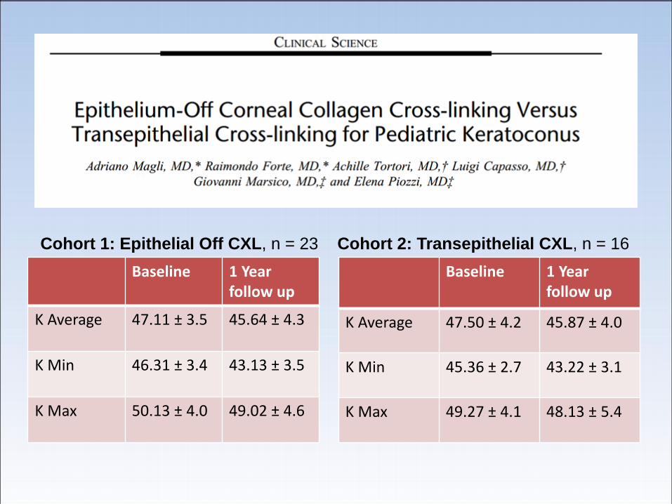
Baseline 1 Year follow up
K Average 47.11 ± 3.5 45.64 ± 4.3
K Min 46.31 ± 3.4 43.13 ± 3.5
K Max 50.13 ± 4.0 49.02 ± 4.6
Cohort 1: Epithelial Off CXL, n = 23
Baseline 1 Year follow up
K Average 47.50 ± 4.2 45.87 ± 4.0
K Min 45.36 ± 2.7 43.22 ± 3.1
K Max 49.27 ± 4.1 48.13 ± 5.4
Cohort 2: Transepithelial CXL, n = 16

Baseline 1 yr follow up Baseline 1 yr follow up
Average K 49.98 ± 4.46 47.95 ± 2.69 48.78 ±3.46 49.37 ± 2.90
Steep K 60.30 ± 5.26 58.10 ± 4.20 61.30 ± 6.29 64.20 ± 4.25
Transepithelial CXL No treatment
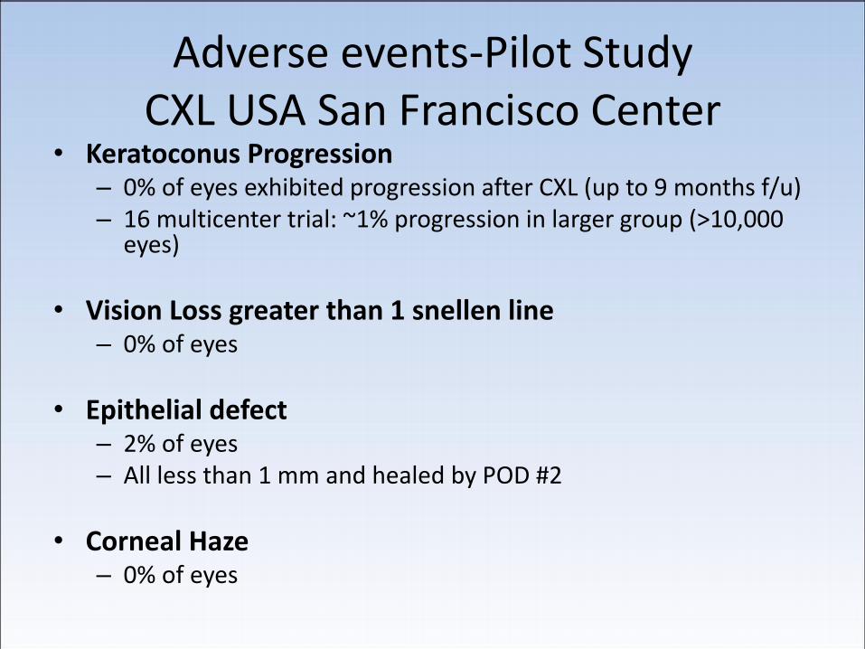
Adverse events-Pilot Study CXL USA San Francisco Center
• Keratoconus Progression – 0% of eyes exhibited progression after CXL (up to 9 months f/u) – 16 multicenter trial: ~1% progression in larger group (>10,000
eyes)
• Vision Loss greater than 1 snellen line – 0% of eyes
• Epithelial defect – 2% of eyes – All less than 1 mm and healed by POD #2
• Corneal Haze – 0% of eyes

OCT Cornea- Patient EW pre-op

6 weeks post op
•Microscopic cellular changes in stroma
•Hypotheis: Aldehyde/Amine covalent bonds
cross-linking / changing collagen structure

4.5 months post op
Cellular changes still present, but not as prominent
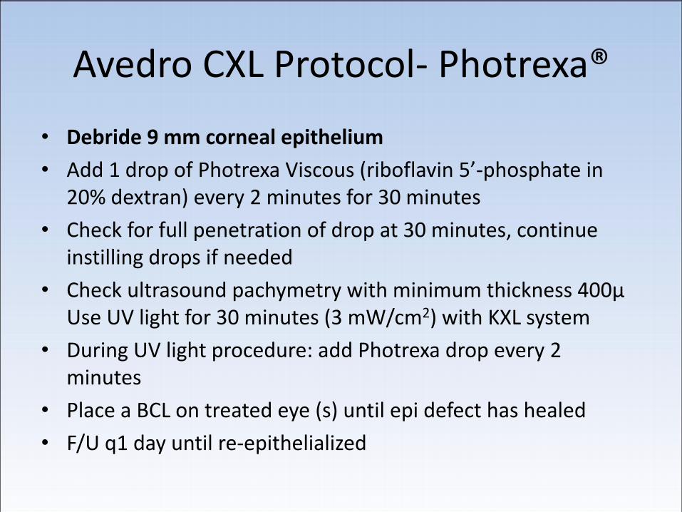
Avedro CXL Protocol- Photrexa®
• Debride 9 mm corneal epithelium
• Add 1 drop of Photrexa Viscous (riboflavin 5’-phosphate in 20% dextran) every 2 minutes for 30 minutes
• Check for full penetration of drop at 30 minutes, continue instilling drops if needed
• Check ultrasound pachymetry with minimum thickness 400µ Use UV light for 30 minutes (3 mW/cm2) with KXL system
• During UV light procedure: add Photrexa drop every 2 minutes
• Place a BCL on treated eye (s) until epi defect has healed
• F/U q1 day until re-epithelialized
